Embed Size (px)
Citation preview

EAC1
Catholic University of Louvain, St - Luc University HospitalHead and Neck Oncology Programme
Mar. 2002
Carcinoma of external auditory canal

EAC2
Catholic University of Louvain, St - Luc University HospitalHead and Neck Oncology Programme
Mar. 2002
Carcinoma of external auditory canal
•• Work-up procedureWork-up procedure
•• StagingStaging
•• Primary treatmentPrimary treatment
•• Follow-upFollow-up
•• Treatment of recurrent and/orTreatment of recurrent and/or metastatic metastatic disease disease
•• References References

EAC3
Catholic University of Louvain, St - Luc University HospitalHead and Neck Oncology Programme
Mar. 2002
Clinical evaluation Evidence Option
� Complete history of the disease� Performance status (Karnofsky / WHO scale)� Examination of external auditory canal� Audiogram� Examination of the VII th nerve� Neck examination� Drawing of any lesions
Type CType CType CType CType CType CType C
Std.Std.Std.Std.Std.Std.Std.
EAC4
Catholic University of Louvain, St - Luc University HospitalHead and Neck Oncology Programme
Mar. 2002
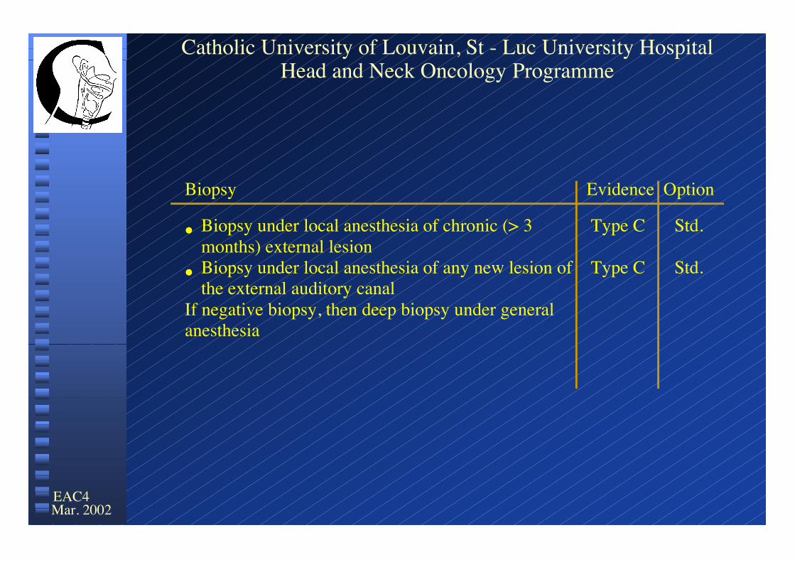
Biopsy Evidence Option
� Biopsy under local anesthesia of chronic (> 3months) external lesion
� Biopsy under local anesthesia of any new lesion ofthe external auditory canal
If negative biopsy, then deep biopsy under generalanesthesia
Type C
Type C
Std.
Std.

EAC5
Catholic University of Louvain, St - Luc University HospitalHead and Neck Oncology Programme
Mar. 2002
Advanced clinical evaluation Evidence Option
� Dental examination by oral surgeon if RxThscheduled
� Others (if required)
Type C
Type C
Std.
Indiv.
EAC6
Catholic University of Louvain, St - Luc University HospitalHead and Neck Oncology Programme
Mar. 2002
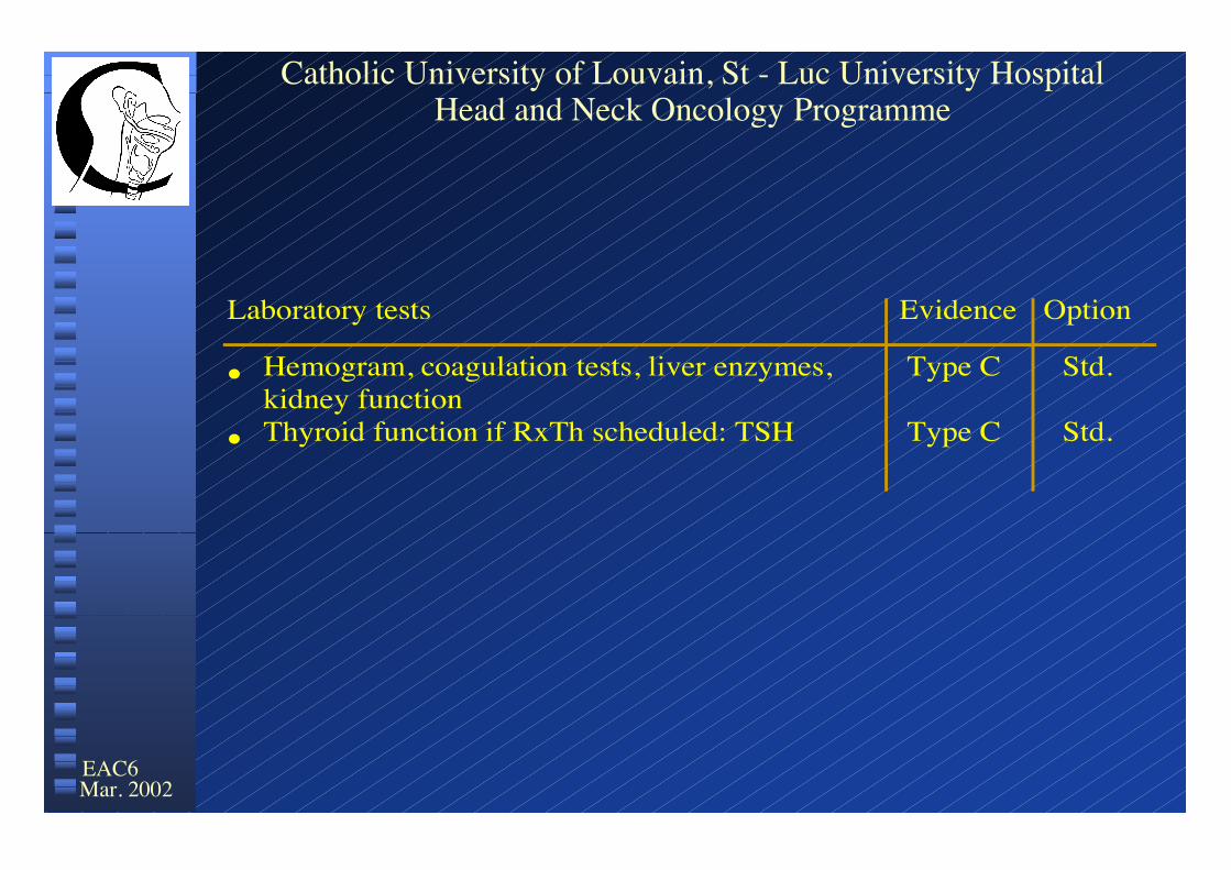
Laboratory tests Evidence Option
� Hemogram, coagulation tests, liver enzymes, kidney function
� Thyroid function if RxTh scheduled: TSH
Type C
Type C
Std.
Std.

EAC7
Catholic University of Louvain, St - Luc University HospitalHead and Neck Oncology Programme
Mar. 2002
Loco-regional imaging Evidence Option
� CT scan without contrast enhancement (bonewindow)1
� MRI with gadolinium enhancement1
Type C
Type C
Std.
Std.
1See guidelines for loco-regional imaging

EAC8
Catholic University of Louvain, St - Luc University HospitalHead and Neck Oncology Programme
Mar. 2002
Pathologic examination Evidence Option
Standards of the British Royal College ofPathologists (endorsed by EORTC)1
Type C Std.
1See pathology guidelines
EAC9
Catholic University of Louvain, St - Luc University HospitalHead and Neck Oncology Programme
Mar. 2002
Carcinoma of external auditory canal
•• Work-up procedureWork-up procedure
•• StagingStaging
•• Primary treatmentPrimary treatment
•• Follow-upFollow-up
•• Treatment of recurrent and/orTreatment of recurrent and/or metastatic metastatic disease disease
•• References References

EAC10
Catholic University of Louvain, St - Luc University HospitalHead and Neck Oncology Programme
Mar. 2002
Staging Evidence Option
� Modified Pittsburgh (revision 2002) classification Type C Std.

EAC11
Catholic University of Louvain, St - Luc University HospitalHead and Neck Oncology Programme
Mar. 2002
� T1: Tumor limited to the external auditory canal without bony erosion or evidence of soft tissue extension
� T2: Tumor with limited external auditory canal bony erosion (not full thickness) or radiographic finding consistent with limited (< 0.5 cm) softtissue involvement
� T3: Tumor eroding the osseous external auditory canal (full thickness) withlimited (< 0.5 cm) soft tissue involvement, or tumor involving middle ear and/or mastoid
� T4: Tumor eroding the cochlea, petrous apex, medical wall of the middle ear, carotid canal, jugular foramen or dura, or with extensive (> 0.5 cm)soft tissue involvement; patients presenting with facial paralysis
- T4a: extracranial extension (> 0.5 cm) in soft tissue or skin
- T4b: Tumor eroding the cochlea, petrous apex, medical wall of the middle ear, carotid canal or jugular foramen
- T4c: extension to the dura

EAC12
Catholic University of Louvain, St - Luc University HospitalHead and Neck Oncology Programme
Mar. 2002
• N status: - N0: no regional lymph node metastasis
- N1: metastasis in regional lymph node(s)
- Nx: regional lymph nodes cannot be assessed
• M status: - M0: no distant metastasis
- M1: distant metastasis
- Mx: distant metastasis cannot be assessed

EAC13
Catholic University of Louvain, St - Luc University HospitalHead and Neck Oncology Programme
Mar. 2002
Carcinoma of external auditory canal
•• Work-up procedureWork-up procedure
•• StagingStaging
•• Primary treatmentPrimary treatment
•• Follow-upFollow-up
•• Treatment of recurrent and/orTreatment of recurrent and/or metastatic metastatic disease disease
•• References References

EAC14
Catholic University of Louvain, St - Luc University HospitalHead and Neck Oncology Programme
Mar. 2002
Primary treatment: general strategy Evidence Option
� T1–T2, N0- Lateral temporal bone resection + parotidectomy + selective ND (level II) ± RxTh1
pN+ on frozen section examination, dissection of levels III-V - RxTh if medical status not suitable for surgery : RxTh� T1-T2, N1
- Lateral temporal bone resection + parotidectomy + ND (selective or radical modified) ± RxTh1
� T3 N0- no extension to middle ear: Lateral temporal bone resection + parotidectomy + selective ND (level II) + RxTh1
- extension to middle ear: subtotal temporal bone resection + dissection of nerve VII + nerve graft + parotidectomy + selective ND (level II) + RxTh1
� T3 N1- no extension to middle ear: Lateral temporal bone resection+ parotidectomy + ND (selective or radical modified) + RxTh1
- extension to middle ear: subtotal temporal bone resection+ dissection of nerve VII + nerve graft + parotidectomy + ND
(selective or radical modified) + RxTh1
Type 3
Type 3
Type 3
Type 3
Type 3
Type 3
Type 3
Std.
Indiv.
Std.
Std.
Std.
Std.
Std.
1 see indication of post-operative RxTH (slide 18)

EAC15
Catholic University of Louvain, St - Luc University HospitalHead and Neck Oncology Programme
Mar. 2002
Primary treatment: general strategy Evidence Option
� T4a, N0- Subtotal temporal bone resection + parotidectomy + selective ND (level II) + RxTh1
pN+ on frozen section examination, dissection of levels III-V - RxTh if medical status not suitable for surgery : RxTh� T4a, N1
- Subtotal temporal bone resection + parotidectomy + ND (selectif or radical modified) + RxTh1
� T4b, any N- Best supportive care- Chemotherapy- Local palliative surgery- Local palliative RxTh- Temporal bone resection + RxTh1
� T4c, any N- Best supportive care- Chemotherapy- Local palliative surgery- Local palliative RxTh
Type 3
Type 3
Type 3
Type CType CType CType CType C
Type CType CType CType C
Std.
Indiv.
Std.
Std.Std.Std.Std.
Indiv.
Std.Std.Std.Std.
1 see indication of post-operative RxTh (slide 18)

EAC16
Catholic University of Louvain, St - Luc University HospitalHead and Neck Oncology Programme
Mar. 2002
Primary treatmentPrimary treatment: : pathologic examination pathologic examination Evidence Evidence OptionOption
Standards of the British Royal Standards of the British Royal College College of of Type C Type C StdStd..
Pathalogists Pathalogists ( ( endorsed endorsed by EORTC )by EORTC )
11See See pathology guidelinespathology guidelines

EAC17
Catholic University of Louvain, St - Luc University HospitalHead and Neck Oncology Programme
Mar. 2002
Indication for post-op RxTh Evidence Option
� Evidence at the "T" level-T2-T4-close margins (< 5mm)-positive margins: R1-macroscopic residual disease: R2-perineural invasion
� Evidence at the "N" level-more than one involved lymph node-extracapsular rupture/soft tissue invasion-more than one involved level-invasion of lymphatic vessels
Type 3Type 3Type 3Type 3Type 3
Type 3Type 3Type 3Type 3
Std.Std.Std.Std.Std.
Std.Std.Std.Std.

EAC18
Catholic University of Louvain, St - Luc University HospitalHead and Neck Oncology Programme
Mar. 2002
Low risk (LR): no adverse featureLow risk (LR): no adverse feature
Intermediate risk (IR): 1 feature other than ECE/STEIntermediate risk (IR): 1 feature other than ECE/STE
High risk (HR): >1 features or ECE/STEHigh risk (HR): >1 features or ECE/STE
Risk factorsRisk factors
• Extracapsular extension / soft tissue extension (ECE/STE)
• (Oral cavity tumors)
• R1 surgical margins
• Nerve invasion
• >1 positive neck nodes
• Positive node in > 1 levels
• Node size > 3 cm
• > 6 week interval between surgery and RxTh

EAC19
Catholic University of Louvain, St - Luc University HospitalHead and Neck Oncology Programme
Mar. 2002
RxTh regimen Evidence Option
� Target volumes- Petrous bone, mastoid, parotid, para- pharyngeal space and level II (N0) or level II-V (N1)
� Technique- conformal radiotherapy- IMRT radiotherapy
� Dose- T and positive neck levels: 70 Gy- prophylactic dose (undissected neck): 50 Gy- high risk (ECE or >1 risk factors): 64 Gy- intermediate risk (1 risk factor other than(ECE): 60 Gy
� Fractionation- daily 2Gy/fraction
Type C
Type 3Type 3
Type CType CType CType C
Type 3
Std.
Std.Invest.
Std.Std.Std.Std.
Std.1See detailled protocol2See guidelines for post-operative radiotherapy

EAC20
Catholic University of Louvain, St - Luc University HospitalHead and Neck Oncology Programme
Mar. 2002
Carcinoma of external auditory canal
•• Work-up procedureWork-up procedure
•• StagingStaging
•• Primary treatmentPrimary treatment
•• Follow-upFollow-up
•• Treatment of recurrent and/orTreatment of recurrent and/or metastatic metastatic disease disease
•• References References

EAC21
Catholic University of Louvain, St - Luc University HospitalHead and Neck Oncology Programme
Mar. 2002
Follow-up Evidence Option
� Clinical examination- local examination, audiogram, fiberoptic examination and neck palpation every 2 months (first 2 years), every 6 months (3rd-5th year), then every year (> 5 years)- dental examination every 6 months, if RxTh
� Loco-regional imaging- NMR at 6, 12 and 24 months
� Laboratory tests-thyroid function (TSH) every year, if RxTh
� Evolution of late toxicity (EORTC/RTOG) scale
Type C
Type C
Type C
Type CType C
Std.
Std.
Std.
Std.Std.

EAC22
Catholic University of Louvain, St - Luc University HospitalHead and Neck Oncology Programme
Mar. 2002
Carcinoma of external auditory canal
•• Work-up procedureWork-up procedure
•• StagingStaging
•• Primary treatmentPrimary treatment
•• Follow-upFollow-up
• Treatment of recurrent and/orTreatment of recurrent and/or metastatic metastatic diseasedisease
• References

EAC23
Catholic University of Louvain, St - Luc University HospitalHead and Neck Oncology Programme
Mar. 2002
Salvage treatment for recurrent disease Evidence Option
� anyT-N0-M0-Surgery ± RxTh-Chemotherapy-Best supportive care
� T0-anyN-M0-ND ± RxTh-RxTh-Chemotherapy-Best supportive care
� AnyT-N1-M0/T4 any NSurgery ± RxThChemotherapyBest supportive care
� MetastasisChemotherapySurgeryBest supportive care
Type 3Type 3Type 3
Type 3Type 3Type 3Type 3
Type 3Type 3Type 3
Type 3Type 3Type 3
Std.Indiv.Indiv.
Indiv.Indiv.Indiv.Indiv.
Indiv.Indiv.Indiv.
Std.Indiv.Indiv.

EAC24
Catholic University of Louvain, St - Luc University HospitalHead and Neck Oncology Programme
Mar. 2002
Carcinoma of external auditory canal
•• Work-up procedureWork-up procedure
•• StagingStaging
•• Primary treatmentPrimary treatment
•• Follow-upFollow-up
• Treatment of recurrent and/orTreatment of recurrent and/or metastatic metastatic disease disease
• References

EAC25
Catholic University of Louvain, St - Luc University HospitalHead and Neck Oncology Programme
Mar. 2002
ReferencesReferences� ARRIAGA M., CURTIN H, TAKAHASHI H, HIRSCH B and KAMERER DB : Staging proposal for external
auditory meatus carcinoma based on preoperative clinical examination and computed tomography findings.Ann Otol Rhinol Laryngol 1990, 99 : 714-721
� ARRIAGA M, HIRSCH BE, KAMERER DB and MYERS EN : Squamous carcinoma of the external auditory meatus .Otolaryngol Head neck Surg 1989, 101 :330-337
� AUSTIN J, STEWART K. and FAWZI N. : Squamous cell carcinoma of the external auditory canal. Arch otolaryngol Head Neck Surg 1994, 120 :1228 - 1232
� JACKLER R. and DRISCOLL C. , Ed. : Tumors of the ear and temporal bone.Lippincot Williams & Wilkins, Philadelphia, 2000
� KUHEL W., HUME C. and SELESNICK S. : Cancer of the external auditory canal and temporal bone. OtolaryngolClin N Am 1996, 29 : 827-852
� MANOLIDIS S, PAPPAS D, VON DOERSTEN P, JACKSON G and GLASSCOCK M Temporal bone and lateralskul base malignancy results . Am J Otol, 1998, 19 : S 1 - S 15
� MOODY S , HIRSCH B and MYERS E : Squamous cell carcinoma of the external auditory canal : an evaluation of astaging system. Am J Otol 2000, 21 : 582-588
� PRASAD S. and JANECKA I. : Efficacy of surgical treatments for squamous cell carcinoma of the temporal bone, alitterature review. Otolaryngol Head Neck Surg 1994, 110 : 270 - 280
� SPECTOR JG : Management of temporal bone carcinomas : a therapeutic analysis of two groups of patients andlong-term followup. Otolaryngol Head Neck Surg 1991, 104 : 58-66
� SHIH L. and CRABTREE J : Carcinoma of the external auditory canal, an update. Laryngoscope 1990, 100: 1215-1218� TRAISSAC L., Ed : Les cancers de l ’oreille, Masson, Paris, 1995� TESTA J , FUKUDA Y and KOWALSKI L. : Prognostic factors in carcinoma of the external auditory canal . Arch
Otolaryngol Head neck Surg, 1997, 123 : 720-724� ZIESKE L. and MYERS E.N. : Squamous cell carcinoma with positive margins Arch otolaryngol Head neck Surg
1986, 112 : 863-866