Embed Size (px)
Citation preview
at SciVerse ScienceDirect
Plant Physiology and Biochemistry 56 (2012) 62e71
Contents lists available
Plant Physiology and Biochemistry
journal homepage: www.elsevier .com/locate/plaphy
Research article
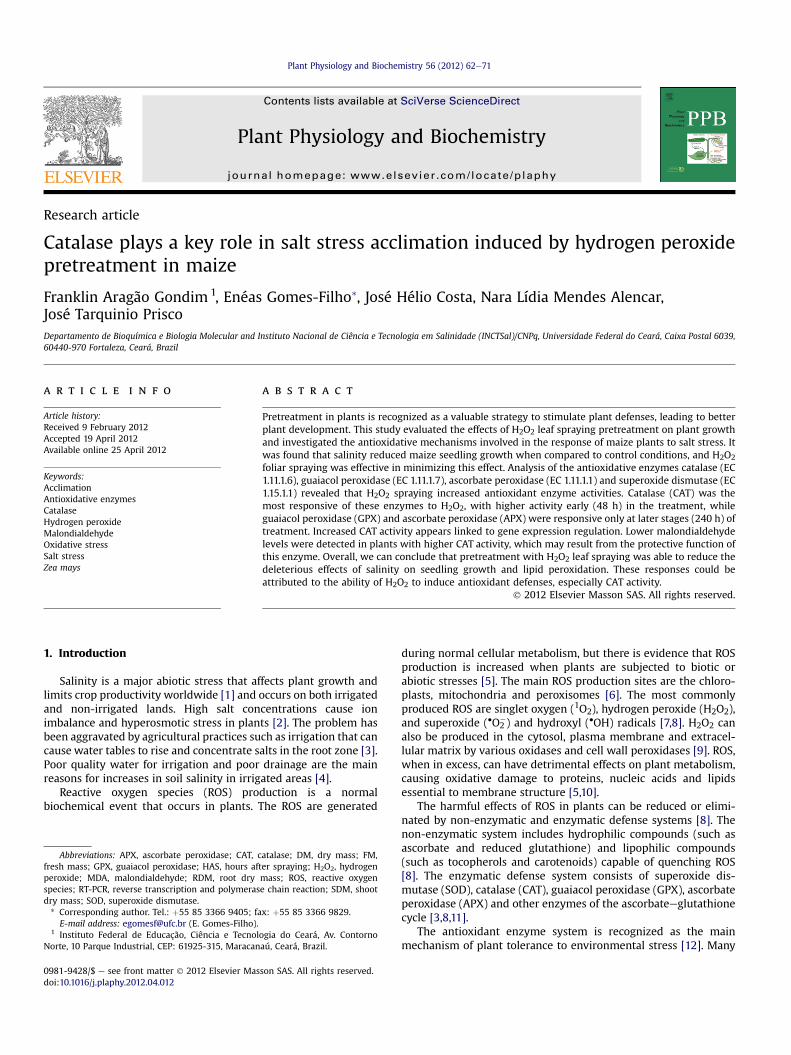
Catalase plays a key role in salt stress acclimation induced by hydrogen peroxidepretreatment in maize
Franklin Aragão Gondim1, Enéas Gomes-Filho*, José Hélio Costa, Nara Lídia Mendes Alencar,José Tarquinio PriscoDepartamento de Bioquímica e Biologia Molecular and Instituto Nacional de Ciência e Tecnologia em Salinidade (INCTSal)/CNPq, Universidade Federal do Ceará, Caixa Postal 6039,60440-970 Fortaleza, Ceará, Brazil
a r t i c l e i n f o
Article history:Received 9 February 2012Accepted 19 April 2012Available online 25 April 2012
Keywords:AcclimationAntioxidative enzymesCatalaseHydrogen peroxideMalondialdehydeOxidative stressSalt stressZea mays
Abbreviations: APX, ascorbate peroxidase; CAT, cfresh mass; GPX, guaiacol peroxidase; HAS, hours afperoxide; MDA, malondialdehyde; RDM, root dryspecies; RT-PCR, reverse transcription and polymerasdry mass; SOD, superoxide dismutase.* Corresponding author. Tel.: þ55 85 3366 9405; fa
E-mail address: [email protected] (E. Gomes-Filho).1 Instituto Federal de Educação, Ciência e Tecnol
Norte, 10 Parque Industrial, CEP: 61925-315, Maracan
0981-9428/$ e see front matter � 2012 Elsevier Masdoi:10.1016/j.plaphy.2012.04.012
a b s t r a c t
Pretreatment in plants is recognized as a valuable strategy to stimulate plant defenses, leading to betterplant development. This study evaluated the effects of H2O2 leaf spraying pretreatment on plant growthand investigated the antioxidative mechanisms involved in the response of maize plants to salt stress. Itwas found that salinity reduced maize seedling growth when compared to control conditions, and H2O2
foliar spraying was effective in minimizing this effect. Analysis of the antioxidative enzymes catalase (EC1.11.1.6), guaiacol peroxidase (EC 1.11.1.7), ascorbate peroxidase (EC 1.11.1.1) and superoxide dismutase (EC1.15.1.1) revealed that H2O2 spraying increased antioxidant enzyme activities. Catalase (CAT) was themost responsive of these enzymes to H2O2, with higher activity early (48 h) in the treatment, whileguaiacol peroxidase (GPX) and ascorbate peroxidase (APX) were responsive only at later stages (240 h) oftreatment. Increased CAT activity appears linked to gene expression regulation. Lower malondialdehydelevels were detected in plants with higher CAT activity, which may result from the protective function ofthis enzyme. Overall, we can conclude that pretreatment with H2O2 leaf spraying was able to reduce thedeleterious effects of salinity on seedling growth and lipid peroxidation. These responses could beattributed to the ability of H2O2 to induce antioxidant defenses, especially CAT activity.
� 2012 Elsevier Masson SAS. All rights reserved.
1. Introduction
Salinity is a major abiotic stress that affects plant growth andlimits crop productivity worldwide [1] and occurs on both irrigatedand non-irrigated lands. High salt concentrations cause ionimbalance and hyperosmotic stress in plants [2]. The problem hasbeen aggravated by agricultural practices such as irrigation that cancause water tables to rise and concentrate salts in the root zone [3].Poor quality water for irrigation and poor drainage are the mainreasons for increases in soil salinity in irrigated areas [4].
Reactive oxygen species (ROS) production is a normalbiochemical event that occurs in plants. The ROS are generated
atalase; DM, dry mass; FM,ter spraying; H2O2, hydrogenmass; ROS, reactive oxygene chain reaction; SDM, shoot
x: þ55 85 3366 9829.
ogia do Ceará, Av. Contornoaú, Ceará, Brazil.
son SAS. All rights reserved.
during normal cellular metabolism, but there is evidence that ROSproduction is increased when plants are subjected to biotic orabiotic stresses [5]. The main ROS production sites are the chloro-plasts, mitochondria and peroxisomes [6]. The most commonlyproduced ROS are singlet oxygen (1O2), hydrogen peroxide (H2O2),and superoxide (�O2
�) and hydroxyl (�OH) radicals [7,8]. H2O2 canalso be produced in the cytosol, plasma membrane and extracel-lular matrix by various oxidases and cell wall peroxidases [9]. ROS,when in excess, can have detrimental effects on plant metabolism,causing oxidative damage to proteins, nucleic acids and lipidsessential to membrane structure [5,10].
The harmful effects of ROS in plants can be reduced or elimi-nated by non-enzymatic and enzymatic defense systems [8]. Thenon-enzymatic system includes hydrophilic compounds (such asascorbate and reduced glutathione) and lipophilic compounds(such as tocopherols and carotenoids) capable of quenching ROS[8]. The enzymatic defense system consists of superoxide dis-mutase (SOD), catalase (CAT), guaiacol peroxidase (GPX), ascorbateperoxidase (APX) and other enzymes of the ascorbateeglutathionecycle [3,8,11].
The antioxidant enzyme system is recognized as the mainmechanism of plant tolerance to environmental stress [12]. Many
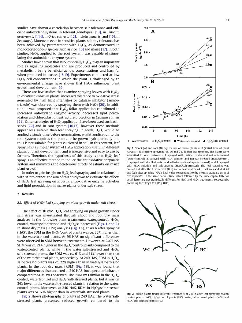
Fig. 1. Shoot (A) and root (B) dry masses of maize plants at 0 (initial time of plantharvest e just before spraying), 48, 96 and 240 h after leaf spraying. The plants weresubmitted to four treatments: 1. sprayed with distilled water and not salt-stressed(water/control), 2. sprayed with H2O2 solution and not salt-stressed (H2O2/control),3. sprayed with distilled water and salt-stressed (water/salt-stressed), and 4. sprayedwith H2O2 solution and salt-stressed (H2O2/salt-stressed). The leaf spraying wascarried out after the first harvest (0 h) and repeated after 24 h. Salt was added at 48and 72 h after spraying (HAS). Each value corresponds to the mean � standard error offive replicates. In the same harvest time values followed by the same capital letter orsmall letter are not statistically different for NaCl and H2O2 treatments, respectively,according to Tukey’s test (P � 0.05).
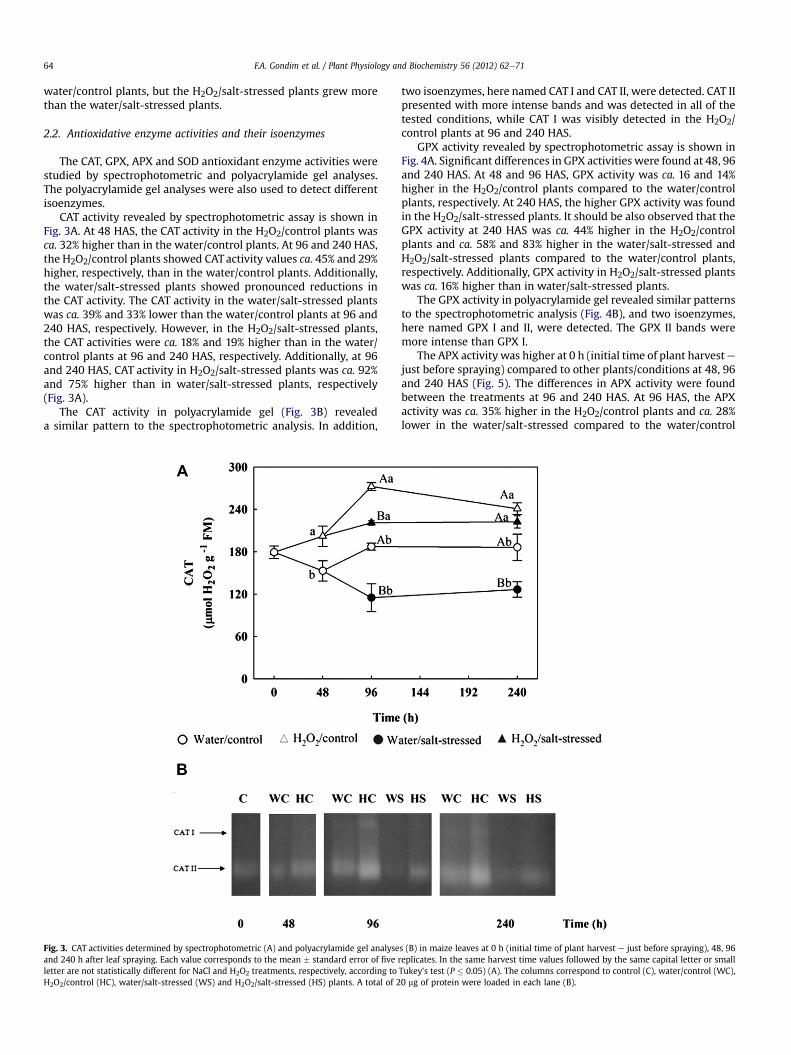
Fig. 2. Maize plants under different treatments at 240 h after leaf spraying: water/control plants (WC); H2O2/control plants (HC); water/salt-stressed plants (WS); andH2O2/salt-stressed plants (HS).
F.A. Gondim et al. / Plant Physiology and Biochemistry 56 (2012) 62e71 63
studies have shown a correlation between salt tolerance and effi-cient antioxidant systems in tolerant genotypes ([13], in Triticumaestivum L. [1,14], in Oriza sativa L. [12], in Beta vulgaris; and [15], inZea mays). Moreover, even in sensitive plants, salinity tolerance hasbeen achieved by pretreatment with H2O2, as demonstrated inmonocotyledonous species such as rice [16] and maize [17]. In bothstudies, H2O2, applied to the root system, was capable of stimu-lating the antioxidant enzyme system.
Studies have shown that ROS, especially H2O2, play an importantrole as signaling molecules and are produced and controlled bymetabolism, being beneficial at low concentrations and harmfulwhen produced in excess [18,19]. Experiments conducted at lowH2O2 cell concentrations in which the plant is challenged by anenvironmental change have shown that H2O2 influences plantgrowth and development [19].
There are few studies that examine spraying leaves with H2O2.In Nicotiana tabacum plants, increased tolerance to oxidative stressgenerated by high light intensities or catalase inhibitor (amino-triazole) was observed by spraying them with H2O2 [20]. In addi-tion, it was proposed that H2O2 foliar application contributed toincreased antioxidant enzyme activity, decreased lipid perox-idation and chloroplast ultrastructure protection in Cucumis sativus[21]. Other strategies of H2O2 application have been used such as inseeds [22] and in root system [16,17], however these methodsappear less suitable than leaf spraying. In seeds, H2O2 would beapplied a single time before germination, whilst application to theroot system requires the plants to be grown hydroponically andthus is not suitable for plants cultivated in soil. In this context, leafspraying is a simpler system of H2O2 application, useful in differentstages of plant development, and is inexpensive and easy to use byfarmers. Therefore, the hypothesis of this study is that H2O2 leafspray is an effective method to induce the antioxidative enzymaticsystem and minimizes the deleterious effects of salinity on maizeplant growth.
In order to gain insight on H2O2 leaf spraying and its relationshipwith salt tolerance, the aim of this study was to evaluate the effectsof H2O2 leaf spraying on growth, antioxidative enzyme activitiesand lipid peroxidation in maize plants under salt stress.
2. Results
2.1. Effect of H2O2 leaf spraying on plant growth under salt stress
The effect of 10 mM H2O2 leaf spraying on plant growth undersalt stress was investigated through shoot and root dry massanalyses in the following plant treatments: water/control, H2O2/control, water/salt-stressed and H2O2/salt-stressed (Figs. 1 and 2).In shoot dry mass (SDM) analyses (Fig. 1A), at 48 h after spraying(HAS), the SDM in the H2O2/control plants was ca. 23% higher thanin the water/control plants. At 96 HAS no significant differenceswere observed in SDM between treatments. However, at 240 HAS,SDMwas ca. 21% higher in the H2O2/control plants compared to thewater/control plants, while in the water/salt-stressed and H2O2/salt-stressed plants, the SDM was ca. 61% and 31% lower than thatof the water/control plants, respectively. At 240 HAS, SDM in H2O2/salt-stressed plants was ca. 22% higher than in water/salt-stressedplants. In the root dry mass (RDM) (Fig. 1B), it was found thatmajor differences also occurred at 240 HAS, but a peculiar behavior,compared to SDM, was observed. The RDMwas similar in the H2O2/control, water/control and H2O2/salt-stressed plants, but it was ca.36% lower in the water/salt-stressed plants in relation to the water/control plants. Moreover, at 240 HAS, RDM in H2O2/salt-stressedplants was ca. 69% higher than in water/salt-stressed plants.
Fig. 2 shows photographs of plants at 240 HAS. The water/salt-stressed plants presented reduced growth compared to the
F.A. Gondim et al. / Plant Physiology and Biochemistry 56 (2012) 62e7164
water/control plants, but the H2O2/salt-stressed plants grew morethan the water/salt-stressed plants.
2.2. Antioxidative enzyme activities and their isoenzymes
The CAT, GPX, APX and SOD antioxidant enzyme activities werestudied by spectrophotometric and polyacrylamide gel analyses.The polyacrylamide gel analyses were also used to detect differentisoenzymes.
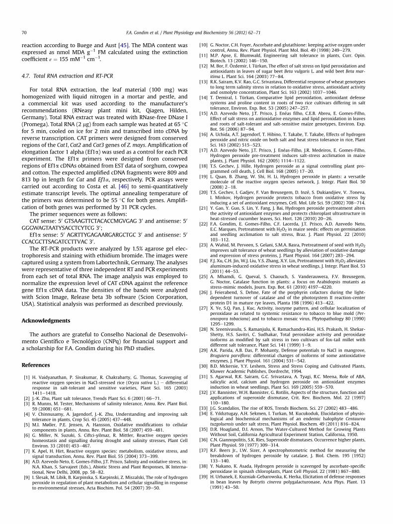
CAT activity revealed by spectrophotometric assay is shown inFig. 3A. At 48 HAS, the CAT activity in the H2O2/control plants wasca. 32% higher than in the water/control plants. At 96 and 240 HAS,the H2O2/control plants showed CAT activity values ca. 45% and 29%higher, respectively, than in the water/control plants. Additionally,the water/salt-stressed plants showed pronounced reductions inthe CAT activity. The CAT activity in the water/salt-stressed plantswas ca. 39% and 33% lower than the water/control plants at 96 and240 HAS, respectively. However, in the H2O2/salt-stressed plants,the CAT activities were ca. 18% and 19% higher than in the water/control plants at 96 and 240 HAS, respectively. Additionally, at 96and 240 HAS, CAT activity in H2O2/salt-stressed plants was ca. 92%and 75% higher than in water/salt-stressed plants, respectively(Fig. 3A).
The CAT activity in polyacrylamide gel (Fig. 3B) revealeda similar pattern to the spectrophotometric analysis. In addition,
Fig. 3. CAT activities determined by spectrophotometric (A) and polyacrylamide gel analyseand 240 h after leaf spraying. Each value corresponds to the mean � standard error of fiveletter are not statistically different for NaCl and H2O2 treatments, respectively, according toH2O2/control (HC), water/salt-stressed (WS) and H2O2/salt-stressed (HS) plants. A total of 2
two isoenzymes, here named CAT I and CAT II, were detected. CAT IIpresented with more intense bands and was detected in all of thetested conditions, while CAT I was visibly detected in the H2O2/control plants at 96 and 240 HAS.
GPX activity revealed by spectrophotometric assay is shown inFig. 4A. Significant differences in GPX activities were found at 48, 96and 240 HAS. At 48 and 96 HAS, GPX activity was ca. 16 and 14%higher in the H2O2/control plants compared to the water/controlplants, respectively. At 240 HAS, the higher GPX activity was foundin the H2O2/salt-stressed plants. It should be also observed that theGPX activity at 240 HAS was ca. 44% higher in the H2O2/controlplants and ca. 58% and 83% higher in the water/salt-stressed andH2O2/salt-stressed plants compared to the water/control plants,respectively. Additionally, GPX activity in H2O2/salt-stressed plantswas ca. 16% higher than in water/salt-stressed plants.
The GPX activity in polyacrylamide gel revealed similar patternsto the spectrophotometric analysis (Fig. 4B), and two isoenzymes,here named GPX I and II, were detected. The GPX II bands weremore intense than GPX I.
The APX activity was higher at 0 h (initial time of plant harvestejust before spraying) compared to other plants/conditions at 48, 96and 240 HAS (Fig. 5). The differences in APX activity were foundbetween the treatments at 96 and 240 HAS. At 96 HAS, the APXactivity was ca. 35% higher in the H2O2/control plants and ca. 28%lower in the water/salt-stressed compared to the water/control
s (B) in maize leaves at 0 h (initial time of plant harvest e just before spraying), 48, 96replicates. In the same harvest time values followed by the same capital letter or smallTukey’s test (P � 0.05) (A). The columns correspond to control (C), water/control (WC),0 mg of protein were loaded in each lane (B).
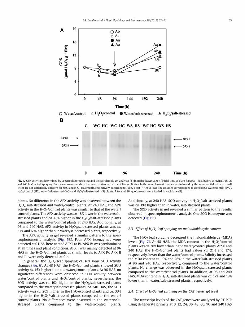
Fig. 4. GPX activities determined by spectrophotometric (A) and polyacrylamide gel analyses (B) in maize leaves at 0 h (initial time of plant harvest e just before spraying), 48, 96and 240 h after leaf spraying. Each value corresponds to the mean � standard error of five replicates. In the same harvest time values followed by the same capital letter or smallletter are not statistically different for NaCl and H2O2 treatments, respectively, according to Tukey’s test (P � 0.05) (A). The columns corresponded to control (C), water/control (WC),H2O2/control (HC), water/salt-stressed (WS) and H2O2/salt-stressed (HS) plants. A total of 20 mg of protein were loaded in each lane (B).
F.A. Gondim et al. / Plant Physiology and Biochemistry 56 (2012) 62e71 65
plants. No difference in the APX activity was observed between theH2O2/salt-stressed and water/control plants. At 240 HAS, the APXactivity in the H2O2/control plants was similar to that of the water/control plants. The APX activity was ca.18% lower in the water/salt-stressed plants and ca. 40% higher in the H2O2/salt-stressed plantscompared to the water/control plants at 240 HAS. Additionally, at96 and 240 HAS, APX activity in H2O2/salt-stressed plants was ca.37% and 69% higher than inwater/salt-stressed plants, respectively.
The APX activity in gel revealed a similar pattern to the spec-trophotometric analysis (Fig. 5B). Four APX isoenzymes weredetected at 0 HAS, here named APX I to IV. APX IV was predominantat all times and plant conditions. APX I was mainly detected at 96HAS in the H2O2/control plants at similar levels to APX IV. APX IIand III were only detected at 0 h.
In general, the H2O2 leaf spraying caused some SOD activitychanges (Fig. 6). At 48 HAS, the H2O2/control plants showed SODactivity ca. 15% higher than the water/control plants. At 96 HAS, nosignificant differences were observed in SOD activity betweenwater/control plants and H2O2/control plants, nevertheless, theSOD activity was ca. 10% higher in the H2O2/salt-stressed plantscompared to the water/salt-stressed plants. At 240 HAS, the SODactivity was ca. 20% higher in the H2O2/control plants and ca. 33%higher in the H2O2/salt-stressed plants compared to the water/control plants. No differences were observed in the water/salt-stressed plants compared to the water/control plants.
Additionally, at 240 HAS, SOD activity in H2O2/salt-stressed plantswas ca. 19% higher than in water/salt-stressed plants.
The SOD activity in gel revealed a similar pattern to the resultsobserved in spectrophotometric analysis. One SOD isoenzyme wasdetected (Fig. 6B).
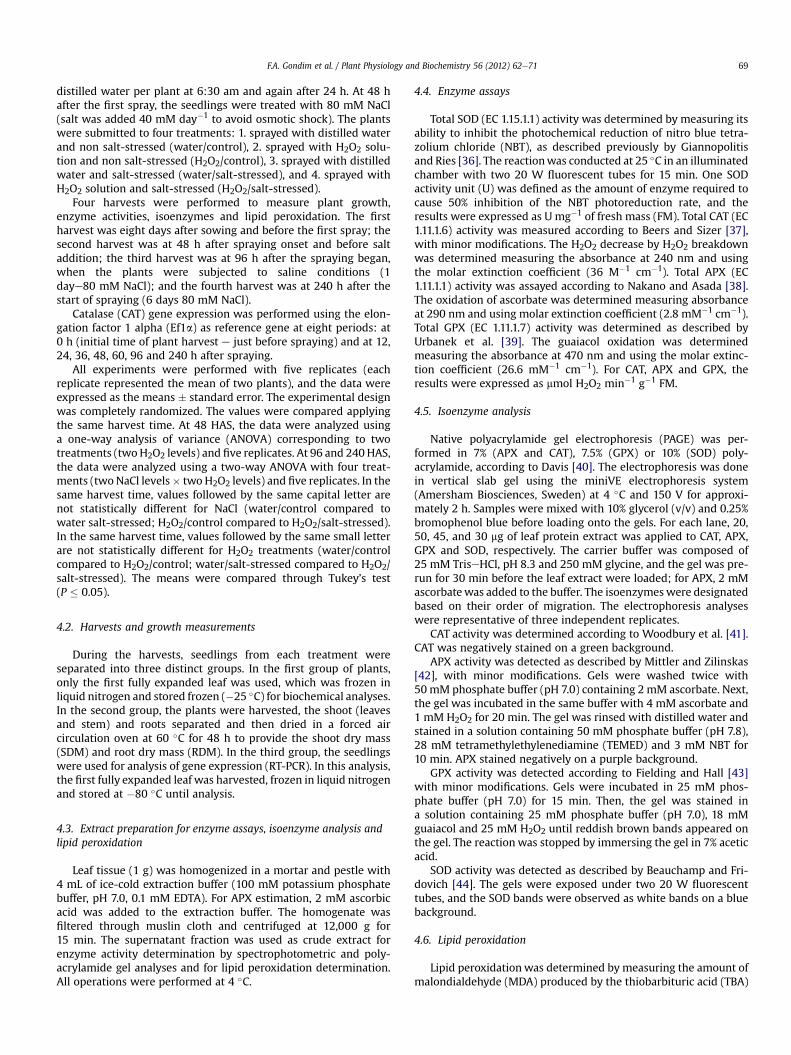
2.3. Effect of H2O2 leaf spraying on malondialdehyde content
The H2O2 leaf spraying decreased the malondialdehyde (MDA)levels (Fig. 7). At 48 HAS, the MDA content in the H2O2/controlplants was ca. 28% lower than in the water/control plants. At 96 and240 HAS, the H2O2/control plants had values ca. 21% and 17%,respectively, lower than thewater/control plants. Salinity increasedthe MDA content ca. 19% and 26% in the water/salt-stressed plantsat 96 and 240 HAS, respectively, compared to the water/controlplants. No change was observed in the H2O2/salt-stressed plantscompared to the water/control plants. In addition, at 96 and 240HAS, MDA content in H2O2/salt-stressed plants was ca.17% and 18%lower than in water/salt-stressed plants, respectively.
2.4. Effect of H2O2 leaf spraying on the CAT transcript level
The transcript levels of the CAT genes were analyzed by RT-PCRusing degenerate primers at 0, 12, 24, 36, 48, 60, 96 and 240 HAS
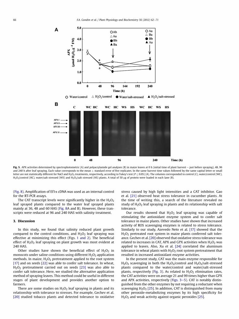
Fig. 5. APX activities determined by spectrophotometric (A) and polyacrylamide gel analyses (B) in maize leaves at 0 h (initial time of plant harvest e just before spraying), 48, 96and 240 h after leaf spraying. Each value corresponds to the mean � standard error of five replicates. In the same harvest time values followed by the same capital letter or smallletter are not statistically different for NaCl and H2O2 treatments, respectively, according to Tukey’s test (P � 0.05) (A). The columns corresponded to control (C), water/control (WC),H2O2/control (HC), water/salt-stressed (WS) and H2O2/salt-stressed (HS) plants. A total of 50 mg of protein were loaded in each lane (B).
F.A. Gondim et al. / Plant Physiology and Biochemistry 56 (2012) 62e7166
(Fig. 8). Amplification of Ef1a cDNAwas used as an internal controlfor the RT-PCR assays.
The CAT transcript levels were significantly higher in the H2O2leaf sprayed plants compared to the water leaf sprayed plantsmainly at 36, 48 and 60 HAS (Fig. 8A and B). However, these tran-scripts were reduced at 96 and 240 HAS with salinity treatment.
3. Discussion
In this study, we found that salinity reduced plant growthcompared to the control conditions, and H2O2 leaf spraying waseffective at minimizing this effect (Figs. 1 and 2). The beneficialeffect of H2O2 leaf spraying on plant growth was most evident at240 HAS.
Other studies have shown the beneficial effect of H2O2 inmonocots under saline conditions using different H2O2 applicationmethods. In maize, H2O2 pretreatment applied to the root system[17] and on seeds [22] was able to confer salt tolerance. In wheat,H2O2 pretreatment carried out on seeds [23] was also able toconfer salt tolerance. Here, we studied the alternative applicationmethod of spraying leaves. This method could be useful in differentstages of plant development and provides another option tofarmers.
There are some studies on H2O2 leaf spraying in plants and itsrelationship with tolerance to stresses. For example, Gechev et al.[20] studied tobacco plants and detected tolerance to oxidative
stress caused by high light intensities and a CAT inhibitor. Gaoet al. [21] observed heat stress tolerance in cucumber plants. Atthe time of writing this, a search of the literature revealed nostudy of H2O2 leaf spraying in plants and its relationship with salttolerance.
Our results showed that H2O2 leaf spraying was capable ofstimulating the antioxidant enzyme system and to confer salttolerance in maize plants. Other studies have shown that increasedactivity of ROS scavenging enzymes is related to stress tolerance.Similarly to our study, Azevedo Neto et al. [17] showed that theH2O2 pretreated root system in maize plants conferred salt toler-ance. Gechev et al. [20] observed that oxidative stress tolerance wasrelated to increases in CAT, APX and GPX activities when H2O2 wasapplied to leaves. Also, Xu et al. [24] correlated the aluminumtolerance in wheat plants with H2O2 root system pretreatment thatresulted in increased antioxidant enzyme activities.
In the present study, CAT was the main enzyme responsible forH2O2 scavenging in both the H2O2/control and H2O2/salt-stressedplants compared to the water/control and water/salt-stressedplants, respectively (Fig. 3). As related to H2O2 elimination rates,the CAT activities were on average 21 and 99 times higher than GPXand APX activities, respectively (Figs. 3e5). CAT is notably distin-guished from the other enzymes by not requiring a reductant whenscavenging H2O2 [25]. In addition, CAT is distinguished from manyother peroxide-metabolizing enzymes by its high specificity forH2O2 and weak activity against organic peroxides [25].
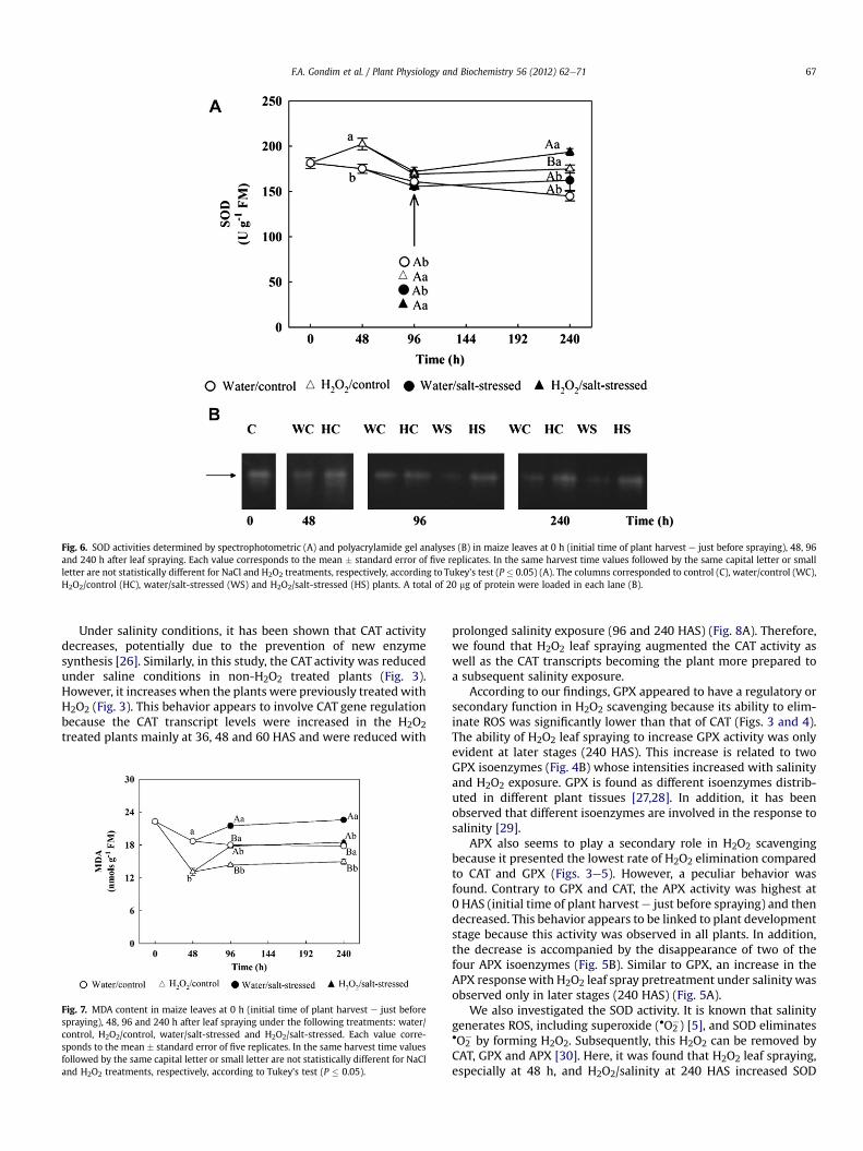
Fig. 6. SOD activities determined by spectrophotometric (A) and polyacrylamide gel analyses (B) in maize leaves at 0 h (initial time of plant harvest e just before spraying), 48, 96and 240 h after leaf spraying. Each value corresponds to the mean � standard error of five replicates. In the same harvest time values followed by the same capital letter or smallletter are not statistically different for NaCl and H2O2 treatments, respectively, according to Tukey’s test (P � 0.05) (A). The columns corresponded to control (C), water/control (WC),H2O2/control (HC), water/salt-stressed (WS) and H2O2/salt-stressed (HS) plants. A total of 20 mg of protein were loaded in each lane (B).
F.A. Gondim et al. / Plant Physiology and Biochemistry 56 (2012) 62e71 67
Under salinity conditions, it has been shown that CAT activitydecreases, potentially due to the prevention of new enzymesynthesis [26]. Similarly, in this study, the CAT activity was reducedunder saline conditions in non-H2O2 treated plants (Fig. 3).However, it increases when the plants were previously treatedwithH2O2 (Fig. 3). This behavior appears to involve CAT gene regulationbecause the CAT transcript levels were increased in the H2O2treated plants mainly at 36, 48 and 60 HAS and were reduced with
Fig. 7. MDA content in maize leaves at 0 h (initial time of plant harvest e just beforespraying), 48, 96 and 240 h after leaf spraying under the following treatments: water/control, H2O2/control, water/salt-stressed and H2O2/salt-stressed. Each value corre-sponds to the mean � standard error of five replicates. In the same harvest time valuesfollowed by the same capital letter or small letter are not statistically different for NaCland H2O2 treatments, respectively, according to Tukey’s test (P � 0.05).
prolonged salinity exposure (96 and 240 HAS) (Fig. 8A). Therefore,we found that H2O2 leaf spraying augmented the CAT activity aswell as the CAT transcripts becoming the plant more prepared toa subsequent salinity exposure.
According to our findings, GPX appeared to have a regulatory orsecondary function in H2O2 scavenging because its ability to elim-inate ROS was significantly lower than that of CAT (Figs. 3 and 4).The ability of H2O2 leaf spraying to increase GPX activity was onlyevident at later stages (240 HAS). This increase is related to twoGPX isoenzymes (Fig. 4B) whose intensities increased with salinityand H2O2 exposure. GPX is found as different isoenzymes distrib-uted in different plant tissues [27,28]. In addition, it has beenobserved that different isoenzymes are involved in the response tosalinity [29].
APX also seems to play a secondary role in H2O2 scavengingbecause it presented the lowest rate of H2O2 elimination comparedto CAT and GPX (Figs. 3e5). However, a peculiar behavior wasfound. Contrary to GPX and CAT, the APX activity was highest at0 HAS (initial time of plant harvest e just before spraying) and thendecreased. This behavior appears to be linked to plant developmentstage because this activity was observed in all plants. In addition,the decrease is accompanied by the disappearance of two of thefour APX isoenzymes (Fig. 5B). Similar to GPX, an increase in theAPX responsewith H2O2 leaf spray pretreatment under salinity wasobserved only in later stages (240 HAS) (Fig. 5A).
We also investigated the SOD activity. It is known that salinitygenerates ROS, including superoxide (�O2
�) [5], and SOD eliminates�O2
� by forming H2O2. Subsequently, this H2O2 can be removed byCAT, GPX and APX [30]. Here, it was found that H2O2 leaf spraying,especially at 48 h, and H2O2/salinity at 240 HAS increased SOD
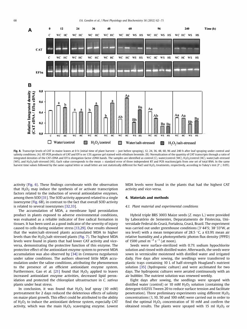
Fig. 8. Transcript levels of CAT in maize leaves at 0 h (initial time of plant harvest e just before spraying), 12, 24, 36, 48, 60, 96 and 240 h after leaf spraying under control andsalinity conditions. (A): RT-PCR products of CAT and EF1a on 1.5% agarose gel stained with ethidium bromide. (B): Normalization of the quantity of CAT transcripts through a ratio ofintegrated densities of the CAT cDNA and EF1a elongation factor cDNA bands. The samples are identified as control (C), water/control (WC), H2O2/control (HC), water/salt-stressed(WS), and H2O2/salt-stressed (HS). Each value corresponds to the mean � standard error of three independent RT and PCR reactions/gels from one set of total RNA. In the sameharvest time values followed by the same capital letter or small letter are not statistically different for NaCl and H2O2 treatments, respectively, according to Tukey’s test (P � 0.05).
F.A. Gondim et al. / Plant Physiology and Biochemistry 56 (2012) 62e7168
activity (Fig. 6). These findings corroborate with the observationthat H2O2 may induce the synthesis of or activate transcriptionfactors related to the induction of several antioxidative enzymes,among them SOD [31]. The SOD activity appeared related to a singleisoenzyme (Fig. 6B), in contrast to the fact that overall SOD activityis related to several isoenzymes [32,33].
The accumulation of MDA, a membrane lipid peroxidationproduct in plants exposed to adverse environmental conditions,was evaluated as a reliable indicator of free radical formation intissues. It has been used as a good indicator of the severity of injurycaused to cells during oxidative stress [13,29]. Our results showedthat the water/salt-stressed plants accumulated MDA to higherlevels than the H2O2/salt-stressed plants (Fig. 7). The highest MDAlevels were found in plants that had lower CAT activity and vice-versa, demonstrating the protective function of this enzyme. Theprotective effect of the antioxidant enzyme system to prevent MDAaccumulation was also observed by [34] in Centaurea tuzgoluensisunder saline conditions. The authors observed little MDA accu-mulation under the saline conditions, attributing the phenomenonto the presence of an efficient antioxidant enzyme system.Furthermore, Gao et al. [21] found that H2O2 applied to leavesincreased antioxidant enzyme activities, decreased lipid perox-idation and protected the chloroplast ultrastructure in C. sativusplants under heat stress.
In conclusion, it was found that H2O2 leaf spray (10 mM)pretreatment for 2 days reduced the deleterious effects of salinityon maize plant growth. This effect could be attributed to the abilityof H2O2 to induce the antioxidant defense system, especially CATactivity, which was the main H2O2 scavenging enzyme. Lowest
MDA levels were found in the plants that had the highest CATactivity and vice-versa.
4. Materials and methods
4.1. Plant material and experimental conditions
Hybrid triple BRS 3003 Maize seeds (Z. mays L.) were providedby Laboratório de Sementes, Deparatamento de Fitotecnia, Uni-versidade Federal do Ceará, Fortaleza, Ceará, Brazil. The experimentwas carried out under greenhouse conditions (3�440S; 38�330W, atsea level) with a mean temperature of 28.5 �C, a 63.9% mean airrelative humidity and a photosynthetic photon flux density (PPFD)of 1500 mmol m�2 s�1 (at noon).
Seeds were surface-sterilized with 0.7% sodium hypochloritesolution and rinsedwith distilled water. Afterwards, the seeds weresown in vermiculite moistened with distilled water and irrigateddaily. Five days after sowing, the seedlings were transferred toplastic trays containing 10 L of half-strength Hoagland’s nutrientsolution [35] (hydroponic culture) and were acclimated for twodays. The hydroponic cultures were aerated continuously with anair bubbler. The nutrient solution was renewed weekly.
Eight days after sowing, the seedlings were sprayed withdistilled water (control) or 10 mM H2O2 solution (containing thedetergent 0.025% Tween 20 to reduce surface tension and facilitatepenetration). Three preliminary experiments using different H2O2concentrations (1, 10, 50 and 100 mM) were carried out in order tofind the optimal H2O2 concentration of 10 mM and confirm theobtained results. The plants were sprayed with 15 ml H2O2 or
F.A. Gondim et al. / Plant Physiology and Biochemistry 56 (2012) 62e71 69
distilled water per plant at 6:30 am and again after 24 h. At 48 hafter the first spray, the seedlings were treated with 80 mM NaCl(salt was added 40 mM day�1 to avoid osmotic shock). The plantswere submitted to four treatments: 1. sprayed with distilled waterand non salt-stressed (water/control), 2. sprayed with H2O2 solu-tion and non salt-stressed (H2O2/control), 3. sprayed with distilledwater and salt-stressed (water/salt-stressed), and 4. sprayed withH2O2 solution and salt-stressed (H2O2/salt-stressed).
Four harvests were performed to measure plant growth,enzyme activities, isoenzymes and lipid peroxidation. The firstharvest was eight days after sowing and before the first spray; thesecond harvest was at 48 h after spraying onset and before saltaddition; the third harvest was at 96 h after the spraying began,when the plants were subjected to saline conditions (1daye80 mM NaCl); and the fourth harvest was at 240 h after thestart of spraying (6 days 80 mM NaCl).
Catalase (CAT) gene expression was performed using the elon-gation factor 1 alpha (Ef1a) as reference gene at eight periods: at0 h (initial time of plant harvest e just before spraying) and at 12,24, 36, 48, 60, 96 and 240 h after spraying.
All experiments were performed with five replicates (eachreplicate represented the mean of two plants), and the data wereexpressed as the means � standard error. The experimental designwas completely randomized. The values were compared applyingthe same harvest time. At 48 HAS, the data were analyzed usinga one-way analysis of variance (ANOVA) corresponding to twotreatments (twoH2O2 levels) and five replicates. At 96 and 240HAS,the data were analyzed using a two-way ANOVA with four treat-ments (twoNaCl levels� two H2O2 levels) and five replicates. In thesame harvest time, values followed by the same capital letter arenot statistically different for NaCl (water/control compared towater salt-stressed; H2O2/control compared to H2O2/salt-stressed).In the same harvest time, values followed by the same small letterare not statistically different for H2O2 treatments (water/controlcompared to H2O2/control; water/salt-stressed compared to H2O2/salt-stressed). The means were compared through Tukey’s test(P � 0.05).
4.2. Harvests and growth measurements
During the harvests, seedlings from each treatment wereseparated into three distinct groups. In the first group of plants,only the first fully expanded leaf was used, which was frozen inliquid nitrogen and stored frozen (�25 �C) for biochemical analyses.In the second group, the plants were harvested, the shoot (leavesand stem) and roots separated and then dried in a forced aircirculation oven at 60 �C for 48 h to provide the shoot dry mass(SDM) and root dry mass (RDM). In the third group, the seedlingswere used for analysis of gene expression (RT-PCR). In this analysis,the first fully expanded leaf was harvested, frozen in liquid nitrogenand stored at �80 �C until analysis.
4.3. Extract preparation for enzyme assays, isoenzyme analysis andlipid peroxidation
Leaf tissue (1 g) was homogenized in a mortar and pestle with4 mL of ice-cold extraction buffer (100 mM potassium phosphatebuffer, pH 7.0, 0.1 mM EDTA). For APX estimation, 2 mM ascorbicacid was added to the extraction buffer. The homogenate wasfiltered through muslin cloth and centrifuged at 12,000 g for15 min. The supernatant fraction was used as crude extract forenzyme activity determination by spectrophotometric and poly-acrylamide gel analyses and for lipid peroxidation determination.All operations were performed at 4 �C.
4.4. Enzyme assays
Total SOD (EC 1.15.1.1) activity was determined by measuring itsability to inhibit the photochemical reduction of nitro blue tetra-zolium chloride (NBT), as described previously by Giannopolitisand Ries [36]. The reactionwas conducted at 25 �C in an illuminatedchamber with two 20 W fluorescent tubes for 15 min. One SODactivity unit (U) was defined as the amount of enzyme required tocause 50% inhibition of the NBT photoreduction rate, and theresults were expressed as U mg�1 of fresh mass (FM). Total CAT (EC1.11.1.6) activity was measured according to Beers and Sizer [37],with minor modifications. The H2O2 decrease by H2O2 breakdownwas determined measuring the absorbance at 240 nm and usingthe molar extinction coefficient (36 M�1 cm�1). Total APX (EC1.11.1.1) activity was assayed according to Nakano and Asada [38].The oxidation of ascorbate was determined measuring absorbanceat 290 nm and using molar extinction coefficient (2.8 mM�1 cm�1).Total GPX (EC 1.11.1.7) activity was determined as described byUrbanek et al. [39]. The guaiacol oxidation was determinedmeasuring the absorbance at 470 nm and using the molar extinc-tion coefficient (26.6 mM�1 cm�1). For CAT, APX and GPX, theresults were expressed as mmol H2O2 min�1 g�1 FM.
4.5. Isoenzyme analysis
Native polyacrylamide gel electrophoresis (PAGE) was per-formed in 7% (APX and CAT), 7.5% (GPX) or 10% (SOD) poly-acrylamide, according to Davis [40]. The electrophoresis was donein vertical slab gel using the miniVE electrophoresis system(Amersham Biosciences, Sweden) at 4 �C and 150 V for approxi-mately 2 h. Samples were mixed with 10% glycerol (v/v) and 0.25%bromophenol blue before loading onto the gels. For each lane, 20,50, 45, and 30 mg of leaf protein extract was applied to CAT, APX,GPX and SOD, respectively. The carrier buffer was composed of25 mM TriseHCl, pH 8.3 and 250 mM glycine, and the gel was pre-run for 30 min before the leaf extract were loaded; for APX, 2 mMascorbatewas added to the buffer. The isoenzymeswere designatedbased on their order of migration. The electrophoresis analyseswere representative of three independent replicates.
CAT activity was determined according to Woodbury et al. [41].CAT was negatively stained on a green background.
APX activity was detected as described by Mittler and Zilinskas[42], with minor modifications. Gels were washed twice with50 mM phosphate buffer (pH 7.0) containing 2 mM ascorbate. Next,the gel was incubated in the same buffer with 4 mM ascorbate and1 mM H2O2 for 20 min. The gel was rinsed with distilled water andstained in a solution containing 50 mM phosphate buffer (pH 7.8),28 mM tetramethylethylenediamine (TEMED) and 3 mM NBT for10 min. APX stained negatively on a purple background.
GPX activity was detected according to Fielding and Hall [43]with minor modifications. Gels were incubated in 25 mM phos-phate buffer (pH 7.0) for 15 min. Then, the gel was stained ina solution containing 25 mM phosphate buffer (pH 7.0), 18 mMguaiacol and 25 mM H2O2 until reddish brown bands appeared onthe gel. The reaction was stopped by immersing the gel in 7% aceticacid.
SOD activity was detected as described by Beauchamp and Fri-dovich [44]. The gels were exposed under two 20 W fluorescenttubes, and the SOD bands were observed as white bands on a bluebackground.
4.6. Lipid peroxidation
Lipid peroxidationwas determined by measuring the amount ofmalondialdehyde (MDA) produced by the thiobarbituric acid (TBA)
F.A. Gondim et al. / Plant Physiology and Biochemistry 56 (2012) 62e7170
reaction according to Buege and Aust [45]. The MDA content wasexpressed as nmol MDA g�1 FM calculated using the extinctioncoefficient ε ¼ 155 mM�1 cm�1.
4.7. Total RNA extraction and RT-PCR
For total RNA extraction, the leaf material (100 mg) washomogenized with liquid nitrogen in a mortar and pestle, anda commercial kit was used according to the manufacturer’srecommendations (RNeasy plant mini kit, Qiagen, Hilden,Germany). Total RNA extract was treated with RNase-free DNase I(Promega). Total RNA (2 mg) from each sample was heated at 65 �Cfor 5 min, cooled on ice for 2 min and transcribed into cDNA byreverse transcription. CAT primers were designed from conservedregions of the Cat1, Cat2 and Cat3 genes of Z. mays. Amplification ofelongation factor 1 alpha (Ef1a) was used as a control for each PCRexperiment. The Ef1a primers were designed from conservedregions of Ef1a cDNAs obtained from EST data of sorghum, cowpeaand cotton. The expected amplified cDNA fragments were 809 and813 bp in length for Cat and Ef1a, respectively. PCR assays werecarried out according to Costa et al. [46] to semi-quantitativelyestimate transcript levels. The optimal annealing temperature ofthe primers was determined to be 55 �C for both genes. Amplifi-cation of both genes was performed by 31 PCR cycles.
The primer sequences were as follows:CAT sense: 50 GTSAAGTTCTACACCMGVGAG 30 and antisense: 50
GGGWAGTAATYSACCTCYTCG 30;Ef1a sense: 50 AGRTTYGAGAARGARGCTGC 30 and antisense: 50
CCACGCTTSAGATCCTTVAC 30.The RT-PCR products were analyzed by 1.5% agarose gel elec-
trophoresis and staining with ethidium bromide. The images werecaptured using a system from Labortechnik, Germany. The analyseswere representative of three independent RT and PCR experimentsfrom each set of total RNA. The image analysis was employed tonormalize the expression level of CAT cDNA against the referencegene Ef1a cDNA data. The densities of the bands were analyzedwith Scion Image, Release beta 3b software (Scion Corporation,USA). Statistical analysis was performed as described previously.
Acknowledgments
The authors are grateful to Conselho Nacional de Desenvolvi-mento Científico e Tecnológico (CNPq) for financial support anda scholarship for F.A. Gondim during his PhD studies.
References
[1] H. Vaidyanathan, P. Sivakumar, R. Chakrabarty, G. Thomas, Scavenging ofreactive oxygen species in NaCl-stressed rice (Oryza sativa L.) e differentialresponse in salt-tolerant and sensitive varieties, Plant Sci. 165 (2003)1411e1418.
[2] J.-K. Zhu, Plant salt tolerance, Trends Plant Sci. 6 (2001) 66e71.[3] R. Munns, M. Tester, Mechanisms of salinity tolerance, Annu. Rev. Plant Biol.
59 (2008) 651e681.[4] V. Chinnusamy, A. Jagendorf, J.-K. Zhu, Understanding and improving salt
tolerance in plants, Crop Sci. 45 (2005) 437e448.[5] M.I. Møller, P.E. Jensen, A. Hansson, Oxidative modifications to cellular
components in plants, Annu. Rev. Plant Biol. 58 (2007) 459e481.[6] G. Miller, N. Suzuki, S. Ciftci-yilmaz, R. Mittler, Reactive oxygen species
homeostasis and signalling during drought and salinity stresses, Plant CellEnviron. 33 (2010) 453e467.
[7] K. Apel, H. Hirt, Reactive oxygen species: metabolism, oxidative stress, andsignal transduction, Annu. Rev. Plant Biol. 55 (2004) 373e399.
[8] A.D. Azevedo Neto, E. Gomes-Filho, J.T. Prisco, Salinity and oxidative stress, in:N.A. Khan, S. Sarvajeet (Eds.), Abiotic Stress and Plant Responses, IK Interna-tional, New Delhi, 2008, pp. 58e82.
[9] I. �Slesak, M. Libik, B. Karpinska, S. Karpinski, Z. Miszalski, The role of hydrogenperoxide in regulation of plant metabolism and cellular signalling in responseto environmental stresses, Acta Biochim. Pol. 54 (2007) 39e50.
[10] G. Noctor, C.H. Foyer, Ascorbate and glutathione: keeping active oxygen undercontrol, Annu. Rev. Plant Physiol. Plant Mol. Biol. 49 (1998) 249e279.
[11] M.P. Apse, E. Blumwald, Engineering salt tolerance in plants, Curr. Opin.Biotech. 13 (2002) 146e150.
[12] M. Bor, F. Özdemir, I. Türkan, The effect of salt stress on lipid peroxidation andantioxidants in leaves of sugar beet Beta vulgaris L. and wild beet Beta mar-itima L. Plant Sci. 164 (2003) 77e84.
[13] R.K. Sairam, K.V. Rao, G.C. Srivastava, Differential response of wheat genotypesto long term salinity stress in relation to oxidative stress, antioxidant activityand osmolyte concentration, Plant Sci. 163 (2002) 1037e1046.
[14] T. Demiral, I. Türkan, Comparative lipid peroxidation, antioxidant defensesystems and proline content in roots of two rice cultivars differing in salttolerance, Environ. Exp. Bot. 53 (2005) 247e257.
[15] A.D. Azevedo Neto, J.T. Prisco, J. Enéas filho, C.E.B. Abreu, E. Gomes-Filho,Effect of salt stress on antioxidative enzymes and lipid peroxidation in leavesand roots of salt-tolerant and salt-sensitive maize genotypes, Environ. Exp.Bot. 56 (2006) 87e94.
[16] A. Uchida, A.T. Jagendorf, T. Hibino, T. Takabe, T. Takabe, Effects of hydrogenperoxide and nitric oxide on both salt and heat stress tolerance in rice, PlantSci. 163 (2002) 515e523.
[17] A.D. Azevedo Neto, J.T. Prisco, J. Enéas-Filho, J.R. Medeiros, E. Gomes-Filho,Hydrogen peroxide pre-treatment induces salt-stress acclimation in maizeplants, J. Plant Physiol. 162 (2005) 1114e1122.
[18] T.S. Gechev, J. Hille, Hydrogen peroxide as a signal controlling plant pro-grammed cell death, J. Cell Biol. 168 (2005) 17e20.
[19] L. Quan, B. Zhang, W. Shi, H. Li, Hydrogen peroxide in plants: a versatilemolecule of the reactive oxygen species network, J. Integr. Plant Biol. 50(2008) 2e18.
[20] T.S. Gechev, I. Gadjev, F. Van Breusegem, D. Inzé, S. Dukiandjiev, V. .Toneva,I. Minkov, Hydrogen peroxide protects tobacco from oxidative stress byinducing a set of antioxidant enzymes, Cell. Mol. Life Sci. 59 (2002) 708e714.
[21] Y. Gao, Y. Guo, S. Lin, Y. Fang, J. Bai, Hydrogen peroxide pretreatment altersthe activity of antioxidant enzymes and protects chloroplast ultrastructure inheat-stressed cucumber leaves, Sci. Hort. 126 (2010) 20e26.
[22] F.A. Gondim, E. Gomes-Filho, C.F. Lacerda, J.T. Prisco, A.D. Azevedo Neto,E.C. Marques, Pretreatment with H2O2 in maize seeds: effects on germinationand seedling acclimation to salt stress, Braz. J. Plant Physiol. 22 (2010)103e112.
[23] A. Wahid, M. Perveen, S. Gelani, S.M.A. Basra, Pretreatment of seed with H2O2improves salt tolerance of wheat seedlings by alleviation of oxidative damageand expression of stress proteins, J. Plant Physiol. 164 (2007) 283e294.
[24] F.J. Xu, C.H. Jin, W.J. Liu, Y.S. Zhang, X.Y. Lin, Pretreatment with H2O2 alleviatesaluminum-induced oxidative stress in wheat seedlings, J. Integr. Plant Biol. 53(2011) 44e53.
[25] A. Mhamdi, G. Queval, S. Chaouch, S. Vanderauwera, F.V. Breusegem,G. Noctor, Catalase function in plants: a focus on Arabidopsis mutants asstress-mimic models, Journ. Exp. Bot. 61 (2010) 4197e4220.
[26] J. Feierabend, S. Dehne, Fate of the porphyrin cofactors during the light-dependent turnover of catalase and of the photosystem II reaction-centerprotein D1 in mature rye leaves, Planta 198 (1996) 413e422.
[27] X. Ye, S.Q. Pan, J. Kuc, Activity, isozyme pattern, and cellular localization ofperoxidase as related to systemic resistance to tobacco to blue mold (Per-onospora tobaciana) and to tobacco mosaic virus, Phytopathology 80 (1990)1295e1299.
[28] N. Sreenivasulu, S. Ramanjulu, K. Ramachandra-Kini, H.S. Prakash, H. Shekar-Shetty, H.S. Savitri, C. Sudhakar, Total peroxidase activity and peroxidaseisoforms as modified by salt stress in two cultivars of fox-tail millet withdifferent salt tolerance, Plant Sci. 141 (1999) 1e9.
[29] A.K. Parida, A.B. Das, P. Mohanty, Defense potentials to NaCl in mangrove,Bruguiera parviflora: differential changes of isoforms of some antioxidativeenzymes, J. Plant Physiol. 161 (2004) 531e542.
[30] B.D. Mckersie, Y.Y. Leshem, Stress and Stress Coping and Cultivated Plants,Kluwer Academic Publishes, Dordrecht, 1994.
[31] S. Agarwal, R.K. Sairam, G.C. Srivastava, A. Tyagi, R.C. Meena, Role of ABA,salicylic acid, calcium and hydrogen peroxide on antioxidant enzymesinduction in wheat seedlings, Plant Sci. 169 (2005) 559e570.
[32] J.V. Bannister, W.H. Bannister, G. Rotilis, Aspects of the structure, function andapplications of superoxide dismutase, Crit. Rev. Biochem. Mol. 22 (1997)110e180.
[33] J.G. Scandalios, The rise of ROS, Trends Biochem. Sci. 27 (2002) 483e486.[34] E. Yıldıztugay, A.H. Sekmen, I. Turkan, M. Kucukoduk, Elucidation of physio-
logical and biochemical mechanisms of an endemic halophyte Centaureatuzgoluensis under salt stress, Plant Physiol. Biochem. 49 (2011) 816e824.
[35] D.R. Hoagland, D.I. Arnon, The Water-Cultured Method for Growing PlantsWithout Soil, California Agricultural Experiment Station, California, 1950.
[36] C.N. Giannopolitis, S.K. Ries, Superoxide dismutases. Occurrence higher plants,Plant Physiol. 59 (1977) 309e314.
[37] R.F. Beers Jr., I.W. Sizer, A spectrophotometric method for measuring thebreakdown of hydrogen peroxide by catalase, J. Biol. Chem. 195 (1952)133e140.
[38] Y. Nakano, K. Asada, Hydrogen peroxide is scavenged by ascorbate-specificperoxidase in spinash chloroplasts, Plant Cell Physiol. 22 (1981) 867e880.
[39] H. Urbanek, E. Kuzniak-Gebarowska, K. Herka, Elicitation of defense responsesin bean leaves by Botrytis cinerea polygalacturonase, Acta Phys. Plant. 13(1991) 43e50.

F.A. Gondim et al. / Plant Physiology and Biochemistry 56 (2012) 62e71 71
[40] B.J. Davis, Disk eletrophoresis-II: method and applications to human serumproteins, Ann. NY Acad. Sci. 121 (1964) 404e427.
[41] W. Woodbury, A.K. Spencer, M.A. Stahmann, An improved procedure usingferricyanide for detecting catalase isozymes, Anal. Biochem. 44 (1971) 301e305.
[42] R. Mittler, B.A. Zilinskas, Detection of ascorbate peroxidase activity in nativegels by inhibition of the ascorbate-dependent reduction of nitroblue tetra-zolium, Anal. Biochem. 212 (1993) 540e546.
[43] J.L. Fielding, J.L. Hall, A biochemical and cytological study of peroxidaseactivity in roots of Pisum sativum, J. Exp. Bot. 29 (1978) 969e981.
[44] C. Beauchamp, I. Fridovich, Superoxide dismutase: improved assays andan assay applicable to acrylamide gels, Anal. Biochem. 44 (1971)276e287.
[45] J.A. Buege, S.D. Aust, Microsomal lipid peroxidation, Methods Enzymol. 52(1978) 302e310.
[46] J.H. Costa, E.F. Mota, M.V. Cambursano, M.A. Lauxmann, L.M.N. de Oliveira,M.D.G. Silva Lima, E.G. Orellano, D. Fernandes de Melo, Stress-induced co-expression of two alternative oxidase (VuAox1 and 2b) genes in Vignaunguiculata, J. Plant Physiol. 167 (2010) 561e570.