Embed Size (px)
Citation preview
accelrys.com
CASE STUDY
1
Understanding the bio-chemical transformation in melanine a skin pigment is a key target or cosmaceutical and chemical manufacturers. Scientists using Materials Studio and Vamp are able to investigate these systems.
Key Products
• DMol3
• VAMP
• Discover
Industry sectors
• Chemicals
• Health&Beauty
Organizations
• Innovene
• UniversityofSanAntonio
• UniversityofCalifornia,Irvine
OPtIcal abSOrPtIOn SPectra Of MelanInS - a cOMParISOn Of theOretIcal and exPerIMental reSUlt
The oligomeric hypothesis is supported
by Atomic Force Microscopy (AFM)
characterizations of synthetic eumelanins,
formed by auto-oxidation or electrochemical
oxidation of dihydroxyindole (DHI).
Comparison of calculated absorption spectra
to experimental spectra demonstrates a
red shift in absorption with oxidation and
stacking of the eumelanin, and validates
the theoretical results. The work will lead
to the development of better sun screen-
based products, aiding the protection
against, and the prevention of, skin cancer.
Melanin is the biopolymer that accounts
for much of the coloration in nature.1,2
In humans, it is responsible for the color
of the hair, skin, and eyes. Yet the optical
absorption spectra corresponding to these
colors have a common feature: a broadband,
structure-less curve that is a monotonically
increasing function of energy.3
The shape of the absorption spectrum
of melanin also appears to relate to skin
cancer in a particular way.4 People with
red hair have a greater risk of skin cancer
from exposure to sunlight. Thus the shape
of the melanin absorption spectrum may
be efficient with regard to protection
against major diseases in humans.
Theoretical work on melanin and its
possible proto-molecules is relatively
sparse due to the size of the molecules
involved and to the unknown nature of
ResearchersatInnovene,theUniversityofSanAntonio,andtheUniversityof
California,Irvine,andAccelryshaveusedMSModeling’sDMol3,VAMP,and
Discovercodestostudytheeffectofaggregationandoxidationontheoptical
absorptionofeumelaninoligomericsheets.
CASE STUDY: MATEriAlS STUDio
2accelrys.com
the overall melanin structure. In previous work, the researchers
performed Density Functional Theory (DFT) calculations on the
monomer models.5 From the optimized structures, absorption
spectra were calculated. The results agreed well with earlier
theoretical work6 as well with published experimental work.7
Then optical spectra of melanin using a particular
structural model that assumes a poly-quinone layer
were calculated.8 From this work it was proposed that
the smooth optical absorption for melanin is obtained
by superposition of all the constituent absorptions.
The structural models used in this previous work were limited
to single sheet oligomers. In this work,9 the simulations include
aggregates of the oligomeric sheets as well as changes in
their oxidation states. These are important considerations
for several reasons. A variety of experimental probes point
to extensive stacking of oligomers in eumelanin.10
Changes in subunit oxidation state are relevant, as the darkening
and bleaching of melanin has been correlated to oxidation/
reduction reactivity, and is believed to be essential in its
natural physiological functions.11 It is therefore of great interest
to evaluate the model for melanin by simulating its optical
properties for oligomeric sheets in various oxidation states
and to compare such spectra with the experimental data.
cOMPUtatIOnal reSUltS
In previous work, an oligomeric model of eumelanin
was developed using the fully oxidized form of melanin.8
Here, this chemical model is extended by including half-
reduced and fully-reduced proto-molecules as well as the
oxidized form. Shown in Fig. 1 are the resulting energy-
optimized hexamers for the three oxidation states.
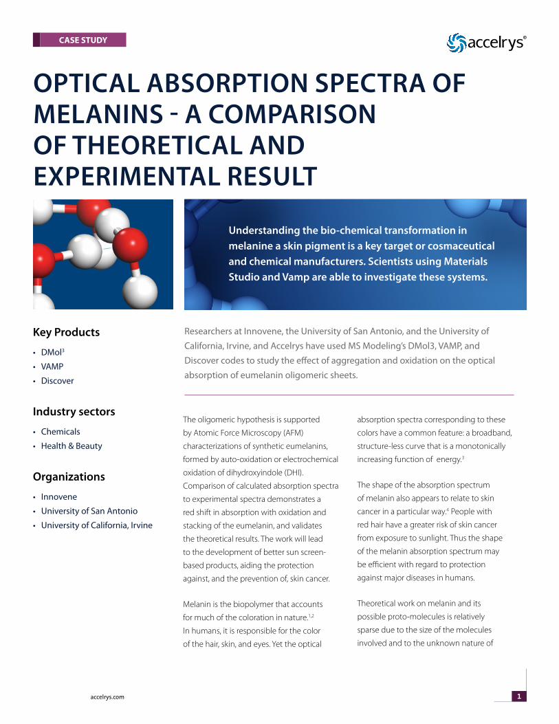
Calculated absorption spectra for the single sheet structures
are shown in Fig. 2. The absorption maximum of the reduced
form is red-shifted as compared to the oxidized form. The
predicted absorption in the red part of the spectrum is
also lower in the reduced form. The absorption spectrum
of the half-reduced form exhibits intermediate behavior,
with absorbance bands over a broad wavelength range.
The large number of possible hydrogen-bonding interactions
precluded simple energy minimization of layered structures in
the reduced and half-reduced forms; therefore, the molecular
dynamics quenching technique was applied to generate a
manifold of low-energy structures for the stacked oligomers.
Only intra-sheet hydrogen bonds are expected for the oxidized
structure, and consequently the quenching procedure led
to well defined low-energy structures in which a stacked
structure is built with the individual sheets somewhat shifted
Figure 1. DFT optimized hexamericDHI melanin structures. (a) Oxidized form, (b) half-reduced form, (c) fully reduced form. Carbon atoms are displayed in gray, oxygen atoms in red, nitrogen atoms in blue, and hydrogen atoms in white.
Figure 2. Calculated absorption spectra for single sheet hexamers of the oxidized, half-reduced and fully reduced melanin proto-molecules.
Figure 3. Calculated absorption spectra for hexamers of the oxidized form. Displayed are the spectra of the non-stacked form (1 sheet) and stacked forms with 2 and 3 sheets.
CASE STUDY: MATEriAlS STUDio
3accelrys.com
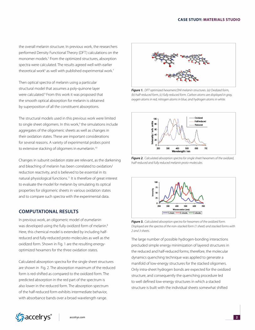
against each other. Optical spectra were calculated for the
oxidized hexamer stacked in 1 through 3 sheets, the results
being displayed in Fig. 3. Overall the spectra become more
redshifted and smooth with increasing number of sheets.
exPerIMental reSUltS
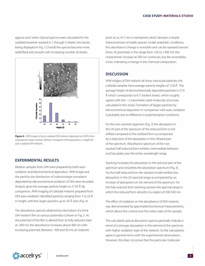
Melanin samples from DHI were prepared by both auto-
oxidation and electrochemical deposition. AFM image and
the particle size distribution of submonolayer eumelanin
deposited by electrochemical oxidation of DHI were recorded.
Analysis gives the average particle height as 27.93 Å. By
comparison, AFM imaging of colloidal melanin prepared from
DHI auto-oxidation identified particles ranging from 7 to 22 Å
in height, with few larger particles up to 35 Å seen (Fig. 4).
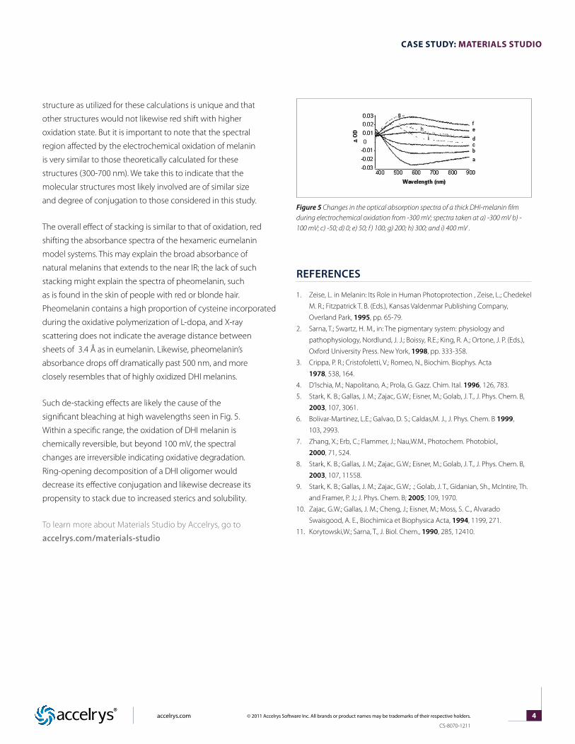
The absorbance spectra obtained by electrolysis of a thick
DHI melanin film at various potentials is shown in Fig. 5. As
the potential of the film is altered from its fully reduced state
at -300 mV, the absorbance increases above 400 nm with
increasing potential. Between -300 and 50 mV, an isobestic
point at ca. 417 nm is maintained, which denotes a simple
interconversion of stable species. Under anaerobic conditions,
this absorbance change is reversible and can be repeated several
times. At potentials in the range from +50 to +300 mV, the
characteristic increase at 500 nm continues, but the reversibility
is lost, indicating a change in the chemical composition.
dIScUSSIOn
AFM images of DHI melanin all show nanoscale particles; the
colloidal samples have average particle heights of 13.8 Å.1 The
average height of electrochemically deposited particles is 27.9
Å which corresponds to 6-7 stacked sheets, which roughly
agrees with the ~ 2 nanometer sized molecular structures
calculated in this study. Formation of bigger particles by
electrochemical deposition in comparison with auto-oxidation
is probably due to difference in polymerization conditions.
For the non-stacked oligomers (Fig. 2) the absorption in
the UV part of the spectrum of the reduced form is red-
shifted compared to the oxidized form accompanied
by a reduction of the absorption in the infrared part
of the spectrum. Absorbance spectrum of the non-
stacked half-reduced form exhibits intermediate behavior
and has peaks over the entire wavelength range.
Stacking increases the absorption in the red-end part of the
spectrum and smoothes the absorption spectrum (Fig. 3).
For the half-reduced form the stacked model exhibits less
absorption in the UV spectral range accompanied by an
increase of absorption on the red end of the spectrum. For
the fully reduced form stacking narrows the spectral range in
which the reduced form absorbs to a region of 300-500 nm.
The effect of oxidation on the absorbance of DHI-melanin
was demonstrated by spectroelectrochemical measurements,
which allows fine control over the redox state of the sample.
The calculated optical absorption spectra generally indicate a
trend of a stronger absorption in the red end of the spectrum
with higher oxidation state of the melanin. So the calculations
agree in general terms with the experimental observations.
However, this does not prove that this particular molecular
Figure 4. AFM images of auto-oxidized DHI melanin deposited on HOPG from isopropanol/ water solution. Bottom: histogram of % population vs. height for auto-oxidized DHI melanin.
CASE STUDY: MATEriAlS STUDio
4accelrys.com © 2011 Accelrys Software Inc. All brands or product names may be trademarks of their respective holders.
CS-8070-1211
structure as utilized for these calculations is unique and that
other structures would not likewise red shift with higher
oxidation state. But it is important to note that the spectral
region affected by the electrochemical oxidation of melanin
is very similar to those theoretically calculated for these
structures (300-700 nm). We take this to indicate that the
molecular structures most likely involved are of similar size
and degree of conjugation to those considered in this study.
The overall effect of stacking is similar to that of oxidation, red
shifting the absorbance spectra of the hexameric eumelanin
model systems. This may explain the broad absorbance of
natural melanins that extends to the near IR; the lack of such
stacking might explain the spectra of pheomelanin, such
as is found in the skin of people with red or blonde hair.
Pheomelanin contains a high proportion of cysteine incorporated
during the oxidative polymerization of L-dopa, and X-ray
scattering does not indicate the average distance between
sheets of 3.4 Å as in eumelanin. Likewise, pheomelanin’s
absorbance drops off dramatically past 500 nm, and more
closely resembles that of highly oxidized DHI melanins.
Such de-stacking effects are likely the cause of the
significant bleaching at high wavelengths seen in Fig. 5.
Within a specific range, the oxidation of DHI melanin is
chemically reversible, but beyond 100 mV, the spectral
changes are irreversible indicating oxidative degradation.
Ring-opening decomposition of a DHI oligomer would
decrease its effective conjugation and likewise decrease its
propensity to stack due to increased sterics and solubility.
To learn more about Materials Studio by Accelrys, go to
accelrys.com/materials-studio
referenceS
1. Zeise, L. in Melanin: Its Role in Human Photoprotection , Zeise, L.; Chedekel
M. R.; Fitzpatrick T. B. (Eds.), Kansas Valdenmar Publishing Company,
Overland Park, 1995, pp. 65-79.
2. Sarna, T.; Swartz, H. M., in: The pigmentary system: physiology and
pathophysiology, Nordlund, J. J.; Boissy, R.E.; King, R. A.; Ortone, J. P. (Eds.),
Oxford University Press. New York, 1998, pp. 333-358.
3. Crippa, P. R.; Cristofoletti, V.; Romeo, N., Biochim. Biophys. Acta
1978, 538, 164.
4. D’Ischia, M.; Napolitano, A.; Prola, G. Gazz. Chim. Ital. 1996, 126, 783.
5. Stark, K. B.; Gallas, J. M.; Zajac, G.W.; Eisner, M.; Golab, J. T., J. Phys. Chem. B,
2003, 107, 3061.
6. Bolivar-Martinez, L.E.; Galvao, D. S.; Caldas,M. J., J. Phys. Chem. B 1999,
103, 2993.
7. Zhang, X.; Erb, C.; Flammer, J.; Nau,W.M., Photochem. Photobiol.,
2000, 71, 524.
8. Stark, K. B.; Gallas, J. M.; Zajac, G.W.; Eisner, M.; Golab, J. T., J. Phys. Chem. B,
2003, 107, 11558.
9. Stark, K. B.; Gallas, J. M.; Zajac, G.W.; .; Golab, J. T., Gidanian, Sh., McIntire, Th.
and Framer, P. J.; J. Phys. Chem. B; 2005; 109, 1970.
10. Zajac, G.W.; Gallas, J. M.; Cheng, J.; Eisner, M.; Moss, S. C., Alvarado
Swaisgood, A. E., Biochimica et Biophysica Acta, 1994, 1199, 271.
11. Korytowski,W.; Sarna, T., J. Biol. Chem., 1990, 285, 12410.
Figure 5 Changes in the optical absorption spectra of a thick DHI-melanin film during electrochemical oxidation from -300 mV; spectra taken at a) -300 mV b) - 100 mV; c) -50; d) 0; e) 50; f ) 100; g) 200; h) 300; and i) 400 mV .