Embed Size (px)
Citation preview
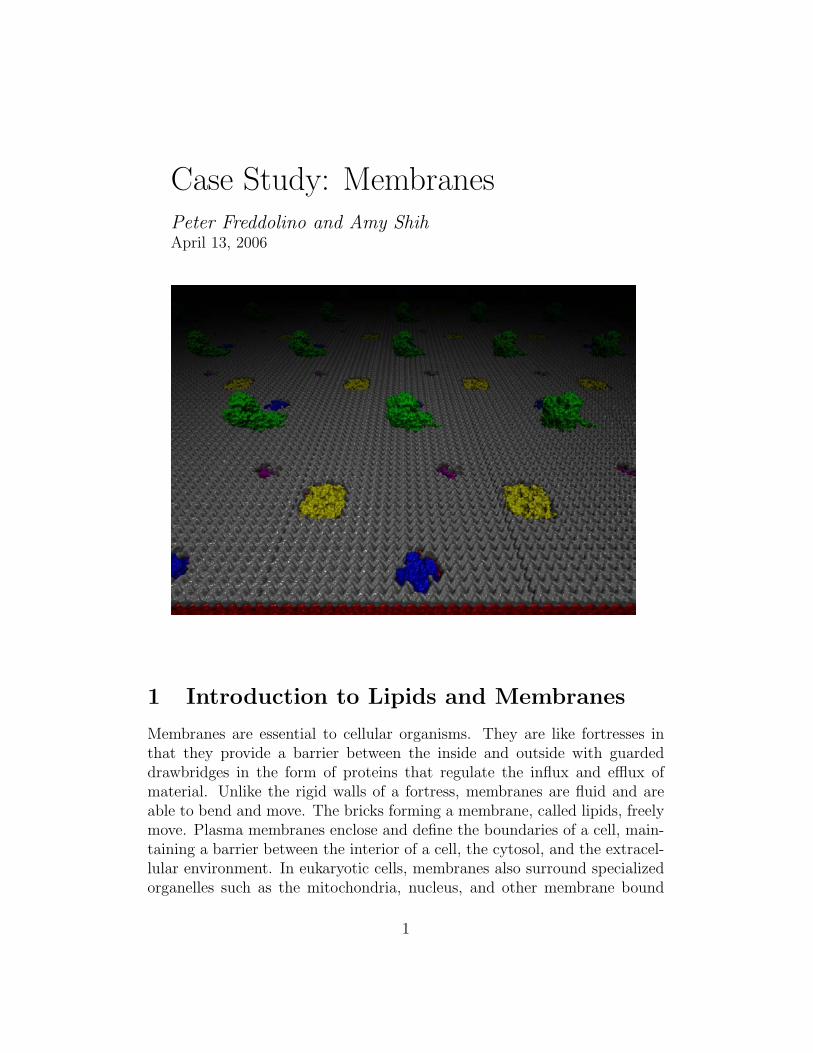
Case Study: MembranesPeter Freddolino and Amy ShihApril 13, 2006
1 Introduction to Lipids and Membranes
Membranes are essential to cellular organisms. They are like fortresses inthat they provide a barrier between the inside and outside with guardeddrawbridges in the form of proteins that regulate the influx and efflux ofmaterial. Unlike the rigid walls of a fortress, membranes are fluid and areable to bend and move. The bricks forming a membrane, called lipids, freelymove. Plasma membranes enclose and define the boundaries of a cell, main-taining a barrier between the interior of a cell, the cytosol, and the extracel-lular environment. In eukaryotic cells, membranes also surround specializedorganelles such as the mitochondria, nucleus, and other membrane bound
1
organelles. Some cells can also have large membrane bound compartmentscalled vacoules which serve a variety of functions including capturing food,sequestering toxic material, surrounding and eliminianting cellualr debris andmaintianing fluid balance (turgor). Vesicles are relatively smaller membranebound compartments which can store, transport or digest cellular productsand waste. For example, cell organelles like the endoplasmic reticulum con-tain membranes seperating the cytoplasm from their lumen. In the case ofthe rough endoplasmic reticulum, ribosomes attach to the surface releasingnewly synthesized proteins, through membrane channels, translocons, eitherinto the lumen or into the membrane itself, in the case of membrane proteins.Another example are lysosomes are membrane-bound digestive vesicles foundinside cells. They contain lysozymes (digestive enzymes) that can break downmacromolecules allowing cells to feed. The membrane of lysosomes are im-pervious to lysozymes allowing cells to break down their food without beingdigested by their own digestive enzymes [12]. Although biological membraneshave diverse functions and compositions, they are all structurally similar inthat they are comprised of a thin, 5-10 nm, layer of lipids and proteins heldtogether by primarily non-covalent interactions.
1.1 Membrane Function
The function of membranes is diverse. Membranes form continuous sheetsenclosing a defined inner compartment. This compartmentalization allowsfor specialized activities with regulated interaction with the surrounding en-vironment. Although membranes form a barrier allowing for compartmen-talization, they do not form an impenetrable barrier. Membranes are selec-tively pervious in that they allow certain molecules through but exclude othermolecules. A pure lipid bilayer is slightly permeable to water, but mostlyimpermeable to water-soluble molecules such as sodium, calcium, and potas-sium. It is the responsibility of membrane proteins to regulate the transportof such molecules. The distribution and types of membrane proteins foundwithin a particular membrane varies, thus allowing for the customizationof each interior space to the specific requirements of the cell or organelle.Imagine in our fortress analogy that drawbridges are manned by guards whocan tell the difference between friend and foe. Thus friendly material can beallowed entrance, while adverse materials can be kept at bay.
2
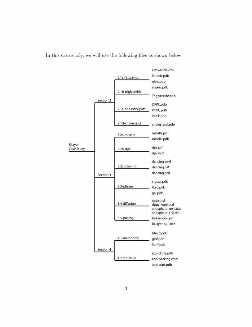
In this case study, we will use the following files as shown below.
3
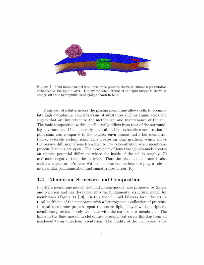
Figure 1: Fluid mosaic model with membrane proteins shown in surface representationembedded in the lipid bilayer. The hydrophobic interior of the lipid bilayer is shown inorange with the hydrophillic head groups shown in blue.
Transport of solutes across the plasma membrane allows cells to accumu-late high cytoplasmic concentrations of substances such as amino acids andsugars that are important to the metabolism and maintenance of the cell.The ionic composition within a cell usually differs from that of the surround-ing environment. Cells generally maintain a high cytosolic concentration ofpotassium ions compared to the exterior environment and a low concentra-tion of cytosolic sodium ions. This creates an ionic gradient, which allowsthe passive diffusion of ions from high to low concentration when membraneprotein channels are open. The movement of ions through channels createsan electric potential difference where the inside of the cell is roughly -70mV more negative then the exterior. Thus the plasma membrane is alsocalled a capacitor. Proteins within membranes, furthermore play a role inintercellular communication and signal transduction [10].
1.2 Membrane Structure and Composition
In 1972 a membrane model, the fluid mosaic-model, was proposed by Singerand Nicolson and has developed into the fundamental structural model formembranes (Figure 1) [10]. In this model, lipid bilayers form the struc-tural backbone of the membrane with a heterogeneous collection of proteins.Integral membrane proteins span the entire lipid bilayer while peripheralmembrane proteins loosely associate with the surface of a membrane. Thelipids in the fluid-mosaic model diffuse laterally, but rarely flip-flop from aninside-out to an outside-in orientation. The fluidity of the membrane is de-
4
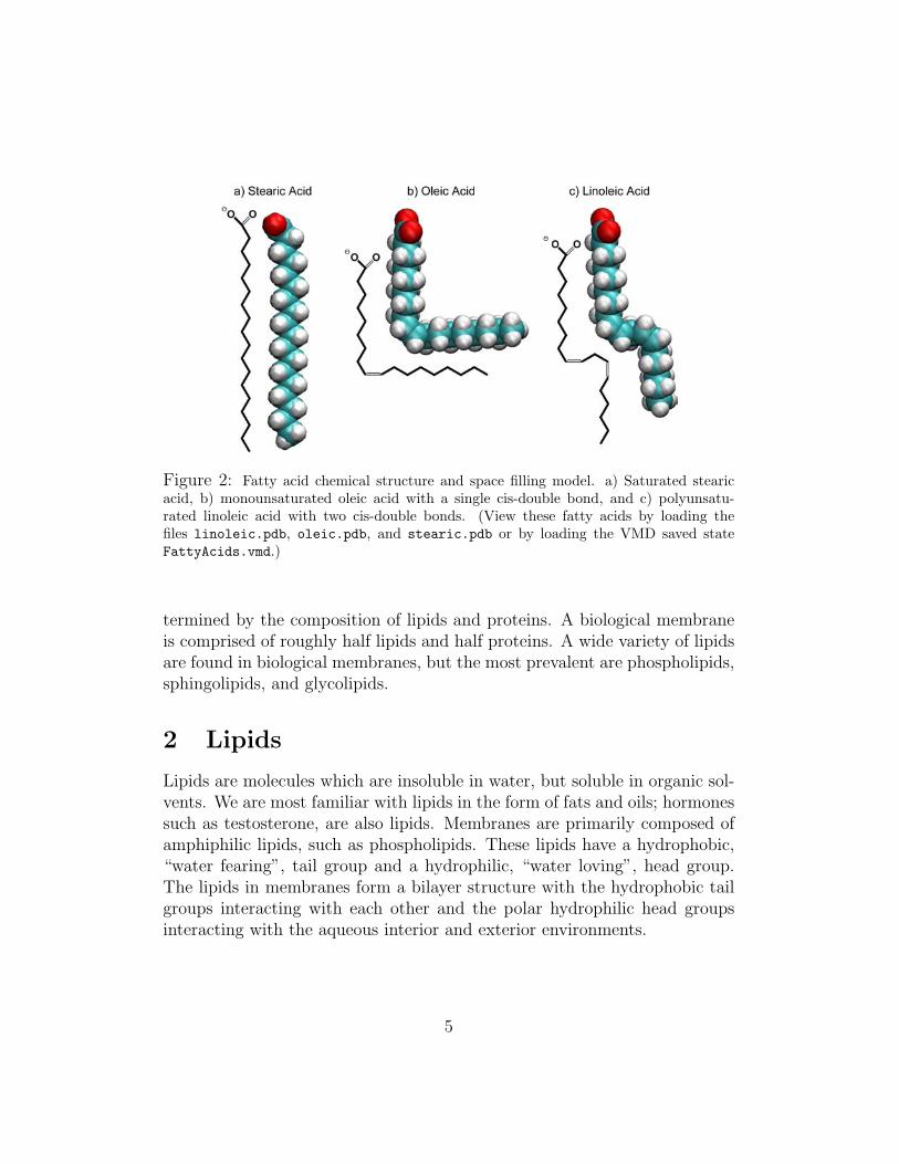
Figure 2: Fatty acid chemical structure and space filling model. a) Saturated stearicacid, b) monounsaturated oleic acid with a single cis-double bond, and c) polyunsatu-rated linoleic acid with two cis-double bonds. (View these fatty acids by loading thefiles linoleic.pdb, oleic.pdb, and stearic.pdb or by loading the VMD saved stateFattyAcids.vmd.)
termined by the composition of lipids and proteins. A biological membraneis comprised of roughly half lipids and half proteins. A wide variety of lipidsare found in biological membranes, but the most prevalent are phospholipids,sphingolipids, and glycolipids.
2 Lipids
Lipids are molecules which are insoluble in water, but soluble in organic sol-vents. We are most familiar with lipids in the form of fats and oils; hormonessuch as testosterone, are also lipids. Membranes are primarily composed ofamphiphilic lipids, such as phospholipids. These lipids have a hydrophobic,“water fearing”, tail group and a hydrophilic, “water loving”, head group.The lipids in membranes form a bilayer structure with the hydrophobic tailgroups interacting with each other and the polar hydrophilic head groupsinteracting with the aqueous interior and exterior environments.
5
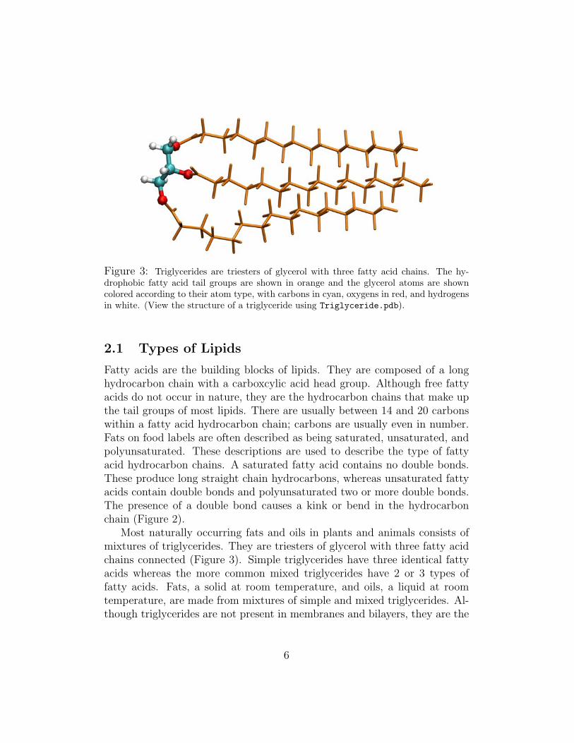
Figure 3: Triglycerides are triesters of glycerol with three fatty acid chains. The hy-drophobic fatty acid tail groups are shown in orange and the glycerol atoms are showncolored according to their atom type, with carbons in cyan, oxygens in red, and hydrogensin white. (View the structure of a triglyceride using Triglyceride.pdb).
2.1 Types of Lipids
Fatty acids are the building blocks of lipids. They are composed of a longhydrocarbon chain with a carboxcylic acid head group. Although free fattyacids do not occur in nature, they are the hydrocarbon chains that make upthe tail groups of most lipids. There are usually between 14 and 20 carbonswithin a fatty acid hydrocarbon chain; carbons are usually even in number.Fats on food labels are often described as being saturated, unsaturated, andpolyunsaturated. These descriptions are used to describe the type of fattyacid hydrocarbon chains. A saturated fatty acid contains no double bonds.These produce long straight chain hydrocarbons, whereas unsaturated fattyacids contain double bonds and polyunsaturated two or more double bonds.The presence of a double bond causes a kink or bend in the hydrocarbonchain (Figure 2).
Most naturally occurring fats and oils in plants and animals consists ofmixtures of triglycerides. They are triesters of glycerol with three fatty acidchains connected (Figure 3). Simple triglycerides have three identical fattyacids whereas the more common mixed triglycerides have 2 or 3 types offatty acids. Fats, a solid at room temperature, and oils, a liquid at roomtemperature, are made from mixtures of simple and mixed triglycerides. Al-though triglycerides are not present in membranes and bilayers, they are the
6
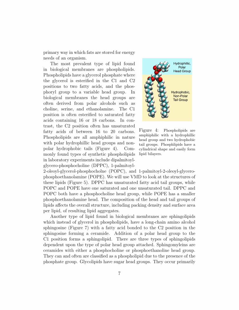
Figure 4: Phospholipids areamphiphilic with a hydrophillichead group and two hydrophobictail groups. Phosphlipids have acylindrical shape and easily formlipid bilayers.
primary way in which fats are stored for energyneeds of an organism.
The most prevalent type of lipid foundin biological membranes are phospholipids.Phospholipids have a glycerol phosphate wherethe glycerol is esterified in the C1 and C2positions to two fatty acids, and the phos-phoryl group to a variable head group. Inbiological membranes the head groups areoften derived from polar alcohols such ascholine, serine, and ethanolamine. The C1position is often esterified to saturated fattyacids containing 16 or 18 carbons. In con-trast, the C2 position often has unsaturatedfatty acids of between 16 to 20 carbons.Phospholipids are all amphiphilic in naturewith polar hydrophillic head groups and non-polar hydrophobic tails (Figure 4). Com-monly found types of synthetic phospholipidsin laboratory experiments include dipalmitoyl-glycero-phosphocholine (DPPC), 1-palmitoyl-2-oleoyl-glycerol-phosphocholne (POPC), and 1-palmitoyl-2-oleoyl-glycero-phosphoethanolamine (POPE). We will use VMD to look at the structures ofthese lipids (Figure 5). DPPC has unsaturated fatty acid tail groups, whilePOPC and POPE have one saturated and one unsaturated tail. DPPC andPOPC both have a phosphocholine head group, while POPE has a smallerphosphoethanolamine head. The composition of the head and tail groups oflipids affects the overall structure, including packing density and surface areaper lipid, of resulting lipid aggregates.
Another type of lipid found in biological membranes are sphingolipidswhich instead of glycerol in phospholipids, have a long-chain amino alcoholsphingosine (Figure 7) with a fatty acid bonded to the C2 position in thesphingosine forming a ceramide. Addition of a polar head group to theC1 position forms a sphingolipid. There are three types of sphingolipidsdependent upon the type of polar head group attached. Sphingomyleins areceramides with either a phosphocholine or phosphoethanoline head group.They can and often are classified as a phospholipid due to the presence of thephosphate group. Glycolipids have sugar head groups. They occur primarily
7
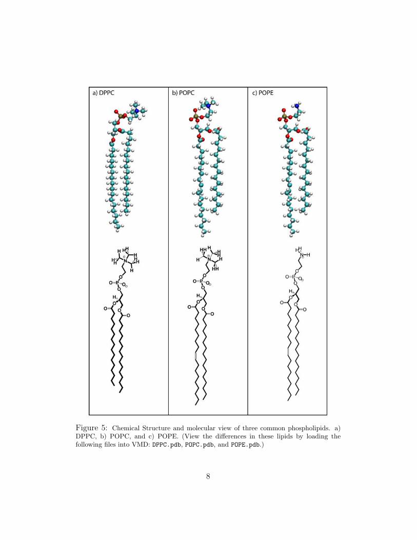
Figure 5: Chemical Structure and molecular view of three common phospholipids. a)DPPC, b) POPC, and c) POPE. (View the differences in these lipids by loading thefollowing files into VMD: DPPC.pdb, POPC.pdb, and POPE.pdb.)
8
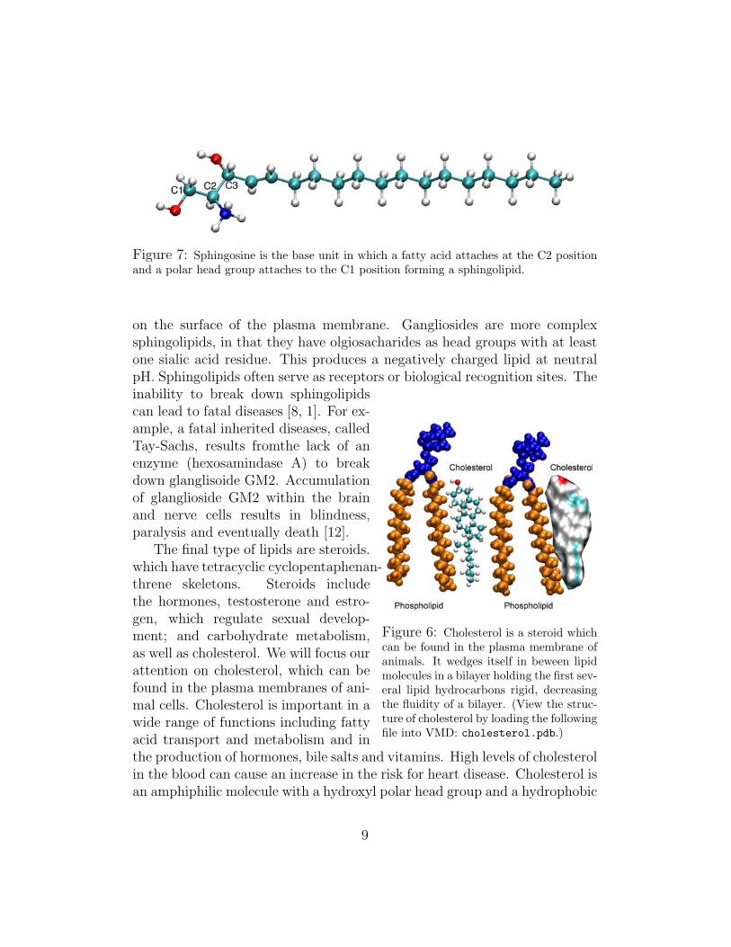
Figure 7: Sphingosine is the base unit in which a fatty acid attaches at the C2 positionand a polar head group attaches to the C1 position forming a sphingolipid.
on the surface of the plasma membrane. Gangliosides are more complexsphingolipids, in that they have olgiosacharides as head groups with at leastone sialic acid residue. This produces a negatively charged lipid at neutralpH. Sphingolipids often serve as receptors or biological recognition sites. The
Figure 6: Cholesterol is a steroid whichcan be found in the plasma membrane ofanimals. It wedges itself in beween lipidmolecules in a bilayer holding the first sev-eral lipid hydrocarbons rigid, decreasingthe fluidity of a bilayer. (View the struc-ture of cholesterol by loading the followingfile into VMD: cholesterol.pdb.)
inability to break down sphingolipidscan lead to fatal diseases [8, 1]. For ex-ample, a fatal inherited diseases, calledTay-Sachs, results fromthe lack of anenzyme (hexosamindase A) to breakdown glanglisoide GM2. Accumulationof glanglioside GM2 within the brainand nerve cells results in blindness,paralysis and eventually death [12].
The final type of lipids are steroids.which have tetracyclic cyclopentaphenan-threne skeletons. Steroids includethe hormones, testosterone and estro-gen, which regulate sexual develop-ment; and carbohydrate metabolism,as well as cholesterol. We will focus ourattention on cholesterol, which can befound in the plasma membranes of ani-mal cells. Cholesterol is important in awide range of functions including fattyacid transport and metabolism and inthe production of hormones, bile salts and vitamins. High levels of cholesterolin the blood can cause an increase in the risk for heart disease. Cholesterol isan amphiphilic molecule with a hydroxyl polar head group and a hydrophobic
9
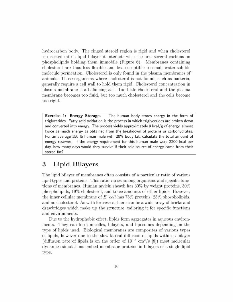
hydrocarbon body. The ringed steroid region is rigid and when cholesterolis inserted into a lipid bilayer it interacts with the first several carbons onphospholipids holding them immobile (Figure 6). Membranes containingcholesterol are thus less flexible and less suseptible to small water-solublemolecule permeation. Cholesterol is only found in the plasma membranes ofanimals. Those organisms where cholesterol is not found, such as bacteria,generally require a cell wall to hold them rigid. Cholesterol concentration inplasma membrane is a balancing act. Too little cholesterol and the plasmamembrane becomes too fluid, but too much cholesterol and the cells becometoo rigid.
Exercise 1: Energy Storage. The human body stores energy in the form oftriglycerides. Fatty acid oxidation is the process in which triglycerides are broken downand converted into energy. The process yields approximately 9 kcal/g of energy, almosttwice as much energy as obtained from the breakdown of proteins or carbohydrates.For an average 150 lb human male with 20% body fat, calculate the total amount ofenergy reserves. If the energy requirement for this human male were 2200 kcal perday, how many days would they survive if their sole source of energy came from theirstored fat?
3 Lipid Bilayers
The lipid bilayer of membranes often consists of a particular ratio of variouslipid types and proteins. This ratio varies among organisms and specific func-tions of membranes. Human mylein sheath has 30% by weight proteins, 30%phospholipids, 19% cholesterol, and trace amounts of other lipids. However,the inner cellular membrane of E. coli has 75% proteins, 25% phospholipids,and no cholesterol. As with fortresses, there can be a wide array of bricks anddrawbridges which make up the structure, tailoring it for specific functionsand environments.
Due to the hydrophobic effect, lipids form aggregates in aqueous environ-ments. They can form micelles, bilayers, and liposomes depending on thetype of lipids used. Biological membranes are composites of various typesof lipids, however due to the slow lateral diffusion of lipids within a bilayer(diffusion rate of lipids is on the order of 10−8 cm2/s [8]) most moleculardynamics simulations embed membrane proteins in bilayers of a single lipidtype.
10
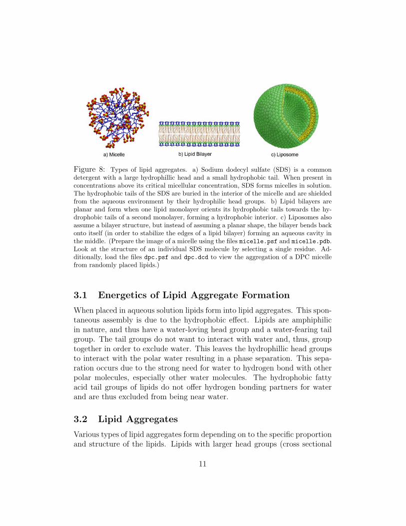
Figure 8: Types of lipid aggregates. a) Sodium dodecyl sulfate (SDS) is a commondetergent with a large hydrophillic head and a small hydrophobic tail. When present inconcentrations above its critical micellular concentration, SDS forms micelles in solution.The hydrophobic tails of the SDS are buried in the interior of the micelle and are shieldedfrom the aqueous environment by their hydrophilic head groups. b) Lipid bilayers areplanar and form when one lipid monolayer orients its hydrophobic tails towards the hy-drophobic tails of a second monolayer, forming a hydrophobic interior. c) Liposomes alsoassume a bilayer structure, but instead of assuming a planar shape, the bilayer bends backonto itself (in order to stabilize the edges of a lipid bilayer) forming an aqueous cavity inthe middle. (Prepare the image of a micelle using the files micelle.psf and micelle.pdb.Look at the structure of an individual SDS molecule by selecting a single residue. Ad-ditionally, load the files dpc.psf and dpc.dcd to view the aggregation of a DPC micellefrom randomly placed lipids.)
3.1 Energetics of Lipid Aggregate Formation
When placed in aqueous solution lipids form into lipid aggregates. This spon-taneous assembly is due to the hydrophobic effect. Lipids are amphiphilicin nature, and thus have a water-loving head group and a water-fearing tailgroup. The tail groups do not want to interact with water and, thus, grouptogether in order to exclude water. This leaves the hydrophillic head groupsto interact with the polar water resulting in a phase separation. This sepa-ration occurs due to the strong need for water to hydrogen bond with otherpolar molecules, especially other water molecules. The hydrophobic fattyacid tail groups of lipids do not offer hydrogen bonding partners for waterand are thus excluded from being near water.
3.2 Lipid Aggregates
Various types of lipid aggregates form depending on to the specific proportionand structure of the lipids. Lipids with larger head groups (cross sectional
11
area) than that of their hydrophobic fatty acid side chain tend to form mi-celles in solution. Micelles are spherical structures in which the hydrophobiclipid tails are buried in the interior core and shielded from water (Figure8a). The polar head groups are at the surface interacting with the aque-ous environment. Micelles require a minimum concentration of lipids, suchas the detergent sodium dodecyl sulfate, in order to form. This minimumconcentration is called the critical micelle concentration.
Another type of lipid aggregate is the bilayer, in which a sheet is formedwith two lipid monolayers. The hydrophobic tails of one lipid monolayer areoriented towards the hydrophobic tails of the second monolayer (Figure 8b).This type of lipid aggregate forms when the lipid head group and tail groupshave roughly similar cross sectional areas. However this type of aggregateleaves exposed hydrophobic tail groups at the edges of the bilayer. To furtherstabilize the structure, lipid bilayers fold back onto themselves and sponta-neously form into liposomes forming an aqueous cavity inside the aggregate(Figure 8c).
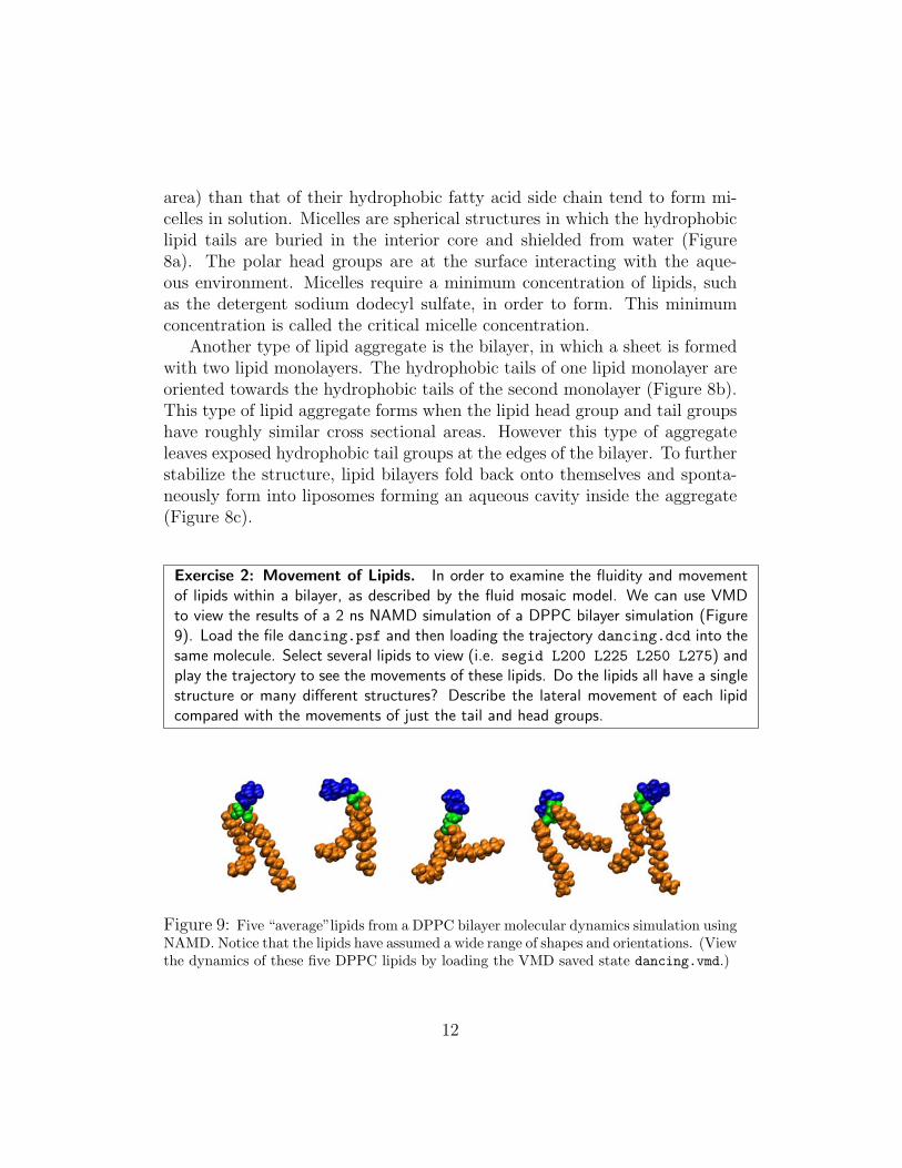
Exercise 2: Movement of Lipids. In order to examine the fluidity and movementof lipids within a bilayer, as described by the fluid mosaic model. We can use VMDto view the results of a 2 ns NAMD simulation of a DPPC bilayer simulation (Figure9). Load the file dancing.psf and then loading the trajectory dancing.dcd into thesame molecule. Select several lipids to view (i.e. segid L200 L225 L250 L275) andplay the trajectory to see the movements of these lipids. Do the lipids all have a singlestructure or many different structures? Describe the lateral movement of each lipidcompared with the movements of just the tail and head groups.
Figure 9: Five “average”lipids from a DPPC bilayer molecular dynamics simulation usingNAMD. Notice that the lipids have assumed a wide range of shapes and orientations. (Viewthe dynamics of these five DPPC lipids by loading the VMD saved state dancing.vmd.)
12
3.3 Lipid Phases
Even within the realm of lipid bilayers, there are a number of different‘phases’ that the lipids can take, analogous to the classical solid-liquid-gasprogression of most substances. In the case of lipids, the three most impor-tant states are the liquid (Lα), gel (L′
β), and crystal states. Depending onthe lipid in question, other states, such as hexagonally packed tubes, mayoccur [17].
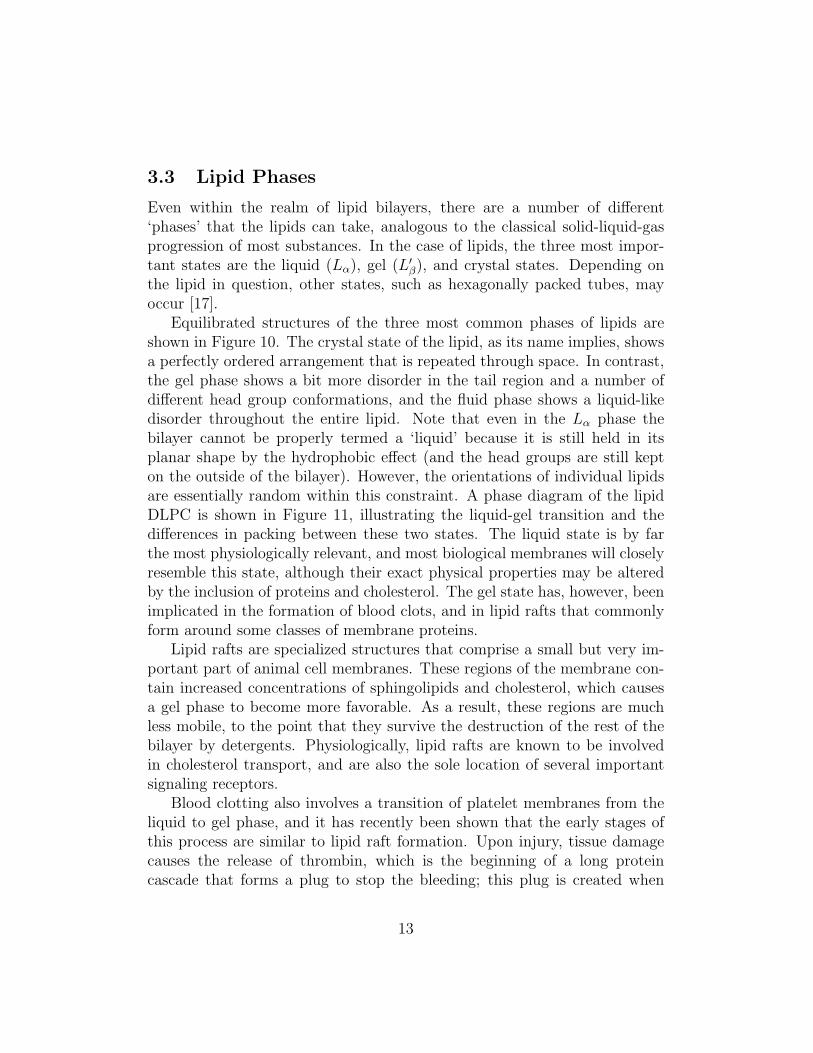
Equilibrated structures of the three most common phases of lipids areshown in Figure 10. The crystal state of the lipid, as its name implies, showsa perfectly ordered arrangement that is repeated through space. In contrast,the gel phase shows a bit more disorder in the tail region and a number ofdifferent head group conformations, and the fluid phase shows a liquid-likedisorder throughout the entire lipid. Note that even in the Lα phase thebilayer cannot be properly termed a ‘liquid’ because it is still held in itsplanar shape by the hydrophobic effect (and the head groups are still kepton the outside of the bilayer). However, the orientations of individual lipidsare essentially random within this constraint. A phase diagram of the lipidDLPC is shown in Figure 11, illustrating the liquid-gel transition and thedifferences in packing between these two states. The liquid state is by farthe most physiologically relevant, and most biological membranes will closelyresemble this state, although their exact physical properties may be alteredby the inclusion of proteins and cholesterol. The gel state has, however, beenimplicated in the formation of blood clots, and in lipid rafts that commonlyform around some classes of membrane proteins.
Lipid rafts are specialized structures that comprise a small but very im-portant part of animal cell membranes. These regions of the membrane con-tain increased concentrations of sphingolipids and cholesterol, which causesa gel phase to become more favorable. As a result, these regions are muchless mobile, to the point that they survive the destruction of the rest of thebilayer by detergents. Physiologically, lipid rafts are known to be involvedin cholesterol transport, and are also the sole location of several importantsignaling receptors.
Blood clotting also involves a transition of platelet membranes from theliquid to gel phase, and it has recently been shown that the early stages ofthis process are similar to lipid raft formation. Upon injury, tissue damagecauses the release of thrombin, which is the beginning of a long proteincascade that forms a plug to stop the bleeding; this plug is created when
13
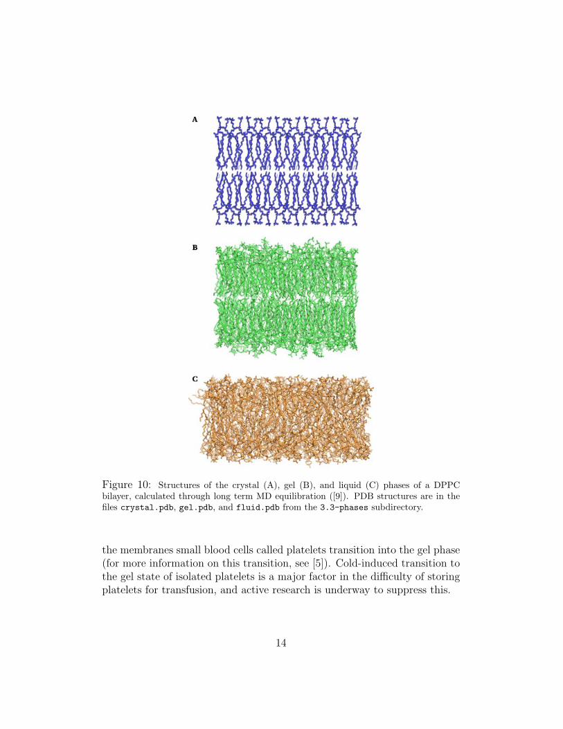
Figure 10: Structures of the crystal (A), gel (B), and liquid (C) phases of a DPPCbilayer, calculated through long term MD equilibration ([9]). PDB structures are in thefiles crystal.pdb, gel.pdb, and fluid.pdb from the 3.3-phases subdirectory.
the membranes small blood cells called platelets transition into the gel phase(for more information on this transition, see [5]). Cold-induced transition tothe gel state of isolated platelets is a major factor in the difficulty of storingplatelets for transfusion, and active research is underway to suppress this.
14
Figure 11: Phase diagram of the lipid DPPC, showing the transition between the gel andliquid phases. A diagram of the crystal phase is shown at right for comparison. Structureswere obtained from [9].
Exercise 3: Physical Properties of Lipid Phases. Load the structures of the threelipid phases illustrated in Figure 10 into VMD from the files crystal.pdb, gel.pdb,and fluid.pdb in the 3.3-phases subdirectory. Each structure contains a total of200 DPPC lipids. Use VMD to calculate the height of the bilayer, surface area perlipid, and density of the bilayer for each of these phases. You should use the commandmeasure minmax $sel to obtain the approximate dimensions of the bilayer in eachcase (be sure that $sel contains only lipids); observe that the bilayer is oriented in thex-z plane, with its height along the y axis. Explain possible reasons for the differencesin these characteristics that you see in these states, based on the packing of lipids ineach phase. Compare the values you obtained with experimental values [11].
3.4 Lipid Diffusion Through and Across the Bilayer
Biological membranes are not rigid structures, and diffusion both in andacross the bilayer is possible for small, apolar molecules. Within the plane
15
of the membrane, most lipids have diffusion coefficients on the order of 10−8
cm2/s. Simple diffusion in two dimensions is governed by the equation:
〈r2〉 = 4Dt (1)
In this equation, 〈r2〉 is the expected value of the square of the distance agiven particle has travelled from its starting point. This means that it wouldbe expected to take on the order of 10 ms for a lipid to diffuse 100 nm inthe plane of the membrane (see Appendix I for a detailed derivation of thediffusion law and the calculations involved for lipid diffusion). This is notan unreasonably long time, and indeed lipid diffusion and mixing are veryrapid on biological time scales. In the world of simulations, however, this isan incredibly lengthy process; a ‘long’ simulation of large biological systemswill generally run for a few tens of nanoseconds, and on such time scales onewould expect lipids to diffuse a distance on the order of a few angstroms.
Exercise 4: Lipid Diffusion in the Bilayer. The results of a molecular dynamicssimulation of a lipid bilayer are contained in the files dppc.psf and dppc equi.dcdfrom the 3.4-diffusion. The mean square deviation of phosphorus atoms as afunction of time has been extracted from this simulation, and can be found in thefile phosphate msd.dat as a set of time/value pairs, with time in picoseconds. Usethis data and equation 1 to calculate the observed diffusion coefficient of lipids inthe bilayer in these simulations. Is this result reasonable? Observe the data fromthis file in graph form; how long from the start of the simulation does it take for thesystem to begin observing the diffusion law? The deviation of five selected phosphorusatoms from their starting position as a function of time has been placed in the filesphosphate#.dat, with # an integer between 1 and 5. Do any of these individualatoms come close to obeying the diffusion law?
In addition to diffusing within the bilayer, it is also conceivable for lipidsto flip across it. In a pure bilayer, however, such an event is almost in-considerably rare; bilayers require around a few weeks to equilibrate in thisway in the absence of proteins. However, lipid flipping is known to be veryimportant for some signaling processes, as well as in properly regulating theleaflet composition of the bilayer. In living cells specialized enzymes calledflippases are known to facilitate this process, bringing lipid flipping into amuch more reasonable time scale. The flipping of lipids and other hydropho-bic molecules contained in the bilayer is important for processes includingproper lipid composition, blood clotting, and resistance of bacteria and can-cer cells to a number of drugs [14, 7].
16
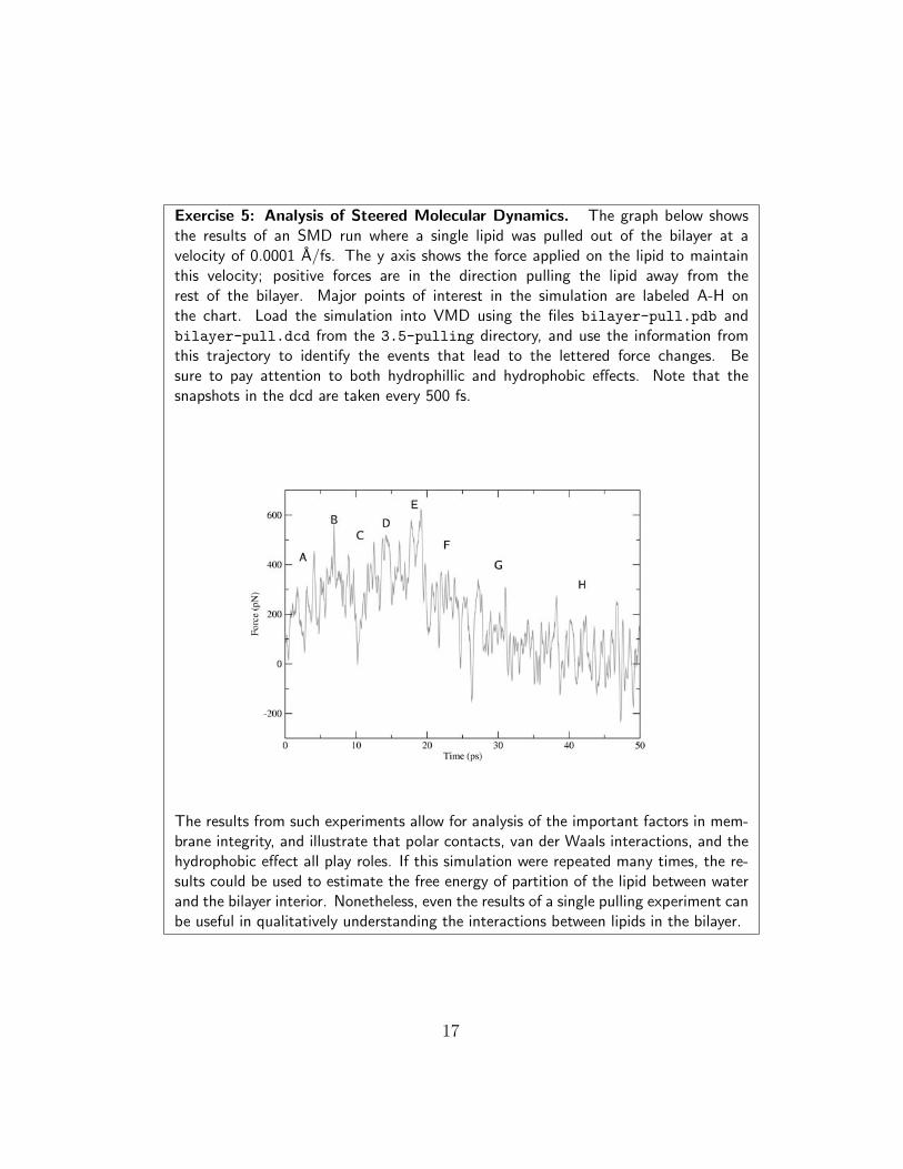
Exercise 5: Analysis of Steered Molecular Dynamics. The graph below showsthe results of an SMD run where a single lipid was pulled out of the bilayer at avelocity of 0.0001 A/fs. The y axis shows the force applied on the lipid to maintainthis velocity; positive forces are in the direction pulling the lipid away from therest of the bilayer. Major points of interest in the simulation are labeled A-H onthe chart. Load the simulation into VMD using the files bilayer-pull.pdb andbilayer-pull.dcd from the 3.5-pulling directory, and use the information fromthis trajectory to identify the events that lead to the lettered force changes. Besure to pay attention to both hydrophillic and hydrophobic effects. Note that thesnapshots in the dcd are taken every 500 fs.
The results from such experiments allow for analysis of the important factors in mem-brane integrity, and illustrate that polar contacts, van der Waals interactions, and thehydrophobic effect all play roles. If this simulation were repeated many times, the re-sults could be used to estimate the free energy of partition of the lipid between waterand the bilayer interior. Nonetheless, even the results of a single pulling experiment canbe useful in qualitatively understanding the interactions between lipids in the bilayer.
17
3.5 Lipid-Lipid Interactions
As discussed in Section 3.1, the primary impetus for lipid bilayer assemblyis the hydrophobic effect, which drives the tails of lipids to aggregate andescape an aqueous medium. Aside from the hydrophobic effect, there area number of other interactions which can take place between neighboringlipids to stabilize the bilayer. In addition to the hydrophobic effect, the longhydrophobic tails of lipids are attracted through van der Waals interactions,which although weak can have a significant impact when there are large areasof surface contact between molecules. In the hydrophillic head region, mostbiological lipids have a negatively charged phosphate group attached to asecond positively charged or polar group. These regions of the lipids interactfavorably with water, but interactions between positive and negative groupson the heads of neighboring lipids can also have a powerful influence on lipidstructure; these interactions have an impact on the surface area of each lipidand cause the heads of most lipids to bend rather than staying perpendicularto the membrane surface.
4 The Bilayer and Embedded Proteins
The ability of the membrane bilayer to block transit of large molecules intoand out of the cell is precisely what makes it useful for separating and pro-tecting the cell from its environment. If all cells were surrounded only by apure lipid bilayer they would be unable to obtain food, maintain an osmoticbalance with the environment, or communicate with other cells. Living cellsaccomplish all these tasks using transmembrane (TM) proteins : a class ofproteins which span the entirety of the lipid bilayer. Such proteins comprisean average of 50% of the mass of biological membranes, and are responsiblefor such tasks as selectively allowing certain substances through the mem-brane, pumping important ions and small molecules into or out of the cell,and activating intracellular signaling pathways in response to extracellularsignals.
4.1 Membrane Protein Structure and Lipid Packing
Most soluble proteins follow a well-studied formula in their structure, con-sisting of a hydrophobic core that lends stability to the folded state, and a
18
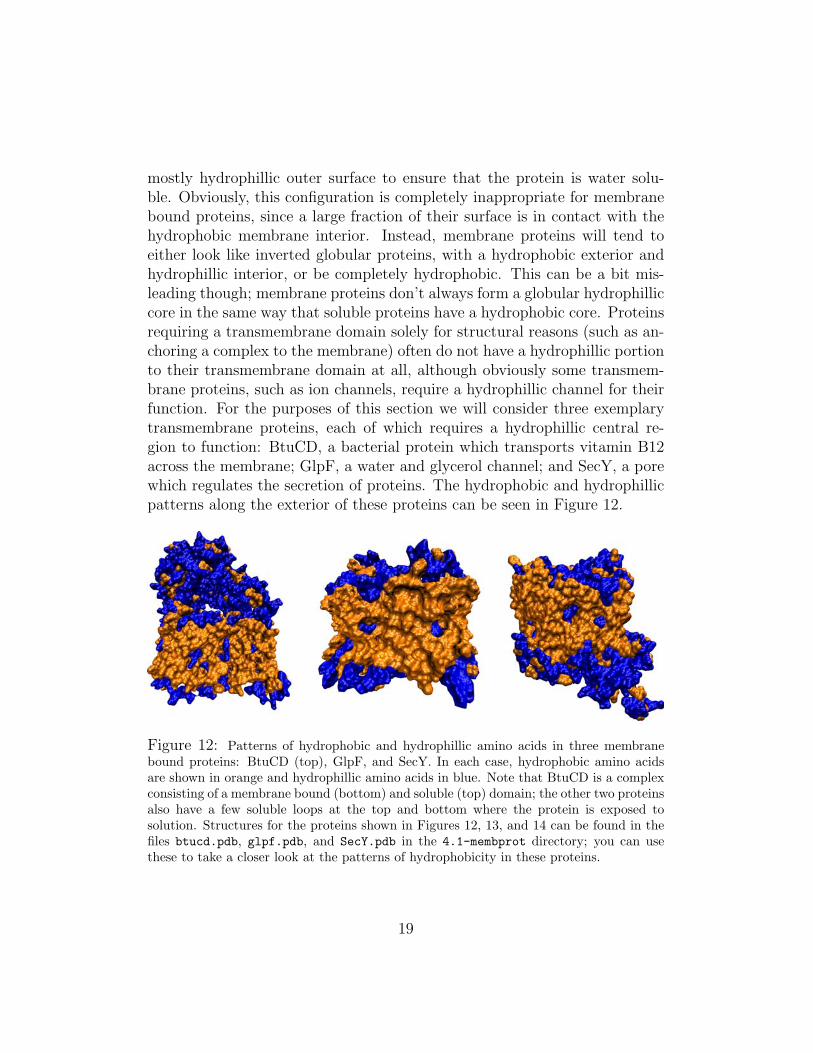
mostly hydrophillic outer surface to ensure that the protein is water solu-ble. Obviously, this configuration is completely inappropriate for membranebound proteins, since a large fraction of their surface is in contact with thehydrophobic membrane interior. Instead, membrane proteins will tend toeither look like inverted globular proteins, with a hydrophobic exterior andhydrophillic interior, or be completely hydrophobic. This can be a bit mis-leading though; membrane proteins don’t always form a globular hydrophilliccore in the same way that soluble proteins have a hydrophobic core. Proteinsrequiring a transmembrane domain solely for structural reasons (such as an-choring a complex to the membrane) often do not have a hydrophillic portionto their transmembrane domain at all, although obviously some transmem-brane proteins, such as ion channels, require a hydrophillic channel for theirfunction. For the purposes of this section we will consider three exemplarytransmembrane proteins, each of which requires a hydrophillic central re-gion to function: BtuCD, a bacterial protein which transports vitamin B12across the membrane; GlpF, a water and glycerol channel; and SecY, a porewhich regulates the secretion of proteins. The hydrophobic and hydrophillicpatterns along the exterior of these proteins can be seen in Figure 12.
Figure 12: Patterns of hydrophobic and hydrophillic amino acids in three membranebound proteins: BtuCD (top), GlpF, and SecY. In each case, hydrophobic amino acidsare shown in orange and hydrophillic amino acids in blue. Note that BtuCD is a complexconsisting of a membrane bound (bottom) and soluble (top) domain; the other two proteinsalso have a few soluble loops at the top and bottom where the protein is exposed tosolution. Structures for the proteins shown in Figures 12, 13, and 14 can be found in thefiles btucd.pdb, glpf.pdb, and SecY.pdb in the 4.1-membprot directory; you can usethese to take a closer look at the patterns of hydrophobicity in these proteins.
19
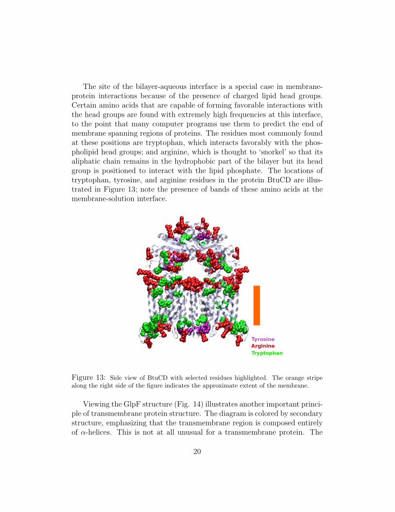
The site of the bilayer-aqueous interface is a special case in membrane-protein interactions because of the presence of charged lipid head groups.Certain amino acids that are capable of forming favorable interactions withthe head groups are found with extremely high frequencies at this interface,to the point that many computer programs use them to predict the end ofmembrane spanning regions of proteins. The residues most commonly foundat these positions are tryptophan, which interacts favorably with the phos-pholipid head groups; and arginine, which is thought to ‘snorkel’ so that itsaliphatic chain remains in the hydrophobic part of the bilayer but its headgroup is positioned to interact with the lipid phosphate. The locations oftryptophan, tyrosine, and arginine residues in the protein BtuCD are illus-trated in Figure 13; note the presence of bands of these amino acids at themembrane-solution interface.
Figure 13: Side view of BtuCD with selected residues highlighted. The orange stripealong the right side of the figure indicates the approximate extent of the membrane.
Viewing the GlpF structure (Fig. 14) illustrates another important princi-ple of transmembrane protein structure. The diagram is colored by secondarystructure, emphasizing that the transmembrane region is composed entirelyof α-helices. This is not at all unusual for a transmembrane protein. The
20
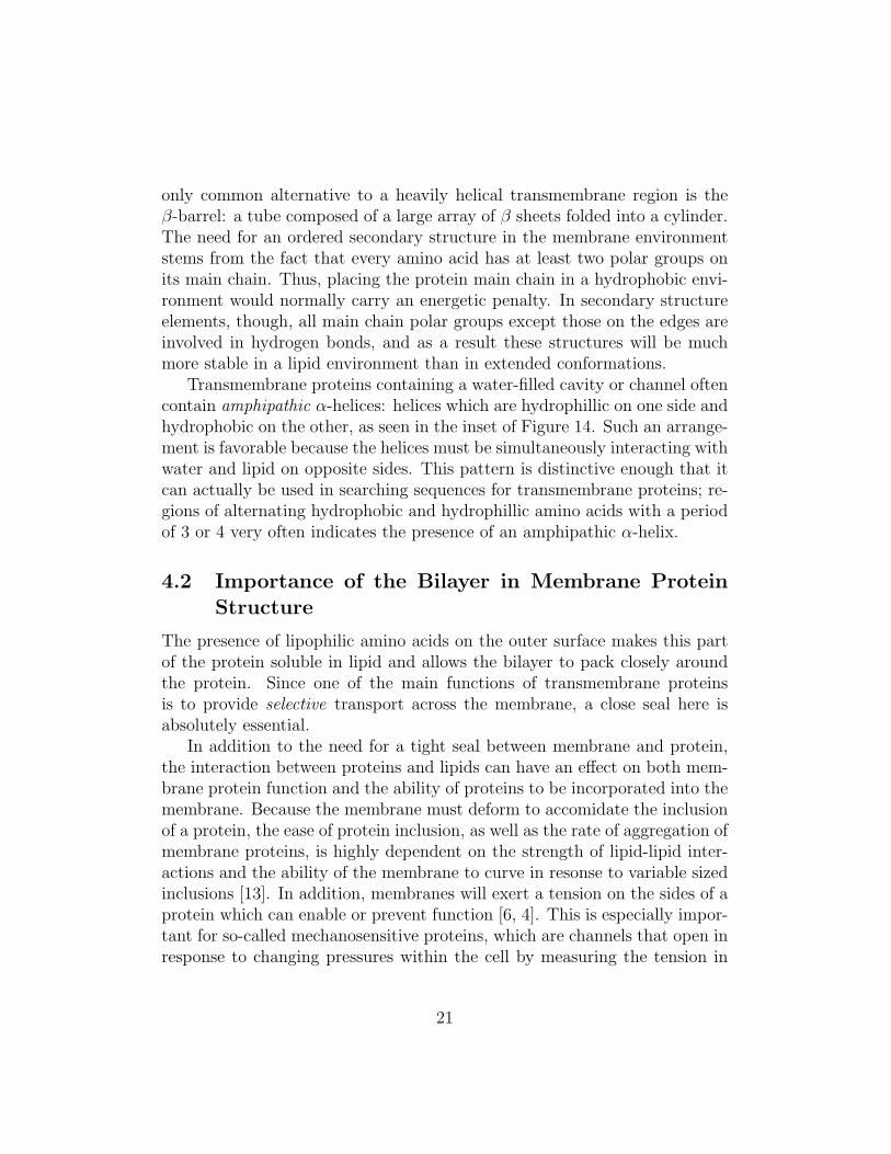
only common alternative to a heavily helical transmembrane region is theβ-barrel: a tube composed of a large array of β sheets folded into a cylinder.The need for an ordered secondary structure in the membrane environmentstems from the fact that every amino acid has at least two polar groups onits main chain. Thus, placing the protein main chain in a hydrophobic envi-ronment would normally carry an energetic penalty. In secondary structureelements, though, all main chain polar groups except those on the edges areinvolved in hydrogen bonds, and as a result these structures will be muchmore stable in a lipid environment than in extended conformations.
Transmembrane proteins containing a water-filled cavity or channel oftencontain amphipathic α-helices: helices which are hydrophillic on one side andhydrophobic on the other, as seen in the inset of Figure 14. Such an arrange-ment is favorable because the helices must be simultaneously interacting withwater and lipid on opposite sides. This pattern is distinctive enough that itcan actually be used in searching sequences for transmembrane proteins; re-gions of alternating hydrophobic and hydrophillic amino acids with a periodof 3 or 4 very often indicates the presence of an amphipathic α-helix.
4.2 Importance of the Bilayer in Membrane ProteinStructure
The presence of lipophilic amino acids on the outer surface makes this partof the protein soluble in lipid and allows the bilayer to pack closely aroundthe protein. Since one of the main functions of transmembrane proteinsis to provide selective transport across the membrane, a close seal here isabsolutely essential.
In addition to the need for a tight seal between membrane and protein,the interaction between proteins and lipids can have an effect on both mem-brane protein function and the ability of proteins to be incorporated into themembrane. Because the membrane must deform to accomidate the inclusionof a protein, the ease of protein inclusion, as well as the rate of aggregation ofmembrane proteins, is highly dependent on the strength of lipid-lipid inter-actions and the ability of the membrane to curve in resonse to variable sizedinclusions [13]. In addition, membranes will exert a tension on the sides of aprotein which can enable or prevent function [6, 4]. This is especially impor-tant for so-called mechanosensitive proteins, which are channels that open inresponse to changing pressures within the cell by measuring the tension in
21
Figure 14: Diagram of the α-helices of GlpF viewed from the outside of the cell look-ing in, with the protein in cartoon representation. One transmembrane helix from eachGlpF monomer has been highlighted, with hydrophobic amino acids colored orange andhydrophillics colored blue. To aid in understanding the organization, the four water-filledpores of the protein are highlighted with green dots, and a red dot indicates the central‘pore’ between the monomers.
the membrane [16].To see an example of lipid packing around a membrane protein, open
the VMD savestate aqp-packing.vmd from the 4.2-structure directory.The structures shown here are snapshots of the aquaporin crystal structurein a POPC bilayer; in the initial snapshot lipids are left in their normalstructure, with a simple hole cut out from the bilayer to make room for theprotein, whereas in the final state, they have been allowed to adjust to thestructure of the protein. In the final snapshot, one can observe that thelipids have packed around the protein, changing from a fairly flat interiorsurface to one that has adapted to the outer surface of the protein to allowfor enhanced interaction. This is best seen by looking at a single TM helix
22
of the protein (for example, the region between residues 210 and 235) andthe lipid immediately adjacent to this region. Observe how the lipid actuallyforms indentations for hydrophobic side chains to fit.
Exercise 6: Energetics of Lipid Packing. The solvent accesible surface area (sasa)of a selection in VMD can be calculated with the measure sasa command. Thestructures for aquaporin that you have been given contain only protein and lipid, whichmeans that the sasa calculated for this structure is the area interacting with water.To calculate the area accessible to water for a given subset of this structure, use thecommand measure sasa 1.4 $all -restrict $selection, where $all is an atomselection for all atoms in the system, and $selection is the atoms you actually wantthe sasa of. What is the difference in the water exposed surface area of hydrophobicside chains between the pre- and post-equilibration structures of aquaporin given here?Assuming that the energy of partition of a hydrophobic side chain from lipid to water is0.04 kJ
mol·A2, what is the difference in energy due solely to the solvation of hydrophobic
side chains between these two structures? Is this enough energy to be significant atroom temperature? Why?
Appendix I: Diffusion
To determine how far a lipid molecule undergoing free diffusion is likely totravel in a given period of time, we can model it as a single particle freelydiffusing in two dimensions. For the sake of simplicity, we assume that itmoves in discrete steps, once every τ units of time, and thus will take its jthstep at t = t0 + jτ . Note that this will essentially duplicate actual physicalcircumstances in the limit τ → 0. We consider each of these steps to be of afixed length L, in a random direction.
For this freely diffusing particle, we wish to calculate 〈x2〉, the expectedmagnitude of the square of the distance that the particle has travelled fromthe starting point, as a function of t. This can be determined by first notingthat ~RN , the position vector of the particle with respect to the origin afterN steps, is equal to (~RN−1 + ~L); where ~L is a random step of length L. We
seek 〈x2〉 after N steps, which is equivalent to ~RN · ~RN . Combining theseidentities,
〈x2〉 = ~RN · ~RN = (~RN−1 + ~L)2
= R2N−1 + 2~RN−1 · L + L2
23
Considered over many trials, 〈~L〉 = 0, since the particle is equally likely totravel in any direction. This in turn means that over many trials 〈RN−1 ·L〉 =
0, and thus, 〈x2〉 = R2N−1 + L2. Note that for N=1, ~RN−1 = 0, since the
particle starts at the origin, so 〈R21〉 = L2. For step 2, then,
〈R22〉 = 〈R2
1〉+ L2 = L2 + L2 = 2L2
Following this pattern inductively, we find that after N steps,
〈x2〉 = 〈R2N〉 = NL2
Since we have defined each step as lasting τ units of time, N = t/τ , and thusthe mean square deviation can be defined as 〈x2〉 = NL2 = tL2
τ.
In practice, the constant L2
Nis replaced by 2nD, where D is defined as the
diffusion coefficient of the molecule (which can be empirically determined)and n is the number of dimensions that the motion occurs in. The diffusioncoefficient is useful not only in mean squared deviation calculation, but alsoin much more general diffusive properties. The inclusion of this factor allowsa variety of different processes for a given system to be connected. A moredetailed discussion of the physical principles involved in diffusion within thebilayer, as well as a description for how the two-dimensional diffusion con-stants of lipids may be measured, is given in [3].
For the case of the lipid bilayer, n = 2, since diffusion occurs in twodimensions, and the biologically relevant diffusion coefficient D has beenmeasured to be on the order of 10−8 cm2/s. For one to calculate, for example,the expected distance for one lipid molecule to travel in 10 ms (as was donein the text), the calculations are as follows:
〈x2〉 = 4Dt = 4 ∗ 10−8cm2/s ∗ 10ms
= 4 ∗ 10−8cm2/s ∗ 10−2s
= 4 ∗ 10−10cm2
Taking the square root of this to obtain the root mean square distance trav-elled, one obtains
√4 ∗ 10−10cm2 = 2 ∗ 10−5 cm = 200 nm.
References
[1] B. Alberts, D. Bray, J. Lewis, M. Raff, K. Roberts, and J. D. Watson.Molecular Biology of the Cell. Garland Publishing, third edition, 1994.
24
[2] G. S. Ayton and G. A. Voth. Mesoscopic lateral diffusion in lipid bilayers.Biophysical Journal, 87:3299–3311, 2004.
[3] A. Brunger, R. Peters, and K. Schulten. Continuous fluorescence mi-crophotolysis to observe lateral diffusion in membranes: Theoreticalmethods and applications. J. Chem. Phys., 82:2147–2160, 1985.
[4] R. S. Cantor. Lipid composition and the lateral pressure profile in bi-layers. Biophys. J., 76:2625–2639, 1999.
[5] J. Craft, K. Epps, S. Brancato, and A. Serfis. Incorporation of bloodclotting factor x into phospholipid model membranes; fluorescence mi-croscopy imaging and subphase effects surrounding the lipid phase tran-sition region. J. Colloid and Interface Science, 268(1):181–187, 2003.
[6] N. Dan and S. A. Safran. Effect of lipid characteristics on the structureof transmembrane proteins. Biophys. J., 75:1410–1414, 1998.
[7] W. T. Doerrler and C. R. H. Raetz. Atpase activity of the msba lipidflippase of Escherichia coli. J. Biol. Chem., 39:36697–36705, 2002.
[8] R. B. Gennis. Biomembranes: Molecular Structure and Function.Springer, first edition, 1989.
[9] H. Heller, M. Schaeffer, and K. Schulten. Molecular dynamics simulationof a bilayer of 200 lipids in the gel and in the liquid-crystal phases. J.Phys. Chem., 97:8343–8360, 1993.
[10] G. Karp. Cell and Molecular Biology: Concepts and Experiments. Wiley,third edition, 2002.
[11] J. F. Nagle and S. Tristram-Nagle. Structure of lipid bilayers. Biochim.Biophys. Acta, 1469:159–195, 2000.
[12] D. L. Nelson and M. M. Cox. Lenhinger Principles of Biochemistry.Worth Publishers, third edition, 2000.
[13] V. Pata and N. Dan. The effect of chain length on protein solubilizationin polymer-based vesicles (polymersomes). Biophys. J., 85:2111–2118,2003.
25
[14] Y. Romsicki and F. J. Sharom. Phospholipid flippase activity ofthe reconstituted p-glycoprotein multidrug transporter. Biochemistry,40:6937–6947, 2001.
[15] D. Voet and J. G. Voet. Biochemistry. Wiley, second edition, 1995.
[16] P. Wiggins and R. Phillips. Membrane-protein interactions inmechanosensitive channels. Biophys. J., 88:880–902, 2005.
[17] P. Yager, J. Chappell, and D. D. Archibald. When lipid bilayers won’tform liposomes: Tubules, helices, and cochleate cylinders. In B. P. Gaberand K. R. K. Easwaran, editors, Biomembrane Structure and Function:The State of the Art, pages 001–019, 1992.
26