Embed Size (px)
Citation preview

Running head: KISE CASE STUDIES THREE AND FOUR 1
Kise Case Studies Three and Four
Shawn Kise BSN, RN
Wright State University
Nursing 7202
KISE CASE STUDIES THREE AND FOUR 2
Kise Cases Studies Three and Four
Case Study Three
1. Which of the following processes can produce postoperative hypotension? Explain your
rationale. Bold the correct answer.
A. Hypovolemia secondary to blood or fluid lossB. SepsisC. Adrenal insufficiencyD. Perioperative myocardial infarctionE. All of the above
All of the above answers to the question are correct. There are many causes of
postoperative hypotension and blood loss due to hemorrhage is the most common complication.
Once a patient starts showing physical signs of hemorrhage they have lost at least 30% or
approximately 1,500 mL other blood volume. These patients should be followed by monitoring
of their vital signs, urine output, and serial hemoglobins to follow the progression of the
bleeding. It may be necessary to take these patients back to surgery to find the source and stop
the area of bleeding. External bleeding sites generally can be treated with simple pressure or
ligation of the bleeding site itself. Large amounts of fluid loss during surgery with under
resuscitation of fluid repletion is another common cause of postop hypotension. These patients
generally require large amounts of fluid resuscitation, larger than the amount lost during surgery.
They should be followed by vigilant vital signs and urinary output to monitor hydration (Kim &
Maxhimer, 2008).
Infection with the possibility of sepsis is always a risk with any type of surgical
procedure. In this case the patient has had a second surgery in a short period of time with a
known intra-abdominal abscess and infection from the first surgery. The second surgery was an
emergent exploratory laparotomy which also increases the risk for sepsis. Sepsis is the most
KISE CASE STUDIES THREE AND FOUR 3
common cause for distributive shock. Arterial vasodilatation is the key hemodynamic
characteristic of sepsis that is created by decrease in total vascular resistance. Hypotension in
these patients results from both hypovolemia and inadequate vasodilatation (Landry & Oliver,
2001). Sepsis induced hypotension that persists after initial fluid resuscitation efforts and/or
patients that have a serum lactate concentration of greater than 4 mmol/L should be considered
as severe sepsis. Prompt recognition and treatment of these patients is necessary to obtain the
most optimal outcomes. Early goal directed therapies are aimed at maintaining a central venous
pressure between 8 – 12 mm Hg, mean arterial pressure greater than or equal to 65 mm Hg, urine
output of greater than or equal to 0.5 mg/kg/hr, and maintaining a superior vena cava
oxygenation saturation of 70% or a mixed venous oxygen saturation of 65%. Targeting
normalization of serum lactate levels in resuscitation efforts is very important as elevated serum
lactate levels are a marker for tissue hypoperfusion (Dillinger et al., 2013). Further treatment
guidelines for the management of severe sepsis can be found in the recently updated surviving
sepsis campaign.
Primary adrenal insufficiency can be a cause of postoperative hypotension. Patients
diagnosed with primary adrenal insufficiency before undergoing surgery are at higher risk for
developing complications due to the high stress environment of surgery. These patients require
additional glucocorticoid doses during surgery or medical illness. Primary adrenal insufficiency
can also be developed from complicated or expensive surgeries. Primary adrenal insufficiency
can also be a result of infection, autoimmune disorder, genetic disorders, bilateral adrenal
hemorrhage, and metastasis (Jung & Inder, 2008). Blood pressure is generally low in patients
with adrenal insufficiency and is also commonly associated with postural dizziness or syncope.
Volume depletion resulting from aldosterone deficiency is responsible for these symptoms. If
KISE CASE STUDIES THREE AND FOUR 4
adrenal insufficiency is suspected due to a patient’s low blood pressure without an identifiable
cause, then a Cortrosyn stimulation test may be administered (Neiman, 2013a). The Cortrosyn
stimulation test will be discussed in more depth in the following question.
Perioperative myocardial infarction is also a complication from surgery that can cause
postoperative hypotension. Other cardiac complications including cardiac death, heart failure, or
ventricular tachycardia may also be seen as a complication postoperatively. Perioperative
myocardial infarction is the most common of these major cardiac complications from noncardiac
surgery. In the POISE trial, perioperative MI was defined by one or more the following findings:
ischemic symptoms, electrocardiogram changes in two consecutive leads, coronary artery
intervention, or evidence of myocardial infarction on cardiac imaging or at the time of autopsy.
Patients that are considered moderate to high risk should be evaluated before surgery with a
cardiac risk assessment tool and the risks versus benefits of noncardiac surgery should be heavily
weighed (Shammash, Kimmel, & Devereaux, 2013).
2. Which of the following is the most appropriate method to diagnose bilateral massive
adrenal hemorrhage? Explain your answer.
A. Cortrosyn stimulation testB. CT scan of adrenal glandsC. CT scan of adrenal glands and Cortrosyn stimulation testD. Random plasma cortisol level
Before widespread use of computed tomography (CT), adrenal hemorrhage was often
diagnosed at time of autopsy or during an exploratory laparotomy. A CT scan of the adrenal
glands with IV contrast is the preferred method for the evaluation of possible adrenal
hemorrhage. If adrenal hemorrhage is present, it will show a round or oval mass in the area of
the adrenal gland and is often seen with periadrenal stranding. Other findings that are also
consistent with adrenal hemorrhage on CT scan are retroperitoneal hemorrhage and crural
KISE CASE STUDIES THREE AND FOUR 5
thickening. The attenuation value seen on the CT scan is dependent on the age of the
hemorrhage with acute or subacute hematomas showing a high attenuation of 50 – 90 Hounsfield
units (Jordan et al., 2012; Simon & Palese, 2009). Adrenal hemorrhage can also be seen on
magnetic resonance imaging (MRI) and ultrasound, but the preferred imaging for bilateral
massive adrenal hemorrhage is CT scan (Kovacs, Lam, & Pater, 2001).
To confirm the diagnosis of bilateral adrenal hemorrhage (BAH) there must be evidence
of biochemical adrenal insufficiency. A Cortrosyn stimulation test is the criterion standard for
the diagnosis of adrenal insufficiency. This test is completed by drawing a baseline cortisol and
aldosterone level in separate tubes. Then 250 mcg of CORTROSYN® is given either
intramuscularly or intravenously. A second separately drawn cortisol and aldosterone levels are
drawn at 30 minutes or 60 minutes post-injection. There is some debate as to whether there is a
need to draw both a 30 minute and 60 minute level since there is documented high sensitivity of
the 30 minute level accurately diagnosing adrenal insufficiency. Once this has been completed
there are two criteria that are necessary for diagnosis. An increase in the baseline cortisol level
of 7 mcg/dL or more and the cortisol level must rise to above 20 mcg/dL or more in 30 or 60
minutes to establish normal adrenal glucocorticoid function. If these criteria are not met, this is
indicative of adrenal insufficiency (Griffing, 2012). The aldosterone levels from the rapid
Cortrosyn stimulation test are helpful to determine between primary and secondary adrenal
insufficiency. In secondary adrenal insufficiency, the aldosterone increments from the baseline
will be normal, greater than or equal to 5 ng/dL, as opposed to primary adrenal insufficiency
where small or no increments are seen. Also in primary adrenal insufficiencies, the plasma
adrenocorticotropin (ACTH) is elevated, where ACTH plasma levels in secondary adrenal
insufficiency are low or inappropriately normal (Fauci et al., 2009).
KISE CASE STUDIES THREE AND FOUR 6
In critically ill patients, treatment should not be delayed to perform in Cortrosyn
stimulation test. Hydrocortisone must not be given within eight hours prior to starting the
Cortrosyn stimulation test. Corticosteroids, prednisone and dexamethasone, do not interfere with
the specific assays for cortisol testing and may be used to treat the patient well diagnostic testing
is being completed (McPhee & Papadakis, 2011a). A random plasma cortisol level can be
helpful in diagnosing adrenal insufficiency. An eight AM cortisol level that is less than 3μm/dL
is diagnostic of adrenal insufficiency. If the eight AM cortisol level is greater than 15μg/dL,
then adrenal insufficiency may be ruled out. Any level that falls between 3μg/dL and 15μg/dL
requires further testing with a Cortrosyn stimulation test (Ioachimescu & Hamrahian, 2013).
3. Which of the following can occur in patients with primary adrenal insufficiency?
Explain your answer.
A. Electrolyte abnormalitiesB. HypotensionC. Mental status changesD. Abdominal painE. All of the above
All of the above answers are seen in patients with primary adrenal insufficiency. Sodium
loss and volume depletion cause hyponatremia in approximately 85 to 90 percent of these
patients. Many of these patients will describe having salt cravings and have continuous
increased thirst. A mild hyperchloremic acidosis occurs in approximately 60 to 65 percent of
patients causing hyperkalemia due to mineralocorticoid deficiency. Hypercalcemia has been
seen but is rare (Neiman, 2013a).
Hypotension is commonly seen in patients with primary adrenal insufficiency. As
previously discussed, this can occur with postural dizziness and syncope. Patients may also
present with having only postural hypotension. Hypotension is primarily due to the volume
KISE CASE STUDIES THREE AND FOUR 7
depletion but as a result from the aldosterone deficiency. Low levels of glucocorticoids cause a
decrease in the adrenal medullary epinephrine synthesis. With decreased levels of serum
epinephrine, compensatory increases in serum norepinephrine concentrations are present. This
could contribute to lower basal systolic blood pressures with an exaggerated increase in heart
rate upon standing (Neiman, 2013a).
Patients with adrenal insufficiency will often present with mental status changes. The
classic presentation of patients with adrenal insufficiency include mental status changes, febrile
or hypothermic, refractory hypotension, and eosinophilia (Simon & Palese, 2009). Patients may
also get impairment of their memory that can progress to confusion, delirium, and stupor.
Psychosis may be present in 20 to 40 percent of patients manifesting by social withdrawal,
irritability, poor judgment, agitation, hallucinations, paranoid delusions, and bizarre or catatonic
posturing (Neiman, 2013a).
Abdominal pain is also closely associated with adrenal insufficiency and is generally
accompanied by nausea, vomiting, and diarrhea that may alternate with constipation. Severe
abdominal pain and vomiting is a cause for concern of adrenal crisis. Weakness, fatigue ability,
weight loss, myalgias, arthralgias, fever, anorexia, anxiety, and mental irritability are all also
associated with adrenal insufficiency and Addison disease (McPhee & Papadakis, 2011a).
4. Which of the following is not a risk factor for developing bilateral massive adrenal
hemorrhage? Explain your rationale.
A. Postoperative stateB. CoagulopathyC. Thromboembolic diseaseD. DiabetesE. Sepsis
KISE CASE STUDIES THREE AND FOUR 8
Of the answer options, diabetes is the only one that is not associated with increased risk
for bilateral massive adrenal hemorrhage (BMAH). Kovacs, Lam, and Prater (2001), conducted
a case-control study to assess the risk factors for BMAH. Diabetes was a marker that was
evaluated and did not show an increased risk for BMAH. Coronary artery disease hypertension
also appeared to be markers for lower risk of BMAH. The authors reported that
thrombocytopenia have the highest independent correlation with risk for BMAH. The use of
heparin by any route for greater than a three-day period and sepsis were also highly associated
independent risk factors for BMAH.
The postoperative state is also associated with BMAH. BMAH has been reported more
often with cardiovascular and orthopedic surgeries. This is most likely due to the common
practice in use of anticoagulants after these surgeries. Coagulopathy, especially with heparin use
of any route or form of greater than three days duration has been shown to increase the risk for
BMAH. Thromboembolic disease and hypercoagulable states such as antiphospholipid
syndrome have been reported to also increase the risk for BMAH. Adrenal hemorrhage has also
been associated with sepsis due to meningococcemia (Waterhouse – Friderichsen syndrome).
Sepsis from Streptococcus pneumoniae, Neisseria gonorrhea, Escherichia coli,, Haemophilus
influenza, and Staphylococcus aureus have all been associated with BMAH (Neiman, 2013b).
Other risk factors that have been stated by case reports include advanced age, serious underlying
medical illness, significant hypotension, spontaneous or iatrogenic coagulopathy’s, certain
prothrombotic disorders, trauma, ACTH administration, vasculitis, adrenal venography,
pheochromocytoma, and any cause of severe stress (Kovacs, Lam, & Pater, 2001).
KISE CASE STUDIES THREE AND FOUR 9
5. Which of the following statements regarding the long-term management of patients with
BMAH is correct? Explain your answer.
A. Glucocorticoid therapy is needed only done acute illness.B. Patient should be discharged on maintenance doses of oral glucocorticoids and mineralocorticoids.C. Patients do not need mineralocorticoid therapy.D. Adrenal function is likely to recover over 4 to 6 months with no further need for glucocorticoids.
Due to the adrenal cortex destruction when BMAH has occurred, it is very unlikely that
the adrenal function will return. This will require lifelong glucocorticoid and mineralocorticoid
therapy. The long-term management of patients with BMAH consists of glucocorticoid
replacement with hydrocortisone at doses starting at 20 to 25 mg per 24 hours. The should be
administered in two to three divided doses, with the first dose administered immediately after
waking up. Monitoring of these patients should include body weight, body mass index, and
signs for under replacement or over replacement. Signs of under replacement include weight
loss, fatigue, nausea, myalgias, and lack of energy. Signs for over replacement include weight
gain, central obesity, stretch marks, osteopenia/osteoporosis, impaired glucose tolerance, and
hypertension (Arlt, 2009).
Mineralocorticoid replacement is only required for patients with primary adrenal
insufficiency. Mineralocorticoid replacement is not required if the patient’s hydrocortisone dose
is greater than 50 mg per 24 hours. Patient should be started on 100 μg of fludrocortisone and
should be administered as a single dose in the morning immediately after waking up. The
fludrocortisone dose may vary between 50 – 250 μg per 24 hours. Patients receiving
mineralocorticoid replacement therapy should also be monitored for over replacement and under
replacement. Postural hypotension greater than or equal to 20 mm Hg may be indicative of
under replacement. Postural hypertension and peripheral edema may be indicative of over
KISE CASE STUDIES THREE AND FOUR 10
replacement. Serum sodium and potassium levels should be routinely monitored and plasma
renin activity should be monitored at least every 2 to 3 years, with significant changes in the
hydrocortisone dose, or if there’s clinical suspicion of over or under replacement. Adrenal
androgen replacement should also be considered in patients with impaired well-being and mood
despite optimal glucocorticoid and mineralocorticoid replacement, and in women with symptoms
and signs of androgen deficiency. The signs of androgen deficiency include dry and itchy skin
as well as reduced libido. For patients requiring adrenal androgen replacement, DHEA may be
given at 20 – 50 mg as a single morning dose. In women you may also consider using
transdermal testosterone to deliver 300 μg per day which would equal two patches per week
(Arlt, 2009).
KISE CASE STUDIES THREE AND FOUR 11
Case Study Four
A 22-year-old Caucasian female presents to the emergency department by ambulance.
She is lethargic but wakes up to verbal stimulus. Her roommate has accompanied her to the
emergency department and states that they were out drinking with some friends the night before.
The friend was unsure of how much the patient had drank the previous night, but did state that
she had gotten sick with two rounds of emesis when they got home to their apartment. When
asked questions, the patient only answered “I can’t remember” and “I don’t know”. The only
history that the friend could give us is that the patient was diabetic and that she had an
appendectomy when she was in high school. She did not know if she was a type I or type II
diabetic but did state that the patient takes insulin. The paramedic on the life squad reported that
the patient was confused at their time of arrival and that she had emesis during the transport to
the hospital. An IV was placed and a 0.9 normal saline bolus was started. They checked a finger
stick blood glucose that read high on the glucometer.
Physical examination revealed a slender Caucasian female in mild distress. The patient
was lethargic but arousable, she had dry mucous membranes, and she had a normal S1 S2 with
no murmurs, rubs, or gallops. Her lung sounds were clear with no adventitious sounds.
Hypoactive bowel sounds were heard in all four quadrants, and the patient complained of some
slight tenderness with palpation to the upper abdomen. Her skin assessment revealed warm dry
skin with no lesions, ecchymosis, or open areas. The patient was able to follow simple
commands and move all extremities without problems. She was able to fully flex and extend the
neck without pain or difficulties. The neurological exam showed no focal deficits other than the
confusion. She was alert and oriented to person only at the time of the exam. The vital signs on
arrival to the emergency department showed a blood pressure of 101/58 mm Hg, heart rate of
110 bpm, respirations of 26 per minute, temperature of 99.0°F orally, and O2 saturation of 98%
KISE CASE STUDIES THREE AND FOUR 12
on room air. A repeat finger stick blood glucose level was drawn in the emergency department
and showed a blood sugar of 496 mg/dL. Blood work was drawn from the patient’s IV and is
pending.
1. What are the differential diagnoses for this patient? Provide rationale.
The highest differential diagnosis for this patient is diabetic ketoacidosis (DKA). The
patient presents with many manifestations of DKA including nausea and vomiting, tachycardia,
low blood pressure, abdominal pain, lethargy, and tachypnea. The classic presentation of a
patient with DKA is Kussmaul respirations with an acetone odor that is described as a fruity
smell. Precipitating events that can induce diabetic ketoacidosis include inadequate insulin
administration, infection, infarction, drugs, and pregnancy. The most common types of
infections that precede DKA are pneumonia, urinary tract infection, gastroenteritis, and sepsis.
Infarction can include cerebral, coronary, mesenteric, and peripheral. The most likely drug to
induce DKA is the use of cocaine (Longo et al, 2012). With the patient’s clinical presentation
and history of diabetes with insulin use, it is likely that the patient is experiencing diabetic
ketoacidosis.
The second differential diagnosis for this patient is hyperglycemic hyperosmolar state
(HHS). HHS is the second most common form of hyperglycemic coma that is seen with severe
hyperglycemia in the absence of significant ketosis. It case test may be more insidious and onset
than DKA, arising over a period of days to weeks with symptoms of fatigue, weakness, polyuria,
and polydipsia. Lethargy and confusion are commonly seen in cases of HHS, and can develop
into convulsions and deep coma. The physical examination is consistent with findings of severe
dehydration with the absence of tachypnea or kussmaul respirations. HHS may be more
common in elderly patients due to reduced intake of fluid from several different reasons
KISE CASE STUDIES THREE AND FOUR 13
including an inappropriate lack of thirst mechanism, nausea and vomiting, and lack of access to
adequate fluids seen in bedridden patients. Laboratory evaluation is necessary to distinguish
between DKA and HHS. HHS is characterized by a markedly higher blood glucose level of
greater than 600 mg/dL, absence of metabolic acidosis, and normal to slightly elevated anion gap
(McPhee & Papadakis, 2011b).
Another differential diagnoses that should be considered is alcohol and/or drug
intoxication. Alcohol intoxication is similar to the patient’s presentation with the disorientation,
nausea and vomiting, and dehydration. The history from the patient’s friend stated that they
were out the night before consuming alcohol. Whether alcohol intoxication is the main cause for
the patient symptomology, it is probably playing a part in the process of her illness. The other
differential diagnosis of an otherwise healthy young individual with this presentation include
infection, lactic acidosis, acute pancreatitis, and hyperchloremic metabolic acidosis (Pischke,
2001).
Case continued
The patient was given a bolus of 10 units of regular insulin. A second 0.9 normal saline
liter bolus was started and blood work was drawn and sent to the lab along with a urine sample.
The blood work revealed a sodium of 128 mEq/L, chloride of 99 mmol/L, potassium of 5.0
mEq/L, calcium of 10.2 mg/dL, BUN of 24 mg/dL, and creatinine of 1.2 mg/dL. Her white
blood count was 13,100 cells/mcL, hemoglobin 13.3 gm/dL, hematocrit 39%, and platelet count
of the hundred and 79,000 per microliter. An EKG was completed and showed sinus tachycardia
with no acute changes. Chest x-ray was negative for any cardiopulmonary process. The
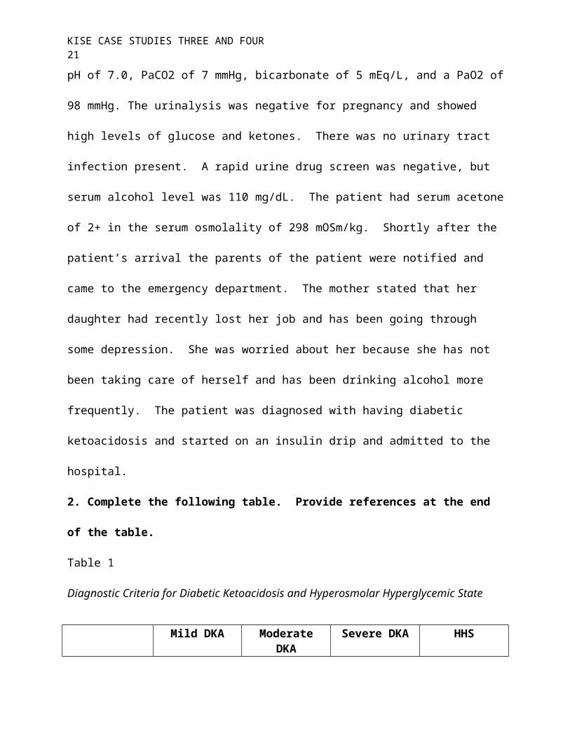
patient’s arterial blood gas revealed a metabolic acidosis with a pH of 7.0, PaCO2 of 7 mmHg,
bicarbonate of 5 mEq/L, and a PaO2 of 98 mmHg. The urinalysis was negative for pregnancy
KISE CASE STUDIES THREE AND FOUR 14
and showed high levels of glucose and ketones. There was no urinary tract infection present. A
rapid urine drug screen was negative, but serum alcohol level was 110 mg/dL. The patient had
serum acetone of 2+ in the serum osmolality of 298 mOSm/kg. Shortly after the patient’s arrival
the parents of the patient were notified and came to the emergency department. The mother
stated that her daughter had recently lost her job and has been going through some depression.
She was worried about her because she has not been taking care of herself and has been drinking
alcohol more frequently. The patient was diagnosed with having diabetic ketoacidosis and
started on an insulin drip and admitted to the hospital.
2. Complete the following table. Provide references at the end of the table.
Table 1
Diagnostic Criteria for Diabetic Ketoacidosis and Hyperosmolar Hyperglycemic State
Mild DKA Moderate DKA Severe DKA HHSPlasma glucose
(mg/dL)˃ 250 ˃ 250 ˃ 250 ˃ 600
Venous or arterial pH
7.25 – 7.30 7.00 to ˂ 7.24 ˂ 7.00 ˃ 7.3
Serum bicarbonate
(mEq/L)15 – 18 10 – 15 ˂ 10 ˃ 18
Urine or serum ketones
Positive Positive Positive Small
Beta- hydroxybutyrat
e
High High High Normal or elevated
Serum osmolality m(Osm/kg)
Variable Variable Variable ˃ 320
Anion gap ˃10 ˃ 12 ˃ 12 ˂12
Mental status Alert Alert/drowsy Stupor/coma Stupor/coma
Note. Table information from (McNaughton, Shelf, & Slovis, 2011; Trachtenberg, 2005)
KISE CASE STUDIES THREE AND FOUR 15
3. Briefly discuss the pathophysiology and development of DKA and how it differs from
HHS?
DKA is primarily seen in type I diabetes but can also occur in type II diabetics as well.
DKA is a result of relative or absolute insulin deficiency with an increase of contrary regulatory
hormone excretion of glucagon, catecholamines, cortisol, and growth hormone. This causes an
increase in hepatic gluconeogenesis and cellular underutilization of glucose and produces a
hyperglycemic state. With insulin deficiency plus an increase in counter regulatory hormones,
especially cortisol and growth hormone, an increase in lipolysis occurs. Lipolysis is the
breakdown of lipids into glycerol and free fatty acids. This causes a hepatic overproduction of
beta-hydroxybutyrate and acetoacetic acid causing an increase in ketone concentration in the
body. This is considered ketoacidosis and leads to an increased anion gap and metabolite
acidosis. When a patient reaches the ketoacidosis stage, this induces nausea and vomiting with
ketonuria present. With ketonemia present, the patient will develop a compensatory tachypnea
and efforts to correct the metabolic acidosis. With the serum blood glucose increasing, osmotic
diuresis occurs causing a loss of electrolytes and glycosuria. As this progresses there is a
decrease in the glomerular filtration rate. This process produces severe hyperglycemia with
intracellular dehydration, volume depletion, and metabolic acidosis that can lead to shock if not
quickly diagnosed and treated (Chiasson et al, 2003; McCance, Huether, Brashers, & Rote, 2010;
Pischke, 2001).
The pathogenesis of hyperglycemic hyperosmolar state differs in that there is only a
relative insulin deficiency as compared to an absolute insulin deficiency seen in DKA. This
inhibits the process of lipolysis and production of free fatty acids eventually leading to the
acidosis that is seen in DKA. In HHS, serum blood glucose levels are more severe than seen in

KISE CASE STUDIES THREE AND FOUR 16
DKA causing a greater increase in water loss and dehydration. When combined with a decrease
fluid intake it creates hyperosmolality that is characteristic of HHS and not DKA. The
differentiation between HHS and DKA can be determined by the presence of excess ketones,
acidosis, the bicarbonate level, and serum osmolality (Kitabchi & Nyenwe, 2006).
4. Should this patient be treated with sodium bicarbonate for her metabolic acidosis?
This patient should not receive sodium bicarbonate treatment for her metabolic acidosis.
The treatment guidelines only recommend sodium bicarbonate if the pH level is below 7.0 after
the first hour of hydration. If the pH is below 7.0 then sodium bicarbonate 100 mmol in 400 mL
of water should be administered at 200 ml/hr every two hours until the pH reaches ˃ 7.0
(American Diabetes Association, 2003). Chua, Schneider, and Bellomo (2011) conducted a
systematic review on sodium bicarbonate use in patients with diabetic ketoacidosis. The
research showed no evidence that sodium bicarbonate was beneficial in glycemic control or
clinical efficacy. There was significant evidence that the use of the sodium bicarbonate in
children increases the risk for cerebral edema and increase the length of hospitalization. There
was also weak evidence that showed a transient paradoxical worsening of ketosis, and increased
the need for potassium supplementation in all patients. Therefore sodium bicarbonate should
only be used in cases of severe metabolic acidosis of a pH less than 7.0.
5. Write a set of admission orders for this patient. Provide references at the end that
support your orders.
Admit to step downDiagnosis: diabetic ketoacidosisSecondary diagnosis: dehydration, ETOH intoxicationCondition: fairActivity: Up to restroom with assist onlyDiet: NPOVitals: 1.) Every hour times four, if stable then every two hours
KISE CASE STUDIES THREE AND FOUR 17
2.) Continuous cardiac monitoringLabs:1.) Finger stick blood glucose levels every hour well on IV insulin drip, every four hours when switch to subcutaneous insulin 2.) Basic metabolic panel every hour until CO2 level is greater than 15 then every 4 hours3.) AM complete metabolic panel and complete blood count with differential4.) Obtained a venous blood gas two hours after admissionHeight/Weight: 66 inches/58.1 kgNursing: 1.) SCDs and pneumatic compression devices while in bed2.) Strict monitoring of intake and output3.) Call if temperature is greater than 101°F4.) Call when anion gap is ˂ 12IV fluids:1.) 0.9% normal saline at 14 mL/kg/hr (800 mL/hr), change fluids to 5% dextrose with 0.45% sodium chloride when blood sugar falls below 250 mg/dLMedications:1.) Insulin drip at 0.1 units/kg/hr (5.8 units/hr), if the serum glucose does not decrease by 50 – 70 mg/dL in the first hour, then double the drip rate hourly until there is a decrease of 50 – 70 mg/dL. Once glucose reaches below 250 mg/dL change rate to 5.8 units/hr.2.) Zofran 4 mg IV every six hours as needed for nausea or vomiting3.) Potassium replacement protocol
K ˂ 3.3 – hold insulin drip, give 20 mEq/hr K until level is ˃ 3.3K ≥ 3.3 – ˂ 5.0 – add 20 mEq to each leader of IV fluidsK ≥ 5.0 – monitor potassium every two hours on the BMP
Thank You,
Shawn Kise RN, ACNP – Student
The guidelines set by the American diabetes Association (2003) was used to formulate
the orders for the administration of IV fluids, insulin, and potassium protocol.
KISE CASE STUDIES THREE AND FOUR 18
References
American Diabetes Association (2003). Hyperglycemic crisis in patients with diabetes mellitus.
Diabetes Care, 26, S 109 – 117. doi: 10.2337/diacare.26.2007.S109
Arlt, W. (2009). The approach to adult with newly diagnosed adrenal insufficiency. Journal of
clinical endocrinology and metabolism, 94, 1059 – 1067. doi: 10.1210/JC.2009 – 0032
Dillinger, R. P., Levy, M. M., Rhodes, A., Annane, D., Gerlach, H., Opal, S., … Moreno, R.
(2013). Surviving sepsis campaign: International guidelines for management of severe
sepsis and septic shock: 2012. Critical Care Medicine, 41, 580 – 637. doi:
10.1097/CCM.0b013e31827e83af
Chiasson, J. L., Aris-Jilwan, N., Belanger, R., Bertrand, S., Beauregard, H., Ekoe, J. M.,
Fournier, H. & Havrankova, J. (2003). Diagnosis and treatment of diabetic ketoacidosis
and hyperglycemic hyperosmolar state. Canadian medical Association Journal, 168, 859
– 866. Retrieved from http://www.cmaj.ca/content/168/7/859.long
Chua, H. R., Schneider, H., & Bellomo, R. (2011). Annals of Intensive Care, 1, 23.
doi:10.1186/2110-5820-1-23
Fauci, A. S., Braunwald, E., Kasper D. L., Hauser, S. L., Longo, D. L., Jameson, L. J., &
Loscalzo, J. (2009). Hypofunction of the adrenal gland. Harrison’s manual of medicine
(pp. 936). New York, NY: McGraw Hill
Griffing, G. T.(2012). Addison disease. Medscape. Retrieved from
http://emedicine.medscape.com/article/116467-overview
Ioachimescu, A. G. & Hamrahian, A. H.(2013). Diseases of the adrenal gland. Cleveland
Clinic: Center for continuing education. Retrieved from
http://www.clevelandclinicmeded.com/medicalpubs/diseasemanagement/endocrinology/
diseases-of-the-adrenal-gland/
KISE CASE STUDIES THREE AND FOUR 19
Jordan, E., Poder, L., Courtier, J., Sai, V., Jung, A., & Coakley, F. V. (2011). Imaging of non-
hemorrhagic adrenal hemorrhage. American Journal of Radiology, 199, W91 – W98,
doi: 10.2214/AJR.11.7973
Jung, C. & Inder, W. J. (2008). Management of adrenal insufficiency during the stress of
medical illness and surgery. The Medical Journal of Australia, 188, 409 – 413.
Retrieved from https://www.mja.com.au/journal/2008/188/7/management-adrenal-
insufficiency-during-stress-medical-illness-and-surgery
Kim, A. W., & Maxhimer, J. B. (2008). Hypotension in the postoperative patient. In J. Myers,
K. Miliken, & Saclarides (Eds.), Common surgical diseases (pp. 395 – 397). doi:
10.1007/978-0-387-75246-4
Kitabchi, A. E. & Nyenwe, E. A. (2006). Hyperglycemic crisis and diabetes mellitus: diabetic
ketoacidosis and hyperglycemic hyperosmolar state. Endocrinology and Metabolism
Clinics of North American, 35, 725 – 751. doi: 10.1016/j.ecl.2006.09.006
Kovacs, K. A., Lam, Y. M., & Pater, J. L.(2001). Bilateral massive adrenal hemorrhage
assessment of punitive risk factors by the case-control method. Medicine, 80, 45 – 53.
Retrieved from http://www.ncbi.nlm.nih.gov/pubmed/11204502
Landry, D. W. & Oliver, J. A.(2001). The pathogenesis of vasodilatory shock. The New
England Journal of Medicine, 345, 588 – 595. Retrieved from
http://www.nejm.org/doi/full/10.1056/NEJMra002709
Longo, D. L., Fauci, A. S., Kasper, D. L., Hauser, S. L., Jameson, J. L., & Loscalzo, J. (2012).
Harrison’s principles of internal medicine 18th ed (pp 2976 – 2978). New York, NY:
McGraw Hill
KISE CASE STUDIES THREE AND FOUR 20
McCance, K. L., Huether, S. E., Brasher, V. L., & Rote, N. S. (2010). Pathophysiology: The
biologic basis for disease in adults and children 6th ed (pp 754 – 758). Maryland Heights,
Missouri: Mosby Elsevier
McCance, C. D., Self, W. H., & Slovis, C.(2011). Diabetes in the emergency Department: Acute
care of diabetes patients. Clinical Diabetes, 29, 51 – 59. doi: 10.2337/diaclin.29.2.51
McPhee, S. J., & Papadakis, M. A. (2011a). Chronic adrenocortical insufficiency. 2011 Current
medical diagnosis and treatment (pp 1108 – 1110). New York, NY: McGraw Hill
McPhee, S. J., & Papadakis, M. A. (2011b). Diabetic ketoacidosis. 2011 Current medical
diagnosis and treatment (pp 2976 – 2979). New York, NY: McGraw Hill
Niemann, K. N. (2013a). Clinical manifestations of adrenal insufficiency in adults.
UpToDate®. Retrieved from http://www.uptodate.com/contents/clinical-manifestations-
of-adrenal-insufficiency-in-adults
Niemann, K. N. (2013b). Causes of primary adrenal insufficiency. UpToDate®. Retrieved
from http://www.uptodate.com/contents/causes-of-primary-adrenal-insufficiency-
addisons-disease?source=see_link
Pischke, M. A. (2001). Diabetic ketoacidosis. Physician Assistant, 25, 42 – 48. Retrieved from
http://www.smccd.net/accounts/felixf/nurs232/dka.pdf
Shammash, J. B., Kimmel, S. E., & Devereaux, P. J. (2013). Perioperative myocardial infarction
after noncardiac surgery. UpToDate®. Retrieved from
http://www.uptodate.com/contents/perioperative-myocardial-infarction-after-noncardiac-
surgery?
source=search_result&search=Perioperative+myocardial+infarction&selectedTitle=1%7
E36
KISE CASE STUDIES THREE AND FOUR 21
Trachtenbarg, D. E. (2005). Diabetic ketoacidosis. American Family Physician, 71, 1705 –
1714. Retrieved from http://www.aafp.org/afp/2005/0501/p1705.html


![APCD Medical Claim File Submission Guide - chiamass.gov · Web viewExternal Codes Source 8 - International Classification of Diseases. varchar[7] ICD Other Diagnosis Code. Other](https://img.pdfslide.us/doc/110x75/5cca04a888c9937c048d7c89/apcd-medical-claim-file-submission-guide-web-viewexternal-codes-source-8-international.jpg)