Embed Size (px)
Citation preview
SR Ashwanirani et al: Case series of ectodermal dysplasia
IJMDS ● www.ijmds.org ● July 2014; 3(2) 507
Case Series Ectodermal dysplasia: familial report of six cases Ashwanirani SR 1, Bijjaragi S2, Suragimath G3, Kulkarni P4, Kulkarni A5, Nimbal A6
ABSTRACT
Ectodermal dysplasia is a hereditary disease characterized by a congenital
dysplasia of one or more ectodermal structures and their accessory appendages.
There are two main types, Hypohidrotic/Christ-Seimens- Tourian Syndrome and
Hidrotic/Clouston syndrome. Common manifestations include defective hair
follicles and eyebrows, frontal bossing with prominent supraorbital ridges, nasal
bridge depression and protuberant lips. Intraorally common findings are
anodontia or hypodontia and conical shaped teeth. The patient may suffer from
dry skin, hyperthermia and unexplained high fever because of the deficiency of
sweat glands. The present article reports unique case series of ectodermal
dysplasia cases in two families, where three generations in the both the families
were affected.
Key-words: Christ-Siemens-Touraine syndrome, ectodermal dysplasia,
hypohidrosis, hypotrichosis, oligodontia
Introduction Ectodermal dysplasia (ED) is a rare heterogeneous group of inherited disorder that share primary defects in the development of two or more tissues derived from ectoderm like skin, hair, nails, eccrine glands and teeth. The disorders are congenital, diffuse and non-progressive; more than 192 distinct disorders have been
described. There are 2 major types, depending on the number and functionality of the sweat glands: X-linked anhidrotic or hypohidrotic, where sweat glands are either absent or significantly reduced in number (Christ-Siemens-Touraine syndrome) second one is hidrotic, where sweat glands are normal and the condition is inherited as autosomal dominant (Clouston’s
1Dr Ashwani Rani SR MDS, Senior Lecturer, Oral Medicine and Radiology [email protected] 2Dr Shobha Bijjaragi MDS, Senior Lecturer, Oral Medicine and Radiology [email protected] 3Dr Girish Suragimath
MDS, Professor & Head, Periodontology School of Dental Sciences, KIMSDU, Karad, Maharashtra, India [email protected] 4Dr Prasad Kulkarni MDS, Professor & Head, Periodontology Kaling Institute of Dental Sciences, KIMS Campus, Bubaneshwar, Orissa, India [email protected] 5Dr Arun Kulkarni MDS, Professor and Head, Periodontology [email protected] 1,3 School of Dental Sciences, KIMSDU, Karad, Maharashtra, India 2,5 MIDSR Dental College, Kasargaon, Latur, Maharashtra, India 6Dr Anand Nimbal MDS, Professor & Head, Orthodontics Bharthi Vidyapeeth Dental College, BVPDC Campus, Sangli, Maharashtra, India [email protected]
Received: 03-12-2013 Revised: 20-01-2014
Accepted: 25-01-2014 Correspondence to:
Dr Ashwini Rani SR +917798250369, +918007467216
SR Ashwanirani et al: Case series of ectodermal dysplasia
IJMDS ● www.ijmds.org ● July 2014; 3(2) 508
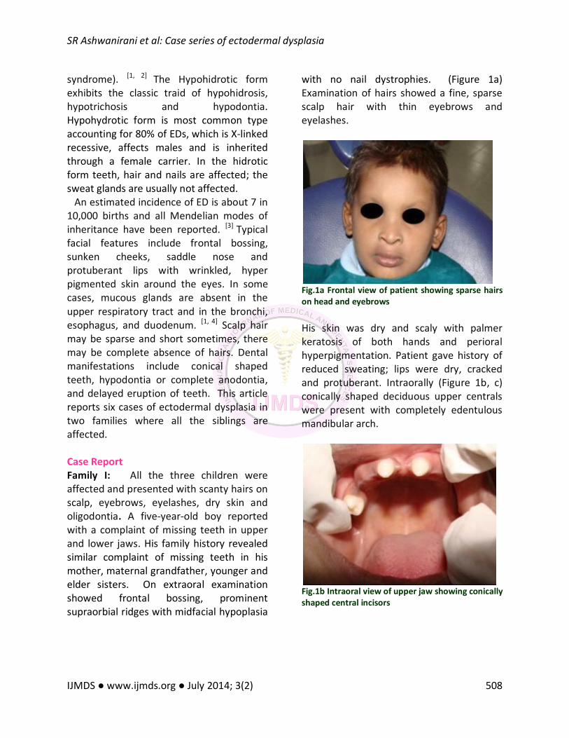
syndrome). [1, 2] The Hypohidrotic form exhibits the classic traid of hypohidrosis, hypotrichosis and hypodontia. Hypohydrotic form is most common type accounting for 80% of EDs, which is X-linked recessive, affects males and is inherited through a female carrier. In the hidrotic form teeth, hair and nails are affected; the sweat glands are usually not affected. An estimated incidence of ED is about 7 in 10,000 births and all Mendelian modes of inheritance have been reported. [3] Typical facial features include frontal bossing, sunken cheeks, saddle nose and protuberant lips with wrinkled, hyper pigmented skin around the eyes. In some cases, mucous glands are absent in the upper respiratory tract and in the bronchi, esophagus, and duodenum. [1, 4] Scalp hair may be sparse and short sometimes, there may be complete absence of hairs. Dental manifestations include conical shaped teeth, hypodontia or complete anodontia, and delayed eruption of teeth. This article reports six cases of ectodermal dysplasia in two families where all the siblings are affected. Case Report Family I: All the three children were affected and presented with scanty hairs on scalp, eyebrows, eyelashes, dry skin and oligodontia. A five-year-old boy reported with a complaint of missing teeth in upper and lower jaws. His family history revealed similar complaint of missing teeth in his mother, maternal grandfather, younger and elder sisters. On extraoral examination showed frontal bossing, prominent supraorbial ridges with midfacial hypoplasia
with no nail dystrophies. (Figure 1a) Examination of hairs showed a fine, sparse scalp hair with thin eyebrows and eyelashes.
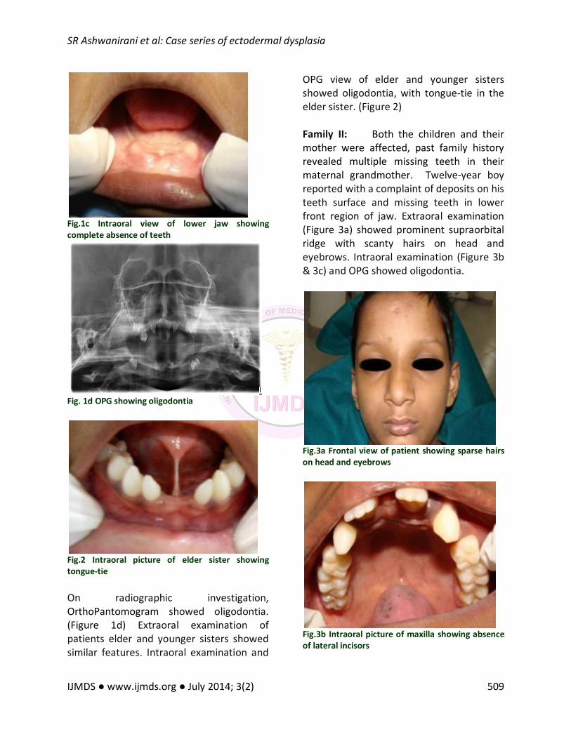
Fig.1a Frontal view of patient showing sparse hairs on head and eyebrows His skin was dry and scaly with palmer keratosis of both hands and perioral hyperpigmentation. Patient gave history of reduced sweating; lips were dry, cracked and protuberant. Intraorally (Figure 1b, c) conically shaped deciduous upper centrals were present with completely edentulous mandibular arch.
Fig.1b Intraoral view of upper jaw showing conically shaped central incisors
SR Ashwanirani et al: Case series of ectodermal dysplasia
IJMDS ● www.ijmds.org ● July 2014; 3(2) 509
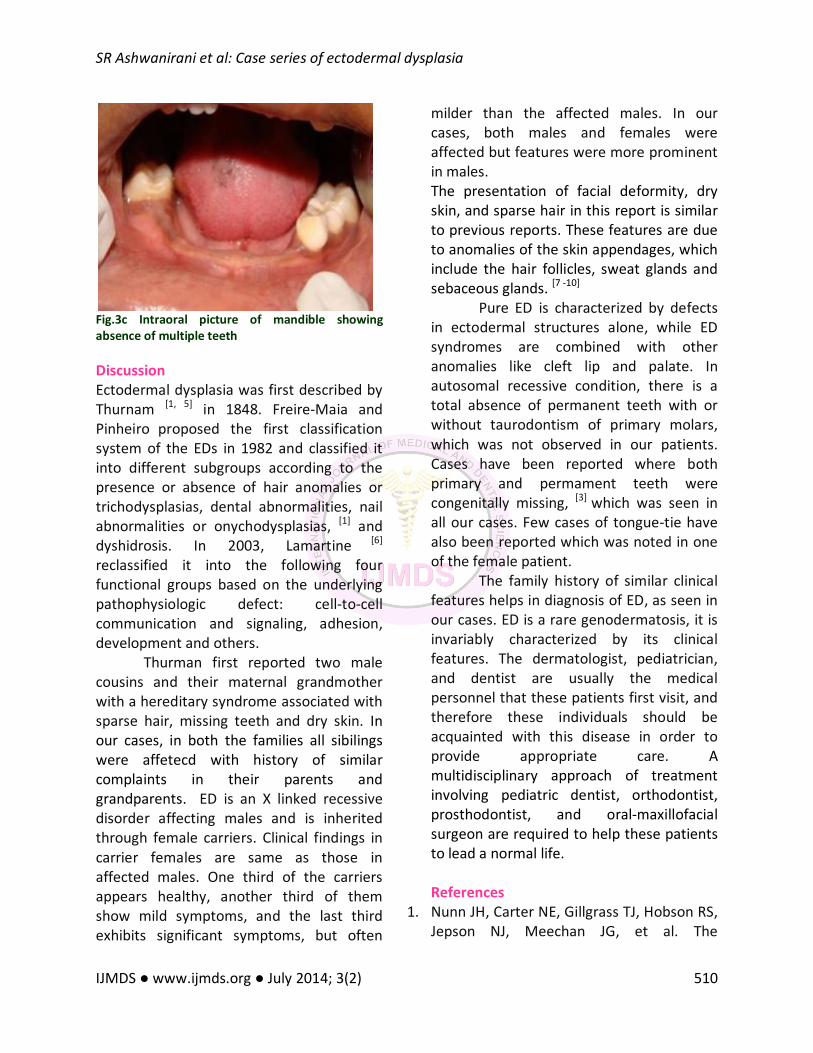
Fig.1c Intraoral view of lower jaw showing complete absence of teeth
Fig. 1d OPG showing oligodontia
Fig.2 Intraoral picture of elder sister showing tongue-tie On radiographic investigation, OrthoPantomogram showed oligodontia. (Figure 1d) Extraoral examination of patients elder and younger sisters showed similar features. Intraoral examination and
OPG view of elder and younger sisters showed oligodontia, with tongue-tie in the elder sister. (Figure 2) Family II: Both the children and their mother were affected, past family history revealed multiple missing teeth in their maternal grandmother. Twelve-year boy reported with a complaint of deposits on his teeth surface and missing teeth in lower front region of jaw. Extraoral examination (Figure 3a) showed prominent supraorbital ridge with scanty hairs on head and eyebrows. Intraoral examination (Figure 3b & 3c) and OPG showed oligodontia.
Fig.3a Frontal view of patient showing sparse hairs on head and eyebrows
Fig.3b Intraoral picture of maxilla showing absence of lateral incisors
SR Ashwanirani et al: Case series of ectodermal dysplasia
IJMDS ● www.ijmds.org ● July 2014; 3(2) 510
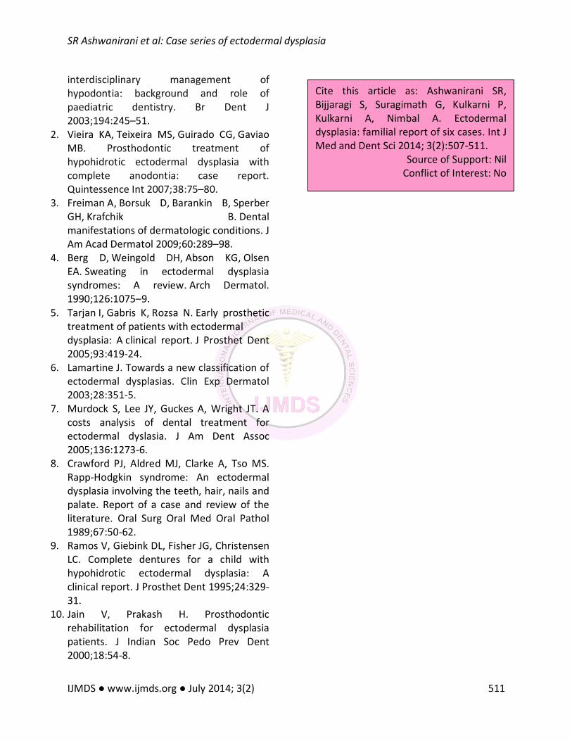
Fig.3c Intraoral picture of mandible showing absence of multiple teeth Discussion Ectodermal dysplasia was first described by Thurnam [1, 5] in 1848. Freire-Maia and Pinheiro proposed the first classification system of the EDs in 1982 and classified it into different subgroups according to the presence or absence of hair anomalies or trichodysplasias, dental abnormalities, nail abnormalities or onychodysplasias, [1] and dyshidrosis. In 2003, Lamartine [6]
reclassified it into the following four functional groups based on the underlying pathophysiologic defect: cell-to-cell communication and signaling, adhesion, development and others.
Thurman first reported two male cousins and their maternal grandmother with a hereditary syndrome associated with sparse hair, missing teeth and dry skin. In our cases, in both the families all sibilings were affetecd with history of similar complaints in their parents and grandparents. ED is an X linked recessive disorder affecting males and is inherited through female carriers. Clinical findings in carrier females are same as those in affected males. One third of the carriers appears healthy, another third of them show mild symptoms, and the last third exhibits significant symptoms, but often
milder than the affected males. In our cases, both males and females were affected but features were more prominent in males. The presentation of facial deformity, dry skin, and sparse hair in this report is similar to previous reports. These features are due to anomalies of the skin appendages, which include the hair follicles, sweat glands and sebaceous glands. [7 -10]
Pure ED is characterized by defects in ectodermal structures alone, while ED syndromes are combined with other anomalies like cleft lip and palate. In autosomal recessive condition, there is a total absence of permanent teeth with or without taurodontism of primary molars, which was not observed in our patients. Cases have been reported where both primary and permament teeth were congenitally missing, [3] which was seen in all our cases. Few cases of tongue-tie have also been reported which was noted in one of the female patient.
The family history of similar clinical features helps in diagnosis of ED, as seen in our cases. ED is a rare genodermatosis, it is invariably characterized by its clinical features. The dermatologist, pediatrician, and dentist are usually the medical personnel that these patients first visit, and therefore these individuals should be acquainted with this disease in order to provide appropriate care. A multidisciplinary approach of treatment involving pediatric dentist, orthodontist, prosthodontist, and oral-maxillofacial surgeon are required to help these patients to lead a normal life. References
1. Nunn JH, Carter NE, Gillgrass TJ, Hobson RS, Jepson NJ, Meechan JG, et al. The
SR Ashwanirani et al: Case series of ectodermal dysplasia
IJMDS ● www.ijmds.org ● July 2014; 3(2) 511
interdisciplinary management of hypodontia: background and role of paediatric dentistry. Br Dent J 2003;194:245–51.
2. Vieira KA, Teixeira MS, Guirado CG, Gaviao MB. Prosthodontic treatment of hypohidrotic ectodermal dysplasia with complete anodontia: case report. Quintessence Int 2007;38:75–80.
3. Freiman A, Borsuk D, Barankin B, Sperber GH, Krafchik B. Dental manifestations of dermatologic conditions. J Am Acad Dermatol 2009;60:289–98.
4. Berg D, Weingold DH, Abson KG, Olsen EA. Sweating in ectodermal dysplasia syndromes: A review. Arch Dermatol. 1990;126:1075–9.
5. Tarjan I, Gabris K, Rozsa N. Early prosthetic treatment of patients with ectodermal dysplasia: A clinical report. J Prosthet Dent 2005;93:419-24.
6. Lamartine J. Towards a new classification of ectodermal dysplasias. Clin Exp Dermatol 2003;28:351-5.
7. Murdock S, Lee JY, Guckes A, Wright JT. A costs analysis of dental treatment for ectodermal dyslasia. J Am Dent Assoc 2005;136:1273-6.
8. Crawford PJ, Aldred MJ, Clarke A, Tso MS. Rapp-Hodgkin syndrome: An ectodermal dysplasia involving the teeth, hair, nails and palate. Report of a case and review of the literature. Oral Surg Oral Med Oral Pathol 1989;67:50-62.
9. Ramos V, Giebink DL, Fisher JG, Christensen LC. Complete dentures for a child with hypohidrotic ectodermal dysplasia: A clinical report. J Prosthet Dent 1995;24:329-31.
10. Jain V, Prakash H. Prosthodontic rehabilitation for ectodermal dysplasia patients. J Indian Soc Pedo Prev Dent 2000;18:54-8.
Cite this article as: Ashwanirani SR, Bijjaragi S, Suragimath G, Kulkarni P, Kulkarni A, Nimbal A. Ectodermal dysplasia: familial report of six cases. Int J Med and Dent Sci 2014; 3(2):507-511.
Source of Support: Nil Conflict of Interest: No