Embed Size (px)
Citation preview
SoutheaSt aSian J trop Med public health
468 Vol 44 No. 3 May 2013
Correspondence: M Chayakulkeeree, Division of Infectious Diseases and Tropical Medicine, Department of Medicine, Faculty of Medicine Siriraj Hospital, Mahidol University, Bangkok Noi, Bangkok 10700, Thailand.Tel: +66 (0) 2419 7784; Fax: +66 (0) 2419 7783E-mail: [email protected]
CASE REPORT
LISTERIA MONOCYTOGENES BRAIN ABSCESS: TWO CASES AND REVIEW OF THE LITERATURE
Samornrod Limmahakhun and Methee Chayakulkeeree
Division of Infectious Diseases and Tropical Medicine, Department of Medicine, Faculty of Medicine Siriraj Hospital, Mahidol University, Bangkok, Thailand
Abstract. Listeria monocytogenes is a gram-positive bacillus that exhibits predilec-tion to infect the central nervous system in immunocompromised individuals; the most common manifestations are meningitis and rhombencephalitis. Liste-rial brain abscesses are rare. We report here two brain abscess cases caused by L. monocytogenes in patients receiving immunosuppressive agents. The first patient presented with left hemiparesis mimicking stroke and the second patient pre-sented with neurological symptoms without fever, which was indistinguishable from brain tumor. In both cases, magnetic resonance spectroscopy (MRS) was performed to differentiate infectious processes from other causes. Diagnosis was made with a positive blood culture in both cases. Listerial DNA was detected in the pus aspirated from the abscess in the first case. Both patients were successfully treated with intravenous ampicillin followed by oral amoxicillin. MRS was useful in differentiating infectious processes from non-infectious causes.
Keywords: Listeria monocytogenes, listeriosis, brain abscess, listerial brain abscess
INTRODUCTION
Listeria monocytogenes is a non-spore forming non-branching gram-positive bacillus, which can cause serious infec-tions in immunocompromised patients, especially in those with a malignancy or using corticosteroid. Common clini-cal manifestations of listeriosis include meningitis and rhombencephalitis (Arm-strong and Fung, 1993). In rare occasions,
this bacterium can cause infections in the parenchyma of the cerebral cortex, resulting in a brain abscess. Ampicillin and trimethoprim/sulfamethoxazole can be used to treat listerial brain abscesses. We report two patients with listerial brain abscesses whose presentations mimicked stroke and brain tumor.
CASE REPORT
Case 1A 68-year-old female presented to
our hospital with low grade fever, severe headache and progressive left-sided weakness 1 week following the second course of chemotherapy which was com-menced 5 weeks earlier to treat breast
liSterial brain abSceSS
Vol 44 No. 3 May 2013 469
cancer. She had underlying hypertension, chronic atrial fibrillation, a history of a right internal capsule cerebral infarction and a history of rheumatoid arthritis she was taking warfarin, simvastatin, atenolol and chloroquine. On examination, her temperature was 39.2ºC and her respira-tory rate was 22/minute. The patient was lethargic but able to follow commands. Neurological examination revealed gaze preference to the right, spastic tone and hyperreflexia on her left side with motor strength of 3/5 on the left and 5/5 on the right. Plantar reflexes exhibited an exten-sor response on the left. No stiffness of the neck was detected. A complete blood count showed a hemoglobin of 10.4 g/dl with a white blood cell count of 10,910 cells/mm3 (neutrophils 82%, lymphocytes 7%, monocytes 11%). The prothrombin time showed on initial normalized ratio (INR) of 1.8. Computed tomography (CT) of the brain after administration of contrast material showed a hypodense lesion without abnormal enhancement
Fig 1–CT brain with contrast media of the first patient showing a hypodense lesion without abnormal enhancement in the right frontotemporoparietal lobe with surrounding vasogenic edema and a 0.4 cm midline shift.
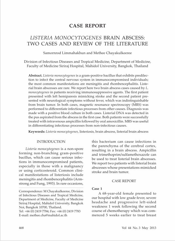
Fig 2–MRI of the brain of the first patient after 1 week of ceftriaxone and clindamycin (T2W, T1W with gadolinium) showing a high SI on T2W, iso-SI on T1W with peripheral rim enhancement in the right frontotemporoparietal region and an increase in the degree of midline shift with right uncal herniation (A). Presence of lactate peak with decreased choline, creatine, and NAA on MRS (B).
of the right frontotemporoparietal lobe with surrounding vasogenic brain edema and midline shift (Fig 1). The brain CT suggested an acute right middle cerebral artery infarction. A chest radiograph showed an alveolar infiltration of the right lower lung field; ceftriaxone and clindamycin were started to treat aspira-tion pneumonia.
Blood cultures taken before initiation of antimicrobial therapy grew out a gram-positive bacterium with a positive motility test at ambient temperature, subsequently

SoutheaSt aSian J trop Med public health
470 Vol 44 No. 3 May 2013
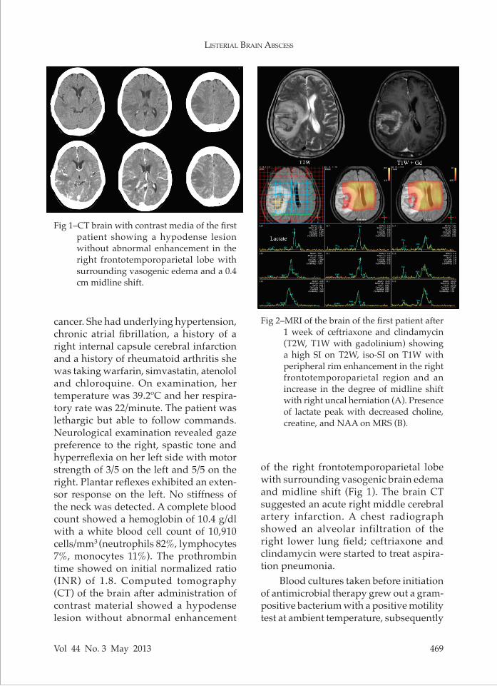
Fig 3–Sequential CT brain images with contrast in the first patient showing a reduction in the abscess size, and a disappearance of rim enhancement after 6 months of antimicrobial therapy. A) At diagnosis; B) at 1 month; C) at 2.5 months; D) at 3.5 months; E) at 6 months.
identified as L. monocytogenes. The patient was treated with intravenous ampicillin (12 grams per day). Magnetic resonance imaging (MRI), angiography (MRA) and spectroscopy (MRS) demonstrated cere- britis and an early brain abscess in the right frontotemporoparietal carebrum with right uncal herniation. The lesion was characterized by isosignal inten-sity (SI) on T1, hypersignal SI on T2 and FLAIR; restrictive diffusion on DWI, peripheral rim enhancement after gado-linium administration, and the presence of a lactate peak, a decreased choline and creatine and a N-Acetyl Aspartate (NAA) peak on MRS (Fig 2). Stereotactic-guided aspiration of the brain was done, due to an increased size in the abscess after five weeks of treatment. Eight milliliters of pus was obtained from the abscess and a Gram’s stain of the pus showed numer-ous polymorphonuclear cells with no or-ganisms. Aerobic and anaerobic cultures yielded no growth. However, molecular identification of the pus using polymerase
chain reaction (PCR) and sequencing iden-tified the DNA of Listeria spp. The patients was treated with intravenous ampicillin for 10 weeks and switched to oral amoxi-cillin (3 grams per day) for 6 months. Her weakness abated within 2 weeks and she could walk without support. A CT of the brain after 6-months of treatment showed nearly complete resolution of the lesion, with residual contrast enhancement of the right parietotemporal region (Fig 3).Case 2
A 47-year-old female with Evan’s syndrome (autoimmune hemolytic ane-mia with idiopathic thrombocytopenic purpura), autoimmune hepatitis, and diabetes mellitus presented with head-ache, a complex partial seizure and pain-ful subcutaneous nodules of 2 weeks duration. She was being treated with prednisolone at 30 mg/day. On physical examination, she was afebrile with painful erythematous subcutaneous nodules of both her cheeks and the submandibular area. Neurological examination revealed impaired attention but was otherwise nor-mal. A complete blood count revealed a hemoglobin of 9.1 g/dl with a white blood cell count of 2,670 cells/mm3 (neutrophils 87%, lymphocytes 8%, monocytes 5%), and a platelet count of 52,000 cells/mm3. A CT of the brain showed effacement of the sulci and gyri in the cortical part of posterior left frontal region and the an-terior right temporal region, with gyral enhancement suggestive of cerebritis (Fig 4). Lumbar puncture was performed and the cerebrospinal fluid (CSF) was normal. CSF cultures for bacteria, fungi, and my-cobacteria were negative. MRI and MRS of the brain revealed conglomerated le-sions with satellite rim enhancing lesions in the cortical and subcortical regions of left middle frontal gyrus and right superior temporal gyrus. Leptomenigeal

liSterial brain abSceSS
Vol 44 No. 3 May 2013 471
Fig 4–CT brain with contrast of the second pa-tient showing effacement of the sulci and gyri of the cortical part of the posterior left frontal region and the anterior right temporal region, and gyral enhance-ment after contrast medium injection. All of these findings were suggestive of cerebritis.
Fig 5–MRI brain of the second patient showing rim-enhancing conglomerated lesions in the cortical-subcortical left middle temporal gyrus and right superior tem-poral gyrus. These lesions appear as a completely dark rim on T2WI showing a uniform enhancing rim after gadolinium administration. Perilesional vasogenic edema is seen. A) T1WI with gadolinium; B) T2WI.
enhancement of the left frontal lobe was also present (Fig 5). A marked elevation in the lipid/lactate peak with normal choline and the NAA peak on MRS was demonstrated. Those findings were more compatible with brain abscess rather than brain tumor. After the MRI result a hemo-culture was obtained and L. monocytogenes was recovered. A subcutaneous nodule was biopsied and revealed neutrophilic panniculitis with negative stains, cultures, and PCR results for microorganisms. The patient was treated with ampicillin 12 grams/day IV and phenytoin. Because of the risk of bleeding, a stereotactic brain biopsy was not done. The patient had no further seizures and the erythematous nodules subsided entirely. A follow-up MRI 4 weeks after treatment showed sig-nificant improvement in the abscess at the left frontal lobe. Ampicillin was continued for 6 weeks and then switched to oral amoxicillin at 4.5 grams daily.
DISCUSSION
L. monocytogenes is a facultative an-aerobic gram-positive non-spore forming rod that is widely distributed in nature and found in multiple ecological sites. Listeria is an intracellular organism that can invade tissues normally resistant to infection, such as the central nervous system (CNS), a gravid uterus or a fetus (Gellin and Broome, 1989). Protection against L. monocytogenes infection is pre-dominantly cell-mediated (Mielke et al, 1997). Therefore, conditions associated with impaired cellular immunity, such as lymphoma, AIDS and corticosteroid

SoutheaSt aSian J trop Med public health
472 Vol 44 No. 3 May 2013
Tabl
e 1
Sum
mar
y of
pub
lishe
d re
port
s of
pat
ient
s w
ith li
ster
ial b
rain
abs
cess
es.
Cas
es
Age
/Sex
U
nder
lyin
g Im
mun
o-
Bloo
d
CSF
Su
rger
y A
ntib
iotic
s use
d O
utco
mes
Re
fere
nces
dise
ases
su
ppre
ssiv
e C
/S
C/S
th
erap
y
1 70
/M
Alc
ohol
ism
N
one
+ +
ND
A
BPC
, GM
, VC
M
Die
d (C
one
et a
l, 20
03)
2 56
/M
AID
S N
one
+ -
ND
A
BPC
, GM
Su
rviv
ed
(Pat
ey et
al,
1989
)3
49/M
Rh
eum
atic
feve
r, a
lcoh
olis
m,
Non
e +
- N
D
PGS
Die
d (B
uchn
er a
nd S
chne
iers
on, 1
968)
diab
etes
mel
litus
4
64/M
D
M, a
ortic
val
ve
Non
e +
- N
D
ABP
C, G
M
Surv
ived
(S
oto
and
Slim
an, 1
992)
repl
acem
ent
5 71
/M
Dia
bete
s mel
litus
, N
one
+ -
ND
A
BPC
, GM
D
ied
(Eck
burg
et a
l, 20
01)
rheu
mat
ic h
eart
dis
ease
,
en
doca
rditi
s 6
56/M
A
IDS
N
one
+ -
ND
A
BPC
, GM
Su
rviv
ed
(Pat
ey et
al,
1989
)7
70/F
C
irrho
sis,
diab
etes
N
one
+ N
D
ND
A
BPC
, ST
Die
d (S
ival
inga
m et
al,
1992
)
m
ellit
us, h
eart
failu
re
8 25
/F
Ulc
erat
ive
colit
is
Non
e N
R N
R N
D
NR
Die
d (L
arss
on a
nd L
inel
l, 19
79)
9 87
/M
Non
e N
one
+ +
ND
PG
S, C
P D
ied
(Spi
lkin
et a
l, 19
68)
10
63/M
N
one
Non
e +
- N
D
ABP
C
Die
d (K
enna
rd et
al,
1979
)11
24
/M
Non
e N
one
+ -
ND
A
BPC
, GM
Su
rviv
ed
(Sm
iata
cz et
al,
2006
)12
53
/F
Non
e N
one
+ -
ND
M
inoc
yclin
e, G
M
Surv
ived
(M
row
ka et
al,
2002
)13
63
/F
Non
e N
one
- -
ND
N
R D
ied
(Bru
n-Bu
isso
n et
al,
1985
)14
43
/F
Non
e N
one
- -
ND
A
BPC
D
ied
(Bru
n-Bu
isso
n et
al,
1985
)15
39
/M
Non
e N
one
NR
- N
D
NR
Die
d (K
wan
tes a
nd Is
aac,
197
1)16
54
/F
Non
e N
one
NR
NR
ND
N
R D
ied
(Lar
sson
and
Lin
ell,
1979
)17
1
1/4/
M
Non
e N
one
NR
NR
NR
Am
oxic
illin
Su
rviv
ed
(Man
cini
et a
l, 19
90)
18
70/M
N
one
Non
e -
- +
ABP
C
Surv
ived
(S
alga
do et
al,
1996
)19
53
/M
Cirr
hosi
s, se
izur
e N
one
+ -
+ PG
S, E
M
Surv
ived
(H
alki
n et
al,
1971
)20
85
/M
Dia
bete
s mel
litus
, gou
t N
one
+ -
+ A
BPC
D
ied
(Bro
wn
et a
l, 19
91)
21
43/M
Sl
eep
apne
a sy
ndro
me,
al
coho
l abu
se
Non
e +
- +
NR
Surv
ived
(D
ouen
and
Bou
rque
, 199
7)22
0/
M
Pron
atis
N
one
+ -
+ A
BPC
, GM
Su
rviv
ed
(Ban
erji
and
Noy
a, 1
999)
23
63/M
M
ultip
le m
yelo
ma
NR
+ +
+ A
BPC
, li
nezo
lid
Surv
ived
(L
eiti
et a
l, 20
05)
24
61/M
D
iabe
tes m
ellit
us
Non
e
NR
NR
+ ST
, CP
Surv
ived
(S
jost
rom
et a
l, 19
95)
25
60/M
H
IV
Non
e N
R N
R +
PGS,
CP
Die
d (H
arris
et a
l, 19
89)
26
68/M
Le
ukem
ia
NR
NR
NR
ND
C
P D
ied
(Lar
sson
et a
l, 19
78)
27
NR/
M
Non
e N
R N
R N
R N
D
NR
Die
d (P
ollo
ck et
al,
1984
)28
2/
M
NR
NR
NR
+ +
NR
Surv
ived
(U
men
ai et
al,
1978
)29
49
/M
Rena
l tra
nspl
ant
AZP
, PSL
+
+ N
D
CP
Die
d (C
rock
er a
nd L
eice
ster
, 197
6)30
16
/M
ALL
C
MT
+ +
ND
PG
S, C
P Su
rviv
ed
(Dyk
es et
al,
1979
)

liSterial brain abSceSS
Vol 44 No. 3 May 2013 473
31
20/M
A
LL
CM
T +
+ N
D
ABP
C, C
P,
EM, G
M
Surv
ived
(H
utch
inso
n an
d H
eyn,
198
3)32
6/
F A
LL
CM
T +
+ N
D
ABP
C, V
CM
, Su
rviv
ed
(Vis
coli
et a
l, 19
91)
ne
tilm
icin
33
40/F
U
lcer
ativ
e co
litis
PS
L +
+ N
D
ABP
C, G
M
Surv
ived
(S
oare
s-Fe
rnan
des e
t al,
2008
)34
58
/F
Mul
tiple
end
ocrin
e
ne
opla
sm 2
A, S
LE
PSL
+ -
ND
PG
S, T
OB
Surv
ived
(T
akan
o et
al,
1999
)35
58
/F
Imm
unob
last
ic
lym
phad
enop
athy
PS
L, C
MT
+ -
ND
A
BPC
Su
rviv
ed
(Mae
zaw
a et
al,
2002
)36
65
/M
Dia
bete
s mel
litus
PS
L +
NR
ND
A
BPC
, GM
Su
rviv
ed
(Wu
et a
l, 20
10)
37
19/M
Ju
veni
le rh
eum
atoi
d ar
thrit
is,
Tetr
alog
y of
Fal
lot
PSL
NR
+ N
D
VC
M, A
BPC
Su
rviv
ed
(Tur
ner e
t al,
1995
)38
55
/M
Rena
l tra
nspl
ant
AZP
, PSL
+
- +
ABP
C
Surv
ived
(L
echt
enbe
rg et
al,
1979
)39
45
/M
Rena
l tra
nspl
ant
AZP
, PSL
+
- +
ABP
C
Surv
ived
(S
tam
et a
l, 19
82)
40
60/F
Rh
eum
atoi
d ar
thrit
is
PSL
+ -
+ A
BPC
Su
rviv
ed
(Upd
ike
et a
l, 19
90)
41
66/F
A
ML,
Cro
hn’s
dis
ease
PS
L +
- +
ABP
C, S
T Su
rviv
ed
(Eck
burg
et a
l, 20
01)
42
47/M
A
IDS,
Mul
tiple
live
r abs
cess
, G
anci
clov
ir +
- +
ABP
C, G
M, V
CM
D
ied
(Con
e et
al,
2003
)
M
.aviu
m b
acte
rem
ia,
CM
V re
tiniti
s 43
54
/F
Sarc
oido
sis
PSL
+ -
+ A
BPC
, GM
D
ied
(Ack
erm
ann
et a
l, 20
01)
44
23/F
IT
P PS
L, C
Y +
- +
ST
Surv
ived
(T
reeb
upac
hats
akul
et a
l, 20
06)
45
58/M
M
ultip
le m
yelo
ma
VAD
+
- +
ABP
C, G
M
Surv
ived
(A
l-Kha
tti a
nd A
l-Taw
fiq, 2
010)
46
55/M
G
liobl
asto
ma
mul
tifor
me
CM
T -
+ +
ABP
C, G
M
Surv
ived
(G
anie
re et
al,
2006
)47
51
/M
Car
diac
tran
spla
nt
AZP
, +
ND
+
ABP
C, G
M
Surv
ived
(E
ckbu
rg et
al,
2001
)
cycl
ospo
rine,
PSL
48
37/M
C
ardi
ac tr
ansp
lant
PS
L,
+ N
D
+ A
BPC
Su
rviv
ed
(Eck
burg
et a
l, 20
01)
cy
clos
porin
e49
56
/F
Aut
oim
mun
e he
patit
is,
AZP
, PSL
+
ND
+
ABP
C, G
M
Surv
ived
(C
one
et a
l, 20
03)
prim
ary
bilia
ry c
irrho
sis
50
50/M
Sa
rcoi
dosi
s PS
L -
- +
ST
Surv
ived
(P
orop
atic
h an
d Ph
illip
s, 19
92)
51
51/F
C
rohn
’s d
isea
se
PSL
- -
+ A
BPC
, GM
Su
rviv
ed
(Ste
fano
vic
et a
l, 20
10)
52
50/M
C
ardi
ac tr
ansp
lant
, dia
bete
s A
ZP,
- -
+ A
BPC
, GM
D
ied
(Eck
burg
et a
l, 20
01)
mel
litus
per
irect
al a
bsce
ss
cycl
ospo
rine,
PSL
53
75/M
C
OPD
PS
L -
ND
+
ABP
C, G
M
Surv
ived
(M
ylon
akis
et a
l, 19
98)
54
77/M
C
LL
CM
T -
NR
+ C
P N
R (C
leve
land
and
Gel
fand
, 199
3)55
58
/M
CLL
C
MT
ND
N
D
+ A
BPC
, GM
Su
rviv
ed
(Dee
and
Lor
ber,
1986
)56
C
hild
A
LL
CM
T N
R N
R +
NR
Surv
ived
(A
ntun
es et
al,
1998
)57
(PR)
58
/F
Brea
st c
ance
r C
MT
+ N
D
+ A
BPC
Su
rviv
ed
58 (P
R)
47/F
Ev
an’s
synd
rom
e,
PS
L +
- N
D
ABP
C
Surv
ived
SL
E, d
iabe
tes m
ellit
us
ABP
C, a
mpi
cilli
n; A
IDS,
acq
uire
d im
mun
e de
ficie
ncy
synd
rom
e; A
LL, a
cute
lym
phob
last
ic le
ukem
ia; A
ML,
acu
te m
yelo
id le
ukem
ia; A
ZP, a
zath
iopr
ine;
CLL
, chr
onic
lym
phoc
ytic
le
ukem
ia; C
MT,
che
mot
hera
py; C
P, c
hlor
amph
enic
ol; C
Y, c
yclo
phos
pham
ide;
EM
, ery
thro
myc
in; G
M, g
enta
mic
in; I
TP, i
mm
une
thro
mbo
cyto
peni
c pu
rpur
a; N
D, n
ot d
one;
NR,
no
t rep
orte
d; P
GS,
pen
icill
in G
sodi
um; P
R, p
rese
nt re
port
; PSL
, pre
dnis
olon
e; S
LE, s
yste
mic
lupu
s ery
them
atos
us; S
T, tr
imet
hopr
ime/
sulfa
met
hoxa
zole
; TO
B, to
bram
ycin
; VA
D,
vinc
ristin
e do
xoru
bici
n an
d de
xam
etha
sone
; VC
M, v
anco
myc
in

SoutheaSt aSian J trop Med public health
474 Vol 44 No. 3 May 2013
use are risk factors for listerial infection (Stam et al, 1982; Southwick and Purich, 1996). Ingestion of L. monocytogenes contaminated food is considered to be the source of nearly all listerial human infections (Lorber, 1997). Once ingested, L. monocytogenes penetrates the Peyer’s patches of the small intestine, but not via the phagocytic microfold or M cells (Pron et al, 1998). The bacterium enters the mes-enteric lymph nodes and then the blood stream, resulting in bacteremia (Cone et al, 2003). Meningitis may occur when the organisms attach to the epithelial cells of the choroid plexus (Schluter et al, 1996). Cerebritis and subsequent brain abscesses result from penetration of the bacterium into the brain parenchyma through the cerebral capillary endothelium (Kirk, 1993; Dramsi et al, 1998); the infected macrophages penetrate these endothelial cells via the cerebral artery (Muller and Weigl, 1992). Listeria displays a tropism for the CNS, with manifestations ranging from meningitis to cerebritis and focal pa-renchymal involvement (Cone et al, 2003). A unique form of listerial parenchymal CNS infection is rhombencephalitis, an acute brainstem infection characterized by asymmetrical cranial nerve palsies, cerebellar dysfunction, hemiparesis, im-pairment of consciousness and possible respiratory failure (Armstrong and Fung, 1993). Listerial abscesses of the cerebral hemisphere are extremely rare, account-ing for approximately 1-10% of listerial CNS infections (Nieman and Lorber, 1980; Lorber, 1997; Cone et al, 2003). Listerial brain abscesses are associated with men-ingitis in up to 38% of the patients (Chun et al, 1986). Involvement of the subcorti-cal grey matter, such as the thalamus and basal ganglia, is more common in listerial brain abscesses (about 21%) than with oth-er agents (Lorber, 1997). Similar to other
bacterial brain abscesses, most patients with listerial brain abscesses have fever, elevated leukocyte counts, headache, and focal neurological signs. Bacteremia is common and found in about 86% of Listeria brain abscess (Dee and Lorber, 1986). Bacteremia is unusual in brain abscesses caused by other organisms, in which it occurs in approximately 11% of cases (Mathisen and Johnson, 1997). Hence, three different features of listerial brain abscesses are: 1) the presence of bacteremia in most patients, 2) a quarter of patients exhibit concomitant meningitis, and 3) subcortical abscesses are usually located in the pons, thalamus, or medulla (Lorber, 1997). However, listerial brain abscesses may mimic other causes of CNS disease, such as strokes or brain tumors; an MRS of the brain may be useful test to distinguish the etiologies (Lai et al, 2002). Choline is a marker of increased cellular turnover and is elevated in tumors and gliosis, but is decreased in abscesses (Gujar et al, 2005). Lactate, a product of anaerobic glycolysis, is detected in any diseased brain, such as with an infarc-tion, tumor or infection (Gujar et al, 2005). NAA, a neuronal marker, and creatine, a measure of energy stores, are decreased in cases of infarctions, tumors, and infections (Gujar et al, 2005).
There have been no controlled trials to establish a drug of choice or duration of therapy for listerial infection (Lorber, 1997). Ampicillin is generally considered as the preferred agent for treatment of lis-teriosis, although its superiority to penicil-lin is questionable. For patients intolerant of penicillins, trimethoprim-sulfamethox-azole is thought to be the best alterna-tive single agent (Winslow and Pankey, 1982; Treebupachatsakul et al, 2006). On the basis of the synergy observed with in vitro and in animal models (Edmiston

liSterial brain abSceSS
Vol 44 No. 3 May 2013 475
and Gordon, 1979), most authorities sug-gest adding gentamicin to ampicillin to treat listerial bacteremia in patients with severely impaired T-cell function and in all cases of listerial meningitis and endocarditis (Nieman and Lorber, 1980; Gellin and Broome, 1989; Cherubin et al, 1991). Linezolid and rifampicin are ac-tive against L. monocytogenes in vitro and cross the blood-brain barrier. Combina-tion treatment involving rifampin and another active antimicrobial may reduce the emergence of resistance to rifampin (Leiti et al, 2005). Leiti et al (2005) reported successful treatment with a combination of linezolid and rifampin in the case of a listerial brain abscess without any ad-verse events. Patients with a brain abscess require treatment for at least six weeks and should be followed up with serial neurological imaging until the abscesses are resolved (Lorber, 1997). The mortality rate in listerial CNS abscesses has been reported to be 40% compared with 17% for other types of brain abscesses (Cone et al, 2003).
To our knowledge, only 56 cases of macroscopic brain abscesses due to L. monocytogenes have been reported be-tween 1968 and 2011 (Table 1). Thirty-nine patients (68%) were men. The mean age was 50±20 years (range 0-87 years). Most patients (80%) had underlying condi-tions, such as a hematological malignancy (23%), autoimmune diseases treated with prednisolone (19%), having undergone solid organ transplant (10%), diabetes mellitus (12%), human immunodeficiency virus (HIV) infection (7%), or others (9%). Two of our patients had an underlying malignancy and an autoimmune disease treated with corticosteroids and immuno-suppressive agents, respectively. Positive blood cultures and CSF cultures were found in 79% and 23%, of cases listed in
Table 1, respectively. Ampicillin-based regimens were used in 37 patients (74%). Twenty-nine patients (51%) underwent surgery. A total of 19 patients died, giv-ing a mortality rate of 33%. Patients who had underlying diseases and who did not undergo surgical interventions were more likely to die (63% and 74%, respectively).
In conclusion, due to the severity of listerial diseases, its predilection for the CNS and high mortality rate, clinicians should retain a high index of suspicion in susceptible patients presenting with CNS infections. Blood cultures are usually positive in patients with listerial brain abscesses. A MRS of the brain may be beneficial to differentiate between a brain tumor and an abscess. Ampicillin should be considered as part of combination an-timicrobial therapy in such patients.
ACKNOWLEDGEMENTS
The authors would like to thank all the Infectious Diseases clinical fellows and residents for providing the clinical information and patient care. The author, Methee Chayakulkeeree, was supported by a “Chalermphrakiat” Grant from the Faculty of Medicine, Siriraj Hospital, Mahidol University, Thailand.
REFERENCES
Ackermann G, Schoen H, Schaumann R, Di-etrich J, Rodloff AC. Rapidly growing tumor-like brain lesion. Infection 2001; 29: 278-9.
Al-Khatti AA, Al-Tawfiq JA. Listeria monocy-togenes brain abscess in a patient with multiple myeloma. J Infect Dev Ctries 2010; 4: 849-51.
Antunes NL, Hariharan S, DeAngelis LM. Brain abscesses in children with cancer. Med Pediatr Oncol 1998; 31: 19-21.
Armstrong RW, Fung PC. Brainstem encepha-
SoutheaSt aSian J trop Med public health
476 Vol 44 No. 3 May 2013
litis (rhombencephalitis) due to Listeria monocytogenes: case report and review. Clin Infect Dis 1993; 16: 689-702.
Banerji A, Noya FJ. Brain abscess associated with neonatal listeriosis. Pediatr Infect Dis J 1999; 18: 305-7.
Brown PH, Ingram CW, van der Horst C. Brain abscess caused by Listeria monocytogenes. Rev Infect Dis 1991; 13: 768-9.
Brun-Buisson CJ, de Gialluly E, Gherardi R, Otterbein G, Gray F, Rapin M. Fatal non-meningitic Listeria rhombencephalitis. Report of two cases. Arch Intern Med 1985; 145: 1982-5.
Buchner LH, Schneierson S. Clinical and labora-tory aspects of Listeria monocytogenes infec-tions. With a report of ten cases. Am J Med 1968; 45: 904-21.
Cherubin CE, Appleman MD, Heseltine PN, Khayr W, Stratton CW. Epidemiological spectrum and current treatment of liste-riosis. Rev Infect Dis 1991; 13: 1108-14.
Chun CH, Johnson JD, Hofstetter M, Raff MJ. Brain abscess. A study of 45 consecutive cases. Medicine (Baltimore) 1986; 65: 415-31.
Cleveland KO, Gelfand MS. Listerial brain ab-scess in a patient with chronic lymphocytic leukemia treated with fludarabine. Clin Infect Dis 1993; 17: 816-7.
Cone LA, Leung MM, Byrd RG, Annunziata GM, Lam RY, Herman BK. Multiple cere-bral abscesses because of Listeria monocy-togenes: three case reports and a literature review of supratentorial listerial brain abscess(es). Surg Neurol 2003; 59: 320-8.
Crocker EF, Leicester J. Cerebral abscess due to Listeria monocytogenes. Med J Aust 1976; 1: 90-2.
Dee RR, Lorber B. Brain abscess due to Listeria monocytogenes: case report and literature review. Rev Infect Dis 1986; 8: 968-77.
Douen AG, Bourque PR. Musical auditory hal-lucinosis from Listeria rhombencephalitis. Can J Neurol Sci 1997; 24: 70-2.
Dramsi S, Levi S, Triller A, Cossart P. Entry of Listeria monocytogenes into neurons occurs by cell-to-cell spread: an in vitro study. Infect Immun 1998; 66: 4461-8.
Dykes A, Baraff LJ, Herzog P. Listeria brain abscess in an immunosuppressed child. J Pediatr 1979; 94: 72-4.
Eckburg PB, Montoya JG, Vosti KL. Brain ab-scess due to Listeria monocytogenes: five cases and a review of the literature. Medi-cine (Baltimore) 2001; 80: 223-35.
Edmiston CE, Jr, Gordon RC. Evaluation of gentamicin and penicillin as a synergistic combination in experimental murine lis-teriosis. Antimicrob Agents Chemother 1979; 16: 862-3.
Ganiere V, Christen G, Bally F, et al. Listeria brain abscess, Pneumocystis pneumonia and Kaposi’s sarcoma after temozolomide. Nat Clin Pract Oncol 2006; 3: 339-43; quiz following 43.
Gellin BG, Broome CV. Listeriosis. JAMA 1989; 261: 1313-20.
Gujar SK, Maheshwari S, Bjorkman-Burtscher I, Sundgren PC. Magnetic resonance spectroscopy. J Neuroophthalmol 2005; 25: 217-26.
Halkin H, Shacked IJ, Altmann G. Brain abscess due to Listeria monocytogenes in a patient with cirrhosis of the liver. Isr J Med Sci 1971; 7: 1192-5.
Harris JO, Marquez J, Swerdloff MA, Magana IA. Listeria brain abscess in the acquired immunodeficiency syndrome. Arch Neurol 1989; 46: 250.
Hutchinson RJ, Heyn RM. Listerial brain ab-scess in a patient with leukemia: success-ful nonsurgical management. Clin Pediatr (Phila) 1983; 22: 312.
Kennard C, Howard AJ, Scholtz C, Swash M. Infection of the brainstem by Listeria mono-cytogenes. J Neurol Neurosurg Psychiatry 1979; 42: 931-3.
Kirk J. Diagnostic ultrastructure of Listeria monocytogenes in human central nervous tissue. Ultrastruct Pathol 1993; 17: 583-92.

liSterial brain abSceSS
Vol 44 No. 3 May 2013 477
Kwantes W, Isaac M. Listeriosis. Br Med J 1971; 4: 296-7.
Lai PH, Ho JT, Chen WL, et al. Brain abscess and necrotic brain tumor: discrimination with proton MR spectroscopy and diffusion-weighted imaging. Am J Neuroradiol 2002; 23: 1369-77.
Larsson S, Cronberg S, Winblad S. Clinical aspects on 64 cases of juvenile and adult listeriosis in Sweden. Acta Med Scand 1978; 204: 503-8.
Larsson S, Linell F. Correlations between clini-cal and postmortem findings in listeriosis. Scand J Infect Dis 1979; 11: 55-8.
Lechtenberg R, Sierra MF, Pringle GF, Shucart WA, Butt KM. Listeria monocytogenes: brain abscess or meningoencephalitis? Neurology 1979; 29: 86-90.
Leiti O, Gross JW, Tuazon CU. Treatment of brain abscess caused by Listeria monocyto-genes in a patient with allergy to penicillin and trimethoprim-sulfamethoxazole. Clin Infect Dis 2005; 40: 907-8.
Lorber B. Listeriosis. Clin Infect Dis 1997; 24: 1-9; quiz 10-1.
Maezawa Y, Hirasawa A, Abe T, et al. Successful treatment of listerial brain abscess: a case report and literature review. Intern Med 2002; 41: 1073-8.
Mancini J, Choux M, Pinsard N. [A cerebral abscess due to Listeria monocytogenes in a 15-month-old infant]. Ann Pediatr (Paris) 1990; 37: 299-302.
Mathisen GE, Johnson JP. Brain absess. Clin Infect Dis 1997; 25: 763-79.
Mielke ME, Peters C, Hahn H. Cytokines in the induction and expression of T-cell-medi-ated granuloma formation and protection in the murine model of listeriosis. Immunol Rev 1997; 158: 79-93.
Mrowka M, Graf LP, Odin P. MRI findings in mesenrhombencephalitis due to Listeria monocytogenes. J Neurol Neurosurg Psychia-try 2002; 73: 775.
Muller WA, Weigl SA. Monocyte-selective transendothelial migration: dissection of
the binding and transmigration phases by an in vitro assay. J Exp Med 1992; 176: 819-28.
Mylonakis E, Hohmann EL, Calderwood SB. Central nervous system infection with Listeria monocytogenes. 33 years’ experi-ence at a general hospital and review of 776 episodes from the literature. Medicine (Baltimore) 1998; 77: 313-36.
Nieman RE, Lorber B. Listeriosis in adults: a changing pattern. Report of eight cases and review of the literature, 1968-1978. Rev Infect Dis 1980; 2: 207-27.
Patey O, Nedelec C, Emond JP, Mayorga R, N’Go N, Lafaix C. Listeria monocytogenes septicemia in an AIDS patient with a brain abscess. Eur J Clin Microbiol Infect Dis 1989; 8: 746-8.
Pollock SS, Pollock TM, Harrison MJ. Infection of the central nervous system by Listeria monocytogenes: a review of 54 adult and ju-venile cases. Q J Med 1984; 53: 331-40.
Poropatich R, Phillips YY. Listerial brain abscess in long-standing sarcoidosis. South Med J 1992; 85: 554-6.
Pron B, Boumaila C, Jaubert F, et al. Compre-hensive study of the intestinal stage of listeriosis in a rat ligated ileal loop system. Infect Immun 1998; 66: 747-55.
Salgado MJ, Damani NN, Llewellyn CG, Ma-loney WJ, Vandorpe RA, Sangalang VE. Magnetic resonance imaging of abscesses of the brain stem and cerebellum com-plicating Listeria monocytogenes rhomb-encephalitis. Can Assoc Radiol J 1996; 47: 431-3.
Schluter D, Chahoud S, Lassmann H, Schumann A, Hof H, Deckert-Schluter M. Intracere-bral targets and immunomodulation of murine Listeria monocytogenes meningoen-cephalitis. J Neuropathol Exp Neurol 1996; 55: 14-24.
Sivalingam JJ, Martin P, Fraimow HS, Yarze JC, Friedman LS. Listeria monocytogenes peri-tonitis: case report and literature review. Am J Gastroenterol 1992; 87: 1839-45.
Sjostrom A, Olsson T, Burman LA, Tarnvik A.

SoutheaSt aSian J trop Med public health
478 Vol 44 No. 3 May 2013
Listerial brain abscess in an immunocom-petent adult with a predisposing intestinal condition. J Infect 1995; 30: 185-6.
Smiatacz T, Kowalik MM, Hlebowicz M. Pro-longed dysphagia due to Listeria rhomb-encephalitis with brainstem abscess and acute polyradiculoneuritis. J Infect 2006; 52: e165-7.
Soares-Fernandes JP, Beleza P, Cerqueira JJ, et al. Simultaneous supratentorial and brainstem abscesses due to Listeria mono-cytogenes. J Neuroradiol 2008; 35: 173-6.
Soto LF, Sliman RJ. Focal seizures in a man with multiple medical problems. Hosp Pract (Off Ed) 1992; 27: 27-9.
Southwick FS, Purich DL. Intracellular patho-genesis of listeriosis. N Engl J Med 1996; 334: 770-6.
Spilkin ES, Rachmaninoff N, Climie AR. Listeria monocytogenes meningitis. Report of 2 cases and review of the literatur. Am J Clin Pathol 1968; 49: 671-6.
Stam J, Wolters EC, van Manen J, Verbeeten B. Listeria monocytogenes abscess in the basal ganglia. J Neurol Neurosurg Psychiatry 1982; 45: 757-9.
Stefanovic A, Reid J, Nadon AC, Grant J. Potential nosocomial acquisition of epi-demic Listeria monocytogenes presenting as multiple brain abscesses resembling nocardiosis. Can J Infect Dis Med Microbiol 2010; 21: 57-60.
Takano A, Adachi H, Mizuno M, Kawamura K, Sobue G. [Direct Gram staining of
blood culture sample enabled the early diagnosis of brain abscess due to Listeria monocytogenes]. Rinsho Shinkeigaku 1999; 39: 1164-7.
Treebupachatsakul P, Srifeungfung S, Chay-akulkeeree M. Brain abscess due to Listeria monocytogenes: first case report in Thailand. J Med Assoc Thai 2006; 89: 1516-20.
Turner D, Fried M, Hoffman M, Paleacu D, Reider I, Yust I. Brainstem abscess and meningitis due to Listeria monocytogenes in an adult with juvenile chronic arthritis. Neurology 1995; 45: 1020-1.
Umenai T, Saitoh Y, Sasaki T, et al. Listeria monocytogenes infection with brain abscess formation–the first case in Miyagi Prefec-ture. Tohoku J Exp Med 1978; 124: 95-6.
Updike WS, Anderson CJ, Lundberg MS, Spru-ance SL. Successful medical treatment of listerial brain abscess. West J Med 1990; 152: 298-301.
Viscoli C, Garaventa A, Ferrea G, Manno G, Taccone A, Terragna A. Listeria monocyto-genes brain abscesses in a girl with acute lymphoblastic leukaemia after late central nervous system relapse. Eur J Cancer 1991; 27: 435-7.
Winslow DL, Pankey GA. In vitro activities of trimethoprim and sulfamethoxazole against Listeria monocytogenes. Antimicrob Agents Chemother 1982; 22: 51-4.
Wu C, Sung SF, Chang RY. Brain abscess due to Listeria monocytogenes : A case report. J Intern Med Taiwan 2010; 21: 221-6.