Embed Size (px)
Citation preview
Case ReportPericardial Effusion-Associated Hyponatremia
Ahmed A. Hanfy, Nicholas T. Manasewitsch , Bryce D. Beutler , Daniel Antwi-Amoabeng,Mohamed Elnaggar, Munadel Awad, and Gurpreet S. Chahal
Department of Internal Medicine, University of Nevada, Reno School of Medicine, Nevada, USA
Correspondence should be addressed to Bryce D. Beutler; [email protected]
Received 3 April 2020; Revised 28 June 2020; Accepted 8 July 2020; Published 13 July 2020
Academic Editor: Ertugrul Ercan
Copyright © 2020 Ahmed A. Hanfy et al. This is an open access article distributed under the Creative Commons AttributionLicense, which permits unrestricted use, distribution, and reproduction in any medium, provided the original work isproperly cited.
Pericardial effusion has been identified as a rare cause of hyponatremia. In most patients, pericardiocentesis results in rapidcorrection. We describe a 67-year-old male who presented with pericardial effusion-associated hyponatremia secondary tocardiac resynchronization therapy-D placement that resolved following evacuation. In addition, we review the literature onpericardial effusion-associated hyponatremia.
1. Introduction
Hyponatremia is defined as a serum sodium concentrationbelow 135mEq/L. It is most commonly asymptomatic butmay manifest with headaches, nausea, seizures, and deathdue to cerebral edema [1].
Pericardial effusion has been identified as a rare causeof hyponatremia. Interestingly, pericardiocentesis oftenresults in rapid and complete reversal of hyponatremia[2–9]. The mechanism underlying pericardial effusion-related hyponatremia remains to be established. However,it has been hypothesized to result from increased anti-diuretic hormone release in the setting of decreased freewater excretion [2, 5, 8].
We describe a 67-year-old male with pericardialeffusion-associated hyponatremia who experienced rapidand complete normalization of serum sodium followingpericardiocentesis.
2. Case Presentation
A 67-year-old male with a history of nonischemic cardiomy-opathy, non-insulin–dependent diabetes mellitus, and hyper-tension presented with progressively worsening dyspnea andcough of three weeks duration. Two months prior to presen-tation, the patient had undergone cardiac resynchronization
therapy-defibrillator (CRT-D) placement for nonischemiccardiomyopathy and left bundle branch block with a left ven-tricular ejection fraction of less than 35%. No operative com-plications were reported.
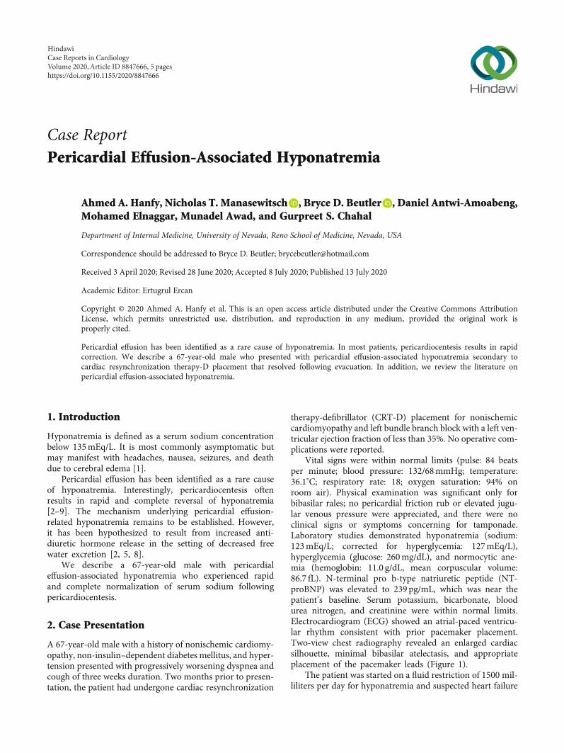
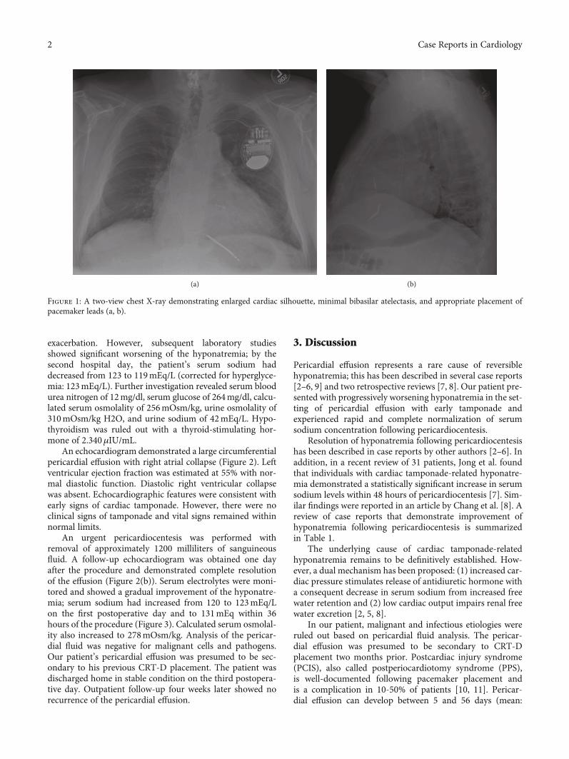
Vital signs were within normal limits (pulse: 84 beatsper minute; blood pressure: 132/68mmHg; temperature:36.1°C; respiratory rate: 18; oxygen saturation: 94% onroom air). Physical examination was significant only forbibasilar rales; no pericardial friction rub or elevated jugu-lar venous pressure were appreciated, and there were noclinical signs or symptoms concerning for tamponade.Laboratory studies demonstrated hyponatremia (sodium:123mEq/L; corrected for hyperglycemia: 127mEq/L),hyperglycemia (glucose: 260mg/dL), and normocytic ane-mia (hemoglobin: 11.0 g/dL, mean corpuscular volume:86.7 fL). N-terminal pro b-type natriuretic peptide (NT-proBNP) was elevated to 239 pg/mL, which was near thepatient’s baseline. Serum potassium, bicarbonate, bloodurea nitrogen, and creatinine were within normal limits.Electrocardiogram (ECG) showed an atrial-paced ventricu-lar rhythm consistent with prior pacemaker placement.Two-view chest radiography revealed an enlarged cardiacsilhouette, minimal bibasilar atelectasis, and appropriateplacement of the pacemaker leads (Figure 1).
The patient was started on a fluid restriction of 1500 mil-liliters per day for hyponatremia and suspected heart failure
HindawiCase Reports in CardiologyVolume 2020, Article ID 8847666, 5 pageshttps://doi.org/10.1155/2020/8847666
exacerbation. However, subsequent laboratory studiesshowed significant worsening of the hyponatremia; by thesecond hospital day, the patient’s serum sodium haddecreased from 123 to 119mEq/L (corrected for hyperglyce-mia: 123mEq/L). Further investigation revealed serum bloodurea nitrogen of 12mg/dl, serum glucose of 264mg/dl, calcu-lated serum osmolality of 256mOsm/kg, urine osmolality of310mOsm/kg H2O, and urine sodium of 42mEq/L. Hypo-thyroidism was ruled out with a thyroid-stimulating hor-mone of 2.340μIU/mL.
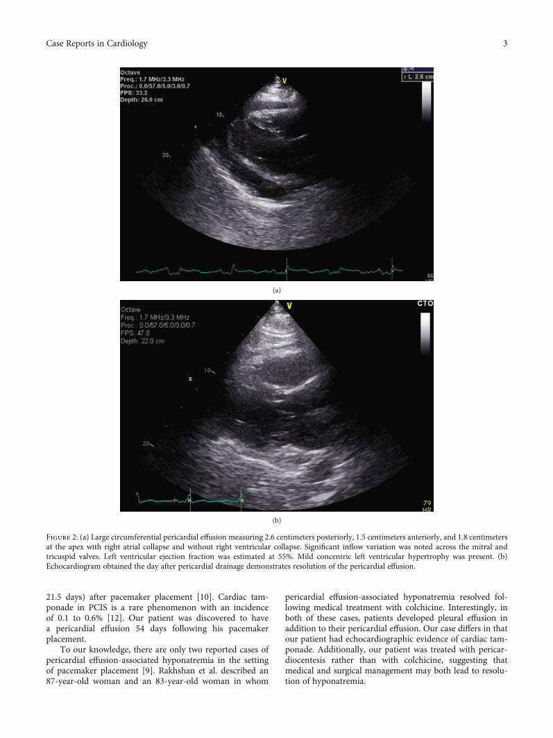
An echocardiogram demonstrated a large circumferentialpericardial effusion with right atrial collapse (Figure 2). Leftventricular ejection fraction was estimated at 55% with nor-mal diastolic function. Diastolic right ventricular collapsewas absent. Echocardiographic features were consistent withearly signs of cardiac tamponade. However, there were noclinical signs of tamponade and vital signs remained withinnormal limits.
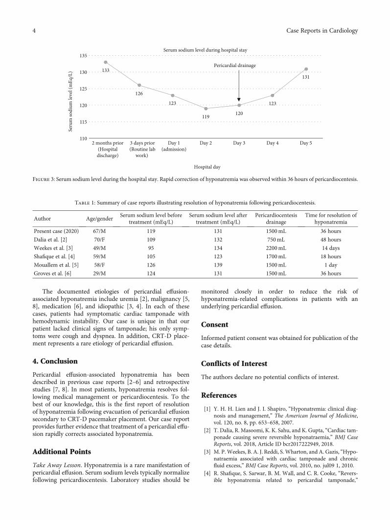
An urgent pericardiocentesis was performed withremoval of approximately 1200 milliliters of sanguineousfluid. A follow-up echocardiogram was obtained one dayafter the procedure and demonstrated complete resolutionof the effusion (Figure 2(b)). Serum electrolytes were moni-tored and showed a gradual improvement of the hyponatre-mia; serum sodium had increased from 120 to 123mEq/Lon the first postoperative day and to 131mEq within 36hours of the procedure (Figure 3). Calculated serum osmolal-ity also increased to 278mOsm/kg. Analysis of the pericar-dial fluid was negative for malignant cells and pathogens.Our patient’s pericardial effusion was presumed to be sec-ondary to his previous CRT-D placement. The patient wasdischarged home in stable condition on the third postopera-tive day. Outpatient follow-up four weeks later showed norecurrence of the pericardial effusion.
3. Discussion
Pericardial effusion represents a rare cause of reversiblehyponatremia; this has been described in several case reports[2–6, 9] and two retrospective reviews [7, 8]. Our patient pre-sented with progressively worsening hyponatremia in the set-ting of pericardial effusion with early tamponade andexperienced rapid and complete normalization of serumsodium concentration following pericardiocentesis.
Resolution of hyponatremia following pericardiocentesishas been described in case reports by other authors [2–6]. Inaddition, in a recent review of 31 patients, Jong et al. foundthat individuals with cardiac tamponade-related hyponatre-mia demonstrated a statistically significant increase in serumsodium levels within 48 hours of pericardiocentesis [7]. Sim-ilar findings were reported in an article by Chang et al. [8]. Areview of case reports that demonstrate improvement ofhyponatremia following pericardiocentesis is summarizedin Table 1.
The underlying cause of cardiac tamponade-relatedhyponatremia remains to be definitively established. How-ever, a dual mechanism has been proposed: (1) increased car-diac pressure stimulates release of antidiuretic hormone witha consequent decrease in serum sodium from increased freewater retention and (2) low cardiac output impairs renal freewater excretion [2, 5, 8].
In our patient, malignant and infectious etiologies wereruled out based on pericardial fluid analysis. The pericar-dial effusion was presumed to be secondary to CRT-Dplacement two months prior. Postcardiac injury syndrome(PCIS), also called postperiocardiotomy syndrome (PPS),is well-documented following pacemaker placement andis a complication in 10-50% of patients [10, 11]. Pericar-dial effusion can develop between 5 and 56 days (mean:
(a) (b)
Figure 1: A two-view chest X-ray demonstrating enlarged cardiac silhouette, minimal bibasilar atelectasis, and appropriate placement ofpacemaker leads (a, b).
2 Case Reports in Cardiology
21.5 days) after pacemaker placement [10]. Cardiac tam-ponade in PCIS is a rare phenomenon with an incidenceof 0.1 to 0.6% [12]. Our patient was discovered to havea pericardial effusion 54 days following his pacemakerplacement.
To our knowledge, there are only two reported cases ofpericardial effusion-associated hyponatremia in the settingof pacemaker placement [9]. Rakhshan et al. described an87-year-old woman and an 83-year-old woman in whom
pericardial effusion-associated hyponatremia resolved fol-lowing medical treatment with colchicine. Interestingly, inboth of these cases, patients developed pleural effusion inaddition to their pericardial effusion. Our case differs in thatour patient had echocardiographic evidence of cardiac tam-ponade. Additionally, our patient was treated with pericar-diocentesis rather than with colchicine, suggesting thatmedical and surgical management may both lead to resolu-tion of hyponatremia.
(a)
(b)
Figure 2: (a) Large circumferential pericardial effusion measuring 2.6 centimeters posteriorly, 1.5 centimeters anteriorly, and 1.8 centimetersat the apex with right atrial collapse and without right ventricular collapse. Significant inflow variation was noted across the mitral andtricuspid valves. Left ventricular ejection fraction was estimated at 55%. Mild concentric left ventricular hypertrophy was present. (b)Echocardiogram obtained the day after pericardial drainage demonstrates resolution of the pericardial effusion.
3Case Reports in Cardiology
The documented etiologies of pericardial effusion-associated hyponatremia include uremia [2], malignancy [5,8], medication [6], and idiopathic [3, 4]. In each of thesecases, patients had symptomatic cardiac tamponade withhemodynamic instability. Our case is unique in that ourpatient lacked clinical signs of tamponade; his only symp-toms were cough and dyspnea. In addition, CRT-D place-ment represents a rare etiology of pericardial effusion.
4. Conclusion
Pericardial effusion-associated hyponatremia has beendescribed in previous case reports [2–6] and retrospectivestudies [7, 8]. In most patients, hyponatremia resolves fol-lowing medical management or pericardiocentesis. To thebest of our knowledge, this is the first report of resolutionof hyponatremia following evacuation of pericardial effusionsecondary to CRT-D pacemaker placement. Our case reportprovides further evidence that treatment of a pericardial effu-sion rapidly corrects associated hyponatremia.
Additional Points
Take Away Lesson. Hyponatremia is a rare manifestation ofpericardial effusion. Serum sodium levels typically normalizefollowing pericardiocentesis. Laboratory studies should be
monitored closely in order to reduce the risk ofhyponatremia-related complications in patients with anunderlying pericardial effusion.
Consent
Informed patient consent was obtained for publication of thecase details.
Conflicts of Interest
The authors declare no potential conflicts of interest.
References
[1] Y. H. H. Lien and J. I. Shapiro, “Hyponatremia: clinical diag-nosis and management,” The American Journal of Medicine,vol. 120, no. 8, pp. 653–658, 2007.
[2] T. Dalia, R. Masoomi, K. K. Sahu, and K. Gupta, “Cardiac tam-ponade causing severe reversible hyponatraemia,” BMJ CaseReports, vol. 2018, Article ID bcr2017222949, 2018.
[3] M. P.Weekes, B. A. J. Reddi, S. Wharton, and A. Gazis, “Hypo-natraemia associated with cardiac tamponade and chronicfluid excess,” BMJ Case Reports, vol. 2010, no. jul09 1, 2010.
[4] R. Shafique, S. Sarwar, B. M. Wall, and C. R. Cooke, “Revers-ible hyponatremia related to pericardial tamponade,”
133
126
123
119 120
123
131
110
115
120
125
130
135
2 months prior(Hospitaldischarge)
3 days prior(Routine lab
work)
Day 1(admission)
Day 2 Day 3 Day 4 Day 5
Seru
m so
dium
leve
l (m
Eq/L
)
Hospital day
Serum sodium level during hospital stay
Pericardial drainage
Figure 3: Serum sodium level during the hospital stay. Rapid correction of hyponatremia was observed within 36 hours of pericardiocentesis.
Table 1: Summary of case reports illustrating resolution of hyponatremia following pericardiocentesis.
Author Age/genderSerum sodium level before
treatment (mEq/L)Serum sodium level after
treatment (mEq/L)Pericardiocentesis
drainageTime for resolution of
hyponatremia
Present case (2020) 67/M 119 131 1500mL 36 hours
Dalia et al. [2] 70/F 109 132 750mL 48 hours
Weekes et al. [3] 49/M 95 134 2200mL 14 days
Shafique et al. [4] 59/M 105 123 1700mL 18 hours
Mouallem et al. [5] 58/F 126 139 1500mL 1 day
Groves et al. [6] 29/M 124 131 1500mL 36 hours
4 Case Reports in Cardiology
American Journal of Kidney Diseases, vol. 50, no. 2, pp. 336–341, 2007.
[5] M. Mouallem, I. Wolf, G. Mindlin, and Z. Farfel, “Pericardialtamponade-associated hyponatremia,” The American Journalof the Medical Sciences, vol. 325, no. 1, pp. 51-52, 2003.
[6] P. H. Groves, A. M. Shah, and S. J. Hutchison, “Hyponatrae-mia secondary to an inappropriately high release of antidiure-tic hormone in cardiac tamponade,” British Heart Journal,vol. 64, no. 3, pp. 206-207, 1990.
[7] B. H. Jong, C. C. Wei, and K. G. Shyu, “Improved hyponatre-mia after pericardial drainage in patients suffering from car-diac tamponade,” BMC Cardiovascular Disorders, vol. 16,no. 1, 2016.
[8] F. K. Chang, Y. C. Lee, and C. H. Chiu, “Hyponatremia inpatients with symptomatic pericardial effusion,” Journal ofthe Chinese Medical Association, vol. 75, no. 10, pp. 509–512,2012.
[9] E. Rakhshan, S. A. Mirabbasi, B. Khalighi, and K. Khalighi,“Pericarditis-induced hyponatremia after cardiac electronicimplantable device (CEID) procedures,” Am J Case Rep.,vol. 16, pp. 245–249, 2015.
[10] W. C. Tsai, C. T. Liou, C. C. Cheng, K. S. Tsai, S. M. Cheng,and W. S. Lin, “Post-cardiac injury syndrome after permanentpacemaker implantation,” Acta Cardiologica Sinica, vol. 28,pp. 53–55, 2012.
[11] I. Zeltser, L. A. Rhodes, R. E. Tanel et al., “Postpericardiotomysyndrome after permanent pacemaker implantation in chil-dren and young adults,” The Annals of Thoracic Surgery,vol. 78, no. 5, pp. 1684–1687, 2004.
[12] G. L. Miller, E. B. Coccio, and S. C. Sharma, “Postpericardiot-omy syndrome and cardiac tamponade following transvenouspacemaker placement,” Clinical Cardiology, vol. 19, no. 3,pp. 255-256, 1996.
5Case Reports in Cardiology


![Challenges in Management of Pericardial Effusion in ... · [5,6]. It has been demonstrated that cardiac tamponade, a serious hemodynamic medical emergency as a result of pericardial](https://img.pdfslide.us/doc/110x75/5ceb108588c993886b8bfeff/challenges-in-management-of-pericardial-effusion-in-56-it-has-been-demonstrated.jpg)