Embed Size (px)
Citation preview
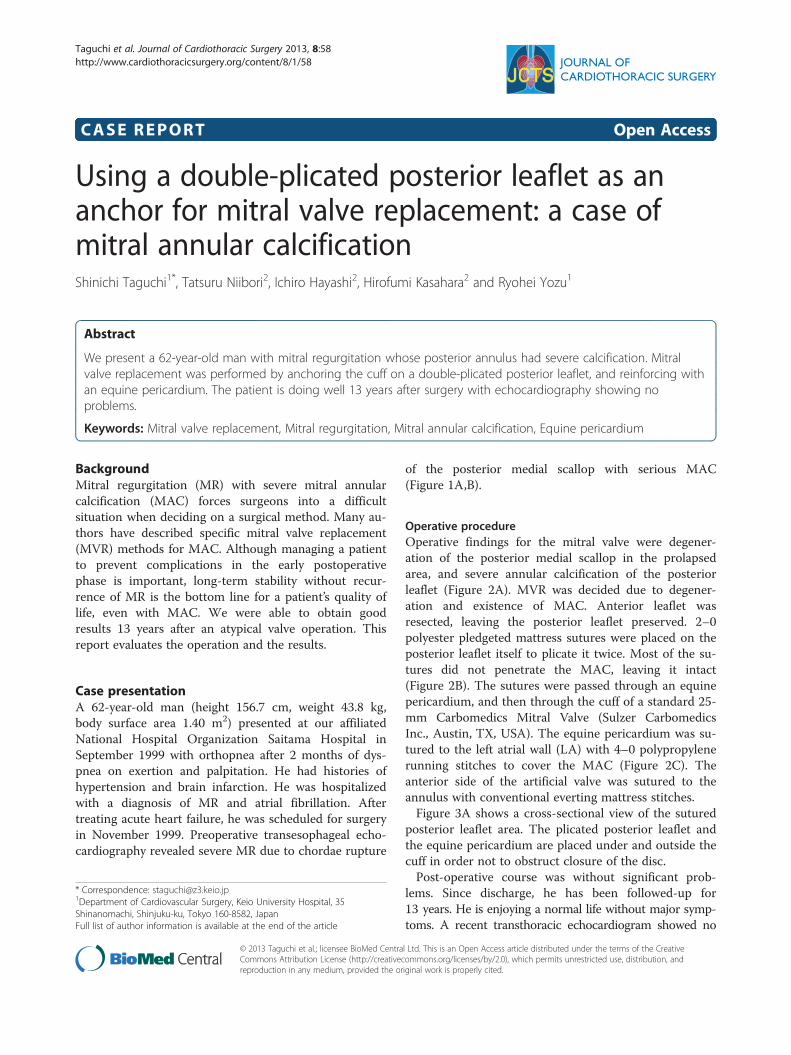
Taguchi et al. Journal of Cardiothoracic Surgery 2013, 8:58http://www.cardiothoracicsurgery.org/content/8/1/58
CASE REPORT Open Access
Using a double-plicated posterior leaflet as ananchor for mitral valve replacement: a case ofmitral annular calcificationShinichi Taguchi1*, Tatsuru Niibori2, Ichiro Hayashi2, Hirofumi Kasahara2 and Ryohei Yozu1
Abstract
We present a 62-year-old man with mitral regurgitation whose posterior annulus had severe calcification. Mitralvalve replacement was performed by anchoring the cuff on a double-plicated posterior leaflet, and reinforcing withan equine pericardium. The patient is doing well 13 years after surgery with echocardiography showing noproblems.
Keywords: Mitral valve replacement, Mitral regurgitation, Mitral annular calcification, Equine pericardium
BackgroundMitral regurgitation (MR) with severe mitral annularcalcification (MAC) forces surgeons into a difficultsituation when deciding on a surgical method. Many au-thors have described specific mitral valve replacement(MVR) methods for MAC. Although managing a patientto prevent complications in the early postoperativephase is important, long-term stability without recur-rence of MR is the bottom line for a patient’s quality oflife, even with MAC. We were able to obtain goodresults 13 years after an atypical valve operation. Thisreport evaluates the operation and the results.
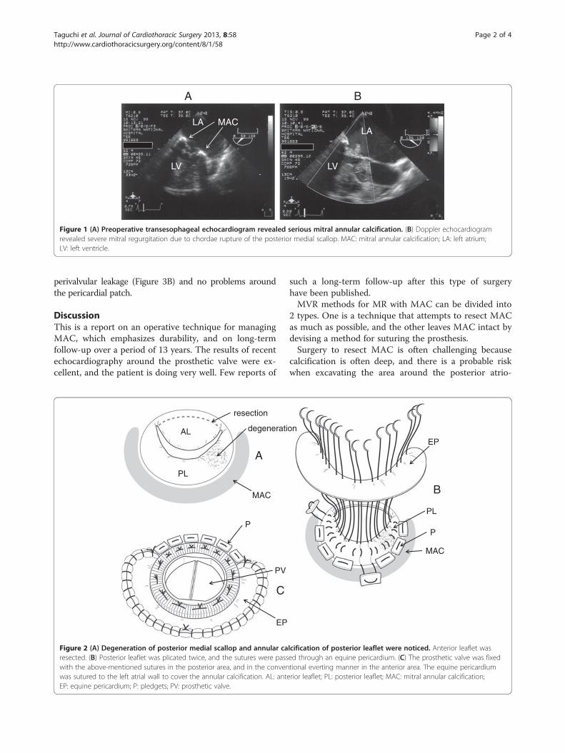
Case presentationA 62-year-old man (height 156.7 cm, weight 43.8 kg,body surface area 1.40 m2) presented at our affiliatedNational Hospital Organization Saitama Hospital inSeptember 1999 with orthopnea after 2 months of dys-pnea on exertion and palpitation. He had histories ofhypertension and brain infarction. He was hospitalizedwith a diagnosis of MR and atrial fibrillation. Aftertreating acute heart failure, he was scheduled for surgeryin November 1999. Preoperative transesophageal echo-cardiography revealed severe MR due to chordae rupture
* Correspondence: [email protected] of Cardiovascular Surgery, Keio University Hospital, 35Shinanomachi, Shinjuku-ku, Tokyo 160-8582, JapanFull list of author information is available at the end of the article
© 2013 Taguchi et al.; licensee BioMed CentraCommons Attribution License (http://creativecreproduction in any medium, provided the or
of the posterior medial scallop with serious MAC(Figure 1A,B).
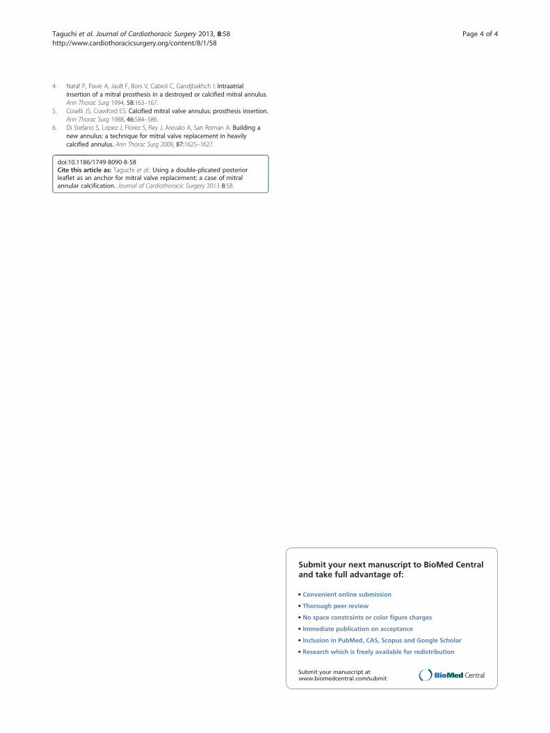
Operative procedureOperative findings for the mitral valve were degener-ation of the posterior medial scallop in the prolapsedarea, and severe annular calcification of the posteriorleaflet (Figure 2A). MVR was decided due to degener-ation and existence of MAC. Anterior leaflet wasresected, leaving the posterior leaflet preserved. 2–0polyester pledgeted mattress sutures were placed on theposterior leaflet itself to plicate it twice. Most of the su-tures did not penetrate the MAC, leaving it intact(Figure 2B). The sutures were passed through an equinepericardium, and then through the cuff of a standard 25-mm Carbomedics Mitral Valve (Sulzer CarbomedicsInc., Austin, TX, USA). The equine pericardium was su-tured to the left atrial wall (LA) with 4–0 polypropylenerunning stitches to cover the MAC (Figure 2C). Theanterior side of the artificial valve was sutured to theannulus with conventional everting mattress stitches.Figure 3A shows a cross-sectional view of the sutured
posterior leaflet area. The plicated posterior leaflet andthe equine pericardium are placed under and outside thecuff in order not to obstruct closure of the disc.Post-operative course was without significant prob-
lems. Since discharge, he has been followed-up for13 years. He is enjoying a normal life without major symp-toms. A recent transthoracic echocardiogram showed no
l Ltd. This is an Open Access article distributed under the terms of the Creativeommons.org/licenses/by/2.0), which permits unrestricted use, distribution, andiginal work is properly cited.
A B
LA
LV
MACLA
LV
Figure 1 (A) Preoperative transesophageal echocardiogram revealed serious mitral annular calcification. (B) Doppler echocardiogramrevealed severe mitral regurgitation due to chordae rupture of the posterior medial scallop. MAC: mitral annular calcification; LA: left atrium;LV: left ventricle.
Taguchi et al. Journal of Cardiothoracic Surgery 2013, 8:58 Page 2 of 4http://www.cardiothoracicsurgery.org/content/8/1/58
perivalvular leakage (Figure 3B) and no problems aroundthe pericardial patch.
DiscussionThis is a report on an operative technique for managingMAC, which emphasizes durability, and on long-termfollow-up over a period of 13 years. The results of recentechocardiography around the prosthetic valve were ex-cellent, and the patient is doing very well. Few reports of
C
A
resection
degeneratiAL
PL
MAC
P
PV
EP
Figure 2 (A) Degeneration of posterior medial scallop and annular caresected. (B) Posterior leaflet was plicated twice, and the sutures were passwith the above-mentioned sutures in the posterior area, and in the convenwas sutured to the left atrial wall to cover the annular calcification. AL: anteEP: equine pericardium; P: pledgets; PV: prosthetic valve.
such a long-term follow-up after this type of surgeryhave been published.MVR methods for MR with MAC can be divided into
2 types. One is a technique that attempts to resect MACas much as possible, and the other leaves MAC intact bydevising a method for suturing the prosthesis.Surgery to resect MAC is often challenging because
calcification is often deep, and there is a probable riskwhen excavating the area around the posterior atrio-
B
on
EP
PL
P
MAC
lcification of posterior leaflet were noticed. Anterior leaflet wased through an equine pericardium. (C) The prosthetic valve was fixedtional everting manner in the anterior area. The equine pericardiumrior leaflet; PL: posterior leaflet; MAC: mitral annular calcification;

A
MACD
CEP
P
PL
LA
LV
B
CMAC
AV
Figure 3 (A) Cross-sectional view of our operation using a standard prosthetic mitral valve. The area around the posterior leaflet and theannular calcification is shown. (B) The systolic phase in the parasternal long-axis view of the two-dimensional echocardiogram, taken more than12 years after surgery, shows no perivalvular leakage. AV: aortic valve; C: cuff; D: disc; P: pledget; EP: equine pericardium; PL: posterior leaflet; MAC:mitral annular calcification; LA: left atrial wall; LV: left ventricular wall.
Taguchi et al. Journal of Cardiothoracic Surgery 2013, 8:58 Page 3 of 4http://www.cardiothoracicsurgery.org/content/8/1/58
ventricular groove of rupture, injury to circumflex artery,and thrombo-embolic events [1]. To prevent theseevents, ultrasonic debridement is often used, andmethods such as using autologous or equine pericar-dium [2], and transferring the anterior leaflet to theposterior area [3] are adopted.The best way to prevent these disadvantages when
resecting MAC is to leave it intact, and to develop newmethods of suturing the prosthetic valve. Many reportshave been published on possible solutions such as:suturing the prosthesis to the LA itself [1], developing acollar for the prosthesis [4], using the leaflets to securethe prosthesis [5], plicating both mitral leaflets and theLA to create a new annulus [6], and using a Dacron graftbetween the LA and the prosthesis.The advantages of our method are that folding the
posterior leaflet twice creates a new annulus above theMAC, and using the equine pericardium reinforcesthe suture and acts as a preventive sheet to avoidperivalvular leakage. By preserving the posterior leaflet,the subvalvular apparatus is also maintained. Ourmethod of suturing the prosthesis to the posterior leafletis similar to that of Di Stefano et al. [6], who plicatedthe mitral leaflets and the atrial wall to create a new an-nulus. Although we used only the equine pericardium-covered posterior leaflet as a new annulus, we plicatedthe leaflet twice. This made the leaflet more solid andsuitable as an anchor for suturing the prosthesis. Equinepericardium was used to reinforce this anchor becausethe leaflet alone might not be structurally sufficient evenif plicated twice.Feindel et al. [2] used pericardium to reconstruct the
MAC resected area and to create a new annulus forMVR. Our usage of pericardium is similar to them inthe aspect of being a part of the new annulus for MVR,but different from them in the aspect of constituting thenew annulus together with the posterior leaflet.
We must mention that this operation was performedin only 1 patient. Although long-term durability of theprocedure was proven in the presented case, multipleexperiences using the procedure are needed for furtherevaluation.
ConclusionsIn summary of the presented case, using the double-plicated posterior leaflet as an anchor in MVR andequine pericardium as a reinforcement resulted in anexcellent long-term outcome for MR with MAC.
ConsentWritten informed consent was obtained from thepatient for publication of this report and any accom-panying images.
Competing interestsThe authors declare that they have no conflicts of interests.
Authors’ contributionsST and TN contributed to the pre-operative planning and operation of thecase. All authors contributed to following up the case. All authors read andapproved the final manuscript.
Author details1Department of Cardiovascular Surgery, Keio University Hospital, 35Shinanomachi, Shinjuku-ku, Tokyo 160-8582, Japan. 2Department ofCardiovascular Surgery, National Hospital Organization Saitama Hospital,Saitama, Japan.
Received: 25 November 2012 Accepted: 26 March 2013Published: 1 April 2013
References1. Atoui R, Lash V, Mohammadi S, Cecere R: Intra-atrial implantation of a
mitral valve prosthesis in a heavily calcified mitral annulus. Eur JCardiothorac Surg 2009, 36:776–778.
2. Feindel CM, Tufail Z, David TE, Ivanov J, Armstrong S: Mitral valve surgeryin patients with extensive calcification of the mitral annulus. J ThoracCardiovasc Surg 2003, 126:777–782.
3. Casselman FP, Gillinov AM, McDonald ML, Cosgrove DM III: Use of theanterior mitral leaflet to reinforce the posterior mitral annulus afterdebridement of calcium. Ann Thorac Surg 1999, 68:261–262.
Taguchi et al. Journal of Cardiothoracic Surgery 2013, 8:58 Page 4 of 4http://www.cardiothoracicsurgery.org/content/8/1/58
4. Nataf P, Pavie A, Jault F, Bors V, Cabrol C, Gandjbakhch I: Intraatrialinsertion of a mitral prosthesis in a destroyed or calcified mitral annulus.Ann Thorac Surg 1994, 58:163–167.
5. Coselli JS, Crawford ES: Calcified mitral valve annulus: prosthesis insertion.Ann Thorac Surg 1988, 46:584–586.
6. Di Stefano S, Lopez J, Florez S, Rey J, Arevalo A, San Roman A: Building anew annulus: a technique for mitral valve replacement in heavilycalcified annulus. Ann Thorac Surg 2009, 87:1625–1627.
doi:10.1186/1749-8090-8-58Cite this article as: Taguchi et al.: Using a double-plicated posteriorleaflet as an anchor for mitral valve replacement: a case of mitralannular calcification. Journal of Cardiothoracic Surgery 2013 8:58.
Submit your next manuscript to BioMed Centraland take full advantage of:
• Convenient online submission
• Thorough peer review
• No space constraints or color figure charges
• Immediate publication on acceptance
• Inclusion in PubMed, CAS, Scopus and Google Scholar
• Research which is freely available for redistribution
Submit your manuscript at www.biomedcentral.com/submit