Embed Size (px)
Citation preview

Idrissi Serhrouchni et al. Diagnostic Pathology 2013, 8:15http://www.diagnosticpathology.org/content/8/1/15
CASE REPORT Open Access
Eccrine carcinoma : a rare cutaneous neoplasmKarima Idrissi Serhrouchni1*, Taoufiq Harmouch1, Laila Chbani1, Hind El Fatemi1, Mohammed Sekal1,Nawal Hammas1, Meriem Soughi2, Loubna Benchat2 and Afaf Amarti1
Abstract
Eccrine carcinoma is an extremely rare malignancy of the skin with few well documented cases reported in theliterature. It is frequently found on the lower extremities, and it equally affects both sexes in the sixth and seventhdecade.In our case, we present a 46- year-old female with a recurring exophytic tumor on the right lower extremity,without local extension.The initial tumor was biopsied, excised and diagnosed as an eccrine carcinoma.Virtual Slides: The virtual slide(s) for this article can be found here: http://www.diagnosticpathology.diagnomx.eu/vs/3568051328673318.
Keywords: Eccrine carcinoma, Sweat gland tumor, Mammary gland
BackgroundMalignant cutaneous adnexal neoplasms are a large and va-ried group, in particular eccrine carcinoma. They are one ofthe most challenging areas of Dermatopathology [1].Eccrine and apocrine neoplasms present a bewildering
array of morphologies which often defy precise classifi-cation [2].The purpose of this case is to discuss the most common
problems concerning the classification of this rare neoplasmand report the aim of the immunohistochemical profiles indifferential diagnosis between a primitive eccrine carcinomaof the skin and a secondary neoplasm.
Case reportIn July 2012, a 45-year-old Moroccan woman presentedto the department of Dermatology of Hassan II Univer-sity Hospital of Fez with a 25-year-history of primaryinfertility, and an 18-month-history of an exophytic massat the posterior right lower extremity, gradually increas-ing in size. She was otherwise healthy and had nosystemic symptoms. Physical examination showed an ul-cerative and bourgeoning mass of 15 cm with bleedingand purulent features (Figure 1). Biological testsincluded a complete blood count, routine blood andurine chemistry were normal, except for elevated LDH.
* Correspondence: [email protected] of Pathology, Hassan II University Hospital, Fez 30000, MoroccoFull list of author information is available at the end of the article
© 2013 Idrissi Serhrouchni et al.; licensee BioMCreative Commons Attribution License (http:/distribution, and reproduction in any medium
Tumor markers as like as CA 19–9, CA 125, CA 15–3were normal. MRI of the leg showed a subcutaneousinfiltrative process coming into contact with the musclefascia.The rest of the radiological examination, including radiog-
raphy of the lung, abdominopelvic ultrasonography, com-puted tomographic thoraco-abdomino-pelvic scan, wasnormal.The mammography and ultrasound examination objecti-
fied a 4 mm measuring benign cystic lesion of the left breastclassified ACR 3, requiring supervision during six months.After biopsy of the lesion, a surgical excision with clear
margins was performed.On gross examination, the tumor was 15 cm in size,
bourgeoning, erythematous and heterogeneous withareas of necrosis and haemorrhage.Microscopic examination of the specimen revealed an
undifferentiated malignant proliferation with nodulararchitecture, located in the deep dermis (Figure 2).Tumor cells made clear cytoplasm with an hyperchro-matic nucleus and a high mitotic activity (Figure 3).Immunohistochemical analysis showed positive stain-
ing for cytokeratine and progesterone receptor. Cytoke-ratine 7 was weakly positive and Ki67 was estimated at95% (Figure 4).In contrast, the tumor cells were not reactive to cyto-
keratine 20, estrogen receptor, LCA, CD10, S 100
ed Central Ltd. This is an Open Access article distributed under the terms of the/creativecommons.org/licenses/by/2.0), which permits unrestricted use,, provided the original work is properly cited.
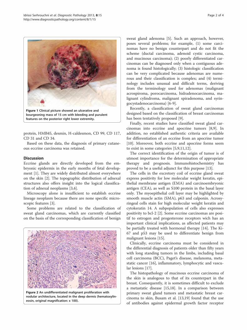
Figure 1 Clinical picture showed an ulcerative andbourgeoning mass of 15 cm with bleeding and purulentfeatures on the posterior right lower extremity.
Idrissi Serhrouchni et al. Diagnostic Pathology 2013, 8:15 Page 2 of 4http://www.diagnosticpathology.org/content/8/1/15
protein, HMB45, desmin, H-caldesmon, CD 99, CD 117,CD 31 and CD 34.Based on these data, the diagnosis of primary cutane-
ous eccrine carcinoma was retained.
DiscussionEccrine glands are directly developed from the em-bryonic epidermis in the early months of fetal develop-ment [1]. They are widely distributed almost everywhereon the skin [2]. The topographic distribution of adnexalstructures also offers insight into the logical classifica-tion of adnexal neoplasms [3,4].Microscopy alone is insufficient to establish eccrine
lineage neoplasm because there are none specific micro-scopic features [3].Some problems are related to the classification of
sweat gland carcinomas, which are currently classifiedon the basis of the corresponding classification of benign
Figure 2 An undifferentiated malignant proliferation withnodular architecture, located in the deep dermis (hematoxylin-eosin, original magnification: x 100).
sweat gland adenoma [5]. Such an approach, however,poses several problems; for example, (1) some carci-nomas have no benign counterpart and do not fit thescheme (ductal carcinoma, adenoid cystic carcinoma,and mucinous carcinoma); (2) poorly differentiated car-cinomas can be diagnosed only when a contiguous ade-noma is found histologically; (3) histologic classificationcan be very complicated because adenomas are nume-rous and their classification is complex; and (4) termi-nology includes unusual and difficult terms, derivingfrom the terminology used for adenomas (malignantacrospiroma, porocarcinoma, hidradenocarcinoma, ma-lignant cylindroma, malignant spiradenoma, and syrin-gocystadenocarcinoma) [6-9].Recently, a classification of sweat gland carcinomas
designed based on the classification of breast carcinomashas been tentatively proposed [9].Finally, recent studies have classified sweat gland car-
cinomas into eccrine and apocrine tumors [8,9]. Inaddition, no established authentic criteria are availablefor differentiation of an eccrine from an apocrine tumor[10]. Moreover, both eccrine and apocrine forms seemto exist in some categories [5,9,11,12].The correct identification of the origin of tumor is of
utmost importance for the determination of appropriatetherapy and prognosis. Immunohistochemistry hasproved to be a useful adjunct for this purpose [13].The cells in the excretory coil of eccrine gland sweat
express positivity for low molecular weight keratin, epi-thelial membrane antigen (EMA) and carcinoembryonicantigen (CEA), as well as S100 protein in the basal layeronly. The myoepithelial cell layer may be highlighted bysmooth muscle actin (SMA), p63 and calponin. Acrosy-ringeal cells stain for high molecular weight keratin andcytokeratin 14. A subpopulation of cells also expressespositivity to bcl-2 [2]. Some eccrine carcinomas are posi-tif to estrogen and progesterone receptors wich has animportant clinical implications, as affected patients maybe partially treated with hormonal therapy [14]. The Ki-67 and p53 may be used to differentiate benign frommalignant lesions [15].Clinically, eccrine carcinoma must be considered in
the differential diagnosis of patients older than fifty yearswith long standing tumors in the limbs, including basalcell carcinoma (BCC), Paget’s disease, melanoma, meta-static cancer [16], inflammatory, lymphocytic and vascu-lar lesions [17].The histopathology of mucinous eccrine carcinoma of
the skin is analogous to that of its counterpart in thebreast. Consequently, it is sometimes difficult to excludea metastatic disease [15,18]. In a comparison betweenprimary sweat gland tumors and metastatic breast car-cinoma to skin, Busam et al. [13,19] found that the useof antibodies against epidermal growth factor receptor

Figure 3 Tumor cells made clear cytoplasm with an hyperchromatic nucleus and a high mitotic activity (hematoxylin-eosin, originalmagnification: A, × 200, B, x 400).
Idrissi Serhrouchni et al. Diagnostic Pathology 2013, 8:15 Page 3 of 4http://www.diagnosticpathology.org/content/8/1/15
strongly decorated 81% of sweat gland tumors, but only17% of metastatic breast cancers (P=0.001). There wasno significant difference between the skin tumors andmetastatic breast carcinoma when antibodies against es-trogen and progesterone receptors were used, but withprogression of disease, androgen receptors (AR) are pre-served with higher frequency than ER/PR in metastaticmammary carcinoma [13]. In another study comparingbenign, malignant primary eccrine and apocrine neo-plasms to metastatic breast carcinoma, only 3.5% ofthese primary adnexal cancers demonstrated HER-2positivity, whereas 10–23% of the breast carcinomaswere positive [13,20]. The authors suggested that thistest may be useful in distinguishing primary skin cancersfrom metastatic breast cancers [20]. Another studyshowed that primary cutaneous neoplasms stainedstrongly for p63 and CK 5/6 with high specificity, whileCK 7 and 20 were not useful [15,21]. When CK 7 waspositive in the cutaneous lesions, it exhibited markedfocality and a specific pattern, while metastatic breastcarcinoma expressed CK 7 diffusely and did not expressp63 or CK 5/6. The usefulness of CK 5/6 was againshown by Plumb et al. [15,22]. Ivan et al. [15,23] pre-sented further evidence of the usefulness of p63 in the
Figure 4 Immunohistochemical study showing reactivity for CK (A), C
diagnosis of adnexal cancer; in their study, none of theexamples of metastatic adenocarcinoma to skin stainedfor p63, whereas virtually all the adnexal carcinomaswere positive.These studies showed that immunohistochemistry
does not distinguish cutaneous eccrine tumours fromcutaneous metastases of breast carcinoma, in which caseclinical and radiological correlation is critical [24].Other differential diagnoses comprise neoplasms with
clear cell differentiation. These include trichilemmal car-cinoma, clear cell BCC and clear cell carcinomas meta-static to the skin. With respect to the latter, thedominant diagnostic considerations are metastatic clearcell carcinoma from thyroid gland and lung. The ex-pression of TTF seen in the great majority of these casesis helpful in resolving this dilemma. Some metastaticthyroid cancers also manifest expression of thyroglobu-lin. Metastatic clear cell carcinoma of renal primary ori-gin manifests a prominent stromal vascularity withhemorrhage and areas of granular necrosis typically evi-dent in biopsy material. Clear cell squamous cell carci-noma of the cervix is a glycogen-rich cancer withintercellular bridge formation characteristic of squa-mous differentiation [15].
K7 (B) and progesterone receptors (C).

Idrissi Serhrouchni et al. Diagnostic Pathology 2013, 8:15 Page 4 of 4http://www.diagnosticpathology.org/content/8/1/15
ConclusionEccrine carcinoma should be considered in the diagnosisof cutaneous malignant tumor with immunostaining forCK7, P63, CK5/6, estrogen and progesterone receptors.
Consent from the patientWritten informed consent was obtained from patient forpublication of this case report.
AbbreviationsLDH: Lactate Dehydrogenase; MRI: Magnetic Resonance Imaging;ACR: American College of Radiology; CK: Cytokeratin; LCA: LeukocyteCommon Antigen.
Competing interestsThe authors declare that they have no competing interests.
Authors’ contributionsAll authors read and approved the final manuscript.
Author details1Department of Pathology, Hassan II University Hospital, Fez 30000, Morocco.2Department of Dermatology, Hassan II University Hospital, Fez 30000,Morocco.
Received: 9 December 2012 Accepted: 27 January 2013Published: 4 February 2013
References1. Montagna W: Embryology and anatomy of the cutaneous adnexa.
J Cutan Pathol 1984, 11:350–351.2. Nidal A, Khaled O, Danny G: Skin adnexal neoplasms: an approach to
tumours of cutaneous sweat glands. J Clin Pathol 2007, 60:145–159.3. Timothy H, Calmont M: Batman and adnexal neoplasms. J Cutan Pathol
2010, 37:401–402.4. Brownstein MH, Shapiro L: The sweat gland adenomas. Int J Dermatol
1975, 14:397–411.5. Carmelo U, Roberto B, Milena P, Adriana S, Chiara A, Augusto G:
Carcinomas of Sweat Glands. Arch Pathol Lab Med 2001, 125:498–505.6. Cooper PH: Carcinomas of sweat glands. Pathol Annu 1987, 22:83–110.7. Santa C: Sweat glands carcinomas: a comprehensive review. Semin Diagn
Pathol 1987, 4:38–74.8. Murphy GF, Elder DE: Non-melanocytic Tumors of the Skin. Washington, DC:
Armed Forces Institute of Pathology; 1991:61–153. Atlas of TumorPathology; 3rd series, fascicle 1.
9. Requena L, Kiryu H, Ackerman AB: Neoplasms With Apocrine Differentiation.Philadelphia, Pa: Lippincott-Raven; 1998:589–855.
10. Abenoza P, Ackerman AB: Neoplasms With Eccrine Differentiation.Philadelphia, Pa: Lea and Febiger; 1990:371–412.
11. Swanson PE, Mazoujian G, Mills SE, Campbell RJ, Wick MR:Immunoreactivity for estrogen receptor protein in sweat gland tumors.Am J Surg Pathol 1991, 15:835–841.
12. Ansai S, Koseki S, Hozumi Y, Kondo S: An immunohistochemical study oflysozyme, CD-15 (Leu M1), and gross cystic disease fluid protein-15 invarious skin tumors: assessment of the specificity and sensitivity ofmarkers of apocrine differentiation. Am J Dermatopathol 1995, 17:249–255.
13. Vinod B, Richard A, Jinobya K: Androgen receptor expression in metastaticadenocarcinoma in females favors a breast primary. Diagn Pathol 2006,1:34.
14. Mirza I, Kloss R, Sieber SC: Malignant eccrine spiradenoma. Arch Pathol LabMed 2002, 126:591–594.
15. Crowson AN, Magro CM, Mihm MC: Malignant adnexal neoplasms.Mod Pathol 2006, 19:93–126.
16. Oliver C, Ashraf E, Bernard R, Armand A, Nadeem C: Eccrine porocarcinomaof the lower extremity: a case report and review of literature. World JSurg Oncol 2011, 9:94.
17. Helena PS, Ventura MP, Orellana ME, Novais GA, Cheema DP, Burnier MN:Histopathological study of lesions of the caruncle: a 15-year singlecenter review. Diagn Pathol 2009, 4:29.
18. Page DL, Anderson TJ, Sakamoto G: Infiltrating carcinoma: majorhistological types. In Diagnostic Histopathology of the Breast. Edited byPage DL, Anderson TJ. Churchill-Livingstone: New York; 1987:193–295.
19. Busam KJ, Tan LK, Granter SR: Epidermal growth factor, estrogen, andprogesterone receptor expression in primary sweat gland carcinomasand primary and metastatic mammary carcinomas. Mod Pathol 1999,12:786–793.
20. Hiatt KM, Pilow JL, Smoller BR: Her-2 expression in cutaneous eccrine andapocrine neoplasms. Mod Pathol 2004, 17:28–32.
21. Qureshi HS, Ormsby AH, Lee MW: The diagnostic utility of p63, CK5/6, CK7, and Ck 20 in distinguishing primary cutaneous adnexal neoplasmsfrom metastatic carcinomas. Cutan Pathol 2004, 31:145–152.
22. Plumb SJ, Argenyi ZB, Stone MS: Cytokeratin 5/6 immunostaining incutaneous adnexal neoplasms and metastatic adenocarcinoma.Am J Dermatopathol 2004, 26:447–451.
23. Ivan D, Hafeez Diwan A, Prieto VG: Expression of p63 in primarycutaneous adnexal neoplasms and adenocarcinoma metastatic to theskin. Mod Pathol 2005, 18:137–142.
24. Wallace ML, Longacre TA, Smoller BR: Estrogen and progesteronereceptors and anti-‐gross cystic disease fluid protein 15 (BRST-‐2) fail todistinguish metastatic breast carcinoma from eccrine neoplasms.Mod Pathol 1995, 8:897–901.
doi:10.1186/1746-1596-8-15Cite this article as: Idrissi Serhrouchni et al.: Eccrine carcinoma : a rarecutaneous neoplasm. Diagnostic Pathology 2013 8:15.
Submit your next manuscript to BioMed Centraland take full advantage of:
• Convenient online submission
• Thorough peer review
• No space constraints or color figure charges
• Immediate publication on acceptance
• Inclusion in PubMed, CAS, Scopus and Google Scholar
• Research which is freely available for redistribution
Submit your manuscript at www.biomedcentral.com/submit


![Ghost cell odontogenic carcinoma: A rare case report and ... · PDF fileGhost cell odontogenic carcinoma [GCOC] is a rare malignant odontogenic epithelial tumor with features of calcifying](https://img.pdfslide.us/doc/110x75/5a9cd2d97f8b9a335c8b5251/ghost-cell-odontogenic-carcinoma-a-rare-case-report-and-cell-odontogenic-carcinoma.jpg)


![Surgical Management of Primary Cutaneous Mucinous Carcinoma · represents 0.005% of all malignant epithelial neoplasms [1]. These adnexal tumours have been thought to be of eccrine](https://img.pdfslide.us/doc/110x75/5f0b6f0f7e708231d4307f6a/surgical-management-of-primary-cutaneous-mucinous-represents-0005-of-all-malignant.jpg)