Embed Size (px)
Citation preview
CASE REPORT
OCULAR SPARGANOSIS IN THAILAND
Somsanguan Ausayakhun', Veera Siriprasert2, Nimit Morakote2 and Kanidta Taweesap'
'Department of Ophthalmology and 2Department of Parasitology, Faculty of Medicine, Chiang Mai University, Chiang Mai, Thailand
Human sparganosis which was first reported by Patrick Manson in 1882 (Beaver, 1984) is a rare parasitic infection caused by the migrating plerocercoid larva or sparganum of a certain group of pseudophyllidean cestodes, namely in the genus Spirometra Mueller, 1937. The adults of these tapeworms are generally found in the intestinal lumen of domestic, feral, or wild canids and felids (Daly, 1982). Spargana, a term originally used to describe any unknown pseudophyllidean larvae, do not have adequate morphological characteristics for differentiation. Therefore, they must be fed to a definitive host (dog or cat) to grow to adult worms which have morphological differences that can be used for taxonomic separation. This, however, has not been accomplished for spargana from most of human sparganoses. Thus, an opinion as to species of the sparganum was usually based on the general nature of the larva, knowledge of adult forms in hosts from that area, or previously reported sparganosis cases from that area. This leads to invalidity of species in many cases because certain forms are sometimes found in the same geographical regions, such as S. mansoni and S. mansonoides in North and South America (Mueller et ai, 1975) and/or knowledge of adult forms in those areas is inadequate. Furthermore, the variability in adult worm morphology as well as lack of communication and agreement among the ivestigators, has led to confusion about the classification of this group of tapeworms.
Human sparganosis has been reported sporadically worldwide, from every continent but most commonly from East and Southeast Asia (Japan, China, Korea, Taiwan, Vietnam, Thailand). There are two types of the disease, namely nonproliferative and proliferative sparganosis. Review of the literature revealed more than 300
Correspondence to Dr Veera Siriprasert, Department of Parasitology, Faculty of Medicine, Chiang Mai University, Chiang Mai 50002, Thailand.
cases of nonproliferative sparganosis in humans but there have been only II well-documented and 2 other suspected cases of proliferative type (La Chance et ai, 1983; Nakamura et ai, 1990).
A 33-year-old female Thai had had history of pain, swelling, and redness of her left eye since June 1990. She went to medical clinics twice and the last doctor suggested she come. to Maharaj Nakhon Chiang Mai Hospital, Faculty of Medicine, Chiang Mai University. At Ophthalmologic OPD, the diagnosis of allergic blepharitis and conjunctivitis was made but showed no clinical improvement after standard treatment. Follow-up examination revealed localized conjunctival swelling in the temporal region of her left eye. Gnathostomiasis was suspected. Palliative therapy was given with some degree of improvement and the patient had been absent for 2 years. She revisited in October 1992. A left subconjunctival mass was noted. Other findings were unremarkable except the presence of Strongyloides stercoralis larvae in the stool. No preoperative hemogram was done. Exploration of the lesion revealed a white ribbon-like cestode larva. ELISA using 36 and 31 kDa sparganum antigens (kindly donated by Dr Yoon Kong) gave a positive antibody value of 0.263 (cut-off = 0.22); 3 days after surgery; which was negative 4 months later. The patient's serum was also tested for gnathostomiasis 7 days postoperatively. The ELISA value was 0.273 (cut-off = 0.44). The WBC count 2 weeks after surgery was 14.1 x 10⁹ /1 of which 2% were eosinophils. She was well on follow-up at 4-month.
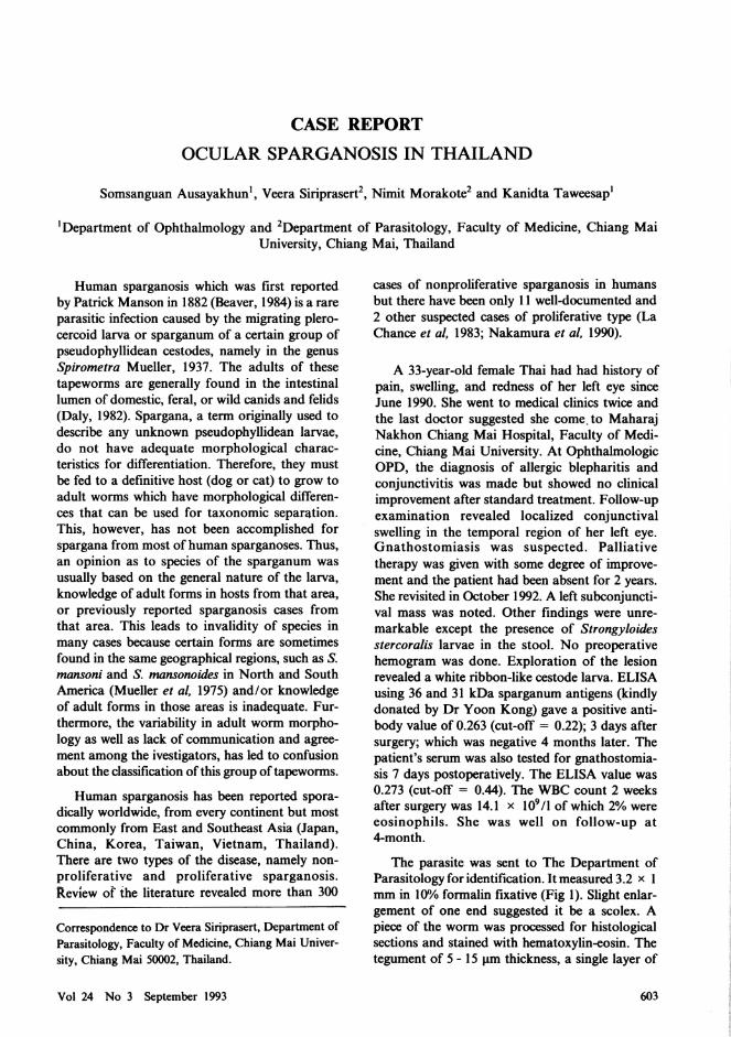
The parasite was sent to The Department of Parasitology for identification. Itmeasured 3.2 x I mm in 10% formalin fixative (Fig I). Slight enlargement of one end suggested it be a scolex. A piece of the worm was processed for histological sections and stained with hematoxylin-eosin. The tegument of 5 - 15 I11ll thickness, a single layer of
Vol 24 No 3 September 1993 603
SOUTHEAST ASIAN J TROP MED PUBLIC HEALTH
Fig I-A Sparganum removed from the patient's eye. Bulging of the end on the left is the parasite's scolex.
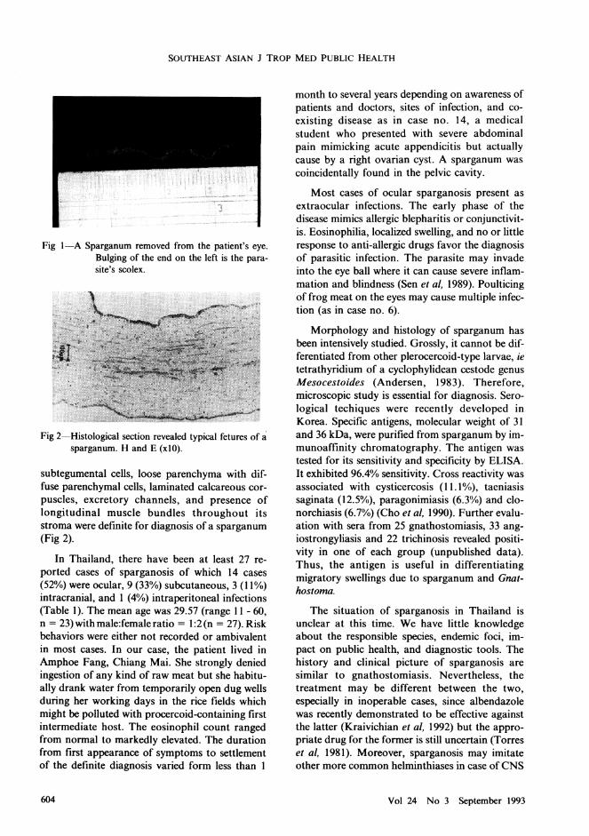
Fig 2-Histological section revealed typical fetures of Ii sparganum. Hand E (xlO).
subtegumental cells, loose parenchyma with diffuse parenchymal cells, laminated calcareous corpuscles, excretory channels, and presence of longitudinal muscle bundles throughout its stroma were definite for diagnosis of a sparganum (Fig 2).
In Thailand, there have been at least 27 reported cases of sparganosis of which 14 cases (52%) were ocular, 9 (33%) subcutaneous, 3 (11%) intracranial, and 1 (4%) intraperitoneal infections (Table 1). The mean age was 29.57 (range 11 - 60, n = 23) with male:female ratio = 1:2(n = 27). Risk behaviors were either not recorded or ambivalent in most cases. In our case, the patient lived in Amphoe Fang, Chiang MaL She strongly denied ingestion of any kind of raw meat but she habitually drank water from temporarily open dug wells during her working days in the rice fields which might be polluted with procercoid-containing first intermediate host. The eosinophil count ranged from normal to markedly elevated. The duration from first appearance of symptoms to settlement of the definite diagnosis varied form less than 1
month to several years depending on awareness of patients and doctors, sites of infection, and coexisting disease as in case no. 14, a medical student who presented with severe abdominal pain mimicking acute appendicitis but actually cause by a right ovarian cyst. A sparganum was coincidentally found in the pelvic cavity.
Most cases of ocular sparganosis present as extraocular infections. The early phase of the disease mimics allergic blepharitis or conjunctivitis. Eosinophilia, localized swelling, and no or little response to anti-allergic drugs favor the diagnosis of parasitic infection. The parasite may invade into the eye ball where it can cause severe inflammation and blindness (Sen et ai, 1989). Poulticing of frog meat on the eyes may cause multiple infection (as in case no. 6).
Morphology and histology of sparganum has been intensively studied. Grossly, it cannot be differentiated from other plerocercoid-type larvae, ie tetrathyridium of a cyclophylidean cestode genus Mesocestoides (Andersen, 1983). Therefore, microscopic study is essential for diagnosis. Serological techiques were recently developed in Korea. Specific antigens, molecular weight of 31 and 36 kDa, were purified from sparganum by immunoaffinity chromatography. The antigen was tested for its sensitivity and specificity by ELISA. It exhibited 96.4% sensitivity. Cross reactivity was associated with cysticercosis (11.1 %), taeniasis saginata (12.5%), paragonimiasis (6.3%) and clonorchiasis (6.7%) (Cho et ai, 1990). Further evaluation with sera from 25 gnathostomiasis, 33 angiostrongyliasis and 22 trichinosis revealed positivity in one of each group (unpublished data). Thus, the antigen is useful in differentiating migratory swellings due to sparganum and Gnathostoma.
The situation of sparganosis in Thailand is unclear at this time. We have little knowledge about the responsible species, endemic foci, impact on public health, and diagnostic tools. The history and clinical picture of sparganosis are similar to gnathostomiasis. Nevertheless, the treatment may be different between the two, especially in inoperable cases, since albendazole was recently demonstrated to be effective against the latter (Kraivichian et ai, 1992) but the appropriate drug for the former is still uncertain (Torres et ai, 1981). Moreover, sparganosis may imitate other more common helminthiases in case of CNS
Vol 24 No 3 September 1993 604
OCULAR SPARGANOSIS IN THAILAND
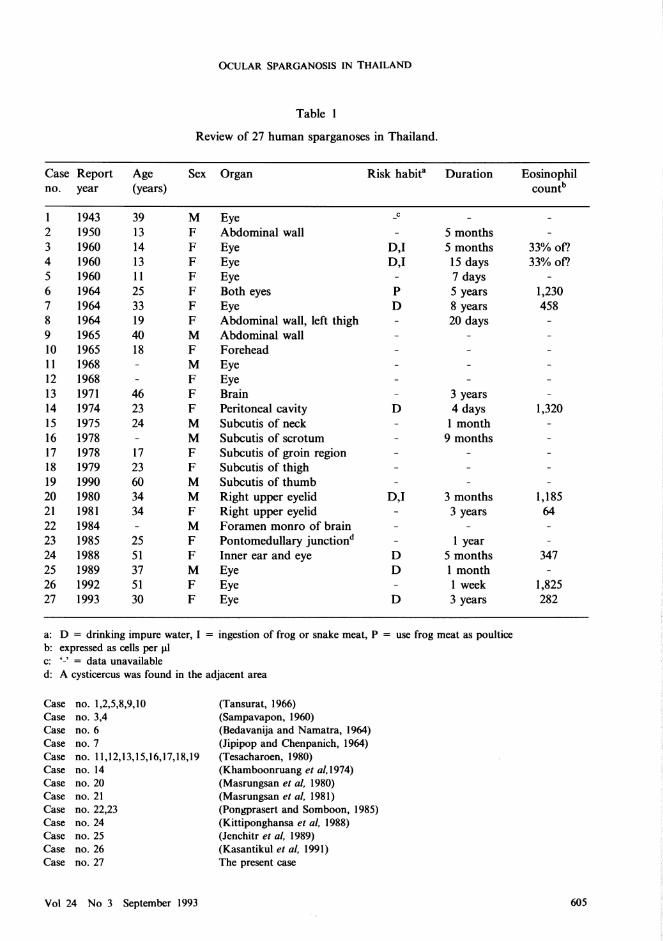
Table I
Review of 27 human sparganoses in Thailand.
Case Report Age Sex Organ Risk habit" Duration Eosinophil no. year (years) countb
1 1943 39 M Eye _c
2 1950 13 F Abdominal wall 5 months 3 1960 14 F Eye D,I 5 months 33% of? 4 1960 13 F Eye D,I 15 days 33% of? 5 1960 11 F Eye 7 days 6 1964 25 F Both eyes P 5 years 1,230 7 1964 33 F Eye D 8 years 458 8 1964 19 F Abdominal wall, left thigh 20 days 9 1965 40 M Abdominal wall 10 1965 18 F Forehead 11 1968 M Eye 12 1968 F Eye 13 1971 46 F Brain 3 years 14 1974 23 F Peritoneal cavity D 4 days 1,320 15 1975 24 M Subcutis of neck 1 month 16 1978 M Subcutis of scrotum 9 months 17 1978 17 F Subcutis of groin region 18 1979 23 F Subcutis of thigh 19 1990 60 M Subcutis of thumb 20 1980 34 M Right upper eyelid D,I 3 months 1,185 21 1981 34 F Right upper eyelid 3 years 64 22 1984 M Foramen monro of brain 23 1985 25 F Pontomedullary junctiond I year 24 1988 51 F Inner ear and eye D 5 months 347 25 1989 37 M Eye D 1 month 26 1992 51 F Eye 1 week 1,825 27 1993 30 F Eye D 3 years 282
a: D = drinking impure water, I = ingestion of frog or snake meat, P = use frog meat as poultice b: expressed as cells per 111 c: '-' = data unavailable d: A cysticercus was found in the adjacent area
Case no. 1,2,5,8,9,10 (Tansurat, 1966) Case no. 3,4 (Sampavapon, 1960) Case no. 6 (Bedavanija and Namatra, 1964) Case no. 7 (Jipipop and Chenpanich, 1964) Case no. 11,12,13,15,16,17,18,19 (Tesacharoen, 1980) Case no. 14 (Khamboonruang et al.1974) Case no. 20 (Masrungsan et al. 1980) Case no. 21 (Masrungsan et al. 1981) Case no. 22,23 (Pongprasert and Somboon, 1985) Case no. 24 (Kittiponghansa et al. 1988) Case no. 25 (Jenchitr et al. 1989) Case no. 26 (Kasantikul et al. 1991) Case no. 27 The present case
Vo124 No 3 September 1993 605

SOUTHEAST ASIAN J TROP MED PUBLIC HEALTH
infection (gnathostomiasis, cysticercosis, angiost'tongyliasis).
ACKNOWLEDGEMENTS
Grateful thanks is made to Dr Y oon Kong, Department of Parasitology, College of Medicine, Chung-Ang University, Seoul, Korea for kindly providing of sparganum antigens and valuable information. The authors also thank Mr. Kasame Lersmanokulchai, Department of Pathology, Faculty of Medicine, Chiang Mai University for technical assistance in histological preparation.
REFERENCE
Andersen KI. Description of musculature differences in spargana of Spirometra (Cestoda; Pseudophyllidea) and tetrathyridia of Mesocestoides (Cestoda; Cyclophyllidea) and their value in identification. J Helminthol 1983; 57 : 331 - 4.
Beaver PC, Jung RC, Cupp EW. Clinical Parasitology. 91h ed. Philadelphia: Lea and Febiger, 1984 : 499 - 504.
Bedavanija A, Namatra B. Bilateral ocular sparganosis. J Med Assoc Thai 1964; 47 : 215 - 9 (Thai).
Cho SY, Kang SY, Kong Y, Purification of antigenic protein of sparganum by immunoaffinity chromatography using a monoclonal antibody. Korean J Parasitol 1990; 280 : 135 - 42.
Daly JJ. Sparganosis. In : Steele JH, eds. CRe's Handbook Series in Zoonoses, Section C : Parasitic Zoonoses, Volume I. Florida: CRC Press, Boca Raton, 1982 : 293 - 312.
Jenchitr W, Chaijukool S, Ying-yuad P. Ocular sparganosis in Lampang.Thai J Ophthalmol 1989; 3 : 21 - 5 (Thai).
Jipipop B, Chenpanich U. Ocular sparganosis with a case report J Inter College Surg Thai 1964; 7: 33-7 (Thai).
Kasantikul V, Raiyawa S, Rattanavijarn C, et al.
Human sparganosis presenting as exophthalmos
and ocular mass Chula Med J 1991; 36 : 47 51.
Khamboonruang C, Premasathian D, Little MD. A case of intra-abdominal sparganosis in Chiang Mai, Thailand, Am J Trop Med Hyg 1974; 23 : 538 - 9.
Kittiponghansa S, Tesana S, Ritch R. Ocular sparganosis: a cause of subconjunctival tumor and deafness. Trop Med Parasito/1988; 39 : 247 - 8.
Kraivichian P, Kulkumthorn M, Yingyourd P, et al. AIbendazole for the treatment of human gnathostomiasis. Trans R Soc Trop Med Hyg 1992; 86 : 418 - 21.
LaChance MA, Clark RM, Connor DH. Proliferating larval cestodiasis: report of a case, Acta Tropica 1983; 40 : 391 -7.
Masrungsan N, Duangratana S, Changwaiwit S. Ocular sparganosis: a case report. Chiang Mai Med Bull 1980; 19: 11-3 (Thai).
Masrungsan N, Thawari M, Kulsu R. Sparganosis of the upper eyelid: a case report. Chiang Mai Med Bull 1981; 20 : 613 - 6 (Thai).
Mueller JF, Froes OM, Fernndez TR. On the occurrence of Spirometra mansonoides in South America. J Parasitol 1975; 61 : 774 - 5.
Nakamura T, Hara M, Matsuoka M, et al. Human proliferative sparganosis: a new Japanese case, Am J Clin Pathol 1990; 94 : 224 - 8.
Pongprasert S, Somboon N. Sparganum mansoni combined with Cysticercus cellulosae infected in pontomedullary junction in the same patient, presented with two clinical states: a case report. Lampang Med Bull 1985; 6 : 225 - 41 (Thai).
Sampavapon V. Ocular-sparganosis: report of two cases. J Med Assoc Thai 1960; 43 : 333 -7 (Thai).
Sen DK, Muller R, Gupta VP, et at. Cestode larva (sparganum) in the anterior chamber of the eye. Trop Geogr Med 1989; 41 : 270 - 3.
Tansurat P. Human sparganosis in Thailand. J Med Assoc Thai 1966; 49 : 391 - 5.
Tesacharoen S. Sparganum in Thai people. J Med Council 1980; 9 : 255 - 62 (Thai).
Torres JR, Noya 00, et al. Treatment of proliferative sparganosis with mebendazole and praziquantel. Trans R Soc Trop Med Hyg 1981; 75: 846-7.
Vol 24 No 3 September 1993 606