Embed Size (px)
Citation preview

Case ReportNoncardiogenic Pulmonary Edema after AmlodipineOverdose without Refractory Hypotension and Bradycardia
M. Hedaiaty, N. Eizadi-Mood, and A. M. Sabzghabaee
Clinical Toxicology Department, Isfahan Clinical Toxicology Research Center, Noor Hospital, Isfahan University ofMedical Sciences, Isfahan 81458-31451, Iran
Correspondence should be addressed to N. Eizadi-Mood; [email protected]
Received 17 January 2015; Accepted 15 April 2015
Academic Editor: Kazuhito Imanaka
Copyright © 2015 M. Hedaiaty et al. This is an open access article distributed under the Creative Commons Attribution License,which permits unrestricted use, distribution, and reproduction in any medium, provided the original work is properly cited.
Amlodipine overdose can be life-threatening when manifesting as noncardiogenic pulmonary edema. Treatment remainschallenging. We describe a case of noncardiogenic pulmonary edema without refractory hypotension and bradycardia afteringestion of 500 milligram amlodipine with suicidal intent. Mechanical ventilation, dexamethasone, atrovent HFA (ipratropium),pulmicort inhalation, and antibiotic therapy were used for the management. Length of hospital stay was 11 days. The patient wasdischarged with full recovery.
1. Introduction
Amlodipine, a dihydropyridine group of calcium channelblockers (CCBs), constitutes the leading form of cardiovascu-lar drug overdose and has been implicated in several deathsresulting from such overdose [1, 2]. It has half-life of 30–50hours with a large volume of distribution (21 liter per kilo-grams) [1]. It has also a lowmetabolic clearance with the adv-antage of using a once-daily dosage [3].
Treating patients with amlodipine overdose can be chal-lenging [4]. Patients severely poisoned can develop profoundrefractory bradycardia, hypotension, acute kidney injury, andeither cardiogenic or noncardiogenic pulmonary edema [5].
Here we report a case of amlodipine overdose with non-cardiogenic pulmonary edema without refractory hypoten-sion and bradycardia which was managed supportively.
2. Case Report
A 36-year-old woman was admitted to our poisoning emer-gency department with recurrent vomiting and generalizedmuscular pain 11 hours after ingestion of 100 tablets of amlo-dipine five milligram. She had a suicidal intent. She had goneto a local health center three hours after the consumption.Gastric decontamination had been performed for her at thatcenter. Then she had been discharged with her own consent.
On admission, she had a blood pressure of 95/60mmHgin the supine position with a pulse rate of 99 per minute andrespiratory rate of 21 per minute (Table 1). She was afebrile,conscious, and anxious. Other cardiac and respiratory man-ifestations were normal. Pulse oximetry (SpO
2) showed 93%
on room air. She denied concomitant consumption of alcoholor any other drugs. Comprehensive toxicology analysis ofurine was negative for opioids, morphine, alcohols, ampheta-mines, and so forth. There were no signs of head trauma orfocal neurologic signs. She was hospitalized in an intensivecare unit with respect to high toxic ingestion. Routine labo-ratory tests on admission were as follows: white blood cells(12.6 × 10⁹ per liter; normal range: 4–10 × 109 per liter);90% neutrophil; serum urea (BUN) 21 milligrams per decili-ter (mg/dL); creatinine (Cr) 1.6mg/dL; serum calcium8.4mg/dL; phosphorus 5.8mg/dL; and glucose plasma level184mg/dL. Liver function tests, sedimentation rate (ESR),and serum electrolytes were within the normal limits. Venousblood gas analysis showed respiratory alkalosis (pH 7.54,carbon dioxide tension 18millimeter ofmercury, Bicarbonate15.1 milimol per liter) (Table 2). Electrocardiography demon-strated sinus tachycardia with normal PR, QRS, and Q-Tintervals. Chest X-ray performed immediately after admis-sion was normal.
After four hours, blood pressure decreased to 85/50 mil-limeter of mercury (mmHg). She received one-liter normal
Hindawi Publishing CorporationCase Reports in Emergency MedicineVolume 2015, Article ID 546012, 4 pageshttp://dx.doi.org/10.1155/2015/546012
2 Case Reports in Emergency Medicine
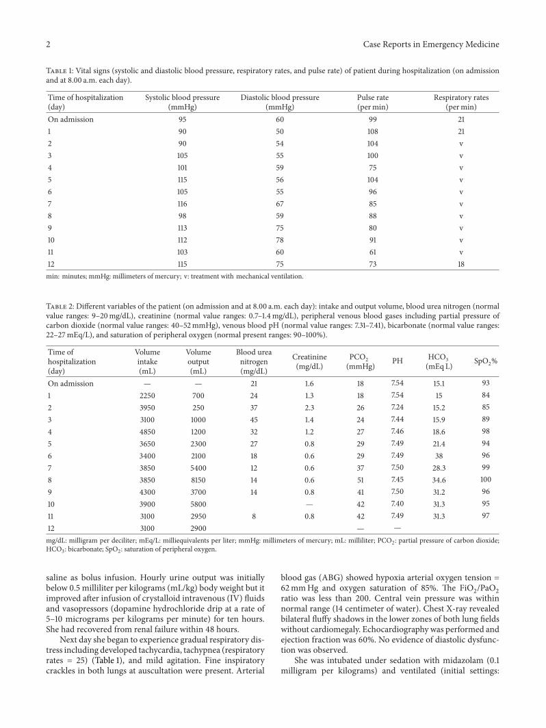
Table 1: Vital signs (systolic and diastolic blood pressure, respiratory rates, and pulse rate) of patient during hospitalization (on admissionand at 8.00 a.m. each day).
Time of hospitalization(day)
Systolic blood pressure(mmHg)
Diastolic blood pressure(mmHg)
Pulse rate(permin)
Respiratory rates(permin)
On admission 95 60 99 211 90 50 108 212 90 54 104 v3 105 55 100 v4 101 59 75 v5 115 56 104 v6 105 55 96 v7 116 67 85 v8 98 59 88 v9 113 75 80 v10 112 78 91 v11 103 60 61 v12 115 75 73 18min: minutes; mmHg: millimeters of mercury; v: treatment with mechanical ventilation.
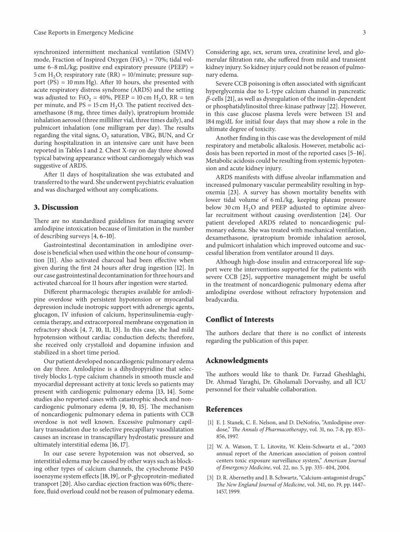
Table 2: Different variables of the patient (on admission and at 8.00 a.m. each day): intake and output volume, blood urea nitrogen (normalvalue ranges: 9–20mg/dL), creatinine (normal value ranges: 0.7–1.4mg/dL), peripheral venous blood gases including partial pressure ofcarbon dioxide (normal value ranges: 40–52mmHg), venous blood pH (normal value ranges: 7.31–7.41), bicarbonate (normal value ranges:22–27mEq/L), and saturation of peripheral oxygen (normal present ranges: 90–100%).
Time ofhospitalization(day)
Volumeintake(mL)
Volumeoutput(mL)
Blood ureanitrogen(mg/dL)
Creatinine(mg/dL)
PCO2(mmHg) PH HCO3
(mEq L) SpO2%
On admission — — 21 1.6 18 7.54 15.1 931 2250 700 24 1.3 18 7.54 15 842 3950 250 37 2.3 26 7.24 15.2 853 3100 1000 45 1.4 24 7.44 15.9 894 4850 1200 32 1.2 27 7.46 18.6 985 3650 2300 27 0.8 29 7.49 21.4 946 3400 2100 18 0.6 29 7.49 38 967 3850 5400 12 0.6 37 7.50 28.3 998 3850 8150 14 0.6 51 7.45 34.6 1009 4300 3700 14 0.8 41 7.50 31.2 9610 3900 5800 — 42 7.40 31.3 9511 3100 2950 8 0.8 42 7.49 31.3 9712 3100 2900 — —mg/dL: milligram per deciliter; mEq/L: milliequivalents per liter; mmHg: millimeters of mercury; mL: milliliter; PCO2: partial pressure of carbon dioxide;HCO3: bicarbonate; SpO2: saturation of peripheral oxygen.
saline as bolus infusion. Hourly urine output was initiallybelow 0.5 milliliter per kilograms (mL/kg) body weight but itimproved after infusion of crystalloid intravenous (IV) fluidsand vasopressors (dopamine hydrochloride drip at a rate of5–10 micrograms per kilograms per minute) for ten hours.She had recovered from renal failure within 48 hours.
Next day she began to experience gradual respiratory dis-tress including developed tachycardia, tachypnea (respiratoryrates = 25) (Table 1), and mild agitation. Fine inspiratorycrackles in both lungs at auscultation were present. Arterial
blood gas (ABG) showed hypoxia arterial oxygen tension =62mmHg and oxygen saturation of 85%. The FiO
2/PaO2
ratio was less than 200. Central vein pressure was withinnormal range (14 centimeter of water). Chest X-ray revealedbilateral fluffy shadows in the lower zones of both lung fieldswithout cardiomegaly. Echocardiography was performed andejection fraction was 60%. No evidence of diastolic dysfunc-tion was observed.
She was intubated under sedation with midazolam (0.1milligram per kilograms) and ventilated (initial settings:
Case Reports in Emergency Medicine 3
synchronized intermittent mechanical ventilation (SIMV)mode, Fraction of Inspired Oxygen (FiO
2) = 70%; tidal vol-
ume 6–8mL/kg; positive end expiratory pressure (PEEP) =5 cm H
2O; respiratory rate (RR) = 10/minute; pressure sup-
port (PS) = 10mmHg). After 10 hours, she presented withacute respiratory distress syndrome (ARDS) and the settingwas adjusted to: FiO
2= 40%, PEEP = 10 cm H
2O, RR = ten
per minute, and PS = 15 cm H2O. The patient received dex-
amethasone (8mg, three times daily), ipratropium bromideinhalation aerosol (threemilliliter vial, three times daily), andpulmicort inhalation (one milligram per day). The resultsregarding the vital signs, O
2saturation, VBG, BUN, and Cr
during hospitalization in an intensive care unit have beenreported in Tables 1 and 2. Chest X-ray on day three showedtypical batwing appearance without cardiomegaly which wassuggestive of ARDS.
After 11 days of hospitalization she was extubated andtransferred to theward. She underwent psychiatric evaluationand was discharged without any complications.
3. Discussion
There are no standardized guidelines for managing severeamlodipine intoxication because of limitation in the numberof describing surveys [4, 6–10].
Gastrointestinal decontamination in amlodipine over-dose is beneficial when usedwithin the one hour of consump-tion [11]. Also activated charcoal had been effective whengiven during the first 24 hours after drug ingestion [12]. Inour case gastrointestinal decontamination for three hours andactivated charcoal for 11 hours after ingestion were started.
Different pharmacologic therapies available for amlodi-pine overdose with persistent hypotension or myocardialdepression include inotropic support with adrenergic agents,glucagon, IV infusion of calcium, hyperinsulinemia-eugly-cemia therapy, and extracorporeal membrane oxygenation inrefractory shock [4, 7, 10, 11, 13]. In this case, she had mildhypotension without cardiac conduction defects; therefore,she received only crystalloid and dopamine infusion andstabilized in a short time period.
Our patient developednoncardiogenic pulmonary edemaon day three. Amlodipine is a dihydropyridine that selec-tively blocks L-type calcium channels in smooth muscle andmyocardial depressant activity at toxic levels so patients maypresent with cardiogenic pulmonary edema [13, 14]. Somestudies also reported cases with catastrophic shock and non-cardiogenic pulmonary edema [9, 10, 15]. The mechanismof noncardiogenic pulmonary edema in patients with CCBoverdose is not well known. Excessive pulmonary capil-lary transudation due to selective precapillary vasodilatationcauses an increase in transcapillary hydrostatic pressure andultimately interstitial edema [16, 17].
In our case severe hypotension was not observed, sointerstitial edemamay be caused by other ways such as block-ing other types of calcium channels, the cytochrome P450isoenzyme system effects [18, 19], or P-glycoprotein-mediatedtransport [20]. Also cardiac ejection fraction was 60%; there-fore, fluid overload could not be reason of pulmonary edema.
Considering age, sex, serum urea, creatinine level, and glo-merular filtration rate, she suffered from mild and transientkidney injury. So kidney injury could not be reason of pulmo-nary edema.
Severe CCB poisoning is often associated with significanthyperglycemia due to L-type calcium channel in pancreatic𝛽-cells [21], as well as dysregulation of the insulin-dependentor phosphatidylinositol three-kinase pathway [22]. However,in this case glucose plasma levels were between 151 and184mg/dL for initial four days that may show a role in theultimate degree of toxicity.
Another finding in this case was the development of mildrespiratory and metabolic alkalosis. However, metabolic aci-dosis has been reported in most of the reported cases [5–16].Metabolic acidosis could be resulting from systemic hypoten-sion and acute kidney injury.
ARDS manifests with diffuse alveolar inflammation andincreased pulmonary vascular permeability resulting in hyp-oxemia [23]. A survey has shown mortality benefits withlower tidal volume of 6mL/kg, keeping plateau pressurebelow 30 cm H
2O and PEEP adjusted to optimize alveo-
lar recruitment without causing overdistention [24]. Ourpatient developed ARDS related to noncardiogenic pul-monary edema. She was treated with mechanical ventilation,dexamethasone, ipratropium bromide inhalation aerosol,and pulmicort inhalation which improved outcome and suc-cessful liberation from ventilator around 11 days.
Although high-dose insulin and extracorporeal life sup-port were the interventions supported for the patients withsevere CCB [25], supportive management might be usefulin the treatment of noncardiogenic pulmonary edema afteramlodipine overdose without refractory hypotension andbradycardia.
Conflict of Interests
The authors declare that there is no conflict of interestsregarding the publication of this paper.
Acknowledgments
The authors would like to thank Dr. Farzad Gheshlaghi,Dr. Ahmad Yaraghi, Dr. Gholamali Dorvashy, and all ICUpersonnel for their valuable collaboration.
References
[1] E. J. Stanek, C. E. Nelson, and D. DeNofrio, “Amlodipine over-dose,”The Annals of Pharmacotherapy, vol. 31, no. 7-8, pp. 853–856, 1997.
[2] W. A. Watson, T. L. Litovitz, W. Klein-Schwartz et al., “2003annual report of the American association of poison controlcenters toxic exposure surveillance system,” American Journalof Emergency Medicine, vol. 22, no. 5, pp. 335–404, 2004.
[3] D. R. Abernethy and J. B. Schwartz, “Calcium-antagonist drugs,”The New England Journal of Medicine, vol. 341, no. 19, pp. 1447–1457, 1999.

4 Case Reports in Emergency Medicine
[4] M. Onge, P. Dube, S. Gosselin et al., “Treatment for calciumchannel blocker poisoning: a systematic review,” Clinical Tox-icology, vol. 52, pp. 926–944, 2014.
[5] C. R. DeWitt and J. C.Waksman, “Pharmacology, pathophysiol-ogy andmanagement of calcium channel blocker and 𝛽-blockertoxicity,” Toxicological Reviews, vol. 23, no. 4, pp. 223–238, 2004.
[6] S. Ghosh and M. Sircar, “Calcium channel blocker overdose:experience with amlodipine,” Indian Journal of Critical CareMedicine, vol. 12, no. 4, pp. 190–193, 2008.
[7] H. Azendour, L. Belyamani, M. Atmani, H. Balkhi, and C.Haimeur, “Severe amlodipine intoxication treated by hyperin-sulinemiaeuglycemia therapy,” Journal of Emergency Medicine,vol. 38, no. 1, pp. 33–35, 2010.
[8] K. Saravu and R. Balasubramanian, “Near-fatal amlodipinepoisoning,” Journal of Association of Physicians of India, vol. 52,pp. 156–157, 2004.
[9] R. Hasson, V. Mulcahy, and H. Tahir, “Amlodipine poisioningcomplicated with acute non-cardiogenic pulmonary oedema,”BMJ Case Reports, vol. 2011, 3 pages, 2011.
[10] V. B. Kute, P. R. Shah, K. R. Goplani, M. R. Gumber, A. V.Vanikar, and H. L. Trivedi, “Successful treatment of refractoryhypotension, noncardiogenic pulmonary edema and acute kid-ney injury after an overdose of amlodipine,” Indian Journal ofCritical Care Medicine, vol. 15, no. 3, pp. 182–184, 2011.
[11] H. Sanaei-Zadeh, “Treatment of amlodipine overdose,” IndianJournal of Critical Care Medicine, vol. 16, no. 3, p. 182, 2012.
[12] G. Jurgens, L. C. Groth Hoegberg, and N. A. Graudal, “Theeffect of activated charcoal on drug exposure in healthy volun-teers: a meta-analysis,” Clinical Pharmacology andTherapeutics,vol. 85, no. 5, pp. 501–505, 2009.
[13] S. K. Shah, S. K. Goswami, R. V. Babu, G. Sharma, and A. G.Duarte, “Management of calcium channel antagonist overdosewith hyperinsulinemia-euglycemia therapy: case series andreview of the literature,” Case Reports in Critical Care, vol. 2012,Article ID 927040, 5 pages, 2012.
[14] S. Vogt, A. Mehlig, P. Hunziker et al., “Survival of severe amlo-dipine intoxication due to medical intensive care,” ForensicScience International, vol. 161, no. 2-3, pp. 216–220, 2006.
[15] K. Naha, J. Suryanarayana, R. A. Aziz, and B. A. Shastry,“Amlodipine poisoning revisited: acidosis, acute kidney injuryand acute respiratory distress syndrome,” Indian Journal ofCritical Care Medicine, vol. 18, no. 7, pp. 467–469, 2014.
[16] T. A. Siddiqi, J. Hill, Y. Huckleberry, and S. Parthasarathy, “Non-cardiogenic pulmonary edema and life-threatening shock dueto calcium channel blocker overdose and itsmanagement: a casereport and a clinical review,” Respiratory Care, vol. 59, no. 2, pp.e15–e21, 2014.
[17] V. H. Humbert Jr., N. J. Munn, and R. F. Hawkins, “Non-cardiogenic pulmonary edema complicating massive diltiazemoverdose,” Chest, vol. 99, no. 1, pp. 258–259, 1991.
[18] P. Gladding, H. Pilmore, and C. Edwards, “Potentially fatalinteraction between diltiazem and statins,” Annals of InternalMedicine, vol. 140, no. 8, article W31, 2004.
[19] D. A. Sica, “Interaction of grapefruit juice and calcium channelblocker,”TheAmerican Journal of Hypertension, vol. 19, no. 7, pp.768–773, 2006.
[20] D. R. Abernethy and J. B. Schwartz, “Calcium-antagonist drugs,”The New England Journal of Medicine, vol. 341, no. 19, pp. 1447–1457, 1999.
[21] G. Devis, G. Somers, E. Van Obberghen, and W. J. Malaisse,“Calcium antagonists and islet function. I. Inhibition of insulinrelease by verapamil,” Diabetes, vol. 24, no. 6, pp. 547–551, 1975.
[22] L. K. Bechtel, D. M. Haverstick, and C. P. Holstege, “Verapamiltoxicity dysregulates the phosphatidylinositol 3-kinase path-way,” Academic Emergency Medicine, vol. 15, no. 4, pp. 368–374,2008.
[23] V.M. Ranieri, G.D. Rubenfeld, B. T.Thompson et al., “Acute res-piratory distress syndrome: the Berlin definition,” The Journalof the American Medical Association, vol. 307, no. 23, pp. 2526–2533, 2012.
[24] D. R. Hess, “Approaches to conventional mechanical ventilationof the patient with acute respiratory distress syndrome,” Respi-ratory Care, vol. 56, no. 10, pp. 1555–1572, 2011.
[25] M. St-Onge, P.-A. Dube, S. Gosselin et al., “Treatment for cal-cium channel blocker poisoning: a systematic review,” ClinicalToxicology, vol. 52, no. 9, pp. 926–944, 2014.

Submit your manuscripts athttp://www.hindawi.com
Stem CellsInternational
Hindawi Publishing Corporationhttp://www.hindawi.com Volume 2014
Hindawi Publishing Corporationhttp://www.hindawi.com Volume 2014
MEDIATORSINFLAMMATION
of
Hindawi Publishing Corporationhttp://www.hindawi.com Volume 2014
Behavioural Neurology
EndocrinologyInternational Journal of
Hindawi Publishing Corporationhttp://www.hindawi.com Volume 2014
Hindawi Publishing Corporationhttp://www.hindawi.com Volume 2014
Disease Markers
Hindawi Publishing Corporationhttp://www.hindawi.com Volume 2014
BioMed Research International
OncologyJournal of
Hindawi Publishing Corporationhttp://www.hindawi.com Volume 2014
Hindawi Publishing Corporationhttp://www.hindawi.com Volume 2014
Oxidative Medicine and Cellular Longevity
Hindawi Publishing Corporationhttp://www.hindawi.com Volume 2014
PPAR Research
The Scientific World JournalHindawi Publishing Corporation http://www.hindawi.com Volume 2014
Immunology ResearchHindawi Publishing Corporationhttp://www.hindawi.com Volume 2014
Journal of
ObesityJournal of
Hindawi Publishing Corporationhttp://www.hindawi.com Volume 2014
Hindawi Publishing Corporationhttp://www.hindawi.com Volume 2014
Computational and Mathematical Methods in Medicine
OphthalmologyJournal of
Hindawi Publishing Corporationhttp://www.hindawi.com Volume 2014
Diabetes ResearchJournal of
Hindawi Publishing Corporationhttp://www.hindawi.com Volume 2014
Hindawi Publishing Corporationhttp://www.hindawi.com Volume 2014
Research and TreatmentAIDS
Hindawi Publishing Corporationhttp://www.hindawi.com Volume 2014
Gastroenterology Research and Practice
Hindawi Publishing Corporationhttp://www.hindawi.com Volume 2014
Parkinson’s Disease
Evidence-Based Complementary and Alternative Medicine
Volume 2014Hindawi Publishing Corporationhttp://www.hindawi.com