Embed Size (px)
Citation preview
Hindawi Publishing CorporationCase Reports in DentistryVolume 2013, Article ID 828373, 4 pageshttp://dx.doi.org/10.1155/2013/828373
Case ReportMultiple Cementoblastoma: A Rare Case Report
G. Iannaci,1 R. Luise,1 G. Iezzi,2 A. Piattelli,2 and A. Salierno3
1 Department of Pathology, Incurabili Hospital ASLNAPOLI1 Centro, Via Maria Longo, 50-80138 Napoli, Italy2 Department of Medical, Oral and Biotechnological Sciences, University of Chieti-Pescara, Chieti, Italy3 Department of Oral Medicine, School of Dentistry of the Second University of the Study of Naples, Naples, Italy
Correspondence should be addressed to G. Iannaci; [email protected]
Received 28 May 2013; Accepted 24 July 2013
Academic Editors: M. B. D. Gaviao, J. A. Shibli, and R. Sorrentino
Copyright © 2013 G. Iannaci et al. This is an open access article distributed under the Creative Commons Attribution License,which permits unrestricted use, distribution, and reproduction in any medium, provided the original work is properly cited.
Benign cementoblastoma is a rare ectomesenchymal odontogenic tumor that originates from the root of the tooth and that ischaracterized by the formation of cementum-like tissue. A 60-year old man was referred to us complaining of pain in his rightjaw. The patient underwent TC dental scan of the mandible, which highlighted the presence of three well-circumscribed, round,unilocular neoformations of radiopaque appearance with a radiotransparent edge, one of which was in close contact with theroots of the lower right second molar. Microscopic examination of the greater sample consisted, in its central portion, of densemineralized acellular trabeculae of basophilic tissue cement-like, devoid of vessels, adhering to the root of the tooth, whileperipherally was observed a zone of vascularized osteoid surrounded, occasionally, by a thin rim of cementoblasts mixed withfibrous tissue and inflammatory elements.This lesionwas diagnosed as cementoblastoma.The second lesion appeared radiologicallyand histologically entirely identical to cementoblastoma, but it did not show the intimate association with the root of involvedtooth. After a careful review of the literature, the diagnosis of residual cementoblastoma was made. The clinicopathologic features,treatment, and prognosis of this rare tumor are here discussed for the young dental practitioner.
1. Introduction
Benign cementoblastoma is a rare lesion of the oral cavity,currently classified by theWorld Health Organization (WHO2005) as an ectomesenchymal odontogenic tumor that origi-nates from the root of the tooth and that is characterized withthe formation of cementum-like tissue [1, 2]. It usually arisesin the first permanent molars in their mandibular regionbut can also be associated with multiple teeth, deciduousteeth, or unerupted molars [3, 4]. The caucasians race andmale sex are more commonly affected than black race andfemale (ratio 2.1 : 1), with a very wide age range and apeak incidence between the second and third decade oflife. Clinically, the lesion presents as a nodular formation,hard-elastic in consistency producing swelling in the alveolarridge area. Paresthesias of the lower lip or a pathologicfracture of the jaw have been rarely reported.The radiologicalfindings show a well-defined radiopaque mass surroundedwith a thin, radiolucent rim of nonmineralized tissue, inintimate association of the root of the involved tooth. Theresorption of the tooth root, the loss of the regular outline
with the obliteration of the periodontal ligament are clinicaland radiological features that can be frequently found [5,6]. We describe a rare case of multiple cementoblastoma: aclassic cementoblastoma in direct continuity with the root ofthe tooth (true cementoma) and a residual cementoblastomalocalized in the edentulous area post-extraction.
2. Case Report
A 60-years-old man was referred to us complaining of painin his right jaw. The examination of the patient’s medicalhistory revealed good general health, absence of systemicdiseases, and smoking habit (10 cigarettes\day). The clinicalexamination showed discrete oral hygiene, thick and flatperiodontal biotype, class II malocclusion with marked lossof vertical size, multiple missing teeth in the site 1.4-1.5-3.5-3.6-3.7-4.5-4.6., presence of swelling, hard elastic consistency,and crepitus on palpation in the region corresponding tothe elements 4.4-4.7 and in the edentulous area 4.6, 4.5. Theorthopantomography revealed on the upper jaw radicular
2 Case Reports in Dentistry
Figure 1: The first orthopantomography revealed the presence of three well-circumscribed, round, unilocular neoformations of radiopaqueappearance with a radiotransparent edge, one of which was in close contact with the roots of the lower right second molar.
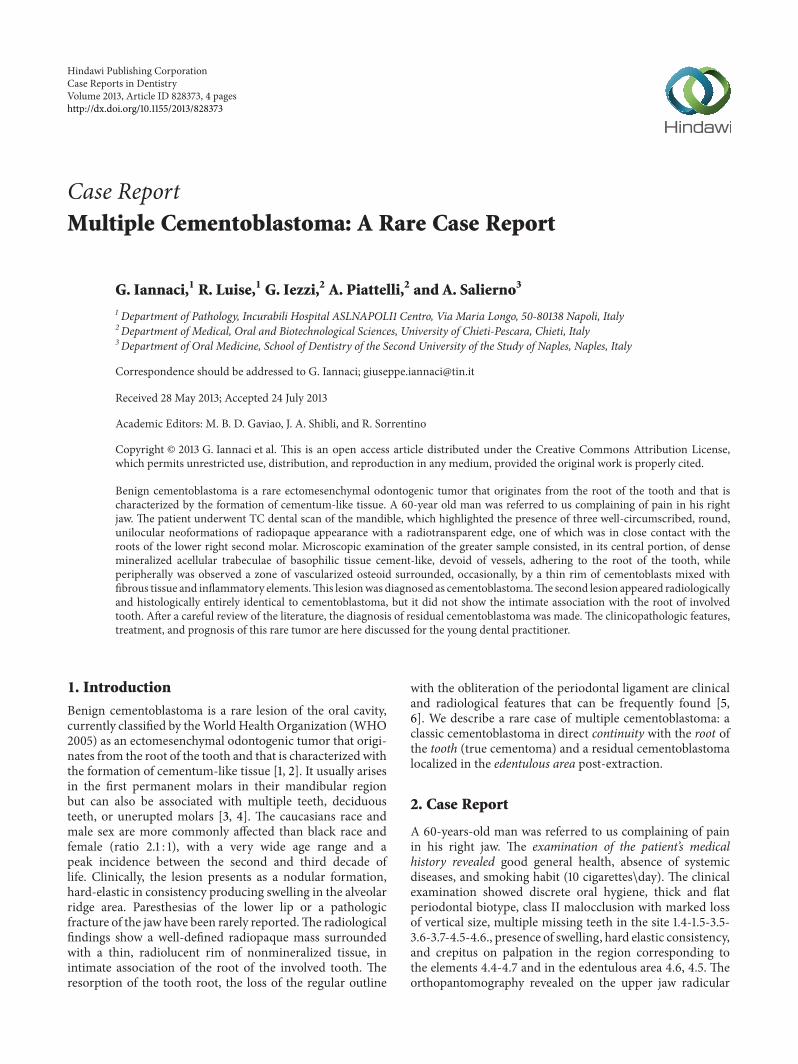
Figure 2: TC dental scan of the mandible.
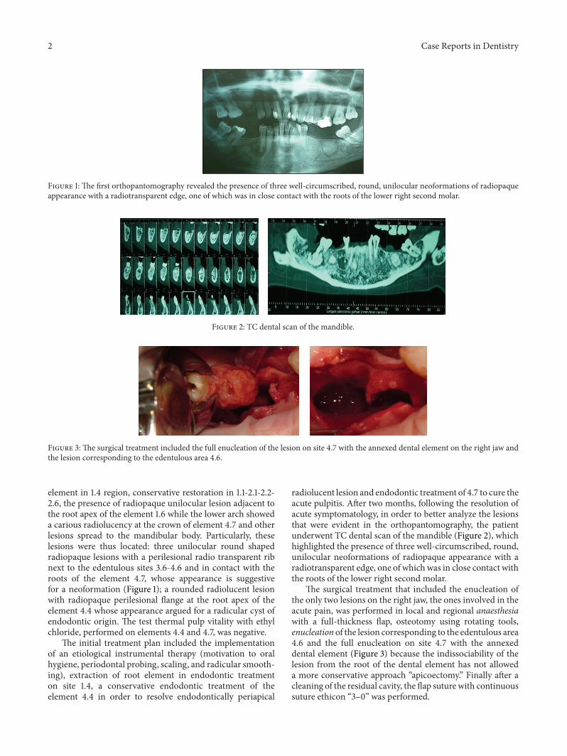
Figure 3: The surgical treatment included the full enucleation of the lesion on site 4.7 with the annexed dental element on the right jaw andthe lesion corresponding to the edentulous area 4.6.
element in 1.4 region, conservative restoration in 1.1-2.1-2.2-2.6, the presence of radiopaque unilocular lesion adjacent tothe root apex of the element 1.6 while the lower arch showeda carious radiolucency at the crown of element 4.7 and otherlesions spread to the mandibular body. Particularly, theselesions were thus located: three unilocular round shapedradiopaque lesions with a perilesional radio transparent ribnext to the edentulous sites 3.6-4.6 and in contact with theroots of the element 4.7, whose appearance is suggestivefor a neoformation (Figure 1); a rounded radiolucent lesionwith radiopaque perilesional flange at the root apex of theelement 4.4 whose appearance argued for a radicular cyst ofendodontic origin. The test thermal pulp vitality with ethylchloride, performed on elements 4.4 and 4.7, was negative.
The initial treatment plan included the implementationof an etiological instrumental therapy (motivation to oralhygiene, periodontal probing, scaling, and radicular smooth-ing), extraction of root element in endodontic treatmenton site 1.4, a conservative endodontic treatment of theelement 4.4 in order to resolve endodontically periapical
radiolucent lesion and endodontic treatment of 4.7 to cure theacute pulpitis. After two months, following the resolution ofacute symptomatology, in order to better analyze the lesionsthat were evident in the orthopantomography, the patientunderwent TC dental scan of the mandible (Figure 2), whichhighlighted the presence of three well-circumscribed, round,unilocular neoformations of radiopaque appearance with aradiotransparent edge, one of which was in close contact withthe roots of the lower right second molar.
The surgical treatment that included the enucleation ofthe only two lesions on the right jaw, the ones involved in theacute pain, was performed in local and regional anaesthesiawith a full-thickness flap, osteotomy using rotating tools,enucleation of the lesion corresponding to the edentulous area4.6 and the full enucleation on site 4.7 with the annexeddental element (Figure 3) because the indissociability of thelesion from the root of the dental element has not alloweda more conservative approach “apicoectomy.” Finally after acleaning of the residual cavity, the flap suture with continuoussuture ethicon “3–0” was performed.
Case Reports in Dentistry 3
Figure 4: Macroscopically the two samples presented as a nodular, hard-elastic in consistency, the largest of which was adherent to the dentalelement.
(a) (b)
(c) (d)
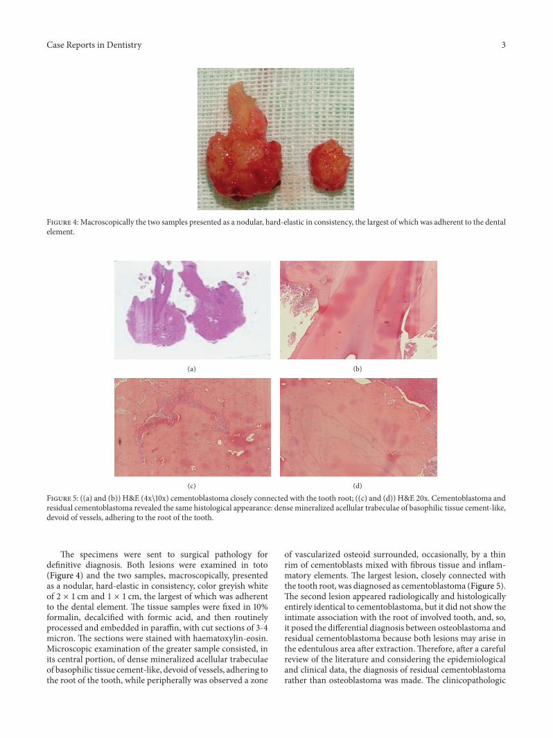
Figure 5: ((a) and (b)) H&E (4x\10x) cementoblastoma closely connected with the tooth root; ((c) and (d)) H&E 20x. Cementoblastoma andresidual cementoblastoma revealed the same histological appearance: dense mineralized acellular trabeculae of basophilic tissue cement-like,devoid of vessels, adhering to the root of the tooth.
The specimens were sent to surgical pathology fordefinitive diagnosis. Both lesions were examined in toto(Figure 4) and the two samples, macroscopically, presentedas a nodular, hard-elastic in consistency, color greyish whiteof 2 × 1 cm and 1 × 1 cm, the largest of which was adherentto the dental element. The tissue samples were fixed in 10%formalin, decalcified with formic acid, and then routinelyprocessed and embedded in paraffin, with cut sections of 3-4micron. The sections were stained with haematoxylin-eosin.Microscopic examination of the greater sample consisted, inits central portion, of dense mineralized acellular trabeculaeof basophilic tissue cement-like, devoid of vessels, adhering tothe root of the tooth, while peripherally was observed a zone
of vascularized osteoid surrounded, occasionally, by a thinrim of cementoblasts mixed with fibrous tissue and inflam-matory elements. The largest lesion, closely connected withthe tooth root, was diagnosed as cementoblastoma (Figure 5).The second lesion appeared radiologically and histologicallyentirely identical to cementoblastoma, but it did not show theintimate association with the root of involved tooth, and, so,it posed the differential diagnosis between osteoblastoma andresidual cementoblastoma because both lesions may arise inthe edentulous area after extraction.Therefore, after a carefulreview of the literature and considering the epidemiologicaland clinical data, the diagnosis of residual cementoblastomarather than osteoblastoma was made. The clinicopathologic
4 Case Reports in Dentistry
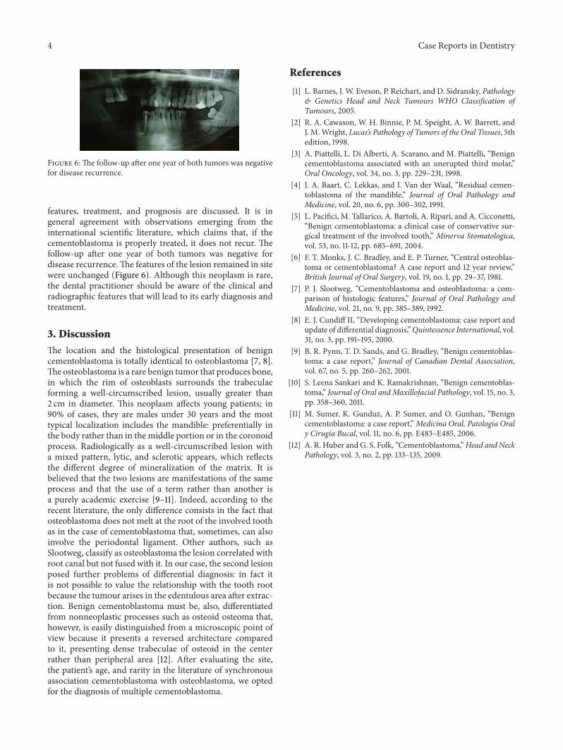
Figure 6: The follow-up after one year of both tumors was negativefor disease recurrence.
features, treatment, and prognosis are discussed. It is ingeneral agreement with observations emerging from theinternational scientific literature, which claims that, if thecementoblastoma is properly treated, it does not recur. Thefollow-up after one year of both tumors was negative fordisease recurrence.The features of the lesion remained in sitewere unchanged (Figure 6). Although this neoplasm is rare,the dental practitioner should be aware of the clinical andradiographic features that will lead to its early diagnosis andtreatment.
3. Discussion
The location and the histological presentation of benigncementoblastoma is totally identical to osteoblastoma [7, 8].The osteoblastoma is a rare benign tumor that produces bone,in which the rim of osteoblasts surrounds the trabeculaeforming a well-circumscribed lesion, usually greater than2 cm in diameter. This neoplasm affects young patients; in90% of cases, they are males under 30 years and the mosttypical localization includes the mandible: preferentially inthe body rather than in themiddle portion or in the coronoidprocess. Radiologically as a well-circumscribed lesion witha mixed pattern, lytic, and sclerotic appears, which reflectsthe different degree of mineralization of the matrix. It isbelieved that the two lesions are manifestations of the sameprocess and that the use of a term rather than another isa purely academic exercise [9–11]. Indeed, according to therecent literature, the only difference consists in the fact thatosteoblastoma does not melt at the root of the involved toothas in the case of cementoblastoma that, sometimes, can alsoinvolve the periodontal ligament. Other authors, such asSlootweg, classify as osteoblastoma the lesion correlated withroot canal but not fused with it. In our case, the second lesionposed further problems of differential diagnosis: in fact itis not possible to value the relationship with the tooth rootbecause the tumour arises in the edentulous area after extrac-tion. Benign cementoblastoma must be, also, differentiatedfrom nonneoplastic processes such as osteoid osteoma that,however, is easily distinguished from a microscopic point ofview because it presents a reversed architecture comparedto it, presenting dense trabeculae of osteoid in the centerrather than peripheral area [12]. After evaluating the site,the patient’s age, and rarity in the literature of synchronousassociation cementoblastoma with osteoblastoma, we optedfor the diagnosis of multiple cementoblastoma.
References
[1] L. Barnes, J. W. Eveson, P. Reichart, and D. Sidransky, Pathology& Genetics Head and Neck Tumours WHO Classification ofTumours, 2005.
[2] R. A. Cawason, W. H. Binnie, P. M. Speight, A. W. Barrett, andJ. M.Wright, Lucas’s Pathology of Tumors of the Oral Tissues, 5thedition, 1998.
[3] A. Piattelli, L. Di Alberti, A. Scarano, and M. Piattelli, “Benigncementoblastoma associated with an unerupted third molar,”Oral Oncology, vol. 34, no. 3, pp. 229–231, 1998.
[4] J. A. Baart, C. Lekkas, and I. Van der Waal, “Residual cemen-toblastoma of the mandible,” Journal of Oral Pathology andMedicine, vol. 20, no. 6, pp. 300–302, 1991.
[5] L. Pacifici, M. Tallarico, A. Bartoli, A. Ripari, and A. Cicconetti,“Benign cementoblastoma: a clinical case of conservative sur-gical treatment of the involved tooth,” Minerva Stomatologica,vol. 53, no. 11-12, pp. 685–691, 2004.
[6] F. T. Monks, J. C. Bradley, and E. P. Turner, “Central osteoblas-toma or cementoblastoma? A case report and 12 year review,”British Journal of Oral Surgery, vol. 19, no. 1, pp. 29–37, 1981.
[7] P. J. Slootweg, “Cementoblastoma and osteoblastoma: a com-parison of histologic features,” Journal of Oral Pathology andMedicine, vol. 21, no. 9, pp. 385–389, 1992.
[8] E. J. Cundiff II, “Developing cementoblastoma: case report andupdate of differential diagnosis,”Quintessence International, vol.31, no. 3, pp. 191–195, 2000.
[9] B. R. Pynn, T. D. Sands, and G. Bradley, “Benign cementoblas-toma: a case report,” Journal of Canadian Dental Association,vol. 67, no. 5, pp. 260–262, 2001.
[10] S. Leena Sankari and K. Ramakrishnan, “Benign cementoblas-toma,” Journal of Oral andMaxillofacial Pathology, vol. 15, no. 3,pp. 358–360, 2011.
[11] M. Sumer, K. Gunduz, A. P. Sumer, and O. Gunhan, “Benigncementoblastoma: a case report,”Medicina Oral, Patologı́a Oraly Cirugı́a Bucal, vol. 11, no. 6, pp. E483–E485, 2006.
[12] A. R. Huber and G. S. Folk, “Cementoblastoma,”Head and NeckPathology, vol. 3, no. 2, pp. 133–135, 2009.

Submit your manuscripts athttp://www.hindawi.com
Hindawi Publishing Corporationhttp://www.hindawi.com Volume 2014
Oral OncologyJournal of
DentistryInternational Journal of
Hindawi Publishing Corporationhttp://www.hindawi.com Volume 2014
Hindawi Publishing Corporationhttp://www.hindawi.com Volume 2014
International Journal of
Biomaterials
Hindawi Publishing Corporationhttp://www.hindawi.com Volume 2014
BioMed Research International
Hindawi Publishing Corporationhttp://www.hindawi.com Volume 2014
Case Reports in Dentistry
Hindawi Publishing Corporationhttp://www.hindawi.com Volume 2014
Oral ImplantsJournal of
Hindawi Publishing Corporationhttp://www.hindawi.com Volume 2014
Anesthesiology Research and Practice
Hindawi Publishing Corporationhttp://www.hindawi.com Volume 2014
Radiology Research and Practice
Environmental and Public Health
Journal of
Hindawi Publishing Corporationhttp://www.hindawi.com Volume 2014
The Scientific World JournalHindawi Publishing Corporation http://www.hindawi.com Volume 2014
Hindawi Publishing Corporationhttp://www.hindawi.com Volume 2014
Dental SurgeryJournal of
Drug DeliveryJournal of
Hindawi Publishing Corporationhttp://www.hindawi.com Volume 2014
Hindawi Publishing Corporationhttp://www.hindawi.com Volume 2014
Oral DiseasesJournal of
Hindawi Publishing Corporationhttp://www.hindawi.com Volume 2014
Computational and Mathematical Methods in Medicine
ScientificaHindawi Publishing Corporationhttp://www.hindawi.com Volume 2014
PainResearch and TreatmentHindawi Publishing Corporationhttp://www.hindawi.com Volume 2014
Preventive MedicineAdvances in
Hindawi Publishing Corporationhttp://www.hindawi.com Volume 2014
EndocrinologyInternational Journal of
Hindawi Publishing Corporationhttp://www.hindawi.com Volume 2014
Hindawi Publishing Corporationhttp://www.hindawi.com Volume 2014
OrthopedicsAdvances in