Embed Size (px)
Citation preview
Hindawi Publishing CorporationCase Reports in Gastrointestinal MedicineVolume 2013, Article ID 568193, 4 pageshttp://dx.doi.org/10.1155/2013/568193
Case ReportMetronidazole Induced Liver Injury: A Rare Immune MediatedDrug Reaction
Dayakar Kancherla,1 Mahesh Gajendran,1 Priyanka Vallabhaneni,2 and Kishore Vipperla1
1 Division of General Internal Medicine, University of Pittsburgh, Pittsburgh, PA 15213, USA2Department of Internal Medicine, St. Luke’s University Hospitals and Health Network, Bethlehem, PA 18015, USA
Correspondence should be addressed to Kishore Vipperla; [email protected]
Received 5 November 2013; Accepted 10 December 2013
Academic Editors: E. Altintas and M. Neri
Copyright © 2013 Dayakar Kancherla et al. This is an open access article distributed under the Creative Commons AttributionLicense, which permits unrestricted use, distribution, and reproduction in any medium, provided the original work is properlycited.
Drug induced liver injury (DILI) can result either from dose-dependent direct hepatotoxicity or from an unpredictable dose-independent idiosyncratic reaction. Incidence of idiosyncraticDILI is estimated to be approximately 10–15 per 100,000 patient years.Here we report an extremely rare case of metronidazole induced delayed immune-allergic hepatocellular liver injurymasqueradingas autoimmune hepatitis. A previously healthy 54-year-old Caucasian male, who was treated with metronidazole for Clostridiumdifficile associated diarrhea, presented 3 months later with right upper quadrant abdominal pain. Laboratory tests revealed totalbilirubin level of 12.7mg/dL, direct bilirubin of 7.2mg/dL, alanine aminotransferase (ALT) of 973 IU/L, aspartate transaminase(AST) of 867 IU/L, alkaline phosphatase (AP) of 96 IU/L, and an INR of 1.9, suggestive of hepatocellular pattern of injury. Adetailedworkup for hepatitis revealed no other etiology. A clinical diagnosis ofmetronidazole induced liver injurywasmade.With apersistent rise in his bilirubin and transaminase levels, the patient was started on oral prednisone. At the 2-week posthospitalizationfollow-up visit, the patient reported a significant improvement in his overall sense of being well and liver functions tests trendeddown substantially (total bilirubin 7.2mg/dL, ALT 420 IU/L, AST 276 IU/L, AP 183 IU/L, and INR 1.5).
1. Introduction
Reactive chemical metabolites formed during hepatic drugmetabolism can incite hepatocellular damage from oxidativestress and mitochondrial dysfunction causing drug inducedliver injury (DILI). DILI can result from either dose-dependent direct hepatotoxicity (e.g., acetaminophen toxic-ity) or from an unpredictable dose-independent idiosyncraticreaction. Genetic polymorphisms in the drug bioactivationand detoxification pathways along with host immunolog-ical factors are responsible for these rare and potentiallyfatal idiosyncratic DILI [1]. Of the several mechanismsproposed to elucidate the mechanism underlying immune-allergic idiosyncratic DILI, the “hapten hypothesis” is themost favored [2]. Drugs and/or their metabolites covalentlybind to host proteins forming drug-protein adducts (i.e.,haptens) that are processed by the antigen-presenting cellsand trigger a T-cell mediated cytotoxicity or B-cell antibody
response. Incidence of idiosyncratic DILI is estimated to beapproximately 10–15 per 100,000 patient years [3]. About 1in 7 cases of acute liver failure are related to an adversedrug reaction, making DILI the most common indication forliver transplantation in USA [4]. Antimicrobials are the mostcommon class (∼45%) of drugs responsible for DILI withamoxicillin-clavulanic acid being the single most commoncausative agent [5]. Advanced age, female sex, drug dose,and the extent of its hepatic drug metabolism are some ofthe identified risk factors for DILI [6]. A “probable” reactionto metronidazole presenting as a cholestatic pattern liverinjury reaction within a few days after initiation and thatresolved shortly after drug cessation has been reported earlier[7]. Here we report an extremely rare case of metronidazoleinduced delayed immune-allergic hepatocellular liver injurymasquerading as autoimmune hepatitis that has not beenpreviously reported to our knowledge.
2 Case Reports in Gastrointestinal Medicine
2. Case Presentation
A previously healthy 54-year-old Caucasian male presentedto our institution with worsening right upper quadrantabdominal pain and jaundice of 3-week duration. He deniedalcohol or tobacco use, and his recent past medical historywas significant only for a dental abscess treated with clin-damycin followed by the development of Clostridium difficileassociated diarrhea (CDAD) approximately 3months prior tothis admission. His CDAD was successfully treated then by a2-week course ofmetronidazole, but the course was staggereddue to the complaints of vague epigastric and right upperquadrant abdominal discomfort associated with nausea thatwere felt to be from drug “intolerance.” But his abdominaldiscomfort, anorexia, and nausea gradually worsened in thesubsequent weeks though he denied having any skin rash,fever, vomiting, or change in bowel habits since his treatmentwith metronidazole. Three weeks ago, he started to noticedarker urine, acholic stools, yellowish skin discoloration, andpruritus. “Abnormal liver function tests” noted on bloodtests ordered by his family physician prompted an in-patientevaluation.
At admission, the patient was afebrile and had stablevital signs. Physical examination revealed a comfortableappearing well-built male who had remarkable icterus andjaundice without any other stigmata of chronic liver diseaseor cirrhosis such as spider nevi, clubbing, or muscle atro-phy. Cardiovascular and respiratory system examination wasnormal. His abdomen was nondistended with normal bowelsounds, but was mildly tender in the right upper quadrantwithout signs of peritonitis, hepatosplenomegaly, or ascites.His mental status was intact and did not exhibit asterixis.Initial blood tests (normal values range in parenthesis)revealed a total bilirubin level of 12.7 (0.3–1.5)mg/dL, directbilirubin of 7.2 (0.1–0.5)mg/dL, alanine aminotransferase(ALT) of 973 (17–63) IU/L, aspartate transaminase (AST)of 867 (15–41) IU/L, alkaline phosphatase (AP) of 96 (38–126) IU/L, and an INR of 1.9 (0.8–1.2) clearly suggestive ofmild hepatic failure with a hepatocellular pattern of injury.His complete blood count and renal function tests werewithin normal limits. A right upper quadrant abdominalDoppler ultrasonography revealed normal hepatic textureand patent hepatic and portal veins. A magnetic resonancecholangiopancreatography (MRCP) scan was remarkable formoderate periportal edema but otherwise normal hepaticmorphology and without any obstruction within the bil-iary tract. However during the gastroenterologist’s personalreview of the MRCP images, a subtle filling defect wassuspected in the proximal CBD prompting an endoscopicretrograde cholangiography (ERCP) which did not reveal anyobstructive process or mass.
During a 4-day hospitalization, a comprehensive lab-oratory workup was performed to rule out acute andchronic liver diseases. Drug toxicity (undetectable serumacetaminophen levels), viral hepatitis (HIV, herpes simplexvirus, hepatitides A, B, C, E antibodies, Epstein Barr virus,and cytomegalovirus PCR), autoimmune hepatitis (anti-nuclear antibody, anti-neutrophil cytoplasmic antibody, anti-M2 mitochondrial antibody, anti-smooth muscle antibody,
P
C
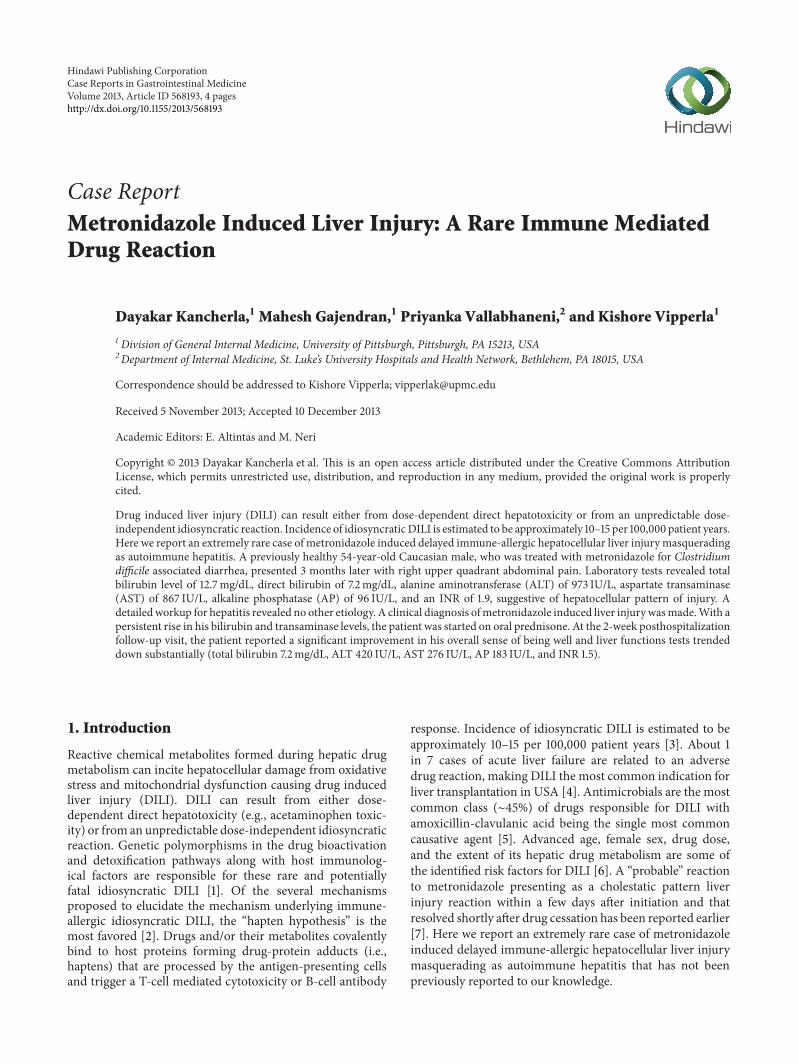
Figure 1:The liver biopsy showed severe portal and lobular hepatitiswith perivenular and bridging necrosis (arrow heads) (H&E stain,original magnification 100x). P: portal tracts, C: central vein.
Figure 2: High-magnification picture of the lobule showing anarea of centrizonal confluent necrosis (arrow) and adjacent viableparenchyma. Scattered mononuclear inflammatory cells and anapoptotic hepatocyte are present in the viable parenchyma (H&Estain, original magnification 400x).
and quantitative immunoglobulin levels), andmetabolic liverdisease (serum ceruloplasmin and alpha-1 antitrypsin levels)tests were unremarkable. Progressively worsening hepatictransaminase levels prompted a liver biopsy for a definitivediagnosis. An ultrasound guided needle biopsy specimenfrom right hepatic lobe showed severe acute to subacutepredominantly portal and lobular hepatitis with confluentperivenular and bridging necrosis strongly suggestive of animmunoallergic etiology related to either a drug reaction orautoimmune hepatitis (Figures 1, 2, and 3). His age, sex, andclinical presentation were not typical of autoimmune hepati-tis, and, moreover, the autoimmune serological workup wasnot corroborative, making a drug reaction to metronidazolethe most likely etiology.
With a persistent rise in his bilirubin and transaminaselevels, the patientwas started on oral steroidswith prednisoneprescribed at a dose of 40mg/day to control hepatic inflam-mation contributing to the hepatocellular injury. He was clin-ically stable and symptomatically comfortable throughoutthe hospitalization without developing signs or symptomsof fulminant hepatic failure. He was discharged home withrecommendation to use a 4-week course of steroids with close

Case Reports in Gastrointestinal Medicine 3
Figure 3: Infiltrating cells in the portal tracts were predominantlylymphocytes admixed with fewer neutrophils, plasma cells (arrows),and rare eosinophils (H&E stain, original magnification 400x).
followup of his liver function tests. At the 2-week posthos-pitalization followup visit, the patient reported a significantimprovement in his overall sense of being well and liverfunctions tests trended down substantially (total bilirubinlevel of 7.2mg/dL, ALT of 420 IU/L, AST of 276 IU/L, AP of183 IU/L, and an INR of 1.5). He was recommended to con-tinue using the steroid for a total of 4 weeks with subsequentgradual tapering of the steroid dose with close monitoringof his liver function test. Excellent response to prednisoneand absence of a preexisting underlying liver disease predictsgood prognosis, but only time would determine if he wouldhave any long-lasting sequelae of DILI.
3. Discussion
Immunoallergic DILI is a form of DILI that typically presents1–3 months after exposure to the drug, the time that typicallyis taken for manifestation of a full-blown delayed immunereaction. DILI can present as a wide spectrum rangingfrom asymptomatic elevation of liver enzymes to fulminanthepatic failure and often mimics other acute or chronic liverdiseases.The abnormal liver enzymes can be categorized intocholestatic, hepatocellular, or mixed injury patterns. It is adiagnosis of exclusion that is established by a strong clinicalsuspicion and a comprehensive workup to rule out othercompeting etiologies. Diagnostic algorithms such as RousselUclaf Causality Assessment Model have been proposed toassist in ascertaining the probability of DILI but are notrobust for general clinical application. Histopathologicalfindings of DILI are nonspecific too but can be valuable innarrowing the differential diagnoses.
Treatment measures mainly involve prompt cessation ofthe offending drug and supportive care. N-Acetyl cysteinehas been used in some cases and was shown to improvetransplant free survival, though mortality reduction benefitwas not observed [8]. Treatment with steroids has been ofunproven benefit in most hepatotoxic drug reactions butoffers a potential therapeutic role when DILI is secondaryto a hypersensitivity reaction and has a severe clinicalcourse or worsening liver function tests with conservativemanagement, identical to our case [9]. Majority of patients
surviving DILI recover completely, but ∼6% persist to havechronic liver disease (chronic DILI), and only 1% develop“cryptogenic” cirrhosis [10]. Interestingly, 22% of patientswith chronic DILI at presentation developed autoimmunehepatitis eventually in one registry, suggesting the possibilityof drug induced increase in susceptibility to developingautoimmune hepatitis.
The wide spread antibiotic usage in the practice of mod-ern medicine has been responsible for a surge in potentiallylife threatening drug reactions. Drug reactions that developimmediately after initiation of inciting agent are easier torecognize and resolve with cessation of the drug. But it isquite challenging to recognize the rare immune mediateddelayed reactions. Albeit unavoidable at the first instance, itis paramount to identify such idiosyncratic DILI to preventrecurrent reactions that are likely to be more severe andrapid in recurrence on rechallenge. Antibiotics have to beused judiciously bearing in mind the risk of DILI even fora widely used drug such as metronidazole, which is generallyconsidered to be quite a safer drug for short-term use. Also,potentially serious adverse effects such as DILI have to beconsidered in the differential diagnosis in the work up forjaundice.
Consent
The authors have obtained an informed written consent fromthe patient.
Conflict of Interests
All the authors declare that there is no conflict of interests.
Authors’ Contribution
All the authors provided major input into the conceptualdevelopment of the report. K. Vipperla helped in takingcare of the patient and obtaining consent. D. Kancherlawrote the first draft of the paper and all the other authorsequally contributed to revising the paper. All authors read andapproved the final manuscript for submission.
Acknowledgments
The authors thank Eizaburo, Sasatomi M.D. (Pathologist),and Michael A. Dunn, M.D. (Gastroenterologist), for theirassistance in providing valuable insight into the diagnosis.
References
[1] D. E. Amacher, “The primary role of hepatic metabolism inidiosyncratic drug-induced liver injury,” Expert Opinion onDrugMetabolism and Toxicology, vol. 8, no. 3, pp. 335–347, 2012.
[2] S. Tujios and R. J. Fontana, “Mechanisms of drug-induced liverinjury: From bedside to bench,” Nature Reviews Gastroenterol-ogy and Hepatology, vol. 8, no. 4, pp. 202–211, 2011.
[3] L. N. Bell and N. Chalasani, “Epidemiology of idiosyncraticdrug-induced liver injury,” Seminars in Liver Disease, vol. 29, no.4, pp. 337–347, 2009.
4 Case Reports in Gastrointestinal Medicine
[4] G. Ostapowicz, R. J. Fontana, F. V. Schioødt et al., “Results of aprospective study of acute liver failure at 17 tertiary care centersin the United States,” Annals of Internal Medicine, vol. 137, no.12, pp. 947–954, 2002.
[5] N.Chalasani, R. J. Fontana,H. L. Bonkovsky et al., “Causes, clin-ical features, and outcomes from a prospective study of drug-induced liver injury in the United States,” Gastroenterology, vol.135, no. 6, pp. 1924–1934, 2008.
[6] M. I. Lucena, R. J. Andrade, N. Kaplowitz et al., “Phenotypiccharacterization of idiosyncratic drug-induced liver injury:Theinfluence of age and sex,” Hepatology, vol. 49, no. 6, pp. 2001–2009, 2009.
[7] E. Bjornsson,H.Nordlinder, and R.Olssson, “Metronidazol as aprobable cause of severe liver injury,” Hepato-Gastroenterology,vol. 49, no. 43, pp. 252–254, 2002.
[8] W. M. Lee, L. S. Hynan, L. Rossaro et al., “Intravenous N-acetylcysteine improves transplant-free survival in early stagenon-acetaminophen acute liver failure,” Gastroenterology, vol.137, no. 3, pp. 856–864, 2009.
[9] A. Giannattasio, M. D’Ambrosi, M. Volpicelli, and R. Iorio,“Steroid therapy for a case of severe drug-induced cholestasis,”Annals of Pharmacotherapy, vol. 40, no. 6, pp. 1196–1199, 2006.
[10] E. Bjornsson and L. Davidsdottir, “The long-term follow-upafter idiosyncratic drug-induced liver injury with jaundice,”Journal of Hepatology, vol. 50, no. 3, pp. 511–517, 2009.

Submit your manuscripts athttp://www.hindawi.com
Stem CellsInternational
Hindawi Publishing Corporationhttp://www.hindawi.com Volume 2014
Hindawi Publishing Corporationhttp://www.hindawi.com Volume 2014
MEDIATORSINFLAMMATION
of
Hindawi Publishing Corporationhttp://www.hindawi.com Volume 2014
Behavioural Neurology
EndocrinologyInternational Journal of
Hindawi Publishing Corporationhttp://www.hindawi.com Volume 2014
Hindawi Publishing Corporationhttp://www.hindawi.com Volume 2014
Disease Markers
Hindawi Publishing Corporationhttp://www.hindawi.com Volume 2014
BioMed Research International
OncologyJournal of
Hindawi Publishing Corporationhttp://www.hindawi.com Volume 2014
Hindawi Publishing Corporationhttp://www.hindawi.com Volume 2014
Oxidative Medicine and Cellular Longevity
Hindawi Publishing Corporationhttp://www.hindawi.com Volume 2014
PPAR Research
The Scientific World JournalHindawi Publishing Corporation http://www.hindawi.com Volume 2014
Immunology ResearchHindawi Publishing Corporationhttp://www.hindawi.com Volume 2014
Journal of
ObesityJournal of
Hindawi Publishing Corporationhttp://www.hindawi.com Volume 2014
Hindawi Publishing Corporationhttp://www.hindawi.com Volume 2014
Computational and Mathematical Methods in Medicine
OphthalmologyJournal of
Hindawi Publishing Corporationhttp://www.hindawi.com Volume 2014
Diabetes ResearchJournal of
Hindawi Publishing Corporationhttp://www.hindawi.com Volume 2014
Hindawi Publishing Corporationhttp://www.hindawi.com Volume 2014
Research and TreatmentAIDS
Hindawi Publishing Corporationhttp://www.hindawi.com Volume 2014
Gastroenterology Research and Practice
Hindawi Publishing Corporationhttp://www.hindawi.com Volume 2014
Parkinson’s Disease
Evidence-Based Complementary and Alternative Medicine
Volume 2014Hindawi Publishing Corporationhttp://www.hindawi.com


![Degradation Kinetics of Metronidazole in Solution[1]](https://img.pdfslide.us/doc/110x75/54f94e5c4a7959d7638b4b82/degradation-kinetics-of-metronidazole-in-solution1.jpg)