Embed Size (px)
Citation preview

Case ReportIschemic Left Ventricular Perforation Covered bya Thrombus in a Patient Presenting with Cerebral Ischemia:Importance of Time-Sensitive Performance and AdequateInterpretation of Bedside Transthoracic Echography
A. J. Fischer,1 P. Lebiedz,2 M. Wiaderek,3 M. Lichtenberg,4 D. Böse,5
S. Martens,6 and F. Breuckmann5
1Department of Cardiovascular Medicine, Division of Electrophysiology, University of Munster, 48149 Munster, Germany2Department of Cardiovascular Medicine, University of Munster, 48149 Munster, Germany3Department of Neurology, Arnsberg Medical Center, 59759 Arnsberg, Germany4Department of Angiology, Arnsberg Medical Center, 59759 Arnsberg, Germany5Department of Cardiology, Arnsberg Medical Center, 59759 Arnsberg, Germany6Department of Cardiothoracic Surgery, Division of Cardiac Surgery, University of Munster, 48149 Munster, Germany
Correspondence should be addressed to F. Breuckmann; [email protected]
Received 25 November 2015; Revised 12 January 2016; Accepted 13 January 2016
Academic Editor: Aristomenis K. Exadaktylos
Copyright © 2016 A. J. Fischer et al. This is an open access article distributed under the Creative Commons Attribution License,which permits unrestricted use, distribution, and reproduction in any medium, provided the original work is properly cited.
If myocardial infarction remains silent, only clinical signs of complications may unveil its presence. Life-threatening complicationsinclude myocardial rupture, thrombus formation, or arterial embolization. In the presented case, a 76-year-old patient wasadmitted with left-sided hemiparesis. In duplex sonography, a critical stenosis of the right internal carotid artery was identifiedand initially but retrospectively incorrectly judged as the potential cause for ischemia. During operative thromboendarterectomy,arterial embolism of the right leg occurred coincidentally, more likely pointing towards a cardioembolic origin. Percutaneousinterventions remained unsuccessful and local fibrinolysis was applied. Delayed bedside echocardiography by an experiencedcardiologist demonstrated a discontinuity of the normal myocardial texture of the left ventricular apex together with an echodense,partly floating structure merely attached by a thin bridge not completely sealing the myocardial defect, accompanied by pericardialeffusion. The patient was immediately transferred to emergency cardiac surgery with extirpation of the thrombus, aortocoronarybypass graft placement, and aneurysmectomy.This didactic case reveals decisive structural shortcomings in patient’s admission andtriage processes and underlines, if performed timely and correctly, the value of transthoracic echocardiography as a noninvasive andcost-effective tool allowing immediate decision-making, which, in this case, led to the correct but almost fatally delayed diagnosis.
1. Introduction
Myocardial infarction is one of the major causes of death inthe industrialized world. It is characterized as an ischemicnecrosis of cardiac cells in a clinical setting consistent withacute myocardial ischemia [1]. Patients with myocardialinfarction may present with different symptoms rangingfrom epigastric pain to typical left thoracic chest pain. Aslittle as 40% of patients present with typical clinical signsof myocardial infarction [2]. Therefore, diagnosis is often
delayed as to the time patients present with clinical signsof complications of myocardial infarction only. There areseveral severe complications worsening the overall prognosis.Myocardial rupture that can appear in the acute settingwith laceration of the myocardium represents a main life-threatening complication. Fibrinolytic agents should not beadministered as it has been shown that cardiac rupture maybe accelerated by thrombolytic therapy, especially in casea thrombus seals the perforated myocardium by adherenceto the pericardium [3]. Another serious complication is left
Hindawi Publishing CorporationCase Reports in Emergency MedicineVolume 2016, Article ID 7565042, 5 pageshttp://dx.doi.org/10.1155/2016/7565042
2 Case Reports in Emergency Medicine
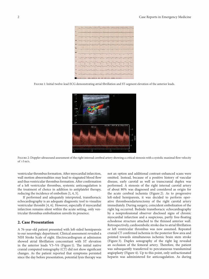
Figure 1: Initial twelve-lead ECG demonstrating atrial fibrillation and ST-segment elevation of the anterior leads.
−3
−3
−2
−2
−1
−1
0
0
(m/s)
1
2
3
4
0
Figure 2: Doppler ultrasound assessment of the right internal cerebral artery showing a critical stenosis with a systolic maximal flow velocityof >3m/s.
ventricular thrombus formation. Aftermyocardial infarction,wall motion abnormalities may lead to stagnated blood flowand thus ventricular thrombus formation. After confirmationof a left ventricular thrombus, systemic anticoagulation isthe treatment of choice in addition to antiplatelet therapy,reducing the incidence of embolism [1, 4, 5].
If performed and adequately interpreted, transthoracicechocardiography is an adequate diagnostic tool to visualizeventricular thrombi [4, 6]. However, especially if myocardialinfarction remains silent within the acute setting, only ven-tricular thrombus embolization unveils its presence.
2. Case Presentation
A 76-year-old patient presented with left-sided hemiparesisto our neurologic department. Clinical assessment revealed aNIH Stroke Scale of eight. Electrocardiogram at admissionshowed atrial fibrillation concomitant with ST elevationin the anterior leads V3–V6 (Figure 1). The initial nativecranial computed tomography (CT) did not show significantchanges. As the patient reported that symptoms persistedsince the day before presentation, potential lysis therapy was
not an option and additional contrast-enhanced scans wereomitted. Instead, because of a positive history of vasculardisease, early carotid as well as transcranial duplex wasperformed. A stenosis of the right internal carotid arteryof about 80% was diagnosed and considered as origin forthe acute cerebral ischemia (Figure 2). As to progressiveleft-sided hemiparesis, it was decided to perform oper-ative thromboendarterectomy of the right carotid arteryimmediately. During surgery, coincident embolization of theright leg occurred. Bedside transthoracic echocardiographyby a nonprofessional observer disclosed signs of chronicmyocardial infarction and a suspicious, partly free-floatingechodense structure attached to the thinned anterior wall.Retrospectively, cardioembolic stroke due to atrial fibrillationor left ventricular thrombus was now assumed. Repeatedcranial CT confirmed ischemia in the posterior flow area andpointed towards simultaneous ischemic brain stem stroke(Figure 3). Duplex sonography of the right leg revealedan occlusion of the femoral artery. Therefore, the patientwas subsequently transferred to percutaneous transluminalangioplasty (Figure 4). Up to this point, only unfractionatedheparin was administered for anticoagulation. As during

Case Reports in Emergency Medicine 3
Figure 3: Contrast-enhanced cranial computed tomographic scanshowing an insult of the right posterior region with hypodensityof the parafalcine parenchyma as well as loss of grey/white matterdifferentiation (black arrow).
the interventional procedure perfusion of the leg remainedimpaired, local fibrinolysis was applied as bail-out. Dueto persisting insufficient perfusion, operative popliteopedalbypass surgery had to be performed finally. In the intensivecare unit, an experienced cardiologist repeated postinter-ventional transthoracic echocardiography. Imaging showeda severely impaired left ventricular function accompaniedby a discontinuity of the normal myocardial texture of theapex together with an echodense, partly floating structuremerely attached by a thin bridge, not completely sealingthe assumed myocardial defect anymore. Simultaneously, aprogression of the pericardial effusion had occurred. Trans-esophageal echocardiography confirmed transthoracic suspi-cion (Figure 5). Imminentmyocardial rupture was feared andurgent cardiac surgery was planned, accepting an elevatedrisk of secondary cerebral bleeding. Preoperative coronarycatheterization showed a complete occlusion of the proxi-mal left anterior descending artery (Figure 6). Cardiac CTimaging showed a thrombus with exophytic componentsinto the left ventricle adherent to a postischemic anterioraneurysm with extremely thinned myocardium (Figure 7).Surgery validated a perforated left ventricle partly covered bya thrombus. An extirpation of the thrombus and coronarybypass graft were performed (Figure 8). The apical aneurysmwas resected. Endomyocardial biopsies revealed thromboticmaterial along with fibrosis.
In the following days, the perfusion of the right legremained critically impaired despite reoperation. Ultimately,an amputation of the right lower limb got necessary. A hemi-paresis persisted. Transthoracic echocardiographic follow-upbefore discharge revealed that the patients’ left ventricularejection fraction had improved without any more thrombusformation.
3. Discussion
Often, clinical signs of myocardial infarction are unspecific,particularly in females, elderly, diabetics, and patients withdementia or suffering from chronic renal disease [1, 7]. As aconsequence, patients may present in a subacute setting withclinical signs of complications as in the presented case.
30∘ RAO
Figure 4: Percutaneous transluminal angiography demonstratingthe occlusion of the right popliteal artery (black arrow).
Figure 5: Transesophageal echocardiographic assessment of the leftventricular thrombus in the two-chamber view. The thrombus ismarked with a white arrow.
45∘ LAO
20∘ cranial
Figure 6: Coronary angiography revealing a complete occlusion ofthe left anterior descending coronary artery marked with the blackarrow.

4 Case Reports in Emergency Medicine
Figure 7: Contrast enhanced ECG-gated chest computed tomo-graphic scan showing loosening of the myocardial anteroapical wallas well as thrombus formation (black arrow).
Figure 8: Intraoperative situs showing an ischemic myocardialperforation of the left ventricle.
Particularly, myocardial infarction of the anterior wallcan lead to thrombus formation because of apical aneurysmand, thus, stagnant blood flow in the apex. In a multicentertrial on patients after acute myocardial infarction who wereconsidered in low tomedium risk for left ventricular thrombi,in 5.1%, a thrombus could be diagnosed within predischargeechocardiogram. In anteriormyocardial infarction, there waseven an incidence of 11.5% [8]. If the thrombus is free-floating within the left ventricle, there is a considerable risk ofarterial embolism. In fact, the risk for cardioembolic eventsis fivefold higher in patients after myocardial infarctionwith detected thrombus formation as compared to patientswithout echocardiographic signs of thrombus formation [5].Even though these cardioembolic events are well known,early bedside transthoracic echocardiogram is, as in ourcase, not yet implemented in clinical practice. Our case,which can be considered as a teaching case, demonstratesthe value of transthoracic echocardiography as a noninvasiveand cost-effective tool for immediate decision-making, atleast within experts’ hands. At initial presentation of thepatient, an ECG was performed where atrial fibrillation as apotential cause for thromboembolism and ST elevation of the
anterior leads as a sign formyocardial ischemia ormyocardialaneurysm were detected. Because of structural failures in acommon setting without central interdisciplinary emergencydepartment but in case of leading neurological symptomsdirect admission to the stroke unit, neurological work-upwas overweighed and the ECG was not given sufficientattention. Even though it has been shown that particularlyin patients with intracerebral hemorrhage but also withischemic stroke ECG changes such as ST-segment elevationcan be present, however, an echocardiography directly atadmission or prior to surgery performed by an experiencedphysicianmay have led tomuch earlier diagnosis of imminentmyocardial rupture, preventing unnecessary surgery of theinternal carotid artery and further systemic embolization[9]. Most dramatically, orientating echocardiography by anoncardiologist evenwas performedwithin the clinical work-up; however, the information gathered was misinterpreted ornot correctly weighted, particularly when local fibrinolysiswas applied in the later course. Even though anticoagulationis the adequate treatment for prevention of thromboembolicevents when left ventricular thrombus has been detected,thrombolytic therapy should not be administered [1, 4]. In thepresented case, inadequate application of fibrinolytic agentsresulted in an almost fatal rupture of the myocardium.
Nonetheless, it would be inappropriate to blame rescuefibrinolytic therapy initiating this sort of medical night-mare. By contrast, the presented case initiated internaldiscussions and review of procedural shortcomings in theaforementioned common patient population, thereby help-ing us to improve processes in our hospital. We learnedthat echocardiography has to be performed early in theclinical course in patients with seemingly unrelated clinicalsymptoms and integrated early ECG and echocardiographyassessment within our stroke unit protocols. Even in acuteand urgent cases, where a delay of operative treatment maypotentially lead to more neurological damage, at least a shortcardiologic consultation should be prompted whenever thereare hints for a cardioembolic origin. Otherwise, potentiallylife-threatening illnesses may be missed.
Conflict of Interests
The authors declare that there is no conflict of interestsregarding the publication of this paper.
References
[1] M. Roffi, C. Patrono, J.-P. Collet et al., “2015 ESC Guidelinesfor the management of acute coronary syndromes in patientspresenting without persistent ST-segment elevation: task forcefor the management of acute coronary syndromes in patientspresentingwithout persistent ST-segment elevation of the Euro-pean Society of Cardiology (ESC),” EuropeanHeart Journal, vol.37, no. 3, pp. 267–315, 2015.
[2] U. Keil, “The Worldwide WHO MONICA Project: results andperspectives,”Gesundheitswesen, vol. 67, supplement 1, pp. S38–S45, 2005.
[3] R. C. Becker, J. M. Gore, C. Lambrew et al., “A compositeview of cardiac rupture in the United States National Registry
Case Reports in Emergency Medicine 5
of Myocardial Infarction,” Journal of the American College ofCardiology, vol. 27, no. 6, pp. 1321–1326, 1996.
[4] L. L. Cregler, “Antithrombotic therapy in left ventricular throm-bosis and systemic embolism,”AmericanHeart Journal, vol. 123,no. 4, part 2, pp. 1110–1114, 1992.
[5] P. T.Vaitkus andE. S. Barnathan, “Embolic potential, preventionand management of mural thrombus complicating anteriormyocardial infarction: a meta-analysis,” Journal of the AmericanCollege of Cardiology, vol. 22, no. 4, pp. 1004–1009, 1993.
[6] J. R. Stratton, G. W. Lighty Jr., A. S. Pearlman, and J. L. Ritchie,“Detection of left ventricular thrombus by two-dimensionalechocardiography: sensitivity, specificity, and causes of uncer-tainty,” Circulation, vol. 66, no. 1, pp. 156–166, 1982.
[7] M. R. Gimenez, M. Reiter, R. Twerenbold et al., “Sex-specificchest pain characteristics in the early diagnosis of acutemyocar-dial infarction,” JAMA InternalMedicine, vol. 174, no. 2, pp. 241–249, 2014.
[8] F. Chiarella, E. Santoro, S. Domenicucci, A. Maggioni, andC. Vecchio, “Predischarge two-dimensional echocardiographicevaluation of left ventricular thrombosis after acute myocardialinfarction in theGISSI-3 study,”American Journal of Cardiology,vol. 81, no. 7, pp. 822–827, 1998.
[9] G. Khechinashvili and K. Asplund, “Electrocardiographicchanges in patients with acute stroke: a systematic review,”Cerebrovascular Diseases, vol. 14, no. 2, pp. 67–76, 2002.

Submit your manuscripts athttp://www.hindawi.com
Stem CellsInternational
Hindawi Publishing Corporationhttp://www.hindawi.com Volume 2014
Hindawi Publishing Corporationhttp://www.hindawi.com Volume 2014
MEDIATORSINFLAMMATION
of
Hindawi Publishing Corporationhttp://www.hindawi.com Volume 2014
Behavioural Neurology
EndocrinologyInternational Journal of
Hindawi Publishing Corporationhttp://www.hindawi.com Volume 2014
Hindawi Publishing Corporationhttp://www.hindawi.com Volume 2014
Disease Markers
Hindawi Publishing Corporationhttp://www.hindawi.com Volume 2014
BioMed Research International
OncologyJournal of
Hindawi Publishing Corporationhttp://www.hindawi.com Volume 2014
Hindawi Publishing Corporationhttp://www.hindawi.com Volume 2014
Oxidative Medicine and Cellular Longevity
Hindawi Publishing Corporationhttp://www.hindawi.com Volume 2014
PPAR Research
The Scientific World JournalHindawi Publishing Corporation http://www.hindawi.com Volume 2014
Immunology ResearchHindawi Publishing Corporationhttp://www.hindawi.com Volume 2014
Journal of
ObesityJournal of
Hindawi Publishing Corporationhttp://www.hindawi.com Volume 2014
Hindawi Publishing Corporationhttp://www.hindawi.com Volume 2014
Computational and Mathematical Methods in Medicine
OphthalmologyJournal of
Hindawi Publishing Corporationhttp://www.hindawi.com Volume 2014
Diabetes ResearchJournal of
Hindawi Publishing Corporationhttp://www.hindawi.com Volume 2014
Hindawi Publishing Corporationhttp://www.hindawi.com Volume 2014
Research and TreatmentAIDS
Hindawi Publishing Corporationhttp://www.hindawi.com Volume 2014
Gastroenterology Research and Practice
Hindawi Publishing Corporationhttp://www.hindawi.com Volume 2014
Parkinson’s Disease
Evidence-Based Complementary and Alternative Medicine
Volume 2014Hindawi Publishing Corporationhttp://www.hindawi.com