Embed Size (px)
Citation preview

Hindawi Publishing CorporationCase Reports in DentistryVolume 2012, Article ID 390874, 4 pagesdoi:10.1155/2012/390874
Case Report
Hemisection: A Window of Hope For Freezing Tooth
Usha Radke,1 Rajesh Kubde,2 and Aditi Paldiwal1
1 Department of Prosthodontics, VSPM’s Dental College & Research Centre, Nagpur 440019, India2 Department of Conservative & Endodontics, VSPM’s Dental College & Research Centre, Nagpur 440019, India
Correspondence should be addressed to Usha Radke, [email protected]
Received 10 June 2012; Accepted 18 July 2012
Academic Editors: M. Feichtinger and M. J. Wahl
Copyright © 2012 Usha Radke et al. This is an open access article distributed under the Creative Commons Attribution License,which permits unrestricted use, distribution, and reproduction in any medium, provided the original work is properly cited.
Advances in dentistry, as well as the increased desire of patients to maintain their dentition, have led to treatment of teeth that oncewould have been removed. Mandibular first molars are the most commonly extracted teeth due to dental caries and periodontaldisease. These teeth are the major standpoint for occlusion, and also have a wide pericemental area. Hence, any defect in the rooteither mesial or distal, extraction is the most common treatment planned. Under specific conditions, only the diseased part of thetooth can be extracted after an endodontic treatment. A modified fixed partial denture design is fabricated to splint the remainingportion of the tooth to adjacent teeth. This procedure though daunting can be easily achieved and maintained successfully.
1. Introduction
A terminal abutment molar with extensive decay may beunsuitable for restoration. In such cases, the treatmentoptions are limited and may include a removable partialdenture or a dental implant to replace the missing tooth [1].Alternatively, if the decay is limited to one root, a hemisectionprocedure may be possible. Periodontal, prosthodontics, andendodontic assessment for appropriate selection of cases isimportant. From a periodontal perspective, this procedureis indicated if there is severe bone loss limited to one rootor involvement of class III furcations that could producea stable root after hemisection. This procedure is alsoappropriate if the patient is unable to perform appropriateoral hygiene in the area. Extensive exposure of the rootsbecause of dehiscence is another indication for excisionof one root. From a restorative standpoint, treatment byhemisection is indicated for failure of an abutment withina fixed prosthesis, provided a portion of the tooth can beretained to act as the abutment for the prosthesis. Otherindications include vertical root fracture confined to a singleroot of a multirooted tooth or any severe destructive processthat is confined to a single root, including caries, externalroot resorption, and trauma. Contraindications include thepresence of a strong abutment tooth adjacent to the proposedhemisection, which could act as an abutment to a prosthesis.The remaining root may be inoperable for the necessary root
canal treatment. Also, fusion or proximity of the roots mayprevent their separation [2].
Hemisection refers to sectioning of a mandibular molarinto two halves followed by removal of the diseased root andits coronal portion [3]. The retained root is endodonticallytreated and the furcations area is made self-cleansable byremoving the lip of root carefully. Since hemisected teeth failby root fractures, it is important to restore them adequatelyby an extracoronal restoration [4]. It is indicated where oneof the root of molar is unsalvageable due to caries, peri-odontitis, or iatrogenic mishaps [5]. It is thus a conservativeoption with acceptable prognosis [6].
2. Case Report
A 35-year-old male patient, reported to the Department ofPeriodontics, with the chief complaint of loose tooth andpain in lower left back tooth region (Figure 1). Pain wasdull aching and intermittent in nature, which aggravated onmastication. On further enquiry, patient did not give anysignificant medical and previous dental history. Extra oralexamination revealed no abnormality.
On intraoral examination, it was found that patienthad fair oral hygiene. On probing lower left mandibularfirst molar, a periodontal pocket of 8–10 mm was foundon buccal and distal surfaces along with grade III furcation
2 Case Reports in Dentistry
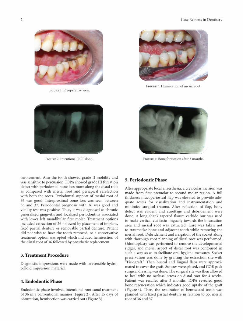
Figure 1: Preoperative view.
Figure 2: Intentional RCT done.
involvement. Also the tooth showed grade II mobility andwas sensitive to percussion. IOPA showed grade III furcationdefect with periodontal bone loss more along the distal rootas compared with mesial root and periapical rarefactionwith both the roots. Periodontal support of mesial root of36 was good. Interproximal bone loss was seen between36 and 37. Periodontal prognosis with 36 was good andvitality test was positive. Thus, it was diagnosed as chronicgeneralised gingivitis and localized periodontitis associatedwith lower left mandibular first molar. Treatment optionsincluded extraction of 36 followed by placement of implant,fixed partial denture or removable partial denture. Patientdid not wish to have the tooth removed, so a conservativetreatment option was opted which included hemisection ofthe distal root of 36 followed by prosthetic replacement.
3. Treatment Procedure
Diagnostic impressions were made with irreversible hydro-colloid impression material.
4. Endodontic Phase
Endodontic phase involved intentional root canal treatmentof 36 in a conventional manner (Figure 2). After 15 days ofobturation, hemisection was carried out (Figure 3).
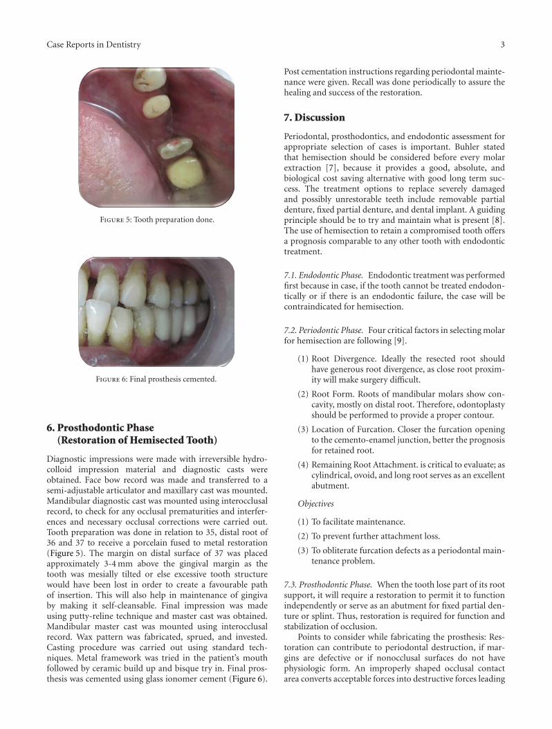
Figure 3: Hemisection of mesial root.
Figure 4: Bone formation after 3 months.
5. Periodontic Phase
After appropriate local anaesthesia, a crevicular incision wasmade from first premolar to second molar region. A fullthickness mucoperiosteal flap was elevated to provide ade-quate access for visualization and instrumentation andminimize surgical trauma. After reflection of flap, bonydefect was evident and curettage and debridement weredone. A long shank tapered fissure carbide bur was usedto make vertical cut facio-lingually towards the bifurcationarea and mesial root was extracted. Care was taken notto traumatize bone and adjacent tooth while removing themesial root. Debridement and irrigation of the socket alongwith thorough root planning of distal root was performed.Odontoplasty was performed to remove the developmentalridges, and mesial aspect of distal root was contoured insuch a way so as to facilitate oral hygiene measures. Socketpreservation was done by grafting the extraction site with“Fisiograft.” Then buccal and lingual flaps were approxi-mated to cover the graft. Sutures were placed, and COE packsurgical dressing was done. The surgical site was then allowedto heal with no occlusal stress on distal root for 4 weeks.Patient was recalled after 3 months. IOPA revealed goodbone regeneration which indicates good uptake of the graft(Figure 4). Then, the restoration of hemisected tooth wasplanned with fixed partial denture in relation to 35, mesialroot of 36 and 37.
Case Reports in Dentistry 3
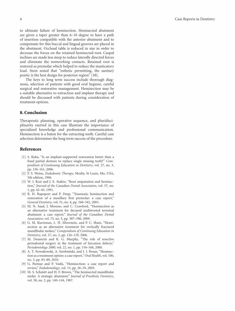
Figure 5: Tooth preparation done.
Figure 6: Final prosthesis cemented.
6. Prosthodontic Phase(Restoration of Hemisected Tooth)
Diagnostic impressions were made with irreversible hydro-colloid impression material and diagnostic casts wereobtained. Face bow record was made and transferred to asemi-adjustable articulator and maxillary cast was mounted.Mandibular diagnostic cast was mounted using interocclusalrecord, to check for any occlusal prematurities and interfer-ences and necessary occlusal corrections were carried out.Tooth preparation was done in relation to 35, distal root of36 and 37 to receive a porcelain fused to metal restoration(Figure 5). The margin on distal surface of 37 was placedapproximately 3-4 mm above the gingival margin as thetooth was mesially tilted or else excessive tooth structurewould have been lost in order to create a favourable pathof insertion. This will also help in maintenance of gingivaby making it self-cleansable. Final impression was madeusing putty-reline technique and master cast was obtained.Mandibular master cast was mounted using interocclusalrecord. Wax pattern was fabricated, sprued, and invested.Casting procedure was carried out using standard tech-niques. Metal framework was tried in the patient’s mouthfollowed by ceramic build up and bisque try in. Final pros-thesis was cemented using glass ionomer cement (Figure 6).
Post cementation instructions regarding periodontal mainte-nance were given. Recall was done periodically to assure thehealing and success of the restoration.
7. Discussion
Periodontal, prosthodontics, and endodontic assessment forappropriate selection of cases is important. Buhler statedthat hemisection should be considered before every molarextraction [7], because it provides a good, absolute, andbiological cost saving alternative with good long term suc-cess. The treatment options to replace severely damagedand possibly unrestorable teeth include removable partialdenture, fixed partial denture, and dental implant. A guidingprinciple should be to try and maintain what is present [8].The use of hemisection to retain a compromised tooth offersa prognosis comparable to any other tooth with endodontictreatment.
7.1. Endodontic Phase. Endodontic treatment was performedfirst because in case, if the tooth cannot be treated endodon-tically or if there is an endodontic failure, the case will becontraindicated for hemisection.
7.2. Periodontic Phase. Four critical factors in selecting molarfor hemisection are following [9].
(1) Root Divergence. Ideally the resected root shouldhave generous root divergence, as close root proxim-ity will make surgery difficult.
(2) Root Form. Roots of mandibular molars show con-cavity, mostly on distal root. Therefore, odontoplastyshould be performed to provide a proper contour.
(3) Location of Furcation. Closer the furcation openingto the cemento-enamel junction, better the prognosisfor retained root.
(4) Remaining Root Attachment. is critical to evaluate; ascylindrical, ovoid, and long root serves as an excellentabutment.
Objectives
(1) To facilitate maintenance.
(2) To prevent further attachment loss.
(3) To obliterate furcation defects as a periodontal main-tenance problem.
7.3. Prosthodontic Phase. When the tooth lose part of its rootsupport, it will require a restoration to permit it to functionindependently or serve as an abutment for fixed partial den-ture or splint. Thus, restoration is required for function andstabilization of occlusion.
Points to consider while fabricating the prosthesis: Res-toration can contribute to periodontal destruction, if mar-gins are defective or if nonocclusal surfaces do not havephysiologic form. An improperly shaped occlusal contactarea converts acceptable forces into destructive forces leading
4 Case Reports in Dentistry
to ultimate failure of hemisection. Hemisected abutmentare given a taper greater than 6–10 degree to have a pathof insertion compatible with the anterior abutment and tocompensate for this buccal and lingual grooves are placed inthe abutment. Occlusal table is reduced in size in order todecrease the forces on the retained hemisected root. Cuspalinclines are made less steep to reduce laterally directed forcesand eliminate the nonworking contacts. Retained root isrestored as premolar which helped to reduce the masticatoryload. Stein noted that “esthetic permitting, the sanitarypontic is the best design for posterior region” [10].
The keys to long term success include thorough diag-nosis, selection of patients with good oral hygiene, carefulsurgical and restorative management. Hemisection may bea suitable alternative to extraction and implant therapy andshould be discussed with patients during consideration oftreatment options.
8. Conclusion
Therapeutic planning, operative sequence, and pluridisci-plinarity exerted in this case illustrate the importance ofspecialized knowledge and professional communication.Hemisection is a baton for the extracting teeth. Careful caseselection determines the long term success of the procedure.
References
[1] S. Koka, “Is an implant-supported restoration better than afixed partial denture to replace single missing teeth?” Com-pendium of Continuing Education in Dentistry, vol. 27, no. 3,pp. 156–161, 2006.
[2] F. S. Weine, Endodontic Therapy, Mosby, St Louis, Mo, USA,5th edition, 1996.
[3] W. J. Kost and J. E. Stakiw, “Root amputation and hemisec-tion,” Journal of the Canadian Dental Association, vol. 57, no.1, pp. 42–45, 1991.
[4] R. H. Rapoport and P. Deep, “Traumatic hemisection andrestoration of a maxillary first premolar: a case report,”General Dentistry, vol. 51, no. 4, pp. 340–342, 2003.
[5] M. N. Saad, J. Moreno, and C. Crawford, “Hemisection asan alternative treatment for decayed multirooted terminalabutment: a case report,” Journal of the Canadian DentalAssociation, vol. 75, no. 5, pp. 387–390, 2009.
[6] G. M. Kurtzman, L. H. Silverstein, and P. C. Shatz, “Hemi-section as an alternative treatment for vertically fracturedmandibular molars,” Compendium of Continuing Education inDentistry, vol. 27, no. 2, pp. 126–129, 2006.
[7] M. Desanctis and K. G. Murphy, “The role of resectiveperiodontal surgery in the treatment of furcation defects,”Periodontology 2000, vol. 22, no. 1, pp. 154–168, 2000.
[8] A. T. Nowakowski, A. Serebnitski, and I. J. Pesun, “Hemisec-tion as a treatment option: a case report,” Oral Health, vol. 100,no. 3, pp. 83–89, 2010.
[9] G. Parmar and P. Vashi, “Hemisection: a case report andreview,” Endodontology, vol. 15, pp. 26–29, 2003.
[10] M. S. Schmitt and H. F. Brown, “The hemisected mandibularmolar. A strategic abutment,” Journal of Prosthetic Dentistry,vol. 58, no. 2, pp. 140–144, 1987.

Submit your manuscripts athttp://www.hindawi.com
Hindawi Publishing Corporationhttp://www.hindawi.com Volume 2014
Oral OncologyJournal of
DentistryInternational Journal of
Hindawi Publishing Corporationhttp://www.hindawi.com Volume 2014
Hindawi Publishing Corporationhttp://www.hindawi.com Volume 2014
International Journal of
Biomaterials
Hindawi Publishing Corporationhttp://www.hindawi.com Volume 2014
BioMed Research International
Hindawi Publishing Corporationhttp://www.hindawi.com Volume 2014
Case Reports in Dentistry
Hindawi Publishing Corporationhttp://www.hindawi.com Volume 2014
Oral ImplantsJournal of
Hindawi Publishing Corporationhttp://www.hindawi.com Volume 2014
Anesthesiology Research and Practice
Hindawi Publishing Corporationhttp://www.hindawi.com Volume 2014
Radiology Research and Practice
Environmental and Public Health
Journal of
Hindawi Publishing Corporationhttp://www.hindawi.com Volume 2014
The Scientific World JournalHindawi Publishing Corporation http://www.hindawi.com Volume 2014
Hindawi Publishing Corporationhttp://www.hindawi.com Volume 2014
Dental SurgeryJournal of
Drug DeliveryJournal of
Hindawi Publishing Corporationhttp://www.hindawi.com Volume 2014
Hindawi Publishing Corporationhttp://www.hindawi.com Volume 2014
Oral DiseasesJournal of
Hindawi Publishing Corporationhttp://www.hindawi.com Volume 2014
Computational and Mathematical Methods in Medicine
ScientificaHindawi Publishing Corporationhttp://www.hindawi.com Volume 2014
PainResearch and TreatmentHindawi Publishing Corporationhttp://www.hindawi.com Volume 2014
Preventive MedicineAdvances in
Hindawi Publishing Corporationhttp://www.hindawi.com Volume 2014
EndocrinologyInternational Journal of
Hindawi Publishing Corporationhttp://www.hindawi.com Volume 2014
Hindawi Publishing Corporationhttp://www.hindawi.com Volume 2014
OrthopedicsAdvances in