Embed Size (px)
Citation preview
Case ReportEpigenetic Alterations in a Gastric Leiomyoma
M. T. Branham,1 M. Pellicer,2 E. Campoy,1 M. Palma,3 A. Correa,4 and M. Roqué1
1 Institute of Histology and Embryology (IHEM-CCT-CONICET) and School of Medical Sciences,National University of Cuyo, M5502JMA Mendoza, Argentina2Pathology Laboratory, J5402EQC San Juan, Argentina3Surgery Unit, San Martin Hospital, E3100 Entre Rıos, Argentina4Surgery Unit, Espanol Hospital, M5501 Mendoza, Argentina
Correspondence should be addressed to M. T. Branham; [email protected]
Received 29 September 2014; Revised 17 November 2014; Accepted 17 November 2014; Published 4 December 2014
Academic Editor: Haruhiko Sugimura
Copyright © 2014 M. T. Branham et al.This is an open access article distributed under the Creative Commons Attribution License,which permits unrestricted use, distribution, and reproduction in any medium, provided the original work is properly cited.
Leiomyomas constitute 2.5% of all resected neoplasms of the stomach. They are usually asymptomatic, but may present mucosalulceration. Aberrant DNAmethylation is a well-defined epigenetic change in human neoplasms; however, gene-acquired methyla-tion may not necessarily be related with a malignant phenotype. In this report we analyzed in a gastric leiomyoma, the methylationstatus of 84CpGI in tumor suppressor and DNA repair genes. We analyzed the tumor center (TC) and tumor periphery (TP)separately. We found aberrant methylation in 2/84CpGI in the TC portion, that is, MLH1 and MSH3, and 5/84CpGI in the TP,that is, MLH1, MSH3, APC, MSH6, and MGMT. The gene with the highest methylation percentage in the TC and TP was MLH1.Given thatMLH1methylation has been associated withmicrosatellite instability, we analyzed the status of the microsatellite Bat-26.We found that neither the TC nor the TP presented instability. The methylation ofMLH1,MGMT, and APC has been described inGISTs, but to the best of our knowledge this is the first time that the methylation of these genes has been associated with gastricleiomyoma. Further research should be conducted to identify reliable molecular markers that could differentiate between GISTsand gastric leiomyomas.
1. Introduction
Gastric leiomyomas account for 2.5% of gastric neoplasms.Although most of them are asymptomatic, patients maypresent upper gastrointestinal hemorrhage [1, 2]. Endoscopi-cally, gastric leiomyomas appear as a large submucosal lesion,and generally endoscopic biopsies are not deep enough tobe of any diagnostic value [3]. Pathologically, most of thesetumors are composed of spindle cells and display smoothmuscle differentiation. Leiomyomas are defined as beingdesmin and actin positive and c-Kit (or CD117) negativetumors [4].
Tumorigenesis is the result of a multistep process charac-terized by the accumulation of genetic and epigenetic alter-ations leading to uncontrolled growth. Among epigeneticmodifications, the most studied event in human neoplasmsis the deregulation of methylation of DNA, giving rise to
widespread changes in the methylome patterns during tumorprogression [5]. The epigenome of tumors is character-ized by global DNA hypomethylation and by gene-specifichypermethylation. Gene silencing by CpG islands (CpGI)hypermethylation in gene promoters can modulate pathwaysthat control the basic function of the cell by acting directly ontumor suppressor genes and caretaker genes and indirectly ononcogenes through their regulators [5].
Gene expression profile studies have demonstrated thatsome genes are hypermethylated in gastric GISTs (gastrointestinal stromal tumors) [6–8], but, to our knowledge,there is no information of the methylation profile of gastricleiomyomas.
The objective of this study was to analyze by methylspecific-multiplex ligation probe amplification (MS-MLPA)the methylation status of tumor suppressor and DNA repairgenes in a gastric leiomyoma.
Hindawi Publishing CorporationCase Reports in Gastrointestinal MedicineVolume 2014, Article ID 371638, 5 pageshttp://dx.doi.org/10.1155/2014/371638
2 Case Reports in Gastrointestinal Medicine
(a)
(b) (c)
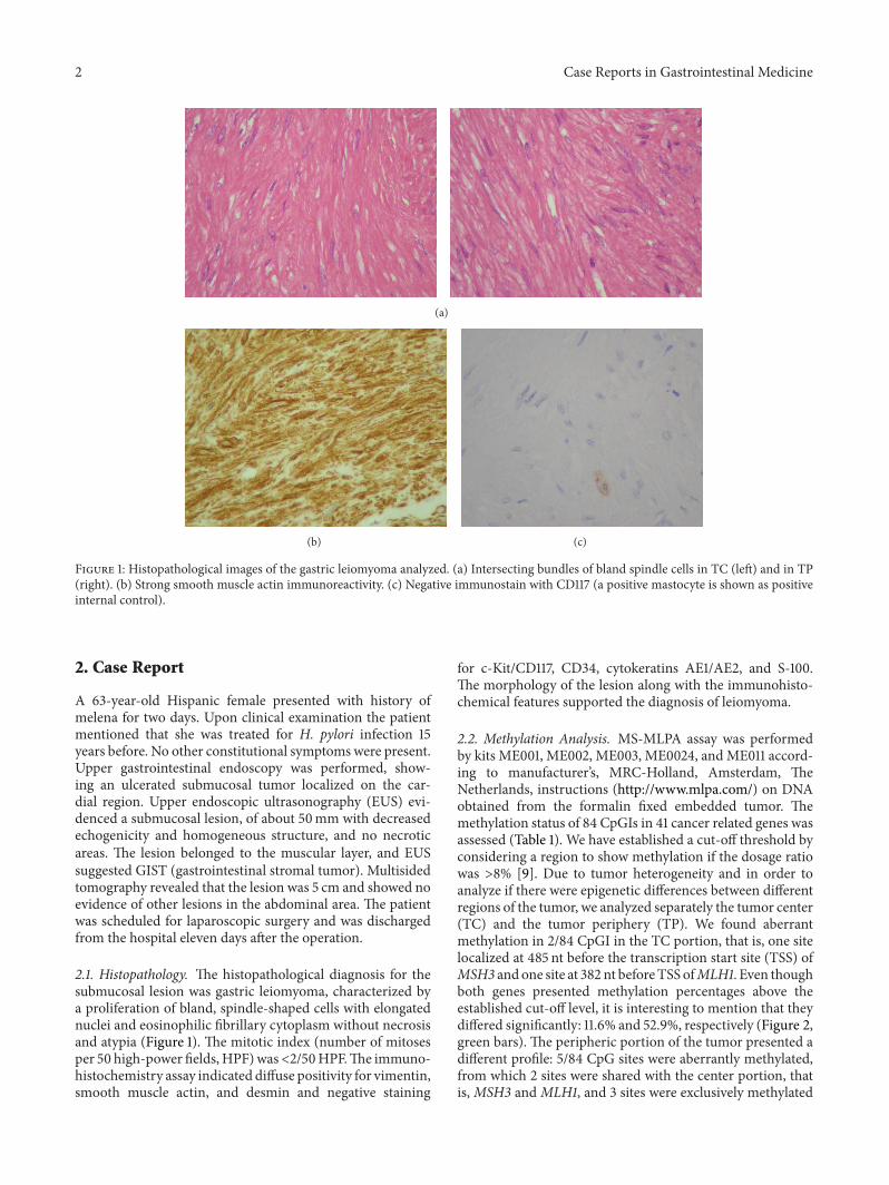
Figure 1: Histopathological images of the gastric leiomyoma analyzed. (a) Intersecting bundles of bland spindle cells in TC (left) and in TP(right). (b) Strong smooth muscle actin immunoreactivity. (c) Negative immunostain with CD117 (a positive mastocyte is shown as positiveinternal control).
2. Case Report
A 63-year-old Hispanic female presented with history ofmelena for two days. Upon clinical examination the patientmentioned that she was treated for H. pylori infection 15years before. No other constitutional symptoms were present.Upper gastrointestinal endoscopy was performed, show-ing an ulcerated submucosal tumor localized on the car-dial region. Upper endoscopic ultrasonography (EUS) evi-denced a submucosal lesion, of about 50mm with decreasedechogenicity and homogeneous structure, and no necroticareas. The lesion belonged to the muscular layer, and EUSsuggested GIST (gastrointestinal stromal tumor). Multisidedtomography revealed that the lesion was 5 cm and showed noevidence of other lesions in the abdominal area. The patientwas scheduled for laparoscopic surgery and was dischargedfrom the hospital eleven days after the operation.
2.1. Histopathology. The histopathological diagnosis for thesubmucosal lesion was gastric leiomyoma, characterized bya proliferation of bland, spindle-shaped cells with elongatednuclei and eosinophilic fibrillary cytoplasm without necrosisand atypia (Figure 1). The mitotic index (number of mitosesper 50 high-power fields, HPF)was<2/50HPF.The immuno-histochemistry assay indicated diffuse positivity for vimentin,smooth muscle actin, and desmin and negative staining
for c-Kit/CD117, CD34, cytokeratins AE1/AE2, and S-100.The morphology of the lesion along with the immunohisto-chemical features supported the diagnosis of leiomyoma.
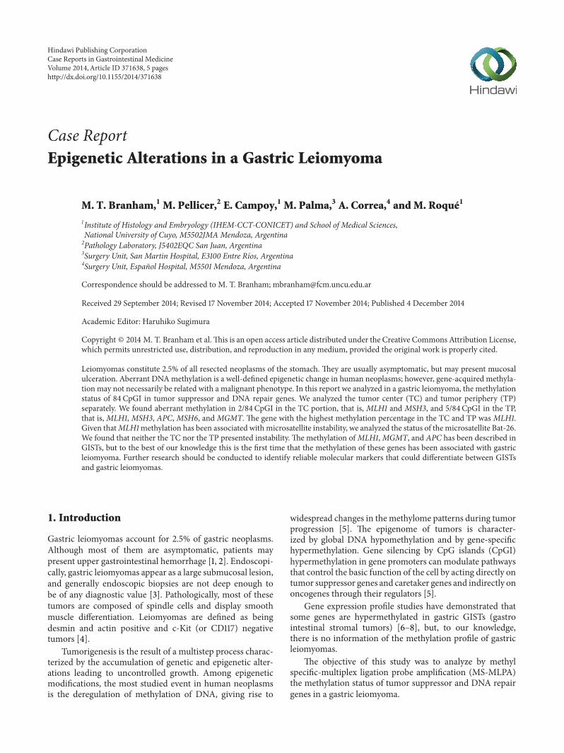
2.2. Methylation Analysis. MS-MLPA assay was performedby kits ME001, ME002, ME003, ME0024, and ME011 accord-ing to manufacturer’s, MRC-Holland, Amsterdam, TheNetherlands, instructions (http://www.mlpa.com/) on DNAobtained from the formalin fixed embedded tumor. Themethylation status of 84 CpGIs in 41 cancer related genes wasassessed (Table 1). We have established a cut-off threshold byconsidering a region to show methylation if the dosage ratiowas >8% [9]. Due to tumor heterogeneity and in order toanalyze if there were epigenetic differences between differentregions of the tumor, we analyzed separately the tumor center(TC) and the tumor periphery (TP). We found aberrantmethylation in 2/84 CpGI in the TC portion, that is, one sitelocalized at 485 nt before the transcription start site (TSS) ofMSH3 andone site at 382 nt beforeTSS ofMLH1. Even thoughboth genes presented methylation percentages above theestablished cut-off level, it is interesting to mention that theydiffered significantly: 11.6% and 52.9%, respectively (Figure 2,green bars). The peripheric portion of the tumor presented adifferent profile: 5/84 CpG sites were aberrantly methylated,from which 2 sites were shared with the center portion, thatis,MSH3 andMLH1, and 3 sites were exclusively methylated

Case Reports in Gastrointestinal Medicine 3
Table 1: List of genes analyzed in the gastric leiomyoma. Genenames and gene symbols are according to the HGNC database.
Gene symbol Gene nameAPC Adenomatous polyposis coliATM Ataxia telangiectasia mutatedBCL2 B-cell CLL/lymphoma 2BNIP3 BCL2/adenovirus E1B 19 kDa interacting protein 3BRCA1 Breast cancer 1BRCA2 Breast cancer 2
CACNA1A Calcium channel, voltage-dependent, P/Q type,alpha 1A subunit
CACNA1G Calcium channel, voltage-dependent, P/Q type,alpha 1B subunit
CADM1 Cell adhesion molecule 1CCND2 Cyclin D2CD44 CD44 moleculeCDH13 Cadherin 13CDKN1B Cyclin-dependent kinase inhibitor 1BCDKN2A Cyclin-dependent kinase inhibitor 2ACHFR Checkpoint with forkhead ring finger domainsCREM cAMP responsive element modulatorDAPK1 Death-associated protein kinase 1DLC1 DLC1 Rho GTPase activating proteinESR1 Estrogen receptor 1FHIT Fragile histidine triadGATA5 GATA binding protein 5GSTP1 Glutathione S-transferase pi 1H2AFX H2A histone family, member XHIC1 Hypermethylated in cancer 1HLTF Helicase-like transcription factor
ID4 Inhibitor of DNA binding 4, dominant negativehelix-loop-helix protein
MGMT O-6-Methylguanine-DNA methyltransferaseMLH1 MutL homolog 1MLH3 MutL homolog 3MSH2 MutS homolog 2MSH3 MutS homolog 3MSH6 MutS homolog 6PAH Phenylalanine hydroxylasePAX5 Paired box 5PAX6 Paired box 6PMS2 PMS2 postmeiotic segregation increased 2PRDM2 PR domain containing 2, with ZNF domainPTCH1 Patched 1PTEN Phosphatase and tensin homologPYCARD PYD and CARD domain containingRARB Retinoic acid receptor betaRASSF1 Ras association domain family member 1ARB1 Retinoblastoma 1
Table 1: Continued.
Gene symbol Gene nameRUNX3 Runt-related transcription factor 3SCGB3A1 Secretoglobin, family 3A, member 1SFRP4 Secreted frizzled-related protein 4SFRP5 Secreted frizzled-related protein 5STK11 Serine/threonine kinase 11TGIF1 TGFB-induced factor homeobox 1THBS1 Thrombospondin 1TIMP3 TIMP metallopeptidase inhibitor 3TP53 Tumor protein p53TP73 Tumor protein p73TWIST1 Twist family bHLH transcription factor 1VHL Von Hippel-Lindau tumor suppressorWT1 Wilms tumor 1
0102030405060708090100
MLH1 APC MSH6 MSH3 MGMT
TCTP
(%)
Figure 2: Genes methylated in a gastric leiomyoma. Grey barsrepresent the genes methylated in the tumor periphery and greenbars represent the genes methylated in the tumor center.
in the tumor periphery, that is, MSH6, MGMT, and APC(Table 2). As a control, MS-MLPA assay was performedin normal tissue (i.e., leucocytes of healthy patients); wedetermined that there was no aberrant methylation in theregions analyzed. The percentages of methylation in theTP were 10.8%, 77%, 14.3%, 9.5%, and 27.7%, respectively(Figure 2, grey bars). Even though the methylated genesdiffered between the TC and the TP, it is interesting tomention that MLH1 was shared by both and presented thehighest percentage of methylation (52.9% and 77%, resp.).To analyze whether methylation affected gene expression,we performed qRT-PCR assays on 2 cell lines (MDA-MB231and MCF-7) which presented different percentages of APCpromotermethylation (0%and 52%, resp.); we confirmed thatthe methylation of APC on the CpG site −21 nt before TSSreduces gene expression (data not shown). The methylationof MLH1 at 382 nt before TSS has been previously shown toprovoke downregulation of gene expression [10].
Given that MLH1 methylation is associated with mic-rosatellite instability (MSI) in sporadic endometrial and
4 Case Reports in Gastrointestinal Medicine
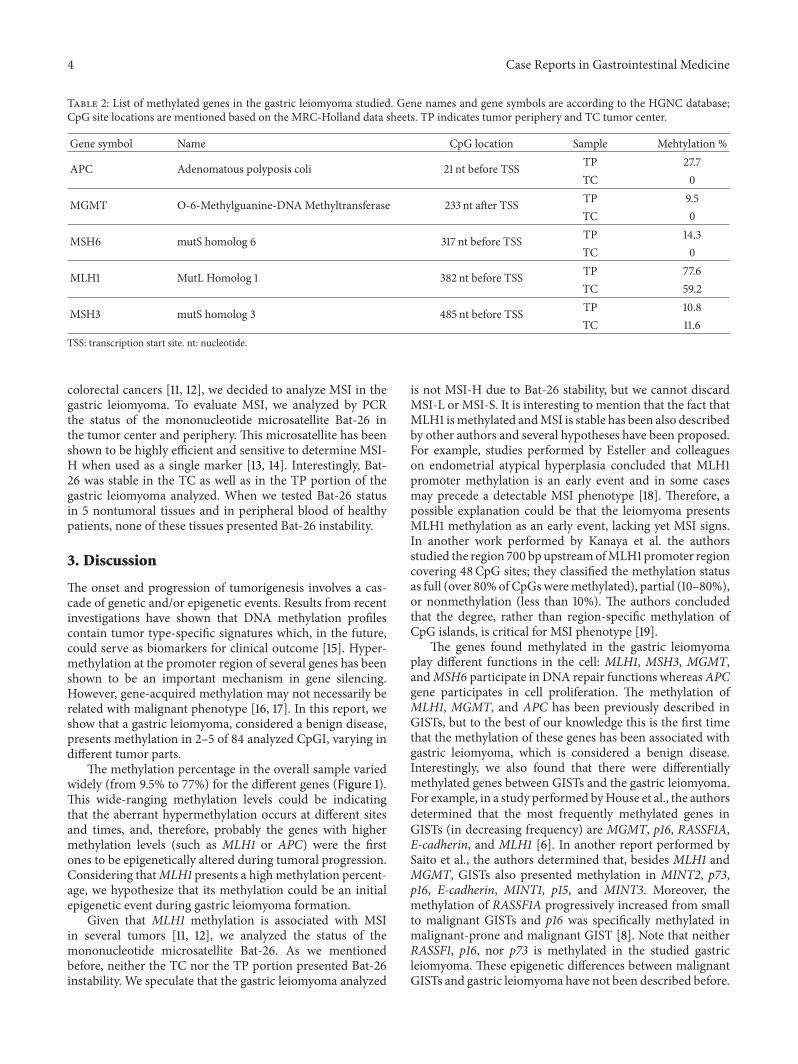
Table 2: List of methylated genes in the gastric leiomyoma studied. Gene names and gene symbols are according to the HGNC database;CpG site locations are mentioned based on the MRC-Holland data sheets. TP indicates tumor periphery and TC tumor center.
Gene symbol Name CpG location Sample Mehtylation %
APC Adenomatous polyposis coli 21 nt before TSS TP 27.7TC 0
MGMT O-6-Methylguanine-DNAMethyltransferase 233 nt after TSS TP 9.5TC 0
MSH6 mutS homolog 6 317 nt before TSS TP 14.3TC 0
MLH1 MutL Homolog 1 382 nt before TSS TP 77.6TC 59.2
MSH3 mutS homolog 3 485 nt before TSS TP 10.8TC 11.6
TSS: transcription start site. nt: nucleotide.
colorectal cancers [11, 12], we decided to analyze MSI in thegastric leiomyoma. To evaluate MSI, we analyzed by PCRthe status of the mononucleotide microsatellite Bat-26 inthe tumor center and periphery. This microsatellite has beenshown to be highly efficient and sensitive to determine MSI-H when used as a single marker [13, 14]. Interestingly, Bat-26 was stable in the TC as well as in the TP portion of thegastric leiomyoma analyzed. When we tested Bat-26 statusin 5 nontumoral tissues and in peripheral blood of healthypatients, none of these tissues presented Bat-26 instability.
3. Discussion
The onset and progression of tumorigenesis involves a cas-cade of genetic and/or epigenetic events. Results from recentinvestigations have shown that DNA methylation profilescontain tumor type-specific signatures which, in the future,could serve as biomarkers for clinical outcome [15]. Hyper-methylation at the promoter region of several genes has beenshown to be an important mechanism in gene silencing.However, gene-acquired methylation may not necessarily berelated with malignant phenotype [16, 17]. In this report, weshow that a gastric leiomyoma, considered a benign disease,presents methylation in 2–5 of 84 analyzed CpGI, varying indifferent tumor parts.
The methylation percentage in the overall sample variedwidely (from 9.5% to 77%) for the different genes (Figure 1).This wide-ranging methylation levels could be indicatingthat the aberrant hypermethylation occurs at different sitesand times, and, therefore, probably the genes with highermethylation levels (such as MLH1 or APC) were the firstones to be epigenetically altered during tumoral progression.Considering thatMLH1 presents a highmethylation percent-age, we hypothesize that its methylation could be an initialepigenetic event during gastric leiomyoma formation.
Given that MLH1 methylation is associated with MSIin several tumors [11, 12], we analyzed the status of themononucleotide microsatellite Bat-26. As we mentionedbefore, neither the TC nor the TP portion presented Bat-26instability. We speculate that the gastric leiomyoma analyzed
is not MSI-H due to Bat-26 stability, but we cannot discardMSI-L or MSI-S. It is interesting to mention that the fact thatMLH1 ismethylated andMSI is stable has been also describedby other authors and several hypotheses have been proposed.For example, studies performed by Esteller and colleagueson endometrial atypical hyperplasia concluded that MLH1promoter methylation is an early event and in some casesmay precede a detectable MSI phenotype [18]. Therefore, apossible explanation could be that the leiomyoma presentsMLH1 methylation as an early event, lacking yet MSI signs.In another work performed by Kanaya et al. the authorsstudied the region 700 bp upstreamofMLH1 promoter regioncovering 48CpG sites; they classified the methylation statusas full (over 80% of CpGsweremethylated), partial (10–80%),or nonmethylation (less than 10%). The authors concludedthat the degree, rather than region-specific methylation ofCpG islands, is critical for MSI phenotype [19].
The genes found methylated in the gastric leiomyomaplay different functions in the cell: MLH1, MSH3, MGMT,andMSH6 participate in DNA repair functions whereasAPCgene participates in cell proliferation. The methylation ofMLH1, MGMT, and APC has been previously described inGISTs, but to the best of our knowledge this is the first timethat the methylation of these genes has been associated withgastric leiomyoma, which is considered a benign disease.Interestingly, we also found that there were differentiallymethylated genes between GISTs and the gastric leiomyoma.For example, in a study performed byHouse et al., the authorsdetermined that the most frequently methylated genes inGISTs (in decreasing frequency) are MGMT, p16, RASSF1A,E-cadherin, and MLH1 [6]. In another report performed bySaito et al., the authors determined that, besides MLH1 andMGMT, GISTs also presented methylation in MINT2, p73,p16, E-cadherin, MINT1, p15, and MINT3. Moreover, themethylation of RASSF1A progressively increased from smallto malignant GISTs and p16 was specifically methylated inmalignant-prone and malignant GIST [8]. Note that neitherRASSF1, p16, nor p73 is methylated in the studied gastricleiomyoma. These epigenetic differences between malignantGISTs and gastric leiomyoma have not been described before.
Case Reports in Gastrointestinal Medicine 5
To the best of our knowledge this is the first report show-ing the methylation profile of a gastric leiomyoma. Furtherresearch should be conducted to identify reliable and accuratemolecular markers that could help in the differentiationbetween GISTs and gastric leiomyomas.
Conflict of Interests
The authors declare no conflict of interests.
Acknowledgments
This work lacked specific grant and was partially supportedbyNational Cancer Institute of Argentina and by theNationalUniversity of Cuyo, Argentina.
References
[1] A. K. Madan, C. T. Frantzides, A. Keshavarzian, and C. Smith,“Laparoscopic wedge resection of gastric leiomyoma,” JSLS, vol.8, no. 1, pp. 77–80, 2004.
[2] B. K. Morgan, C. Compton, M. Talbert, W. J. Gallagher, and W.C.Wood, “Benign smoothmuscle tumors of the gastrointestinaltract: a 24-year experience,”Annals of Surgery, vol. 211, no. 1, pp.63–66, 1990.
[3] A.-W. N. Meshikhes, E. A. Al-Khalaf, and S. H. Al-Bahar, “Gas-tric leiomyoma. Is there an association with Helicobacter pyl-ori?” Saudi Medical Journal, vol. 25, no. 11, pp. 1758–1759, 2004.
[4] M. Miettinen, L. H. Sobin, and M. Sarlomo-Rikala, “Immuno-histochemical spectrum of GISTs at different sites and theirdifferential diagnosis with a reference to CD117 (KIT),”ModernPathology, vol. 13, no. 10, pp. 1134–1142, 2000.
[5] M. Esteller, “Molecular origins of cancer: epigenetics in cancer,”The New England Journal of Medicine, vol. 358, no. 11, pp. 1148–1159, 2008.
[6] M. G. House, M. Guo, D. T. Efron et al., “Tumor suppressorgene hypermethylation as a predictor of gastric stromal tumorbehavior,” Journal of Gastrointestinal Surgery, vol. 7, no. 8, pp.1004–1014, 2003.
[7] Y. Okamoto, A. Sawaki, S. Ito et al., “Aberrant DNA methyla-tion associated with aggressiveness of gastrointestinal stromaltumour,” Gut, vol. 61, no. 3, pp. 392–401, 2012.
[8] K. Saito, S. Sakurai, T. Sano et al., “Aberrant methylation statusof known methylation-sensitive CpG islands in gastrointestinalstromal tumors without any correlation to the state of c-kitand PDGFRA gene mutations and their malignancy,” CancerScience, vol. 99, no. 2, pp. 253–259, 2008.
[9] M. T. Branham, D.M.Marzese, S. R. Laurito et al., “Methylationprofile of triple-negative breast carcinomas,”Oncogenesis, vol. 1,no. 7, article e17, 2012.
[10] L. H. Jensen, A. A. Rasmussen, L. Byriel et al., “Regulation ofMLH1 mRNA and protein expression by promoter methylationin primary colorectal cancer: a descriptive and prognosticcancer marker study,” Cellular Oncology, vol. 36, no. 5, pp. 411–419, 2013.
[11] X. Li, X. Yao, Y. Wang et al., “MLH1 promoter methylation fre-quency in colorectal cancer patients and related clinicopatho-logical and molecular features,” PLoS ONE, vol. 8, no. 3, ArticleID e59064, 2013.
[12] I. Zighelboim, P. J. Goodfellow, F. Gao et al., “Microsatelliteinstability and epigenetic inactivation of MLH1 and outcomeof patients with endometrial carcinomas of the endometrioidtype,” Journal of Clinical Oncology, vol. 25, no. 15, pp. 2042–2048, 2007.
[13] M. S. Cicek, N. M. Lindor, S. Gallinger et al., “Quality assess-ment and correlation of microsatellite instability and immuno-histochemical markers among population—and clinic-basedcolorectal tumors: results from the colon cancer family registry,”Journal of Molecular Diagnostics, vol. 13, no. 3, pp. 271–283, 2011.
[14] K. Imai and H. Yamamoto, “Carcinogenesis and microsatelliteinstability: the interrelationship between genetics and epigenet-ics,” Carcinogenesis, vol. 29, no. 4, pp. 673–680, 2008.
[15] A. Al-Kaabi, L. W. van Bockel, A. J. Pothen, and S. M. Willems,“P16𝐼𝑁𝐾4𝐴 and p14𝐴𝑅𝐹 gene promoter hypermethylation as prog-nostic biomarker in oral and oropharyngeal squamous cell car-cinoma: a review,”DiseaseMarkers, vol. 2014, Article ID 260549,8 pages, 2014.
[16] A. O. O. Chan, S. K. Lam, B. C.-Y. Wong, Y.-L. Kwong, A.Rashid, and G. Tamura, “Gene methylation in non-neoplasticmucosa of gastric cancer: age orHelicobacter pylori related?”TheAmerican Journal of Pathology, vol. 163, no. 1, pp. 370–373, 2003.
[17] D. M. Marzese, F. E. Gago, J. I. Orozco, O. M. Tello, M. Roque,and L. M. Vargas-Roig, “Aberrant DNA methylation of cancer-related genes in giant breast fibroadenoma: a case report,”Journal of Medical Case Reports, vol. 5, article 516, 2011.
[18] M. Esteller, R. Levine, S. B. Baylin, L. H. Ellenson, and J. G.Herman, “MLH1 promoter hypermethylation is associated withthemicrosatellite instability phenotype in sporadic endometrialcarcinomas,” Oncogene, vol. 17, no. 18, pp. 2413–2417, 1998.
[19] T. Kanaya, S. Kyo, Y. Maida et al., “Frequent hypermethylationof MLH1 promoter in normal endometrium of patients withendometrial cancers,” Oncogene, vol. 22, no. 15, pp. 2352–2360,2003.

Submit your manuscripts athttp://www.hindawi.com
Stem CellsInternational
Hindawi Publishing Corporationhttp://www.hindawi.com Volume 2014
Hindawi Publishing Corporationhttp://www.hindawi.com Volume 2014
MEDIATORSINFLAMMATION
of
Hindawi Publishing Corporationhttp://www.hindawi.com Volume 2014
Behavioural Neurology
EndocrinologyInternational Journal of
Hindawi Publishing Corporationhttp://www.hindawi.com Volume 2014
Hindawi Publishing Corporationhttp://www.hindawi.com Volume 2014
Disease Markers
Hindawi Publishing Corporationhttp://www.hindawi.com Volume 2014
BioMed Research International
OncologyJournal of
Hindawi Publishing Corporationhttp://www.hindawi.com Volume 2014
Hindawi Publishing Corporationhttp://www.hindawi.com Volume 2014
Oxidative Medicine and Cellular Longevity
Hindawi Publishing Corporationhttp://www.hindawi.com Volume 2014
PPAR Research
The Scientific World JournalHindawi Publishing Corporation http://www.hindawi.com Volume 2014
Immunology ResearchHindawi Publishing Corporationhttp://www.hindawi.com Volume 2014
Journal of
ObesityJournal of
Hindawi Publishing Corporationhttp://www.hindawi.com Volume 2014
Hindawi Publishing Corporationhttp://www.hindawi.com Volume 2014
Computational and Mathematical Methods in Medicine
OphthalmologyJournal of
Hindawi Publishing Corporationhttp://www.hindawi.com Volume 2014
Diabetes ResearchJournal of
Hindawi Publishing Corporationhttp://www.hindawi.com Volume 2014
Hindawi Publishing Corporationhttp://www.hindawi.com Volume 2014
Research and TreatmentAIDS
Hindawi Publishing Corporationhttp://www.hindawi.com Volume 2014
Gastroenterology Research and Practice
Hindawi Publishing Corporationhttp://www.hindawi.com Volume 2014
Parkinson’s Disease
Evidence-Based Complementary and Alternative Medicine
Volume 2014Hindawi Publishing Corporationhttp://www.hindawi.com