Embed Size (px)
Citation preview
Case ReportEarly Stage Relapsing Polychondritis Diagnosed byNasal Septum Biopsy
Takaaki Kobayashi,1 Sandra Moody,2,3 Masafumi Komori,4
Akira Jibatake,5 and Makito Yaegashi4
1Department of Internal Medicine, Mount Sinai Beth Israel, New York, NY, USA2Department of Post-Graduate Education, Kameda Medical Center, Japan3Division of Geriatrics, University of California, San Francisco, CA, USA4Department of General Medicine, Kameda Medical Center, Japan5Department of Rheumatology and Allergy, Kameda Medical Center, Japan
Correspondence should be addressed to Takaaki Kobayashi; takaaki [email protected]
Received 1 November 2015; Accepted 20 December 2015
Academic Editor: Joaquim Mullol
Copyright © 2015 Takaaki Kobayashi et al. This is an open access article distributed under the Creative Commons AttributionLicense, which permits unrestricted use, distribution, and reproduction in any medium, provided the original work is properlycited.
Relapsing polychondritis is a rare inflammation of cartilaginous tissues, the diagnosis of which is usually delayed by a mean periodof 2.9 years from symptom onset. We present the case of a 36-year-old man with nasal pain and fever. Physical examination ofthe nose was grossly unremarkable, but there was significant tenderness of the nasal bridge. Acute sinusitis was initially diagnoseddue to thickened left frontal sinus mucosa on computed tomography (CT); however, there was no improvement after antibioticintake. Repeat CT showed edematous inflammation of the nasal septum; biopsy of this site demonstrated erosion and infiltrationof lymphocytes, plasma cells, eosinophils, and neutrophils in the hyaline cartilage. Relapsing polychondritis was confirmed by themodifiedMcAdam’s criteria and can be diagnosed at an early stage by nasal septum biopsy; it should be considered as a differentialdiagnosis in patients presenting with nasal symptoms alone or persistent sinus symptoms.
1. Introduction
Relapsing polychondritis (RP) is an immune-mediated con-dition involving cartilaginous structures and other tissuesthroughout the body. RP is a rare but severe systemicdisease often misdiagnosed before the appearance of specificsymptoms such as auricular inflammation or saddle nosedeformity. No specific test is available; therefore, RP isclinically diagnosed. The mean delay in diagnosis is 2.9years from symptom onset [1]. RP is associated with otherautoimmune diseases such as ulcerative colitis (UC), evenyears after diagnosis [2].
2. Case
A 36-year-old man presented with the gradual onset of nasalpain and fever. One month before admission, he noticed anabnormal sensation around his nasal bridge, followed by pro-gression to worsening pain. A fewweeks before admission, he
was prescribed oral faropenem for fever by his primary carephysician, but his symptoms did not improve; this promptedhim to consult our outpatient internal medicine clinic forfurther evaluation and treatment.
His past medical history was significant for small intesti-nal obstruction requiring surgery during childhood, thedetails of which were not available. He was in moderatedistress due to the nasal pain. Physical examination revealedblood pressure of 112/60mmHg; pulse rate of 64 beats/min;respiratory rate of 14 breaths/min; body temperature of36.4∘C; oxygen saturation of 98% in room air; and nasalbridge tenderness. Remarkable laboratory findings includedwhite blood cell count, 15,500/𝜇L; hemoglobin, 11.7 g/dL;ALT, 53U/L; CRP, 16.4mg/dL; and ESR, 70mm/h. Computedtomography (CT) revealed a thickened left frontal sinusmucosa.
A tentative diagnosis of acute sinusitis was made basedon the nasal bridge tenderness and CT findings. Oral amox-icillin/clavulanic acid was prescribed for 10 days but without
Hindawi Publishing CorporationCase Reports in MedicineVolume 2015, Article ID 307868, 4 pageshttp://dx.doi.org/10.1155/2015/307868
2 Case Reports in Medicine
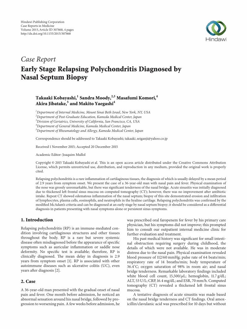
Figure 1: CT scan of a 36-year-oldmanwith RP.The nasal septum isedematous and inflamed. CT, computed tomography; RP, relapsingpolychondritis.
Figure 2: Photomicrograph of the nasal septum biopsy in a 36-year-old man with RP.The hyaline cartilage is eroded and infiltratedby lymphocytes, plasma cells, eosinophils, and neutrophils. (Hema-toxylin and eosin stain, ×100). RP, relapsing polychondritis.
any improvement. The nose pain became worse, and thepatient was admitted for further evaluation. After admission,generalized joint pain and severe diarrhea developed. Repeatsinus CT revealed resolving mucosal thickening, but thenasal septum and cartilage were noted to be edematous andinflamed (Figure 1). Contrast magnetic resonance imagingconfirmed the same findings. All serological tests, includingthose for rheumatoid factor and anti-neutrophil cytoplas-mic antibodies (ANCA), were negative. Due to a suspicionof RP, the patient underwent nasal septum biopsy, whichshowed erosion and infiltration of lymphocytes, plasmacells, eosinophils, and neutrophils in the hyaline cartilage(Figure 2). The symptoms plus the biopsy findings fulfilledthemodifiedMcAdam’s criteria for RP.Chest CTwas negativefor laryngotracheobronchial wall thickening, luminal nar-rowing, and cartilaginous calcification. Pulmonary functiontests showed an obstructive pattern of the upper airwaysduring inspiration. Retinal examination was unremarkable.Echocardiography and electrocardiography were also withinnormal limits.
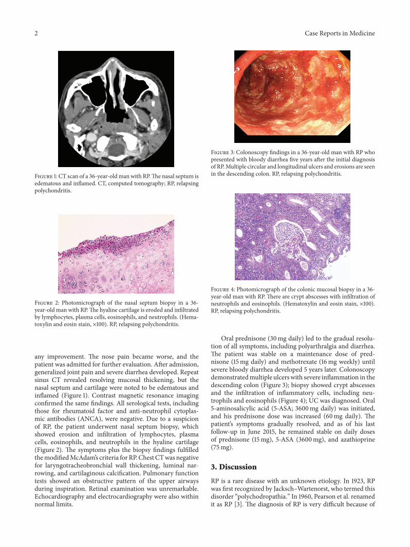
Figure 3: Colonoscopy findings in a 36-year-old man with RP whopresented with bloody diarrhea five years after the initial diagnosisof RP.Multiple circular and longitudinal ulcers and erosions are seenin the descending colon. RP, relapsing polychondritis.
Figure 4: Photomicrograph of the colonic mucosal biopsy in a 36-year-old man with RP. There are crypt abscesses with infiltration ofneutrophils and eosinophils. (Hematoxylin and eosin stain, ×100).RP, relapsing polychondritis.
Oral prednisone (30mg daily) led to the gradual resolu-tion of all symptoms, including polyarthralgia and diarrhea.The patient was stable on a maintenance dose of pred-nisone (15mg daily) and methotrexate (16mg weekly) untilsevere bloody diarrhea developed 5 years later. Colonoscopydemonstratedmultiple ulcerswith severe inflammation in thedescending colon (Figure 3); biopsy showed crypt abscessesand the infiltration of inflammatory cells, including neu-trophils and eosinophils (Figure 4); UC was diagnosed. Oral5-aminosalicylic acid (5-ASA; 3600mg daily) was initiated,and his prednisone dose was increased (60mg daily). Thepatient’s symptoms gradually resolved, and as of his lastfollow-up in June 2015, he remained stable on daily dosesof prednisone (15mg), 5-ASA (3600mg), and azathioprine(75mg).
3. Discussion
RP is a rare disease with an unknown etiology. In 1923, RPwas first recognized by Jacksch–Wartenorst, who termed thisdisorder “polychodropathia.” In 1960, Pearson et al. renamedit as RP [3]. The diagnosis of RP is very difficult because of
Case Reports in Medicine 3
nonspecific symptoms, especially in early stages. Trenthamand Le reported that the mean delay from symptom onset-to-diagnosis was 2.9 years, and one-third of patients wentto more than five physicians before successful diagnosis [1].In our case, we successfully diagnosed RP within 2 monthsof symptom onset, by performing a nasal septum biopsy todifferentiate between Wegener’s granulomatosis and RP. RPcan mimic acute sinusitis, as in our patient, and should beconsidered in the differential diagnosis of persistent sinussymptoms such as fever and paranasal sinus tenderness.
There is no specific test for RP, and it is usually diag-nosed by a combination of clinical and biopsy findings. In1976, McAdam et al. established the diagnostic criteria thatrequired the presence of more than three of the followingsix symptoms: auricular chondritis, polyarthritis, nasal chon-dritis, ocular inflammation, respiratory tract chondritis, andaudiovestibular damage [2]. Our patient initially had onlynasal symptoms. Although peripheral joint pain developedafter admission, it did not meet the original McAdam’scriteria. However, he fulfilled the modified McAdam’s cri-teria for RP [4, 5], which required more than one of sixsymptoms plus histological confirmation or chondritis attwo or more separate anatomic locations that is responsiveto corticosteroids and/or dapsone. The initial change inhistology involves the loss of basophilia in the cartilagematrix, corresponding to the loss of matrix proteoglycans.Additionally, a reduced number of chondrocytes and theinfiltration of lymphocytes, neutrophils, and plasma cells inthe cartilage–soft tissue interface are seen in areas of cartilagedestruction [1]. The biopsy findings described in our caseconfirmed a diagnosis of RP. Therefore, we believe that thebiopsy of affected regions is very helpful in obtaining an earlydiagnosis of RP.
Unilateral external ear inflammation, the most commonpresenting feature of RP, is seen in approximately 40% ofpatients and eventually appears in nearly 90% of patients [6].Our patient only hadnasal symptoms; therewere no auricularor periauricular symptoms. Nasal chondritis is present atthe time of diagnosis in 24% of patients and is seen atsome stage of the disease in 53% of patients [7]. Saddlenose deformity is a typical RP finding, but our patient’s nosewas grossly unremarkable. Mucosal thickening on sinus CTraised the suspicion of Wegener’s granulomatosis, which hasbeen reported to occur in association with RP [8]. However,Wegener’s granulomatosis usually has positive serology forANCA and granulomas on biopsy specimens, findings thatwere not present in our patient.
Laryngotracheal disease occurs in more than half ofpatients and laryngotracheal stricture in nearly one quar-ter. Respiratory tract involvement accounts for almost 50%of deaths from RP [2]. Our patient did not show anyabnormality between the inspiratory and expiratory phaseson chest CT, but the flow–volume curve on spirometryrevealed a variable extrathoracic upper airway obstructivepattern. This patient may have some degree of damageof the extrathoracic tracheal cartilages; therefore, regularpulmonary examinations are needed, and careful attentionshould be given to complaints of hoarseness, cough, dyspnea,choking sensations, wheezing, or stridor.
Other diseases associated with RP, including rheumaticdisease, thyroid disease, and UC, have been reported; infact, 25%–35% of patients with RP were found to havesome associated autoimmune disease [2]. Five years afterthe diagnosis of RP, our patient was diagnosed with UC.Similar cases of RP patients developing UC, or vice versa,have been reported [9]. Coexistence of other autoimmunediseases, includingUC, should be suspected if new symptomssuch as severe diarrhea or bloody stools develop.
In conclusion, we encountered a case of RP that wasdiagnosed early by nasal septum biopsy. RP should beconsidered in patients who present with nasal symptomsalone orwith persistent sinus symptoms.Coexistence of otherautoimmune diseases should always be suspected if a patientwith RP develops new symptoms.
Consent
Written informed consent was obtained from the patient forpublication of this case report and accompanying images.
Conflict of Interests
The authors declare that there is no conflict of interestsregarding the publication of this paper.
Authors’ Contribution
SandraMoody andMakito Yaegashi were amajor contributorin writing the paper. Masafumi Komori and Akira Jibatakeperformed the histological examination of the nose and joint.Takaaki Kobayashi was primary writer of the paper. Allauthors read and approved the final paper.
Acknowledgments
The authors acknowledge the support of all members of thegeneral medicine department at Kameda Medical Center.
References
[1] D. E. TrenthamandC.H. Le, “Relapsing polychondritis,”Annalsof Internal Medicine, vol. 129, no. 2, pp. 114–122, 1998.
[2] L. P. McAdam, M. A. O’Hanlan, R. Bluestone, and C. M.Pearson, “Relapsing polychondritis: prospective study of 23patients and a review of the literature,” Medicine, vol. 55, no. 3,pp. 193–215, 1976.
[3] C. M. Pearson, H. M. Kline, and V. D. Newcomer, “Relapsingpolychondritis,”The New England Journal of Medicine, vol. 263,pp. 51–58, 1960.
[4] C. J. Michet Jr., C. H. McKenna, H. S. Luthra, and W.M. O’Fallon, “Relapsing polychondritis. Survival and predic-tive role of early disease manifestations,” Annals of InternalMedicine, vol. 104, no. 1, pp. 74–78, 1986.
[5] J. M. Damiani and H. L. Levine, “Relapsing polychondritis—report of ten cases,” The Laryngoscope, vol. 89, no. 6, pp. 929–946, 1979.
4 Case Reports in Medicine
[6] P. D. Kent, C. J. Michet Jr., and H. S. Luthra, “Relapsingpolychondritis,” Current Opinion in Rheumatology, vol. 16, no.1, pp. 56–61, 2004.
[7] B. L. Isaak, T. J. Liesegang, and C. J. Michet Jr., “Ocular andsystemic findings in relapsing polychondritis,” Ophthalmology,vol. 93, no. 5, pp. 681–689, 1986.
[8] K. Handrock and W. L. Gross, “Relapsing polychondritis as asecondary phenomenon of primary systemic vasculitis,”Annalsof the Rheumatic Diseases, vol. 52, no. 12, pp. 895–897, 1993.
[9] P. Karmacharya, R. Pathak, P. Shrestha, and R. Alweis, “The redhearing: swollen ear in a patient with ulcerative colitis,” Journalof Community Hospital Internal Medicine Perspectives, vol. 4,2014.

Submit your manuscripts athttp://www.hindawi.com
Stem CellsInternational
Hindawi Publishing Corporationhttp://www.hindawi.com Volume 2014
Hindawi Publishing Corporationhttp://www.hindawi.com Volume 2014
MEDIATORSINFLAMMATION
of
Hindawi Publishing Corporationhttp://www.hindawi.com Volume 2014
Behavioural Neurology
EndocrinologyInternational Journal of
Hindawi Publishing Corporationhttp://www.hindawi.com Volume 2014
Hindawi Publishing Corporationhttp://www.hindawi.com Volume 2014
Disease Markers
Hindawi Publishing Corporationhttp://www.hindawi.com Volume 2014
BioMed Research International
OncologyJournal of
Hindawi Publishing Corporationhttp://www.hindawi.com Volume 2014
Hindawi Publishing Corporationhttp://www.hindawi.com Volume 2014
Oxidative Medicine and Cellular Longevity
Hindawi Publishing Corporationhttp://www.hindawi.com Volume 2014
PPAR Research
The Scientific World JournalHindawi Publishing Corporation http://www.hindawi.com Volume 2014
Immunology ResearchHindawi Publishing Corporationhttp://www.hindawi.com Volume 2014
Journal of
ObesityJournal of
Hindawi Publishing Corporationhttp://www.hindawi.com Volume 2014
Hindawi Publishing Corporationhttp://www.hindawi.com Volume 2014
Computational and Mathematical Methods in Medicine
OphthalmologyJournal of
Hindawi Publishing Corporationhttp://www.hindawi.com Volume 2014
Diabetes ResearchJournal of
Hindawi Publishing Corporationhttp://www.hindawi.com Volume 2014
Hindawi Publishing Corporationhttp://www.hindawi.com Volume 2014
Research and TreatmentAIDS
Hindawi Publishing Corporationhttp://www.hindawi.com Volume 2014
Gastroenterology Research and Practice
Hindawi Publishing Corporationhttp://www.hindawi.com Volume 2014
Parkinson’s Disease
Evidence-Based Complementary and Alternative Medicine
Volume 2014Hindawi Publishing Corporationhttp://www.hindawi.com