Embed Size (px)
Citation preview

Case ReportDental Management of a Patient withNager Acrofacial Dysostosis
R. BozatlJoLlu and A. P. MünevveroLlu
Pediatric Dentistry, Faculty of Dentistry, Istanbul Medipol University, 34083 Istanbul, Turkey
Correspondence should be addressed to A. P. Munevveroglu; [email protected]
Received 4 August 2015; Accepted 8 September 2015
Academic Editor: Wasiu L. Adeyemo
Copyright © 2015 R. Bozatlıoglu and A. P. Munevveroglu. This is an open access article distributed under the Creative CommonsAttribution License, which permits unrestricted use, distribution, and reproduction in any medium, provided the original work isproperly cited.
Nager syndrome is a rare syndrome resulting from developmental abnormalities of the first and second branchial arches. Nagersyndrome is rare and mostly sporadic.Themain clinical features consist of craniofacial, limb, and musculoskeletal morphogenesis.These findings included malar hypoplasia, maxillomandibular hypoplasia, micrognathia, downslanting palpebral fissures, cleftpalate, ear anomalies, hypoplastic thumb, short forearm, proximal radioulnar synostosis, atrial septal defect, lower limb deformities,and flat nasal bridge.The prevalence is unknown; about 100 cases of Nager syndrome have been published up to now. Patients withNager syndrome are found worldwide among all racial and ethnic groups. Trismus and glossoptosis resulting in oropharyngealairway narrowing cause life-threatening respiratory distress for patients with Nager syndrome. In this case report, dentalrehabilitation of a 10-year-old child with Nager syndrome is presented.
1. Introduction
Nager syndrome is a rare syndrome resulting from devel-opmental abnormalities of the first and second branchialarches and is mostly sporadic; however, autosomal dominantor autosomal recessive inheritance has been reported. Nagersyndrome has an alteration of the 9q32 chromosome, 1q12q21deletion. The prevalence is unknown; about 100 cases ofNager syndrome have been published up to now. Patientswith Nager syndrome are found worldwide among all racialand ethnic groups [1–3].
The main clinical features consist of craniofacial, limb,and musculoskeletal morphogenesis. Oropharyngeal airwaynarrowing causes life-threatening respiratory distress forpatients with Nager syndrome. The craniofacial abnormal-ities include zygomatic and maxillomandibular hypoplasia,micrognathia, downwards palpebral fissures, absence of thelower lid eyelashes, lower lid coloboma, flat nasal bridge, cleftlip and palate, short soft palate, low-set and posterior-rotatedear, ear canal atresia, lack of development of the internaland external ear, hearing problems, speech problems, andfeeding problems. Respiration and feeding problems are dueto the mandibular hypoplasia with micrognathia, tongue and
severely restricted jaw opening. The hearing loss is due tothe severity of the ear abnormalities. There may be speechproblems due to the impaired hearing. The musculoskeletalabnormalities include preaxial upper-limb deformities, radialdefect, radioulnar synostosis, short forearm, and absenceof digits. Cardiovascular anomalies include Fallot tetralogyand/or ventricular septum defect. Nager syndrome doesnot affect a child’s intelligence [1–4]. In these patients withrestricted jaw opening, chewing is not possible, and oralhygiene is a major problem. Severe dental decay without theoption of adequate treatment is very common. Owing to thehand and limb abnormalities, manipulating implements maybe difficult, and self-care may not be possible [4]. In additionto enamel hypoplasia, oligodontia and dental malocclusioncan be detected [1–3].
The purpose of this case report is to present oral findingsand dental treatment of a patient with Nager syndrome.
2. Case Report
A 10-year-old boywas referred to YeditepeUniversity, Facultyof Dentistry, Department of Pedodontics, in Istanbul withcomplaint of dental caries. The patient was born to a healthy
Hindawi Publishing CorporationCase Reports in DentistryVolume 2015, Article ID 984732, 4 pageshttp://dx.doi.org/10.1155/2015/984732

2 Case Reports in Dentistry
Figure 1: Craniofacial anomalies present included hemifacial atrophy,maxillomandibular hypoplasia, severemicrognathia,malar hypoplasia,flat nasal bridge, and left ear canal atresia.
Figure 2: Lack of development of the internal and external ear with related hearing problems and severe mandibular hypoplasia withretrognathia.
mother and healthy father at 40 weeks of gestation.The preg-nancy of mother was uncomplicated. The mother’s historyregarding alcohol, smoking, and drug abuse was negative.Family history was not noted regarding craniofacial disorder.He has two normal sisters. A diagnosis of Nager syndromewas made around the age of 2 years when facial and limbanomalies became obvious.
Craniofacial anomalies present in the patient includehemifacial atrophy, maxillomandibular hypoplasia, severemicrognathia, restricted jaw opening, malar hypoplasia, flatnasal bridge, cleft palate, shortened soft palate, uvula atresia,left ear canal atresia, and lack of development of the internaland external ear (Figures 1 and 2). Other features found in thepatient were respiration, feeding, and speech problems aswellas short forearm and difficulty in fully extending the elbows.There was no evidence of mental retardation.
Intraoral and radiographic examination revealed that oralhygiene is a major problem. He had numerous carious teeth(#16, #55, #53, #26, #65, #63, #36, #75, #74, #46, #85, #84,and #83) and hypomineralisation teeth #16, #26, #36, and#46, restricted mouth opening, severe maxillomandibular
hypoplasia with severe micrognathia, and space deficiency inmaxillary and mandibular arch (Figures 3, 4, and 5).
Pulpectomy and composite restoration was performed(#53 and #83). Teeth #16, #55, #26, #65, #63, #36, #75, #74, #46,#85, and #84were extracted. Teeth #15, #14, #25, #24, #34, #45,and #44 were restored with fissure sealant followed by topicalfluoride application (Figure 6). Oral hygiene education wasgiven to the patient andhis parents.Orthodontic consultationwas held. The patient and his parents were instructed aboutseverity of orthodontic treatment. The patient was resched-uled to visit every three months because of high caries risk.
3. Discussion
Most of the patients with Nager syndrome are mostly spo-radic. These abnormalities can be diagnosed prenatally withthe help of ultrasonography. Perinatal mortality is about 20%because of oropharyngeal airway narrowing [1, 5].The patientwas born to a healthy mother and healthy father at 40 weeksof gestation.The pregnancy of themother was uncomplicatedand she did not have any harmful habits. He has two normal
Case Reports in Dentistry 3
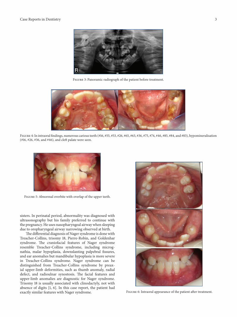
Figure 3: Panoramic radiograph of the patient before treatment.
Figure 4: In intraoral findings, numerous carious teeth (#16, #55, #53, #26, #65, #63, #36, #75, #74, #46, #85, #84, and #83), hypomineralisation(#16, #26, #36, and #46), and cleft palate were seen.
Figure 5: Abnormal overbite with overlap of the upper teeth.
sisters. In perinatal period, abnormality was diagnosed withultrasonography but his family preferred to continue withthe pregnancy. He uses nasopharyngeal airwaywhen sleepingdue to oropharyngeal airway narrowing observed at birth.
The differential diagnosis of Nager syndrome is donewithTreacher-Collins, trisomy 18, Pierre-Robin, and Goldenharsyndrome. The craniofacial features of Nager syndromeresemble Treacher-Collins syndrome, including microg-nathia, malar hypoplasia, downslanting palpebral fissures,and ear anomalies but mandibular hypoplasia is more severein Treacher-Collins syndrome. Nager syndrome can bedistinguished from Treacher-Collins syndrome by preax-ial upper-limb deformities, such as thumb anomaly, radialdefect, and radioulnar synostosis. The facial features andupper-limb anomalies are diagnostic for Nager syndrome.Trisomy 18 is usually associated with clinodactyly, not withabsence of digits [1, 6]. In this case report, the patient hadexactly similar features with Nager syndrome. Figure 6: Intraoral appearance of the patient after treatment.

4 Case Reports in Dentistry
Mandibular malformations, missing joint structures,restriction in jaw movements, and limb abnormalities causeoral hygiene deficiency. In this case report, the patient hadlimitedmouth opening andmultiple carious teeth. Restrictedmouth opening also made dental treatment extremely dif-ficult and requires treatment under general anaesthetic.Treatment under general anaesthesia can be challenging inNager syndrome because of oropharyngeal airway narrow-ing. Severe airway obstruction is derived from mandibu-lar hypoplasia with micrognathia, retroplaced tongue, andstrabismus. Moreover, hearing and speech difficulty lead tocommunication problems with patients [7]. The patient hasmandibular hypoplasia and severe micrognathia. He hadthree general anesthesia attempts for treatment of oropharyn-geal airway narrowing but one of the three was achieved bysurgical and anaesthetic team. Moreover he has hearing andspeech problems but intelligence is of normal level. In thiscase report, dental treatments of the patient were performedunder local anesthesia.
4. Conclusion
Nager syndrome is a rare disorder which inhibits treat-ment under general anaesthesia because of oropharyngealairway narrowing. It is important that these patients receivecomprehensive prevention and regular dental review in aneffort to improve their oral health and therefore avoid,when possible, the need for dental treatment under generalanaesthesia. Patients withNager syndrome should be referredto pedodontics for oral hygiene instruction, dietary advice,and fluoride prevention.
Conflict of Interests
The authors declare that there is no conflict of interestsregarding the publication of this paper.
References
[1] J.-L. Lin, “Nager syndrome: a case report,” Pediatrics andNeonatology, vol. 53, no. 2, pp. 147–150, 2012.
[2] N. Y. P. Bernal, N. Villanueva, and H. Rincon II, “Osteogenicmandibular distraction in Nager’s syndrome. Case report,”Revista Mexicana de Ortodoncia, vol. 1, no. 1, pp. 44–53, 2013.
[3] S. A. Fakhim, N. Shahid, and M. Mousaviagdas, “A case report:nager acrofacial dysostosis,” Iranian Journal of Otorhinolaryn-gology, vol. 24, no. 66, pp. 45–50, 2012.
[4] K. Vargervik, “Mandibular malformations: growth character-istics and management in hemifacial microsomia and Nagersyndrome,” Acta Odontologica Scandinavica, vol. 56, no. 6, pp.331–338, 1998.
[5] R. Thapa, S. Pramanik, M. Mukhopadhyay, and A. Ghosh,“Nager acrofacial dysostosis: an unusual association with bothupper and lower eyelid colobomas,” Indian Journal of Pediatrics,vol. 73, no. 7, pp. 631–632, 2006.
[6] S. K. Bhatia and M. M. Collard, “The dental managementof a patient with Nager syndrome: a case report,” Journal ofDisability and Oral Health, vol. 12, no. 1, pp. 43–46, 2011.
[7] M. T. McDonald and J. L. Gorski, “Nager acrofacial dysostosis,”Journal of Medical Genetics, vol. 30, no. 9, pp. 779–782, 1993.

Submit your manuscripts athttp://www.hindawi.com
Hindawi Publishing Corporationhttp://www.hindawi.com Volume 2014
Oral OncologyJournal of
DentistryInternational Journal of
Hindawi Publishing Corporationhttp://www.hindawi.com Volume 2014
Hindawi Publishing Corporationhttp://www.hindawi.com Volume 2014
International Journal of
Biomaterials
Hindawi Publishing Corporationhttp://www.hindawi.com Volume 2014
BioMed Research International
Hindawi Publishing Corporationhttp://www.hindawi.com Volume 2014
Case Reports in Dentistry
Hindawi Publishing Corporationhttp://www.hindawi.com Volume 2014
Oral ImplantsJournal of
Hindawi Publishing Corporationhttp://www.hindawi.com Volume 2014
Anesthesiology Research and Practice
Hindawi Publishing Corporationhttp://www.hindawi.com Volume 2014
Radiology Research and Practice
Environmental and Public Health
Journal of
Hindawi Publishing Corporationhttp://www.hindawi.com Volume 2014
The Scientific World JournalHindawi Publishing Corporation http://www.hindawi.com Volume 2014
Hindawi Publishing Corporationhttp://www.hindawi.com Volume 2014
Dental SurgeryJournal of
Drug DeliveryJournal of
Hindawi Publishing Corporationhttp://www.hindawi.com Volume 2014
Hindawi Publishing Corporationhttp://www.hindawi.com Volume 2014
Oral DiseasesJournal of
Hindawi Publishing Corporationhttp://www.hindawi.com Volume 2014
Computational and Mathematical Methods in Medicine
ScientificaHindawi Publishing Corporationhttp://www.hindawi.com Volume 2014
PainResearch and TreatmentHindawi Publishing Corporationhttp://www.hindawi.com Volume 2014
Preventive MedicineAdvances in
Hindawi Publishing Corporationhttp://www.hindawi.com Volume 2014
EndocrinologyInternational Journal of
Hindawi Publishing Corporationhttp://www.hindawi.com Volume 2014
Hindawi Publishing Corporationhttp://www.hindawi.com Volume 2014
OrthopedicsAdvances in