Embed Size (px)
Citation preview
441
CASE REPORT
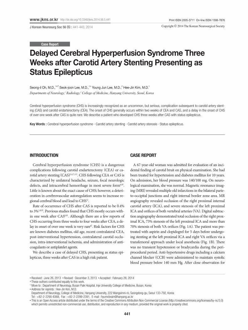
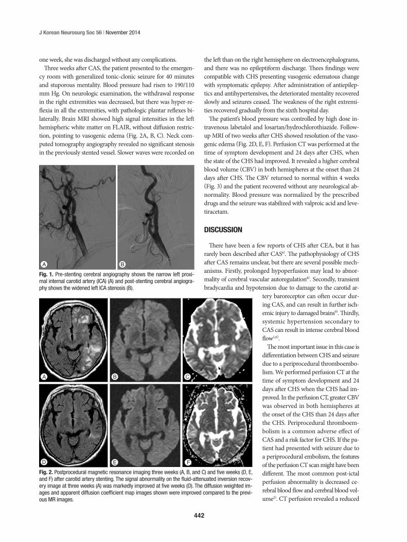
A 67-year-old woman was admitted for evaluation of an inci-dental finding of carotid bruit on physical examination. She had been treated for hypertension and diabetes mellitus for 10 years. On admission, her blood pressure was 140/100 mg. On neuro-logical examination, she was normal. Magnetic resonance imag-ing (MRI) revealed multiple old infarctions in the bilateral parie-to-occipital junctions and right internal border zone area. MR angiography revealed occlusion of the right proximal internal carotid artery (ICA), and severe stenosis of the left proximal ICA and orifices of both vertebral arteries (VA). Digital subtrac-tion angiography demonstrated total occlusion of the right prox-imal ICA, 73% stenosis of the left proximal ICA and more than 70% stenosis of both VA orifices (Fig. 1A). The patient was pre-treated with aspirin and clopidogrel for 5 days before undergo-ing stenting at the left proximal ICA and right VA orifices via a transfemoral approach under local anesthesia (Fig. 1B). There was no transient hypotension or bradycardia during the peri-procedural period. Anti-hypertensive drugs including a calcium channel blocker (CCB) were administered to maintain systolic blood pressure below 140 mm Hg. After close observation for
INTRODUCTION
Cerebral hyperperfusion syndrome (CHS) is a dangerous complications following carotid endarterectomy (CEA) or ca-rotid artery stenting (CAS)3,4,7,11). CHS following CEA or CAS is characterized by unilateral headache, seizure, focal neurologic defects, and intracerebral hemorrhage in most severe form6,9). Little is known about the exact cause of CHS; however, a deteri-oration in cerebrovascular autoregulation seems to increase re-gional cerebral blood and lead to CHS1).
Rate of occurrence of CHS after CAS is reported to be 0.4% to 3%1,8,9). Previous studies found that CHS mostly occurs with-in one week after CAS6,9). Although there are a few reports of CHS occurring from three weeks to four weeks after CEA, a de-lay in onset of over one week is very rare6). Risk factors for CHS are known diabetes mellitus, old age, recent contralateral CEA, post-interventional hypertension, contralateral carotid occlu-sion, intra-interventional ischemia, and administration of anti-coagulants or antiplatelet agents.
We describe a case of delayed CHS, presenting as status epi-lepticus, three weeks after CAS in a high risk patient.
Delayed Cerebral Hyperperfusion Syndrome Three Weeks after Carotid Artery Stenting Presenting as Status Epilepticus
Seong-il Oh, M.D.,1*† Seok-joon Lee, M.D.,1* Young Jun Lee, M.D.,2 Hee-Jin Kim, M.D.1
Departments of Neurology,1 Radiology,2 College of Medicine, Hanyang University, Seoul, Korea
Cerebral hyperperfusion syndrome (CHS) is increasingly recognized as an uncommon, but serious, complication subsequent to carotid artery stent-ing (CAS) and carotid endarterectomy (CEA). The onset of CHS generally occurs within two weeks of CEA and CAS, and a delay in the onset of CHS of over one week after CAS is quite rare. We describe a patient who developed CHS three weeks after CAS with status epilepticus.
Key Words : Cerebral hyperperfusion syndrome · Carotid artery stenting · Carotid artery stenosis · Status epilepticus.
Case Report
• Received : June 26, 2013 • Revised : December 3, 2013 • Accepted : February 28, 2014* These outhors contributed equally to this work.† Move to : Department of Neurology, Busan Paik Hospital, Inje University College of Medicine, Busan, Korea• Address for reprints : Hee-Jin Kim, M.D. Department of Neurology, College of Medicine, Hanyang University, 222 Wangsimni-ro, Seongdong-gu, Seoul 133-792, Korea Tel : +82-2-2290-8368, Fax : +82-2-2299-2391, E-mail : [email protected]• This is an Open Access article distributed under the terms of the Creative Commons Attribution Non-Commercial License (http://creativecommons.org/licenses/by-nc/3.0) which permits unrestricted non-commercial use, distribution, and reproduction in any medium, provided the original work is properly cited.
J Korean Neurosurg Soc 56 (5) : 441-443, 2014
http://dx.doi.org/10.3340/jkns.2014.56.5.441
Copyright © 2014 The Korean Neurosurgical Society
Print ISSN 2005-3711 On-line ISSN 1598-7876www.jkns.or.kr
442
J Korean Neurosurg Soc 56 | November 2014
the left than on the right hemisphere on electroencephalograms, and there was no epileptiform discharge. Thees findings were compatible with CHS presenting vasogenic edematous change with symptomatic epilepsy. After administration of antiepilep-tics and antihypertensives, the deteriorated mentality recovered slowly and seizures ceased. The weakness of the right extremi-ties recovered gradually from the sixth hospital day.
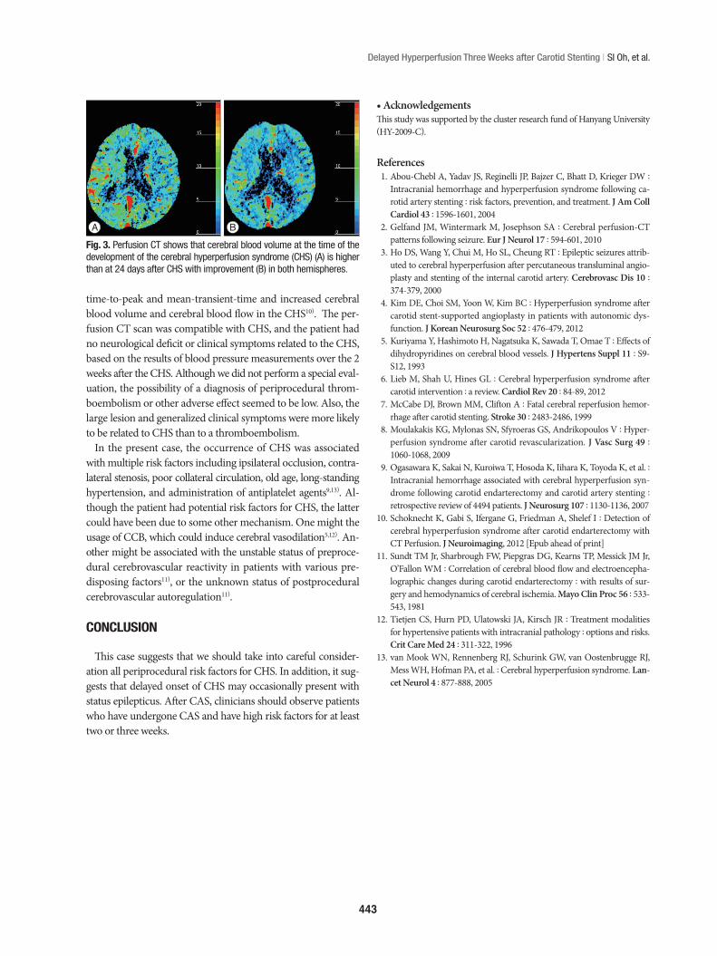
The patient’s blood pressure was controlled by high dose in-travenous labetalol and losartan/hydrochlorothiazide. Follow-up MRI of two weeks after CHS showed resolution of the vaso-genic edema (Fig. 2D, E, F). Perfusion CT was performed at the time of symptom development and 24 days after CHS, when the state of the CHS had improved. It revealed a higher cerebral blood volume (CBV) in both hemispheres at the onset than 24 days after CHS. The CBV returned to normal within 4 weeks (Fig. 3) and the patient recovered without any neurological ab-normality. Blood pressure was normalized by the prescribed drugs and the seizure was stabilized with valproic acid and leve-tiracetam.
DISCUSSION
There have been a few reports of CHS after CEA, but it has rarely been described after CAS6). The pathophysiology of CHS after CAS remains unclear, but there are several possible mech-anisms. Firstly, prolonged hypoperfusion may lead to abnor-mality of cerebral vascular autoregulation8). Secondly, transient bradycardia and hypotension due to damage to the carotid ar-
tery baroreceptor can often occur dur-ing CAS, and can result in further isch-emic injury to damaged brains9). Thirdly, systemic hypertension secondary to CAS can result in intense cerebral blood flow1,6).
The most important issue in this case is differentiation between CHS and seizure due to a periprocedural thromboembo-lism. We performed perfusion CT at the time of symptom development and 24 days after CHS when the CHS had im-proved. In the perfusion CT, greater CBV was observed in both hemispheres at the onset of the CHS than 24 days after the CHS. Periprocedural thromboem-bolism is a common adverse effect of CAS and a risk factor for CHS. If the pa-tient had presented with seizure due to a periprocedural embolism, the features of the perfusion CT scan might have been different. The most common post-ictal perfusion abnormality is decreased ce-rebral blood flow and cerebral blood vol-ume2). CT perfusion revealed a reduced
one week, she was discharged without any complications. Three weeks after CAS, the patient presented to the emergen-
cy room with generalized tonic-clonic seizure for 40 minutes and stuporous mentality. Blood pressure had risen to 190/110 mm Hg. On neurologic examination, the withdrawal response in the right extremities was decreased, but there was hyper-re-flexia in all the extremities, with pathologic plantar reflexes bi-laterally. Brain MRI showed high signal intensities in the left hemispheric white matter on FLAIR, without diffusion restric-tion, pointing to vasogenic edema (Fig. 2A, B, C). Neck com-puted tomography angiography revealed no significant stenosis in the previously stented vessel. Slower waves were recorded on
BA
Fig. 1. Pre-stenting cerebral angiography shows the narrow left proxi-mal internal carotid artery (ICA) (A) and post-stenting cerebral angiogra-phy shows the widened left ICA stenosis (B).
BA C
D E FFig. 2. Postprocedural magnetic resonance imaging three weeks (A, B, and C) and five weeks (D, E, and F) after carotid artery stenting. The signal abnormality on the fluid-attenuated inversion recov-ery image at three weeks (A) was markedly improved at five weeks (D). The diffusion weighted im-ages and apparent diffusion coefficient map images shown were improved compared to the previ-ous MR images.
443
Delayed Hyperperfusion Three Weeks after Carotid Stenting | SI Oh, et al.
• AcknowledgementsThis study was supported by the cluster research fund of Hanyang University (HY-2009-C).
References 1. Abou-Chebl A, Yadav JS, Reginelli JP, Bajzer C, Bhatt D, Krieger DW :
Intracranial hemorrhage and hyperperfusion syndrome following ca-rotid artery stenting : risk factors, prevention, and treatment. J Am Coll Cardiol 43 : 1596-1601, 2004
2. Gelfand JM, Wintermark M, Josephson SA : Cerebral perfusion-CT patterns following seizure. Eur J Neurol 17 : 594-601, 2010
3. Ho DS, Wang Y, Chui M, Ho SL, Cheung RT : Epileptic seizures attrib-uted to cerebral hyperperfusion after percutaneous transluminal angio-plasty and stenting of the internal carotid artery. Cerebrovasc Dis 10 : 374-379, 2000
4. Kim DE, Choi SM, Yoon W, Kim BC : Hyperperfusion syndrome after carotid stent-supported angioplasty in patients with autonomic dys-function. J Korean Neurosurg Soc 52 : 476-479, 2012
5. Kuriyama Y, Hashimoto H, Nagatsuka K, Sawada T, Omae T : Effects of dihydropyridines on cerebral blood vessels. J Hypertens Suppl 11 : S9-S12, 1993
6. Lieb M, Shah U, Hines GL : Cerebral hyperperfusion syndrome after carotid intervention : a review. Cardiol Rev 20 : 84-89, 2012
7. McCabe DJ, Brown MM, Clifton A : Fatal cerebral reperfusion hemor-rhage after carotid stenting. Stroke 30 : 2483-2486, 1999
8. Moulakakis KG, Mylonas SN, Sfyroeras GS, Andrikopoulos V : Hyper-perfusion syndrome after carotid revascularization. J Vasc Surg 49 : 1060-1068, 2009
9. Ogasawara K, Sakai N, Kuroiwa T, Hosoda K, Iihara K, Toyoda K, et al. : Intracranial hemorrhage associated with cerebral hyperperfusion syn-drome following carotid endarterectomy and carotid artery stenting : retrospective review of 4494 patients. J Neurosurg 107 : 1130-1136, 2007
10. Schoknecht K, Gabi S, Ifergane G, Friedman A, Shelef I : Detection of cerebral hyperperfusion syndrome after carotid endarterectomy with CT Perfusion. J Neuroimaging, 2012 [Epub ahead of print]
11. Sundt TM Jr, Sharbrough FW, Piepgras DG, Kearns TP, Messick JM Jr, O’Fallon WM : Correlation of cerebral blood flow and electroencepha-lographic changes during carotid endarterectomy : with results of sur-gery and hemodynamics of cerebral ischemia. Mayo Clin Proc 56 : 533-543, 1981
12. Tietjen CS, Hurn PD, Ulatowski JA, Kirsch JR : Treatment modalities for hypertensive patients with intracranial pathology : options and risks. Crit Care Med 24 : 311-322, 1996
13. van Mook WN, Rennenberg RJ, Schurink GW, van Oostenbrugge RJ, Mess WH, Hofman PA, et al. : Cerebral hyperperfusion syndrome. Lan-cet Neurol 4 : 877-888, 2005
time-to-peak and mean-transient-time and increased cerebral blood volume and cerebral blood flow in the CHS10). The per-fusion CT scan was compatible with CHS, and the patient had no neurological deficit or clinical symptoms related to the CHS, based on the results of blood pressure measurements over the 2 weeks after the CHS. Although we did not perform a special eval-uation, the possibility of a diagnosis of periprocedural throm-boembolism or other adverse effect seemed to be low. Also, the large lesion and generalized clinical symptoms were more likely to be related to CHS than to a thromboembolism.
In the present case, the occurrence of CHS was associated with multiple risk factors including ipsilateral occlusion, contra-lateral stenosis, poor collateral circulation, old age, long-standing hypertension, and administration of antiplatelet agents9,13). Al-though the patient had potential risk factors for CHS, the latter could have been due to some other mechanism. One might the usage of CCB, which could induce cerebral vasodilation5,12). An-other might be associated with the unstable status of preproce-dural cerebrovascular reactivity in patients with various pre-disposing factors11), or the unknown status of postprocedural cerebrovascular autoregulation11).
CONCLUSION
This case suggests that we should take into careful consider-ation all periprocedural risk factors for CHS. In addition, it sug-gests that delayed onset of CHS may occasionally present with status epilepticus. After CAS, clinicians should observe patients who have undergone CAS and have high risk factors for at least two or three weeks.
Fig. 3. Perfusion CT shows that cerebral blood volume at the time of the development of the cerebral hyperperfusion syndrome (CHS) (A) is higher than at 24 days after CHS with improvement (B) in both hemispheres.
BA