Embed Size (px)
Citation preview
Hindawi Publishing CorporationCase Reports in Oncological MedicineVolume 2013, Article ID 697251, 4 pageshttp://dx.doi.org/10.1155/2013/697251
Case ReportChoriocarcinoma Syndrome: A Case Report anda Literature Review
Khaled Baagar,1 Fahmi Yousef Khan,1 and Einas AlKuwari2
1 Department of Medicine, Hamad General Hospital, P.O. Box 3050, Doha, Qatar2 Department of Pathology and Laboratory Medicine, Hamad General Hospital, P.O. Box 3050, Doha, Qatar
Correspondence should be addressed to Khaled Baagar; [email protected]
Received 9 April 2013; Accepted 8 May 2013
Academic Editors: Y.-J. Chen, C. Gennatas, F. A. Mauri, and Y. Yokoyama
Copyright © 2013 Khaled Baagar et al. This is an open access article distributed under the Creative Commons Attribution License,which permits unrestricted use, distribution, and reproduction in any medium, provided the original work is properly cited.
A 35-year-old Qatari man presented to our hospital with a 4-month history of mild abdominal pain, weight loss, and jaundice.He was found to have central intra-abdominal mass and a single testis in the scrotum. His investigations showed cholestaticjaundice and very high level of 𝛽-HCG (1131379 IU/L). CT scans of the chest and abdomen showed a huge pelvic-abdominalmass with extensive retroperitoneal lymphadenopathy, in addition to liver and lung metastases. CT-guided Tru-Cut biopsy of themass showed mixed germ cell tumor. Chemotherapy was refused by the patient and his family. In the following days, the patientbled from his liver metastases leading to hemorrhagic shock, hemorrhage from metastatic sites of choriocarcinoma containingtumors is named choriocarcinoma syndrome. He was transferred to the medical intensive care unit, where he was intubatedand resuscitated. Embolization of the right hepatic artery was done, but failed to control the bleeding, which continued withdevelopment of disseminated intravascular coagulopathy and a severe abdominal compartment syndrome, and eventually thepatient died.
1. Introduction
Testicular neoplasms comprise the most common solidmalignancy affecting men between the ages of 15 and 35,but they only represent almost 1% of all solid tumors inmales. Approximately two to three new cases per 100,000males are reported in the United States each year, and95 percent of all primary testicular tumors are germ celltumors [1, page 1574]. This rare tumor can be compli-cated by a very rare, life threatening complication whichis choriocarcinoma syndrome with only few cases reportedworldwide. Choriocarcinoma syndrome entails hemorrhagefrom metastatic sites of choriocarcinoma associated with asignificant rise of beta-human chorionic gonadotropin (𝛽-HCG) [2]. In this report, we present a case of choriocar-cinoma syndrome in a 35-year-old Qatari man, to drawattention of our physicians to the importance of consid-ering this syndrome while dealing with patients who havemassive and advanced testicular tumors with high 𝛽-HCG,as this syndrome is life-threatening and needs urgent treat-ment.
2. Case Report
A 35-year-old male, not known to have any chronic ill-ness, presented with a 4-month history of mild, dull, andintermittent abdominal pain, associated with jaundice, lossof appetite, and weight loss of 10 kilograms, as well as a one-month history of abdominal swelling.
On examination, he had normal vital signs, but hewas pale and jaundiced. There were no palpable lymphnodes. There was central intra-abdominal mass 15 cm indiameter, firm, and tender with smooth surface. He hadsingle right testicle in the scrotum. Laboratory investiga-tions (Table 1) showed white blood cells: 14.7 × 1000/mm3(normal: 4 × 1000–10 × 1000/mm3), hemoglobin: 7.4 g/dL(normal: 13–17 g/dL), and platelets: 480 × 1000/mm3 (nor-mal: 150 × 1000–400 × 1000/mm3). Blood chemistry andrenal function were within normal limits. Aspartate amino-transferase: 53 u/L (normal: 12–39 u/L), alanine aminotrans-ferase: 16 u/L (normal: 0–40 u/L), alkaline phosphatase:472 u/L (normal: 40–129 u/L), gamma glutamyl transpepti-dase: 155U/L (normal: 11–50U/L), bilirubin: 115 umol/L then
2 Case Reports in Oncological Medicine
Table 1: Investigations done during the patient hospitalization.
Component Uponpresentation In MICU Reference range
WBC 14.7 ×1000/mm3 8.4 × 1000/mm3 4 × 1000–10 ×
1000/mm3
Hemoglobin 7.4 2.5 13–17 g/dL
Platelets 480 ×1000/mm3
113 × 1000/mm3,then
47 × 1000/mm3
150×1000–400×1000/mm3
Creatinine 118 222 62–124 umol/LBUN 8.3 11.5 1.7–8.3mmol/LPotassium 4.6 5.2 3.6–5.1mmol/LSodium 134 134 135–145mmol/LChloride 98 98 96–110mmol/LBicarbonate 22 9 24–30mmol/LAST 53 4425 12–39 u/LALT 16 670 0–40 u/LALP 472 1396 40–129 u/LGGT 155 11–50 U/LBilirubin 115 then 330 722 3.5–24 umol/LDirectbilirubin 306 Up to 7 umol/L
INR 1.1 1.4 then 2.3APTT 28 31 then 49.20 26–38.5 seconds𝛽-HCG 1131379 0–5 IU/LLDH 2331 240–480U/LAFP 1.4 0–5 IU/mLCA19-9 9 0–37U/mLCEA 1.2 0–3Ug/LFT4 49.4 Up to 20 pmol/LTSH 0.01 0.45–4.5 𝜇/LLactic acid 5, then 14.41 0.5–2.2mmol/LWBC: white blood cells, BUN: blood urea nitrogen, AST: aspartateaminotransferase, ALT: alanine aminotransferase, ALP: alkaline phophatase,GGT: gamma glutamyl transpeptidase, INR: international normalized ratio,APTT: activated partial thromboplastin time, 𝛽-HCG: 𝛽-human chorionicgonadotropin, LDH: lactate dehydrogenase, AFP: alpha-fetoprotein, CA19-9: carbohydrate antigen 19-9, CEA: carcinoembryonic antigen, FT4: freethyroxine, and TSH: thyroid stimulating hormone.
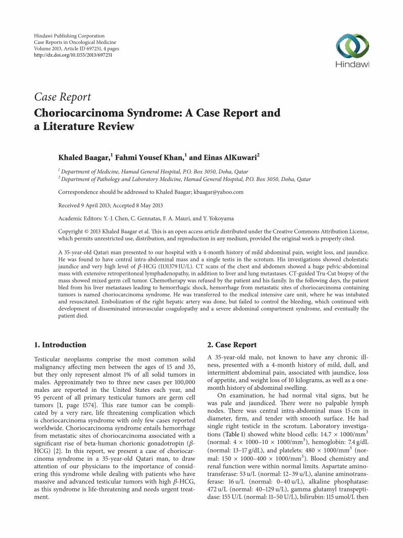
330 umol/L (normal: 3.5–24 umol/L), and direct bilirubin:306 umol/L (normal: up to 7 umol/L). Tumor markers: 𝛽-HCG: 1131379 IU/L (normal: 0–5 IU/L), LDH: 2331U/L (nor-mal: 240–480U/L), AFP: 1.4 IU/mL (normal: 0–5 IU/mL),CA19-9: 9U/mL (normal: 0–37U/mL) CEA: 1.2Ug/L (nor-mal: 0–3Ug/L). FT4: 49.4 pmol/L (normal up to 20 pmol/L)and TSH: 0.01𝜇/L (normal 0.45–4.5𝜇/L). CT scans of chest,abdomen, and pelvis with contrast showed huge pelvic-abdominal mass measuring 19 × 14.7 × 22 cm with exten-sive retroperitoneal lymphadenopathy, and liver and lungmetastases (Figure 1). CT-guided Tru-Cut biopsy of the massshowed a mixed germ cell tumor (Figures 2(a) and 2(b)) withpredominantly seminomatous component (CD117 positive,Figure 3) and foci consistent with choriocarcinoma (𝛽-HCG
Figure 1: CT abdomen at presentation showed huge mass.
positive, Figure 4). Despite a high risk of intratumor bleedingattributed to tumor size and the oncologist’s recommendationfor immediate chemotherapy, the patient and his familyrefused chemotherapy as they planned to travel abroadfor a second opinion. On the following days, his condi-tion deteriorated and his level of consciousness decreased;his BP was 70/40mmHg, pulse rate 140/min, respiratoryrate 35/min, and oxygen saturation 91% with nonrebreath-ing mask on 15 liters oxygen/min. So, he was transferredimmediately to the medical intensive care unit (MICU)where he was intubated. Laboratory investigations (Table 1)showed white blood cells: 8.4 × 1000/mm3, hemoglobin:2.5 g/dL, and platelets: 113 × 1000/mm3. Bun: 11.5mmol/L(normal: 1.7–8.3mmol/L), creatinine: 222 umol/L (normal:62–124 umol/L), K: 5.2mmol/L (normal: 3.6–5.1mmol/L),Na: 134mmol/L (normal: 135–145mmol/L), bicarbonate:9mmol/L (normal: 24–30mmol/L), Cl: 98mmol/L (nor-mal: 96–110mmol/L), bilirubin: 722 umol/L, and lactate:5mmol/L (normal: 0.5–2.2mmol/L). INR: 1.4, APTT: 31seconds (normal: 26–38.5 seconds). ABG: PH: 7.128, PO
2:
270, PCO2: 22, HCO
3: 8, and oxygen saturation: 99%. The
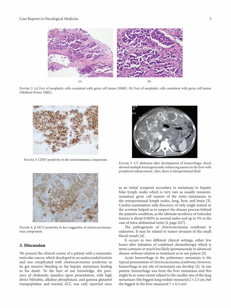
patient was given intravenous fluids, packed red blood cells,fresh frozen plasma and cryopricepitate transfusions, andvasopressors. CT angiogram showed multiple enhancinghepatic metastasis with extensive blood leaking and poolingin the liver and to a lesser extent in the pelvic mass withlarge amount of intraperitoneal fluid (most likely hemoperi-toneum) (Figure 5). So, angiography and embolization of theright Hepatic artery with gelfoam were done.
Despite these interventions, the bleeding continued.Hemoglobin and blood pressure continued dropping, andhepatic surgeon advised for conservative treatment as thepatient was at high risk for bleeding (INR 2.3 and aPTT 49.20seconds).Moreover, the patient developedDIC, evidenced byprolonged INR, aPTT, and low platelets, and activated factorseven was given.
The patient developed anuria as his intra-abdominalpressure reached 50–55mmHg. Surgery team was consultedfor decompression laparotomy, but they thought it was uselessand would not help the patient. At the end, the patientdied secondary to hemorrhagic shock, DIC, and abdominalcompartment syndrome with renal shutdown.
Case Reports in Oncological Medicine 3
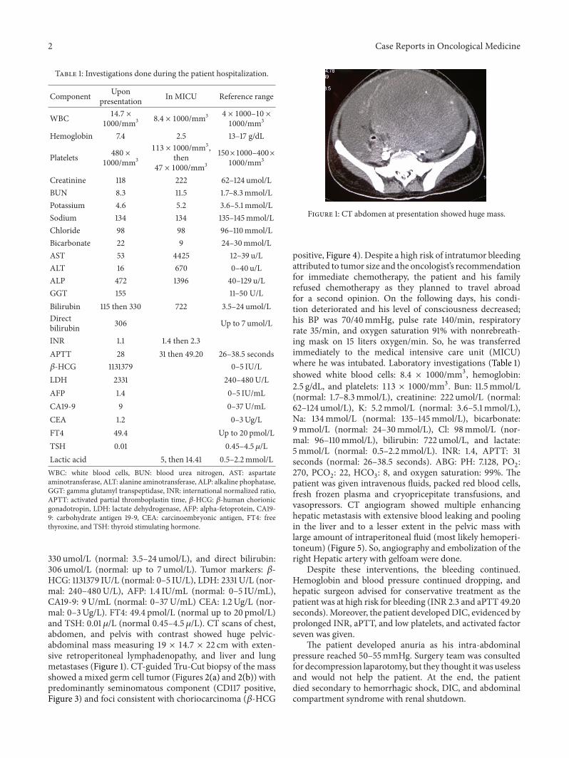
(a) (b)
Figure 2: (a) Foci of neoplastic cells consistent with germ cell tumor (H&E). (b) Foci of neoplastic cells consistent with germ cell tumor(Medium Power, H&E).
Figure 3: CD117 positivity in the seminomatous component.
Figure 4: 𝛽-HCG positivity in foci suggestive of choriocarcinoma-tous component.
3. Discussion
We present the clinical course of a patient with a metastatictesticular cancer, which developed in an undescended testicleand was complicated with choriocarcinoma syndrome ashe got massive bleeding in the hepatic metastases leadingto his death. To the best of our knowledge, the pres-ence of cholestatic jaundice upon presentation, with highdirect bilirubin, alkaline phosphatase, and gamma glutamyltranspeptidase and normal ALT, was only reported once
Figure 5: CT abdomen after development of hemorrhagic shockshowedmultiple heterogeneously enhancingmasses in the liver withperipheral enhancement. Also, there is intraperitoneal fluid.
as an initial symptom secondary to metastasis to hepatichilar lymph nodes which is very rare as usually nonsemi-nomatous germ cell tumors of the testis metastasize tothe retroperitoneal lymph nodes, lung, liver, and brain [3].Careful examination with discovery of only single testicle inthe scrotum helped us to suspect the disease process behindthe patient’s condition, as the ultimate incidence of testiculartumors is about 0.002% in normal males and up to 5% in thecase of intra-abdominal testes [1, page 1127].
The pathogenesis of choriocarcinoma syndrome isunknown. It may be related to tumor invasion of the smallblood vessels [4].
It occurs in two different clinical settings, either fewhours after initiation of combined chemotherapy which ismore common ormuch less likely spontaneously in advanceddisease without relation to treatment as in our patient [5].
Acute hemorrhage in the pulmonary metastasis is thetypical presentation of choriocarcinoma syndrome; however,hemorrhage at any site of metastasis can develop [2]. In ourpatient, hemorrhage was from the liver metastases and thismight be to some extent related to the smaller size of the lungmetastases (the biggest lung nodule measured 2× 2.5 cm, butthe biggest in the liver measured 7 × 6.3 cm).

4 Case Reports in Oncological Medicine
Our patient was found to have severe hyperthyroidism.In general, hyperthyroidism rarely develops as a paraneo-plastic syndrome. It presents in 3.5% of the patients withdisseminated nonseminomatous germ cell tumors and 50%of the patients with HCG above 50000 IU/L versus 0%of the patients with HCG below this level [6]. Thyroidfunction should be checked in patients with high HCG,as hyperthyroidism symptoms resemble those of advancedmetastatic disease and it can be missed. The mechanism ofthe hyperthyroidism is probably due to the ability of HCGto stimulate the TSH receptors, as it has an identical alphasubunit to that of the TSH [6]. Choriocarcinoma syndromehas a poor prognosis, particularly in patients with 𝛽-HCGlevel above 50000 IU/L [4].
This syndrome is a big challenge, where patients withdisseminated disease treated with primary chemotherapyfollowed by surgery have a 5-year disease-free survival rate upto 80% [1, page 1575].The usual chemotherapy protocol is theBEP therapy (bleomycin, etoposide, and cisplatinum). Chori-ocarcinoma syndrome needs early recognition and urgenttreatment which is usually multimodal consisting of medicaltreatment, usually in the intensive care unit, to stabilize thepatient and surgical intervention when appropriate [7].
In conclusion, this case draws attention of our physiciansto the importance of considering this syndromewhile dealingwith patients who have massive and advanced testiculartumorswith high𝛽-HCG, as this syndrome is life-threateningand needs urgent treatment.
Conflict of Interests
The authors declare that they have no conflict of interests.
References
[1] J. M. Stephen,M. Papadakis, andM.W. Rabow,CurrentMedicalDiagnosis and Treatment, McGraw-Hill Medical, San Francisco,Calif, USA, 50th edition, 2011.
[2] C. J. Logothetis, M. L. Samuels, and D. E. Selig, “Cyclicchemotherapy with cyclophosphamide, doxorubicin, and cis-platin plus vinblastine and bleomycin in advanced germinaltumors. Results with 100 patients,” The American Journal ofMedicine, vol. 81, no. 2, pp. 219–227, 1986.
[3] M.Ahsaini, F. Tazi, S.Mellas et al., “Pure choriocarcinomaof thetestis presenting with jaundice: a case report and review of theliterature,” Journal of Medical Case Reports, vol. 6, p. 269, 2012.
[4] M. Shintaku, M. H. Hwang, and R. Amitani, “Primary chori-ocarcinoma of the long manifesting as diffuse alveolar hemor-rhage,” Archives of Pathology and Laboratory Medicine, vol. 130,no. 4, pp. 540–543, 2006.
[5] R. J. Motzer and G. J. Bosl, “Hemorrhage: a complication ofmetastatic testicular choriocarcinoma,” Urology, vol. 30, no. 2,pp. 119–122, 1987.
[6] S. F. Oosting, E. C. de Haas, T. P. Links et al., “Prevalenceof paraneoplastic hyperthyroidism in patients with metastaticnon-seminomatous germ-cell tumors,” Annals of Oncology, vol.21, no. 1, pp. 104–108, 2010.
[7] M. Tatokoro, S. Kawakami, M. Sakura, T. Kobayashi, K.Kihara, and H. Akamatsu, “Successful management of life-threatening choriocarcinoma syndrome with rupture of pul-monary metastatic foci causing hemorrhagic shock,” Interna-tional Journal of Urology, vol. 15, no. 3, pp. 263–264, 2008.

Submit your manuscripts athttp://www.hindawi.com
Stem CellsInternational
Hindawi Publishing Corporationhttp://www.hindawi.com Volume 2014
Hindawi Publishing Corporationhttp://www.hindawi.com Volume 2014
MEDIATORSINFLAMMATION
of
Hindawi Publishing Corporationhttp://www.hindawi.com Volume 2014
Behavioural Neurology
EndocrinologyInternational Journal of
Hindawi Publishing Corporationhttp://www.hindawi.com Volume 2014
Hindawi Publishing Corporationhttp://www.hindawi.com Volume 2014
Disease Markers
Hindawi Publishing Corporationhttp://www.hindawi.com Volume 2014
BioMed Research International
OncologyJournal of
Hindawi Publishing Corporationhttp://www.hindawi.com Volume 2014
Hindawi Publishing Corporationhttp://www.hindawi.com Volume 2014
Oxidative Medicine and Cellular Longevity
Hindawi Publishing Corporationhttp://www.hindawi.com Volume 2014
PPAR Research
The Scientific World JournalHindawi Publishing Corporation http://www.hindawi.com Volume 2014
Immunology ResearchHindawi Publishing Corporationhttp://www.hindawi.com Volume 2014
Journal of
ObesityJournal of
Hindawi Publishing Corporationhttp://www.hindawi.com Volume 2014
Hindawi Publishing Corporationhttp://www.hindawi.com Volume 2014
Computational and Mathematical Methods in Medicine
OphthalmologyJournal of
Hindawi Publishing Corporationhttp://www.hindawi.com Volume 2014
Diabetes ResearchJournal of
Hindawi Publishing Corporationhttp://www.hindawi.com Volume 2014
Hindawi Publishing Corporationhttp://www.hindawi.com Volume 2014
Research and TreatmentAIDS
Hindawi Publishing Corporationhttp://www.hindawi.com Volume 2014
Gastroenterology Research and Practice
Hindawi Publishing Corporationhttp://www.hindawi.com Volume 2014
Parkinson’s Disease
Evidence-Based Complementary and Alternative Medicine
Volume 2014Hindawi Publishing Corporationhttp://www.hindawi.com
![Choriocarcinoma syndrome complicating a mixed testicular ...choriocarcinoma are very rare (0, 3% of all GCT) [8]. βHCG is always secreted by choriocarcinoma and plays an important](https://img.pdfslide.us/doc/110x75/5e366cd2a1f24370d80dcb00/choriocarcinoma-syndrome-complicating-a-mixed-testicular-choriocarcinoma-are.jpg)