Embed Size (px)
Citation preview
ACG CASE REPORTS JOURNAL
acgcasereports.gi.org ACG Case Reports Journal | Volume 3 | Issue 2 | January 2016118
CASE REPORT | BILIARY
No Stone Left Unturned: Using Choledocholithiasis to Open a Papillary Stenosis via a Choledochodudenal FistulaSara West, DO1, and M. Joshua Shellenberger, DO2
1Department of Internal Medicine, Geisinger Medical Center, Danville, PA2Department of Gastroenterology and Hepatology, Geisinger Medical Center, Danville, PA
AbstractIn a patient found to have cholelithiasis and choledocholithiasis, a choledochoduodenal fistula was used to gain access to the bile duct. Due to severe stenosis and atrophy of the major papilla, cannulation was not possible. Stones were purposely impacted in the native ampulla to cause bulging and stretching of the stenosis. Once the stenosis was stretched, the bile and pancreatic duct were accessed via the native ampulla, allowing for stone removal.
IntroductionA choledochoduodenal fistula (CDF) is an abnormal connection between the common bile duct (CBD) and the duodenum. CDF may be incidentally discovered during endoscopic retrograde cholangiopancreatography (ERCP) and is associated with a past history of CBD stones, recurrent biliary tract infections, and less commonly associated with peptic ulcer disease, malignancy, and trauma.1,2
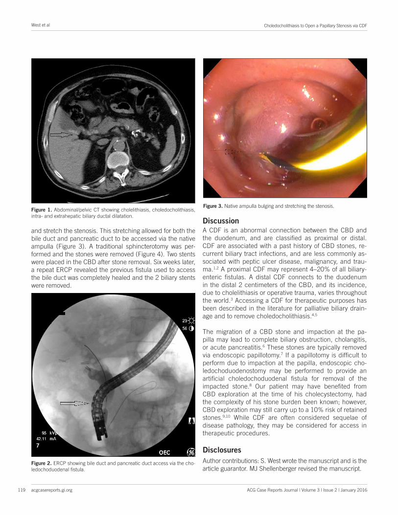
Case ReportA 77-year-old man presented with 2 days of abdominal pain, nausea, and vomiting. Physical exam revealed mild tenderness to palpation in the lower abdomen. Labs showed a leukocytosis of 13.45 K/μL and creatinine of 1.5 mg/dL. Liver function tests and lipase were normal. Abdominal/pelvic computed tomography (CT) without contrast showed extensive cholelithiasis and choledocholithiasis resulting in intra- and extrahepatic biliary ductal dilatation; the CBD measured 1.4 cm (Figure 1).
ERCP showed a CDF and associated severe stenosis and atrophy of the major papilla. Due to the stenosis, can-nulation through the native ampulla was not possible, so the CDF was used to access the bile duct. The fistula was enlarged by making a cut with a sphincterotome. To avoid a peritoneal leak, a limited fistulotomy was per-formed, which hindered complete removal of the stones. Multiple stones were present, but not all were removed, and a stent was placed.
Following ERCP, the patient underwent an uncomplicated laparoscopic cholecystectomy. His pain and nausea resolved completely. The stent was removed during repeat ERCP at 2-month follow-up, and revealed large stones in the bile duct. The fistula was again noted in the mid-bile duct and across a duodenal fold, making it impossible to simply extend the prior fistulotomy to the native ampulla without perforation. Due to the severe stenosis of the native ampulla, the wire could not pass through the ampulla in an anterograde fashion. The stones could not be removed via the fistula. The stones naturally flowed distally and a balloon was placed above to sweep them down to the native ampulla (Figure 2). Once the stones were impacted, this caused the native ampulla to bulge
ACG Case Rep J 2016;3(2):118-120. doi:10.14309/crj.2016.19. Published online: January 20, 2016.
Correspondence: Sara West, 100 North Academy Avenue, Danville, PA 17822 ([email protected]).
Copyright: © 2015 West et al. This work is licensed under a Creative Commons Attribution-NonCommercial-NoDerivatives 4.0 International License. To view a copy of this license, visit http://creativecommons.org/licenses/by-nc-nd/4.0.
Choledocholithiasis to Open a Papillary Stenosis via CDF
acgcasereports.gi.org ACG Case Reports Journal | Volume 3 | Issue 2 | January 2016
West et al
119
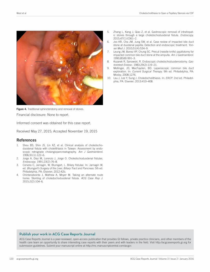
and stretch the stenosis. This stretching allowed for both the bile duct and pancreatic duct to be accessed via the native ampulla (Figure 3). A traditional sphincterotomy was per-formed and the stones were removed (Figure 4). Two stents were placed in the CBD after stone removal. Six weeks later, a repeat ERCP revealed the previous fistula used to access the bile duct was completely healed and the 2 biliary stents were removed.
DiscussionA CDF is an abnormal connection between the CBD and the duodenum, and are classified as proximal or distal. CDF are associated with a past history of CBD stones, re-current biliary tract infections, and are less commonly as-sociated with peptic ulcer disease, malignancy, and trau-ma.1,2 A proximal CDF may represent 4–20% of all biliary-enteric fistulas. A distal CDF connects to the duodenum in the distal 2 centimeters of the CBD, and its incidence, due to cholelithiasis or operative trauma, varies throughout the world.3 Accessing a CDF for therapeutic purposes has been described in the literature for palliative biliary drain-age and to remove choledocholithiasis.4,5
The migration of a CBD stone and impaction at the pa-pilla may lead to complete biliary obstruction, cholangitis, or acute pancreatitis.6 These stones are typically removed via endoscopic papillotomy.7 If a papillotomy is difficult to perform due to impaction at the papilla, endoscopic cho-ledochoduodenostomy may be performed to provide an artificial choledochoduodenal fistula for removal of the impacted stone.8 Our patient may have benefited from CBD exploration at the time of his cholecystectomy, had the complexity of his stone burden been known; however, CBD exploration may still carry up to a 10% risk of retained stones.9,10 While CDF are often considered sequelae of disease pathology, they may be considered for access in therapeutic procedures.
Disclosures
Author contributions: S. West wrote the manuscript and is the article guarantor. MJ Shellenberger revised the manuscript.
Figure 1. Abdominal/pelvic CT showing cholelithiasis, choledocholithiasis, intra- and extrahepatic biliary ductal dilatation.
Figure 3. Native ampulla bulging and stretching the stenosis.
Figure 2. ERCP showing bile duct and pancreatic duct access via the cho-ledochoduodenal fistula.
Publish your work in ACG Case Reports JournalACG Case Reports Journal is a peer-reviewed, open-access publication that provides GI fellows, private practice clinicians, and other members of the health care team an opportunity to share interesting case reports with their peers and with leaders in the field. Visit http://acgcasereports.gi.org for submission guidelines. Submit your manuscript online at http://mc.manuscriptcentral.com/acgcr.
West et al
acgcasereports.gi.org
Choledocholithiasis to Open a Papillary Stenosis via CDF
120 ACG Case Reports Journal | Volume 3 | Issue 2 | January 2016
Financial disclosure: None to report.
Informed consent was obtained for this case report.
Received May 27, 2015; Accepted November 19, 2015 References1. Sheu BS, Shin JS, Lin XZ, et al. Clinical analysis of choledocho-
duodenal fistula with cholelithiasis in Taiwan: Assessment by endo-scopic retrograde cholangiopancreatography. Am J Gastroenterol. 1996;91(1):122–6.
2. Jorge A, Diaz M, Lorenzo J, Jorge O. Choledochoduodenal fistulas. Endoscopy. 1991;23(2):76–8.
3. Corvera C, Jarnagin, W, Blumgart, L. Biliary fistulae. In: Jarnagin W, ed. Blumgart’s Surgery of the Liver, Biliary Tract and Pancreas. 5th ed. Philadelphia, PA: Elsevier; 2012:42b.
4. Chintanabointa J, Mathew A, Moyer M. Taking an alternate route home: Stenting of choledochoduodenal fistula. ACG Case Rep J. 2015;2(2):104–6.
5. Zhang L, Kang J, Qiao Z, et al. Gastroscopic removal of intrahepat-ic stones through a large choledochoduodenal fistula. Endoscopy. 2015;47(1):E361–2.
6. Joo KR, Cha JM, Jung SW, et al. Case review of impacted bile duct stone at duodenal papilla: Detection and endoscopic treatment. Yon-sei Med J. 2010;51(4):534–9.
7. Leung JW, Banez VP, Chung SC. Precut (needle knife) papillotomy for impacted common bile duct stone at the ampulla. Am J Gastroenterol. 1990;85(8):991–3.
8. Kozarek R, Sanowski, R. Endoscopic choledochoduodenostomy. Gas-trointest Endosc. 1983;29(2):119–21.
9. Mellinger, JD, MacFayden, BD. Laparoscopic common bile duct exploration. In: Current Surgical Therapy. 9th ed. Philadelphia, PA: Mosby; 2008:1276.
10. Lau J, Lee Y, Sung J. Choledocholithiasis. In: ERCP. 2nd ed. Philadel-phia, PA: Elsevier; 2013:410–408.
Figure 4. Traditional sphincterotomy and removal of stones.