Embed Size (px)
Citation preview

Case ReportArginine-Restricted Therapy Resistant Bilateral Macular EdemaAssociated with Gyrate Atrophy
Sibel Doguizi, Mehmet Ali Sekeroglu, Mustafa Alpaslan Anayol, and Pelin Yilmazbas
Ulucanlar Eye Training and Research Hospital, Department of Ophthalmology, 06240 Ankara, Turkey
Correspondence should be addressed to Sibel Doguizi; [email protected]
Received 24 October 2015; Accepted 23 November 2015
Academic Editor: Takaaki Hayashi
Copyright © 2015 Sibel Doguizi et al. This is an open access article distributed under the Creative Commons Attribution License,which permits unrestricted use, distribution, and reproduction in any medium, provided the original work is properly cited.
Introduction. Gyrate atrophy is a rare genetical metabolic disorder affecting vision. Here, we report a 9-year-old boy with gyrateatrophy associated with bilateral macular edema at the time of diagnosis and the effect of long term metabolic control on macularedema. Case Presentation. A 9-year-old boy presented with a complaint of low visual acuity (best corrected visual acuity: 20/80in both eyes, refractive error: −12.00D). Dilated fundus examination revealed multiple bilateral, sharply defined, and scallopedchorioretinal atrophy areas in the midperipheral and peripheral zone. Spectral-domain optical coherence tomography revealedbilateral cystoid macular edema in both eyes. Serum ornithine level was high (622𝜇mol/L). An arginine-restricted diet reducedserum ornithine level (55 𝜇mol/L). However, visual findings includingmacular edema remained unchanged in 2 years of follow-up.Conclusion. Arginine-restricted diet did not improve macular edema in our patient with gyrate atrophy. A more comprehensiveunderstanding of the underlying factors for macular edema will lead to the development of effective therapies.
1. Introduction
Gyrate atrophy is a rare, genetically determined, autoso-mal recessive, metabolic disorder associated with increasedplasma ornithine, caused by the deficiency of the vitamin B6-dependent enzyme ornithine ketoacid aminotransferase [1].Patients typically report night blindness, loss of peripheralvision, or low visual acuity in the first or second decade oflife, and these complaints are accompanied by the appearanceof sharply demarcated circular areas of chorioretinal atrophy.As it is a progressive chorioretinal degenerative disorder,the chorioretinal lesions increase in size and number withincreasing age. The macula is apparently the most resis-tant to disease progression [2]. Myopia [2], early posteriorsubcapsular cataract [2], choroidal neovascularization [3],cystoid macular edema [4], epiretinal membrane [4], andmacular hole [5] may also accompany chorioretinal atrophiclesions. In this report, we describe a child with gyrateatrophy associated with bilateral macular edema at the timeof diagnosis, which did not improve within 2 years even afterstrict metabolic control of the disease.
2. Case Presentation
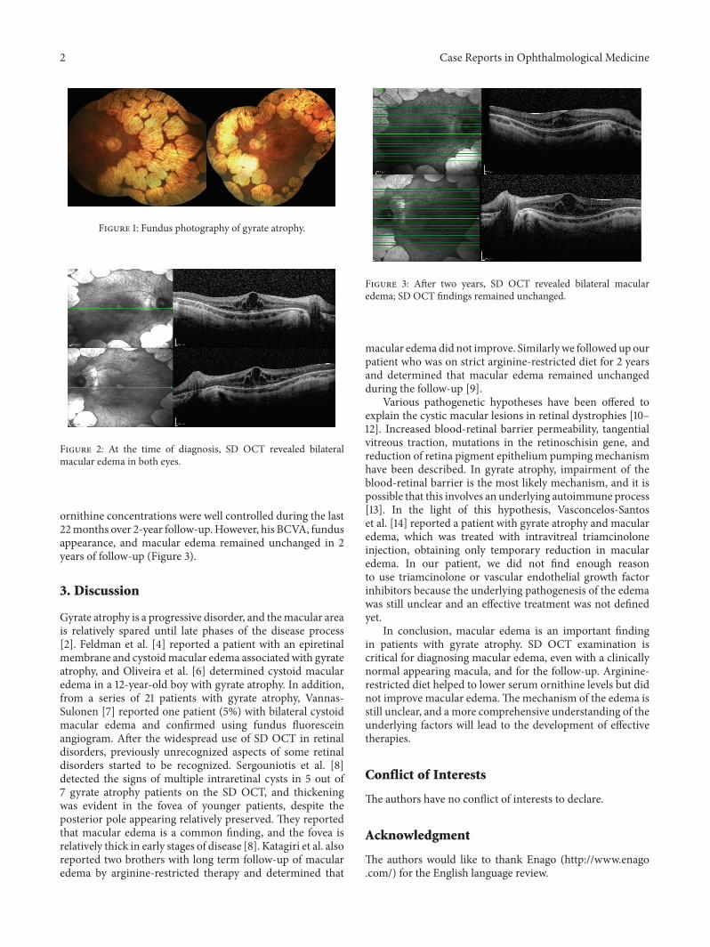
A 9-year-old boy presented with a complaint of low visualacuity. Best corrected visual acuity (BCVA) was 20/80 inboth eyes, with a refractive error of −12.00D. Anteriorsegment examinations were normal bilaterally. Dilated fun-dus examination revealed multiple bilateral, sharply defined,and scalloped chorioretinal atrophy areas in the midperiph-eral and peripheral zone (Figure 1). Spectral-domain opticalcoherence tomography (SD OCT) revealed bilateral cystoidmacular edema in both eyes (Figure 2). With these findings,the gyrate atrophy is the probable diagnosis. Routine bloodtests were normal, but amino acid analysis revealed a highserum ornithine level (622𝜇mol/L), which helps to makethe definite diagnosis. In addition, ornithine level was highin the urine analysis (234 nmol/mg creatinine). Also allfamily members were examined and no similar findingswere found (parents and two sisters). The patient consultedwith a pediatric metabolic disease specialist. After startingto consume an arginine-restricted diet, serum ornithinelevel reduced within two months (55𝜇mol/L). His serum
Hindawi Publishing CorporationCase Reports in Ophthalmological MedicineVolume 2015, Article ID 137270, 3 pageshttp://dx.doi.org/10.1155/2015/137270
2 Case Reports in Ophthalmological Medicine
Figure 1: Fundus photography of gyrate atrophy.
Figure 2: At the time of diagnosis, SD OCT revealed bilateralmacular edema in both eyes.
ornithine concentrations were well controlled during the last22months over 2-year follow-up.However, his BCVA, fundusappearance, and macular edema remained unchanged in 2years of follow-up (Figure 3).
3. Discussion
Gyrate atrophy is a progressive disorder, and themacular areais relatively spared until late phases of the disease process[2]. Feldman et al. [4] reported a patient with an epiretinalmembrane and cystoidmacular edema associatedwith gyrateatrophy, and Oliveira et al. [6] determined cystoid macularedema in a 12-year-old boy with gyrate atrophy. In addition,from a series of 21 patients with gyrate atrophy, Vannas-Sulonen [7] reported one patient (5%) with bilateral cystoidmacular edema and confirmed using fundus fluoresceinangiogram. After the widespread use of SD OCT in retinaldisorders, previously unrecognized aspects of some retinaldisorders started to be recognized. Sergouniotis et al. [8]detected the signs of multiple intraretinal cysts in 5 out of7 gyrate atrophy patients on the SD OCT, and thickeningwas evident in the fovea of younger patients, despite theposterior pole appearing relatively preserved. They reportedthat macular edema is a common finding, and the fovea isrelatively thick in early stages of disease [8]. Katagiri et al. alsoreported two brothers with long term follow-up of macularedema by arginine-restricted therapy and determined that
Figure 3: After two years, SD OCT revealed bilateral macularedema; SD OCT findings remained unchanged.
macular edemadid not improve. Similarlywe followed up ourpatient who was on strict arginine-restricted diet for 2 yearsand determined that macular edema remained unchangedduring the follow-up [9].
Various pathogenetic hypotheses have been offered toexplain the cystic macular lesions in retinal dystrophies [10–12]. Increased blood-retinal barrier permeability, tangentialvitreous traction, mutations in the retinoschisin gene, andreduction of retina pigment epithelium pumping mechanismhave been described. In gyrate atrophy, impairment of theblood-retinal barrier is the most likely mechanism, and it ispossible that this involves an underlying autoimmune process[13]. In the light of this hypothesis, Vasconcelos-Santoset al. [14] reported a patient with gyrate atrophy and macularedema, which was treated with intravitreal triamcinoloneinjection, obtaining only temporary reduction in macularedema. In our patient, we did not find enough reasonto use triamcinolone or vascular endothelial growth factorinhibitors because the underlying pathogenesis of the edemawas still unclear and an effective treatment was not definedyet.
In conclusion, macular edema is an important findingin patients with gyrate atrophy. SD OCT examination iscritical for diagnosing macular edema, even with a clinicallynormal appearing macula, and for the follow-up. Arginine-restricted diet helped to lower serum ornithine levels but didnot improve macular edema.Themechanism of the edema isstill unclear, and a more comprehensive understanding of theunderlying factors will lead to the development of effectivetherapies.
Conflict of Interests
The authors have no conflict of interests to declare.
Acknowledgment
The authors would like to thank Enago (http://www.enago.com/) for the English language review.

Case Reports in Ophthalmological Medicine 3
References
[1] M. J. Potter and E. L. Berson, “Diagnosis and treatment of gyrateatrophy,” International Ophthalmology Clinics, vol. 33, no. 2, pp.229–236, 1993.
[2] K. K. Takki and R. C. Milton, “The natural history of gyrateatrophy of the choroid and retina,” Ophthalmology, vol. 88, no.4, pp. 292–301, 1981.
[3] F. Marano, A. F. Deutman, A. J. L. G. Pinckers, and A. L.Aandekerk, “Gyrate atrophy and choroidal neovascularization,”Archives of Ophthalmology, vol. 114, no. 10, article 1295, 1996.
[4] R. B. Feldman, S. S. Mayo, D.M. Robertson, J. D. Jones, and J. A.Rostvold, “Epiretinalmembranes and cystoidmacular edema ingyrate atrophy of the choroid and retina,” Retina, vol. 9, no. 2,pp. 139–142, 1989.
[5] Y. R. Sharma, D. V. Singh, R. V. Azad, and N. Pal, “Gyrateatrophy with bilateral full thickness macular hole,” Eye, vol. 20,no. 6, pp. 745–747, 2006.
[6] T. L. Oliveira, R. E. Andrade, C. Muccioli, J. Sallum, andR. Belfort Jr., “Cystoid macular edema in gyrate atrophy ofthe choroid and retina: a fluorescein angiography and opticalcoherence tomography evaluation,” The American Journal ofOphthalmology, vol. 140, no. 1, pp. 147–149, 2005.
[7] K. Vannas-Sulonen, “Progression of gyrate atrophy of thechoroid and retina. A long-term follow-up by fluoresceinangiography,” Acta Ophthalmologica, vol. 65, no. 1, pp. 101–109,1987.
[8] P. I. Sergouniotis, A. E. Davidson, E. Lenassi, S. R. Devery, A.T. Moore, and A. R. Webster, “Retinal structure, function, andmolecular pathologic features in gyrate atrophy,” Ophthalmol-ogy, vol. 119, no. 3, pp. 596–605, 2012.
[9] S. Katagiri, T. Gekka, T. Hayashi et al., “OAT mutations andclinical features in two Japanese brothers with gyrate atrophy ofthe choroid and retina,” Documenta Ophthalmologica, vol. 128,no. 2, pp. 137–148, 2014.
[10] G. A. Fishman, J. Cunha-Vaz, and T. Salzano, “Vitreous fluo-rophotometry in patients with retinitis pigmentosa,”Archives ofOphthalmology, vol. 99, no. 7, pp. 1202–1207, 1981.
[11] M. Larsen, C. B. Engler, M. Haim, and H. Lund-Andersen,“Blood-retina barrier permeability is independent of tracesubstance lipid solubility in retinitis pigmentosa and in thehealthy eye,” International Ophthalmology, vol. 21, no. 4, pp.229–234, 1997.
[12] H. Ozdemir, M. Karacorlu, and S. Karacorlu, “Intravitreal tri-amcinolone acetonide for treatment of cystoidmacular oedemain patients with retinitis pigmentosa,” Acta OphthalmologicaScandinavica, vol. 83, no. 2, pp. 248–251, 2005.
[13] S. Salvatore, G. A. Fishman, and M. A. Genead, “Treatment ofcystic macular lesions in hereditary retinal dystrophies,” Surveyof Ophthalmology, vol. 58, no. 6, pp. 560–584, 2013.
[14] D. V. Vasconcelos-Santos, E. P. Magalhaes, and M. B. Nehemy,“Macular edema associated with gyrate atrophy managed withintravitreal triamcinolone: a case report,”Arquivos Brasileiros deOftalmologia, vol. 70, no. 5, pp. 858–861, 2007.

Submit your manuscripts athttp://www.hindawi.com
Stem CellsInternational
Hindawi Publishing Corporationhttp://www.hindawi.com Volume 2014
Hindawi Publishing Corporationhttp://www.hindawi.com Volume 2014
MEDIATORSINFLAMMATION
of
Hindawi Publishing Corporationhttp://www.hindawi.com Volume 2014
Behavioural Neurology
EndocrinologyInternational Journal of
Hindawi Publishing Corporationhttp://www.hindawi.com Volume 2014
Hindawi Publishing Corporationhttp://www.hindawi.com Volume 2014
Disease Markers
Hindawi Publishing Corporationhttp://www.hindawi.com Volume 2014
BioMed Research International
OncologyJournal of
Hindawi Publishing Corporationhttp://www.hindawi.com Volume 2014
Hindawi Publishing Corporationhttp://www.hindawi.com Volume 2014
Oxidative Medicine and Cellular Longevity
Hindawi Publishing Corporationhttp://www.hindawi.com Volume 2014
PPAR Research
The Scientific World JournalHindawi Publishing Corporation http://www.hindawi.com Volume 2014
Immunology ResearchHindawi Publishing Corporationhttp://www.hindawi.com Volume 2014
Journal of
ObesityJournal of
Hindawi Publishing Corporationhttp://www.hindawi.com Volume 2014
Hindawi Publishing Corporationhttp://www.hindawi.com Volume 2014
Computational and Mathematical Methods in Medicine
OphthalmologyJournal of
Hindawi Publishing Corporationhttp://www.hindawi.com Volume 2014
Diabetes ResearchJournal of
Hindawi Publishing Corporationhttp://www.hindawi.com Volume 2014
Hindawi Publishing Corporationhttp://www.hindawi.com Volume 2014
Research and TreatmentAIDS
Hindawi Publishing Corporationhttp://www.hindawi.com Volume 2014
Gastroenterology Research and Practice
Hindawi Publishing Corporationhttp://www.hindawi.com Volume 2014
Parkinson’s Disease
Evidence-Based Complementary and Alternative Medicine
Volume 2014Hindawi Publishing Corporationhttp://www.hindawi.com


![Arginine...Arginine vasotocin ([8-arginine]-oxytocin) (AVT), the primary antidiuretic principle in submammalian vertebrates, has been reported to be present in mammalian pituitary](https://img.pdfslide.us/doc/110x75/5e81a7e1761a1c6f5832a8ca/arginine-arginine-vasotocin-8-arginine-oxytocin-avt-the-primary-antidiuretic.jpg)