Embed Size (px)
Citation preview
Case ReportAcute Paraplegia as a Presentation of Aortic Saddle Embolism
Lisandro Irizarry, Anton Wray, and Kim Guishard
Department of Emergency Medicine, Wyckoff Heights Medical Center, Brooklyn, NY 11237, USA
Correspondence should be addressed to Lisandro Irizarry; [email protected]
Received 29 April 2016; Revised 23 August 2016; Accepted 25 September 2016
Academic Editor: Serdar Kula
Copyright © 2016 Lisandro Irizarry et al. This is an open access article distributed under the Creative Commons AttributionLicense, which permits unrestricted use, distribution, and reproduction in any medium, provided the original work is properlycited.
Background. Acute onset paraplegia has a myriad of causes most often of a nonvascular origin. Vascular etiologies are infrequentcauses and most often associated with postsurgical complications. Objective. To describe the occurrence and possible mechanismfor aortic saddle embolism as a rare cause of acute paraplegia.Case Report. Described is a case of a 46-year-old female who presentedwith the sudden onset of nontraumatic low back pain with rapidly progressive paraplegia which was subsequently determined tobe of vascular origin.
1. Introduction
Lowback pain is a commonpresenting complaint comprisingapproximately 3% of all emergency department visits, themajority of which are the result of injuries sustained at home[1]. A small percentage of similar individuals presenting in aprimary care setting develop concomitant neurologic symp-toms [2]. The onset of nontraumatic paraplegia represents asignificant event for the patient and an emergent diagnosticchallenge for the clinician. The consequence for the patientfrom either a delay or misdiagnosis can be catastrophic. Theetiology of paraplegia is broad and includes disorders of thespinal cord which encompass external compression, infec-tion, ischemia, and other nonspinal disorders. A structuredapproach to early intervention andmanagement is imperativeif neurologic function is to be preserved. Presented is a rarecase of aortic saddle embolism associated with the suddenonset of low back pain and acute onset paraplegia.
2. Case Report
A46-year-old female presented to the emergency department(ED) with a complaint of the sudden onset of low back pain.The symptoms began shortly prior to arrival when the patientexperienced sudden onset of severe pain upon standing froma seated position. The pain was rated 10/10 and was localizedacross the low lumbar area with radiation to both legs. Thepatient took no analgesics and self-transported to the ED.
On ED presentation, the patient was noted to be unableto ambulate secondary to pain. Her initial vital signs were asfollows: blood pressure of 177/99; heart rate of 88 beats perminute and regular; respiratory rate of 18 breaths per minutewith an oxygenation saturation of 99% on room air; and anoral temperature of 99∘F. The patient was an obese femalewith a history of untreated pernicious anemia and significantsmoking history. The patient had not seen a physician inmore than three years and had no recent illness or injuries.Examination revealed diffuse lumbar tenderness with nor-mal lower extremity strength and gross sensation. Detailedsensory examination could not be undertaken due to patientdistress at the time of evaluation. Posterior tibial and dorsalispedis pulses were noted to be present bilaterally. Reflexeswere found to be 2+ symmetrically with no evidence ofsaddle anesthesia. Shortly after presentation, the patient wasnoted to have urinated on herself because of a sense of“urgency.” Given this new finding, it was decided to imme-diately perform a noncontrast lumbar CT which showed noevidence of acute abnormalities. The patient was providedanalgesics and a decisionwasmade tomonitor pain response.
Forty-five minutes after the analgesics were given, thepatient experienced new onset of right lower extremity weak-ness. An emergent lumbarMRIwas performedwhich showedno acute abnormalities.The patient continued to complain ofsevere back pain andprogressivelyworsening lower extremityweakness which now included both lower extremities withthe new onset of paraesthesias. Reexamination revealed
Hindawi Publishing CorporationCase Reports in Emergency MedicineVolume 2016, Article ID 1250153, 4 pageshttp://dx.doi.org/10.1155/2016/1250153
2 Case Reports in Emergency Medicine
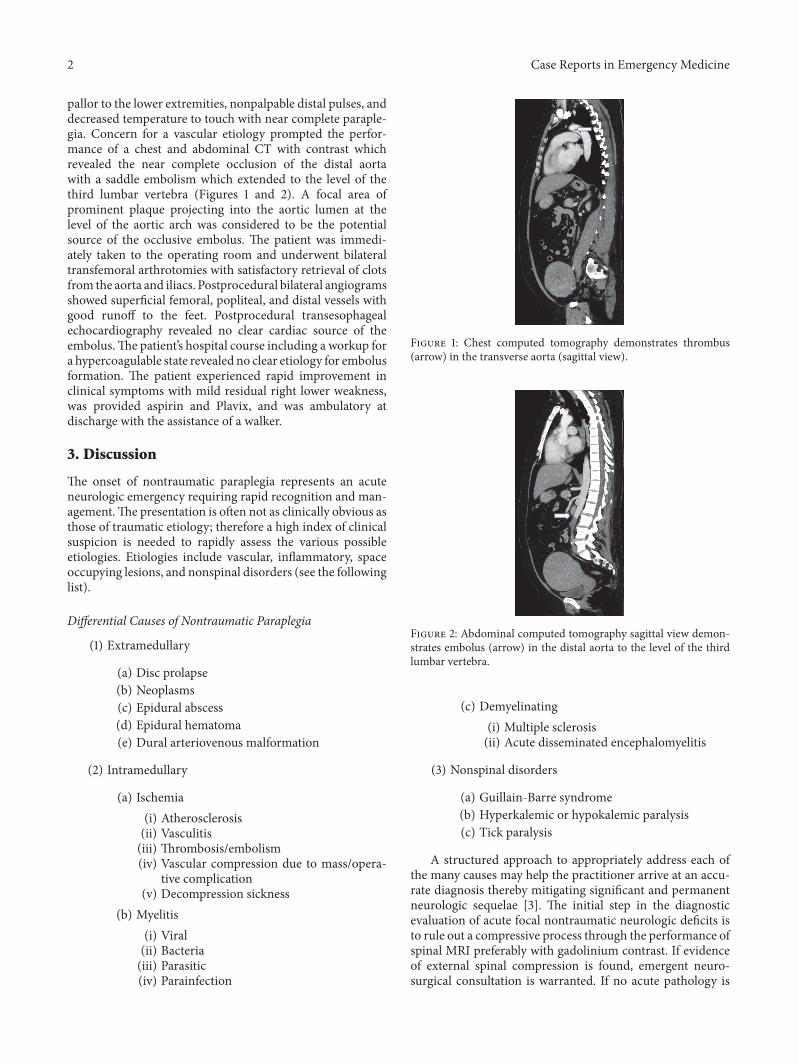
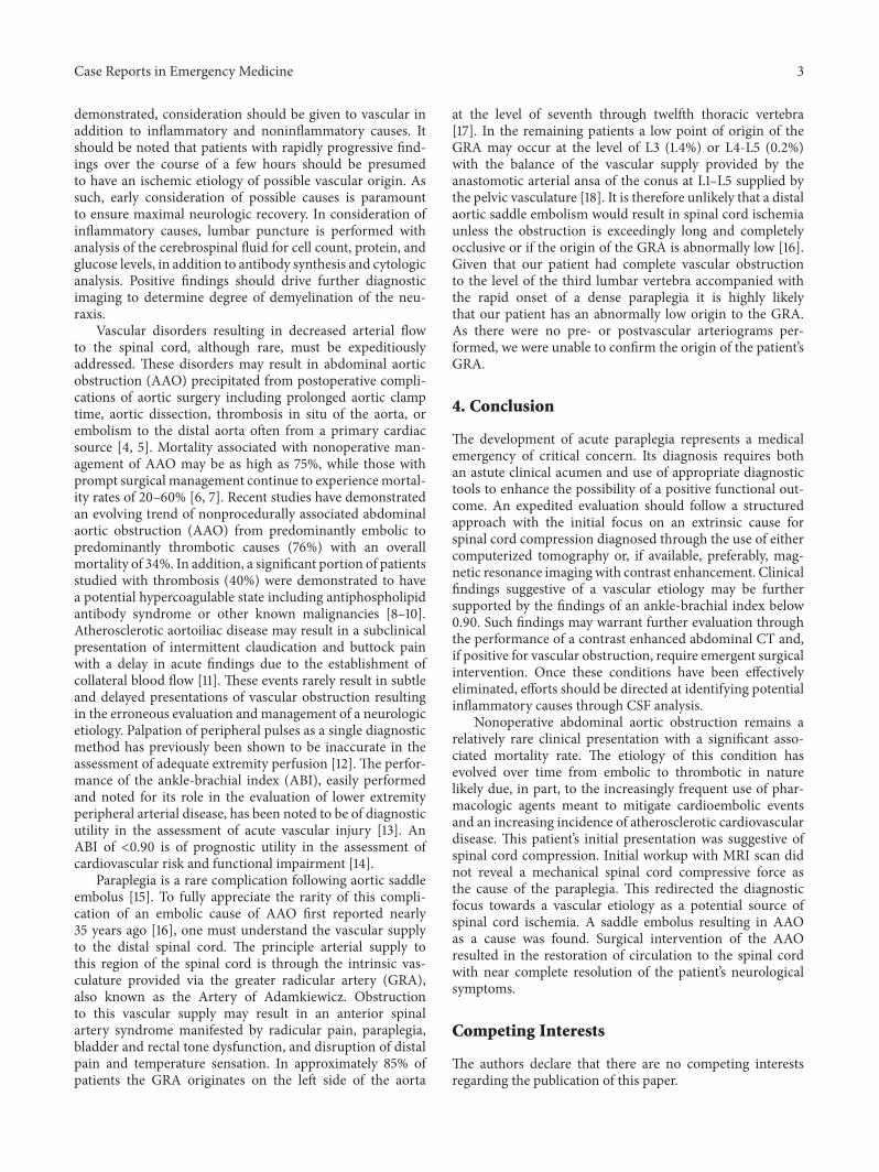
pallor to the lower extremities, nonpalpable distal pulses, anddecreased temperature to touch with near complete paraple-gia. Concern for a vascular etiology prompted the perfor-mance of a chest and abdominal CT with contrast whichrevealed the near complete occlusion of the distal aortawith a saddle embolism which extended to the level of thethird lumbar vertebra (Figures 1 and 2). A focal area ofprominent plaque projecting into the aortic lumen at thelevel of the aortic arch was considered to be the potentialsource of the occlusive embolus. The patient was immedi-ately taken to the operating room and underwent bilateraltransfemoral arthrotomies with satisfactory retrieval of clotsfrom the aorta and iliacs. Postprocedural bilateral angiogramsshowed superficial femoral, popliteal, and distal vessels withgood runoff to the feet. Postprocedural transesophagealechocardiography revealed no clear cardiac source of theembolus.The patient’s hospital course including a workup fora hypercoagulable state revealed no clear etiology for embolusformation. The patient experienced rapid improvement inclinical symptoms with mild residual right lower weakness,was provided aspirin and Plavix, and was ambulatory atdischarge with the assistance of a walker.
3. Discussion
The onset of nontraumatic paraplegia represents an acuteneurologic emergency requiring rapid recognition and man-agement.The presentation is often not as clinically obvious asthose of traumatic etiology; therefore a high index of clinicalsuspicion is needed to rapidly assess the various possibleetiologies. Etiologies include vascular, inflammatory, spaceoccupying lesions, and nonspinal disorders (see the followinglist).
Differential Causes of Nontraumatic Paraplegia
(1) Extramedullary
(a) Disc prolapse(b) Neoplasms(c) Epidural abscess(d) Epidural hematoma(e) Dural arteriovenous malformation
(2) Intramedullary
(a) Ischemia(i) Atherosclerosis(ii) Vasculitis(iii) Thrombosis/embolism(iv) Vascular compression due to mass/opera-
tive complication(v) Decompression sickness
(b) Myelitis(i) Viral(ii) Bacteria(iii) Parasitic(iv) Parainfection
Figure 1: Chest computed tomography demonstrates thrombus(arrow) in the transverse aorta (sagittal view).
Figure 2: Abdominal computed tomography sagittal view demon-strates embolus (arrow) in the distal aorta to the level of the thirdlumbar vertebra.
(c) Demyelinating(i) Multiple sclerosis(ii) Acute disseminated encephalomyelitis
(3) Nonspinal disorders
(a) Guillain-Barre syndrome(b) Hyperkalemic or hypokalemic paralysis(c) Tick paralysis
A structured approach to appropriately address each ofthe many causes may help the practitioner arrive at an accu-rate diagnosis thereby mitigating significant and permanentneurologic sequelae [3]. The initial step in the diagnosticevaluation of acute focal nontraumatic neurologic deficits isto rule out a compressive process through the performance ofspinal MRI preferably with gadolinium contrast. If evidenceof external spinal compression is found, emergent neuro-surgical consultation is warranted. If no acute pathology is
Case Reports in Emergency Medicine 3
demonstrated, consideration should be given to vascular inaddition to inflammatory and noninflammatory causes. Itshould be noted that patients with rapidly progressive find-ings over the course of a few hours should be presumedto have an ischemic etiology of possible vascular origin. Assuch, early consideration of possible causes is paramountto ensure maximal neurologic recovery. In consideration ofinflammatory causes, lumbar puncture is performed withanalysis of the cerebrospinal fluid for cell count, protein, andglucose levels, in addition to antibody synthesis and cytologicanalysis. Positive findings should drive further diagnosticimaging to determine degree of demyelination of the neu-raxis.
Vascular disorders resulting in decreased arterial flowto the spinal cord, although rare, must be expeditiouslyaddressed. These disorders may result in abdominal aorticobstruction (AAO) precipitated from postoperative compli-cations of aortic surgery including prolonged aortic clamptime, aortic dissection, thrombosis in situ of the aorta, orembolism to the distal aorta often from a primary cardiacsource [4, 5]. Mortality associated with nonoperative man-agement of AAO may be as high as 75%, while those withprompt surgical management continue to experiencemortal-ity rates of 20–60% [6, 7]. Recent studies have demonstratedan evolving trend of nonprocedurally associated abdominalaortic obstruction (AAO) from predominantly embolic topredominantly thrombotic causes (76%) with an overallmortality of 34%. In addition, a significant portion of patientsstudied with thrombosis (40%) were demonstrated to havea potential hypercoagulable state including antiphospholipidantibody syndrome or other known malignancies [8–10].Atherosclerotic aortoiliac disease may result in a subclinicalpresentation of intermittent claudication and buttock painwith a delay in acute findings due to the establishment ofcollateral blood flow [11]. These events rarely result in subtleand delayed presentations of vascular obstruction resultingin the erroneous evaluation and management of a neurologicetiology. Palpation of peripheral pulses as a single diagnosticmethod has previously been shown to be inaccurate in theassessment of adequate extremity perfusion [12]. The perfor-mance of the ankle-brachial index (ABI), easily performedand noted for its role in the evaluation of lower extremityperipheral arterial disease, has been noted to be of diagnosticutility in the assessment of acute vascular injury [13]. AnABI of <0.90 is of prognostic utility in the assessment ofcardiovascular risk and functional impairment [14].
Paraplegia is a rare complication following aortic saddleembolus [15]. To fully appreciate the rarity of this compli-cation of an embolic cause of AAO first reported nearly35 years ago [16], one must understand the vascular supplyto the distal spinal cord. The principle arterial supply tothis region of the spinal cord is through the intrinsic vas-culature provided via the greater radicular artery (GRA),also known as the Artery of Adamkiewicz. Obstructionto this vascular supply may result in an anterior spinalartery syndrome manifested by radicular pain, paraplegia,bladder and rectal tone dysfunction, and disruption of distalpain and temperature sensation. In approximately 85% ofpatients the GRA originates on the left side of the aorta
at the level of seventh through twelfth thoracic vertebra[17]. In the remaining patients a low point of origin of theGRA may occur at the level of L3 (1.4%) or L4-L5 (0.2%)with the balance of the vascular supply provided by theanastomotic arterial ansa of the conus at L1–L5 supplied bythe pelvic vasculature [18]. It is therefore unlikely that a distalaortic saddle embolism would result in spinal cord ischemiaunless the obstruction is exceedingly long and completelyocclusive or if the origin of the GRA is abnormally low [16].Given that our patient had complete vascular obstructionto the level of the third lumbar vertebra accompanied withthe rapid onset of a dense paraplegia it is highly likelythat our patient has an abnormally low origin to the GRA.As there were no pre- or postvascular arteriograms per-formed, we were unable to confirm the origin of the patient’sGRA.
4. Conclusion
The development of acute paraplegia represents a medicalemergency of critical concern. Its diagnosis requires bothan astute clinical acumen and use of appropriate diagnostictools to enhance the possibility of a positive functional out-come. An expedited evaluation should follow a structuredapproach with the initial focus on an extrinsic cause forspinal cord compression diagnosed through the use of eithercomputerized tomography or, if available, preferably, mag-netic resonance imaging with contrast enhancement. Clinicalfindings suggestive of a vascular etiology may be furthersupported by the findings of an ankle-brachial index below0.90. Such findings may warrant further evaluation throughthe performance of a contrast enhanced abdominal CT and,if positive for vascular obstruction, require emergent surgicalintervention. Once these conditions have been effectivelyeliminated, efforts should be directed at identifying potentialinflammatory causes through CSF analysis.
Nonoperative abdominal aortic obstruction remains arelatively rare clinical presentation with a significant asso-ciated mortality rate. The etiology of this condition hasevolved over time from embolic to thrombotic in naturelikely due, in part, to the increasingly frequent use of phar-macologic agents meant to mitigate cardioembolic eventsand an increasing incidence of atherosclerotic cardiovasculardisease. This patient’s initial presentation was suggestive ofspinal cord compression. Initial workup with MRI scan didnot reveal a mechanical spinal cord compressive force asthe cause of the paraplegia. This redirected the diagnosticfocus towards a vascular etiology as a potential source ofspinal cord ischemia. A saddle embolus resulting in AAOas a cause was found. Surgical intervention of the AAOresulted in the restoration of circulation to the spinal cordwith near complete resolution of the patient’s neurologicalsymptoms.
Competing Interests
The authors declare that there are no competing interestsregarding the publication of this paper.
4 Case Reports in Emergency Medicine
References
[1] B. R. Waterman, P. J. Belmont Jr., and A. J. Schoenfeld, “Lowback pain in the United States: incidence and risk factors forpresentation in the emergency setting,” Spine Journal, vol. 12,no. 1, pp. 63–70, 2012.
[2] R. A. Deyo and J. N. Weinstein, “Low back pain,”The New Eng-land Journal of Medicine, vol. 344, no. 5, pp. 363–370, 2001.
[3] G. C. Roman, “Proposed diagnostic criteria and nosology ofacute transverse myelitis,”Neurology, vol. 60, no. 4, pp. 730–731,2003.
[4] D. Rosenthal and Southern Association for Vascular Surgery,“Spinal cord ischemia after abdominal aortic operation: is itpreventable?” Journal of Vascular Surgery, vol. 30, no. 3, pp. 391–399, 1999.
[5] J. Inamasu, S. Hori, M. Yokoyama, T. Funabiki, K. Aoki, and N.Aikawa, “Paraplegia caused by painless acute aortic dissection,”Spinal Cord, vol. 38, no. 11, pp. 702–704, 2000.
[6] S. S. N. Wong, G. Roche-Nagle, and G. Oreopoulos, “Acutethrombosis of an abdominal aortic aneurysm presenting ascauda equina syndrome,” Journal of Vascular Surgery, vol. 57, no.1, pp. 218–220, 2013.
[7] H. Yamamoto, F. Yamamoto, F. Tanaka et al., “Acute occlusionof the abdominal aorta with concomitant internal iliac arteryocclusion,” Annals of Thoracic and Cardiovascular Surgery, vol.17, no. 4, pp. 422–427, 2011.
[8] C. D. Dossa, A. D. Shepard, D. J. Reddy et al., “Acute aorticocclusion. A 40-year experience,” Archives of Surgery, vol. 129,no. 6, pp. 603–608, 1994.
[9] J. D. Crawford, K. H. Perrone, V. W. Wong et al., “A modernseries of acute aortic occlusion,” Journal of Vascular Surgery, vol.59, no. 4, pp. 1044–1050, 2014.
[10] R. L. Mcclain and S.-L. Pai, “Acute aortic occlusion presentingas paraplegia: a catastrophic complication in an elective surgicalpatient,” A&A Case Reports, vol. 1, no. 4, pp. 64–66, 2013.
[11] C.-H. Lai, C.-H. Wang, S.-Y. Wu, and H.-M. Shih, “Aortoiliacocclusive disease presenting as sudden onset paraplegia,”Annalsof Vascular Surgery, vol. 28, no. 5, pp. 1321.e5–1321.e7, 2014.
[12] M. Lundin, J.-P. Wiksten, T. Perakyla et al., “Distal pulsepalpation: is it reliable?” World Journal of Surgery, vol. 23, no.3, pp. 252–255, 1999.
[13] W. J. Mills, D. P. Barei, and P. McNair, “The value of theankle-brachial index for diagnosing arterial injury after kneedislocation: a prospective study,” Journal of Trauma—Injury,Infection & Critical Care, vol. 56, no. 6, pp. 1261–1265, 2004.
[14] V. Aboyans, M. H. Criqui, P. Abraham et al., “Measurementand interpretation of the Ankle-Brachial Index: a scientificstatement from the American Heart Association,” Circulation,vol. 126, no. 24, pp. 2890–2909, 2012.
[15] J. R. Silver and P. H. Buxton, “Spinal stroke,” Brain, vol. 97, no.3, pp. 539–550, 1974.
[16] A. P. Dickson, S. K. Lum, andA. S.Whyte, “Paraplegia followingsaddle embolism,”British Journal of Surgery, vol. 71, no. 4, p. 321,1984.
[17] G. K. Triantafyllopoulos, M. Athanassacopoulos, C. Maltezos,and S. G. Pneumaticos, “Acute infrarenal aortic thrombosis pre-sentingwith flaccid paraplegia,” Spine, vol. 36, no. 15, pp. E1042–E1045, 2011.
[18] A. Shaw, H. Anwar, J. Targett, and K. Lafferty, “Cauda equinasyndrome versus saddle embolism,” Annals of the Royal Collegeof Surgeons of England, vol. 90, no. 6, pp. W6–W8, 2008.

Submit your manuscripts athttp://www.hindawi.com
Stem CellsInternational
Hindawi Publishing Corporationhttp://www.hindawi.com Volume 2014
Hindawi Publishing Corporationhttp://www.hindawi.com Volume 2014
MEDIATORSINFLAMMATION
of
Hindawi Publishing Corporationhttp://www.hindawi.com Volume 2014
Behavioural Neurology
EndocrinologyInternational Journal of
Hindawi Publishing Corporationhttp://www.hindawi.com Volume 2014
Hindawi Publishing Corporationhttp://www.hindawi.com Volume 2014
Disease Markers
Hindawi Publishing Corporationhttp://www.hindawi.com Volume 2014
BioMed Research International
OncologyJournal of
Hindawi Publishing Corporationhttp://www.hindawi.com Volume 2014
Hindawi Publishing Corporationhttp://www.hindawi.com Volume 2014
Oxidative Medicine and Cellular Longevity
Hindawi Publishing Corporationhttp://www.hindawi.com Volume 2014
PPAR Research
The Scientific World JournalHindawi Publishing Corporation http://www.hindawi.com Volume 2014
Immunology ResearchHindawi Publishing Corporationhttp://www.hindawi.com Volume 2014
Journal of
ObesityJournal of
Hindawi Publishing Corporationhttp://www.hindawi.com Volume 2014
Hindawi Publishing Corporationhttp://www.hindawi.com Volume 2014
Computational and Mathematical Methods in Medicine
OphthalmologyJournal of
Hindawi Publishing Corporationhttp://www.hindawi.com Volume 2014
Diabetes ResearchJournal of
Hindawi Publishing Corporationhttp://www.hindawi.com Volume 2014
Hindawi Publishing Corporationhttp://www.hindawi.com Volume 2014
Research and TreatmentAIDS
Hindawi Publishing Corporationhttp://www.hindawi.com Volume 2014
Gastroenterology Research and Practice
Hindawi Publishing Corporationhttp://www.hindawi.com Volume 2014
Parkinson’s Disease
Evidence-Based Complementary and Alternative Medicine
Volume 2014Hindawi Publishing Corporationhttp://www.hindawi.com