Embed Size (px)
Citation preview
Case ReportA Rare Case of Melanosis of the Hard Palate Mucosa ina Patient with Chronic Myeloid Leukemia
Umberto Romeo,1 Gaspare Palaia,1 Paolo Junior Fantozzi,1
Gianluca Tenore,1 and Daniela Bosco2
1Department of Oral and Maxillofacial Sciences, “Sapienza” University of Rome, Via Caserta 6, 00161 Rome, Italy2Department of Radiological, Oncological and Anatomo-Pathological Sciences, “Sapienza” University of Rome,Viale Regina Elena 324, 00161 Rome, Italy
Correspondence should be addressed to Gaspare Palaia; [email protected]
Received 31 May 2015; Revised 1 September 2015; Accepted 6 September 2015
Academic Editor: Noam Yarom
Copyright © 2015 Umberto Romeo et al. This is an open access article distributed under the Creative Commons AttributionLicense, which permits unrestricted use, distribution, and reproduction in any medium, provided the original work is properlycited.
Imatinib Mesylate, also known as Gleevec or ST1-571, is a tyrosine-kinase inhibitor used as the gold standard medication for thechronic myeloid leukemia (CML); Imatinib has indeed deeply revolutionized the CML therapy allowing most patients to have agood quality of life. Despite its beneficial effects, Imatinib has significant side effects such as mucosal pigmentation. A 72-year-oldfemale having an Imatinib induced mucosal pigmentation is presented: she has been treated with Imatinib since 2003 and only in2014 discovered, during a routine dental visit, having a pigmented lesion on her hard palate mucosa. Histopathologically, the lesionshows the deposition of fine dark brown spherical bodies within the lamina propria and cloaked in between the collagen fibers.There was no sign of inflammation, hyperplasia, or hemorrhage in the tissue.
1. Introduction
Pigmented lesions of the oral cavity are common oral lesionsthat may present as a solitary lesion, as a manifestation ofsystemic pathology, or as a collateral effect of drug therapy[1]. The etiology of oral pigmentation may range from simplebenign lesions, such as a blue nevus, to complex and malig-nant disorders, such as oral melanoma. The differential diag-nosis includes physiological pigmentation such as those typ-ically seen in African Americans, postinflammatory melano-sis, amalgam tattoo, smoking related pigmentation, bluenevus, and malignant melanoma. Peutz-Jeghers syndrome,Addison disease, and some other rare diseases, such as poly-ostotic dysplasia, hyperthyroidism, and Nelson syndrome,could be also associated with an oral pigmented lesion.
Moreover, a number of drugs, such as antimalarials,tetracyclines, and antiretroviral and chemotherapeutic drugs,may also cause oral pigmentations. One of these chemother-apeutic drugs, Imatinib Mesylate, also known as Gleevec orST1-571, is a tyrosine-kinase inhibitor that targets Bcr-Ablprotein, c-Kit, and platelet derived growth factor receptor
and is used as a first-line treatment for chronic myeloidleukemia (CML) and gastrointestinal stromal tumor (GIST)[2, 3]. Chronic myeloid leukemia is a slow-growing tumorof white blood cells, characterized by an unregulated growthof the myeloid precursor cells and its accumulation both inthe bone marrow and the lymphoid organs. Chronic myeloidleukemia is more common inmales than females and appearsmore in patients between 25 and 60 years. The cause of CMLis the translocation of regions of the BCR and ABL genes toform a BCR-ABL fusion gene. In at least 90 percent of cases,this event is a reciprocal translocation termed t(9;22), whichforms the Philadelphia (Ph) chromosome. The product ofthe BCR-ABL gene, the BCR-ABL protein, is a constitutivelyactive protein tyrosine kinase with an important role in theregulation of cell growth [4]; its presence is a strong indicatorfor CML (since 95% of people with CML have it) but notsufficient on its own for a diagnosis, because 30% of peoplewith ALL will also show Ph+ in their genome. The 9;22translocation leads to a chimeric gene fusion through thebonding of Abl-1 (Abelson) gene located on chromosome9, with a part of the Bcr (breakpoint cluster region) on a
Hindawi Publishing CorporationCase Reports in DentistryVolume 2015, Article ID 817094, 3 pageshttp://dx.doi.org/10.1155/2015/817094
2 Case Reports in Dentistry
Figure 1: Diffuse, blue-grey pigmented lesion of the hard palatemucosa of our case.
truncated chromosome 22. This way, BCR-ABL acts as anoncogene that overexpresses a tyrosine-kinase protein thatstimulates the leukemic growth of myeloblasts [5]. Chronicmyeloid leukemia has been treated for years with bothhydroxyurea and bone marrow transplant, which, until thediscovery of Imatinib, has been the only successful therapy in70% of the cases.
With the introduction of Imatinib Mesylate (ST1-571 orGleevec) the therapeutic options have significantly improved.This treatment modality allows most patients to have agood quality of life when compared to the other chemother-apy drugs [6–8]. Despite its enormous therapeutic effects,Gleevec has very common adverse effects including nau-sea, fluid retention, lowered resistance to infection (dueto neutropenia), and, sometimes, congestive cardiac failure.Dermatological side effects have also been very common suchas rash, superficial edema, GVH-like disease, erythroderma,and lichenoid eruptions. On the other hand, intraoral sideeffects seem to be very rare, such as lichenoid reactions anddental and oral mucosal pigmentation [9].
Herein we report a case of oral pigmentation involvingthe hard palate mucosa in a patient on Imatinib therapy.
2. Case Report
A 72-year-old Spanish woman was referred in October 2014from her regular dentist to the Unit of Oral Medicine,Sapienza University of Rome, for evaluation of a pigmentedlesion on the hard palate mucosa. The lesion, discoveredduring a routine dental examination, appeared as a single,flat, painless lesion, with a blue-grey color, blurred edges, andwas located in the center of the hard palate mucosa. Therewere areas of physiologic mucosa above the palatal raphe(Figure 1). The patient was unaware of the lesion, and shestated that she had no history of trauma to the hard palate.
Her medical history was significant for gastroesophagealreflux disease (GERD), diuresis, and CML. She is beingtreated with Imatinib Mesilate 400mg per day since 2003 forCML. She is also taking medications for GERD (esomepra-zole) and for diuresis (amiloride HCl-hydrochlorothiazide).
The patient stated she has never taken minocycline,hydroxyurea, and/or antimalarial drugs. Furthermore shereports she has never smoked or drank alcohol and has never
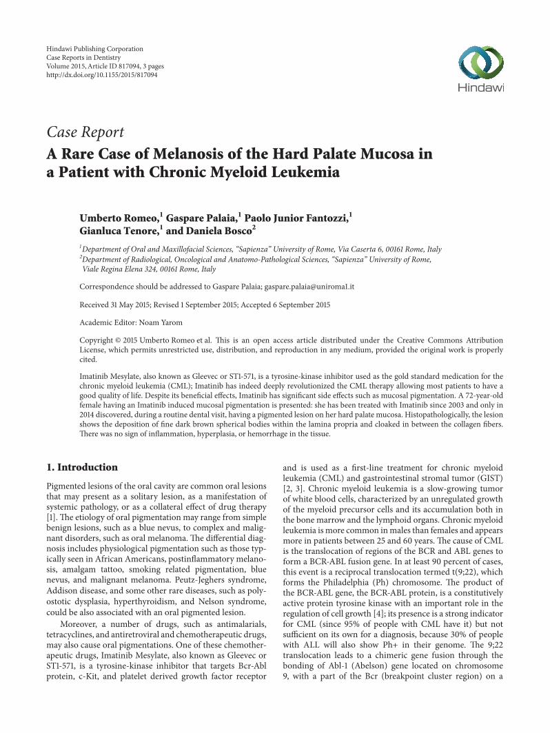
Figure 2: Palatal mucosa with a minor salivary gland containingpigment within the lamina propria (HE ×40).
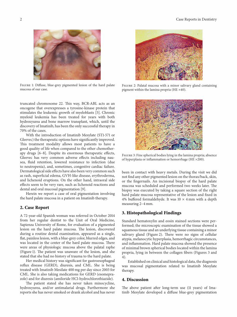
Figure 3: Fine spherical bodies lying in the lamina propria; absenceof hyperplasia or inflammation or hemorrhage (HE ×200).
been in contact with heavy metals. During the visit we didnot find any other pigmented lesion on the thorax/back, skin,or the fingernails. An incisional biopsy of the hard palatemucosa was scheduled and performed two weeks later. Thebiopsy was executed by taking a square section of the righthard palate mucosa representative of the lesion and fixed in4% buffered formaldehyde. It was 10 × 4mm with a depthmeasuring 2–4mm.
3. Histopathological Findings
Standard hematoxylin and eosin stained sections were per-formed; the microscopic examination of the tissue showed asquamous tissue and an underlying tissue containing aminorsalivary gland (Figure 2). There were no signs of cellularatypia, melanocytic hyperplasia, hemorrhagic circumstances,and inflammation. Hard palate mucosa showed the presenceof minimal brown spherical bodies located within the laminapropria, lying in between the collagen fibers (Figures 3 and4).
Established on clinical and histological data, the diagnosiswas mucosal pigmentation related to Imatinib Mesylatetherapy.
4. Discussion
The above patient after long-term use (11 years) of Ima-tinib Mesylate developed a diffuse blue-grey pigmentation
Case Reports in Dentistry 3
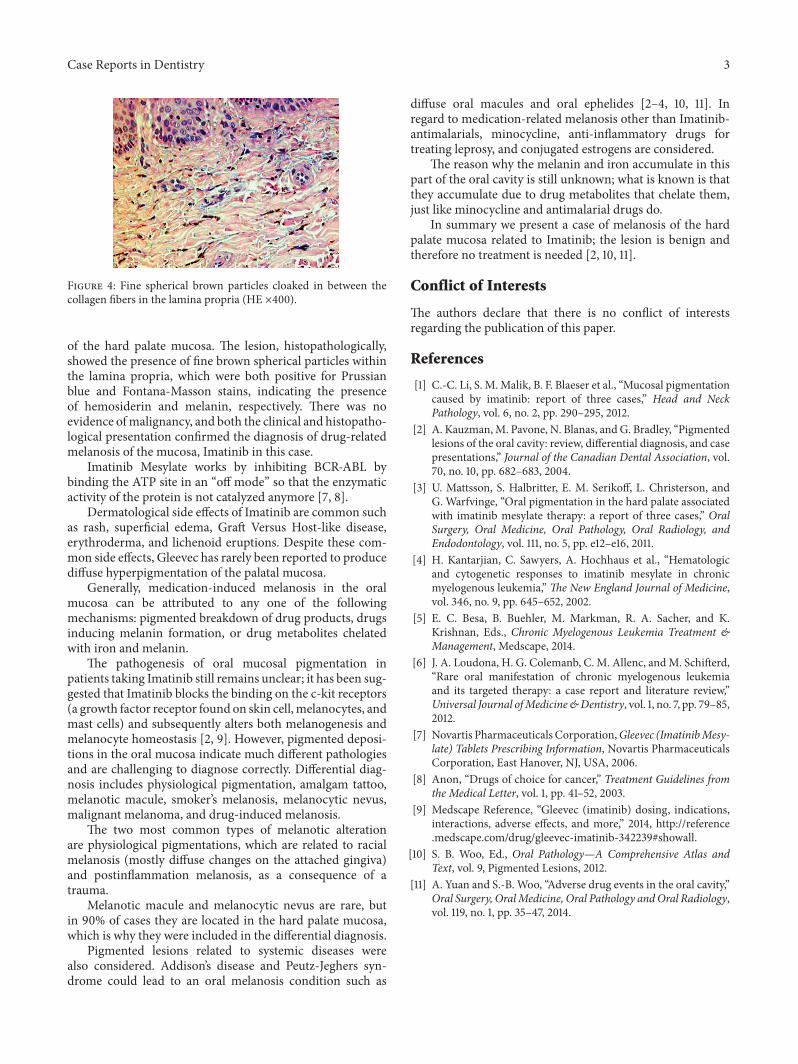
Figure 4: Fine spherical brown particles cloaked in between thecollagen fibers in the lamina propria (HE ×400).
of the hard palate mucosa. The lesion, histopathologically,showed the presence of fine brown spherical particles withinthe lamina propria, which were both positive for Prussianblue and Fontana-Masson stains, indicating the presenceof hemosiderin and melanin, respectively. There was noevidence ofmalignancy, and both the clinical and histopatho-logical presentation confirmed the diagnosis of drug-relatedmelanosis of the mucosa, Imatinib in this case.
Imatinib Mesylate works by inhibiting BCR-ABL bybinding the ATP site in an “off mode” so that the enzymaticactivity of the protein is not catalyzed anymore [7, 8].
Dermatological side effects of Imatinib are common suchas rash, superficial edema, Graft Versus Host-like disease,erythroderma, and lichenoid eruptions. Despite these com-mon side effects, Gleevec has rarely been reported to producediffuse hyperpigmentation of the palatal mucosa.
Generally, medication-induced melanosis in the oralmucosa can be attributed to any one of the followingmechanisms: pigmented breakdown of drug products, drugsinducing melanin formation, or drug metabolites chelatedwith iron and melanin.
The pathogenesis of oral mucosal pigmentation inpatients taking Imatinib still remains unclear; it has been sug-gested that Imatinib blocks the binding on the c-kit receptors(a growth factor receptor found on skin cell,melanocytes, andmast cells) and subsequently alters both melanogenesis andmelanocyte homeostasis [2, 9]. However, pigmented deposi-tions in the oral mucosa indicate much different pathologiesand are challenging to diagnose correctly. Differential diag-nosis includes physiological pigmentation, amalgam tattoo,melanotic macule, smoker’s melanosis, melanocytic nevus,malignant melanoma, and drug-induced melanosis.
The two most common types of melanotic alterationare physiological pigmentations, which are related to racialmelanosis (mostly diffuse changes on the attached gingiva)and postinflammation melanosis, as a consequence of atrauma.
Melanotic macule and melanocytic nevus are rare, butin 90% of cases they are located in the hard palate mucosa,which is why they were included in the differential diagnosis.
Pigmented lesions related to systemic diseases werealso considered. Addison’s disease and Peutz-Jeghers syn-drome could lead to an oral melanosis condition such as
diffuse oral macules and oral ephelides [2–4, 10, 11]. Inregard to medication-related melanosis other than Imatinib-antimalarials, minocycline, anti-inflammatory drugs fortreating leprosy, and conjugated estrogens are considered.
The reason why the melanin and iron accumulate in thispart of the oral cavity is still unknown; what is known is thatthey accumulate due to drug metabolites that chelate them,just like minocycline and antimalarial drugs do.
In summary we present a case of melanosis of the hardpalate mucosa related to Imatinib; the lesion is benign andtherefore no treatment is needed [2, 10, 11].
Conflict of Interests
The authors declare that there is no conflict of interestsregarding the publication of this paper.
References
[1] C.-C. Li, S. M.Malik, B. F. Blaeser et al., “Mucosal pigmentationcaused by imatinib: report of three cases,” Head and NeckPathology, vol. 6, no. 2, pp. 290–295, 2012.
[2] A. Kauzman,M. Pavone, N. Blanas, and G. Bradley, “Pigmentedlesions of the oral cavity: review, differential diagnosis, and casepresentations,” Journal of the Canadian Dental Association, vol.70, no. 10, pp. 682–683, 2004.
[3] U. Mattsson, S. Halbritter, E. M. Serikoff, L. Christerson, andG. Warfvinge, “Oral pigmentation in the hard palate associatedwith imatinib mesylate therapy: a report of three cases,” OralSurgery, Oral Medicine, Oral Pathology, Oral Radiology, andEndodontology, vol. 111, no. 5, pp. e12–e16, 2011.
[4] H. Kantarjian, C. Sawyers, A. Hochhaus et al., “Hematologicand cytogenetic responses to imatinib mesylate in chronicmyelogenous leukemia,” The New England Journal of Medicine,vol. 346, no. 9, pp. 645–652, 2002.
[5] E. C. Besa, B. Buehler, M. Markman, R. A. Sacher, and K.Krishnan, Eds., Chronic Myelogenous Leukemia Treatment &Management, Medscape, 2014.
[6] J. A. Loudona, H. G. Colemanb, C. M. Allenc, andM. Schifterd,“Rare oral manifestation of chronic myelogenous leukemiaand its targeted therapy: a case report and literature review,”Universal Journal ofMedicine&Dentistry, vol. 1, no. 7, pp. 79–85,2012.
[7] Novartis Pharmaceuticals Corporation,Gleevec (ImatinibMesy-late) Tablets Prescribing Information, Novartis PharmaceuticalsCorporation, East Hanover, NJ, USA, 2006.
[8] Anon, “Drugs of choice for cancer,” Treatment Guidelines fromthe Medical Letter, vol. 1, pp. 41–52, 2003.
[9] Medscape Reference, “Gleevec (imatinib) dosing, indications,interactions, adverse effects, and more,” 2014, http://reference.medscape.com/drug/gleevec-imatinib-342239#showall.
[10] S. B. Woo, Ed., Oral Pathology—A Comprehensive Atlas andText, vol. 9, Pigmented Lesions, 2012.
[11] A. Yuan and S.-B. Woo, “Adverse drug events in the oral cavity,”Oral Surgery, OralMedicine, Oral Pathology andOral Radiology,vol. 119, no. 1, pp. 35–47, 2014.

Submit your manuscripts athttp://www.hindawi.com
Hindawi Publishing Corporationhttp://www.hindawi.com Volume 2014
Oral OncologyJournal of
DentistryInternational Journal of
Hindawi Publishing Corporationhttp://www.hindawi.com Volume 2014
Hindawi Publishing Corporationhttp://www.hindawi.com Volume 2014
International Journal of
Biomaterials
Hindawi Publishing Corporationhttp://www.hindawi.com Volume 2014
BioMed Research International
Hindawi Publishing Corporationhttp://www.hindawi.com Volume 2014
Case Reports in Dentistry
Hindawi Publishing Corporationhttp://www.hindawi.com Volume 2014
Oral ImplantsJournal of
Hindawi Publishing Corporationhttp://www.hindawi.com Volume 2014
Anesthesiology Research and Practice
Hindawi Publishing Corporationhttp://www.hindawi.com Volume 2014
Radiology Research and Practice
Environmental and Public Health
Journal of
Hindawi Publishing Corporationhttp://www.hindawi.com Volume 2014
The Scientific World JournalHindawi Publishing Corporation http://www.hindawi.com Volume 2014
Hindawi Publishing Corporationhttp://www.hindawi.com Volume 2014
Dental SurgeryJournal of
Drug DeliveryJournal of
Hindawi Publishing Corporationhttp://www.hindawi.com Volume 2014
Hindawi Publishing Corporationhttp://www.hindawi.com Volume 2014
Oral DiseasesJournal of
Hindawi Publishing Corporationhttp://www.hindawi.com Volume 2014
Computational and Mathematical Methods in Medicine
ScientificaHindawi Publishing Corporationhttp://www.hindawi.com Volume 2014
PainResearch and TreatmentHindawi Publishing Corporationhttp://www.hindawi.com Volume 2014
Preventive MedicineAdvances in
Hindawi Publishing Corporationhttp://www.hindawi.com Volume 2014
EndocrinologyInternational Journal of
Hindawi Publishing Corporationhttp://www.hindawi.com Volume 2014
Hindawi Publishing Corporationhttp://www.hindawi.com Volume 2014
OrthopedicsAdvances in