Embed Size (px)
Citation preview
Int J Clin Exp Pathol 2014;7(5):2624-2635www.ijcep.com /ISSN:1936-2625/IJCEP0000366
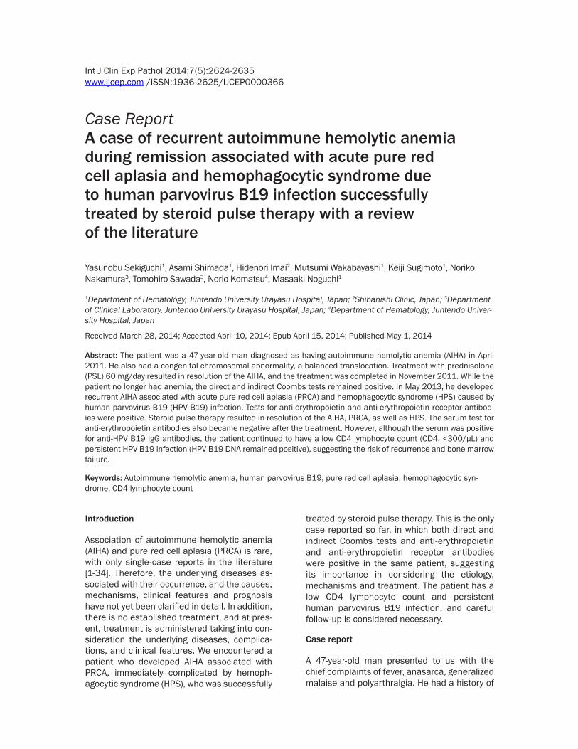
Case Report A case of recurrent autoimmune hemolytic anemia during remission associated with acute pure red cell aplasia and hemophagocytic syndrome due to human parvovirus B19 infection successfully treated by steroid pulse therapy with a review of the literature
Yasunobu Sekiguchi1, Asami Shimada1, Hidenori Imai2, Mutsumi Wakabayashi1, Keiji Sugimoto1, Noriko Nakamura3, Tomohiro Sawada3, Norio Komatsu4, Masaaki Noguchi1
1Department of Hematology, Juntendo University Urayasu Hospital, Japan; 2Shibanishi Clinic, Japan; 3Department of Clinical Laboratory, Juntendo University Urayasu Hospital, Japan; 4Department of Hematology, Juntendo Univer-sity Hospital, Japan
Received March 28, 2014; Accepted April 10, 2014; Epub April 15, 2014; Published May 1, 2014
Abstract: The patient was a 47-year-old man diagnosed as having autoimmune hemolytic anemia (AIHA) in April 2011. He also had a congenital chromosomal abnormality, a balanced translocation. Treatment with prednisolone (PSL) 60 mg/day resulted in resolution of the AIHA, and the treatment was completed in November 2011. While the patient no longer had anemia, the direct and indirect Coombs tests remained positive. In May 2013, he developed recurrent AIHA associated with acute pure red cell aplasia (PRCA) and hemophagocytic syndrome (HPS) caused by human parvovirus B19 (HPV B19) infection. Tests for anti-erythropoietin and anti-erythropoietin receptor antibod-ies were positive. Steroid pulse therapy resulted in resolution of the AIHA, PRCA, as well as HPS. The serum test for anti-erythropoietin antibodies also became negative after the treatment. However, although the serum was positive for anti-HPV B19 IgG antibodies, the patient continued to have a low CD4 lymphocyte count (CD4, <300/µL) and persistent HPV B19 infection (HPV B19 DNA remained positive), suggesting the risk of recurrence and bone marrow failure.
Keywords: Autoimmune hemolytic anemia, human parvovirus B19, pure red cell aplasia, hemophagocytic syn-drome, CD4 lymphocyte count
Introduction
Association of autoimmune hemolytic anemia (AIHA) and pure red cell aplasia (PRCA) is rare, with only single-case reports in the literature [1-34]. Therefore, the underlying diseases as- sociated with their occurrence, and the causes, mechanisms, clinical features and prognosis have not yet been clarified in detail. In addition, there is no established treatment, and at pres-ent, treatment is administered taking into con-sideration the underlying diseases, complica-tions, and clinical features. We encountered a patient who developed AIHA associated with PRCA, immediately complicated by hemoph- agocytic syndrome (HPS), who was successfully
treated by steroid pulse therapy. This is the only case reported so far, in which both direct and indirect Coombs tests and anti-erythropoietin and anti-erythropoietin receptor antibodies were positive in the same patient, suggesting its importance in considering the etiology, mechanisms and treatment. The patient has a low CD4 lymphocyte count and persistent human parvovirus B19 infection, and careful follow-up is considered necessary.
Case report
A 47-year-old man presented to us with the chief complaints of fever, anasarca, generalized malaise and polyarthralgia. He had a history of
Hemolytic anemia with pure red cell aplasia
2625 Int J Clin Exp Pathol 2014;7(5):2624-2635
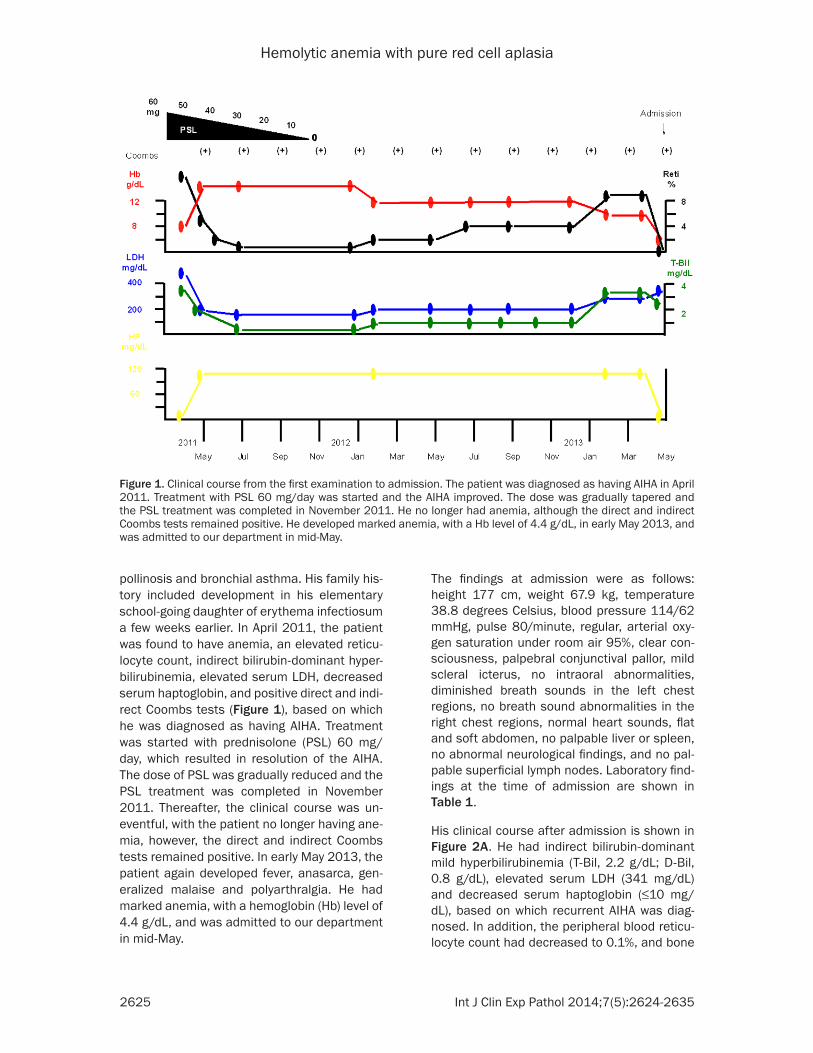
pollinosis and bronchial asthma. His family his-tory included development in his elementary school-going daughter of erythema infectiosum a few weeks earlier. In April 2011, the patient was found to have anemia, an elevated reticu-locyte count, indirect bilirubin-dominant hyper-bilirubinemia, elevated serum LDH, decreased serum haptoglobin, and positive direct and indi-rect Coombs tests (Figure 1), based on which he was diagnosed as having AIHA. Treatment was started with prednisolone (PSL) 60 mg/day, which resulted in resolution of the AIHA. The dose of PSL was gradually reduced and the PSL treatment was completed in November 2011. Thereafter, the clinical course was un- eventful, with the patient no longer having ane-mia, however, the direct and indirect Coombs tests remained positive. In early May 2013, the patient again developed fever, anasarca, gen-eralized malaise and polyarthralgia. He had marked anemia, with a hemoglobin (Hb) level of 4.4 g/dL, and was admitted to our department in mid-May.
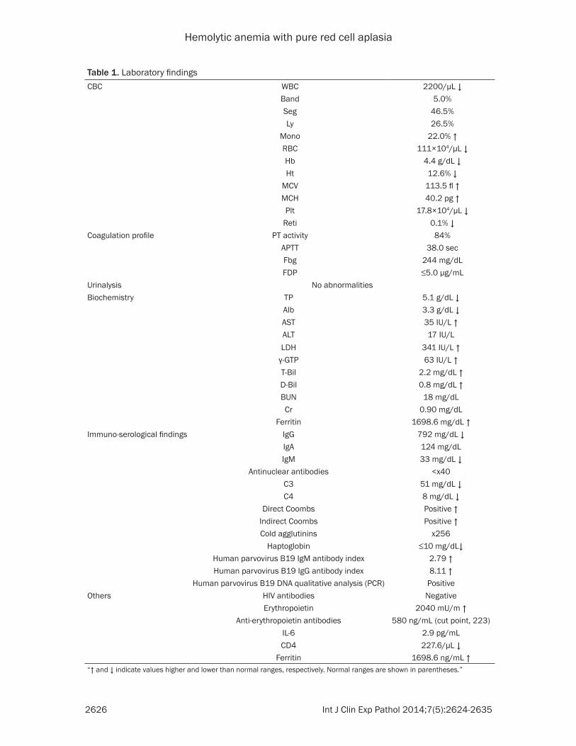
The findings at admission were as follows: height 177 cm, weight 67.9 kg, temperature 38.8 degrees Celsius, blood pressure 114/62 mmHg, pulse 80/minute, regular, arterial oxy-gen saturation under room air 95%, clear con-sciousness, palpebral conjunctival pallor, mild scleral icterus, no intraoral abnormalities, diminished breath sounds in the left chest regions, no breath sound abnormalities in the right chest regions, normal heart sounds, flat and soft abdomen, no palpable liver or spleen, no abnormal neurological findings, and no pal-pable superficial lymph nodes. Laboratory find-ings at the time of admission are shown in Table 1.
His clinical course after admission is shown in Figure 2A. He had indirect bilirubin-dominant mild hyperbilirubinemia (T-Bil, 2.2 g/dL; D-Bil, 0.8 g/dL), elevated serum LDH (341 mg/dL) and decreased serum haptoglobin (≤10 mg/dL), based on which recurrent AIHA was diag-nosed. In addition, the peripheral blood reticu-locyte count had decreased to 0.1%, and bone
Figure 1. Clinical course from the first examination to admission. The patient was diagnosed as having AIHA in April 2011. Treatment with PSL 60 mg/day was started and the AIHA improved. The dose was gradually tapered and the PSL treatment was completed in November 2011. He no longer had anemia, although the direct and indirect Coombs tests remained positive. He developed marked anemia, with a Hb level of 4.4 g/dL, in early May 2013, and was admitted to our department in mid-May.
Hemolytic anemia with pure red cell aplasia
2626 Int J Clin Exp Pathol 2014;7(5):2624-2635
Table 1. Laboratory findingsCBC WBC 2200/µL ↓
Band 5.0%Seg 46.5%Ly 26.5%
Mono 22.0% ↑RBC 111×104/µL ↓Hb 4.4 g/dL ↓Ht 12.6% ↓
MCV 113.5 fl ↑MCH 40.2 pg ↑Plt 17.8×10⁴/µL ↓
Reti 0.1% ↓Coagulation profile PT activity 84%
APTT 38.0 secFbg 244 mg/dLFDP ≤5.0 µg/mL
Urinalysis No abnormalitiesBiochemistry TP 5.1 g/dL ↓
Alb 3.3 g/dL ↓AST 35 IU/L ↑ALT 17 IU/LLDH 341 IU/L ↑
γ-GTP 63 IU/L ↑T-Bil 2.2 mg/dL ↑D-Bil 0.8 mg/dL ↑BUN 18 mg/dLCr 0.90 mg/dL
Ferritin 1698.6 mg/dL ↑Immuno-serological findings IgG 792 mg/dL ↓
IgA 124 mg/dLIgM 33 mg/dL ↓
Antinuclear antibodies <x40C3 51 mg/dL ↓C4 8 mg/dL ↓
Direct Coombs Positive ↑Indirect Coombs Positive ↑Cold agglutinins x256
Haptoglobin ≤10 mg/dL↓Human parvovirus B19 IgM antibody index 2.79 ↑Human parvovirus B19 IgG antibody index 8.11 ↑
Human parvovirus B19 DNA qualitative analysis (PCR) PositiveOthers HIV antibodies Negative
Erythropoietin 2040 mU/m ↑Anti-erythropoietin antibodies 580 ng/mL (cut point, 223)
IL-6 2.9 pg/mLCD4 227.6/µL ↓
Ferritin 1698.6 ng/mL ↑“↑ and ↓ indicate values higher and lower than normal ranges, respectively. Normal ranges are shown in parentheses.”
Hemolytic anemia with pure red cell aplasia
2627 Int J Clin Exp Pathol 2014;7(5):2624-2635
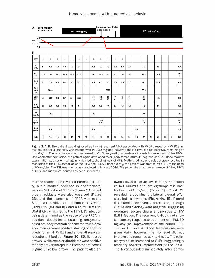
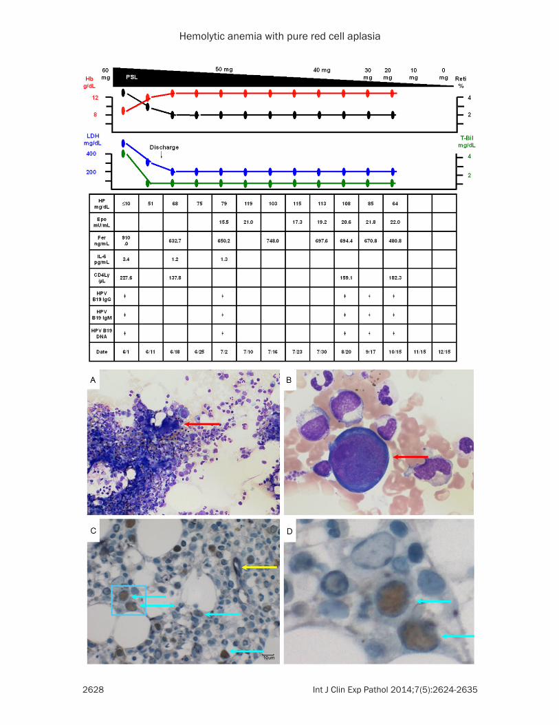
marrow examination revealed normal cellulari-ty, but a marked decrease in erythroblasts, with an M/E ratio of 117.25 (Figure 3A). Giant proerythroblasts were also observed (Figure 3B), and the diagnosis of PRCA was made. Serum was positive for anti-human parvovirus (HPV) B19 IgM and IgG and also for HPV B19 DNA (PCR), which led to the HPV B19 infection being determined as the cause of the PRCA. In addition, double-immunostaining (enzyme-la- beled antibody method) of bone marrow biopsy specimens showed positive staining of erythro-blasts for anti-HPV B19 and anti-erythropoietin receptor antibodies (Figure 3C, 3D, light blue arrows), while some erythroblasts were positive for only anti-erythropoietin receptor antibodies (Figure 3, yellow arrow). The patient also sh-
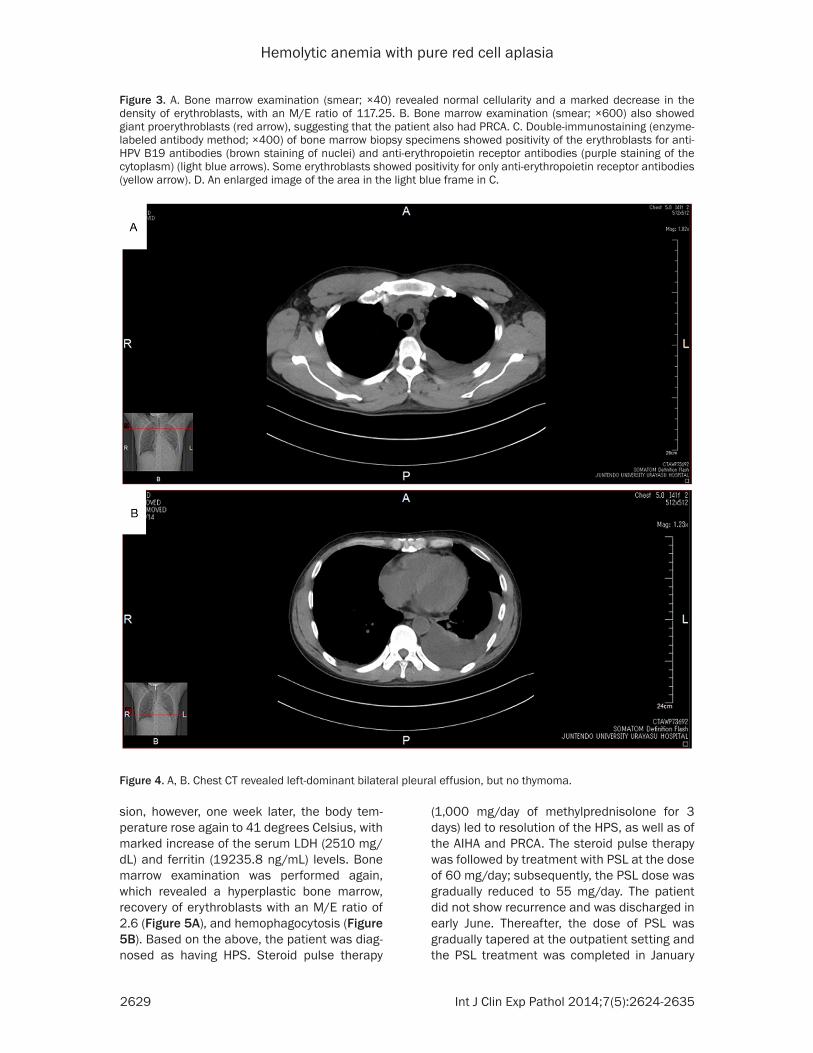
owed elevated serum levels of erythropoietin (2,040 mU/mL) and anti-erythropoietin anti-bodies (580 ng/mL) (Table 1). Chest CT revealed left-dominant bilateral pleural effu-sion, but no thymoma (Figure 4A, 4B). Pleural fluid examination revealed an exudate, although culture and cytology were negative, suggesting exudative reactive pleural effusion due to HPV B19 infection. The recurrent AIHA did not show satisfactory response to treatment with PSL 30 mg/day (no improvement of the serum LDH, T-Bil or HP levels). Blood transfusions were given daily, however, the Hb level did not improve and remained at 4 to 6 g/dL. The retic-ulocyte count increased to 0.4%, suggesting a tendency towards improvement of the PRCA. The fever subsided immediately after admis-
Figure 2. A, B. The patient was diagnosed as having recurrent AIHA associated with PRCA caused by HPV B19 in-fection. The recurrent AIHA was treated with PSL 30 mg/day, however, the Hb level did not improve, remaining at 4 to 6 g/dL. The reticulocyte count increased to 0.4%, suggesting a tendency towards improvement of the PRCA. One week after admission, the patient again developed fever (body temperature 41 degrees Celsius). Bone marrow examination was performed again, which led to the diagnosis of HPS. Methylprednisolone pulse therapy resulted in resolution of the HPS, as well as of the AIHA and PRCA. Subsequently, the patient was treated with PSL at the dose of 60 mg/day. The PSL treatment was completed in January 2014. The patient has had no recurrence of AIHA, PRCA or HPS, and his clinical course has been uneventful.
Hemolytic anemia with pure red cell aplasia
2628 Int J Clin Exp Pathol 2014;7(5):2624-2635
Hemolytic anemia with pure red cell aplasia
2629 Int J Clin Exp Pathol 2014;7(5):2624-2635
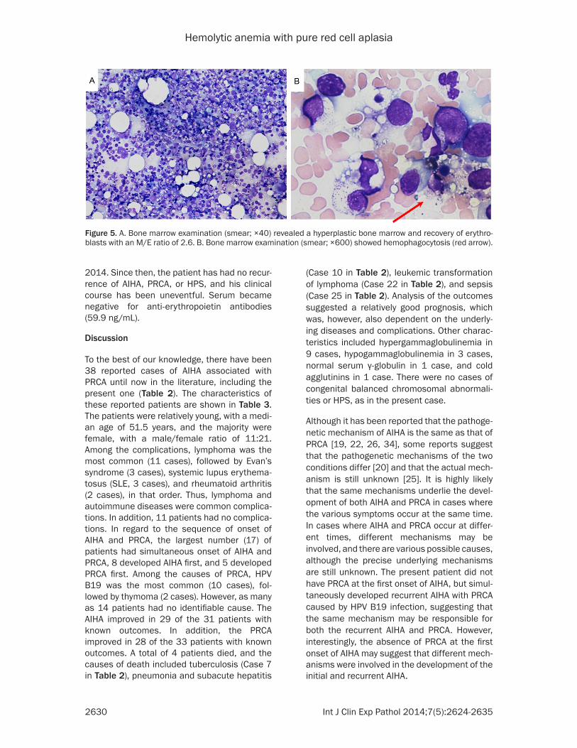
sion, however, one week later, the body tem-perature rose again to 41 degrees Celsius, with marked increase of the serum LDH (2510 mg/dL) and ferritin (19235.8 ng/mL) levels. Bone marrow examination was performed again, which revealed a hyperplastic bone marrow, recovery of erythroblasts with an M/E ratio of 2.6 (Figure 5A), and hemophagocytosis (Figure 5B). Based on the above, the patient was diag-nosed as having HPS. Steroid pulse therapy
(1,000 mg/day of methylprednisolone for 3 days) led to resolution of the HPS, as well as of the AIHA and PRCA. The steroid pulse therapy was followed by treatment with PSL at the dose of 60 mg/day; subsequently, the PSL dose was gradually reduced to 55 mg/day. The patient did not show recurrence and was discharged in early June. Thereafter, the dose of PSL was gradually tapered at the outpatient setting and the PSL treatment was completed in January
Figure 3. A. Bone marrow examination (smear; ×40) revealed normal cellularity and a marked decrease in the density of erythroblasts, with an M/E ratio of 117.25. B. Bone marrow examination (smear; ×600) also showed giant proerythroblasts (red arrow), suggesting that the patient also had PRCA. C. Double-immunostaining (enzyme-labeled antibody method; ×400) of bone marrow biopsy specimens showed positivity of the erythroblasts for anti-HPV B19 antibodies (brown staining of nuclei) and anti-erythropoietin receptor antibodies (purple staining of the cytoplasm) (light blue arrows). Some erythroblasts showed positivity for only anti-erythropoietin receptor antibodies (yellow arrow). D. An enlarged image of the area in the light blue frame in C.
Figure 4. A, B. Chest CT revealed left-dominant bilateral pleural effusion, but no thymoma.
Hemolytic anemia with pure red cell aplasia
2630 Int J Clin Exp Pathol 2014;7(5):2624-2635
2014. Since then, the patient has had no recur-rence of AIHA, PRCA, or HPS, and his clinical course has been uneventful. Serum became negative for anti-erythropoietin antibodies (59.9 ng/mL).
Discussion
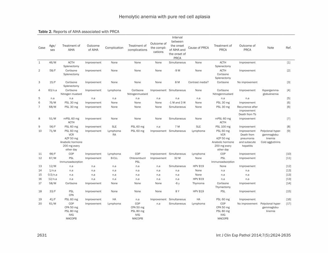
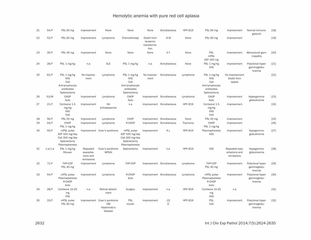
To the best of our knowledge, there have been 38 reported cases of AIHA associated with PRCA until now in the literature, including the present one (Table 2). The characteristics of these reported patients are shown in Table 3. The patients were relatively young, with a medi-an age of 51.5 years, and the majority were female, with a male/female ratio of 11:21. Among the complications, lymphoma was the most common (11 cases), followed by Evan’s syndrome (3 cases), systemic lupus erythema-tosus (SLE, 3 cases), and rheumatoid arthritis (2 cases), in that order. Thus, lymphoma and autoimmune diseases were common complica-tions. In addition, 11 patients had no complica-tions. In regard to the sequence of onset of AIHA and PRCA, the largest number (17) of patients had simultaneous onset of AIHA and PRCA, 8 developed AIHA first, and 5 developed PRCA first. Among the causes of PRCA, HPV B19 was the most common (10 cases), fol-lowed by thymoma (2 cases). However, as many as 14 patients had no identifiable cause. The AIHA improved in 29 of the 31 patients with known outcomes. In addition, the PRCA improved in 28 of the 33 patients with known outcomes. A total of 4 patients died, and the causes of death included tuberculosis (Case 7 in Table 2), pneumonia and subacute hepatitis
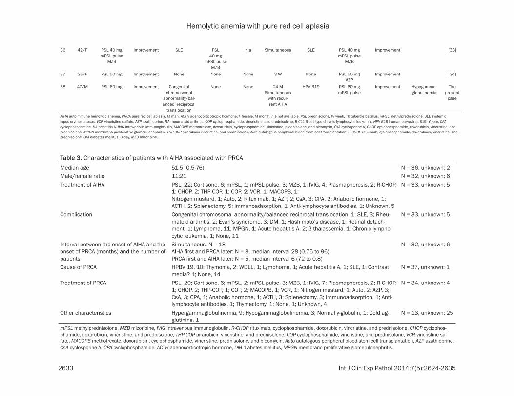
(Case 10 in Table 2), leukemic transformation of lymphoma (Case 22 in Table 2), and sepsis (Case 25 in Table 2). Analysis of the outcomes suggested a relatively good prognosis, which was, however, also dependent on the underly-ing diseases and complications. Other charac-teristics included hypergammaglobulinemia in 9 cases, hypogammaglobulinemia in 3 cases, normal serum γ-globulin in 1 case, and cold agglutinins in 1 case. There were no cases of congenital balanced chromosomal abnormali-ties or HPS, as in the present case.
Although it has been reported that the pathoge-netic mechanism of AIHA is the same as that of PRCA [19, 22, 26, 34], some reports suggest that the pathogenetic mechanisms of the two conditions differ [20] and that the actual mech-anism is still unknown [25]. It is highly likely that the same mechanisms underlie the devel-opment of both AIHA and PRCA in cases where the various symptoms occur at the same time. In cases where AIHA and PRCA occur at differ-ent times, different mechanisms may be involved, and there are various possible causes, although the precise underlying mechanisms are still unknown. The present patient did not have PRCA at the first onset of AIHA, but simul-taneously developed recurrent AIHA with PRCA caused by HPV B19 infection, suggesting that the same mechanism may be responsible for both the recurrent AIHA and PRCA. However, interestingly, the absence of PRCA at the first onset of AIHA may suggest that different mech-anisms were involved in the development of the initial and recurrent AIHA.
Figure 5. A. Bone marrow examination (smear; ×40) revealed a hyperplastic bone marrow and recovery of erythro-blasts with an M/E ratio of 2.6. B. Bone marrow examination (smear; ×600) showed hemophagocytosis (red arrow).
Hemolytic anemia with pure red cell aplasia
2631 Int J Clin Exp Pathol 2014;7(5):2624-2635
Table 2. Reports of AIHA associated with PRCA
Case Age/sex
Treatment of AIHA
Outcome of AIHA Complication Treatment of
complications
Outcome of the compli-
cations
Interval between the onset
of AIHA and the onset of
PRCA
Cause of PRCA Treatment of PRCA
Outcome of PRCA Note Ref.
1 46/M ACTHSplenectomy
Improvement None None None Simultaneous None ACTHSplenectomy
Improvement [1]
2 58/F CortisoneSplenectomy
Improvement None None None -9 M None ACTHCortisone
Splenectomy
Improvement [2]
3 15/F CortisoneSplenectomy
Improvement None None None 8 M Contrast media? Cortisone No improvement [3]
4 63/n.a CortisoneNitrogen mustard
Improvement Lymphoma CortisoneNitrogenmustard
Improvement Simultaneous None CortisoneNitrogenmustard
Improvement Hypergamma-globulinemia
[4]
5 n.a n.a n.a n.a n.a n.a n.a n.a n.a n.a [5]6 76/M PSL 30 mg Improvement None None None -1 M and 3 W None PSL 30 mg Improvement [6]7 68/M PSL 30 mg Improvement None None None Simultaneous None PSL 30 mg Recurrence after
improvementDeath from Tb
[6]
8 51/M mPSL 60 mgACTH
Improvement None None None Simultaneous None mPSL 60 mgACTH
Improvement [7]
9 56/F PSL 60 mg Improvement SLE PSL 60 mg n.a 7 M SLE PSL 100 mg Improvement [8]10 71/M PSL 60 mg
VCRAZP 50 mg
Anabolic hormone 200 mg every
other day
Improvement LymphomaRA
PSL 60 mg Improvement Simultaneous Lymphoma PSL 60 mgVCR
AZP 50 mgAnabolic hormone
200 mg every other day
ImprovementDeath from pneumonia
and subacute hepatitis
Polyclonal hyper-gammaglobu-
linemiaCold agglutinins
[9]
11 66/F COP Improvement Lymphoma COP Improvement Simultaneous Lymphoma COP Improvement [10]12 67/M PSL
ImmunoadsorptionImprovement B-CLL Chlorambucil
PSLImprovement 32 M None PSL
ImmunoadsorptionImprovement [11]
13 12/M n.a n.a n.a n.a n.a Simultaneous HPV B19 None Improvement [12]14 1/n.a n.a n.a n.a n.a n.a n.a None n.a n.a [13]15 0.5/n.a n.a n.a n.a n.a n.a n.a None n.a n.a [13]16 12/n.a n.a n.a n.a n.a n.a n.a HPV B19 n.a n.a [13]17 58/M Cortisone Improvement None None None -6 y Thymoma Cortisone
ThymectomyImprovement [14]
18 33/F PSLCPA
Improvement None None None 8 Y HPV B19 PSL Improvement [15]
19 41/F PSL 60 mg Improvement HA n.a Improvement Simultaneous HA PSL 60 mg Improvement [16]20 61/M COP
CPA 50 mgPSL 80 mg
IVIGMACOPB
Improvement Lymphoma COPCPA 50 mgPSL 80 mg
IVIGMACOPB
n.a Simultaneous Lymphoma COPCPA 50 mgPSL 80 mg
IVIGMACOPB
No improvement Polyclonal hyper-gammaglobu-
linemia
[17]
Hemolytic anemia with pure red cell aplasia
2632 Int J Clin Exp Pathol 2014;7(5):2624-2635
21 54/F PSL 60 mg Improvement None None None Simultaneous HPV B19 PSL 60 mg Improvement Normal immuno-globulin
[18]
22 52/F PSL 60 mg Improvement Lymphoma Chemotherapy Death from leukemic
transforma-tion
-6 M None PSL 60 mg Improvement [19]
23 35/F PSL 50 mg Improvement None None None 4 Y None PSLmPSL
AZP 100 mg
Improvement Monoclonal gam-mopathy
[20]
24 28/F PSL 1 mg/kg n.a SLE PSL 1 mg/kg n.a Simultaneous None PSL 1 mg/kgIVIG
Improvement Polyclonal hyper-gammaglobu-
linemia
[21]
25 62/F PSL 1 mg/kgIVIGCsA
Anti-lymphocyte antibodies
Splenectomy
No improve-ment
Lymphoma PSL 1 mg/kgIVIGCsA
Anti-lymphocyte antibodies
Splenectomy
No improve-ment
Simultaneous Lymphoma PSL 1 mg/kgIVIGCsA
Anti-lymphocyte antibodies
Splenectomy
No improvementDeath from
sepsis
[22]
26 53/M CHOPAuto
Improvement Lymphoma CHOPAuto
Improvement Simultaneous Lymphoma CHOPAuto
Improvement Hypergamma-globulinemia
[23]
27 21/F Cortisone 1.5 mg/kg
IVIGCsA
Improvement HAβ-thalassemia
n.a Improvement Simultaneous HPV B19 Cortisone 1.5 mg/kg
IVIGCsA
Improvement [24]
28 56/F PSL 50 mg Improvement Lymphoma CHOP Improvement Simultaneous None PSL 50 mg Improvement [25]29 54/F CHOP
PSL 1 mg/kgImprovement Lymphoma R-CHOP Improvement Simultaneous Thymoma CHOP
PSL 1 mg/kgImprovement [26]
30 40/F mPSL pulseAZP 100 mg/dayCsA 300 mg/day
SplenectomyPlasmapheresis
Improvement Evan’s syndrome mPSL pulseAZP 100 mg/dayCsA 300 mg/day
SplenectomyPlasmapheresis
Improvement 6 y HPV B19 PlasmapheresisIVIG
Improvement Hypogamma-globulinemia
[27]
31 n.a/n.a PSL 1 mg/kgRituxan
Repeated exacerba-tions and
remissions
Evan’s syndromeMPGN
Splenectomy Improvement n.a HPV B19 IVIG Repeated exac-erbations and
remissions
Hypogamma-globulinemia
[28]
32 71/F THP-COPPSL 40 mg
Improvement Lymphoma THP-COP Improvement Simultaneous Lymphoma THP-COPPSL 40 mg
Improvement Polyclonal hyper-gammaglobu-
linemia
[29]
33 54/F mPSL pulsePlasmapheresis
R-CHOPAuto
Improvement Lymphoma R-CHOPAuto
Improvement Simultaneous Lymphoma mPSL pulsePlasmapheresis
R-CHOPAuto
Improvement Polyclonal hyper-gammaglobu-
linemia
[30]
34 28/F Cortisone 15-20 mgIVIG
n.a Retinal detach-ment
Surgery Improvement n.a HPV B19 Cortisone 15-20 mgIVIG
n.a [31]
35 33/F mPSL pulsePSL 60 mg
Improvement Evan’s syndromeDM
Hashimoto’s disease
PSLInsulin
Improvement -22 D
HPV B19 PSLCsA
Improvement Polyclonal hyper-gammaglobu-
linemia
[32]
Hemolytic anemia with pure red cell aplasia
2633 Int J Clin Exp Pathol 2014;7(5):2624-2635
36 42/F PSL 40 mgmPSL pulse
MZB
Improvement SLE PSL40 mg
mPSL pulseMZB
n.a Simultaneous SLE PSL 40 mgmPSL pulse
MZB
Improvement [33]
37 26/F PSL 50 mg Improvement None None None 3 W None PSL 50 mgAZP
Improvement [34]
38 47/M PSL 60 mg Improvement Congenital chromosomal
abnormality/bal-anced reciprocal
translocation
None None 24 MSimultaneous
with recur-rent AIHA
HPV B19 PSL 60 mgmPSL pulse
Improvement Hypogamma-globulinemia
The present
case
AIHA autoimmune hemolytic anemia, PRCA pure red cell aplasia, M man, ACTH adenocorticotropic hormone, F female, M month, n.a not available, PSL prednisolone, W week, Tb tubercle bacillus, mPSL methylprednisolone, SLE systemic lupus erythematosus, VCR vincristine sulfate, AZP azathioprine, RA rheumatoid arthritis, COP cyclophosphamide, vincristine, and prednisolone, B-CLL B cell-type chronic lymphocytic leukemia, HPV B19 human parvovirus B19, Y year, CPA cyclophosphamide, HA hepatitis A. IVIG intravenous immunoglobulin, MACOPB methotrexate, doxorubicin, cyclophosphamide, vincristine, prednisolone, and bleomycin, CsA cyclosporine A, CHOP cyclophosphamide, doxorubicin, vincristine, and prednisolone, MPGN membrano proliferative glomerulonephritis, THP-COP pirarubicin vincristine, and prednisolone, Auto autologous peripheral blood stem cell transplantation, R-CHOP rituximab, cyclophosphamide, doxorubicin, vincristine, and prednisolone, DM diabetes mellitus, D day, MZB mizoribine.
Table 3. Characteristics of patients with AIHA associated with PRCAMedian age 51.5 (0.5-76) N = 36, unknown: 2Male/female ratio 11:21 N = 32, unknown: 6Treatment of AIHA PSL, 22; Cortisone, 6; mPSL, 1; mPSL pulse, 3; MZB, 1; IVIG, 4; Plasmapheresis, 2; R-CHOP,
1; CHOP, 2; THP-COP, 1; COP, 2; VCR, 1; MACOPB, 1; Nitrogen mustard, 1; Auto, 2; Rituximab, 1; AZP, 2; CsA, 3; CPA, 2; Anabolic hormone, 1; ACTH, 2; Splenectomy, 5; Immunoadsorption, 1; Anti-lymphocyte antibodies, 1; Unknown, 5
N = 33, unknown: 5
Complication Congenital chromosomal abnormality/balanced reciprocal translocation, 1; SLE, 3; Rheu-matoid arthritis, 2; Evan’s syndrome, 3; DM, 1; Hashimoto’s disease, 1; Retinal detach-ment, 1; Lymphoma, 11; MPGN, 1; Acute hepatitis A, 2; β-thalassemia, 1; Chronic lympho-cytic leukemia, 1; None, 11
N = 33, unknown: 5
Interval between the onset of AIHA and the onset of PRCA (months) and the number of patients
Simultaneous, N = 18AIHA first and PRCA later: N = 8, median interval 28 (0.75 to 96)PRCA first and AIHA later: N = 5, median interval 6 (72 to 0.8)
N = 32, unknown: 6
Cause of PRCA HPBV 19, 10; Thymoma, 2; WDLL, 1; Lymphoma, 1; Acute hepatitis A, 1; SLE, 1; Contrast media? 1; None, 14
N = 37, unknown: 1
Treatment of PRCA PSL, 20; Cortisone, 6; mPSL, 2; mPSL pulse, 3; MZB, 1; IVIG, 7; Plasmapheresis, 2; R-CHOP, 1; CHOP, 2; THP-COP, 1; COP, 2; MACOPB, 1; VCR, 1; Nitrogen mustard, 1; Auto, 2; AZP, 3; CsA, 3; CPA, 1; Anabolic hormone, 1; ACTH, 3; Splenectomy, 3; Immunoadsorption, 1; Anti-lymphocyte antibodies, 1; Thymectomy, 1; None, 1; Unknown, 4
N = 34, unknown: 4
Other characteristics Hypergammaglobulinemia, 9; Hypogammaglobulinemia, 3; Normal γ-globulin, 1; Cold ag-glutinins, 1
N = 13, unknown: 25
mPSL methylprednisolone, MZB mizoribine, IVIG intravenous immunoglobulin, R-CHOP rituximab, cyclophosphamide, doxorubicin, vincristine, and prednisolone, CHOP cyclophos-phamide, doxorubicin, vincristine, and prednisolone, THP-COP pirarubicin vincristine, and prednisolone, COP cyclophosphamide, vincristine, and prednisolone, VCR vincristine sul-fate, MACOPB methotrexate, doxorubicin, cyclophosphamide, vincristine, prednisolone, and bleomycin, Auto autologous peripheral blood stem cell transplantation, AZP azathioprine, CsA cyclosporine A, CPA cyclophosphamide, ACTH adenocorticotropic hormone, DM diabetes mellitus, MPGN membrano proliferative glomerulonephritis.
Hemolytic anemia with pure red cell aplasia
2634 Int J Clin Exp Pathol 2014;7(5):2624-2635
It was considered that in the present patient, antibodies against mature erythrocytes (direct and indirect Coombs tests) caused the AIHA, and that anti-erythropoietin and anti-erythro-poietin receptor antibodies inhibited the differ-entiation and maturation of erythroblasts, causing PRCA. There have been reports of patients showing positive test results for anti-erythropoietin antibodies [21, 33] and anti-erythropoietin receptor antibodies [35], where-as ours is the only reported case in which both anti-erythropoietin and anti-erythropoietin rec- eptor antibodies were found in the same patient, suggesting the importance of this case report in elucidating the etiology and develop-ing treatment.
The present patient continues to show seropos-itivity for anti-HPV B19 IgM and IgG and HPV B19 DNA, suggesting persistent infection with HPV B19 (Figure 2B). In addition, the CD4 lym-phocyte count is reduced (159.1 to 227.6/µL; Figure 2B). Therefore, he is at an elevated risk of recurrent PRCA [27] and bone marrow failure due to persistent infection with HPV B19 [15], and requires careful follow-up. The cause of the decreased CD4 lymphocyte count is unknown, however, Katori et al. pointed out that the high steroid doses may have exacerbated the PRCA [ref]. Ito et al. reported that in a patient with persistent HPV B19 infection treated with high-dose immunoglobulin (HDIVIG), the serum became positive for anti-HPV B19 IgG and the CD4 lymphocyte count increased to ≥300/µL, resulting in the elimination of HPV B19. These findings suggest that we should also probably consider HDIVIG treatment in case of recur-rence in the present patient.
Disclosure of conflict of interest
None.
Address correspondence to: Yasunobu Sekiguchi, Department of Hematology, Juntendo University Urayasu Hospital, 2-1-1, Tomioka, Urayasu, Chiba Prefecture, Japan. Tel: 047-353-3111; Fax: 047-381-5054; E-mail: [email protected]
References
[1] Davis LJ, Kennedy AC, Baikie AG, Brown A. Hae-molytic anaemias of various types treated with ACTH and cortisone; report of ten cases, in-cluding one of acquired type in which erythro-
poietic arrest occurred during a crisis. Glasgow Med J 1952; 33: 263-85.
[2] Eisemann G, Dameshek W. Splenectomy for pure red-cell hypoplastic (aregenerative) ane-mia associated with autoimmune hemolytic disease; report of a case. N Engl J Med 1954; 251: 1044-1048.
[3] Seip M. Aplastic crisis in a case of immuno-hemolytic anemia. Acta Med Scand 1955; 153: 137-42.
[4] Bove JR. Combined erythroid hypoplasia and symptomatic hemolytic anemia; report of a case. N Engl J Med 1956; 255: 135-136.
[5] Hennemann HH, Falck I. [Combinations of aplastic with hemolytic syndromes]. Acta Hae-matol 1957; 18: 219-228. German.
[6] Burston J, Husain OA, Hutt MS, Tanner EI. Two cases of auto-immune haemolysis and apla-sia. Br Med J 1959; 1: 83-86.
[7] Meyer LM, Bertcher RW. Acquired hemolytic anemia and transient erythroid hypoplasia of bone marrow. Am J Med 1960; 28: 606-608.
[8] Meyer RJ, Hoffman R, Zanjani ED. Autoimmune hemolytic anemia and periodic pure red cell aplasia in systemic lupus erythematosus. Am J Med 1978; 65: 342-345.
[9] Mannoji M, Shimoda M, Koresawa S, Yamada O, Togawa A, Yawata Y, Umemura H, Kozuru M. [A case of angioimmunoblastic lymphadenop-athy with dysproteinemia associated with auto-immune hemolytic anemia and pure red cell aplasia--with special references to its patho-genesis (author’s transl)]. Rinsho Ketsueki 1981; 22: 1751-1758. Japanese.
[10] Hirosawa S, Kamiyama R, Dan K, Kuriya S, No-mura T. [A case malignant lymphoma associ-ated with pure red cell aplasia]. Rinsho Ket-sueki 1982; 23: 1463-1467. Japanese.
[11] Mangan KF, Besa EC, Shadduck RK, Tedrow H, Ray PK. Demonstration of two distinct antibo-dies in autoimmune hemolytic anemia with re-ticulocytopenia and red cell aplasia. Exp He-matol 1984; 12: 788-793.
[12] Bertrand Y, Lefrere JJ, Leverger G, Courouce AM, Feo C, Clark M, Schaison G, Soulier JP. Au-toimmune haemolytic anaemia revealed by hu-man parvovirus linked erythroblastopenia. Lancet 1985; 2: 382-383.
[13] Lefrère JJ, Couroucé AM, Bertrand Y, Girot R, Soulier JP. Human parvovirus and aplastic cri-sis in chronic hemolytic anemias: a study of 24 observations. Am J Hematol 1986; 23: 271-275.
[14] Taniguchi S, Shibuya T, Morioka E, Okamura T, Okamura S, Inaba S, Niho Y. Demonstration of three distinct immunological disorders on erythropoiesis in a patient with pure red cell aplasia and autoimmune haemolytic anaemia associated with thymoma. Br J Haematol 1988; 68: 473-477.
Hemolytic anemia with pure red cell aplasia
2635 Int J Clin Exp Pathol 2014;7(5):2624-2635
[15] Tomiyama J, Adachi Y, Hanada T, Matsunaga Y. Human parvovirus B19-induced aplastic crisis in autoimmune haemolytic anaemia. Br J Hae-matol 1988; 69: 288-289.
[16] Gundersen SG, Bjoerneklett A, Bruun JN. Se-vere erythroblastopenia and hemolytic anemia during a hepatitis A infection. Scand J Infect Dis 1989; 21: 225-228.
[17] Vukelja SJ, Krishnan J, Link CM, Salvado AJ, Knight RD. Resolution of pure red cell aplasia and lymphoma: response to intravenous gam-maglobulin and combination chemotherapy. Am J Hematol 1989; 32: 129-133.
[18] Chitnavis VN, Patou G, Makar YF, Kendra JR. B19 parvovirus induced red cell aplasia com-plicating acute cold antibody mediated haemo-lytic anaemia. Br J Haematol 1990; 76: 433-434.
[19] Suzuki A, Takahashi T, Taniguchi A, Kotake C, Seo T, Toda T, Kobayashi K, Tsukamoto N, Fu-kumoto M. Pure red cell aplasia associated with non-Hodgkin’s lymphoma and hemolytic anemia. Jpn J Clin Oncol 1991; 21: 384-387.
[20] Tohda S, Nara N, Tanikawa S, Imai Y, Murakami N, Aoki N. Pure red cell aplasia following auto-immune haemolytic anaemia. Cell-mediated suppression of erythropoiesis as a possible pathogenesis of pure red cell aplasia. Acta Haematol 1992; 87: 98-102.
[21] Linardaki GD, Boki KA, Fertakis A, Tzioufas AG. Pure red cell aplasia as presentation of sys-temic lupus erythematosus: antibodies to erythropoietin. Scand J Rheumatol 1999; 28: 189-191.
[22] Zeidman A, Fradin Z, Barac Y, Bendayan D, Mit-telman M, Orlin J. Splenic lymphoma present-ing as warm autoimmune hemolytic anemia associated with pure red cell aplasia. Vox Sang 2000; 78: 126-129.
[23] Katayama H, Takeuchi M, Yoshino T, Munema-sa M, Tada A, Soda R, Takahashi K. Epstein-Barr virus associated diffuse large B-cell lym-phoma complicated by autoimmune hemolytic anemia and pure red cell aplasia. Leuk Lym-phoma 2001; 42: 539-542.
[24] Chehal A, Sharara AI, Haidar HA, Haidar J, Ba-zarbachi A. Acute viral hepatitis A and parvovi-rus B19 infections complicated by pure red cell aplasia and autoimmune hemolytic anemia. J Hepatol 2002; 37: 163-165.
[25] Toyota S, Nakamura N, Dan K. [Coexistence of pure red cell aplasia and autoimmune hemo-lytic anemia occurring during remission of ma-lignant lymphoma]. Rinsho Ketsueki 2002; 43: 493-495. Japanese.
[26] Nakashima Y, Abe Y, Ohtsuka R, Tachikawa Y, Nagasawa E, Nishimura J, Ohshima K, Nawata H, Muta K. [Follicular lymphoma complicated with autoimmune hemolytic anemia and pure red cell aplasia]. Rinsho Ketsueki 2004; 45: 1208-1210. Japanese.
[27] Ito S, Oyake T, Uchiyama T, Sugawara T, Murai K, Ishida Y. Successful treatment with cyclos-porine and high-dose gamma immunoglobulin for persistent parvovirus B19 infection in a pa-tient with refractory autoimmune hemolytic anemia. Int J Hematol 2004; 80: 250-253.
[28] Katori H, Hoshino J, Sawa N, Tagami T, Ubara Y, Takemoto F, Hara S, Hara S, Takaichi N. [A case of membranoproliferative glomerulonephritis type I associated with pure red cell aplasia due to parvovirus B19 infection]. Jin-en Shorei Ken-kyu 2005; 21: 120. Japanese.
[29] Mizobe T, Tsukada J, Higashi T, Iwashige A, Ota T, Kawano I, Kubota A, Matsuura A, Morimoto H, Ogawa R, Toda Y, Tanaka Y. [Angioimmuno-blastic T-cell lymphoma accompanied by pure red cell aplasia]. Rinsho Ketsueki 2005; 46: 211-216. Japanese.
[30] Kuroda H, Matsunaga T, Iyama S, Takimoto R, Shirao S, Kida M, Watanabe H, Konuma Y, Hi-rayama Y, Kohda K, Niitsu Y. [De novo CD5-positive diffuse large B-cell lymphoma associ-ated with autoimmune hemolytic anemia presenting as erythroid hypoplasia]. Rinsho Ketsueki 2006; 47: 633-638. Japanese.
[31] Suzuki J, Goto H, Usui M, Sakai J. Serous reti-nal detachment in a patient with aplastic ane-mia associated with parvovirus B19 infection. Graefes Arch Clin Exp Ophthalmol 2007; 245: 324-326.
[32] Toyokawa Y, Kingetsu I, Yasuda C, Yasuda J, Yo-shida K, Kurosaka D, Yamada A. A case of pure red cell aplasia complicated by Evans syn-drome. Mod Rheumatol 2007; 17: 333-337.
[33] Hara A, Wada T, Kitajima S, Toyama T, Okumu-ra T, Kitagawa K, Iwata Y, Sakai N, Furuichi K, Higuchi M, Kaneko S. Combined pure red cell aplasia and autoimmune hemolytic anemia in systemic lupus erythematosus with anti-eryth-ropoietin autoantibodies. Am J Hematol 2008; 83: 750-752.
[34] Saha M, Ray S, Kundu S, Chakrabarti P. Pure red cell aplasia following autoimmune hemo-lytic anemia: an enigma. J Postgrad Med 2013; 59: 51-53.
[35] Mladenovic J, Farber N, Burton JD, Zanjani ED, Jacob HS. Antibody to the erythropoietin recep-tor in pure red cell aplasia. Blood 1985; 66 Suppl 1: 122.