Embed Size (px)
Citation preview

Case Presentation Emmanuel Amulraj, M.D
SUNY Downstate Medical CenterMarch 2006

History26M presented to the renal service with history of progressive chronic fatigue , myalgia and bone pains since 3 years.
Patient denied any fevers, cardiac or pulmonary symptoms.

HistoryPast Medical Hx
HTN ESRDOn Hemodialysis since 3 years
Past surgical HxAV fistula in the right arm 5 years agoRt knee arthoscopyS/p parathyroidectomy

Past surgical HxPatient underwent Parathyroidectomy for
Persistently elevated PTH levels > 1000Hypercalcemia Calciphylaxix
Operative Finding 3 hyperplastic parathyroids identified and excised on exploration
Right superior and inferior parathyroidsLeft superior parathyroid
retropharyngeal and esophageal spaces explored Traced RLN into the chest Opened the carotid sheath Thymus examined Autotransplanted the parathyroid on the left forearm .
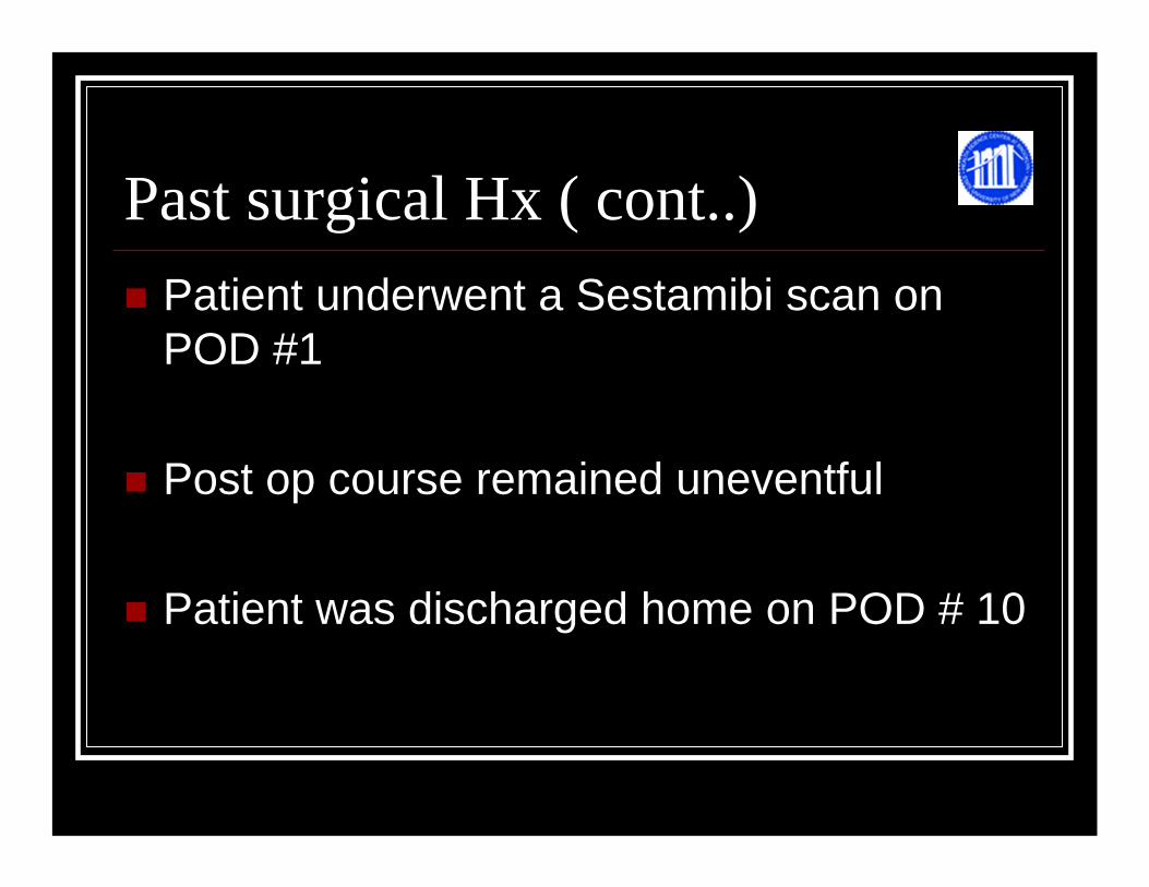
Past surgical Hx ( cont..)Patient underwent a Sestamibi scan on POD #1
Post op course remained uneventful
Patient was discharged home on POD # 10

Technetium-99m-sestamibi

Physical Examination HEEN :
Old cervical scar No palpable mass
CHEST : S1 , S2 N ; RRRB/l breath sounds equal , no crepts
Abdomen: Soft , no palpable mass , no tenderness
Right arm : functional AV fistula
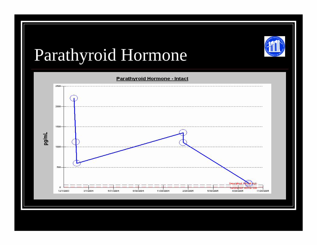
Parathyroid Hormone
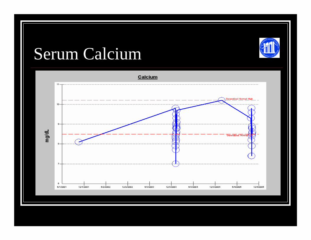
Serum Calcium

Patient showed an initial drop in the PTH levels postoperatively
However on further clinical visits , symptoms of Bone pains and myalgia returned
PTH level continued to raise on further clinical visits
Patient had an outpatient MRI and ultrasound examination

MRI – T2 images

Ultrasound - (L) thyroid gland Ultrasound of the left thyroid demonstrating a soild nodule in the lower lobe.
Operative Procedure Patient underwent left thyroid lobectomy An intraoperative frozen section confirmed parathyroid tissue A portion of the parathyroid was then autoimplanted in the patient’s right arm.
Final Pathology :Intrathyroidal hyperparathyroid tissue

Post- Operative Course Post operative course remained uneventful PTH levels returned to normal levels Calcium levels gradually returned to normal Patient continued to receive Ca rich dialsylate during dialysis.
Recurrent Hyper-Parathyroidism

HistorySir Richard Owen, the curator of the Natural History Museum.
discovered them in England in 1852 when he was dissecting a rhinoceros that had died in the London Zoo.
Some thirty years later in 1880, Ivar Sandstrom. a medical student working in Uppsala, described the glands in man.

Metabolism and calcium PTH – Secreted by chief cells ; 84 AA
N-terminal - active (1/2 life 3-4 min)C-terminal – inert ( 1/2 life longer ) ; cleaved in chief cells .
Binds to receptors and stimulates cyclic AMP production
Targets : Bones , Kidneys , Intestines

PTH physiologyIncreases serum and urinary Ca resorption
Decreases serum PO4 resorption
Increases osteoclastic activity and inhibits osteoblastic activity
Increases GI tract Ca absorption through Vit D
Increases renal conversion of Vit D3

PTH physiology

Embryology
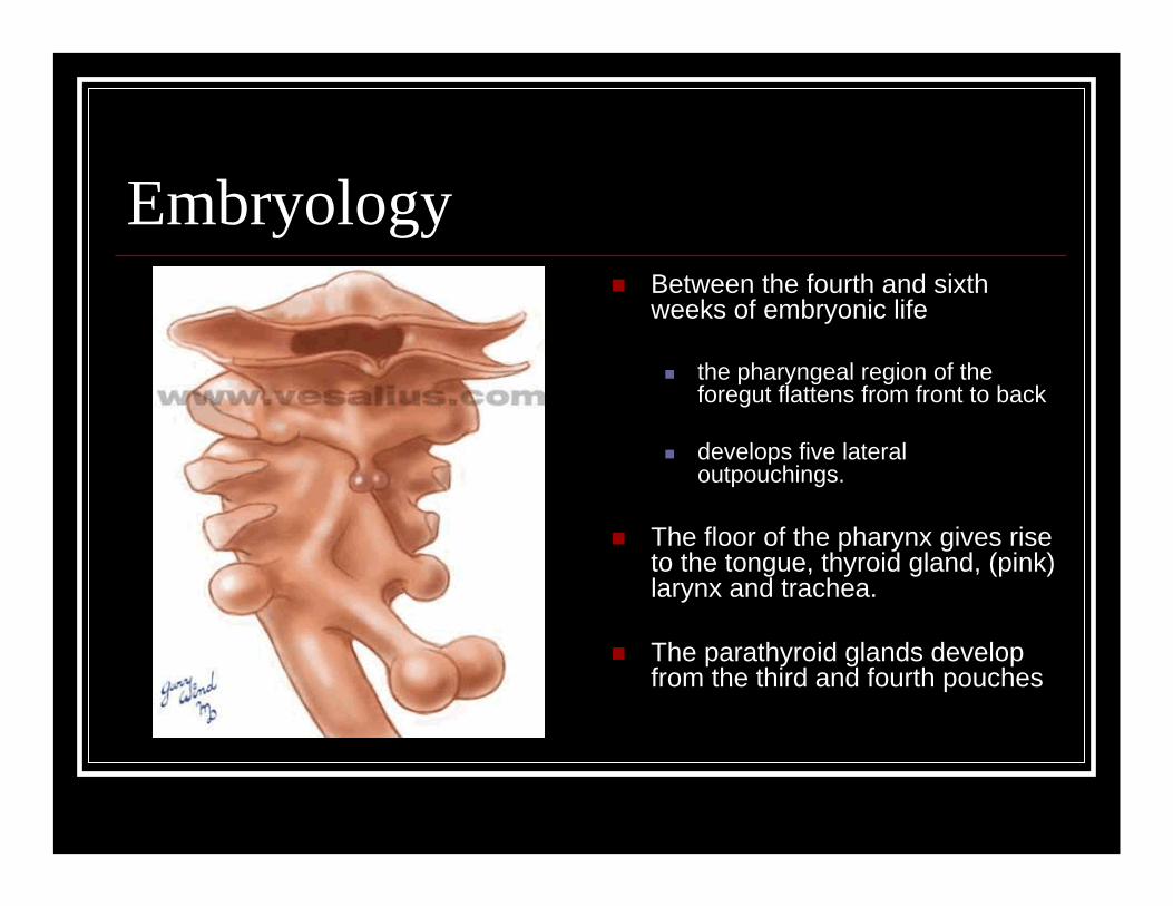
Embryology Between the fourth and sixth weeks of embryonic life
the pharyngeal region of the foregut flattens from front to back
develops five lateral outpouchings.
The floor of the pharynx gives rise to the tongue, thyroid gland, (pink) larynx and trachea.
The parathyroid glands develop from the third and fourth pouches

Embryology (cont..)The third branchial pouch gives rise to
the inferior parathyroid glands (dark blue) in close association with the primordia of the the thymus gland (orange).
As the thymus descends to the anterior mediastinum, parathyroids III follow along,
ultimately coming into contact with the developing thyroid caudal to parathyroids IV (yellow).

Embryology (cont..)The parathyroid glands derived from pouch IV take a more direct route to come in contact with the thyroid
and become the more cephalad or superior glands.
A portion of pouch IV (light blue) contributes a lateral C-cell component to the thyroid.
The parathyroids usually (~80%) lie near the posterolateral capsule of the thyroid lobes

Parathyroid Anatomy Superior Parathryoids – 4th Branchial pouch with thyroid – “ Constant”
Inferior Parathyroids – 3rd branchial pouch with thymus – “Travel”

Superior Parathyroid glands The superior parathyroid glands are most commonly found
the middle third of the thyroid lobe
at the level of the cricothyroidjunction
near the point where the recurrent laryngeal nerve passes beneath the inferior pharyngeal constrictor to enter the larynx

Inferior Parathyroid glands The inferior glands are usually found
near the lower pole of the thyroid lobe
below the lobe in the thyro-thymic ligament.
They commonly lie below the inferior thyroid artery anterior to the recurrent laryngeal nerve.
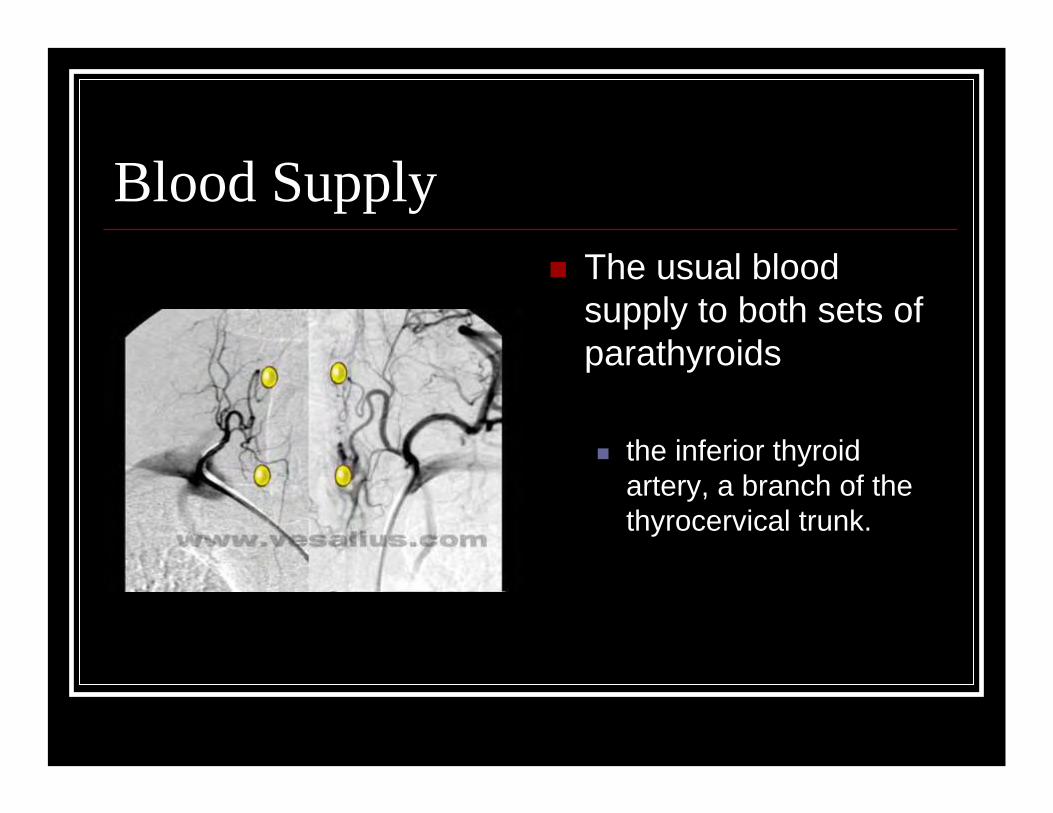
Blood Supply The usual blood supply to both sets of parathyroids
the inferior thyroid artery, a branch of the thyrocervical trunk.

? Number of Parathyroids80-90% of individuals have four parathyroid glands.
About 4% of individuals have three and 4% have five.
Fewer than one percent have two, six or seven.

Hyperparathyroidism Primary
80-90% Solitary adenoma 10-15% Hyperplasia 3 – 5% Double adenoma 1% Parathyroid cancer
SecondaryHyperplasia
Tertiary Autonomous hyperplasia

Symptoms Fatigue Exhaustion Weakness PolydypsiaPolyuriaNocturiaBone painConstipation
DepressionMemory loss Joint pain Loss of appetite Nausea Heartburn Pruritus
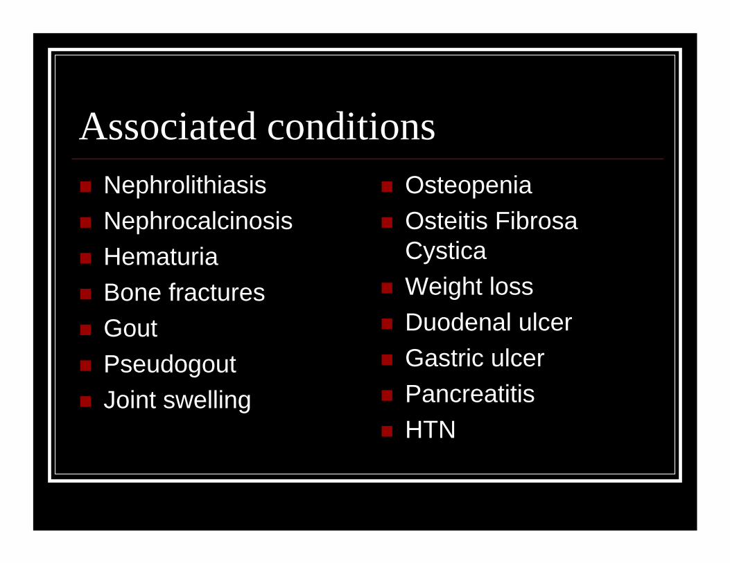
Associated conditionsNephrolithiasisNephrocalcinosisHematuriaBone fractures Gout PseudogoutJoint swelling
OsteopeniaOsteitis FibrosaCysticaWeight loss Duodenal ulcer Gastric ulcer PancreatitisHTN

Diagnosis Hypercalcemia – total or Ionized Elevated intact PTH –(1-84)Other Data :
Elevated or normal urinary Calcium Low serum PO4Increase in serum Cl (Cl/PO4 >33)Increase in serum Alk PhosBone density abnormalities

Guidelines for Surgery NIH consensus (October 29-31, 1990)
Age <50Serum Calcium >12mg/dlHypercalcuria > 400mg/g creatinineSevere Manifestations – bones, moans and stones Decrease in bone density ( <2SD for age )Progressive renal insufficiency

Parathyroidectomy in end stage renal disease
Severe hypercalcemia.
Progressive and debilitating hyperparathyroid bone disease as defined by radiographic or histologic evaluation.
Pruritus that does not respond to medical or dialytictherapy.
Otherwise unexplained symptomatic myopathy.
Progressive extraskeletal calcification or calciphylaxis that are usually associated with hyperphosphatemia that is refractory to oral phosphate binders.
In this setting, PTH-induced release of phosphate from bone contributes to the persistent elevation in the serum phosphate concentration.
Parathyroidectomy will tend to minimize further calcification by lowering the serum calcium and phosphate concentrations

ParathyroidectomyDespite advances in care, a subset of patients with end-stage renal disease still have marked elevations in serum parathyroid hormone levels.
Data are lacking regarding whether chronic elevations of PTH in asymptomatic patients warrant parathyroidectomy.
Surgery is often performed when patients develop refractory hyperparathyroidism (often with serum PTH concentrations above 800 pg/mL).
K/DOQI Clinical Practice Guidelines for Bone Metabolism and Disease in Chronic Kidney Disease. Am J Kidney Dis 2003; 42(Suppl 3):S1.
Tertiary hyperparathyroidismTertiary hyperparathyroidism if hypercalcemia is present is due to hyperfunctioning parathyroid tissue that does not respond appropriately to physiological regulation or to medical therapy with oral calcium salts and calcitrol.

Tertiary hyperparathyroidismfactors are thought to be involved in the pathogenesis of refractory hyperparathyroidism. These include:
Delayed and/or inadequate therapy Persistent hyperphosphatemia
Acquired abnormalities, which may be the most important, include increases in parathyroid gland mass due to polyclonal parathyroid cell proliferation (diffuse hyperplasia) and monoclonal expansion of adenomatous-like tissue.

PathophysiologyWhen exposed to stimuli
hypocalcemia, calcitrol deficiencyHyperphosphatemia
Parathyroid cells quickly respond with increased mitotic activity, leading to parathyroid gland hyperplasia.

Regression of established hyperplasia occurs slowly, perhaps due to a low rate of apoptotic cell death in the parathyroid glands.
This may explain why significant hyperplasia is sometimes irreversible.
Pathologic study of parathyroid glands in tertiary hyperparathyroidism. Krause MW; Hedinger CE Hum Pathol 1985 Aug;16(8):772-84.
The hyperparathyroidism of chronic renal failure: a disorder of growth. Parfitt AM Kidney Int 1997 Jul;52(1)
Polyclonal hyperplasia leads to refractory hyperparathyroidism because each cell may have a low amount of nonsuppressible basal PTH secretion.
In this setting, persistent hyperparathyroidism results from the summation of the contributions from the markedly increased number of cells

Aluminum Exposure
The degree of aluminum exposure and the presence of other forms of renal osteodystrophy (such as adynamic/aplastic disease) should also be considered when evaluating patients for parathyroidectomy.
Both aluminum-associated low turnover bone disease and aplastic/adynamic bone disease can mimic hyperparathyroidism by causing hypercalcemia, bone pain, and similar radiographic features

Surgical Approach
Bilateral neck explorationIdentification of all parathyroid glands Decision to excise based on Morphology
Focused neck explorationRelies on preoperative Imaging Intraoperative assessment of parathyroid function (ie. IOPTH monitoring )
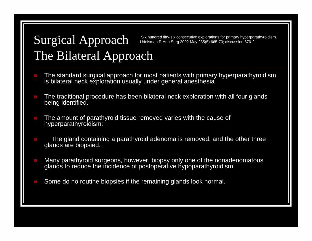
Surgical Approach The Bilateral Approach
The standard surgical approach for most patients with primary hyperparathyroidism is bilateral neck exploration usually under general anesthesia
The traditional procedure has been bilateral neck exploration with all four glands being identified.
The amount of parathyroid tissue removed varies with the cause of hyperparathyroidism:
The gland containing a parathyroid adenoma is removed, and the other three glands are biopsied.
Many parathyroid surgeons, however, biopsy only one of the nonadenomatousglands to reduce the incidence of postoperative hypoparathyroidism.
Some do no routine biopsies if the remaining glands look normal.
Six hundred fifty-six consecutive explorations for primary hyperparathyroidism.Udelsman R Ann Surg 2002 May;235(5):665-70; discussion 670-2.

ParathyroidectomyFor hyperplasia involving all four glands, three and one-half glands are removed
leaving one-half of the most normal-appearing gland marked with a clip.
In patients with multiple endocrine neoplasia type 1total parathyroidectomy with forearm autotransplantation is performed in some centers because of the high recurrence rate

Morphological identification of the abnormal gland

Parathyroidectomy /The Bilateral Approach
If surgeon identifies 3 normal glands and cannot find the 4th gland:
Explore retropharyngeal and esophageal spaces Trace RLN into the chest Open the carotid sheath ThymectomyConsider ipsilateral thyroid lobectomy DO NOT perform sternotomy
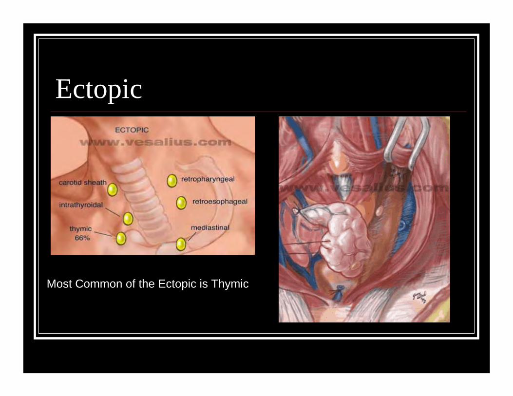
Ectopic
Most Common of the Ectopic is Thymic

Surgical Exploration

Parathyroid Surgery3 surgical procedures have been employed for the treatment of refractory hyperparathyroidism:
Subtotal parathyroidectomyTotal parathyroidectomy with autotransplantationTotal parathyroidectomy

Subtotal ParathyroidectomySubtotal parathyroidectomy involves excision of all identifiable parathyroid tissue except for 40 to 60 mg of the least hyperplastic gland.
Drawbacks to subtotal parathyroidectomya substantial risk of persistent recurrent disease, which is complicated by greater morbidity if repeat neck exploration is required.
For these reasons, some prefer total parathyroidectomywith autotransplantation of small amounts of resected parathyroid tissue
parathyroidectomyThe incidence of reoperation for moderate to severe recurrent hyperparathyroidism is similar with both methods, ranging from 6 to 14 percentRothmund, M, Wagner, PK, Schark, C.
Subtotal parathyroidectomy versus total parathyroidectomy and autotransplantation in secondary hyperparathyroidism: a randomized trial. World J Surg 1991; 15:745.
Kaye, M, D'Amour, P, Henderson, J. Elective total parathyroidectomy without autotransplant in end-stage renal disease. Kidney Int 1989; 35:1390.
Recurrent hyperparathyroidism is much more likely to occur with autografting of nodular hyperplastic tissue
Gagne, ER, Urena, P, Leite-Silva, S, et al. Short- and long-term efficacy of total parathyroidectomy with immediate autografting compared with subtotal parathyroidectomy in hemodialysis patients. J Am Soc Nephrol
1992; 3:1008
The frequency of recurrence was 33 percent with nodular versus only 4 percent with diffuse hyperplasia.
Tominaga, Y, Tanaka, Y, Sato, K, et al. Recurrent renal hyperparathyroidism and DNA analysis of autografted parathyroid tissue. World J Surg 1992; 16:595.

Drawbacks of total Parathyroidectomywithout autotransplantation
Development of adynamic bone disease and intractable osteomalacia
Permanent hypoparathyroidism
Impaired bone healing in the absence of PTH and its anabolic effects
Need for long-term use of calcium and vitamin D.

Refractory parathyroidisim
In spite of these reservations, a majority of patients who undergo total parathyroidectomy have measurable PTH levels at long-term follow-up and no demonstrable bone disease.
One report examined the long-term effects of total parathyroidectomy in 20 patients with refractory secondary hyperparathyroidism.
Six patients had persistent hypocalcemiafive were asymptomatic, andone patient, who was not compliantSerum PTH concentrations were
less than normal – 6 Normal – 7Increased -7
Stracke, S, Jehle, PM, Sturm, D, et al. Clinical course after total parathyroidectomy without autotransplantation in patients with end-stage renal failure. Am J Kidney Dis 1999; 33:304.

The success rate of reoperative surgery without preoperative localization is only 60 percent
this can be improved to 95 percent or more with localization.
It should be noted that localization studies identifying a single adenoma do not exclude the existence of abnormal parathyroid glands in other locations
almost 30 percent of patients undergoing re-operation have multiple gland hyperplasia.


Re -OperationFive to 10 percent of patients undergoing surgery for hyperparathyroidism have persistent disease.
The approach is different in patients with recurrent or persistent hyperparathyroidism because of the differences in etiologies and in surgical morbidity due to fibrosis as compared with un-operated patients.
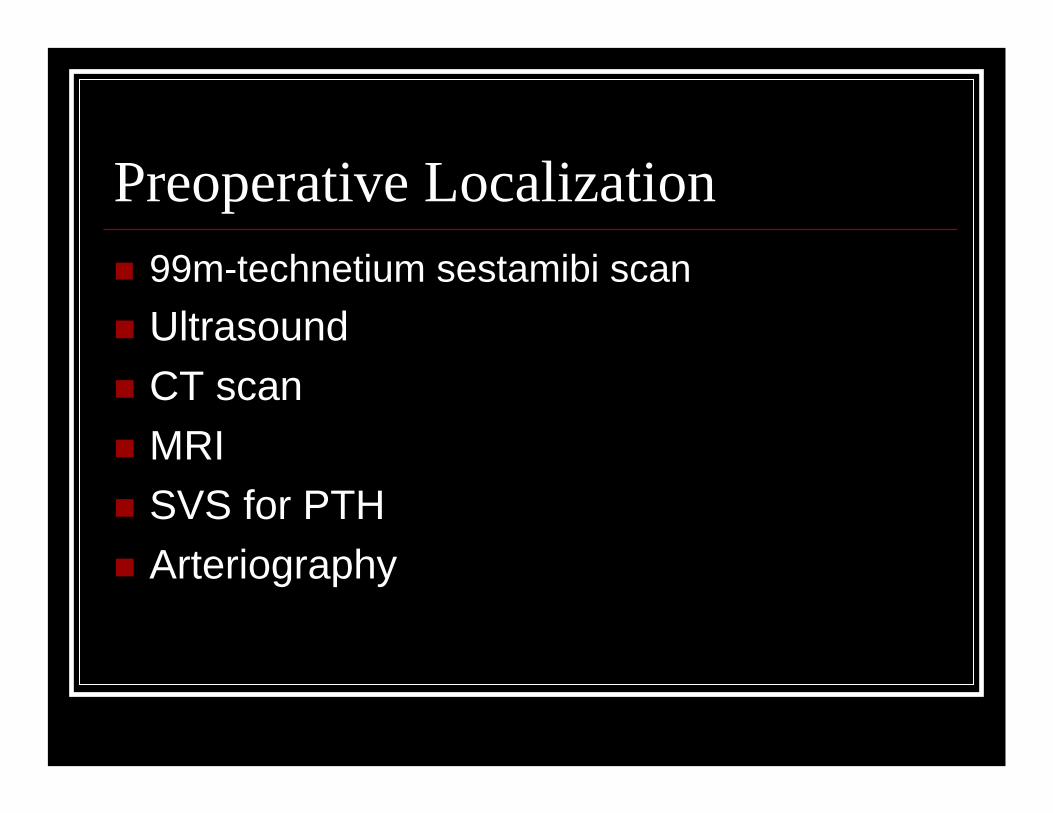
Preoperative Localization 99m-technetium sestamibi scan Ultrasound CT scan MRI SVS for PTH Arteriography

Ultrasonographic localization

99m-technetium sestamibi scanThe results have been somewhat better with 99m-technetium sestamibi
often in combination with a subtraction thyroid scan using 123-I-iodine or with sestamibi double phase studies.
This technique has a predictive value that can range up to 90 to 100 percent for solitary adenomas.
The results with a related radionuclide, technetium-99m tetrofosmin, are similar.

99m-technetium sestamibi scan

99m-technetium sestamibi scan

99m-technetium sestamibi scanSestamibi scanning may detect multiple involved glands or a mediastinal adenoma.
However, scanning for hyperplastic glands or double adenomas is less accurate.
The specificity of sestamibi scanning can be enhanced with delayed (two-hour) imaging and three-dimensional imaging obtained by single photon emission computed tomography (SPECT).
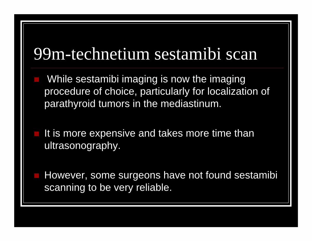
99m-technetium sestamibi scanWhile sestamibi imaging is now the imaging procedure of choice, particularly for localization of parathyroid tumors in the mediastinum.
It is more expensive and takes more time than ultrasonography.
However, some surgeons have not found sestamibiscanning to be very reliable.

Sestamibi in Primary Vs Secondary HyperparathyroidisimPreoperative imaging with the parathyroid technetium-99m-sestamibi and I-123 subtraction
scintigraphy technique in 11 patients with secondary hyperparathyroidism referred for first parathyroid surgery
41 of 45 glands found at surgery were detected by preoperative scanning (91 percent sensitivity), with no false positive scans.The technique was also able to identify ectopic and supernumerary parathyroid glands.
Hindie, E, Urena, P, Jeanguillaume, C, et al. Preoperative imaging of parathyroid glands with technetium 99m-labeled sestamibi and iodine-123 subtraction scanning in secondary hyperparathyroidism. Lancet 1999; 353:2200.
The sensitivity of technetium-99m-sestamibi and I-123 subtraction single photon emission computed tomography (SPECT) was assessed in a series of 19 patients with renal failure and secondary hyperparathyroidism.
Scanning correctly identified 57 of 74 hyperplastic glands found at surgery, resulting in a sensitivity of 77 percent.
Neumann, DR, Esselstyn, CB, Madera, A, et al. Parathyroid detection in secondary hyperparathyroidism with 123I/99mTc-sestamibi subtraction single photon emission computed tomography. J Clin Endocrinol Metab 1998; 83:3867.

In a study of 21 consecutive patientsThe results using the combination of dual phase dual isotope iodine 123/technetium Tc 99m Sestamibi scintigraphy plus high resolution ultrasonography was compared to that found with surgical and histolopathologic findings.
The combination of both techniques detected 88 percent of all hyperplastic parathyroid glands (with surgery producing a success rate of 99 percent). Most missed glands with ultrasonography were of low weight, while scintigraphy missed glands located superiorly.
Perie, S, Fessi, H, Tassart, M, et al. Usefulness of combination of high-resolution ultrasonography and dual-phase dual-isotope iodine 123/technetium Tc 99m sestamibi scintigraphy for the preoperative localization of hyperplasticparathyroid glands in renal hyperparathyroidism.Am J Kidney Dis 2005; 45:344.

MRI localization

Angiogram
Ectopic Parathyroids
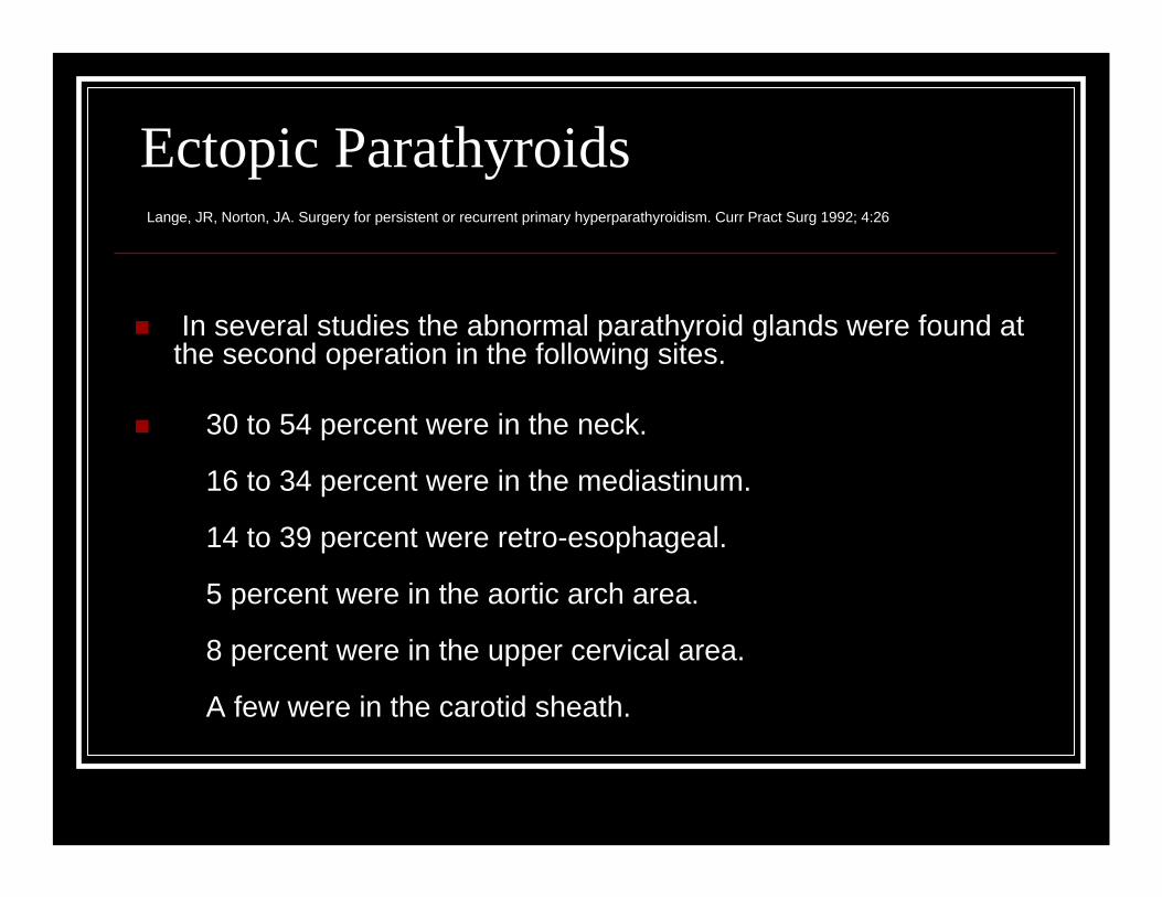
In several studies the abnormal parathyroid glands were found at the second operation in the following sites.
30 to 54 percent were in the neck.
16 to 34 percent were in the mediastinum.
14 to 39 percent were retro-esophageal.
5 percent were in the aortic arch area.
8 percent were in the upper cervical area.
A few were in the carotid sheath.
Lange, JR, Norton, JA. Surgery for persistent or recurrent primary hyperparathyroidism. Curr Pract Surg 1992; 4:26

Complications 1% Recurrent laryngeal nerve palsy 1% Hematoma 0.5% permanent hypothyroidism

Intrathyroidal parathyroid glands can be a cause of failed cervical exploration
for hyperparathyroidism. McIntyre RC Jr, Eisenach JH, Pearlman NW, Ridgeway CE, Liechty RD.Am J Surg. 1997 Dec;174(6):750-3; discussion 753-4.
BACKGROUND:The incidence of intrathyroidal parathyroid glands remains
controversial.
The purpose of this study was to determine the incidence in a series of patients with hyperparathyroidism.
METHODS: 309 patients underwent parathyroidectomy.
Patients were divided into two groups:uniglandular disease versus hyperplasia.

RESULTS: 6% had abnormal intrathyroidal parathyroid glands.
The incidence was 3% in patients with uniglandular disease versus 15% in those with hyperplasia.
With a mean follow-up of 54 months, 12 patients are eucalcemic, 5 have persistent hypocalcemia, and 1 has recurrent hypercalcemia. There were no recurrent laryngeal nerve injuries.
CONCLUSIONS: These data suggest that an intrathyroidal adenoma is an uncommon cause of failure, whereas abnormal intrathyroidal parathyroid tissue may be a more common cause of failure in patients with hyperplasia.
Unilateral approach
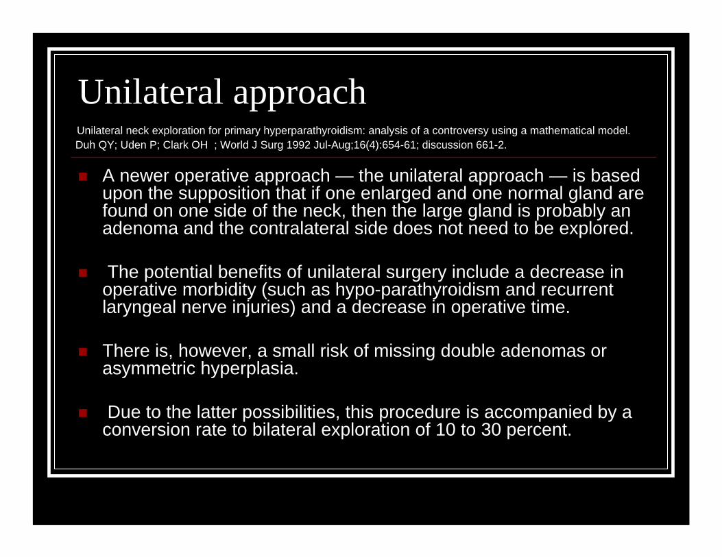
A newer operative approach — the unilateral approach — is based upon the supposition that if one enlarged and one normal gland are found on one side of the neck, then the large gland is probably an adenoma and the contralateral side does not need to be explored.
The potential benefits of unilateral surgery include a decrease in operative morbidity (such as hypo-parathyroidism and recurrent laryngeal nerve injuries) and a decrease in operative time.
There is, however, a small risk of missing double adenomas or asymmetric hyperplasia.
Due to the latter possibilities, this procedure is accompanied by a conversion rate to bilateral exploration of 10 to 30 percent.
Unilateral neck exploration for primary hyperparathyroidism: analysis of a controversy using a mathematical model.Duh QY; Uden P; Clark OH ; World J Surg 1992 Jul-Aug;16(4):654-61; discussion 661-2.

Primary hyperparathyroidism in the 1990s. Choice of surgical procedures for this disease. Kaplan EL; Yashiro T; Salti G Ann Surg 1992 Apr;215(4):300-17
The theoretical advantages of this unilateral approach decrease
in operative morbidity rates hypoparathyroidism nerve injuries--and a decrease in operative time.
Furthermore, proponents argue that if persistent hyperparathyroidism occurs, the second side can be easily explored because it was previously untouched.
In the hands of several expert parathyroid surgeons, excellent results have been achieved.

Minimally invasive parathyroidectomy
Minimally invasive parathyroidectomy is a modification of the unilateral approach.
It consists of preoperative imaging (with technetium-99m-sestamibi)
confirmation of the removal of a single gland by measuring serum PTH in the operating room before withdrawal of anesthesia.
This takes advantage of the short plasma half-life of PTH (three to four minutes) and a rapid assay for PTH.
Late parathyroid function after successful parathyroidectomy guided by intraoperative hormone assay (QPTH) compared with the standard bilateral neck exploration.Carneiro DM; Irvin GL 3rd; Surgery 2000 Dec;128(6):925-9;discussion 935-6.
Minimally invasive parathyroidectomy: 50 consecutive cases. Delbridge LW; Dolan SJ; Hop TT; Robinson BG; Wilkinson MR; Reeve TS Med J Aust 2000 May 1;172(9):418-22.
OBJECTIVE: To determine the effectiveness and outcomes of minimally invasive parathyroidectomy.
DESIGN: Prospective, non-randomised, non-blinded trial.
PATIENTS: 50 consecutive patients who underwent minimally invasive parathyroidectomy for primary hyperparathyroidism, and 150 consecutive patients undergoing open parathyroidectomyover the same period.

RESULTS: cure (normocalcaemia) 84%. 14% required conversion to an open procedure
1 patient had persistent hyperparathyroidism after minimally invasive parathyroidectomy which was cured at subsequent open reoperation.
Open parathyroidectomy was successful in 147 of 150 patients (98%) at initial operation
Intraoperative measurement of parathyroid hormone levels by a quick technique in 23 of the patients (13 having minimally invasive and 10 open procedures) correctly identified the presence of multiple-gland disease.

Conventional Versus MIP
Objective : To review the outcomes of 656 consecutive parathyroid explorations performed by a single surgeon and to compare the results of conventional and minimally invasive parathyroidectomy (MIP) techniques.
Method : Traditional surgery for primary hyperparathyroidism (HPTH) involves bilateral cervical exploration, which is usually accomplished under general endotrachealanesthesia.
The MIP technique involves preoperative localization with sestamibi scanssurgeon-administered cervical block anesthesia directed exploration through a small incisionintraoperative rapid parathyroid hormone assaydischarge within 2 to 3 hours of surgery.
Six hundred fifty-six consecutive explorations for primary hyperparathyroidism.Udelsman R et al / Ann Surg 2002 May;235(5):665-70; discussion 670-2.

RESULTS:61% were performed using the standard technique and 255 patients 39% were selected for MIP.
The success rate for the entire series was 98%, with no significant differences comparing traditional and MIP techniques.
The overall complication rate of 2.3% 3.0% in the standard 1.2% in the MIP groups
MIP was associated with approximately a 50% reduction in operating time, a 7fold reduction in length of hospital stay, and a mean cost savings of $2,693 per procedure, which represents nearly a 50% reduction in total hospital charges.
In a series of 40 patients, for example, unilateral exploration guided by the scan would have failed in 10 percent.
In addition, the specificity of sestamibi scans has been questioned because uptake has been demonstrated in coexisting adenomas and multinodular goiters in patients with hyperparathyroidism.
False positive scans have also been attributed to misinterpretation of the scan by the surgeon.
This may be eliminated by sestamibi-radioguided surgery.
Irvin, GL, Prudhomme, DL, Deriso, GT et al.A new approach to parathyroidectomy. Ann Surg 1994; 219:574.

Minimally invasive radioguidedparathyroidectomy
Objective :Minimally invasive radioguidedparathyroidectomy (MIRP) combines
technetium sestamibi scanintraoperative gamma probe methylene blue dyemeasurement of circulating parathyroid hormone (PTH) levels.
Minimally invasive radioguided parathyroidectomy.
Flynn MB; Bumpous JM; Schill K; McMasters KM; J Am Coll Surg 2000 Jul;191(1):24-31.

Minimally invasive radioguided parathyroidectomy. Flynn MB; Bumpous JM; Schill K; McMasters KM J Am Coll Surg 2000 Jul;191(1):24-31.
STUDY DESIGN: All patients presented with biochemically proved primary hyperparathyroidism.A technetium sestamibi scan was performed preoperatively.
Technetium sestamibi and methylene blue dye (7.5 mg/kg) were administered IV on the day of operation. Operative dissection was directed by the gamma probe. Blood samples for PTH assay were obtained before and after excision of an abnormal gland. When an appropriate decrease in the PTH assay was obtained, the exploration was concluded. Persistent PTH elevation instigated further neck exploration.

Results:36 consecutive patients were explored for untreated primary hyperparathyroidism and 3 for recurrent hyperparathyroidism.
Hypercalcemia was corrected in all 39 patients.
A single adenoma was found in 32 of 36 patients with untreated primary hyperparathyroidism, and a single abnormal gland was identified in all of those with recurrent hyperparathyroidism.

Persistently elevated PTH prompted further exploration in two patients, identifying a second abnormal gland in 1 and hyperplasia in the other.
Minor local complications occurred in 8% of the patients.
44% of the patients were discharged on the day of operation.
83% within 23 hours after the initial neck exploration for primary hyperparathyroidism.

CONCLUSIONS: MIRP is a safe and effective procedure, resulting in the correction of hypercalcemia in all patients. The combination of intraoperative gamma probe and methylene blue dye allows rapid identification of the abnormal gland with minimal dissection through a small incision. PTH assay after excision provides biochemical confirmation that the abnormal gland has been removed. Most patients undergoing MIRP can be treated on an outpatient basis. Low postoperative complications, a small incision, and rapid return to normal activities resulted in very high patient acceptance of the procedure.

Ablation TechniquesAn occasional patient who needs treatment but is not a candidate for surgery, or who has an adenoma in the mediastinum, might be considered for ablation
Options :angiographic ablation ablation with ethanol injected with ultrasound guidance.
Success rates of 66 percent at up to four years have been reported for angiographic ablation.
Arteriographic ablation of cervical parathyroid adenomas.Pallotta JA; Sacks BA; Moller DE; Eisenberg H; J Clin Endocrinol Metab 1989 Dec;69(6):1249-55.
ConclusionAt present,
Bilateral neck exploration by a skilled parathyroid surgeon without preoperative localization is our procedure of choice in patients with primary hyperparathyroidism undergoing initial surgery.
Exceptions are high risk elderly patients, and those who cannot undergo general anesthesia
Unilateral surgery is an alternative in patients with one enlarged and one normal gland on one side of the neck, and is an attractive option in high-risk patients
ConclusionThere is a significant body of literature surrounding the impact on mortality of elevated parathyroid hormone levels, hyperphosphatemia, vascular calcification, and other clinical features associated with derangements of parathyroid function in end-stage renal disease.
However, a paucity of data exists concerning survival following parathyroidectomy.
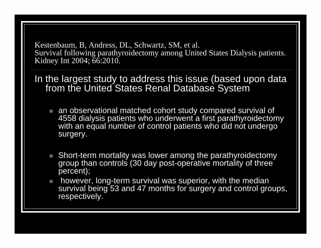
Kestenbaum, B, Andress, DL, Schwartz, SM, et al. Survival following parathyroidectomy among United States Dialysis patients. Kidney Int 2004; 66:2010.
In the largest study to address this issue (based upon data from the United States Renal Database System
an observational matched cohort study compared survival of 4558 dialysis patients who underwent a first parathyroidectomywith an equal number of control patients who did not undergo surgery.
Short-term mortality was lower among the parathyroidectomygroup than controls (30 day post-operative mortality of three percent);however, long-term survival was superior, with the median
survival being 53 and 47 months for surgery and control groups, respectively.