Embed Size (px)
Citation preview
VISIT WWW.MEDEDICUS.COM FOR ONLINE TESTING AND INSTANT CME CERTIFICATE.
Case Debates in the Management of
Jointly sponsored by The New York Eye and Ear Infirmary and MedEdicus LLC.
This continuing medical education activity is supported throughan unrestricted educational grant from Genentech, Inc.
Age-Related MacularDegeneration andDiabetic Macular Edema
HIGHLIGHTS FROM A CME SYMPOSIUM HELD DURING THEAMERICAN ACADEMY OF OPHTHALMOLOGY 2012 MEETING
The CME symposium was not affiliated with the official program of the AAO/APAO Joint Meeting.
CME MONOGRAPH
ORIGINAL RELEASE: APRIL 1, 2013
LAST REVIEW: MARCH 20, 2013
EXPIRATION: APRIL 30, 2014
Distributed with

2
LEARNING METHOD AND MEDIUMThis educational activity consists of a supplement and ten (10)study questions. The participant should, in order, read the learningobjectives contained at the beginning of this supplement, read thesupplement, answer all questions in the post test, and completethe Activity Evaluation/Credit Request form. To receive credit forthis activity, please follow the instructions provided on the posttest and Activity Evaluation/Credit Request form. This educationalactivity should take a maximum of 2 hours to complete.
CONTENT SOURCEThis continuing medical education (CME) activity captures contentfrom a CME symposium held on November 11, 2012, in Chicago,Illinois.
ACTIVITY DESCRIPTIONThrough case applications, this monograph will teach learners therecent developments in the treatment of neovascular AMD and DME.In AMD, clinical trial data (CATT2, VIEW 2-year data, HARBOR,IVAN), and a new anti-VEGF agent (aflibercept) provide furtherinsight to optimal dosing of the anti-VEGF agents. In DME, recentclinical trial results (RISE, RIDE), new treatments (ranibizumab), andmajor DRCR.net trial findings have been released.
TARGET AUDIENCEThis activity is intended for retina specialists, retina specialistfellows, and comprehensive ophthalmologists caring for patientswith age-related macular degeneration (AMD) and diabeticmacular edema (DME).
LEARNING OBJECTIVESUpon completion of this activity, participants will be better able to:Apply the results of recent clinical trials for dosing anti-vascularendothelial growth factor (VEGF) therapy in specific patientswith neovascular AMD and DMEEvaluate differences between anti-VEGF agents with regard toefficacy, dosing, pharmacokinetic parameters, and safety whenused for patients with AMD and DMESelect appropriate therapeutic measures and regimens forpatients with AMD and DME
ACCREDITATION STATEMENTThis activity has been planned and implemented in accordancewith the Essential Areas and Policies of the Accreditation Councilfor Continuing Medical Education through the joint sponsorship ofThe New York Eye and Ear Infirmary and MedEdicus LLC. TheNew York Eye and Ear Infirmary is accredited by the ACCME toprovide continuing medical education for physicians.
AMA CREDIT DESIGNATION STATEMENTThe New York Eye and Ear Infirmary designates this enduringmaterial for a maximum of 2.0 AMA PRA Category 1 Credits™.Physicians should claim only the credit commensurate with theextent of their participation in the activity.
GRANTOR STATEMENTThis continuing medical education activity is supported through anunrestricted educational grant from Genentech, Inc.
DISCLOSURE POLICY STATEMENTIt is the policy of The New York Eye and Ear Infirmary that thefaculty and anyone in a position to control activity content discloseany real or apparent conflicts of interest relating to the topics ofthis educational activity, and also disclose discussions ofunlabeled/unapproved uses of drugs or devices during theirpresentation(s). The New York Eye and Ear Infirmary hasestablished policies in place that will identify and resolve allconflicts of interest prior to this educational activity. Fulldisclosure of faculty/planners and their commercial relationships,if any, follows.
DISCLOSURES David S. Boyer, MD, had a financial agreement or affiliationduring the past year with the following commercial interests in theform of Consultant/Advisory Board: Alcon, Inc; Allegro Ophthalmics,LLC; Allergan, Inc; Bayer; Genentech, Inc; Glaukos Corporation;GlaxoSmithKline; iCo Therapeutics Inc; Neurotech; Novartis;Optos; Ora; Pfizer Inc; QLT Phototherapeutics, Inc; RegeneronPharmaceuticals, Inc; and Spire; Fees for promotional, advertising ornon-CME services received directly from commercial interest or theirAgents (eg, Speakers Bureaus): Alcon, Inc; Allergan, Inc; Eyetech Inc;Genentech, Inc; Novartis; and Pfizer Inc; Ownership Interest: AllegroOphthalmics, LLC.
David S. Boyer, MD—PROGRAM CO-CHAIRClinical Professor of OphthalmologyKeck School of MedicineUniversity of Southern CaliforniaLos Angeles, CaliforniaSenior PartnerRetina-Vitreous Associates Medical GroupBeverly Hills, California
David M. Brown, MD—PROGRAM CO-CHAIRClinical Research CoordinatorGreater Houston Retina ResearchRetina Consultants of HoustonHouston, Texas
Neil M. Bressler, MD*Chief, Retina DivisionThe Wilmer Eye InstituteThe Johns Hopkins HospitalThe James P. Gills Professor of OphthalmologyThe Johns Hopkins University School of MedicineBaltimore, Maryland
Michael S. Ip, MDAssociate ProfessorUniversity of WisconsinSchool of Medicine and Public HealthDepartment of Ophthalmology and Visual SciencesMadison, Wisconsin
Carl D. Regillo, MD, FACSDirector, Retina ServiceWills Eye InstituteProfessor of OphthalmologyThomas Jefferson UniversityPhiladelphia, Pennsylvania
FACULTY
*Participation by Dr Neil M. Bressler in this activity does not constitute or imply endorsement by The Johns Hopkins University, The Johns Hopkins Hospital, or The Johns Hopkins Health System.
3
INTRODUCTIONIn a busy retina practice, neovascular age-related macular degeneration (AMD) and diabetic macularedema (DME) represent 2 of the most common conditions we treat. In recent years, with the adventof drugs that inhibit the activity of vascular endothelial growth factor (VEGF), the treatmentparadigm for both these conditions has evolved dramatically. We now have 3 potent widely used anti-VEGF agents to choose from and a growing number of clinical trials to guide our clinical practicepatterns. In this program, we bring you highlights from a continuing medical education symposium atwhich expert faculty presented and discussed cases representative of the patients we see in the officeevery day. Our objective is to illustrate how we apply the lessons from clinical trials to clinicalpractice, and how we develop treatment plans for individual patients with AMD and DME.
—David S. Boyer, MD, and David M. Brown, MDProgram Co-Chairs
Neil M. Bressler, MD, had a financial agreement or affiliationduring the past year with the following commercial interests in the form of Consultant/Advisory Board (spouse): GlaxoSmithKline;Contracted Research: Abbott Medical Optics Inc; Allergan USA, Inc;Bausch + Lomb Incorporated; Bristol-Myers Squibb Company; CarlZeiss Meditec, Inc; ForSight Labs, LLC; Genentech, Inc; GenzymeCorporation; Lumenis Inc; Notal Vision; Novartis Pharma AG;Optovue, Inc; Pfizer Inc; Quark Biotech, Inc; and RegeneronPharmaceuticals, Inc; Contracted Research (spouse): Bausch + LombIncorporated; Genentech, Inc; Notal Vision; Novartis Pharma AG;Regeneron Pharmaceuticals, Inc; and ThromboGenics NV.
David M. Brown, MD, had a financial agreement or affiliationduring the past year with the following commercial interests in the form of Honoraria: Genentech, Inc; and RegeneronPharmaceuticals, Inc; Consultant/Advisory Board: Alimera Sciences;Allergan, Inc; Genentech, Inc; Novartis; RegeneronPharmaceuticals, Inc; and ThromboGenics NV; Fees for promotional,advertising or non-CME services received directly from commercialinterest or their Agents (eg, Speakers Bureaus): Genentech, Inc; andRegeneron Pharmaceuticals, Inc; Contracted Research: Alcon, Inc;Alimera Sciences; Allergan, Inc; Bayer; Eli Lilly and Company;Genentech, Inc; GlaxoSmithKline; Novartis; RegeneronPharmaceuticals, Inc; and ThromboGenics NV.
Michael S. Ip, MD, had a financial agreement or affiliation duringthe past year with the following commercial interests in the formof Consultant/Advisory Board: Eyetech Inc; Genentech, Inc; NicOx; Notal Vision; QLT Phototherapeutics, Inc; RegeneronPharmaceuticals, Inc; and Sirion Therapeutics, Inc; ContractedResearch: Allergan, Inc.
Carl D. Regillo, MD, had a financial agreement or affiliation duringthe past year with the following commercial interests in the formof Consultant/Advisory Board: Alimera Sciences; Allergan, Inc;Genentech, Inc: GlaxoSmithKline; and Regeneron Pharmaceuticals,Inc; Contracted Research: Alimera Sciences; Allergan, Inc; Genentech,Inc; GlaxoSmithKline; and Regeneron Pharmaceuticals, Inc.
PEER REVIEW DISCLOSURE John Sorenson, MD, has no relevant commercial relationships todisclose .
EDITORIAL SUPPORT DISCLOSURESTony Realini, MD; Cynthia Tornallyay, RD, MBA, CCMEP; KimberlyCorbin, CCMEP; Barbara Aubel; and Vivian Fransen, MPA, have norelevant commercial relationships to disclose.
DISCLOSURE ATTESTATIONThe contributing physicians listed above have attested to thefollowing:1) that the relationships/affiliations noted will not bias or
otherwise influence their involvement in this activity;2) that practice recommendations given relevant to the companies
with whom they have relationships/affiliations will be supportedby the best available evidence or, absent evidence, will beconsistent with generally accepted medical practice; and
3) that all reasonable clinical alternatives will be discussed whenmaking practice recommendations.
OFF-LABEL DISCUSSIONThis activity includes investigational or off-label discussion ofaflibercept for DME and of bevacizumab for AMD and DME. Pleaserefer to the official prescribing information for discussion ofapproved indications, contraindications, and warnings.
FOR DIGITAL EDITIONSSystem Requirements:If you are viewing this activity online, please ensure the computeryou plan to use meets the following requirements:● Operating System: Windows or Macintosh● Media Viewing Requirements: Flash Player or Adobe Reader● Supported Browsers: Microsoft Internet Explorer, Firefox,Google Chrome, Safari, and Opera
● A good Internet connectionThe New York Eye and Ear Infirmary Privacy &Confidentiality PoliciesCME policies: http://www.nyee.edu/cme-enduring.htmlHospital policies: http://www.nyee.edu/website-privacy.html
CME Provider Contact InformationFor questions about this activity, call 212-979-4383.
TO OBTAIN AMA PRA CATEGORY 1 CREDIT™To obtain AMA PRA Category 1 Credit™ for this activity, read the material in its entirety and consult referenced sources asnecessary. Complete the evaluation form along with the post test answer box within this supplement. Remove the ActivityEvaluation/Credit Request page from the printed supplement orprint the Activity Evaluation/Credit Request page from the DigitalEdition. Return via mail or fax to Kim Corbin, Director, ICME, TheNew York Eye and Ear Infirmary, 310 East 14th Street, New York,NY 10003 or fax to (212) 353-5703. Your certificate will bemailed to the address that you provide on the evaluation form.Please allow 3 weeks for mailed/faxed forms to be processed.There are no fees for participating in and receiving CME credit forthis activity.
Alternatively, we offer instant certificate processing and supportGreen CME. Please take this post test and evaluation online bygoing to www.MedEdicus.com, Educational Activities tab, andclicking the Post-Test & CME Certificate button. Upon passing,you will receive your certificate immediately. You must score 70%or higher to receive credit for this activity, and may take the testup to 2 times. Upon registering and successfully completing thepost test, your certificate will be made available online and youcan print it or file it.
DISCLAIMERThe views and opinions expressed in this educational activity arethose of the faculty and do not necessarily represent the views ofThe New York Eye and Ear Infirmary; MedEdicus LLC;Genentech, Inc; or Retina.
This CME activity is copyrighted to MedEdicus LLC ©2013. All rights reserved.
4
AGE-RELATED MACULARDEGENERATIONCASE 1: Treat-and-Extend Approach
—Carl D. Regillo, MD
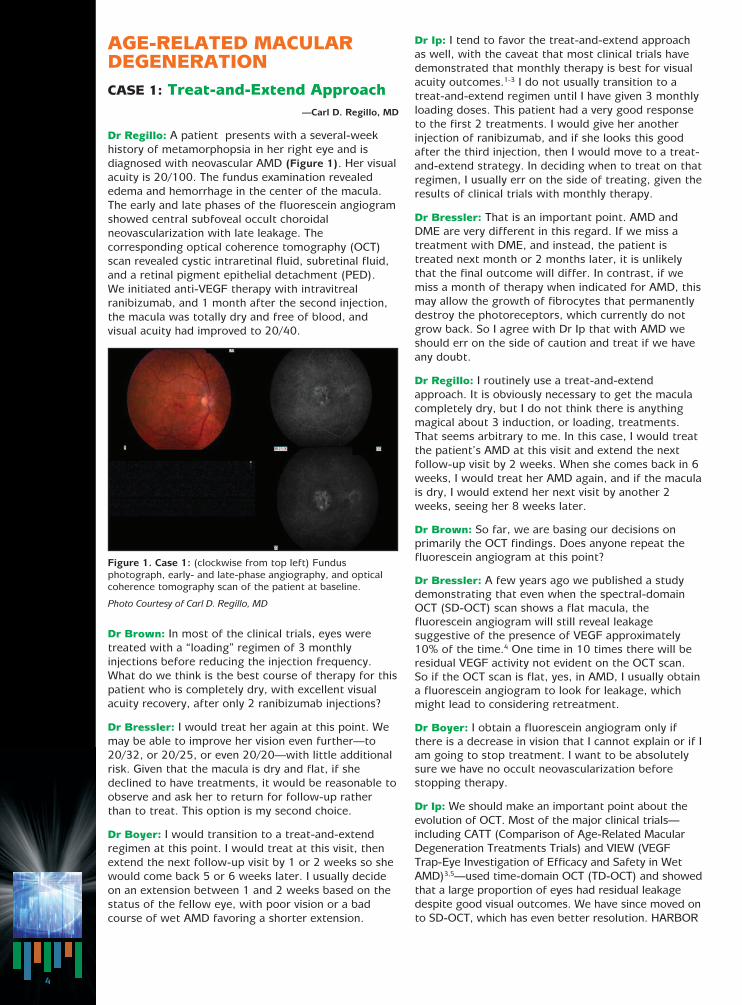
Dr Regillo: A patient presents with a several-weekhistory of metamorphopsia in her right eye and isdiagnosed with neovascular AMD (Figure 1). Her visualacuity is 20/100. The fundus examination revealededema and hemorrhage in the center of the macula.The early and late phases of the fluorescein angiogramshowed central subfoveal occult choroidalneovascularization with late leakage. Thecorresponding optical coherence tomography (OCT)scan revealed cystic intraretinal fluid, subretinal fluid,and a retinal pigment epithelial detachment (PED). We initiated anti-VEGF therapy with intravitrealranibizumab, and 1 month after the second injection,the macula was totally dry and free of blood, andvisual acuity had improved to 20/40.
Dr Brown: In most of the clinical trials, eyes weretreated with a “loading” regimen of 3 monthlyinjections before reducing the injection frequency.What do we think is the best course of therapy for thispatient who is completely dry, with excellent visualacuity recovery, after only 2 ranibizumab injections?
Dr Bressler: I would treat her again at this point. Wemay be able to improve her vision even further—to20/32, or 20/25, or even 20/20—with little additionalrisk. Given that the macula is dry and flat, if shedeclined to have treatments, it would be reasonable toobserve and ask her to return for follow-up ratherthan to treat. This option is my second choice.
Dr Boyer: I would transition to a treat-and-extendregimen at this point. I would treat at this visit, thenextend the next follow-up visit by 1 or 2 weeks so shewould come back 5 or 6 weeks later. I usually decideon an extension between 1 and 2 weeks based on thestatus of the fellow eye, with poor vision or a badcourse of wet AMD favoring a shorter extension.
Dr Ip: I tend to favor the treat-and-extend approach as well, with the caveat that most clinical trials havedemonstrated that monthly therapy is best for visualacuity outcomes.1-3 I do not usually transition to atreat-and-extend regimen until I have given 3 monthlyloading doses. This patient had a very good responseto the first 2 treatments. I would give her anotherinjection of ranibizumab, and if she looks this goodafter the third injection, then I would move to a treat-and-extend strategy. In deciding when to treat on thatregimen, I usually err on the side of treating, given theresults of clinical trials with monthly therapy.
Dr Bressler: That is an important point. AMD andDME are very different in this regard. If we miss atreatment with DME, and instead, the patient istreated next month or 2 months later, it is unlikelythat the final outcome will differ. In contrast, if wemiss a month of therapy when indicated for AMD, thismay allow the growth of fibrocytes that permanentlydestroy the photoreceptors, which currently do notgrow back. So I agree with Dr Ip that with AMD weshould err on the side of caution and treat if we haveany doubt.
Dr Regillo: I routinely use a treat-and-extendapproach. It is obviously necessary to get the maculacompletely dry, but I do not think there is anythingmagical about 3 induction, or loading, treatments.That seems arbitrary to me. In this case, I would treatthe patient’s AMD at this visit and extend the nextfollow-up visit by 2 weeks. When she comes back in 6weeks, I would treat her AMD again, and if the maculais dry, I would extend her next visit by another 2weeks, seeing her 8 weeks later.
Dr Brown: So far, we are basing our decisions onprimarily the OCT findings. Does anyone repeat thefluorescein angiogram at this point?
Dr Bressler: A few years ago we published a studydemonstrating that even when the spectral-domainOCT (SD-OCT) scan shows a flat macula, thefluorescein angiogram will still reveal leakagesuggestive of the presence of VEGF approximately10% of the time.4 One time in 10 times there will beresidual VEGF activity not evident on the OCT scan. So if the OCT scan is flat, yes, in AMD, I usually obtaina fluorescein angiogram to look for leakage, whichmight lead to considering retreatment.
Dr Boyer: I obtain a fluorescein angiogram only ifthere is a decrease in vision that I cannot explain or if Iam going to stop treatment. I want to be absolutelysure we have no occult neovascularization beforestopping therapy.
Dr Ip: We should make an important point about theevolution of OCT. Most of the major clinical trials—including CATT (Comparison of Age-Related MacularDegeneration Treatments Trials) and VIEW (VEGF Trap-Eye Investigation of Efficacy and Safety in WetAMD)3,5—used time-domain OCT (TD-OCT) and showedthat a large proportion of eyes had residual leakagedespite good visual outcomes. We have since moved onto SD-OCT, which has even better resolution. HARBOR
Figure 1. Case 1: (clockwise from top left) Fundusphotograph, early- and late-phase angiography, and opticalcoherence tomography scan of the patient at baseline.
Photo Courtesy of Carl D. Regillo, MD
5
(Study of Ranibizumab Administered Monthly on an As-Needed Basis in Patients With Subfoveal NeovascularAge-Related Macular Degeneration) is among the firststudies to use SD-OCT.6 It demonstrated that residualfluid is even more prevalent than when we use TD-OCT.So even in patients with great visual responses, we maystill see residual fluid using SD-OCT. We do not yetknow what the significance of this finding is, orwhether it necessitates that we become moreaggressive in our therapy. Having said that, if the OCTscan shows residual fluid, it does not differentiate ifthere is active leakage or if old fluid is simply not beingresorbed. Fluorescein angiography can reveal activeleakage. In this case, however, I am unsure what valuean angiogram would have. This is an occult lesion, and Iexpect such lesions to continue to have some leakageand staining for a very long period of time, even foryears. So even if I saw leakage, I do not think it wouldchange my treatment strategy.
Dr Brown: Dr Regillo, please tell us what happened tothe patient in your case.
Dr Regillo: To recap, the patient’s macula was dryaccording to the OCT scan after 2 monthly injectionsof ranibizumab, with improved visual acuity from20/100 to 20/40. I treated her again and extended hernext follow-up visit to 6 weeks. At that visit, hermacula remained dry and her vision had improved to20/25. I treated her again and extended her next visitby 2 more weeks to 8 weeks. Her visual acuityremained at 20/30 and she had dry macula, so weextended her next visit to 10 weeks. At 10 weeks, shehad a recurrence with fluid, according to the OCT scan,and her visual acuity decreased to 20/60.
Dr Bressler: This is a critical point with the treat-and-extend approach. You cannot know the limits of safeextension for a given patient until that patientexperiences a recurrence in AMD symptoms.
Dr Brown: What is the upper time limit you wouldever extend between visits?
Dr Boyer: Twelve weeks.
Dr Bressler: Yes, 12 weeks.
Dr Ip: Twelve weeks, I agree.
Dr Regillo: Twelve weeks for me as well.
Dr Brown: I have a slightly different view. If themacula is dry after 10 weeks without therapy, I willconsider the possibility that I do not need to treat anymore. I will then evaluate monthly when the patient isnot receiving therapy. I will order an angiogram to besure I am not missing occult leakage.
Dr Boyer: What data from clinical trials guide ourchoices to treat less often than monthly?
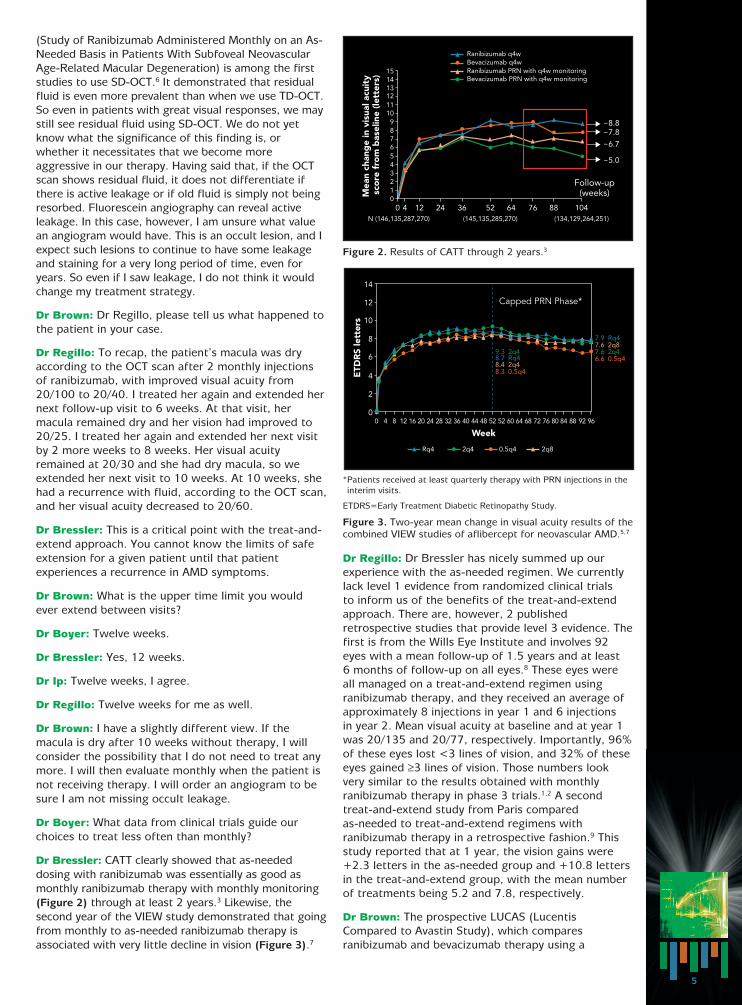
Dr Bressler: CATT clearly showed that as-neededdosing with ranibizumab was essentially as good asmonthly ranibizumab therapy with monthly monitoring(Figure 2) through at least 2 years.3 Likewise, thesecond year of the VIEW study demonstrated that goingfrom monthly to as-needed ranibizumab therapy isassociated with very little decline in vision (Figure 3).7
Dr Regillo: Dr Bressler has nicely summed up ourexperience with the as-needed regimen. We currentlylack level 1 evidence from randomized clinical trials to inform us of the benefits of the treat-and-extendapproach. There are, however, 2 publishedretrospective studies that provide level 3 evidence. Thefirst is from the Wills Eye Institute and involves 92eyes with a mean follow-up of 1.5 years and at least 6 months of follow-up on all eyes.8 These eyes were all managed on a treat-and-extend regimen usingranibizumab therapy, and they received an average ofapproximately 8 injections in year 1 and 6 injections in year 2. Mean visual acuity at baseline and at year 1was 20/135 and 20/77, respectively. Importantly, 96%of these eyes lost <3 lines of vision, and 32% of theseeyes gained ≥3 lines of vision. Those numbers lookvery similar to the results obtained with monthlyranibizumab therapy in phase 3 trials.1,2 A secondtreat-and-extend study from Paris compared as-needed to treat-and-extend regimens withranibizumab therapy in a retrospective fashion.9 Thisstudy reported that at 1 year, the vision gains were+2.3 letters in the as-needed group and +10.8 lettersin the treat-and-extend group, with the mean numberof treatments being 5.2 and 7.8, respectively.
Dr Brown: The prospective LUCAS (LucentisCompared to Avastin Study), which comparesranibizumab and bevacizumab therapy using a
Figure 2. Results of CATT through 2 years.3
*Patients received at least quarterly therapy with PRN injections in theinterim visits.
ETDRS=Early Treatment Diabetic Retinopathy Study.
Figure 3. Two-year mean change in visual acuity results of thecombined VIEW studies of aflibercept for neovascular AMD.5,7
1514131211109876543210
0 4 12 24 36 52
Follow-up(weeks)
~8.8~7.8
~6.7
~5.0
Mea
n ch
ang
e in
vis
ual a
cuit
y sc
ore
fro
m b
asel
ine
(lett
ers)
(145,135,285,270)
Ranibizumab q4w
(134,129,264,251)N (146,135,287,270)
64 76 88 104
Ranibizumab PRN with q4w monitoringBevacizumab q4w
Bevacizumab PRN with q4w monitoring
14
12
10
8
6
4
2
00 4 8 12 16 20 24 28 32 36 52 52484440
9.3 2q4
Capped PRN Phase*
8.7 Rq48.4 2q48.3 0.5q4
7.6 2q46.6 0.5q4
7.6 2q87.9 Rq4
ETD
RS
lett
ers
Week60 76 8872 80 846864 9692
Rq4 2q80.5q42q4
6
treat-and-extend regimen, is ongoing with 450patients. There also is a treat-and-extend trialsponsored by Novartis under way in Canada. We will have prospective level 1 evidence on using atreat-and-extend regimen in the future.
Dr Boyer: What about the use of bevacizumab on anas-needed basis?
Dr Bressler: I do not do that. Bevacizumab needs to be dosed every month within a group of patients for me to be confident that the vision outcomes would beequivalent to the use of monthly ranibizumab therapy.An as-needed regimen with bevacizumab will lead to a clinically relevant amount of vision loss in somepatients more frequently than if every-4-weeksranibizumab were given. But since we cannot reliablydetermine which patients will lose vision and never getit back, the only way to be confident that our patientswho are given bevacizumab therapy will have equivalentresults to ranibizumab every 4 weeks for AMDtreatment is to dose bevacizumab monthly. In CATT at 2 years, the point estimate was an average 4-letterdifference between as-needed bevacizumab andmonthly ranibizumab therapy, favoring ranibizumab.3
The 95% confidence interval around that point estimatewould include differences greater than 5 letters. I amnot confident that the results of using as-neededbevacizumab therapy through 2 years are equivalent toevery-4-weeks ranibizumab therapy.
Dr Brown: How did this case conclude?
Dr Regillo: I retreated the patient’s AMD and broughther back in 8 weeks, which was less than the maximalextension of 10 weeks, when she had a recurrence.Her visual acuity improved to 20/30, and her maculawas dry. I extended her visits to 10 weeks and then to 12 weeks, and she has been well maintained on a 12-week treat-and-extend schedule for 2 years.
CASE 2: Consequences of MonitoringLess Frequently Than Every Month
—Neil M. Bressler, MD
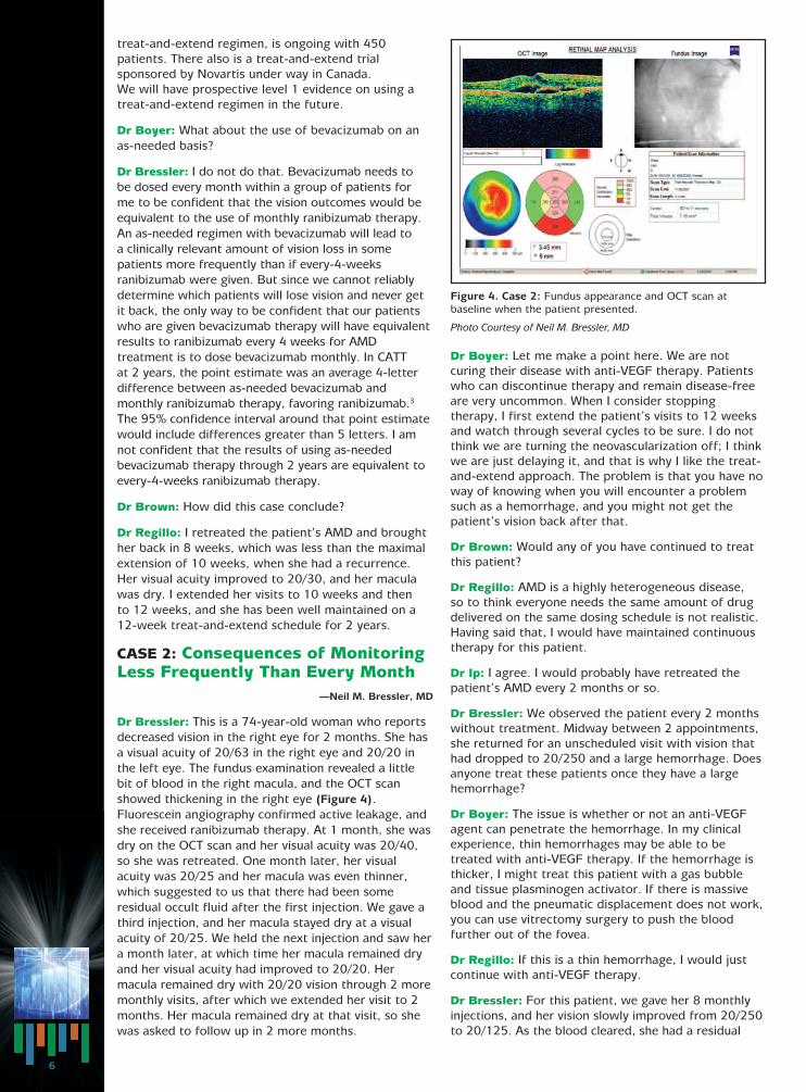
Dr Bressler: This is a 74-year-old woman who reportsdecreased vision in the right eye for 2 months. She hasa visual acuity of 20/63 in the right eye and 20/20 inthe left eye. The fundus examination revealed a littlebit of blood in the right macula, and the OCT scanshowed thickening in the right eye (Figure 4).Fluorescein angiography confirmed active leakage, andshe received ranibizumab therapy. At 1 month, she wasdry on the OCT scan and her visual acuity was 20/40,so she was retreated. One month later, her visualacuity was 20/25 and her macula was even thinner,which suggested to us that there had been someresidual occult fluid after the first injection. We gave athird injection, and her macula stayed dry at a visualacuity of 20/25. We held the next injection and saw hera month later, at which time her macula remained dryand her visual acuity had improved to 20/20. Hermacula remained dry with 20/20 vision through 2 moremonthly visits, after which we extended her visit to 2months. Her macula remained dry at that visit, so shewas asked to follow up in 2 more months.
Dr Boyer: Let me make a point here. We are notcuring their disease with anti-VEGF therapy. Patientswho can discontinue therapy and remain disease-freeare very uncommon. When I consider stoppingtherapy, I first extend the patient’s visits to 12 weeksand watch through several cycles to be sure. I do notthink we are turning the neovascularization off; I thinkwe are just delaying it, and that is why I like the treat-and-extend approach. The problem is that you have noway of knowing when you will encounter a problemsuch as a hemorrhage, and you might not get thepatient’s vision back after that.
Dr Brown: Would any of you have continued to treatthis patient?
Dr Regillo: AMD is a highly heterogeneous disease, so to think everyone needs the same amount of drugdelivered on the same dosing schedule is not realistic.Having said that, I would have maintained continuoustherapy for this patient.
Dr Ip: I agree. I would probably have retreated thepatient’s AMD every 2 months or so.
Dr Bressler: We observed the patient every 2 monthswithout treatment. Midway between 2 appointments,she returned for an unscheduled visit with vision thathad dropped to 20/250 and a large hemorrhage. Doesanyone treat these patients once they have a largehemorrhage?
Dr Boyer: The issue is whether or not an anti-VEGFagent can penetrate the hemorrhage. In my clinicalexperience, thin hemorrhages may be able to betreated with anti-VEGF therapy. If the hemorrhage isthicker, I might treat this patient with a gas bubbleand tissue plasminogen activator. If there is massiveblood and the pneumatic displacement does not work,you can use vitrectomy surgery to push the bloodfurther out of the fovea.
Dr Regillo: If this is a thin hemorrhage, I would justcontinue with anti-VEGF therapy.
Dr Bressler: For this patient, we gave her 8 monthlyinjections, and her vision slowly improved from 20/250to 20/125. As the blood cleared, she had a residual
Figure 4. Case 2: Fundus appearance and OCT scan atbaseline when the patient presented.
Photo Courtesy of Neil M. Bressler, MD
7
scar there from the fibrocytes that grew into theretina. This case argues in favor of monthly monitoring,not necessarily monthly treatment; but checking herprogression once a month gives us the best chance ofdetecting when something is coming back.
Dr Boyer: I do not believe that monthly follow-up willreduce these catastrophic hemorrhages. We all havepatients whom we examine monthly for whom the OCTscans and angiograms look fine, and they still bleed.
Dr Regillo: If you manage a sufficient number ofpatients with AMD over time, you are going to see anoccasional big hemorrhage even just a week after thelast injection.
Dr Bressler: Absolutely, as well as retinal pigmentepithelium tears with loss of vision, or geographicatrophy developing. My point is that even in trials withas-needed dosing as infrequently as every 3 months,patients were still evaluated monthly through 2years.3,5,10 There was an opportunity to catch smallareas of hemorrhage or leakage and to re-treat everymonth. With as-needed or treat-and-extend regimens,we are not seeing these patients between visits thatare more than a month apart. This does not meanlarge hemorrhages will not still occur, even a weekafter injection. However, without monthly monitoring,we are losing the opportunity to catch a small thing,such as new asymptomatic leakage, before it becomesa big thing, such as a large hemorrhage. Extendingbeyond monthly monitoring is a practice pattern thatis different from what was done in the clinical trials.Whether it results in poorer outcomes than were seenin those trials remains to be seen.
CASE 3: Pigment EpithelialDetachment
—Michael S. Ip, MD
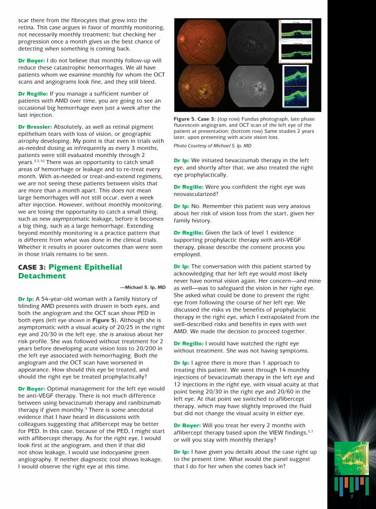
Dr Ip: A 54-year-old woman with a family history ofblinding AMD presents with drusen in both eyes, andboth the angiogram and the OCT scan show PED inboth eyes (left eye shown in Figure 5). Although she isasymptomatic with a visual acuity of 20/25 in the righteye and 20/30 in the left eye, she is anxious about herrisk profile. She was followed without treatment for 2years before developing acute vision loss to 20/200 inthe left eye associated with hemorrhaging. Both theangiogram and the OCT scan have worsened inappearance. How should this eye be treated, andshould the right eye be treated prophylactically?
Dr Boyer: Optimal management for the left eye wouldbe anti-VEGF therapy. There is not much differencebetween using bevacizumab therapy and ranibizumabtherapy if given monthly.3 There is some anecdotalevidence that I have heard in discussions withcolleagues suggesting that aflibercept may be betterfor PED. In this case, because of the PED, I might startwith aflibercept therapy. As for the right eye, I wouldlook first at the angiogram, and then if that did not show leakage, I would use indocyanine greenangiography. If neither diagnostic tool shows leakage, I would observe the right eye at this time.
Dr Ip: We initiated bevacizumab therapy in the lefteye, and shortly after that, we also treated the righteye prophylactically.
Dr Regillo: Were you confident the right eye wasneovascularized?
Dr Ip: No. Remember this patient was very anxiousabout her risk of vision loss from the start, given herfamily history.
Dr Regillo: Given the lack of level 1 evidencesupporting prophylactic therapy with anti-VEGFtherapy, please describe the consent process youemployed.
Dr Ip: The conversation with this patient started byacknowledging that her left eye would most likelynever have normal vision again. Her concern—and mineas well—was to safeguard the vision in her right eye.She asked what could be done to prevent the right eye from following the course of her left eye. Wediscussed the risks vs the benefits of prophylactictherapy in the right eye, which I extrapolated from thewell-described risks and benefits in eyes with wetAMD. We made the decision to proceed together.
Dr Regillo: I would have watched the right eyewithout treatment. She was not having symptoms.
Dr Ip: I agree there is more than 1 approach totreating this patient. We went through 14 monthlyinjections of bevacizumab therapy in the left eye and12 injections in the right eye, with visual acuity at thatpoint being 20/30 in the right eye and 20/60 in theleft eye. At that point we switched to aflibercepttherapy, which may have slightly improved the fluidbut did not change the visual acuity in either eye.
Dr Boyer: Will you treat her every 2 months withaflibercept therapy based upon the VIEW findings,5,7
or will you stay with monthly therapy?
Dr Ip: I have given you details about the case right upto the present time. What would the panel suggestthat I do for her when she comes back in?
Figure 5. Case 3: (top row) Fundus photograph, late phasefluorescein angiogram, and OCT scan of the left eye of thepatient at presentation; (bottom row) Same studies 2 yearslater, upon presenting with acute vision loss.
Photo Courtesy of Michael S. Ip, MD
8
Dr Bressler: There were no patients enrolled in theVIEW trial who initially received ranibizumab orbevacizumab therapy and then were switched toaflibercept therapy, so we have little data to guide ushere. In this case, because the patient is stable, I wouldmonitor her carefully and repeat injections as needed.
Dr Ip: I may treat her every 2 months, particularlybecause she is receiving bilateral injections. Would youtreat both eyes in this patient at the same visit?
Dr Bressler: Typically, it is more convenient for thepatient if we treat both eyes at the same visit than to schedule separate visits. Patients with DME morecommonly need both eyes treated than do patientswith AMD. Regardless, I prefer to treat only 1 eye thefirst time, just to familiarize these patients with theprocess. Once they are convinced that the treatment isworthwhile, I will treat both eyes at the same visit. ButI make sure to use separate preparations and treateach procedure separately to maximize safety.
Dr Ip: I presented this case because there is theperception that eyes with PED have a poorer prognosisthan do eyes with other types of neovascular changes.We do not know much about these patients becausethey were excluded from the major clinical trials. Thereis 1 small study in the literature looking at this issue11;and the researchers reported results relatively similarto those seen with the ranibizumab phase 3 studies,but the strength of evidence is weak. This patient hada very good response. So patients with PED should beaddressed on a case-by-case basis.
DIABETIC MACULAR EDEMACASE 4: Worsening Eye Disease in aSystemically Improving Patient
—David M. Brown, MD
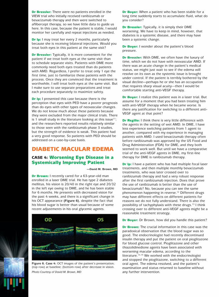
Dr Brown: I recently cared for a 63-year-old manenrolled in a laser DME trial. He has type 2 diabetesmellitus, his vision is 20/40 in the right eye and 20/32in the left eye owing to DME, and he has been stablefor 6 months. He presents with decreased vision forthe past 4 weeks, and there is a significant change inhis OCT appearance (Figure 6), despite the fact thathis blood sugar is better than usual because of somerecent adjustments in his oral glycemic agents.
Dr Boyer: When a patient who has been stable for along time suddenly starts to accumulate fluid, what doyou consider?
Dr Bressler: Typically, it is simply their DMEworsening. We have to keep in mind, however, thatdiabetes is a systemic disease, and there may havebeen a systemic change.
Dr Boyer: I wonder about the patient’s bloodpressure.
Dr Bressler: With DME, we often have the luxury oftime, which we do not have with neovascular AMD. Ifthere was an acute change in the patient’s medicalstatus, we might just wait to see if the edema willresolve on its own as the systemic issue is broughtunder control. If the patient is terribly bothered by thevisual decline—perhaps he or she has an occupationthat requires sharp visual acuity—then I would becomfortable starting anti-VEGF therapy.
Dr Boyer: I realize this patient was in a laser trial. Butassume for a moment that you had been treating himwith anti-VEGF therapy when he became worse. Isthere any justification for switching to a different anti-VEGF agent at that point?
Dr Regillo: I think there is very little difference withthe agents in the setting of wet AMD. In DME, I haveless experience switching patients from 1 agent toanother, compared with my experience in managingpatients with AMD. I used bevacizumab therapy oftenbefore ranibizumab was approved by the US Food andDrug Administration (FDA) for DME, and they bothseemed to work well. But until we have a comparativetrial of the anti-VEGF agents in DME, my first-linetherapy for DME is ranibizumab therapy.
Dr Ip: I have a patient who has had multiple focal lasertreatments, and then multiple monthly bevacizumabtreatments, who was later crossed over toranibizumab therapy and had a very robust responseafter the first ranibizumab injection. Does that provethe use of ranibizumab is better than the use ofbevacizumab? No, because you can see the samephenomenon happening in reverse.12 Different drugsmay have different effects on different patients forreasons we do not fully understand. There is also thepossibility of tachyphylaxis with these drugs.13 I thinkcrossing over to different anti-VEGF agents might be areasonable treatment strategy.
Dr Boyer: Dr Brown, how did you handle this patient?
Dr Brown: The crucial information in this case was theparadoxical observation that the blood sugar was sogood. The endocrinologist had recently discontinuedinsulin therapy and put the patient on oral pioglitazonefor blood glucose control. Pioglitazone and otherthiazolidinedione agents have been associated withworsening macular edema, according to theliterature.14,15 We worked with the endocrinologist and stopped the pioglitazone, switching to a differentoral agent. The edema resolved, and the patient’sexamination and status returned to baseline withoutany further intervention.
Figure 6. Case 4: OCT images of the patient’s presentation;(top row) at baseline; (bottom row) after decrease in vision.
Photo Courtesy of David M. Brown, MD
9
Dr Boyer: Many of our patients with diabetes areusing thiazolidinedione agents—these are known bythe brand names Avandia® or Actos®, and they arenearly ubiquitous in our patients. How can we identifythe patients in our practices who might just need tohave their oral hypoglycemic medications adjusted inorder to improve their DME?
Dr Brown: The patients in whom worsening macularedema is attributable to thiazolidinediones tend toretain fluid systemically, not just in their retinas.14 Askpatients on these drugs if they have gained any weightrecently or if they have swelling in their feet or ankles.Those are the patients in whom discontinuing that oralhypoglycemic agent is most likely to improve theirDME. They also might benefit systemically because thefluid burden can induce congestive heart failure.
Dr Regillo: It is important to always review themedication list for all patients with diabetes. Adjustingdiabetes medication for a patient with DME is an easysolution to resolving worsening macular edema.
CASE 5: VEGF Inhibition for Diabetic Macular Edema
—David S. Boyer, MD
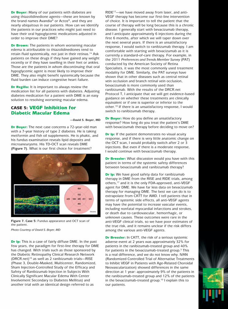
Dr Boyer: The next case concerns a 72-year-old manwith a 7-year history of type 2 diabetes. He is takingmetformin and fish oil supplements. He is phakic, andhis fundus examination reveals lipid deposits andmicroaneurysms. His TD-OCT scan reveals DME (Figure 7). What is our first choice for treatment?
Dr Ip: This is a case of fairly diffuse DME. In the pastfew years, the paradigm for first-line therapy for DMEhas changed. With trials such as those sponsored bythe Diabetic Retinopathy Clinical Research Network(DRCR.net)16 as well as 2 ranibizumab trials—RISE(Phase 3, Double-Masked, Multicenter, Randomized,Sham Injection-Controlled Study of the Efficacy andSafety of Ranibizumab Injection in Subjects WithClinically Significant Macular Edema With CenterInvolvement Secondary to Diabetes Mellitus) andanother trial with an identical design referred to as
RIDE17—we have moved away from laser, and anti-VEGF therapy has become our first-line intervention of choice. It is important to tell the patient that thecourse of therapy will be long because this is a chronicdisease. I generally start with bevacizumab therapy,and I anticipate approximately 6 injections during thefirst 6 months, after which we will taper down overthe next several years. If there is an unsatisfactoryresponse, I would switch to ranibizumab therapy. I amcomfortable with starting with bevacizumab as it iscurrently a standard-of-care therapy. For example, inthe 2011 Preferences and Trends Member Survey (PAT)conducted by the American Society of RetinaSpecialists, it was the most commonly used treatmentmodality for DME. Similarly, the PAT surveys haveshown that in other diseases such as central retinalvein occlusion and branch retinal vein occlusion,bevacizumab is more commonly used thanranibizumab. With the results of the DRCR.netProtocol T, I anticipate that we will get evidence-basedguidance on whether these treatments are clinicallyequivalent or if one is superior or inferior to theother.18 If there is an unsatisfactory response, I wouldswitch to ranibizumab therapy.
Dr Boyer: How do you define an unsatisfactoryresponse? How long do you treat the patient’s DMEwith bevacizumab therapy before deciding to move on?
Dr Ip: If the patient demonstrates no visual acuityresponse, and if there is very little anatomic change onthe OCT scan, I would probably switch after 2 or 3injections. But even if there is a moderate response, I would continue with bevacizumab therapy.
Dr Bressler: What discussion would you have with thispatient in terms of the systemic safety differencesbetween bevacizumab and ranibizumab therapy?
Dr Ip: We have good safety data for ranibizumabtherapy in DME from the RISE and RIDE trials, amongothers,17 and it is the only FDA-approved, anti-VEGFagent for DME. We have far less data on bevacizumabtherapy for managing DME. The best we can do is toextrapolate from CATT for AMD. I tell patients that interms of systemic side effects, all anti-VEGF agentsmay have the potential to increase vascular events,including nonfatal myocardial infarctions and strokes,or death due to cardiovascular, hemorrhagic, orunknown causes. These outcomes were rare in theanti-VEGF clinical trials, so we have poor estimates ofthe true risk, and it remains unclear if the risk differsamong the various anti-VEGF agents.
Dr Bressler: In CATT, the risk of a serious systemicadverse event at 2 years was approximately 32% forpatients in the ranibizumab-treated group and 40% for patients in the bevacizumab-treated group.3 This is a real difference, and we do not know why. IVAN(Randomized Controlled Trial of Alternative Treatmentsto Inhibit VEGF in Patients with Age-Related ChoroidalNeovascularization) showed differences in the samedirection at 1 year: approximately 9% of the patients inthe ranibizumab-treated group and 12% of the patientsin the bevacizumab-treated group.10 I explain this toour patients.
Figure 7. Case 5: Fundus appearance and OCT scan of the patient.
Photo Courtesy of David S. Boyer, MD
10
Dr Boyer: What dose of ranibizumab are we using for DME?
Dr Regillo: The approved dose of ranibizumab is 0.3mg for DME and 0.5 mg for AMD and macular edemaassociated with retinal vein occlusion. The phase 3DME trials evaluated both 0.3- and 0.5-mg doses ofranibizumab; results were essentially equivalent,17 sothe FDA approved the lowest effective dose.
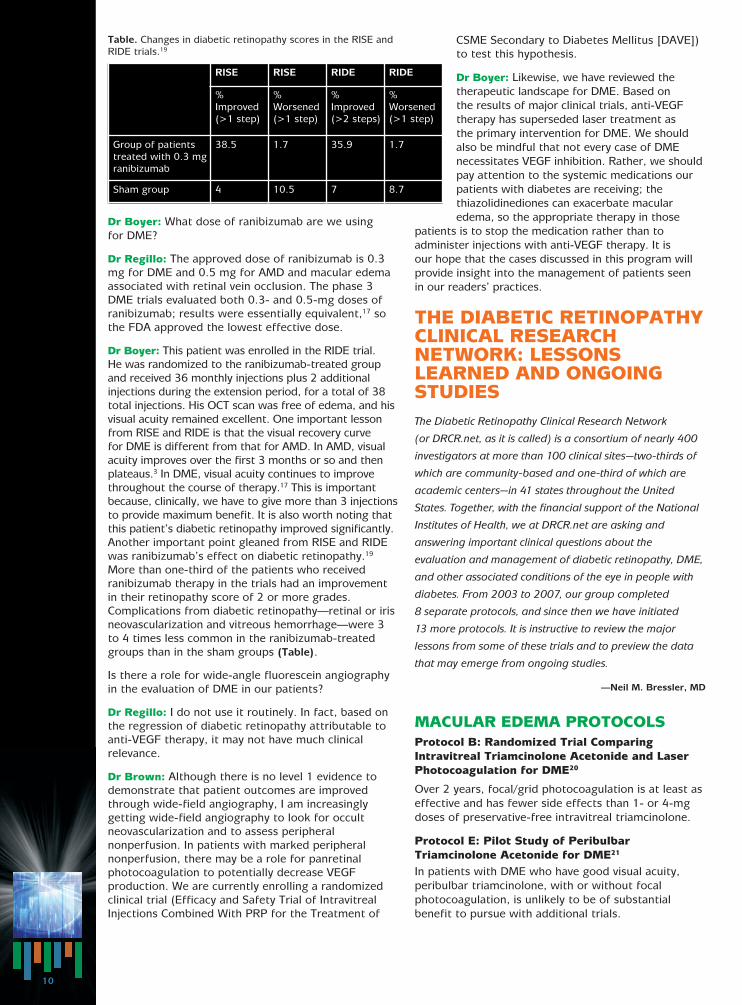
Dr Boyer: This patient was enrolled in the RIDE trial. He was randomized to the ranibizumab-treated groupand received 36 monthly injections plus 2 additionalinjections during the extension period, for a total of 38total injections. His OCT scan was free of edema, and hisvisual acuity remained excellent. One important lessonfrom RISE and RIDE is that the visual recovery curve for DME is different from that for AMD. In AMD, visualacuity improves over the first 3 months or so and thenplateaus.3 In DME, visual acuity continues to improvethroughout the course of therapy.17 This is importantbecause, clinically, we have to give more than 3 injectionsto provide maximum benefit. It is also worth noting thatthis patient’s diabetic retinopathy improved significantly.Another important point gleaned from RISE and RIDEwas ranibizumab’s effect on diabetic retinopathy.19
More than one-third of the patients who receivedranibizumab therapy in the trials had an improvementin their retinopathy score of 2 or more grades.Complications from diabetic retinopathy—retinal or irisneovascularization and vitreous hemorrhage—were 3to 4 times less common in the ranibizumab-treatedgroups than in the sham groups (Table).
Is there a role for wide-angle fluorescein angiographyin the evaluation of DME in our patients?
Dr Regillo: I do not use it routinely. In fact, based onthe regression of diabetic retinopathy attributable toanti-VEGF therapy, it may not have much clinicalrelevance.
Dr Brown: Although there is no level 1 evidence todemonstrate that patient outcomes are improvedthrough wide-field angiography, I am increasinglygetting wide-field angiography to look for occultneovascularization and to assess peripheralnonperfusion. In patients with marked peripheralnonperfusion, there may be a role for panretinalphotocoagulation to potentially decrease VEGFproduction. We are currently enrolling a randomizedclinical trial (Efficacy and Safety Trial of IntravitrealInjections Combined With PRP for the Treatment of
CSME Secondary to Diabetes Mellitus [DAVE])to test this hypothesis.
Dr Boyer: Likewise, we have reviewed thetherapeutic landscape for DME. Based on the results of major clinical trials, anti-VEGFtherapy has superseded laser treatment as the primary intervention for DME. We should also be mindful that not every case of DMEnecessitates VEGF inhibition. Rather, we shouldpay attention to the systemic medications ourpatients with diabetes are receiving; thethiazolidinediones can exacerbate macularedema, so the appropriate therapy in those
patients is to stop the medication rather than toadminister injections with anti-VEGF therapy. It is our hope that the cases discussed in this program willprovide insight into the management of patients seen in our readers’ practices.
THE DIABETIC RETINOPATHYCLINICAL RESEARCHNETWORK: LESSONSLEARNED AND ONGOINGSTUDIESThe Diabetic Retinopathy Clinical Research Network
(or DRCR.net, as it is called) is a consortium of nearly 400
investigators at more than 100 clinical sites—two-thirds of
which are community-based and one-third of which are
academic centers—in 41 states throughout the United
States. Together, with the financial support of the National
Institutes of Health, we at DRCR.net are asking and
answering important clinical questions about the
evaluation and management of diabetic retinopathy, DME,
and other associated conditions of the eye in people with
diabetes. From 2003 to 2007, our group completed
8 separate protocols, and since then we have initiated
13 more protocols. It is instructive to review the major
lessons from some of these trials and to preview the data
that may emerge from ongoing studies.
—Neil M. Bressler, MD
MACULAR EDEMA PROTOCOLSProtocol B: Randomized Trial ComparingIntravitreal Triamcinolone Acetonide and LaserPhotocoagulation for DME20
Over 2 years, focal/grid photocoagulation is at least aseffective and has fewer side effects than 1- or 4-mgdoses of preservative-free intravitreal triamcinolone.
Protocol E: Pilot Study of PeribulbarTriamcinolone Acetonide for DME21
In patients with DME who have good visual acuity,peribulbar triamcinolone, with or without focalphotocoagulation, is unlikely to be of substantialbenefit to pursue with additional trials.
Table. Changes in diabetic retinopathy scores in the RISE and RIDE trials.19
RISE RISE RIDE RIDE
%Improved(>1 step)
%Worsened(>1 step)
%Improved(>2 steps)
%Worsened(>1 step)
Group of patientstreated with 0.3 mgranibizumab
38.5 1.7 35.9 1.7
Sham group 4 10.5 7 8.7
11
Protocol H: Phase 2 Randomized Trial ofBevacizumab for DME22
The results demonstrated that intravitrealbevacizumab can reduce DME in some eyes, but thestudy was not designed to determine whether or notthe treatment was superior to focal/grid laser.
Protocol I: Laser-Ranibizumab-TriamcinoloneStudy for DME16
Intravitreal ranibizumab with prompt or deferred (≥24 weeks) focal/grid laser is more effective through2 years in increasing visual acuity compared withfocal/grid laser treatment alone for the treatment ofDME involving the central macula, although there is asmall risk of endophthalmitis. Ranibizumab should beconsidered for patients with characteristics similar tothose enrolled in this trial, including DME involving thecenter of the macula and decreased visual acuity.
Protocol K: The Course of Response to FocalPhotocoagulation for DME23
Sixteen weeks after focal/grid laser for DME in eyeswith a definite reduction, but not complete resolution,of central edema, it is likely that somewhere between23% and 63% of these eyes will continue to improvewithout additional treatment.
DIABETIC RETINOPATHYPROTOCOLSProtocol F: Observational Study of theDevelopment of DME Following Scatter LaserPhotocoagulation24
Clinically meaningful differences are unlikely in OCTthickness or visual acuity following the application ofpan-retinal photocoagulation (PRP) in 1 sittingcompared with 4 sittings spread over 12 weeks. Theseresults suggest PRP costs to some patients, in termsof travel and lost productivity, as well as to eye careproviders could be reduced.
Protocol J: Laser-Ranibizumab-TriamcinoloneStudy for DME + PRP25
The addition of 1 intravitreal triamcinolone injection or2 monthly intravitreal ranibizumab injections in eyesreceiving focal/grid laser for DME and PRP forproliferative diabetic retinopathy is associated withbetter visual acuity and decreased macular edema by14 weeks. Whether continued long-term intravitrealtreatment is beneficial cannot be determined from this study.
OCT AND RETINAL THICKENINGPROTOCOLSProtocol C: Temporal Variation in OCTMeasurements of DME26
Although on average there are slight decreases inretinal thickening during the day, most eyes with DMEhave little meaningful change in OCT central subfield(CSF) thickening or visual acuity between 8:00 AM and4:00 PM. Also, reproducibility of retinal thickness inDME was better for CSF thickness than for center
point measurements. A change in CSF thicknessexceeding 11% is likely to be real.
Protocol G: Subclinical DME Study27
While subclinical DME (detected on OCT but not onclinical examination) may be uncommon, this studysuggests that between approximately 25% and 50% of the eyes with subclinical DME will progress to moredefinite thickening or be judged to need treatment forDME within 2 years after its identification.
ONGOING STUDIESProtocol N: An Evaluation of IntravitrealRanibizumab for Vitreous Hemorrhage Due toProliferative Diabetic RetinopathyOBJECTIVE: To determine if intravitreal injections ofranibizumab decrease the proportion of eyes in which vitrectomy is performed compared with salineinjections in eyes presenting with vitreous hemorrhagefrom proliferative diabetic retinopathy (PDR).
Protocol R: A Phase II Evaluation of TopicalNSAIDs in Eyes With Non-Central-Involved DMEOBJECTIVES: To assess the effects of topicalnonsteroidal anti-inflammatory drugs (NSAIDs) onmacular retina volume compared with placebo in eyeswith non-central-involved DME; and to assess theeffects of topical NSAIDs on central subfield thicknessand to compare the progression of non-central-involved DME to central-involved DME as determinedby OCT and stereoscopic fundus photographs.
Protocol S: Prompt PRP vs IntravitrealRanibizumab With Deferred PRP for PDR.OBJECTIVE: To determine if visual acuity outcomes at 2years in eyes with PDR that receive anti-VEGF therapywith deferred PRP are non-inferior to those in eyesthat receive standard prompt PRP therapy. And, if so,how many injections are needed, and are the resultsassociated with better visual fields, better reports onvisual function questionnaires, and different costs?
Protocol T: A Comparative Effectiveness Study ofIntravitreal Aflibercept, Bevacizumab, andRanibizumab for DMEOBJECTIVE: To compare the efficacy and safety ofintravitreal (1) aflibercept, (2) bevacizumab, and (3)ranibizumab when given to treat central-involved DME;specifically, the primary outcome is to assess if any ofthese 3 anti-VEGF products is superior to the otherswith respect to mean changes in visual acuity.
GENES IN DIABETIC RETINOPATHYPROJECTOBJECTIVE: To create a repository of genetic materialand clinical phenotype information as a resource forthe research community; the database may provide the opportunity to assess genetic susceptibility andresistance to diabetic retinopathy and also variantsimpacting visually important biomarkers for macularedema and neovascularization.
12
RETINA LITERATURE ROUND-UP:A Summary of Relevant Research Published Sincethe 2012 AAO Meeting
Xu L, Lu T, Tuomi L, Jumbe N, Lu J, Eppler S, KueblerP, Damico-Beyer LA, Joshi A. Pharmacokinetics ofranibizumab in patients with neovascular age-relatedmacular degeneration—a population approach[published online ahead of print January 29, 2013].Invest Ophthalmol Vis Sci. doi:10.1167/iovs.12-10260.
Xu et al conducted a pharmacokinetics study tobetter characterize systemic exposure to ranibizumabadministered via intravitreal injection in patients withneovascular AMD. The study involved 2993 serumsamples from 674 patients with AMD who hadparticipated in 1 of 5 clinical trials (phases 1 through3) of single or multiple intravitreal injections ofranibizumab (at doses ranging from 0.3-2.0mg/injection) administered biweekly, monthly, orquarterly for up to 24 months. The investigatorsreported that the pharmacokinetics followed first-order absorption into and first-order elimination fromthe systemic circulation. They found that the vitreouselimination half-life was 9 days and the systemicelimination half-life was approximately 2 hours, with asystemic-to-vitreous exposure ratio of approximately 1 to 90,000. When administered monthly or quarterly,serum concentrations of ranibizumab followingintravitreal doses of 0.3 mg and 0.5 mg remained too low to inhibit VEGF-A-induced endothelial cellproliferation. They concluded that systemicranibizumab exposure following intravitreal injectionwas very low because of rapid elimination once themolecule moved from the vitreous to the systemiccirculation.
Liew G, Mitchell P, Wong TY, Rochtchina E, Wang JJ.The association of aspirin use with age-related maculardegeneration. JAMA Intern Med. 2013;173(4):258-264.
Liew et al conducted a 15-year prospective analysisof data from a population-based cohort study inAustralia to explore whether long-term aspirin usecontributes to a higher risk for developing AMD.Overall, 2389 subjects participated; of these, 257(10.8%) subjects were regular aspirin users and 63(2.6%) subjects developed neovascular AMD. Regularaspirin users had a higher risk of developing incidentneovascular AMD, with the 15-year cumulativeincidence being 9.3% in users and 3.7% in nonusers.This equated to an odds ratio (OR) of 2.46 (95%confidence interval [CI], 1.25-4.83) for people whowere regular aspirin users vs those who were notregular aspirin users to develop neovascular AMD, evenafter adjusting for potential confounders such as age,sex, smoking, history of cardiovascular disease, systolicblood pressure, and body mass index. The investigatorsalso saw a significant dose-response effect(multivariate-adjusted P=.01 for trend). Interestingly,aspirin use did not seem to be related to the incidenceof geographic atrophy (multivariate-adjusted OR, 0.99;95% CI, 0.59-1.65). The authors concluded that regularaspirin users were at increased risk of developingneovascular AMD, even when cardiovascular diseaseand smoking were accounted for.
Klein BE, Howard KP, Gangnon RE, Dreyer JO, Lee KE,Klein R. Long-term use of aspirin and age-relatedmacular degeneration. JAMA. 2012;308(23):2469-2478.
In the long-running longitudinal population-basedBeaver Dam Eye Study in Wisconsin, Klein et al alsoevaluated a possible association between aspirin useand the development of AMD. In that study, 4926subjects aged 43 to 86 years at the time of enrollmentwere examined every 5 years for 2 decades. At eachexamination, they were asked if they had used aspirinat least 2 times a week on a regular basis for morethan 3 months. They also underwent retinalphotography. After a mean follow-up of 14.8 years,the investigators reported 512 new cases of early AMDand 117 new cases of late AMD. Subjects whoreported being regular aspirin users at the visit 10years prior to the retinal photographs had a higherrate of developing late-stage AMD than patients whowere not regular aspirin users (1.76% vs 1.03%,respectively; hazard ratio, 1.63 [95% CI, 1.01-2.63];P=.05); the association was significant for thedevelopment of neovascular AMD (HR, 2.20 [95% CI,1.20-4.15]; P=.01) but not for geographic atrophy(HR, 0.66 [95% CI, 0.25-1.95]; P=.45). There was nodetectable association between aspirin use and thedevelopment of early-stage AMD. The authorsconcluded that regular aspirin use 10 years prior toevaluation was associated with an increased risk ofdeveloping neovascular AMD.
Mitta VP, Christen WG, Glynn RJ, Semba RD, RidkerPM, Rimm EB, Hankinson SE, Schaumberg DA. C-reactive protein and the incidence of maculardegeneration: pooled analysis of 5 cohorts [publishedonline ahead of print February 7, 2013]. JAMAOphthalmol. doi:10.1001/jamaophthalmol.2013.2303.
Mitta et al pooled the data from 5 ongoingprospective cohort studies (Women’s Health Study,Physicians’ Health Study, Women’s Antioxidant andFolic Acid Cardiovascular Study, Nurses’ Health Study,and Health Professionals Follow-up Study) toinvestigate the relationship between high-sensitivity C-reactive protein (hsCRP) and future risk of AMD.Serum levels of hsCRP were determined at baselinewhen subjects enrolled in their respective cohortstudy; for purposes of analysis, subjects wereclassified based on high (≥3 mg/L) or low (≤1 mg/L)hsCRP levels. The investigators identified 647 incidentcases of AMD and then selected age- and sex-matchedcontrols for each AMD case to conduct a nested case-control study. Once cigarette smoking—a potentialconfounder—was adjusted for, participants with highcompared with low hsCRP levels had a significantlyincreased risk of incident AMD (OR, 1.49; 95% CI,1.06-2.08) and neovascular AMD (OR, 1.84; 95% CI,1.14-2.98). The authors concluded that hsCRP levelspredict future risk of AMD, and they suggested thatsubjects with high hsCRP levels may benefit fromintensified surveillance for AMD development andencouragement to undertake lifestyle modifications to minimize their risk profile.
13
Muni RH, Kohly RP, Lee EQ, Manson JE, Semba RD,Schaumberg DA. Prospective study of inflammatorybiomarkers and risk of diabetic retinopathy in theDiabetes Control and Complications Trial [publishedonline ahead of print Feburary 7, 2013]. JAMAOphthalmol. doi:10.1001/jamaophthalmol.2013.2299.
Muni et al explored the relationship between serumlevels of hsCRP and the development and progressionof diabetic retinopathy, clinically significant macularedema (CSME), retinal hard exudates, and proliferativediabetic retinopathy in 1441 subjects with type 1diabetes participating in the Diabetes Control andComplications Trial (DCCT) cohort, which has beenongoing for well over 20 years. Serum hsCRP levelswere classified into 5 groups based on the quintiles of the distribution of values measured at baseline. The investigators found a statistically significantassociation between hsCRP level and risk of CSME,with a relative risk (RR) for the top vs bottom quintileof 1.83 (95% CI, 0.94-3.55; P for trend=.01) and alsofor the development of retinal hard exudates, with theRR for the top vs bottom quintile of hsCRP level being1.78 (95% CI, 0.98-3.25; P for trend=.004). Theauthors concluded that baseline hsCRP level may beassociated with risk of developing CSME and macularhard exudate in the DCCT cohort.
Arevalo JF, Lasave AF, Wu L, Diaz-Llopis M, Gallego-Pinazo R, Alezzandrini AA, Berrocal MH; for the Pan-American Collaborative Retina Study Group(PACORES). Intravitreal bevacizumab plus grid laserphotocoagulation or intravitreal bevacizumab or gridlaser photocoagulation for diffuse diabetic macularedema: results of the Pan-American CollaborativeRetina Study Group at 24 months. Retina. 2013;33(2):403-413.
Arevalo et al conducted a retrospective,interventional, comparative, multicenter study toevaluate the anatomical and functional outcomes inpatients with diffuse diabetic macular edema treatedwith 1 of 3 regimens: primary intravitreal bevacizumab(IVB) alone (Group A; n=141 eyes), grid laserphotocoagulation alone (Group B; n=120 eyes), or IVBplus laser (Group C; n=157 eyes). All 3 treatmentssignificantly improved best-corrected visual acuityfrom baseline (P<.0001), with IVB producing bettervisual improvement than laser (P=.013).
Similarly, all 3 treatments produced a decrease incentral macular thickness from baseline (P<.0001),with greater central macular thickness decreases seenin Group A than in Groups B and C (P<.001). Theauthors concluded that primary IVB monotherapy wassuperior to laser alone in stabilizing or improving best-corrected visual acuity in patients with diffuse DME.
14
1. Rosenfeld PJ, Brown DM, Heier JS, et al. Ranibizumabfor neovascular age-related macular degeneration. N Engl J Med. 2006;355:1419-1431.
2. Brown DM, Kaiser PK, Michels M, et al. Ranibizumabversus verteporfin for neovascular age-related maculardegeneration. N Engl J Med. 2006;355:1432-1444.
3. Martin DF, Maguire MG, Fine SL, et al. Ranibizumaband bevacizumab for treatment of neovascular age-related macular degeneration: two-year results.Ophthalmology. 2012;119:1388-1398.
4. Khurana RN, Dupas B, Bressler NM. Agreement oftime-domain and spectral-domain optical coherencetomography with fluorescein leakage from choroidalneovascularization. Ophthalmology. 2010;117:1376-1380.
5. Heier JS, Brown DM, Chong V, et al. Intravitrealaflibercept (VEGF Trap-Eye) in wet age-relatedmacular degeneration. Ophthalmology. 2012;119:2537-2548.
6. Suner IJ, Yau L, Lai P. HARBOR Study: One-yearresults of efficacy and safety of 2.0 mg versus 0.5 mgranibizumab in patients with subfoveal choroidalneovascularization secondary to age-related maculardegeneration. Presented at: Association for Researchin Vision and Ophthalmology Annual Meeting; May 6-9, 2012; Fort Lauderdale, FL.
7. Two year results of phase 3 studies with EYLEA™(aflibercept) injection in wet AMD show sustainedimprovement in visual acuity [press release].Tarrytown, NY: Regeneron Pharmaceuticals, Inc;December 5, 2011. http://investor.regeneron.com/releasedetail.cfm?ReleaseID= 629800. AccessedDecember 23, 2012.
8. Gupta OP, Shienbaum G, Patel AH, Fecarotta C, Kaiser RS, Regillo CD. A treat and extend regimenusing ranibizumab for neovascular age-related macular degeneration clinical and economic impact.Ophthalmology. 2010;117:2134-2140.
9. Oubraham H, Cohen SY, Samimi S, et al. Inject andextend dosing versus dosing as needed: a comparativeretrospective study of ranibizumab in exudative age-related macular degeneration. Retina. 2011;31:26-30.
10. Chakravarthy U, Harding SP, Rogers CA, et al.Ranibizumab versus bevacizumab to treat neovascularage-related macular degeneration: one-year findingsfrom the IVAN randomized trial. Ophthalmology.2012;119:1399-1411.
11. Arora S, McKibbin M. One-year outcome afterintravitreal ranibizumab for large, serous pigmentepithelial detachment secondary to age-relatedmacular degeneration. Eye (Lond). 2011;25:1034-1038.
12. Gasperini JL, Fawzi AA, Khondkaryan A, et al.Bevacizumab and ranibizumab tachyphylaxis in thetreatment of choroidal neovascularisation. Br JOphthalmol. 2012;96:14-20.
13. Binder S. Loss of reactivity in intravitreal anti-VEGFtherapy: tachyphylaxis or tolerance? Br J Ophthalmol.2012;96:1-2.
14. Ryan EH Jr, Han DP, Ramsay RC, et al. Diabeticmacular edema associated with glitazone use. Retina.2006;26:562-570.
15. Fong DS, Contreras R. Glitazone use associated withdiabetic macular edema. Am J Ophthalmol. 2009;147:583-586.e1.
16. Elman MJ, Aiello LP, Beck RW, et al. Randomized trialevaluating ranibizumab plus prompt or deferred laseror triamcinolone plus prompt laser for diabeticmacular edema. Ophthalmology. 2010;117:1064-1077.e35.
17. Nguyen QD, Brown DM, Marcus DM, et al.Ranibizumab for diabetic macular edema: results from2 phase III randomized trials: RISE and RIDE.Ophthalmology. 2012;119:789-801.
18. American Society of Retina Specialists. Jumper JM,Mittra RA, eds. 2011 Preferences and Trends MemberSurvey. Chicago, IL.
19. Boyer D. Long-term efficacy and safety ofranibizumab in diabetic macular edema: 36-monthresults from RISE and RIDE. Presented at: The RetinaSociety Annual Meeting; October 4-7, 2012;Washington, DC.
20. Beck RW, Edwards AR, Aiello LP, et al. Three-yearfollow-up of a randomized trial comparing focal/gridphotocoagulation and intravitreal triamcinolone fordiabetic macular edema. Arch Ophthalmol. 2009;127:245-251.
21. Chew E, Strauber S, Beck R, et al. Randomized trial ofperibulbar triamcinolone acetonide with and withoutfocal photocoagulation for mild diabetic macularedema: a pilot study. Ophthalmology. 2007;114:1190-1196.
22. Scott IU, Edwards AR, Beck RW, et al. A phase IIrandomized clinical trial of intravitreal bevacizumabfor diabetic macular edema. Ophthalmology.2007;114:1860-1867.
23. The course of response to focal/grid photocoagulationfor diabetic macular edema. Retina. 2009;29:1436-1443.
24. Brucker AJ, Qin H, Antoszyk AN, et al. Observationalstudy of the development of diabetic macular edemafollowing panretinal (scatter) photocoagulation givenin 1 or 4 sittings. Arch Ophthalmol. 2009;127:132-140.
25. Googe J, Brucker AJ, Bressler NM, et al. Randomizedtrial evaluating short-term effects of intravitrealranibizumab or triamcinolone acetonide on macularedema after focal/grid laser for diabetic macularedema in eyes also receiving panretinalphotocoagulation. Retina. 2011;31:1009-1027.
26. Danis RP, Glassman AR, Aiello LP, et al. Diurnalvariation in retinal thickening measurement by opticalcoherence tomography in center-involved diabeticmacular edema. Arch Ophthalmol. 2006;124:1701-1707.
27. Bressler NM, Miller KM, Beck RW, et al. Observationalstudy of subclinical diabetic macular edema. Eye (Lond).2012;26:833-840.
REFERENCES

15
1. Which of the following is not a typical location toobserve blood in neovascular AMD?A. IntraretinalB. SubretinalC. PreretinalD. Beneath the retinal pigment epithelium
2. How many every-4-weeks injections of VEGF therapywere given in the first year of the phase 3, randomized,clinical trials comparing aflibercept to ranibizumab?A. 0B. 1C. 3D. 6
3. Which of the following statements is true regardingtreat-and-extend therapeutic regimens?A. The regimen has been shown to be non-inferior or
equivalent to monthly treatmentB. The regimen typically consists of gradually
increasing the duration between visits andevaluation for possible treatment at each visit
C. The regimen typically consists of graduallyincreasing the duration between visits andtreatment at each visit
D. The regimen typically consists of monthlyevaluations, but treatment only when the patientis getting worse
4. Which of the following is a key difference betweenfluorescein angiography and OCT?A. OCT identifies VEGF-driven leakage from
capillaries better than fluorescein angiographyB. Fluorescein angiography provides better
quantitative information on retinal thickening than OCT
C. OCT shows the presence or absence of edemawhile fluorescein angiography identifies VEGF-driven leakage from capillaries
D. None of the above
5. Which of the following statements is true regardingCATT (Comparison of AMD Treatments Trials)?A. At 2 years, as-needed ranibizumab confidently
appears to be equivalent to every-4-weeksranibizumab, with differences that are not likely tobe clinically relevant
B. At 2 years, as-needed bevacizumab appears to beequivalent to monthly ranibizumab, withdifferences that are not likely to be clinicallyrelevant
C. Both A and BD. Neither A nor B
6. Which of the following hypoglycemic agents has beenassociated with macular edema?A. InsulinB. PioglitazoneC. MetforminD. All the above
7. Which of the following statements is true regardingfocal/grid laser photocoagulation in the treatment ofDME involving the center of the macula and causingvision impairment?A. The treatment in randomized clinical trials is at
least as effective as intravitreal corticosteroids butwith fewer side effects as evaluated by DRCR.net
B. The treatment is not as effective as intravitrealranibizumab but without the risk ofendophthalmitis as evaluated by DRCR.net
C. The treatment, when combined with intravitrealranibizumab at the onset of anti-VEGF therapy,provides superior visual acuity results comparedwith intravitreal ranibizumab alone
D. None of the above is true
8. Which of the following is the FDA-approved dose of an intravitreal anti-VEGF medication for thecorresponding indication?A. 0.3 mg ranibizumab for DMEB. 0.5 mg ranibizumab for DMEC. 0.3 mg ranibizumab for macular edema associatedwith retinal vein occlusion
D. 0.5 mg aflibercept for AMD
9. Which of the following statements is true regarding theRISE and RIDE phase 3 trials of ranibizumab for DME?A. Visual acuity recovers over the first 3 months and
then plateausB. Eyes treated with ranibizumab demonstrated
worsening of diabetic retinopathy severityC. Complications of diabetic retinopathy, such asvitreous hemorrhage and iris neovascularization,were less common in eyes treated withranibizumab than in those in the sham group
D. All the above
Which of the following statements is true regardingDRCR.net?A. It is made up solely of academic centers in the
United StatesB. It is funded solely through philanthropic donationsC. It conducts trials to answer important clinicalquestions about the evaluation and managementof diabetic retinopathy and associated conditions
D. All the above
CME POST TEST QUESTIONSTo obtain AMA PRA Category 1 Credit™ for this activity, complete the CME Post Test by writing the best answer to eachquestion in the Answer Box located on the Activity Evaluation/Credit Request form on the following page. Alternatively,you can complete the CME Post Test online at http://www.MedEdicus.com, Educational Activities tab, and click thePost-Test & CME Certificate button. See detailed instructions at To Obtain AMA PRA Category 1 Credit™ on page 3.
10.
To receive AMA PRA Category 1 Credit™, you must complete this Evaluation form and the Post Test. Record your answers to the Post Test in the Answer Boxlocated below. Mail or Fax this completed page to The New York Eye and Ear Infirmary–ICME, 310 East 14th Street, New York, NY 10003 (Fax: 212-353-5703).Your comments help us to determine the extent to which this educational activity has met its stated objectives, assess future educational needs, and create timelyand pertinent future activities. Please provide all the requested information below. This ensures that your certificate is filled out correctly and is mailed to the properaddress. It also enables us to contact you about future CME activities. Please print clearly or type. Illegible submissions cannot be processed.
PARTICIPANT INFORMATION (Please Print) � Home � Office
Last Name _____________________________________________________________________ First Name ________________________________________
Specialty __________________________________________ Degree � MD � DO � OD � PharmD � RPh � NP � RN � PA � Other ________
Institution _________________________________________________________________________________________________________________________
Street Address ____________________________________________________________________________________________________________________
City ________________________________________ State _____________________ ZIP Code ____________________ Country ______________________
E-mail ______________________________________ Phone ______________________________________ Fax _____________________________________
Please note: We do not sell or share e-mail addresses. They are used strictly for conducting post-activity follow-up surveys to assess the impact of thiseducational activity on your practice.
Learner Disclosure: To ensure compliance with the US Centers for Medicare and Medicaid Services regarding gifts to physicians, The New York Eye and Ear Infirmary Institute for CME requires that you disclose whether or not you have any financial, referral, and/or other relationship with our institution. CME certificatescannot be awarded unless you answer this question. For additional information, please call NYEE ICME at 212-979-4383. Thank you.
�Yes � No I and/or my family member have a financial relationship with The New York Eye and Ear Infirmary and/or refer Medicare/Medicaid patients to it.
� I certify that I have participated in the entire activity and claim 2.0 AMA PRA Category 1 Credits™.
Signature Required __________________________________________________________________ Date Completed ______________________________
OUTCOMES MEASUREMENT
�Yes � No Did you perceive any commercial bias in any part of this activity? IMPORTANT! If you answered “Yes,” we urge you to be specific about where the bias occurred so we can address the perceived bias with the contributor and/or in the subject matter in future activities.
_________________________________________________________________________________________________________________________________
Circle the number that best reflects your opinion on the degree to which the following learning objectives were met:5 = Strongly Agree 4 = Agree 3 = Neutral 2 = Disagree 1 = Strongly Disagree
Upon completion of this activity, I am better able to:
• Apply the results of recent clinical trials for dosing anti-vascular endothelial 5 4 3 2 1growth factor (VEGF) therapy in specific patients with neovascular AMD and DME
• Evaluate differences between anti-VEGF agents with regard to efficacy, dosing, 5 4 3 2 1pharmacokinetic parameters, and safety when used for patients with AMD and DME
• Select appropriate therapeutic measures and regimens for patients with AMD and DME 5 4 3 2 1
1. Please list one or more things, if any, you learned from participating in this educational activity that you did not already know. ____________________________
_________________________________________________________________________________________________________________________________
2. As a result of the knowledge gained in this educational activity, how likely are you to implement changes in your practice?4=definitely will implement changes 3=likely will implement changes 2=likely will not implement any changes 1=definitely will not make any changes
5 4 3 2 1
Please describe the change(s) you plan to make: __________________________________________________________________________________________
_________________________________________________________________________________________________________________________________
3. Related to what you learned in this activity, what barriers to implementing these changes or achieving better patient outcomes do you face?_________________________________________________________________________________________________________________________________
_________________________________________________________________________________________________________________________________
4. Please check the Core Competencies (as defined by the Accreditation Council for Graduate Medical Education) that were enhanced for you through participationin this activity. � Patient Care � Practice-Based Learning and Improvement � Professionalism
� Medical Knowledge � Interpersonal and Communication Skills � Systems-Based Practice
5. What other topics would you like to see covered in future CME programs? ___________________________________________________________________________
_________________________________________________________________________________________________________________________________
ADDITIONAL COMMENTS __________________________________________________________________________________________________________
_________________________________________________________________________________________________________________________________
_________________________________________________________________________________________________________________________________
1 2 3 4 5 6 7 8 9 10
POST TEST ANSWER BOX
ACTIVITY EVALUATION/CREDIT REQUESTCase Debates in the Management of Age-Related Macular Degeneration and Diabetic Macular Edema
ORIGINAL RELEASE: APRIL 1, 2013
LAST REVIEW: MARCH 20, 2013
EXPIRATION: APRIL 30, 2014