Embed Size (px)
Citation preview
Chestclinic
CASE BASED DISCUSSIONS
A young girl with apparent treatment-resistantasthma treated at Bambino Gesù children’s hospitalFrancesca Petreschi,1 Nicola Ullmann,1 Sergio Bottero,2 Paolo Tomà,3
Alessandro Inserra,4 Paola Francalanci,5 Anna Maria Zicari,6 Renato Cutrera1
▸ Additional material ispublished online only. To viewplease visit the journal online(http://dx.doi.org/10.1136/thoraxjnl-2013-204880).1Respiratory Unit, BambinoGesù Children’s Hospital,IRCCS, Rome, Italy2ENT Department, BambinoGesù Children’s Hospital,IRCCS, Rome, Italy3Department of Imaging,Bambino Gesù Children’sHospital, IRCCS, Rome, Italy4Department of PediatricSurgery, Bambino GesùChildren’s Hospital, IRCCS,Rome, Italy5Department of Pathology,Bambino Gesù Children’sHospital, IRCCS, Rome, Italy6Department of Pediatrics,“Sapienza” University ofRome, Rome, Italy
Correspondence toDr Nicola Ullmann, RespiratoryUnit, Bambino Gesù Children’sHospital, IRCCS, PiazzaSant’Onofrio 4, Rome 00100,Italy; [email protected]
Received 27 November 2013Revised 3 December 2013Accepted 4 December 2013Published Online First24 December 2013
To cite: Petreschi F,Ullmann N, Bottero S, et al.Thorax 2014;69:396–398.
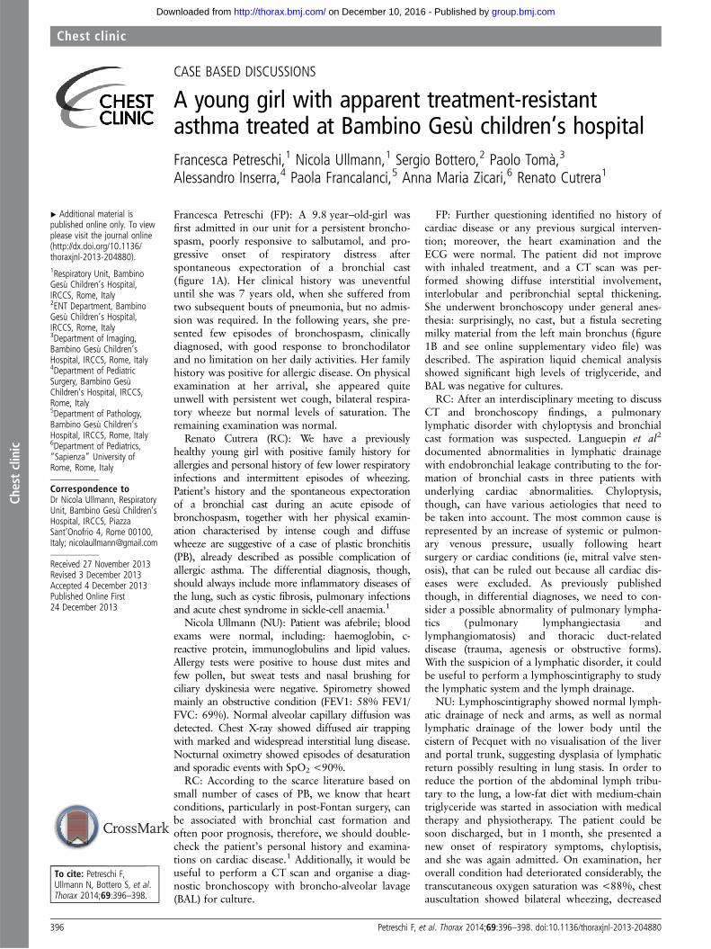
Francesca Petreschi (FP): A 9.8 year–old-girl wasfirst admitted in our unit for a persistent broncho-spasm, poorly responsive to salbutamol, and pro-gressive onset of respiratory distress afterspontaneous expectoration of a bronchial cast(figure 1A). Her clinical history was uneventfuluntil she was 7 years old, when she suffered fromtwo subsequent bouts of pneumonia, but no admis-sion was required. In the following years, she pre-sented few episodes of bronchospasm, clinicallydiagnosed, with good response to bronchodilatorand no limitation on her daily activities. Her familyhistory was positive for allergic disease. On physicalexamination at her arrival, she appeared quiteunwell with persistent wet cough, bilateral respira-tory wheeze but normal levels of saturation. Theremaining examination was normal.Renato Cutrera (RC): We have a previously
healthy young girl with positive family history forallergies and personal history of few lower respiratoryinfections and intermittent episodes of wheezing.Patient’s history and the spontaneous expectorationof a bronchial cast during an acute episode ofbronchospasm, together with her physical examin-ation characterised by intense cough and diffusewheeze are suggestive of a case of plastic bronchitis(PB), already described as possible complication ofallergic asthma. The differential diagnosis, though,should always include more inflammatory diseases ofthe lung, such as cystic fibrosis, pulmonary infectionsand acute chest syndrome in sickle-cell anaemia.1
Nicola Ullmann (NU): Patient was afebrile; bloodexams were normal, including: haemoglobin, c-reactive protein, immunoglobulins and lipid values.Allergy tests were positive to house dust mites andfew pollen, but sweat tests and nasal brushing forciliary dyskinesia were negative. Spirometry showedmainly an obstructive condition (FEV1: 58% FEV1/FVC: 69%). Normal alveolar capillary diffusion wasdetected. Chest X-ray showed diffused air trappingwith marked and widespread interstitial lung disease.Nocturnal oximetry showed episodes of desaturationand sporadic events with SpO2 <90%.RC: According to the scarce literature based on
small number of cases of PB, we know that heartconditions, particularly in post-Fontan surgery, canbe associated with bronchial cast formation andoften poor prognosis, therefore, we should double-check the patient’s personal history and examina-tions on cardiac disease.1 Additionally, it would beuseful to perform a CT scan and organise a diag-nostic bronchoscopy with broncho-alveolar lavage(BAL) for culture.
FP: Further questioning identified no history ofcardiac disease or any previous surgical interven-tion; moreover, the heart examination and theECG were normal. The patient did not improvewith inhaled treatment, and a CT scan was per-formed showing diffuse interstitial involvement,interlobular and peribronchial septal thickening.She underwent bronchoscopy under general anes-thesia: surprisingly, no cast, but a fistula secretingmilky material from the left main bronchus (figure1B and see online supplementary video file) wasdescribed. The aspiration liquid chemical analysisshowed significant high levels of triglyceride, andBAL was negative for cultures.RC: After an interdisciplinary meeting to discuss
CT and bronchoscopy findings, a pulmonarylymphatic disorder with chyloptysis and bronchialcast formation was suspected. Languepin et al2
documented abnormalities in lymphatic drainagewith endobronchial leakage contributing to the for-mation of bronchial casts in three patients withunderlying cardiac abnormalities. Chyloptysis,though, can have various aetiologies that need tobe taken into account. The most common cause isrepresented by an increase of systemic or pulmon-ary venous pressure, usually following heartsurgery or cardiac conditions (ie, mitral valve sten-osis), that can be ruled out because all cardiac dis-eases were excluded. As previously publishedthough, in differential diagnoses, we need to con-sider a possible abnormality of pulmonary lympha-tics (pulmonary lymphangiectasia andlymphangiomatosis) and thoracic duct-relateddisease (trauma, agenesis or obstructive forms).With the suspicion of a lymphatic disorder, it couldbe useful to perform a lymphoscintigraphy to studythe lymphatic system and the lymph drainage.NU: Lymphoscintigraphy showed normal lymph-
atic drainage of neck and arms, as well as normallymphatic drainage of the lower body until thecistern of Pecquet with no visualisation of the liverand portal trunk, suggesting dysplasia of lymphaticreturn possibly resulting in lung stasis. In order toreduce the portion of the abdominal lymph tribu-tary to the lung, a low-fat diet with medium-chaintriglyceride was started in association with medicaltherapy and physiotherapy. The patient could besoon discharged, but in 1 month, she presented anew onset of respiratory symptoms, chyloptisis,and she was again admitted. On examination, heroverall condition had deteriorated considerably, thetranscutaneous oxygen saturation was <88%, chestauscultation showed bilateral wheezing, decreased
396 Petreschi F, et al. Thorax 2014;69:396–398. doi:10.1136/thoraxjnl-2013-204880
Chest clinic
group.bmj.com on December 10, 2016 - Published by http://thorax.bmj.com/Downloaded from

Chestclinic
air entry bilaterally, and diffuse crackles. Chest X-ray showedleft lower lobe opacity, persistent wide interstitial lung diseaseand initial pleural effusion. Spirometry showed severe restrictivedefect (FEV1 42%, FEV1/FVC: 93%). Bronchoscopy revealedmany white secretions and a thick cast into the lower bronchusthat was removed. The cast consisted of fibrin with few lympho-cytes and some eosinophilic inflammatory cells. All culturesremained negative.
RC: Since the lymphoscintigraphy was consistent with a formof lymphatic disorder, possibly a thoracic duct anomaly withlymph stasis in the lung (previously suggested by CT images),we decided to start a supportive treatment and a low-fat dietwith medium-chain triglycerides. Unfortunately, the benefit wasjust partial with rapid formation of another bronchial cast,which was immediately removed with bronchoscopy.3 The hist-ology of the bronchial sample would suggest, according toSeear’s cast classification, of a combination between a type 2cast (acellular and more common in children with underlyingcardiac defects) and a type 1, because of the partial inflamma-tory composition, probably secondary to the patient’s allergicasthmatic condition.4 In literature, it has been hypothesised thata better outcome is possible in patients with type 1 casts withgood response to standard anti-inflammatory therapy.1 In ourpatient though, the absence of a clear inflammatory cast’s com-position, and the poor response to classic asthma treatmentsuggest a more complex underlying pathological conditionresponsible of the recurrence of cast formation. Various therap-ies for bronchial casts have been reported, but the evidenceremains anecdotal.3 According to fibrin cast histology, a treat-ment with nebulised tissue plasminogen activator (Alteplase)could be started following the clinical response.
FP: A plasminogen activator inhaled treatment was adminis-tered with rapid improvement, but 2 weeks later, her clinicalconditions became poorer, oxygen treatment was needed, andshe presented a radiological worsening with onset of chyloperi-cardium (figure 1C,E). A total parenteral nutrition, with the aimof completely eliminate fats from diet, was then started, andagain a rapid benefit was achieved.
RC: Concerning her repeated chylous effusion and episodesof chyloptisis, despite all medical treatments, and the lack of anavailable, definitive long-term solution, a surgical approachneeds to be discussed in a multidisplinary meeting. As
previously seen in the differential diagnosis, we have to considerpossible abnormality of pulmonary lymphatics and, before a sur-gical decision is made, we should better identify the lungpattern and discriminate between pulmonary lymphangiectasiaor lymphangiomatosis.
Paola Francalanci—pathologist: The family consented toperform a lung biopsy, and the tissue sample showed a histo-logical condition characterised by normal number but signifi-cantly dilated lymphatic vessels (H&E, D2–40 immunostaining).These findings are suggestive of a pulmonary lymphangectasia(figure1D), but a differentiation between a primary or a second-ary condition is not possible.
RC: Late symptom’s onset, patient’s clinical history and lym-phoscintigraphy’s findings possibly suggest secondary lymphan-gectasia determined by lymph stasis as a consequence of athoracic duct anomaly. This suspect along with the recurrentclinical worsening, due to continuous lymph congestion of thelungs, justify a surgical attempt with a transabdominal ligationof the thoracic duct.
FP: Surgery was performed, and the patient presented a dra-matic clinical improvement, medical treatments were graduallydiscontinued, ECG showed a complete resolution of chyloperi-cardium, FEV1 improved by 33%, and the young girl was soondischarged with no treatment at all. One year later, her clinicalconditions and lung function remains optimal (FEV1: 92%), norecurrence of cast formation or chyloptisis has been referred,and finally she is back to her usual activities and free diet.
RC: PB is an uncommon potentially fatal disease of theairways, often associated with cyanotic heart condition, butunfortunately, only palliative options are available. The patho-genesis of PB is not fully understood, but chyle in bronchialcast, suggests abnormal lymphatic flow.5 Our patient presentedwith intermittent respiratory distress, recurrent chyloptysis andbronchial cast formation. The investigations oriented towards alymphatic abnormality. We hypothesised that a thoracic-ductdysplasia could have been responsible for an increase of resist-ance downstream the dysplastic tract, with retrograde flow andconsequent formation of a lymphatic leakage into the left mainbronchus through a bronchial fistula. The transabdominal ductligation allowed complete recovery of lung function and thepatient’s usual activities, without need for any medicaltreatment.
Figure 1 (A) Bronchial cast.(B) Bronchial fistula secreting milkymaterial under pressure.(C) CT scan of the thoraxdemonstrating left lower lobe opacityand pleural effusion. (D) Lung biopsycompatible with pulmonarylymphangiectasia. (E) CT scanshowing chylopericardium.
Petreschi F, et al. Thorax 2014;69:396–398. doi:10.1136/thoraxjnl-2013-204880 397
Chest clinic
group.bmj.com on December 10, 2016 - Published by http://thorax.bmj.com/Downloaded from

Chestclinic
To our best knowledge, this is the first case of paediatric PB,without an underlying congenital cardiac disease, which wastreated successfully with a thoracic duct ligation. Given furtherinnovative, is the transabdominal duct ligation (vs thoracic), lessinvasive and effectively safer when the exact location of ductobstruction is uncertain. We believe our case can be a usefulexample for all forms of PB with an increased intraductallymphatic pressure for obstruction or malformation of the thor-acic duct.
Acknowledgements We thank Professor Matthias Griese for his expertise andhelp in the management of this case.
Contributors FP: treated the patient and contributed to the manuscript; NU:conceived the manuscript and treated the patient; SB: performed all bronchoscopiesand provided figure and video of secreting fistula; PT: performed all radiologicalimages and contributed to the radiological aspects of discussion; AI: performed thesurgical intervention and contributed to the decisional aspects of discussion; PF:reviewed the histopathology and contributed to the pathological aspects ofdiscussion; AMZ: treated the patient initially and contributed to the manuscript; RC:
treated the patient, contributed to the manuscript and the clinical aspects ofdiscussion and final decision.
Competing interests None.
Patient consent Obtained.
Ethics approval Obtained.
Provenance and peer review Not commissioned; internally peer reviewed.
REFERENCES1 Brogan T, Finn L, Pyskaty J Jr. et al. Plastic bronchitis in children: a case series and
review of the medical literature. Pediatr Pulmonol 2002;34:482–7.2 Languepin J, Scheinmann P, Mahut B, et al. Bronchial casts in children with
cardiopathies: the role of pulmonary lymphatic abnormalities. Pediatr Pulmonol1999;28:329–36.
3 Kruger J, Shpringer C, Picard E, et al. Thoracic air leakage in the presentation of castbronchitis. Chest 2009;136:615–17.
4 Seear M, Hui H, Magee F, et al. Bronchial casts in children: a proposed classificationbased on nine cases and a review of the literature. Am J Respir Crit Care Med1997;155:364–70.
5 Ezmigna DR, Morgan WJ, Witte MH, et al. Lymphoscintigraphy in plastic bronchitis,a pediatric case report. Pediatr Pulmonol 2013;48:515–18.
398 Petreschi F, et al. Thorax 2014;69:396–398. doi:10.1136/thoraxjnl-2013-204880
Chest clinic
group.bmj.com on December 10, 2016 - Published by http://thorax.bmj.com/Downloaded from

Bambino Gesù children's hospitaltreatment-resistant asthma treated at A young girl with apparent
CutreraAlessandro Inserra, Paola Francalanci, Anna Maria Zicari and Renato Francesca Petreschi, Nicola Ullmann, Sergio Bottero, Paolo Tomà,
doi: 10.1136/thoraxjnl-2013-2048802014 69: 396-398 originally published online December 24, 2013Thorax
http://thorax.bmj.com/content/69/4/396Updated information and services can be found at:
These include:
MaterialSupplementary
0.DC1.htmlhttp://thorax.bmj.com/content/suppl/2013/12/24/thoraxjnl-2013-20488Supplementary material can be found at:
References #BIBLhttp://thorax.bmj.com/content/69/4/396
This article cites 5 articles, 0 of which you can access for free at:
serviceEmail alerting
box at the top right corner of the online article. Receive free email alerts when new articles cite this article. Sign up in the
CollectionsTopic Articles on similar topics can be found in the following collections
(218)Ear, nose and throat/otolaryngology (526)Drugs: respiratory system
(525)Cystic fibrosis (235)Bronchitis
(562)Pneumonia (respiratory medicine) (579)Pneumonia (infectious disease)
(97)Lung infection (843)Child health
(1273)TB and other respiratory infections (559)Interstitial lung disease
(1782)Asthma (812)Radiology (diagnostics)
(676)Cardiothoracic surgery
Notes
http://group.bmj.com/group/rights-licensing/permissionsTo request permissions go to:
http://journals.bmj.com/cgi/reprintformTo order reprints go to:
http://group.bmj.com/subscribe/To subscribe to BMJ go to:
group.bmj.com on December 10, 2016 - Published by http://thorax.bmj.com/Downloaded from


![[1.1E] The Apparent Secret](https://img.pdfslide.us/doc/110x75/62ab570bc3c87f77520dcc48/11e-the-apparent-secret.jpg)