Embed Size (px)
Citation preview
case records of the massachusetts general hospital
T h e n e w e ngl a nd j o u r na l o f m e dic i n e
n engl j med 368;21 nejm.org may 23, 2013 2015
Founded by Richard C. CabotNancy Lee Harris, m.d., Editor Eric S. Rosenberg, m.d., EditorJo-Anne O. Shepard, m.d., Associate Editor Alice M. Cort, m.d., Associate EditorSally H. Ebeling, Assistant Editor Emily K. McDonald, Assistant Editor
Case 16-2013: A 12-Year-Old Girl with Irritability, Hypersomnia,
and Somatic SymptomsSuzanne L. Bender, M.D., Nicole A. Sherry, M.D., and Ricard Masia, M.D., Ph.D.
From the Departments of Psychiatry (S.L.B.), Pediatrics (N.A.S.), and Pathol-ogy (R.M.), Massachusetts General Hos-pital; and the Departments of Psychiatry (S.L.B.), Pediatrics (N.A.S.), and Pathol-ogy (R.M.), Harvard Medical School — both in Boston.
N Engl J Med 2013;368:2015-24.DOI: 10.1056/NEJMcpc1208145Copyright © 2013 Massachusetts Medical Society.
Pr esen tation of C a se
Dr. Elizabeth G. Pinsky (Psychiatry): A 12-year-old girl was seen in the outpatient psy-chiatry clinic of this hospital because of severe irritability, hypersomnia, and mul-tiple somatic symptoms.
The patient had celiac disease but had been otherwise well until approximately 8 months earlier, when she became increasingly irritable and reported daily stom-achaches, tingling and pain in her arms and legs, dizziness, anorexia, and severe fatigue; she also became increasingly somnolent, sleeping up to 13 hours per night. She had frequent angry outbursts directed at her mother and sister, and acted out physically on occasion. She became increasingly isolated from her friends and lost interest in activities. Her performance in school deteriorated, and she failed mathematics. Approximately 4.5 months before this presentation, red-cell indexes and results of thyroid-function tests were normal, and testing for heterophile an-tibody was negative; other test results are shown in Table 1. She was referred to the outpatient gastroenterology, neurology, and psychiatry clinics of this hospital.
The patient was born to a 39-year-old mother by cesarean section because of maternal preeclampsia, and her childhood development was normal. A diagnosis of celiac disease had been made at the age of 8 years, when she presented with abdominal pain and constipation; elevated levels of tissue transglutaminase anti-body (70 U per milliliter) and endomysial antibody (80 U per milliliter) were de-tected, and examination of a biopsy specimen of the duodenum showed changes consistent with celiac disease. She adhered to a gluten-free diet and had been otherwise well. She had a history of anxiety and depression; she had no response to a trial of sertraline but did have a response to both escitalopram, begun 2 years before this evaluation, and 7 months of cognitive behavioral therapy. Sixteen months before this evaluation, the first of three episodes of severe vomiting re-quiring intravenous hydration occurred; subsequent episodes occurred 6 months and 4 months before this evaluation, each preceded by fever and viral symptoms. There was no associated headache, and neurologic evaluation including an electro-encephalogram was reportedly normal.
The New England Journal of Medicine Downloaded from nejm.org at University of Notre Dame Aus on May 23, 2013. For personal use only. No other uses without permission.
Copyright © 2013 Massachusetts Medical Society. All rights reserved.
T h e n e w e ngl a nd j o u r na l o f m e dic i n e
n engl j med 368;21 nejm.org may 23, 20132016
The patient’s mother noted that the patient had a long history of daydreaming. She had not begun to menstruate. She had no history of head injury, loss of consciousness, urinary symptoms, hospitalizations, or surgery. Her only medication was 10 mg daily of escitalopram. She had no known allergies. She lived with her parents and younger sister. Her mother had thyroid disease; maternal and paternal aunts, a paternal uncle,
and her paternal grandmother had celiac dis-ease; a paternal uncle had bipolar disorder; and other relatives had anxiety, depression, or atten-tion deficit–hyperactivity disorder (ADHD). There was no family history of cystic fibrosis, inflam-matory bowel disease, liver disease, pancreatitis, or diabetes mellitus.
On examination, the patient was slim and ap-peared exhausted, without apparent physical dis-
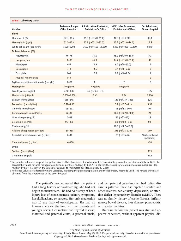
Table 1. Laboratory Data.*
VariableReference Range, Other Hospital†
4.5 Mo before Evaluation, Pediatrician’s Office
6 Wk after Evaluation, Pediatrician’s Office
On Admission, Other Hospital
Blood
Hematocrit (%) 32.1–38.7 35.3 (ref 35.0–45.0) 40.9 (ref 34–40) 40.3
Hemoglobin (g/dl) 11.3–13.4 11.9 (ref 11.5–15.5) 13.7 (ref 11.0–16.0) 13.8
White-cell count (per mm3) 5520–9290 3600 (ref 4500–13,500) 5260 (ref 4000–10,800) 5070
Differential count (%)
Neutrophils 46–76 39.1 45.0 (ref 30.0–85.0) 39
Lymphocytes 8–39 45.9 44.7 (ref 15.0–55.0) 45
Monocytes 4–7 9.9 6.7 (ref 0–10.0) 7
Eosinophils 1–3 4.5 3.4 (ref 0–5.0) 6
Basophils 0–1 0.6 0.2 (ref 0–2.0) 1
Atypical lymphocytes 0–4 2
Erythrocyte sedimentation rate (mm/hr) 0–20 3 7 8
Heterophile Negative Negative Negative
Free thyroxine (ng/dl) 0.80–1.90 0.9 (ref 0.9–1.4) 1.25
Thyrotropin (μU/ml) 0.700–5.700 3.43 9.64 6.820
Sodium (mmol/liter) 135–148 133 (ref 137–145) 126
Potassium (mmol/liter) 3.20–4.50 5.2 (ref 3.5–5.1) 5.55
Chloride (mmol/liter) 99–111 93 (ref 98–107) 94
Carbon dioxide (mmol/liter) 22–30 26.0 (ref 22.0–30.0) 23
Urea nitrogen (mg/dl) 5–18 22 (ref 7–17) 18
Creatinine (mg/dl) 0.3–1.0 0.6 (ref 0.5–1.0) 0.5
Calcium (mg/dl) 10.6 (ref 8.3–10.3)
Alkaline phosphatase (U/liter) 60–335 235 (ref 38–126) 209
Aspartate aminotransferase (U/liter) 2–40 63 (ref 15–46) 58 (hemolyzed specimen)
Creatine kinase (U/liter) 4–150 476
Urine
Sodium (mmol/liter) 119
Creatinine (mg/dl) 67.4
* Ref denotes reference range at the pediatrician’s office. To convert the values for free thyroxine to picomoles per liter, multiply by 12.87. To convert the values for urea nitrogen to millimoles per liter, multiply by 0.357. To convert the values for creatinine to micromoles per liter, multiply by 88.4. To convert the values for calcium to millimoles per liter, multiply by 0.250.
† Reference values are affected by many variables, including the patient population and the laboratory methods used. The ranges shown are obtained from the laboratories at the other hospital.
The New England Journal of Medicine Downloaded from nejm.org at University of Notre Dame Aus on May 23, 2013. For personal use only. No other uses without permission.
Copyright © 2013 Massachusetts Medical Society. All rights reserved.
case records of the massachusetts gener al hospital
n engl j med 368;21 nejm.org may 23, 2013 2017
tress. The blood pressure was 83/52 mm Hg, the pulse 102 beats per minute, the head circumfer-ence 53.2 cm, the weight 35.4 kg, and the height 149.9 cm, with a body-mass index (the weight in kilograms divided by the square of the height in meters) of 15.8 (12th percentile for age). The skin was freckled, with multiple dark nevi and slight hyperpigmentation in the axillae. Results of cranial-nerve testing and funduscopic examina-tion were normal. Strength was full throughout. Reflexes were brisk, and plantar responses were flexor. Clonus (2 to 3 beats, unsustained) on the right foot occurred, but the finding was not eas-ily repeated. The patient initially rocked on her feet during tandem gait but later skipped and jumped without difficulty. Results of sensory ex-amination and Romberg testing were normal. When talking with the psychiatrist, the patient focused primarily on her pain and fatigue. She appeared withdrawn and disengaged, with a sad and apathetic affect. Her speech was sparse, slow, and soft at times, and eye contact and spontaneous movements were limited. Her three wishes were to have a cell phone so she could call her mother when she became nervous while waiting for her mother to pick her up at school, to have different friends, and to have school be different. There was no delusional content, sui-cidal ideation, or hallucinations. In the waiting area, she fell asleep, awakening to gentle shak-ing but not to voice. Magnetic resonance imag-ing (MRI) of the head was normal. Diagnoses of major depressive disorder and generalized anxi-ety disorder were made.
Weekly therapy sessions were begun. During the next 3 weeks, the administration of escitalo-pram was changed to bedtime; the administra-tion of bupropion was begun and gradually in-creased to 75 mg daily. The patient continued to have worsening dizziness, lightheadedness, head-aches, stomachaches, and musculoskeletal pain. She had two episodes of vomiting at school, with nonbloody, nonbilious emesis and dry heaves, and her oral intake decreased.
On repeat examination in the psychiatry clinic 6 weeks after the initial evaluation, the patient ap-peared listless and pallid. Her weight was 34.0 kg. She was referred to her pediatrician for further evaluation. On examination at the pediatrician’s office 5 days later, the weight was 32.9 kg. Blood levels of platelets, glucose, total protein, albumin, total and direct bilirubin, alanine aminotrans-
ferase, and C-reactive protein were normal; other test results are shown in Table 1. Bupropion was stopped, and she was referred to the emergency department of another hospital.
On examination, the blood pressure was 87/45 mm Hg, the pulse 92 beats per minute with the patient in a supine position and 124 beats per minute while she was standing, the tympanic temperature 36.4°C, and the weight 33.5 kg. The remainder of the examination was normal. An electrocardiogram reportedly showed sinus rhythm at 100 beats per minute, with a normal axis, a prolonged QT interval corrected for heart rate of 505 msec, a T-wave inversion in lead III, and bi-phasic T waves in lead V3. Test results are shown in Table 1. The patient was admitted to the other hospital.
Additional diagnostic tests were performed.
Differ en ti a l Di agnosis
Dr. Suzanne L. Bender: I am aware of the diagnosis in this case. This 12-year-old girl with celiac dis-ease adhered to her gluten-free diet. She reported many somatic symptoms and had hypersomnia but appeared in no physical distress. No medical illness had been found by her pediatrician, gas-troenterologist, or neurologist. She was irritable, with frequent angry outbursts at home, and had academic difficulty at school. She was referred to child psychiatry for evaluation. Because some med-ical conditions take time to become apparent, a multidisciplinary team was established to coor-dinate ongoing diagnosis and treatment in this child with somatic symptoms that had no clear cause but that impaired daily functioning.
Somatic symptoms
Emotional distress may be affecting this patient’s bodily function. This is well-described in irrita-ble bowel syndrome as the “brain–gut axis.” Psy-chosocial influences affect gut motility and may increase visceral sensitivity. A change in gut mo-tility and visceral sensitivity may in turn increase anxiety and depression in some patients.1-3 There is an ongoing conversation between the brain and the body. When I talk to families in my clinic about this mind–body interaction, I explain that this is why we have a neck. In addition, some chil-dren may have difficulty distinguishing emotional pain from physical pain. All they know is that they don’t feel well, and then this is translated, in
The New England Journal of Medicine Downloaded from nejm.org at University of Notre Dame Aus on May 23, 2013. For personal use only. No other uses without permission.
Copyright © 2013 Massachusetts Medical Society. All rights reserved.
T h e n e w e ngl a nd j o u r na l o f m e dic i n e
n engl j med 368;21 nejm.org may 23, 20132018
their minds, into feeling sick. Emotional distress expressed as somatic symptoms is a new type of referred pain — pain that is referred from brain to body.
Pediatric patients with somatization are likely to have psychopathological symptoms, family dysfunction, and poor performance and atten-dance at school.4 Children with abdominal pain who have a negative medical workup may have been exposed to a traumatic event and may have underlying anxiety.5 In addition, the mothers of such children often have higher rates of anxiety, depression, and various physical ailments, as compared with a control population.5,6
It is notable that one of this patient’s three wishes was not a wish to feel physically better. Her wishes “to have different friends” and “to have school be different” signal that these areas in her life are causing distress and perhaps her experience of pain.
Irritable Mood
Evaluation of pediatric irritable mood includes an assessment of a child’s functioning at home and at school. This patient’s frequent fights with her mother and sister and concerns about her peer group could both cause and result in her current irritability. Although this patient had anxiety, her symptoms could not be explained by anxiety alone. Persons with anxiety usually pre-sent with sleeplessness or restlessness, and not with hypersomnia and psychomotor retardation.
Academic struggles may also decrease self-esteem and emotional resiliency. This patient’s grades dropped, she failed mathematics, and she expressed concern about school. A consideration in this case is attention-deficit disorder, inatten-tive type. Young girls with this disorder who do not have behavioral issues are often first diag-nosed during middle school, when organiza-tional challenges increase. The results of neuro-psychological testing could show whether this child has a nonverbal learning disorder, defined on a test such as the Wechsler Intelligence Scale for Children, fourth edition, as a verbal compre-hension score that is much greater than the per-ceptual reasoning score. Difficulties with organi-zation, social skills, and academics, specifically mathematics, are often seen in children with this disorder.7
This patient’s irritability could be due to a medical condition.8 Anemia, thyroid dysfunction,
mononucleosis, and disorders involving calcium metabolism are associated with depressive symp-toms; these disorders have already been consid-ered in this case and do not seem to explain her symptoms. She has celiac disease, and adoles-cents with celiac disease have a higher lifetime prevalence of major depressive disorders before diagnosis and before adoption of a gluten-free diet than do matched controls.9 Celiac disease with malabsorption may cause vitamin B12 and folate deficiencies, both of which are associated with mood disorders.10,11 An additional screen-ing blood chemical profile would be useful for identifying abnormal liver and renal function and abnormal levels of electrolytes, vitamins, and glucose, which may be associated with mood symptoms (Tables 2 and 3).11 Irritability may be due to the effects of a substance (e.g., a drug of abuse, a medication, or a toxin).8 Although sur-reptitious substance abuse could account for all this patient’s symptoms, it would be highly un-likely in a well-supervised child whose only medication is escitalopram.
Depression
On a review of this patient’s symptoms, she meets all the criteria for a major depressive episode (i.e., for more than 2 weeks, she has had persis-tent irritable mood and five or more of the fol-lowing neurovegetative symptoms: low energy, decreased appetite, hypersomnia, diminished in-terest in daily activities, psychomotor retardation, and decreased concentration). She reported no sad-ness, feelings of worthlessness, or suicidal ide-ation, but children often have trouble recognizing and reporting their emotional state. Alexithymia (the inability to express one’s feelings) does not rule out depression. Child psychiatrists base their diagnoses on observation and on reports from parents, as well as on the child’s self-report. Prepubertal patients with depression commonly present with somatic symptoms, irritability, or social withdrawal, whereas adolescents are more likely to have hypersomnia or psychomotor retarda-tion. This patient, straddling the worlds of child-hood and adolescence, had all these symptoms.8
This patient did not have a recent loss or trauma to account for her symptoms. It was pos-sible that she was in the depressive phase of an emerging pediatric bipolar disorder, since she had some risk factors, including an early onset of de-pression, atypical depression with hypersomnia,
The New England Journal of Medicine Downloaded from nejm.org at University of Notre Dame Aus on May 23, 2013. For personal use only. No other uses without permission.
Copyright © 2013 Massachusetts Medical Society. All rights reserved.
case records of the massachusetts gener al hospital
n engl j med 368;21 nejm.org may 23, 2013 2019
and an extensive family history of mood disor-ders. However, the absence of adverse effects (e.g., agitation) associated with her use of esci-talopram argues against a current diagnosis of bipolar illness.12-15
The patient had several risk factors for major depressive disorder, including a family history of depression,16 the presence of another nonaffective psychiatric disorder predating the depression (anx-iety), female sex (after puberty), and multiple major life stressors (including a physical illness, family conflict, and poor performance in school).
The more risk factors a patient has, the more likely it is that depression will develop.17,18
While I continued to collect laboratory data, I made a preliminary diagnosis in this patient of treatment-resistant major depressive disorder. More information would clarify the potential diagnosis of attention-deficit disorder, inatten-tive type.
Management of Childhood Depression
Both medications and psychotherapy are used to treat childhood depression. Many different psy-
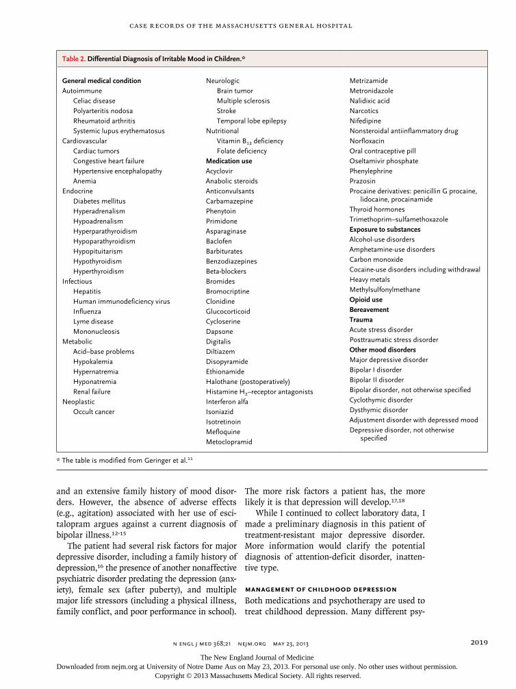
Table 2. Differential Diagnosis of Irritable Mood in Children.*
General medical conditionAutoimmune
Celiac diseasePolyarteritis nodosaRheumatoid arthritisSystemic lupus erythematosus
CardiovascularCardiac tumorsCongestive heart failureHypertensive encephalopathyAnemia
EndocrineDiabetes mellitusHyperadrenalismHypoadrenalismHyperparathyroidismHypoparathyroidismHypopituitarismHypothyroidismHyperthyroidism
InfectiousHepatitisHuman immunodeficiency virusInfluenzaLyme diseaseMononucleosis
MetabolicAcid–base problemsHypokalemiaHypernatremiaHyponatremiaRenal failure
NeoplasticOccult cancer
NeurologicBrain tumorMultiple sclerosisStrokeTemporal lobe epilepsy
NutritionalVitamin B12 deficiencyFolate deficiency
Medication useAcyclovirAnabolic steroidsAnticonvulsantsCarbamazepinePhenytoinPrimidoneAsparaginaseBaclofenBarbituratesBenzodiazepinesBeta-blockersBromidesBromocriptineClonidineGlucocorticoidCycloserineDapsoneDigitalisDiltiazemDisopyramideEthionamideHalothane (postoperatively)Histamine H2–receptor antagonistsInterferon alfaIsoniazidIsotretinoinMefloquineMetoclopramid
MetrizamideMetronidazoleNalidixic acid NarcoticsNifedipineNonsteroidal antiinflammatory drugNorfloxacinOral contraceptive pillOseltamivir phosphatePhenylephrinePrazosinProcaine derivatives: penicillin G procaine,
lidocaine, procainamideThyroid hormonesTrimethoprim–sulfamethoxazoleExposure to substancesAlcohol-use disordersAmphetamine-use disordersCarbon monoxideCocaine-use disorders including withdrawalHeavy metalsMethylsulfonylmethaneOpioid useBereavementTraumaAcute stress disorderPosttraumatic stress disorderOther mood disordersMajor depressive disorderBipolar I disorderBipolar II disorderBipolar disorder, not otherwise specifiedCyclothymic disorderDysthymic disorderAdjustment disorder with depressed moodDepressive disorder, not otherwise
specified
* The table is modified from Geringer et al.11
The New England Journal of Medicine Downloaded from nejm.org at University of Notre Dame Aus on May 23, 2013. For personal use only. No other uses without permission.
Copyright © 2013 Massachusetts Medical Society. All rights reserved.
T h e n e w e ngl a nd j o u r na l o f m e dic i n e
n engl j med 368;21 nejm.org may 23, 20132020
chotherapies (e.g., psychodynamic psychothera-py, family therapy, interpersonal psychotherapy, and cognitive behavioral therapy) are thought to be effective in the treatment of pediatric depres-sion.19-22 Cognitive behavioral therapy and relax-ation techniques have been useful in the treat-ment of chronic pain.23
Only two selective serotonin-reuptake inhibitors (SSRIs) are approved by the Food and Drug Ad-ministration (FDA) — fluoxetine for the treatment of children 8 years of age or older, and escitalo-pram for the treatment of children 12 years of age or older. The FDA issued a black-box warn-ing in 2004 that some children, adolescents, and young adults may have an increased risk of sui-cidal thoughts or behavior while taking antide-pressants.24 One meta-analysis showed that the increased risk in suicidal thoughts was between 1% and 3%.25 Another meta-analysis concluded that the clinical benefits associated with the use of antidepressants in children with depression outweighed the risks.26
In patients who have not previously received treatment, evidence supports the use of fluoxetine combined with cognitive behavioral therapy for the safest and fastest clinical response.27,28 This patient’s depression continued despite psycho-therapy and two trials of an SSRI. Although venlafaxine has been studied as an alternative antidepressant agent for adolescents with treat-ment-resistant depression, it has a higher inci-dence of side effects, including suicidality, over the long term than do SSRIs.29-31
Studies of treatment-resistant depression in
adults provide guidance for medications for this child with treatment-resistant depression. Aug-menting an antidepressant with either buspirone or sustained-release bupropion results in a 30% increase in the remission rate; therefore, it is a reasonable next step for this child, who had treatment-resistant major depressive disorder and no response to two trials of an SSRI.20,32,33 Bu-propion works through noradrenergic and dopa-minergic mechanisms and affects different neu-rotransmitters from those affected by SSRIs. If the administration of bupropion were added, this patient could continue on escitalopram to treat her anxiety. Bupropion is also used to treat at-tentional disorders and may help reduce the pa-tient’s reported daydreaming.34 I have found bu-propion to be very effective in the treatment of disengaged adolescents with somatic symptoms who have not responded to an SSRI.
The clinical approach to major depressive disorder and multiple somatic symptoms in this treatment-resistant child needs to be inclusive and comprehensive. Further laboratory studies are warranted to screen for a medical condition that is fueling the mood disorder (Table 3). I rec-ommended weekly cognitive behavioral therapy (for problem-solving and cognitive restructuring), the practice of relaxation techniques (to help with chronic pain),23 and the use of a psychody-namic approach for identifying and discussing the patient’s feelings about her friends, family, and school. Escitalopram and bupropion were administered. I recommended evaluation for pos-sible attention-deficit disorder, as well as neuro-psychological testing to identify any learning disorders.
Despite these approaches, the patient’s so-matic symptoms worsened, and she began to lose weight and appear ill. She was urgently re-ferred back to her pediatrician.
DR . SUZ A NNE L . BENDER’S DI AGNOSIS
Major depressive disorder and high suspicion for a concurrent medical illness.
Pathol o gic a l Discussion
Dr. Ricard Masia: The patient had undergone upper gastrointestinal endoscopy at 8 years of age. Dif-fuse mildly scalloped mucosa was found in the
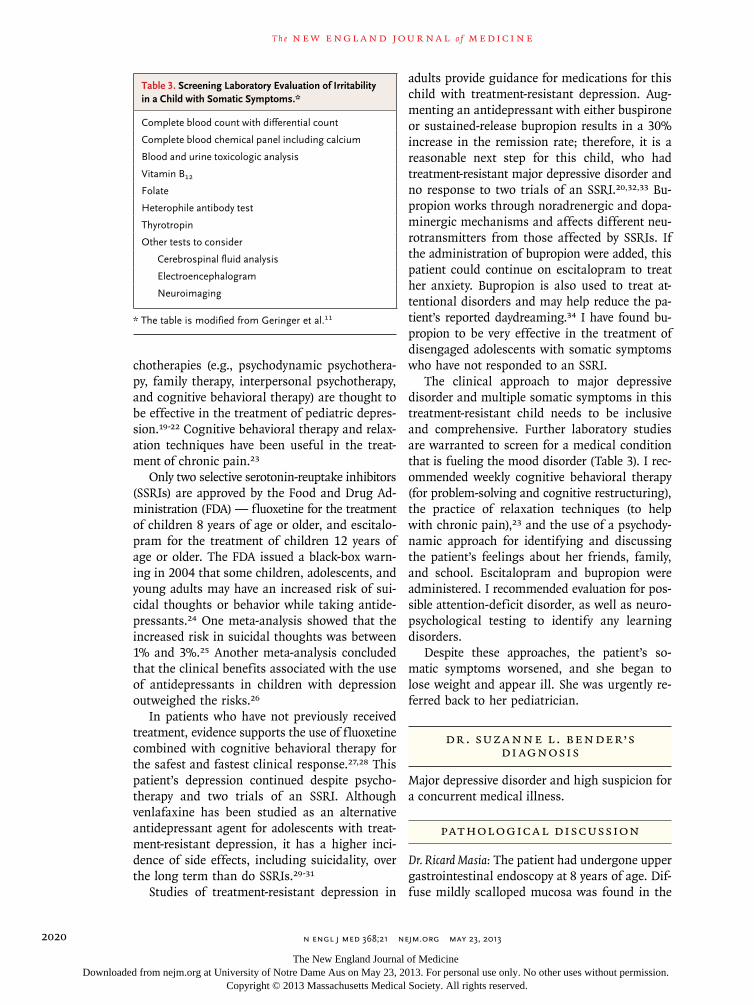
Table 3. Screening Laboratory Evaluation of Irritability in a Child with Somatic Symptoms.*
Complete blood count with differential count
Complete blood chemical panel including calcium
Blood and urine toxicologic analysis
Vitamin B12
Folate
Heterophile antibody test
Thyrotropin
Other tests to consider
Cerebrospinal fluid analysis
Electroencephalogram
Neuroimaging
* The table is modified from Geringer et al.11
The New England Journal of Medicine Downloaded from nejm.org at University of Notre Dame Aus on May 23, 2013. For personal use only. No other uses without permission.
Copyright © 2013 Massachusetts Medical Society. All rights reserved.
case records of the massachusetts gener al hospital
n engl j med 368;21 nejm.org may 23, 2013 2021
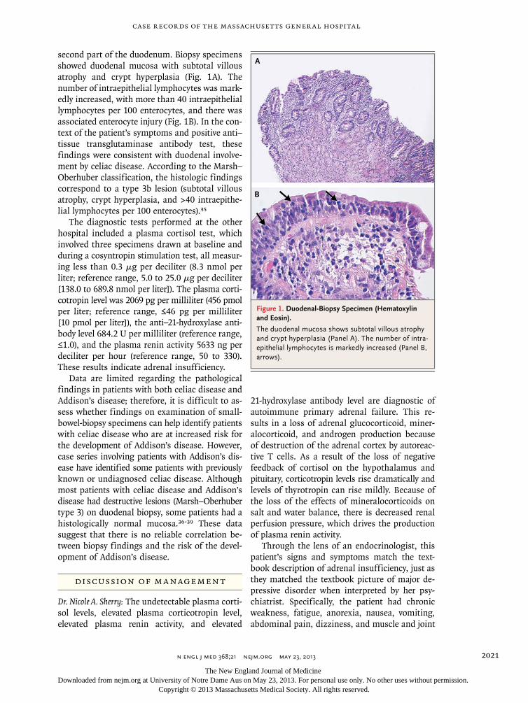
second part of the duodenum. Biopsy specimens showed duodenal mucosa with subtotal villous atrophy and crypt hyperplasia (Fig. 1A). The number of intraepithelial lymphocytes was mark-edly increased, with more than 40 intraepithelial lymphocytes per 100 enterocytes, and there was associated enterocyte injury (Fig. 1B). In the con-text of the patient’s symptoms and positive anti–tissue transglutaminase antibody test, these findings were consistent with duodenal involve-ment by celiac disease. According to the Marsh–Oberhuber classification, the histologic findings correspond to a type 3b lesion (subtotal villous atrophy, crypt hyperplasia, and >40 intraepithe-lial lymphocytes per 100 enterocytes).35
The diagnostic tests performed at the other hospital included a plasma cortisol test, which involved three specimens drawn at baseline and during a cosyntropin stimulation test, all measur-ing less than 0.3 μg per deciliter (8.3 nmol per liter; reference range, 5.0 to 25.0 μg per deciliter [138.0 to 689.8 nmol per liter]). The plasma corti-cotropin level was 2069 pg per milliliter (456 pmol per liter; reference range, ≤46 pg per milliliter [10 pmol per liter]), the anti–21-hydroxylase anti-body level 684.2 U per milliliter (reference range, ≤1.0), and the plasma renin activity 5633 ng per deciliter per hour (reference range, 50 to 330). These results indicate adrenal insufficiency.
Data are limited regarding the pathological findings in patients with both celiac disease and Addison’s disease; therefore, it is difficult to as-sess whether findings on examination of small-bowel-biopsy specimens can help identify patients with celiac disease who are at increased risk for the development of Addison’s disease. However, case series involving patients with Addison’s dis-ease have identified some patients with previously known or undiagnosed celiac disease. Although most patients with celiac disease and Addison’s disease had destructive lesions (Marsh–Oberhuber type 3) on duodenal biopsy, some patients had a histologically normal mucosa.36-39 These data suggest that there is no reliable correlation be-tween biopsy findings and the risk of the devel-opment of Addison’s disease.
Discussion of M a nagemen t
Dr. Nicole A. Sherry: The undetectable plasma corti-sol levels, elevated plasma corticotropin level, elevated plasma renin activity, and elevated
21-hydroxylase antibody level are diagnostic of autoimmune primary adrenal failure. This re-sults in a loss of adrenal glucocorticoid, miner-alocorticoid, and androgen production because of destruction of the adrenal cortex by autoreac-tive T cells. As a result of the loss of negative feedback of cortisol on the hypothalamus and pituitary, corticotropin levels rise dramatically and levels of thyrotropin can rise mildly. Because of the loss of the effects of mineralocorticoids on salt and water balance, there is decreased renal perfusion pressure, which drives the production of plasma renin activity.
Through the lens of an endocrinologist, this patient’s signs and symptoms match the text-book description of adrenal insufficiency, just as they matched the textbook picture of major de-pressive disorder when interpreted by her psy-chiatrist. Specifically, the patient had chronic weakness, fatigue, anorexia, nausea, vomiting, abdominal pain, dizziness, and muscle and joint
A
B
Figure 1. Duodenal-Biopsy Specimen (Hematoxylin and Eosin).
The duodenal mucosa shows subtotal villous atrophy and crypt hyperplasia (Panel A). The number of intra-epithelial lymphocytes is markedly increased (Panel B, arrows).
The New England Journal of Medicine Downloaded from nejm.org at University of Notre Dame Aus on May 23, 2013. For personal use only. No other uses without permission.
Copyright © 2013 Massachusetts Medical Society. All rights reserved.
T h e n e w e ngl a nd j o u r na l o f m e dic i n e
n engl j med 368;21 nejm.org may 23, 20132022
pains. She had weight loss, hyperpigmentation, hypotension, hyponatremia, hyperkalemia, hy-percalcemia, anemia, and eosinophilia. Even more striking, she had had episodes of acute adrenal crises with profound deterioration dur-ing intercurrent viral illnesses over the preced-ing year and a half that had required three hos-pitalizations.
The diagnosis of Addison’s disease is often missed for some time, as it was in this case. In one study, only 47% of the cases were diagnosed within 1 year after initial symptoms and more than 20% were diagnosed more than 5 years after initial symptoms.40 Thirty percent of the patients had seen five physicians before the di-agnosis of Addison’s disease was made. More than 80% had had a previous incorrect diagno-sis, 50% of which were psychiatric disorders and 31% of which were gastrointestinal diseases.
In this case, there is an important clue to the diagnosis. This patient had a personal history of celiac disease and a family history of both celiac disease and autoimmune thyroid disease. This constellation of diseases is highly suggestive of the autoimmune polyglandular syndrome type 2 and should alert the clinician that the patient is at high risk for other autoimmune disorders, in-cluding autoimmune adrenal insufficiency, thy-roid disease, and celiac disease.41
After the diagnosis of Addison’s disease was made, the patient was treated with hydrocorti-sone (9 mg per square meter of body-surface area divided into three doses daily) and fludro-cortisone (0.1 mg daily). Although the somatic symptoms resolved, she continued to have psy-chiatric symptoms. It has been recognized that for patients with adrenal insufficiency who ad-here to current treatment regimens, the quality of life can be impaired,42 with a perception of reduced general health and vitality and increased fatigue as compared with population norms. Pa-tients with autoimmune polyendocrine syndromes have lower scores in these measures on subjec-tive health-assessment questionnaires than do those with adrenal insufficiency in isolation.43,44
The administration of adrenal androgens that have not been traditionally replaced may improve the quality of life. Several short-term studies have shown that the administration of dehydroepi-androsterone improves psychological health,45,46 but the results are mixed, and longer-term stud-ies have not shown as pronounced an effect.43,47
Dr. Pinsky: The patient was discharged from the other hospital taking only escitalopram and glucocorticoid-replacement and mineralocorticoid-replacement therapy. The nausea, vomiting, pain, and weakness diminished rapidly, and her appe-tite quickly improved; her mother reported that the patient ate two candy bars on the way home from the hospital. Fatigue persisted but was improved. The hope was that her symptoms had all been related to underlying adrenal insuffi-ciency and that she might eventually be able to discontinue escitalopram.
After approximately 3 months of glucocorticoid-replacement therapy, the patient continued to be socially withdrawn and anxious, was still sleep-ing 12 to 14 hours a day, and struggled with inattention and academic faltering. Neuropsy-chiatric testing was performed and was sugges-tive of ADHD, although assessment of attention was confounded by depressed mood and anxiety, as well as her recent medical illness. The admin-istration of bupropion was resumed. After several weeks, symptoms of withdrawal, anxiety, ener-gy, and attention improved. At the end of the academic year, the patient was the champion of the science fair.
Dr. Sherry: Although the focus of current treat-ment studies has been finding a better physio-logical match of hormonal replacement to help with psychiatric symptoms, this patient’s symp-toms were successfully treated with a combina-tion of traditional hormonal replacement and psychopharmacologic approaches.
Dr. Nancy Lee Harris (Pathology): Are there any comments or questions?
Dr. Joshua Roffman (Psychiatry): In retrospect, were there any red flags in the patient’s presen-tation that suggested a medical component to her symptoms that we should look for when evaluating depression in children?
Dr. Bender: At first, this patient looked tired but otherwise physically well and she had a negative medical workup. Many patients with a similar presentation get better with psychiatric treatment. Some medical diagnoses become clear as symptoms evolve over time, which is why on-going coordinated care between psychiatry and pediatrics is so critical for patients with somatic symptoms. A key moment was when the patient, who had appeared well, started to look sick. Dr. Pinsky recognized that her condition had changed and referred her for additional evaluation.
The New England Journal of Medicine Downloaded from nejm.org at University of Notre Dame Aus on May 23, 2013. For personal use only. No other uses without permission.
Copyright © 2013 Massachusetts Medical Society. All rights reserved.
case records of the massachusetts gener al hospital
n engl j med 368;21 nejm.org may 23, 2013 2023
A Physician: Were her parents angry that it took 16 months for a diagnosis to be made?
The Patient’s Father: For a very long time, our daughter has had continuing mental and physi-cal problems. It was difficult and traumatic, but we’re very thankful for all the doctors who even-tually made the diagnosis and brought her to where she is today.
Dr. Harris: I think it’s instructive to learn of the usual delay in the diagnosis of Addison’s disease. There are so many possible causes of all these symptoms that Addison’s disease is not on the top of anyone’s differential diagnosis.
Dr. Harland Winter (Pediatric Gastroenterology): Was the axillary hyperpigmentation a potential clue to adrenal insufficiency?
Dr. Sherry: Yes, hyperpigmentation in Addison’s disease is due to an increase in melanocyte-stimulating hormone. The axillae are a typical location for hyperpigmentation, which is most often diffuse.
Dr. Robin M. Jones (Neurology): The patient was referred to me from gastroenterology because of recurrent nausea and vomiting, which had been attributed to the cyclic vomiting syndrome at an outside institution. An electroencephalogram was
reportedly normal. Because of the axillary freck-ling, I entertained the diagnosis of neurofibro-matosis 1. I referred her for an ophthalmologic examination to look for Lisch nodules and or-dered a brain MRI to rule out an intracranial process that might account for the nausea and vomiting.
Dr. Sherry: Addison’s disease can be associated with other skin findings, such as more gener-alized hyperpigmentation, especially in sun-exposed areas, whereas neurofibromatosis can be associated with café au lait lesions. In this patient with no other skin findings, a distinction between Addison’s disease and neurofibromato-sis could not be made on the basis of axillary freckling alone.
A nat omic a l Di agnosis
Celiac disease, Addison’s disease, and major de-pressive disorder.
Presented at Psychiatry Grand Rounds.Dr. Sherry reports receiving consulting fees and grant support
through her institution from MacroGenics. No other potential conflict of interest relevant to this article was reported.
Disclosure forms provided by the authors are available with the full text of this article at NEJM.org.
References
1. Elsenbruch S. Abdominal pain in irri-table bowel syndrome: a review of putative psychological, neural and neuro-immune mechanisms. Brain Behav Immun 2011; 25:386-94.2. Drossman DA. The functional gastro-intestinal disorders and the Rome II pro-cess. Gut 1999;45:Suppl 2:II1-II5.3. Koloski NA, Jones M, Kalantar J, Welt-man M, Zaguirre J, Talley NJ. The brain–gut pathway in functional gastrointesti-nal disorders is bidirectional: a 12-year prospective population-based study. Gut 2012;61:1284-90.4. Campo JV, Jansen-McWilliams L, Comer DM, Kelleher KJ. Somatization in pediatric primary care: association with psychopathology, functional impairment, and use of services. J Am Acad Child Ado-lesc Psychiatry 1999;38:1093-101.5. Wasserman AL, Whitington PF, Rivara FP. Psychogenic basis for abdominal pain in children and adolescents. J Am Acad Child Adolesc Psychiatry 1988;27:179-84.6. Campo JV, Bridge J, Lucas A, et al. Physical and emotional health of mothers of youth with functional abdominal pain. Arch Pediatr Adolesc Med 2007;161:131-7.7. Braaten E, Felopulos G. Nonverbal learning disorders and Asperger syn-drome. In: Braaten E, Felopolus G, eds.
Straight talk about psychological testing for kids. New York: Guilford Press, 2004: 151-77.8. Diagnostic and statistical manual of mental disorders, 4th ed. rev.: DSM-IV-R. Washington, DC: American Psychiatric Association, 2000.9. Pynnönen PA, Isometsä ET, Aronen ET, Verkasalo MA, Savilahti E, Aalberg VA. Mental disorders in adolescents with celiac disease. Psychosomatics 2004;45: 325-35.10. García-Manzanares A, Lucendo AJ. Nutritional and dietary aspects of celiac disease. Nutr Clin Pract 2011;26:163-73.11. Geringer ES, Querques J, Kolodziej MS, Burns TE, Stern TA. Diagnosis and treatment of depression in the intensive care unit patient. In: Irwin RS, Rippe JM, eds. Irwin and Rippe’s intensive care medicine. 7th ed. Philadelphia: Lippincott Williams & Wilkins, 2012:2087-98.12. Akiskal HS, Maser JD, Zeller PJ, et al. Switching from ‘unipolar’ to bipolar II: an 11-year prospective study of clinical and temperamental predictors in 559 pa-tients. Arch Gen Psychiatry 1995;52: 114-23.13. Geller B, Fox LW, Clark KA. Rate and predictors of prepubertal bipolarity dur-ing follow-up of 6- to 12-year-old de-
pressed children. J Am Acad Child Ado-lesc Psychiatry 1994;33:461-8.14. Forty L, Smith D, Jones L, et al. Clini-cal differences between bipolar and uni-polar depression. Br J Psychiatry 2008; 192:388-9.15. Benazzi F. Classifying mood disor-ders by age-at-onset instead of polarity. Prog Neuropsychopharmacol Biol Psychi-atry 2009;33:86-93.16. Weissman MM, Wickramaratne P, Nomura Y, et al. Families at high and low risk for depression: a 3-generation study. Arch Gen Psychiatry 2005;62:29-36.17. Lewinsohn PM, Rohde P, Seeley JR. Major depressive disorder in older adoles-cents: prevalence, risk factors, and clini-cal implications. Clin Psychol Rev 1998; 18:765-94.18. Rohde P, Lewinsohn PM, Seeley JR. Comorbidity of unipolar depression: II. Comorbidity with other mental disorders in adolescents and adults. J Abnorm Psy-chol 1991;100:214-22.19. Weisz JR, McCarty CA, Valeri SM. Ef-fects of psychotherapy for depression in children and adolescents: a meta-analy-sis. Psychol Bull 2006:132:132-49. 20. Choe CJ, Emslie GJ, Mayes TL. De-pression. Child Adolesc Psychiatric Clin N Am 2012;21:807-29.
The New England Journal of Medicine Downloaded from nejm.org at University of Notre Dame Aus on May 23, 2013. For personal use only. No other uses without permission.
Copyright © 2013 Massachusetts Medical Society. All rights reserved.
n engl j med 368;21 nejm.org may 23, 20132024
case records of the massachusetts gener al hospital
21. Birmaher B, Brent D, Bernet W, et al. Practice parameter for the assessment and treatment of children and adolescents with depressive disorders. J Am Acad Child Adolesc Psychiatry 2007;46:1503-26.22. Hofmann SG, Asnaani A, Vonk IJJ, Sawyer AT, Fang A. The efficacy of cogni-tive behavioral therapy: a review of meta-analyses. Cognit Ther Res 2012;36:427-40.23. Palermo TM, Eccleston C, Lewan-dowski AS, Williams AC, Morley S. Ran-domized controlled trials of psychologi-cal therapies for management of chronic pain in children and adolescents: an up-dated meta-analytic review. Pain 2010; 148:387-97.24. Antidepressant medications for chil-dren and adolescents: information for parents and caregivers. Bethesda, MD: National Institute of Mental Health, 2011 (http://www.nimh.nih.gov/health/topics/child-and-adolescent-mental-health).25. Hammad TA, Laughren T, Racoosin J. Suicidality in pediatric patients treated with antidepressant drugs. Arch Gen Psy-chiatry 2006;63:332-9.26. Bridge JA, Iyengar S, Salary CB, et al. Clinical response and risk for reported suicidal ideation and suicide attempts in pediatric antidepressant treatment: a meta-analysis of randomized controlled trials. JAMA 2007;297:1683-96.27. March J, Silva S, Petrycki S, et al. Fluoxetine, cognitive-behavioral therapy, and their combination for adolescents with depression: Treatment for Adolescents With Depression Study (TADS) random-ized controlled trial. JAMA 2004;292: 807-20.28. March JS, Silva S, Petrycki S, et al. The Treatment for Adolescents With Depres-sion Study (TADS): long-term effective-ness and safety outcomes. Arch Gen Psy-chiatry 2007;64:1132-43. [Erratum, Arch Gen Psychiatry 2008;65:101.]29. Brent DA, Emslie GJ, Clarke GN, et al. Predictors of spontaneous and systemati-cally assessed suicidal adverse events in
the Treatment of SSRI-Resistant Depres-sion in Adolescents (TORDIA) study. Am J Psychiatry 2009;166:418-26.30. Vitiello B, Emslie G, Clarke G, et al. Long-term outcome of adolescent depres-sion initially resistant to selective sero-tonin reuptake inhibitor treatment: a fol-low-up study of the TORDIA sample. J Clin Psychiatry 2011;72:388-96.31. Brent D, Emslie G, Clarke G, et al. Switching to another SSRI or to venlafax-ine with or without cognitive behavioral therapy for adolescents with SSRI-resis-tant depression: the TORDIA randomized controlled trial. JAMA 2008;299:901-13.32. Trivedi MH, Fava M, Wisniewski SR, et al. Medication augmentation after the failure of SSRIs for depression. N Engl J Med 2006;354:1243-52.33. Hughes CW, Emslie GJ, Crismon ML, et al. Texas Children’s Medication Algo-rithm Project: update from Texas Consen-sus Conference Panel on Medication Treatment of Childhood Major Depressive Disorder. J Am Acad Child Adolesc Psy-chiatry 2007;46:667-86.34. Barrickman LL, Perry PJ, Allen AJ, et al. Bupropion versus methylphenidate in the treatment of attention-deficit hyper-activity disorder. J Am Acad Child Adolesc Psychiatry 1995;34:649-57.35. Oberhuber G, Granditsch G, Vogel-sang H. The histopathology of coeliac disease: time for a standardized report scheme for pathologists. Eur J Gastroen-terol Hepatol 1999;11:1185-94.36. O’Leary C, Walsh CH, Wieneke P, et al. Coeliac disease and autoimmune Ad-dison’s disease: a clinical pitfall. QJM 2002;95:79-82.37. Myhre AG, Aarsetøy H, Undlien DE, Hovdenak N, Aksnes L, Husebye ES. High frequency of coeliac disease among pa-tients with autoimmune adrenocortical failure. Scand J Gastroenterol 2003;38: 511-5.38. Biagi F, Campanella J, Soriani A, Vailati A, Corazza GR. Prevalence of coe-
liac disease in Italian patients affected by Addison’s disease. Scand J Gastroenterol 2006;41:302-5.39. Betterle C, Lazzarotto F, Spadaccino AC, et al. Celiac disease in North Italian patients with autoimmune Addison’s dis-ease. Eur J Endocrinol 2006;154:275-9.40. Bleicken B, Hahner S, Ventz M, Quink-ler M. Delayed diagnosis of adrenal insuf-ficiency is common: a cross-sectional study in 216 patients. Am J Med Sci 2010; 339:525-31.41. Eisenbarth GS, Gottlieb PA. Autoim-mune polyendocrine syndromes. N Engl J Med 2004;350:2068-79.42. Løväs K, Loge JH, Husebye ES. Subjec-tive health status in Norwegian patients with Addison’s disease. Clin Endocrinol (Oxf) 2002;56:581-8.43. Gurnell EM, Hunt PJ, Curran SE, et al. Long-term DHEA replacement in primary adrenal insufficiency: a randomized, con-trolled trial. J Clin Endocrinol Metab 2008;93:400-9.44. Hahner S, Loeffler M, Fassnacht M, et al. Impaired subjective health status in 256 patients with adrenal insufficiency on standard therapy based on cross-sec-tional analysis. J Clin Endocrinol Metab 2007;92:3912-22.45. Arlt W, Callies F, van Vlijmen JC, et al. Dehydroepiandrosterone replacement in women with adrenal insufficiency. N Engl J Med 1999;341:1013-20.46. Hunt PJ, Gurnell EM, Huppert FA, et al. Improvement in mood and fatigue af-ter dehydroepiandrosterone replacement in Addison’s disease in a randomized, double blind trial. J Clin Endocrinol Metab 2000;85:4650-6.47. Alkatib AA, Cosma M, Elamin MB, et al. A systematic review and meta-analysis of randomized placebo-controlled trials of DHEA treatment effects on quality of life in women with adrenal insufficiency. J Clin Endocrinol Metab 2009;94:3676-81.Copyright © 2013 Massachusetts Medical Society.
Lantern Slides Updated: Complete PowerPoint Slide Sets from the Clinicopathological Conferences
Any reader of the Journal who uses the Case Records of the Massachusetts General Hospital as a teaching exercise or reference material is now eligible to receive a complete set of PowerPoint slides, including digital images, with identifying legends, shown at the live Clinicopathological Conference (CPC) that is the basis of the Case Record. This slide set contains all of the images from the CPC, not only those published in the Journal. Radiographic, neurologic, and cardiac studies, gross specimens, and photomicrographs, as well as unpublished text slides, tables, and diagrams, are included. Every year 40 sets are produced, averaging 50-60 slides per set. Each set is supplied on a compact disc and is mailed to coincide with the publication of the Case Record.
The cost of an annual subscription is $600, or individual sets may be purchased for $50 each. Application forms for the current subscription year, which began in January, may be obtained from the Lantern Slides Service, Department of Pathology, Massachusetts General Hospital, Boston, MA 02114 (telephone 617-726-2974) or e-mail [email protected].
The New England Journal of Medicine Downloaded from nejm.org at University of Notre Dame Aus on May 23, 2013. For personal use only. No other uses without permission.
Copyright © 2013 Massachusetts Medical Society. All rights reserved.