Embed Size (px)
Citation preview

Case 1
• 18 yo woman came to ER with a 5-day history of severe abdominal pain
• Localized, intermittent, sharp, epigastric and periumbilical pain associated with mild nausea but no vomiting for the past 6 months / pain usually starting prior to the beginning of a menstrual cycle, lasting for the length of the cycle
• Discoloration of urine during the episodes • PMH: 2 previous hospital admissions for
hyponatremia work-up

• PMH: spells of confusion with indeterminate jerking spasms of the upper extremities and facial muscles
• No sensitivity to the sun • PE: mild tenderness on deep palpation of the
abdomen / tachycardia / symmetrical motor weakness of the arms
• Stools were heme (-) • Na: 132 mEq/L (135-145 mEq/L) • Clinical diagnosis? • AIP (Acute Intermittent Porphyria)

What are porphyrias?
• Rare, inherited or acquired diseases resulting from enzyme deficiencies that lead to heme pathway intermediates accumulation
• Inheritance pattern for most of them is AD with variable penetrance – majority of affected persons do not exhibit clinical disease
• ♀ >♂
• Identification of the defect is important for providing genetic counselling / advice on how to avoid precipitating factors

Anatomical classification of porphyrias
• Hepatic:
Intermitent Acute Porphyria (IAP)
Hereditary Coproporphyria (HCP)
Variegate Porphyria (VP)
Porphyria Cutanea Tarda (PCT)
• Erythropoietic:
Congenital Erythropoietic Porphyria (CEP)
Erythropoietic Protoporphyria (EPP)

Clinical Classification of Porphyrias
• Acute Porphyrias • Non-Acute Porphyrias
(neurological) (skin photosensitivity)
Acute Intermitent Porphyria Variegate Porphyria
Hereditary Coproporphyria
Porphyria Cutanea Tarda Erythropoietic Protoporphyria
Congenital Erythropoietic Porphyria

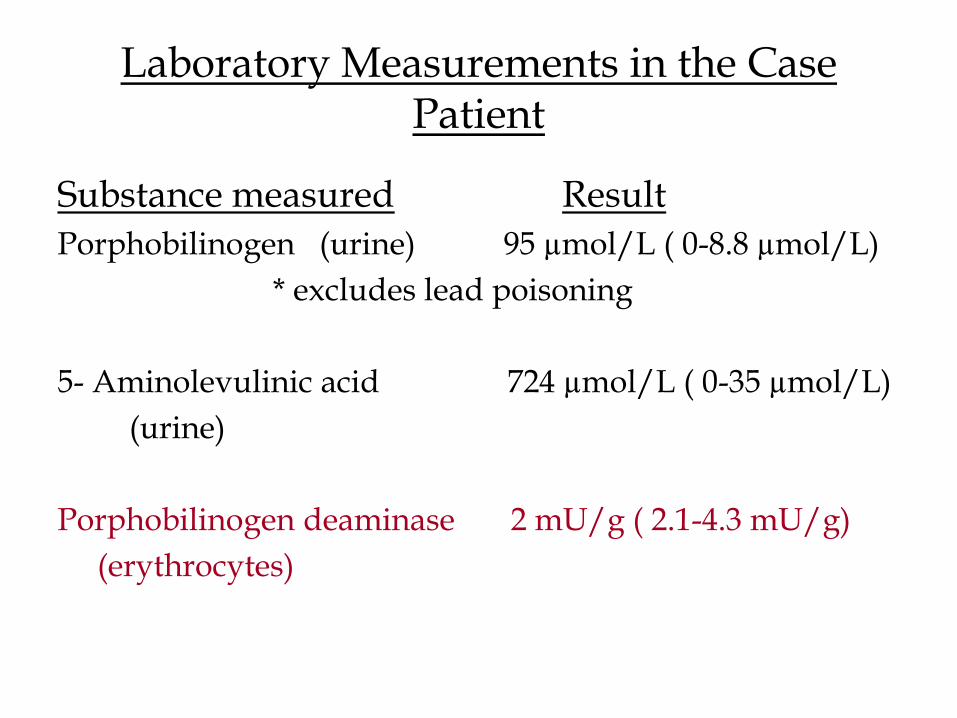
Laboratory Measurements in the Case Patient
Substance measured Result
Porphobilinogen (urine) 95 µmol/L ( 0-8.8 µmol/L)
* excludes lead poisoning
5- Aminolevulinic acid 724 µmol/L ( 0-35 µmol/L)
(urine)
Porphobilinogen deaminase 2 mU/g ( 2.1-4.3 mU/g)
(erythrocytes)

How should I work-up this case?
Step 1: Is this an acute (neurological) case or a non-acute (cutaneous) case?
Acute case
Step 2: Qualitative PBG urine screening test (Hoesch test- Ehrlich’s reagent -or Watson-Schwartz test)
Magenta color → (+) → quantitative assay
Step 3: Fecal Porphyrin Screening test
Negative (AIP) Positive (VP or HCP)

Case explanation
• Severe abdominal pain (80%),confusion, jerking spasms – ALA and PBG (neurotoxins) accumulate in tissues → acute neurovisceral symptoms
• Tachycardia (80%) - release of catecholamines during attack /sudden death
• Menstrual cycle – estrogen is a precipitating factor
• Discoloration of urine – accumulation of fluorescent porphyrins
• Hyponatremia – inappropriate secretion of antidiuretic hormone

• Urine (early morning): - 20°C
• Feces: - 20°C
• Whole blood: 4°C
• Erythrocytes: - 20°C
• Plasma: - 20°C
Subsequent Hospital Course
• Treatment: oral analgesics, antiemetics, intravenous administration of dextrose and hematin (cimetidine is another option)
• Placed on high-carbohydrate diet, instructed to avoid long periods without eating and to use medications only after consulting physician

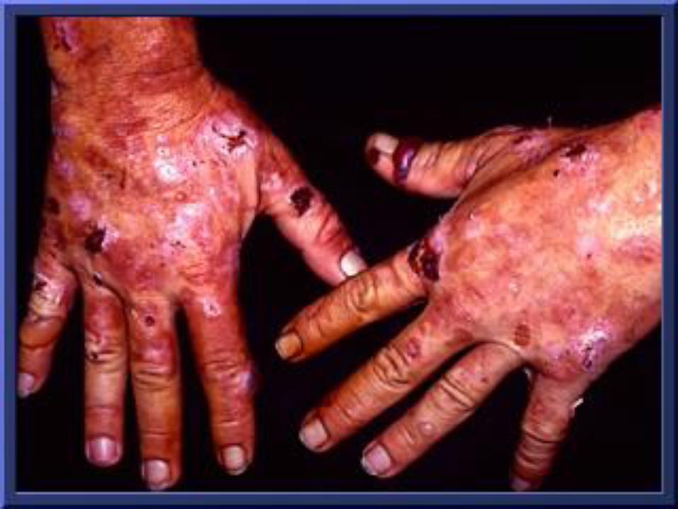
Case 2 • 40 yo landscaper presents with painful blisters
on the back of his hands shortly after the landscaping season began
• Discoloration of urine
• Denied any recent exposure to new soaps, detergents or medications
• PMH: partial complex seizure disorder that begun 3 years after head trauma (taking phenytoin)
• Average weekly ethanol intake: 18 12-oz cans of beer
• PE: besides the blisters, was noted to have hypertrichosis

Laboratory Measurements in the Case Patient
• Fasting Glucose: 159 mg/dL (70-105 mg/dL) • Alanine aminotransferase:135 U/L (<45 U/L) • Aspartate aminotransferase:100 U/L (<41 U/L) • Alkaline phosphatase: 161 U/L (30-115 U/L) • Ferritin: 989 ng/mL (19-260 ng/mL)

• Clinical diagnosis?
• Bullous pemphigoid
• Herpetic infections
• Staphylococcal infections
• Contact dermatitis
• Chemical burns
• Pemphigus vulgaris
• Porphyria Cutanea Tarda (type I – sporadic)


24-hour urine collection
• Uroporphyrin: 1000 µg (<27)
• 5-carboxyporphyrin: 120 (<5)
• 6-carboxyporphyrin: 120 (<3)
• 7-carboxyporphyrin: 720 (<6)
• Coproporphyrin: 67 (<72)
___________________________________
• HCV antibody (+) – active disease confirmed – viral load of more than 1 milion particles of RNA by PCR
How should I work-up this case? Step 1: Is this an acute (neurological) case or a non-
acute (cutaneous) case?
Cutaneous case
Step 2: Urine porphyrin screening test (qualitative)
Positive ( now you have to quantify)
Step 3: ↑ Uro (PCT) ↑ Copro (VP, HCP)
Step 4: Free Erythrocyte Protoporphyrin
Normal (PCT) ↑ (Erythropoietic Protoporphyria)

Case explanation
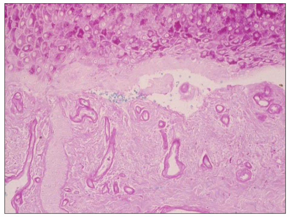
• Painful blisters – ultraviolet light transforms (oxidizes) accumulated porphyrins in the skin into toxins that cause skin fragility
• Hypertrichosis - hair bulb keratinocytes are activated by the dual action of light and porphyrins
• Alcohol - induces ALA synthase, the first enzyme in heme biosynthesis, and inhibits the latter enzymes such as ferrochelatase

• HCV (also HIV) – triggering factors /specific anticytosolic antibodies are associated with liver damage
• Hepatic iron overload (siderosis) - present in nearly every case of PCT /↑ iron, ferritin, transferrin saturation, ↓ TIBC
• Diabetes mellitus - present in 15-20% of PCT patients.

Subsequent clinical course
• Instructed to avoid exposure to direct sunlight, avoid ethanol, and other precipitating drugs (such as phenitoin). Switched to Gabapentin
• Removal of one unit of blood with phlebotomy weekly or biweekly to reduce iron levels
• Antimalarial agent (hydroxychloroquine or chloroquine) biweekly (antimalarials complex with porphyrin and promote the excretion into bile)