Embed Size (px)
Citation preview
CARTILAGE TISSUE.
PREMED II LECTURE.
Introduction
• Specialised connective tissue with a large amount of matrix & few cells.
• The matrix has collagen & elastic fibers embedded in a ground substance rich in proteoglycans & glycoproteins.
• Matrix is less pliable & firm, allowing the tissue to bear mechanical stress without permanent distortion.
• Cartilage,as a specialized connective tissue can act as a biomechanical spring.
• Essential for growth,development of bone.• *long bones before and after birth.
Functions of Cartilage Tissue
• Specialized CT in which the firm consistency of the extracellular matrix allows the tissue to bear mechanical stresses without permanent distortion
• Supports soft tissues.• Shock-absorbing because it is resilient.• Smooth surface allows sliding against it.• Essential for growth, development of bone.
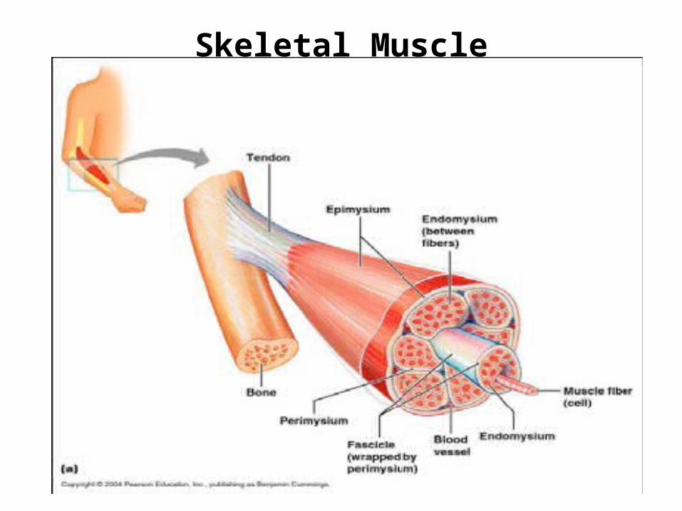
Skeletal Muscle
Characteristics
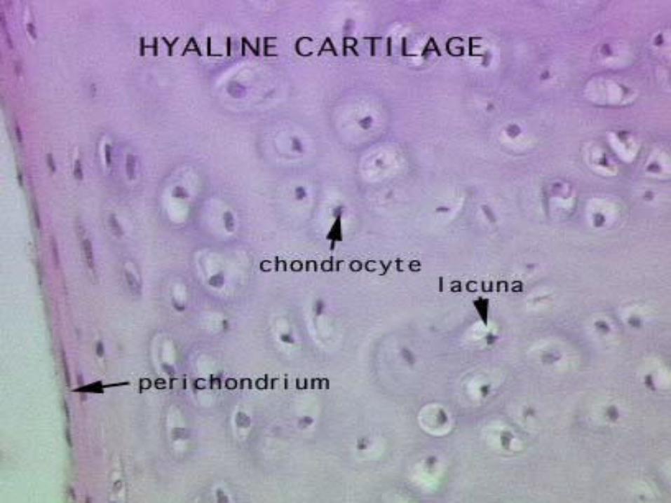
• Chondrocytes (-blasts)– Located in lacunae
• Extensive extra-cellular matrix– Fibers, ground substance– Collagen, hyaluronic acid, proteoglycans,
glycoproteins, elastic (in elastic cartilage)– Macromolecules, water, fibers bind together and
give firm, flexible properties to tissue.• No blood, nerve supply• Low metabolic rate.
• Chondrocytes respire under low oxygen tension.
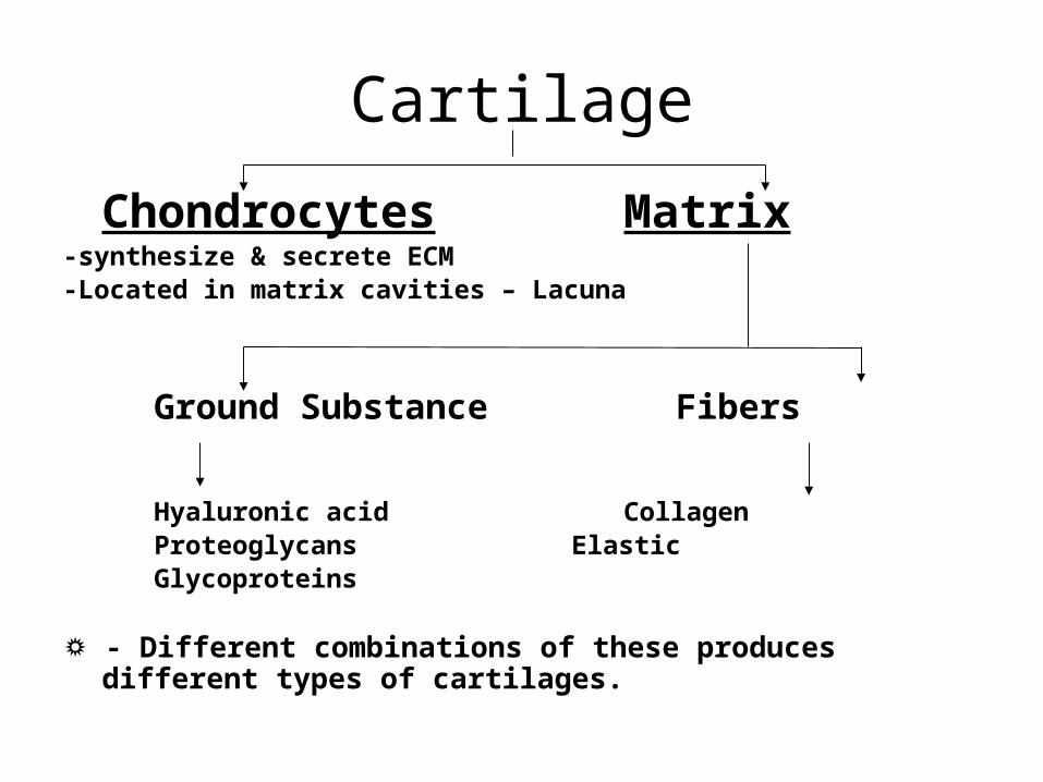
Cartilage
Chondrocytes Matrix-synthesize & secrete ECM-Located in matrix cavities – Lacuna
Ground Substance Fibers
Hyaluronic acid CollagenProteoglycans ElasticGlycoproteins
☼ - Different combinations of these produces different types of cartilages.
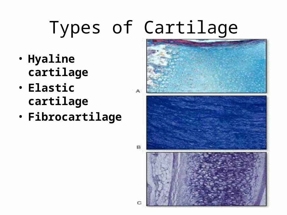
Types of Cartilage
• Hyaline cartilage• Elastic cartilage• Fibrocartilage
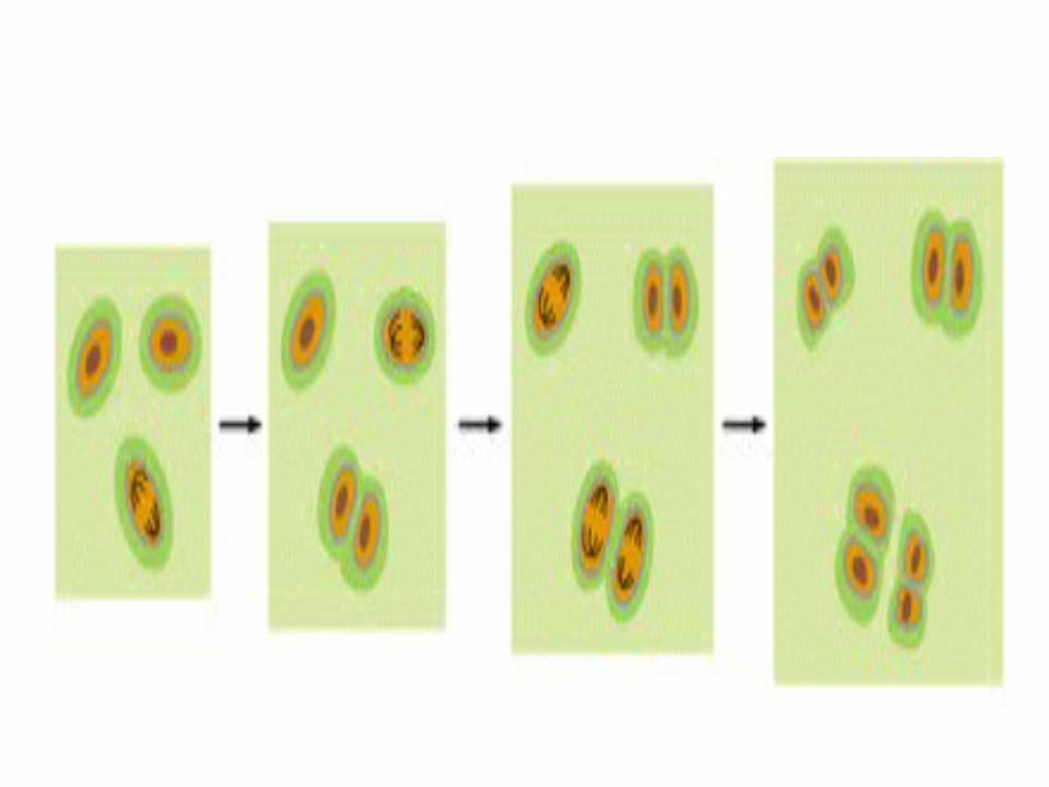
Chondrocytes• Elliptical (young) or round (mature) cells formed from
chondroblasts.• Farther in, they are round & appear in groups of upto 8 cells
originating from mitotic divisions of a single chondrocyte. These groups are called as Isogenous.
• Surrounded by a space called the Lacuna.• During routine histologic preparation, the cells shrink &
therefore lacunae are visible. In living tissue, the cells fill the lacunae completely.
• Synthesize collagen & the extracellular matrix.• Since cartilage is avascular, chondrocytes produce energy
from anaerobic glycolysis.
Proteoglycan, hyaluronic acid and chondronectin make up the extracellular. Matrix.Collagen that is synthesized is basically type-II collagen
At the periphery of hyaline cartilage, young chondrocytes have an elliptical shape ,with the long axis parallel to the surface.
Futher in,they are round and may appear in groups of up to 8 cells originating from mitotic divisions of a single chondrocyte.These groups are called ISOGENOUS.
Chondrocytes
• Function is dependant on hormones.• Growth hormone, thyroxin, testosterone,
somatomedin C – stimulate• Cortisone, hydrocortisone, estradiol – inhibit• **chondrocyte function depends on
hormonal balance**
Influence of hormones
• GH, TH, testosterone accelerates synthesis of glycosaminoglycans.
• Cortisone, estradiol inhibits synthesis.• Growth depends mainly on GH (somatotropin)– Stimulates liver to produce somatomedin C– Somatomedin C acts on cartilage cell, stimulating
growth
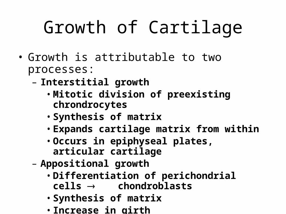
Growth of Cartilage
• Growth is attributable to two processes:– Interstitial growth• Mitotic division of preexisting chrondrocytes• Synthesis of matrix• Expands cartilage matrix from within• Occurs in epiphyseal plates, articular cartilage
– Appositional growth• Differentiation of perichondrial cells chondroblasts• Synthesis of matrix• Increase in girth
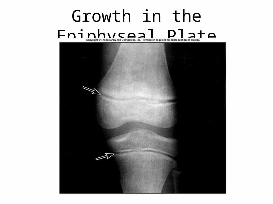
Growth in the Epiphyseal Plate
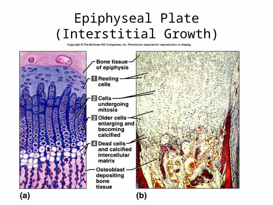
Epiphyseal Plate(Interstitial Growth)
http://web.indstate.edu/thcme/mwking/glycans.html
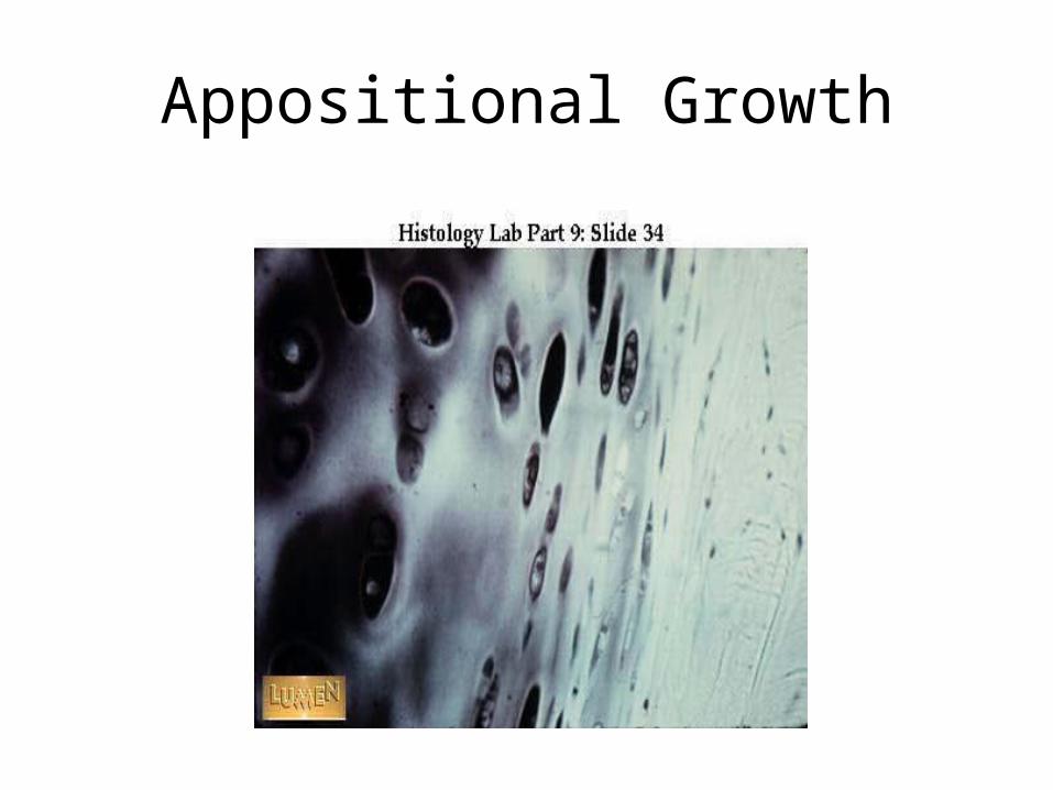
Appositional Growth
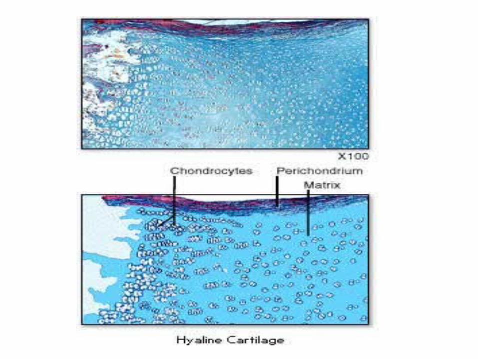
Hyaline Cartilage• Most abundant• Matrix is ‘glassy’ (translucent)
Matrix:• Collagen embedded in a hydrated gel of proteoglycans &
glycoproteins.• Contains primarily type II collagen.• Proteoglycans contain chondroitin sulfate & keratan sulfate.• High content of water bound to the GAGs acts a shock absorber &
biomechanical spring.• Chondronectin is a glycoprotein important of cell-matrix adhesion.• Matrix surrounding the chondrocyte is rich in GAGs & poor in
collagen – territorial/capsular matrix. Stains differently (more basophilic) than the rest of the matrix.
• Has a perichondrium (except articular cartilage)
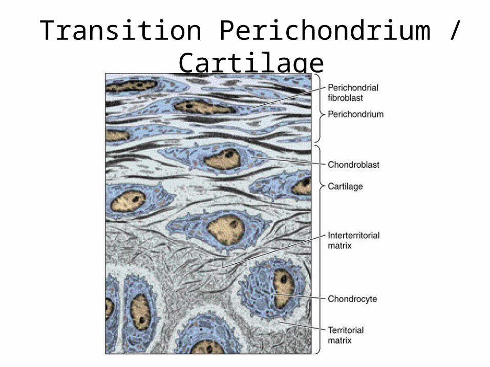
Transition Perichondrium / Cartilage

Hyaline Cartilage• Most abundant• Matrix is ‘glassy’ (translucent)
Matrix:• Collagen embedded in a hydrated gel of proteoglycans &
glycoproteins.• Contains primarily type II collagen.• Proteoglycans contain chondroitin sulfate & keratan sulfate.• High content of water bound to the GAGs acts a shock absorber &
biomechanical spring.• Chondronectin is a glycoprotein important of cell-matrix adhesion.• Matrix surrounding the chondrocyte is rich in GAGs & poor in
collagen – territorial/capsular matrix. Stains differently (more basophilic) than the rest of the matrix.
• Has a perichondrium (except articular cartilage)
Function & Location• It is rigid – allows flexibility & compression.• Has ability to grow rapidly in the fetus, thus forms a
model for bone development.• Shock absorption & friction reduction in joints.
Locations:• Epiphyseal plate• Nose, larynx, trachea, bronchi• Articular cartilage, costal cartilage• **One important property of hyaline cartilage is
that it can ossify at a later stage.
Articular cartilage is sustained by diffusion of oxygen and nutrients from the synovial fluid.
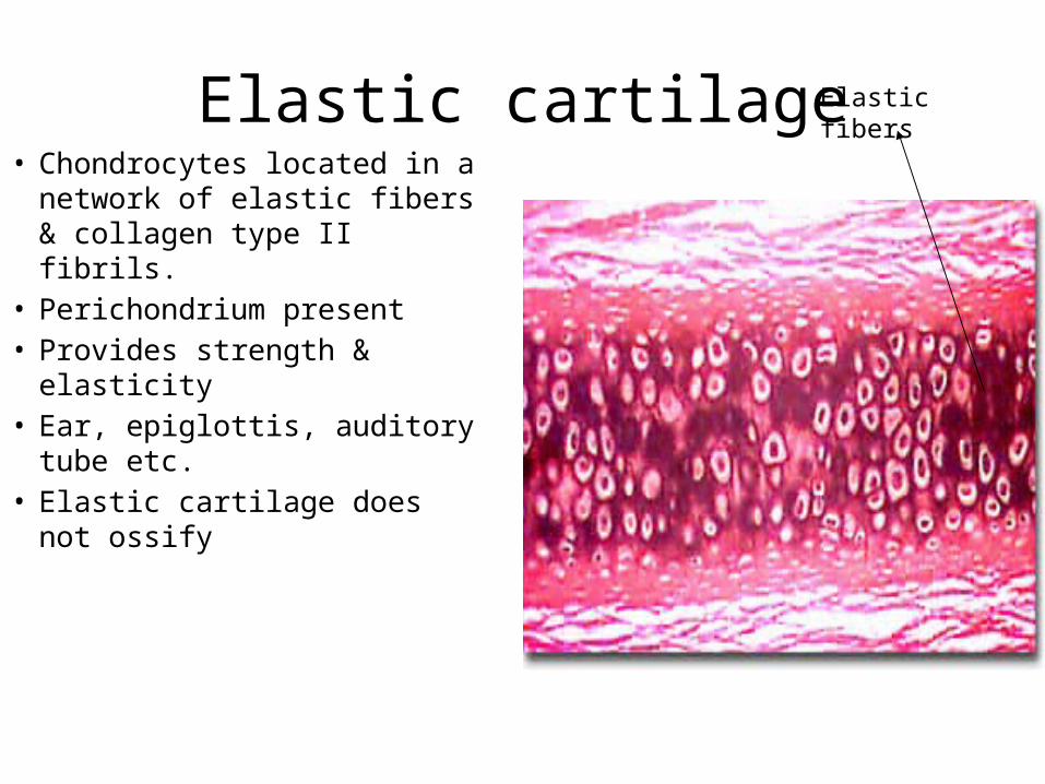
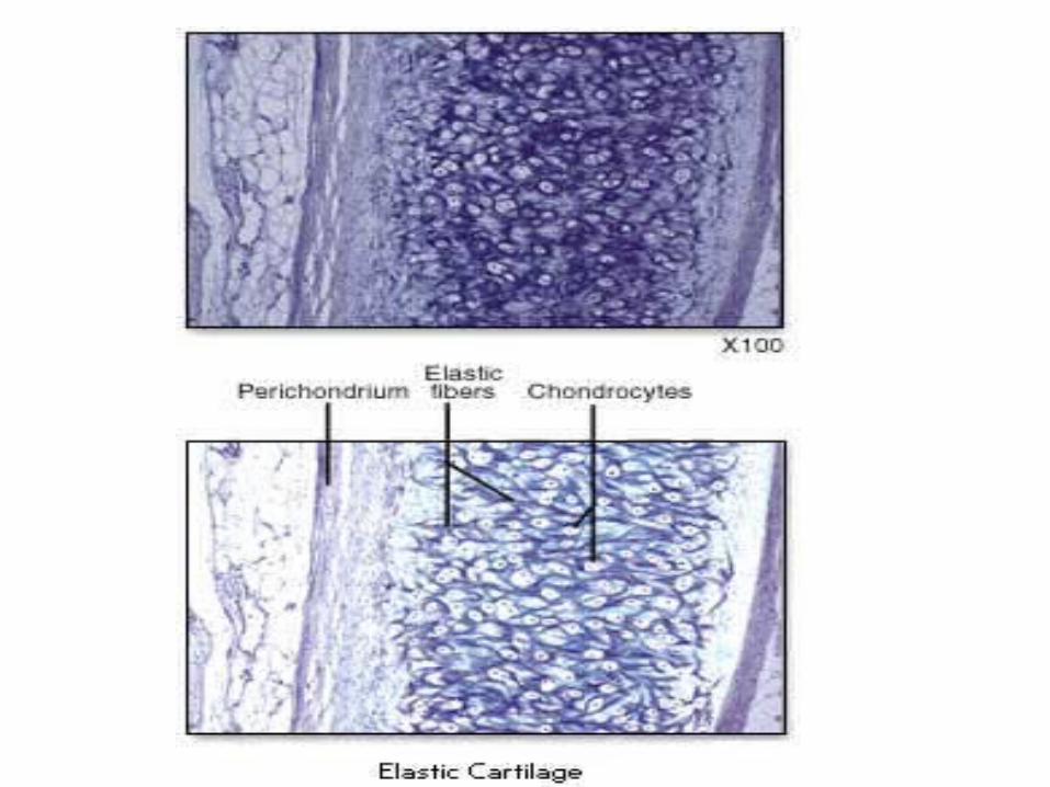
Elastic cartilage• Chondrocytes located in a
network of elastic fibers & collagen type II fibrils.
• Perichondrium present• Provides strength & elasticity• Ear, epiglottis, auditory tube
etc.• Elastic cartilage does not
ossify
Elastic fibers
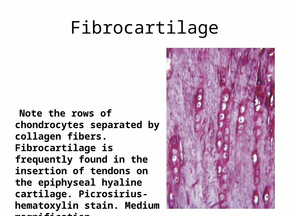
Fibrocartilage
Note the rows of chondrocytes separated by collagen fibers. Fibrocartilage is frequently found in the insertion of tendons on the epiphyseal hyaline cartilage. Picrosirius-hematoxylin stain. Medium magnification.
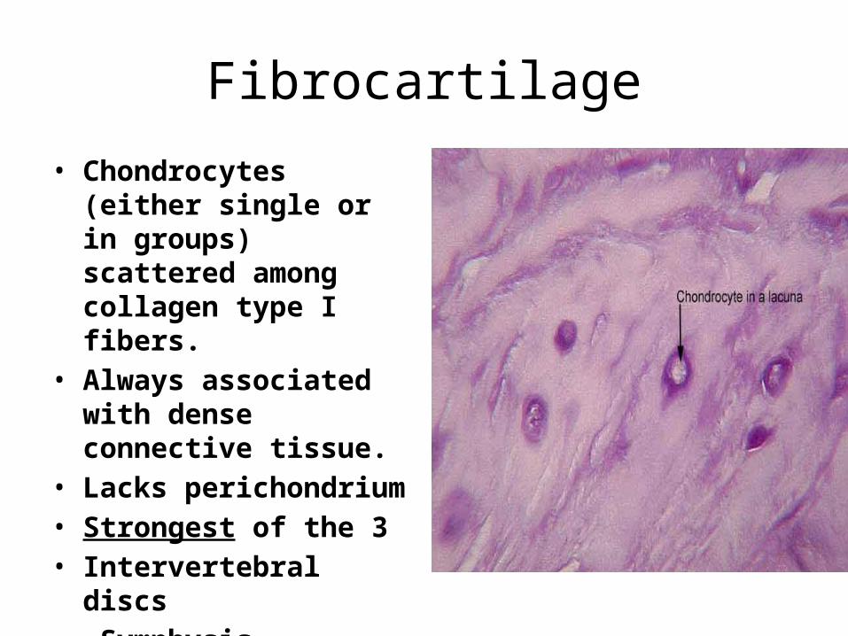
Fibrocartilage
• Chondrocytes (either single or in groups) scattered among collagen type I fibers.
• Always associated with dense connective tissue.
• Lacks perichondrium• Strongest of the 3• Intervertebral discs• Symphysis
****Note that it lacks a perichondrium****
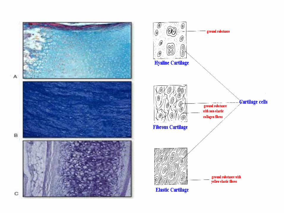
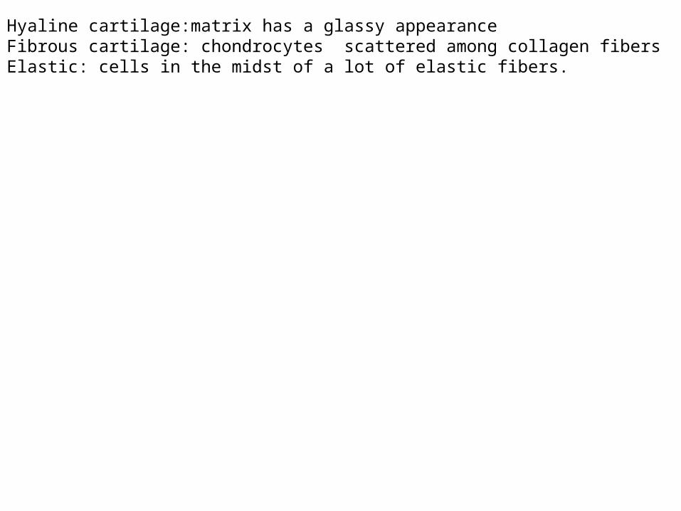
Hyaline cartilage:matrix has a glassy appearanceFibrous cartilage: chondrocytes scattered among collagen fibersElastic: cells in the midst of a lot of elastic fibers.
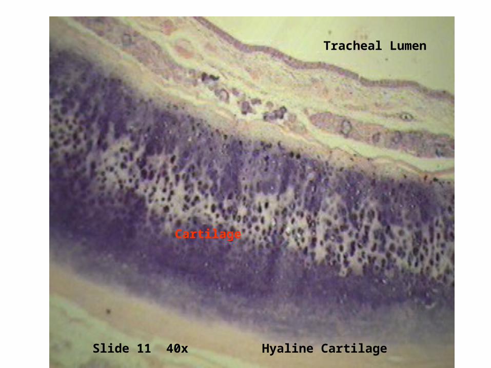
Slide 11 40x Hyaline Cartilage
Cartilage
Tracheal Lumen
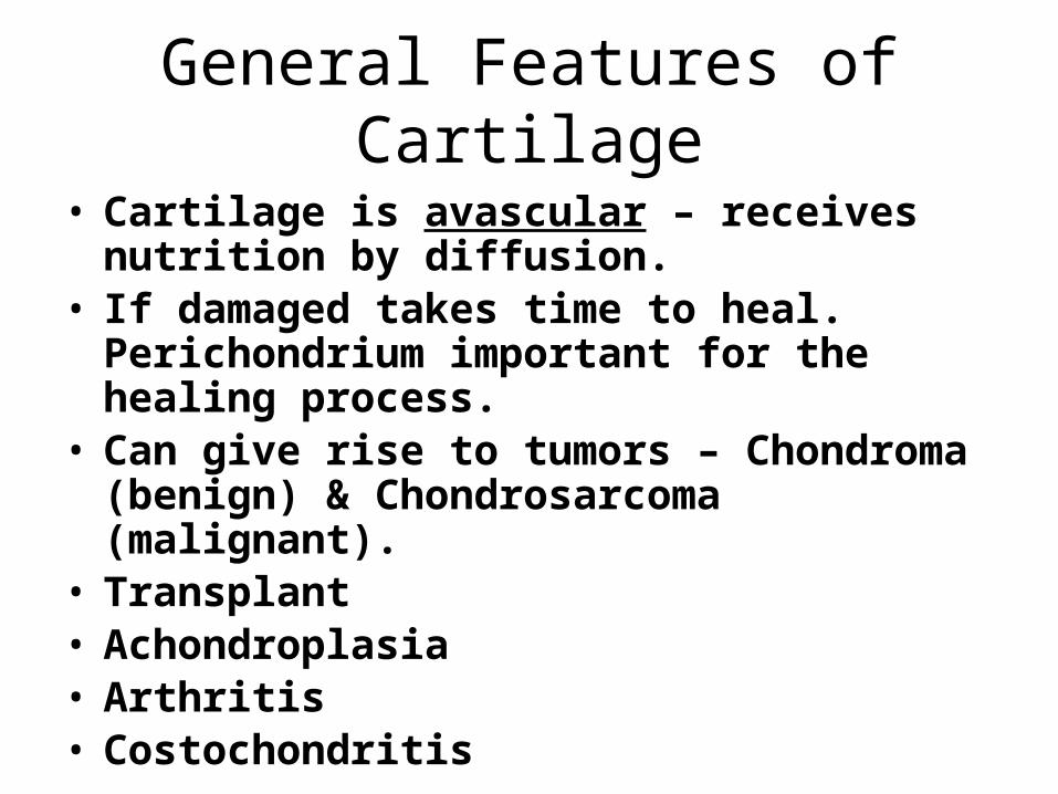
General Features of Cartilage
• Cartilage is avascular – receives nutrition by diffusion.
• If damaged takes time to heal. Perichondrium important for the healing process.
• Can give rise to tumors – Chondroma (benign) & Chondrosarcoma (malignant).
• Transplant• Achondroplasia • Arthritis• Costochondritis