Embed Size (px)
Citation preview
Histopathological Analysis of Salmonella ChronicCarriage in the Mouse Hepatopancreatobiliary SystemGeoffrey Gonzalez-Escobedo1, Krista M. D. La Perle2, John S. Gunn1*
1 Departments of Microbiology and Microbial Infection and Immunity, Center for Microbial Interface Biology, The Ohio State University, Columbus, Ohio, UnitedStates of America, 2 Department of Veterinary Biosciences, Comparative Pathology and Mouse Phenotyping Shared Resource, The Ohio State University,Columbus, Ohio, United States of America
Abstract
Salmonella Typhi asymptomatic chronic carriage represents a challenge for the diagnosis and prevention of typhoidfever in endemic areas. Such carriers are thought to be reservoirs for further spread of the disease. Gallbladdercarriage has been demonstrated to be mediated by biofilm formation on gallstones and by intracellular persistence inthe gallbladder epithelium of mice. In addition, both gallstones and chronic carriage have been associated withchronic inflammation and the development of gallbladder carcinoma. However, the pathogenic relationship betweentyphoid carriage and the development of pre-malignant and/or malignant lesions in the hepatopancreatobiliarysystem as well as the host-pathogen interactions occurring during chronic carriage remains unclear. In this study, wemonitored the histopathological features of chronic carriage up to 1 year post-infection. Chronic cholecystitis andhepatitis ranging from mild to severe were present in infected mice regardless of the presence of gallstones. Biliaryepithelial hyperplasia was observed more commonly in the gallbladder of mice with gallstones (uninfected orinfected). However, pre-malignant lesions, atypical hyperplasia and metaplasia of the gallbladder and exocrinepancreas, respectively, were only associated with chronic Salmonella carriage. This study has implications regardingthe role of Salmonella chronic infection and inflammation in the development of pre-malignant lesions in theepithelium of the gallbladder and pancreas that could lead to oncogenesis.
Citation: Gonzalez-Escobedo G, La Perle KMD, Gunn JS (2013) Histopathological Analysis of Salmonella Chronic Carriage in the MouseHepatopancreatobiliary System. PLoS ONE 8(12): e84058. doi:10.1371/journal.pone.0084058
Editor: Herbert B. Tanowitz, Albert Einstein College of Medicine, United States of America
Received September 25, 2013; Accepted November 18, 2013; Published December 12, 2013
Copyright: © 2013 Gonzalez-Escobedo et al. This is an open-access article distributed under the terms of the Creative Commons Attribution License,which permits unrestricted use, distribution, and reproduction in any medium, provided the original author and source are credited.
Funding: This work was supported by a Pelotonia Graduate Fellowship (http://cancer.osu.edu/research/researcheducation/pelotoniafellowshipprogram/pages/index.aspx?ref=go) and a grant from the US National Institutes of Health (AI066208). The funders had no role in study design, data collection andanalysis, decision to publish, or preparation of the manuscript.
Competing interests: The authors have declared that no competing interests exist.
* E-mail: [email protected]
Introduction
Typhoid or enteric fever, caused primarily by Salmonellaenterica subsp. enterica serovar Typhi (S. Typhi), is a humansystemic disease that is responsible for an estimated 21 millionnew infections per year resulting in approximately 200,000deaths worldwide [1]. It is an important health problem indeveloping countries and poses a significant risk to travelers.After ingestion through contaminated water or food, bacteriacross the intestinal epithelial barrier, migrate into themesenteric lymph nodes, replicate in the reticulo-endothelialsystem and spread systemically producing significantinflammation and acute disease [2-6] with life-threateningcomplications including intestinal hemorrhage and perforation,septicemia and meningitis [7-9]. During this systemic infection,S. Typhi can reach the gallbladder from the liver and establishan acute infection with inflammation (cholecystitis) orchronically persist in this organ. As clinical evidence,inflammation of the gallbladder and bile ducts as well as
sonographic gallbladder abnormalities have been reported inacute and chronic typhoid fever patients [7,10-13]. Althoughhepatomegaly is encountered in approximately 30%-50% oftyphoid patients with or without clinical manifestations [6,14],severe hepatic involvement concomitant with acute hepatitis isseen in 1-26% of typhoid fever patients [15] and the mortalityrate due to typhoid hepatitis is reported to be between20%-33% [16,17].
It is estimated that between 3-5% of typhoid fever patientsbecome chronic carriers with the gallbladder being the primarysite of carriage [7,18,19]. Because S. Typhi is a human specificpathogen, these carriers serve as a critical reservoir for furtherspread of the disease through bacterial shedding in feces,which is a sporadic and intermittent event [10,20]. Chronicinfections can persist for decades and although highlycontagious, they are typically asymptomatic, makingidentification of carriers within a population difficult [21,22].
Particularly in areas of high endemicity, the carrier state hasbeen highly associated with pre-existing hepatobiliary disease
PLOS ONE | www.plosone.org 1 December 2013 | Volume 8 | Issue 12 | e84058
including cholelithiasis (presence of gallstones in thegallbladder), biliary obstruction, intrahepatic cholestasis due toCaroli’s disease, biliary cirrhosis, hepatic hematoma,echinococcal cysts and amoebic abscesses [7,23].Approximately 80-90% of chronically infected carriers havegallstones [24-28]. We have shown that Salmonella can formbiofilms on the surface of cholesterol gallstones in thegallbladder of mice and human carriers, and this biofilmformation has been demonstrated to be a mechanism ofpersistence and chronic colonization in the gallbladder [29].The biofilm state can alter the host-pathogen interaction and isoften associated with a reduction of the host inflammatoryresponse that has been referred to as a “silent chronicinflammation” [30].
In addition to the complications related to the acute phase ofthe disease, especially in the ileum and lymph organs [9],typhoid carriage complications include chronic hepatitis, acuteor chronic cholecystitis, cholangitis, chronic diarrhea and rarely,pancreatitis [6,27]. However, the development of gallbladdercancer is the most severe complication associated with chroniccarriage. Gallbladder cancer is the fifth most commonmalignancy of the gastrointestinal tract and the most commonand aggressive type among the biliary tract malignancies [31].Unfortunately, because of the delayed clinical presentationrelative to pathologic progression, most of the gallbladdercarcinomas are in an advanced stage when diagnosed andmetastasis to the liver and regional lymph nodes are common[32]. Although infrequent in most Western countries, suchcancers are highly prevalent in Chile, India and Pakistan[31,33] where there is a coincident increase in gallstonedisease and typhoid fever [33,34].
Chronic carriers have an approximately 8-14 fold increasedrisk of developing gallbladder carcinoma and approximately150-fold increased risk of developing hepatobiliary carcinomathan non-carriers [34-41]. Moreover, among patients withgallstones, the chronic typhoid carrier state was shown to bethe primary independent risk factor for the development ofgallbladder cancer [34]. Mortality in typhoid carriers as a resultof hepatobiliary carcinomas has been reported to be between3-6% [42]. It has been hypothesized that bacterial degradationof bile salts and chronic cholecystitis related to gallstonespromotes gallbladder carcinomas [43]. In addition togallbladder carcinomas, typhoid carrier patients have increasedrisks of pancreatic carcinoma [5,27,36].
In this study, we demonstrate that Salmonella not onlypersist in the gallbladder and liver of chronically infected micebut also cause chronic-active inflammation that can vary frommild to severe. In addition, chronically infected mice showedepithelial changes such as atypical hyperplasia/dysplasia andmetaplasia in the gallbladder and pancreas as early as 3months post-infection. Although gallstone disease andsubsequent chronic inflammation are known risk factors for thedevelopment of human gallbladder and pancreas cancer[33,44,45], this is the first prospective study that describes theinflammation patterns and epithelial changes occurring duringSalmonella chronic carriage in the absence or presence ofgallstones.
Materials and Methods
Ethics StatementMice were housed and used in strict accordance with
guidelines established by The Ohio State UniversityInstitutional Animal Care and Use Committee (IACUC), and allefforts were made to minimize animal suffering. The workperformed in this study was approved by the OSU IACUC.
The Ohio State University Animal Care and Use Program isaccredited by The Association for the Assessment andAccreditation of Laboratory Animal Care International(AAALAC). The protocol identification number is 2009A0057.All research activities conform to the statutes of the AnimalWelfare Act and the guidelines of the Public Health Service asissued in the Guide for the Care and Use of LaboratoryAnimals (revised 1996).
Bacterial strainsWild-type strain of S. enterica serovar Typhimurium (S.
Typhimurium) ATCC14028 was used in this study. Because S.Typhi is a human-restricted pathogen, in vivo studies of S.Typhi pathogenesis typically involve a mouse model ofinfection using S. Typhimurium. The pathological features ofthe course of mouse infection with S. Typhimurium are similarto those of human infection with S. Typhi [46].
Mice infections and bacteria enumerationWe used a murine model of typhoid chronic infection using
six-eight week old 129X1/SvJ mice (Jackson Laboratories,ME). We previously developed this animal model to studySalmonella chronic infections in the gallbladder thatcorroborated parallel human studies [29]. S. Typhimurium hasbeen demonstrated to persist in the tissues of this mouse strainup to 1 year post-infection [47], but does not typically result in alethal infection. This is due in part to the presence of a wild-type copy of the gene encoding the natural resistance-associated macrophage protein 1 (Nramp1 or Slc11a1).NRAMP1 is a crucial factor in controlling the replication ofintracellular bacteria [48]. It exerts this role by stimulatingexpression of lipocalin-2, which in turn scavenges iron-loadedbacteria siderophores and mediates iron efflux frommacrophages [49].
Female 129X/SvJ mice (n=160) were fed a normal diet(Harlan laboratories, IN) (n=80) or a lithogenic diet containing1% cholesterol (Sigma, MO) and 0.5% cholic acid (Sigma)(n=80) for nine weeks to induce cholesterol gallstonesformation. Mice were inoculated intraperitoneally with 104 S.Typhimurium or left uninfected as controls. For the remainingperiod, all mice were fed a normal diet (Harlan laboratories).Mice were sacrificed at 3, 6, 9 and 12 months post-infection(mpi). Thus, each time point comprised four groups (n=10 pergroup): uninfected mice fed a normal diet (group 1), uninfectedmice fed a lithogenic diet (group 2), Salmonella infected micefed a normal diet (group 3) and Salmonella infected mice fed alithogenic diet (group 4). Spleen and feces from all mice ofeach group (n=10) and liver, pancreas, gallbladder, bile,gallstones from 3 mice of each group were homogenizedand/or diluted in 1X phosphate buffered saline (PBS) for
Histopathological Analysis of Salmonella Carriage
PLOS ONE | www.plosone.org 2 December 2013 | Volume 8 | Issue 12 | e84058
bacterial enumeration on Salmonella-Shigella agar (DifcoTM,Becton Dickinson, MD). In the rare event when mice fed alithogenic diet did not develop gallstones, they were excludedfrom the study.
Histopathology of the hepatopancreatobiliary system ofchronically infected mice
Gallbladder, liver and pancreas (from the 7 mice thatremained from each group) were fixed in 10% neutral bufferedformalin phosphate (Fisher Scientific, MA) for 72 hours. Fixedtissues from groups 1 and 2 (n=3-5 each) and groups 3 and 4(n=5 each) were randomly selected for further evaluation.Tissues were processed by routine methods and embedded in
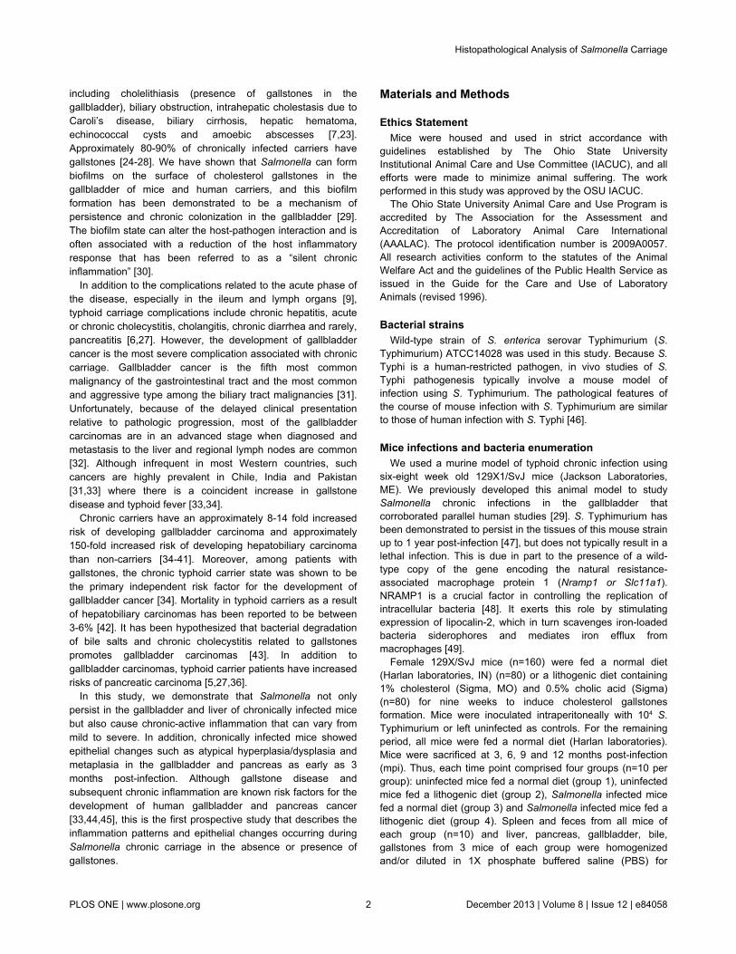
paraffin wax. Sections (4 µm) were stained with hematoxylinand eosin (HE), and evaluated with an Olympus BX45 lightmicroscope with attached DP25 digital camera (B & BMicroscopes Limited, Pittsburg, PA) by a veterinary pathologist(KMDL) certified by the American College of VeterinaryPathologists (ACVP). Inflammatory lesions were scoredaccording to a modified grading scheme [50] (Table 1).
Immunohistochemistry (IHC)Paraffin sections from the tissues noted above were
deparaffinized with xylene and graded ethanols, hydrated todistilled water and stained using the avidin-biotin complex(ABC) method by the OSU Comparative Pathology and Mouse
Table 1. Histopathological scoring of the hepatopancreatobiliary system during chronic Salmonella carriage in mice.
Tissue-specific inflammation
Liver Gallbladder Pancreas
0 Normal 0 Normal 0 Normal
1Focal to multifocal portal/lobular/perivascularlymphohistiocytic ± neutrophilic infiltrates; nonecrosis
1Focal to multifocal serosal/adventitialneutrophilic ± lymphohistiocyticinflammation
1Focal to multifocal lymphocytic aggregates limited tothe interstitium/around blood vessels/within adjacentadipose tissue
2Multifocal to widespread portal/lobularlymphohistiocytic ± neutrophilic inflammation withnecrosis of individual hepatocytes
2Widespread transmural neutrophilic ±lymphohistiocytic inflammation
2
Focal to multifocal neutrophilic ± lymphohistiocyticinflammation with the interstitium or duct with orwithout lymphocytic aggregates in the interstitium/around blood vessels/within adjacent adipose tissue
3Multifocal to widespread portal/lobularlymphohistiocytic ± neutrophilic inflammation withfocally extensive hepatocellular necrosis
3
Necrosuppurative inflammation of lumen± wall with or without transmuralneutrophilic ± lymphohistiocyticinflammation
3
Necrosuppurative inflammation obliterating ducts/vessels with or without lymphocytic aggregates inthe interstitium/around blood vessels/within adjacentadipose tissue
4Multifocal to widespread portal/lobularlymphohistiocytic ± neutrophilic inflammation withMF coalescing areas of hepatocellular necrosis
Applicable to all tissues
Hyalinosis Bacteria visible by HE staining Vascular changesAtypical lymphocytic aggregates& mitotic figures
0 Absent 0 No 0 Absent 0 Absent
1
Focal to multifocalepithelial cytoplasmichyaline material ±crystals; no epithelialhyperplasia
1 Yes 1Fibrin thrombosis ± dystrophicmineralization
1 Focal
2
Multifocal epithelialcytoplasmic hyalinematerial ± crystals withassociated epithelialhyperplasia
2
Fibrinoid vascular degeneration ±myointimal hypertrophy ± muralinflammation with or without thrombosis/dystrophic mineralization
2 Multifocal
3
Widespread to diffuseepithelial cytoplasmichyaline material ± crystlaswith associated epithelialhyperplasia
Modified from Fadl et al. [50]doi: 10.1371/journal.pone.0084058.t001
Histopathological Analysis of Salmonella Carriage
PLOS ONE | www.plosone.org 3 December 2013 | Volume 8 | Issue 12 | e84058
Phenotyping Shared Resource. Briefly, sections were treatedwith proteinase K for 5 min, rinsed, treated with hydrogenperoxide for 10 min, rinsed and blocked with serum-free proteinfor 10 min, incubated for 30 min with anti-LPS Salmonella(1/500 dil.; Novus Biologicals, CO), and rinsed and incubatedfor 30 min with mouse adsorbed biotinylated rabbit anti-mouseantibody (1/1000 dil.) (Vector laboratories, CA). Samples wereincubated with Vector RTU ABC Elite complex for 30 min,rinsed and incubated with chromagen (DAB) for 5 min,counterstained with hematoxylin, rinsed and treated with 1%ammonium hydroxide, dehydrated in ethanol, cleared in xyleneand mounted on coverslips.
Statistical AnalysisStudent’s t-test analysis was performed to detect statistically
significant differences between means of bacteria enumerationand of histology scores (p<0.5). This was performed bycomparison between experimental groups (± infected, ±gallstones) and between time points.
Results
Salmonella was recovered at all time points from thefeces and spleens of mice without gallstones, butinconsistently observed in other tissues/fluids.
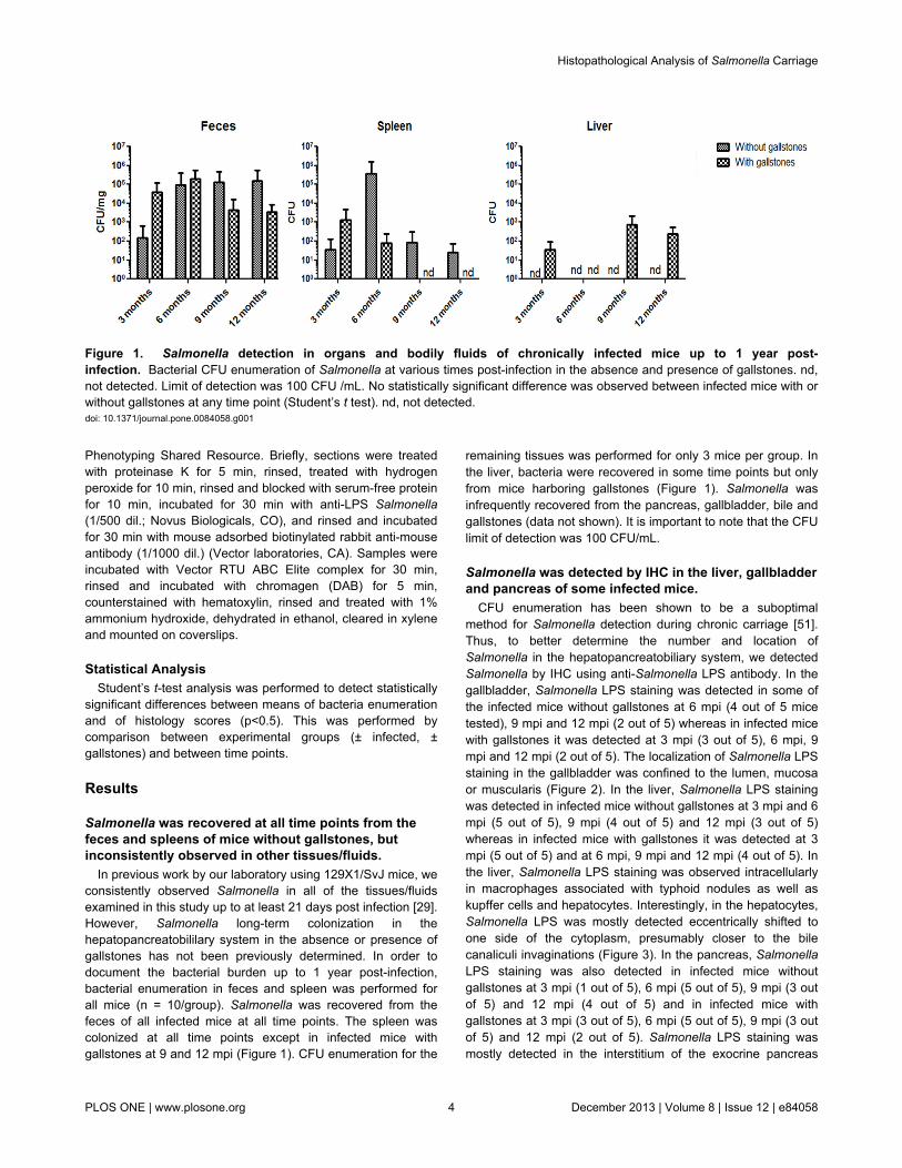
In previous work by our laboratory using 129X1/SvJ mice, weconsistently observed Salmonella in all of the tissues/fluidsexamined in this study up to at least 21 days post infection [29].However, Salmonella long-term colonization in thehepatopancreatobililary system in the absence or presence ofgallstones has not been previously determined. In order todocument the bacterial burden up to 1 year post-infection,bacterial enumeration in feces and spleen was performed forall mice (n = 10/group). Salmonella was recovered from thefeces of all infected mice at all time points. The spleen wascolonized at all time points except in infected mice withgallstones at 9 and 12 mpi (Figure 1). CFU enumeration for the
remaining tissues was performed for only 3 mice per group. Inthe liver, bacteria were recovered in some time points but onlyfrom mice harboring gallstones (Figure 1). Salmonella wasinfrequently recovered from the pancreas, gallbladder, bile andgallstones (data not shown). It is important to note that the CFUlimit of detection was 100 CFU/mL.
Salmonella was detected by IHC in the liver, gallbladderand pancreas of some infected mice.
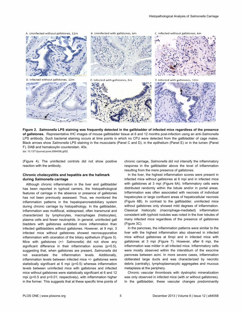
CFU enumeration has been shown to be a suboptimalmethod for Salmonella detection during chronic carriage [51].Thus, to better determine the number and location ofSalmonella in the hepatopancreatobiliary system, we detectedSalmonella by IHC using anti-Salmonella LPS antibody. In thegallbladder, Salmonella LPS staining was detected in some ofthe infected mice without gallstones at 6 mpi (4 out of 5 micetested), 9 mpi and 12 mpi (2 out of 5) whereas in infected micewith gallstones it was detected at 3 mpi (3 out of 5), 6 mpi, 9mpi and 12 mpi (2 out of 5). The localization of Salmonella LPSstaining in the gallbladder was confined to the lumen, mucosaor muscularis (Figure 2). In the liver, Salmonella LPS stainingwas detected in infected mice without gallstones at 3 mpi and 6mpi (5 out of 5), 9 mpi (4 out of 5) and 12 mpi (3 out of 5)whereas in infected mice with gallstones it was detected at 3mpi (5 out of 5) and at 6 mpi, 9 mpi and 12 mpi (4 out of 5). Inthe liver, Salmonella LPS staining was observed intracellularlyin macrophages associated with typhoid nodules as well askupffer cells and hepatocytes. Interestingly, in the hepatocytes,Salmonella LPS was mostly detected eccentrically shifted toone side of the cytoplasm, presumably closer to the bilecanaliculi invaginations (Figure 3). In the pancreas, SalmonellaLPS staining was also detected in infected mice withoutgallstones at 3 mpi (1 out of 5), 6 mpi (5 out of 5), 9 mpi (3 outof 5) and 12 mpi (4 out of 5) and in infected mice withgallstones at 3 mpi (3 out of 5), 6 mpi (5 out of 5), 9 mpi (3 outof 5) and 12 mpi (2 out of 5). Salmonella LPS staining wasmostly detected in the interstitium of the exocrine pancreas
Figure 1. Salmonella detection in organs and bodily fluids of chronically infected mice up to 1 year post-infection. Bacterial CFU enumeration of Salmonella at various times post-infection in the absence and presence of gallstones. nd,not detected. Limit of detection was 100 CFU /mL. No statistically significant difference was observed between infected mice with orwithout gallstones at any time point (Student’s t test). nd, not detected.doi: 10.1371/journal.pone.0084058.g001
Histopathological Analysis of Salmonella Carriage
PLOS ONE | www.plosone.org 4 December 2013 | Volume 8 | Issue 12 | e84058
(Figure 4). The uninfected controls did not show positivereaction with the antibody.
Chronic cholecystitis and hepatitis are the hallmarkduring Salmonella carriage
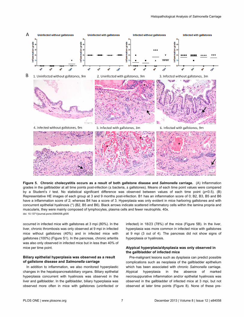
Although chronic inflammation in the liver and gallbladderhas been reported in typhoid carriers, the histopathologicalfeatures of carriage in the absence or presence of gallstoneshas not been previously assessed. Thus, we monitored theinflammation patterns in the hepatopancreatobiliary systemduring chronic carriage by histopathology. In the gallbladder,inflammation was multifocal, widespread, often transmural andcharacterized by lymphocytes, macrophages (histiocytes),plasma cells and fewer neutrophils. In general, uninfected gallbladders with gallstones exhibited more inflammation thaninfected gallbladders without gallstones. However, at 9 mpi, 3infected mice without gallstones showed necrosuppurativeinflammation with ulceration of the biliary epithelium (Figure 5).Mice with gallstones (+/- Salmonella) did not show anysignificant difference in their inflammation scores (p<0.5),suggesting that, when gallstones are present, Salmonella didnot exacerbate the inflammation levels. Additionally,inflammation levels between infected mice +/- gallstones werestatistically significant at 6 mpi (p<0.5) whereas inflammationlevels between uninfected mice with gallstones and infectedmice without gallstones were statistically significant at 6 and 12mpi (p<0.5 and p<0.01; respectively), with inflammation higherin the former. This suggests that at these specific time points of
chronic carriage, Salmonella did not intensify the inflammatoryresponse in the gallbladder above the level of inflammationresulting from the mere presence of gallstones.
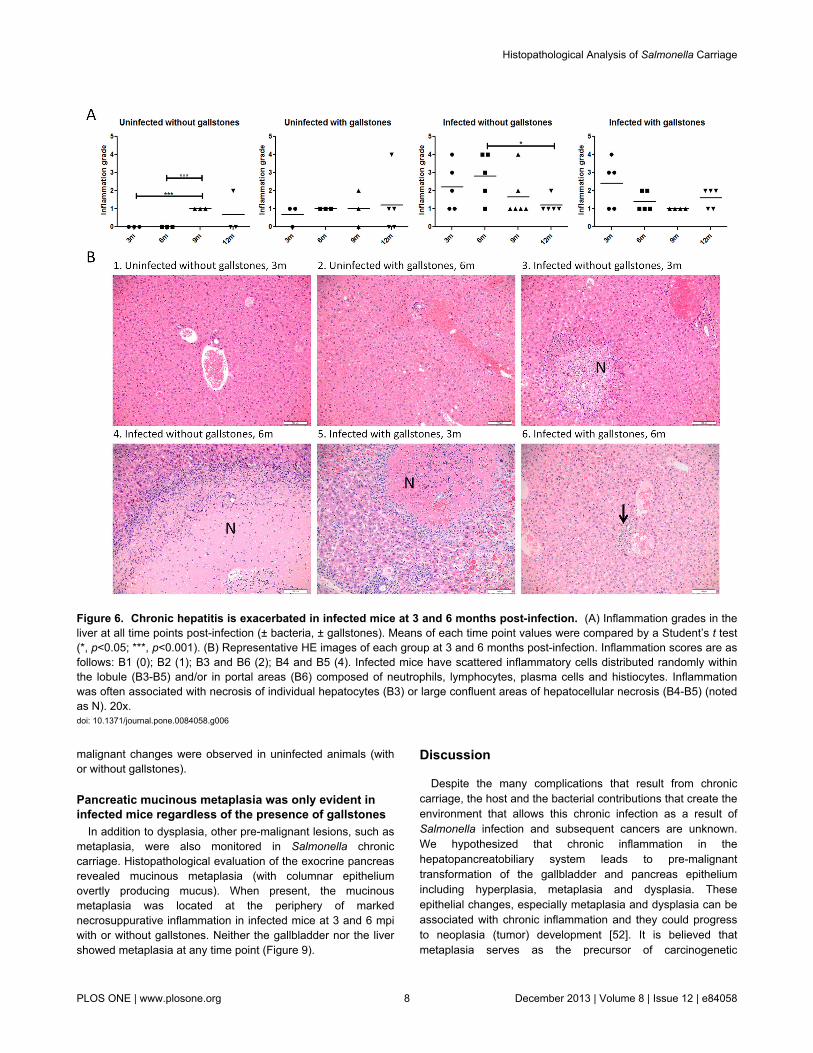
In the liver, the highest inflammation scores were present ininfected mice without gallstones at 6 mpi and in infected micewith gallstones at 3 mpi (Figure 6A). Inflammatory cells weredistributed randomly within the lobule and/or in portal areas.Inflammation was often associated with necrosis of individualhepatocytes or large confluent areas of hepatocellular necrosis(Figure 6B). In contrast to the gallbladder, uninfected micewithout gallstones only showed mild degrees of inflammation.Classical histiocytic (macrophage-mediated) inflammationconsistent with typhoid nodules was noted in the liver lobules ofmany infected mice regardless of the presence of gallstones(Figure 3C).
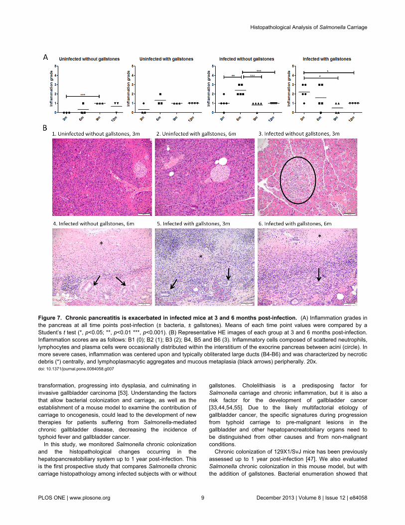
In the pancreas, the inflammation patterns were similar to theliver with the highest inflammation also observed in infectedmice without gallstones at 6mpi and in infected mice withgallstones at 3 mpi (Figure 7). However, after 6 mpi, theinflammation was milder in all infected mice. Inflammatory cellswere mostly observed within the interstitium of the exocrinepancreas between acini. In more severe cases, inflammationobliterated large ducts and was characterized by necroticdebris (centrally), lymphoplasmacytic aggregates and mucousmetaplasia at the periphery.
Chronic vascular thrombosis with dystrophic mineralizationwas only observed in infected mice (with or without gallstones).In the gallbladder, these vascular changes predominantly
Figure 2. Salmonella LPS staining was frequently detected in the gallbladder of infected mice regardless of the presenceof gallstones. Representative IHC images of mouse gallbladder tissue at 6 and 12 months post-infection using an anti-SalmonellaLPS antibody. Such bacterial staining occurs at time points in which no CFU were detected from the gallbladder of cage mates.Black arrows show Salmonella LPS staining in the muscularis (Panel C and D), in the epithelium (Panel E) or in the lumen (PanelF). DAB and hematoxylin counterstain. 40x.doi: 10.1371/journal.pone.0084058.g002
Histopathological Analysis of Salmonella Carriage
PLOS ONE | www.plosone.org 5 December 2013 | Volume 8 | Issue 12 | e84058
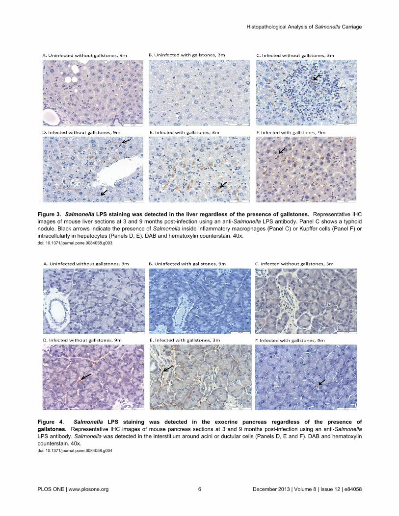
Figure 3. Salmonella LPS staining was detected in the liver regardless of the presence of gallstones. Representative IHCimages of mouse liver sections at 3 and 9 months post-infection using an anti-Salmonella LPS antibody. Panel C shows a typhoidnodule. Black arrows indicate the presence of Salmonella inside inflammatory macrophages (Panel C) or Kupffer cells (Panel F) orintracellularly in hepatocytes (Panels D, E). DAB and hematoxylin counterstain. 40x.doi: 10.1371/journal.pone.0084058.g003
Figure 4. Salmonella LPS staining was detected in the exocrine pancreas regardless of the presence ofgallstones. Representative IHC images of mouse pancreas sections at 3 and 9 months post-infection using an anti-SalmonellaLPS antibody. Salmonella was detected in the interstitium around acini or ductular cells (Panels D, E and F). DAB and hematoxylincounterstain. 40x.doi: 10.1371/journal.pone.0084058.g004
Histopathological Analysis of Salmonella Carriage
PLOS ONE | www.plosone.org 6 December 2013 | Volume 8 | Issue 12 | e84058
occurred in infected mice with gallstones at 3 mpi (80%). In theliver, chronic thrombosis was only observed at 9 mpi in infectedmice without gallstones (40%) and in infected mice withgallstones (100%) (Figure S1). In the pancreas, chronic arteritiswas also only observed in infected mice but in less than 40% ofmice per time point.
Biliary epithelial hyperplasia was observed as a resultof gallstone disease and Salmonella carriage
In addition to inflammation, we also monitored hyperplasticchanges in the hepatopancreatobiliary organs. Biliary epithelialhyperplasia concurrent with hyalinosis was observed in theliver and gallbladder. In the gallbladder, biliary hyperplasia wasobserved more often in mice with gallstones (uninfected or
infected) in 18/23 (78%) of the mice (Figure 5B). In the liver,hyperplasia was more common in infected mice with gallstonesat 9 mpi (3 out of 4). The pancreas did not show signs ofhyperplasia or hyalinosis.
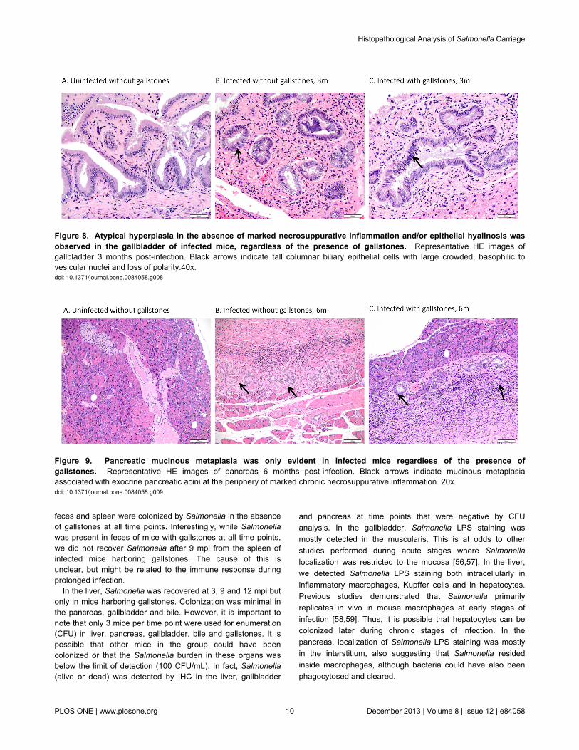
Atypical hyperplasia/dysplasia was only observed inthe gallbladder of infected mice
Pre-malignant lesions such as dysplasia can predict possiblecomplications such as neoplasia of the gallbladder epitheliumwhich has been associated with chronic Salmonella carriage.Atypical hyperplasia in the absence of markednecrosuppurative inflammation and/or epithelial hyalinosis wasobserved in the gallbladder of infected mice at 3 mpi, but notobserved at later time points (Figure 8). None of these pre-
Figure 5. Chronic cholecystitis occurs as a result of both gallstone disease and Salmonella carriage. (A) Inflammationgrades in the gallbladder at all time points post-infection (± bacteria, ± gallstones). Means of each time point values were comparedby a Student’s t test. No statistical significant difference was observed between values of each time point (p<0.5). (B)Representative HE images of each group at 3 and 9 months post-infection. B1 has an inflammation score of 0; B2, B3, B5 and B6have a inflammation score of 2; whereas B4 has a score of 3. Hyperplasia was only evident in mice harboring gallstones and withconcurrent epithelial hyalinosis (*) (B2, B5 and B6). Black arrows indicate scattered inflammatory cells within the lamina propria andmuscularis, they were mainly composed of lymphocytes, plasma cells and fewer neutrophils. 40x.doi: 10.1371/journal.pone.0084058.g005
Histopathological Analysis of Salmonella Carriage
PLOS ONE | www.plosone.org 7 December 2013 | Volume 8 | Issue 12 | e84058
malignant changes were observed in uninfected animals (withor without gallstones).
Pancreatic mucinous metaplasia was only evident ininfected mice regardless of the presence of gallstones
In addition to dysplasia, other pre-malignant lesions, such asmetaplasia, were also monitored in Salmonella chroniccarriage. Histopathological evaluation of the exocrine pancreasrevealed mucinous metaplasia (with columnar epitheliumovertly producing mucus). When present, the mucinousmetaplasia was located at the periphery of markednecrosuppurative inflammation in infected mice at 3 and 6 mpiwith or without gallstones. Neither the gallbladder nor the livershowed metaplasia at any time point (Figure 9).
Discussion
Despite the many complications that result from chroniccarriage, the host and the bacterial contributions that create theenvironment that allows this chronic infection as a result ofSalmonella infection and subsequent cancers are unknown.We hypothesized that chronic inflammation in thehepatopancreatobiliary system leads to pre-malignanttransformation of the gallbladder and pancreas epitheliumincluding hyperplasia, metaplasia and dysplasia. Theseepithelial changes, especially metaplasia and dysplasia can beassociated with chronic inflammation and they could progressto neoplasia (tumor) development [52]. It is believed thatmetaplasia serves as the precursor of carcinogenetic
Figure 6. Chronic hepatitis is exacerbated in infected mice at 3 and 6 months post-infection. (A) Inflammation grades in theliver at all time points post-infection (± bacteria, ± gallstones). Means of each time point values were compared by a Student’s t test(*, p<0.05; ***, p<0.001). (B) Representative HE images of each group at 3 and 6 months post-infection. Inflammation scores are asfollows: B1 (0); B2 (1); B3 and B6 (2); B4 and B5 (4). Infected mice have scattered inflammatory cells distributed randomly withinthe lobule (B3-B5) and/or in portal areas (B6) composed of neutrophils, lymphocytes, plasma cells and histiocytes. Inflammationwas often associated with necrosis of individual hepatocytes (B3) or large confluent areas of hepatocellular necrosis (B4-B5) (notedas N). 20x.doi: 10.1371/journal.pone.0084058.g006
Histopathological Analysis of Salmonella Carriage
PLOS ONE | www.plosone.org 8 December 2013 | Volume 8 | Issue 12 | e84058
transformation, progressing into dysplasia, and culminating ininvasive gallbladder carcinoma [53]. Understanding the factorsthat allow bacterial colonization and carriage, as well as theestablishment of a mouse model to examine the contribution ofcarriage to oncogenesis, could lead to the development of newtherapies for patients suffering from Salmonella-mediatedchronic gallbladder disease, decreasing the incidence oftyphoid fever and gallbladder cancer.
In this study, we monitored Salmonella chronic colonizationand the histopathological changes occurring in thehepatopancreatobiliary system up to 1 year post-infection. Thisis the first prospective study that compares Salmonella chroniccarriage histopathology among infected subjects with or without
gallstones. Cholelithiasis is a predisposing factor forSalmonella carriage and chronic inflammation, but it is also arisk factor for the development of gallbladder cancer[33,44,54,55]. Due to the likely multifactorial etiology ofgallbladder cancer, the specific signatures during progressionfrom typhoid carriage to pre-malignant lesions in thegallbladder and other hepatopancreatobiliary organs need tobe distinguished from other causes and from non-malignantconditions.
Chronic colonization of 129X1/SvJ mice has been previouslyassessed up to 1 year post-infection [47]. We also evaluatedSalmonella chronic colonization in this mouse model, but withthe addition of gallstones. Bacterial enumeration showed that
Figure 7. Chronic pancreatitis is exacerbated in infected mice at 3 and 6 months post-infection. (A) Inflammation grades inthe pancreas at all time points post-infection (± bacteria, ± gallstones). Means of each time point values were compared by aStudent’s t test (*, p<0.05; **, p<0.01 ***, p<0.001). (B) Representative HE images of each group at 3 and 6 months post-infection.Inflammation scores are as follows: B1 (0); B2 (1); B3 (2); B4, B5 and B6 (3). Inflammatory cells composed of scattered neutrophils,lymphocytes and plasma cells were occasionally distributed within the interstitium of the exocrine pancreas between acini (circle). Inmore severe cases, inflammation was centered upon and typically obliterated large ducts (B4-B6) and was characterized by necroticdebris (*) centrally, and lymphoplasmacytic aggregates and mucous metaplasia (black arrows) peripherally. 20x.doi: 10.1371/journal.pone.0084058.g007
Histopathological Analysis of Salmonella Carriage
PLOS ONE | www.plosone.org 9 December 2013 | Volume 8 | Issue 12 | e84058
feces and spleen were colonized by Salmonella in the absenceof gallstones at all time points. Interestingly, while Salmonellawas present in feces of mice with gallstones at all time points,we did not recover Salmonella after 9 mpi from the spleen ofinfected mice harboring gallstones. The cause of this isunclear, but might be related to the immune response duringprolonged infection.
In the liver, Salmonella was recovered at 3, 9 and 12 mpi butonly in mice harboring gallstones. Colonization was minimal inthe pancreas, gallbladder and bile. However, it is important tonote that only 3 mice per time point were used for enumeration(CFU) in liver, pancreas, gallbladder, bile and gallstones. It ispossible that other mice in the group could have beencolonized or that the Salmonella burden in these organs wasbelow the limit of detection (100 CFU/mL). In fact, Salmonella(alive or dead) was detected by IHC in the liver, gallbladder
and pancreas at time points that were negative by CFUanalysis. In the gallbladder, Salmonella LPS staining wasmostly detected in the muscularis. This is at odds to otherstudies performed during acute stages where Salmonellalocalization was restricted to the mucosa [56,57]. In the liver,we detected Salmonella LPS staining both intracellularly ininflammatory macrophages, Kupffer cells and in hepatocytes.Previous studies demonstrated that Salmonella primarilyreplicates in vivo in mouse macrophages at early stages ofinfection [58,59]. Thus, it is possible that hepatocytes can becolonized later during chronic stages of infection. In thepancreas, localization of Salmonella LPS staining was mostlyin the interstitium, also suggesting that Salmonella residedinside macrophages, although bacteria could have also beenphagocytosed and cleared.
Figure 8. Atypical hyperplasia in the absence of marked necrosuppurative inflammation and/or epithelial hyalinosis wasobserved in the gallbladder of infected mice, regardless of the presence of gallstones. Representative HE images ofgallbladder 3 months post-infection. Black arrows indicate tall columnar biliary epithelial cells with large crowded, basophilic tovesicular nuclei and loss of polarity.40x.doi: 10.1371/journal.pone.0084058.g008
Figure 9. Pancreatic mucinous metaplasia was only evident in infected mice regardless of the presence ofgallstones. Representative HE images of pancreas 6 months post-infection. Black arrows indicate mucinous metaplasiaassociated with exocrine pancreatic acini at the periphery of marked chronic necrosuppurative inflammation. 20x.doi: 10.1371/journal.pone.0084058.g009
Histopathological Analysis of Salmonella Carriage
PLOS ONE | www.plosone.org 10 December 2013 | Volume 8 | Issue 12 | e84058
In previous studies, we showed that acute hepatitis andcholecystitis (neutrophilic and histiocytic) occurred up to 2months post-infection [51]. Sonographic findings includingmucosal irregularities, edema with distention and thickening ofthe gallbladder, suggesting cholecystitis, have been reported inacute typhoid fever patients without gallstones [12]. In fact,histiocytic inflammation and granuloma formation occurs in thegallbladder and liver during typhoid fever [27]. In this study,chronic cholecystitis and hepatitis (mostly lymphohistiocyticand plasmacytic) were the predominant findings duringSalmonella carriage up to 1 year post-infection, which has alsobeen reported in human typhoid carriers [23,28,60,61].
Corroborating previous studies using mouse models fed alithogenic diet [62-64], the mere presence of gallstones causedchronic cholecystitis, in general causing more inflammationthan the presence of Salmonella. This suggests that in mostcases, Salmonella chronic colonization does not induce stronginfiltration of immune cells. However, we observed severeinflammation (necrosuppurative) in some infected mice butwithout gallstones, implying that this is a heterogenousprocess. Interestingly, the levels of inflammation were notexacerbated in the gallbladder of infected mice with gallstones,suggesting that Salmonella does not dramatically increase pre-existing inflammation.
In the liver and pancreas, the inflammation patterns weredifferent than in the gallbladder because here, the presence ofSalmonella alone caused more inflammation than the presenceof gallstones alone (especially at 3 and 6 mpi.). This is anexpected observation considering that gallstones are typicallypresent in the gallbladder and bile ducts, and only infrequentlyfound in the pancreatic duct. One of the pathologicalobservations commonly seen in the liver of infected mice wasthe presence of focal/multifocal necrosis and typhoid nodules,which are reported pathological features of typhoid fever alongwith infrequent focal granulomas in the liver and spleen[15,65,66]. Typhoid nodules are primarily aggregates of alteredmacrophages that phagocytose bacteria, erythrocytes anddegenerated lymphocytes, sometimes with central necrosis.They can also contain plasma cells and lymphocytes andatypically, neutrophils [14,65]. Since these nodules are mainlycomprised of altered macrophages, this may indicate thathistiocytic inflammation occurred early during infection (before3 months post-infection in this model). In addition, in the liverwe also observed mineralized chronic vascular thrombi.Venous thrombosis is a consequence of severe cases oftyphoid fever [15].
Acute and chronic pancreatitis, in some cases withabscesses, has been reported in typhoid fever patients[27,67-70]. In this study, we observed lymphohistiocytic andnecrosuppurative inflammation in the pancreas of infected miceat 3 and 6 mpi. In comparison with the gallbladder and liver,chronic inflammation in the pancreas was milder after 6mpi.
Chronic inflammation with oxidative stress caused bypersistent bacterial infection and production of bacterial toxinsand metabolites has been associated with biliarycarcinogenesis [71]. At the molecular level, it has beenproposed that chronic inflammation of the gallbladder leads toan allele-specific mutation with loss of p53 gene heterozygosity
and overexpression of p53 protein [72,73]. This mutation isconsidered to result in malignant transformation of thegallbladder mucosa because p53 is a recognized tumorsuppressor [74]. In addition, cyclooxygenase 2 (COX-2), whichis also associated with gallbladder carcinomas, is also inducedduring gallbladder inflammation [44,75]. Thus, chronicinflammation can lead to molecular disturbances in the cellcycle of the gallbladder mucosa that contribute to gallbladdercancer development [45].
Hyperplastic changes in the epithelium of the gallbladder andbile ducts of the liver were frequently noted in mice harboringgallstones, independent of Salmonella infection. This increasedcell proliferation as a result of cholelithiasis has beenpreviously reported [76,77]. However, typically this was seenconcurrently with hyalinosis in affected tissues and/orinflammation. Hyalinosis is a common lesion in some strains ofmice whereby epithelial cells in various tissues including thenasal cavity, lung, glandular stomach, gallbladder, bile andpancreatic ducts, and ureter accumulate intensely eosinophiliccytoplasm that may also be associated with spiculareosinophilic crystals. It is thought that crystals form due to localincreased concentrations of the chitinase-like protein Chi3l3(formerly Ym-1/Ym-2) secondary to neutrophil degranulationduring repeated episodes of inflammation. The crystals areresistant to degradation, and the inflammation and crystals aresufficient in and of themselves to induce epithelial hyperplasia[78,79]. Biliary hyperplasia in the liver was observed more oftenin infected mice with gallstones at 9 mpi.
Dysplasia is a pre-malignant lesion that can progress toneoplasia. In fact, more than 80% of invasive gallbladdercarcinomas have adjacent areas of dysplasia [53]. Atypicalhyperplasia/dysplasia not associated with chronicnecrosuppurative inflammation or epithelial hyalinosis wasobserved in the gallbladder of infected mice regardless of thepresence of gallstones only at 3 mpi. In fact, this is the first timethat atypical hyperplasia/dysplasia has been shown as a resultof Salmonella chronic infection. It is widely reported thatgallstones are the major risk factor for developing gallbladdercancer [80]. We hypothesized that chronic inflammation of thegallbladder or bile ducts as a result of Salmonella infection mayalso lead to pre-malignant lesions or carcinogenesis.Considering that these proliferative changes are more likely tobe a physiological response to inflammation and that we didnot see atypical hyperplasia/dysplasia after 3 mpi, we cannotpredict if these changes ultimately progress to neoplasia inthese mice. However, this could be due to scarce long term (>3months) Salmonella colonization of the gallbladder in ourmodel under the chosen experimental conditions. Thus, webelieve that in human chronic carriers with permanent bacterialcolonization in the gallbladder, chronic inflammation anddysplasia could occur and progress to neoplasia.
In addition to the epithelial changes observed in thegallbladder, acinar-ductal mucinous metaplasia was observedin the pancreas (and only the pancreas) of infected mice.Acinar-ductal mucinous metaplasia occurred adjacent to aciniat the periphery of necrosuppurative inflammation;morphologically, it is commonly identified as mucinousmetaplastic epithelia mixed with acinar cells in the pancreatic
Histopathological Analysis of Salmonella Carriage
PLOS ONE | www.plosone.org 11 December 2013 | Volume 8 | Issue 12 | e84058
lobules. This type of metaplasia is commonly seen in chronicpancreatitis and in the pancreatic parenchyma adjacent toductal carcinoma [81]. The severe inflammation observed inthe pancreas of infected mice seemed to originate within theducts but then obliterated the ducts and extended into the acini.Mucinous metaplasia is likely an attempt to regenerate the ductbut the acinar cells are also irritated from the inflammation.Although Salmonella was usually detected in the liver duringchronic disease, this persistence did not induce epithelialchanges such as atypical hyperplasia/dysplasia and metaplasiathat were observed in the gallbladder and pancreas,respectively.
In conclusion, chronic inflammation in organs of thehepatopancreatobiliary system was a predominant observationin our model of chronic carriage. Inflammation patterns differedin the gallbladder compared with the liver and pancreas. In thegallbladder, the presence of gallstones caused more robustand ongoing inflammation than the mere presence ofSalmonella. However, in the liver and pancreas, the presenceof Salmonella caused more inflammation. Interestingly, in allorgans, infected mice with gallstones did not show increasedinflammation in comparison with the other groups (except in thepancreas at 3 mpi.). This inflammation could explain theepithelial changes observed in the pancreas (mucinousmetaplasia). In contrast, the atypical hyperplasia observed inthe gallbladder was not associated with inflammation but thiscould have happened before 3 mpi. Although the merepresence of gallstones did not cause these epithelial changes,the low level of Salmonella colonization of these organsmonths post-infection could explain the absence of moredysplastic changes that could better correlate chronic carriage
with oncogenesis. In the future, we will attempt to alter themodel system to permit longer chronic hepatopancreatobiliarycolonization, allowing for histological studies that likely bettercorrelate with changes that occur during long-term humanchronic typhoid carriage.
Supporting Information
Figure S1. Dystrophic mineralization of venous fibrinthrombi was only present in the gallbladder and liver ofinfected mice with gallstones Representative HE liver (A, C)and gallbladder adjacent to the liver (B) at 3 and 9 monthspost-infection, respectively. Note the mineralized thrombi inPanels B and C surrounded by portal aggregates oflymphocytes, plasma cells and neutrophils. 20x.(TIF)
Acknowledgements
We would like to thank Florinda Jaynes and Sarah Chaneyfrom the College of Veterinary Medicine at The Ohio StateUniversity for their contributions to IHC slides preparation andinterpretation, respectively.
Author Contributions
Conceived and designed the experiments: JSG GGE.Performed the experiments: GGE. Analyzed the data: GGEKMDLP. Contributed reagents/materials/analysis tools:KMDLP. Wrote the manuscript: GGE.
References
1. Crump JA, Luby SP, Mintz ED (2004) The global burden of typhoidfever. Bull World Health Organ 82: 346-353. PubMed: 15298225.
2. Jepson MA, Clark MA (2001) The role of M cells in Salmonellainfection. Microbes Infect 3: 1183-1190. doi:10.1016/S1286-4579(01)01478-2. PubMed: 11755406.
3. Vazquez-Torres A, Jones-Carson J, Bäumler AJ, Falkow S, Valdivia Ret al. (1999) Extraintestinal dissemination of Salmonella by CD18-expressing phagocytes. Nature 401: 804-808. doi:10.1038/44593.PubMed: 10548107.
4. Wain J, Diep TS, Ho VA, Walsh AM, Nguyen TT et al. (1998)Quantitation of bacteria in blood of typhoid fever patients andrelationship between counts and clinical features, transmissibility, andantibiotic resistance. J Clin Microbiol 36: 1683-1687. PubMed:9620400.
5. Vladoianu IR, Chang HR, Pechère JC (1990) Expression of hostresistance to Salmonella Typhi and Salmonella Typhimurium: bacterialsurvival within macrophages of murine and human origin. MicrobPathog 8: 83-90. doi:10.1016/0882-4010(90)90072-X. PubMed:2190062.
6. Crum NF (2003) Current trends in typhoid fever. Curr GastroenterolRep 5: 279-286. doi:10.1007/s11894-003-0064-0. PubMed: 12864957.
7. Cohen JI, Bartlett JA, Corey GR (1987) Extra-intestinal manifestationsof Salmonella infections. Medicine (Baltimore) 66: 349-388. PubMed:3306260.
8. Parry CM, Hien TT, Dougan G, White NJ, Farrar JJ (2002) Typhoidfever. N Engl J Med 347: 1770-1782. doi:10.1056/NEJMra020201.PubMed: 12456854.
9. Everest P, Wain J, Roberts M, Rook G, Dougan G (2001) Themolecular mechanisms of severe typhoid fever. Trends Microbiol 9:316-320. doi:10.1016/S0966-842X(01)02067-4. PubMed: 11435104.
10. Vogelsang TM, Boe J (1948) Temporary and chronic carriers ofSalmonella Typhi and Salmonella Paratyphi BJ Hyg (Lond) 46:252-261.
11. Vaishnavi C, Singh S, Kochhar R, Bhasin D, Singh G et al. (2005)Prevalence of Salmonella enterica serovar Typhi in bile and stool ofpatients with biliary diseases and those requiring biliary drainage forother purposes. Jpn J Infect Dis 58: 363-365. PubMed: 16377868.
12. Shetty PB, Broome DR (1998) Sonographic analysis of gallbladderfindings in Salmonella enteric fever. J Ultrasound Med 17: 231-237.PubMed: 9544606.
13. Mateen MA, Saleem S, Rao PC, Reddy PS, Reddy DN (2006)Ultrasound in the diagnosis of typhoid fever. Indian J Pediatr 73:681-685. doi:10.1007/BF02898444. PubMed: 16936362.
14. Ramachandran S, Godfrey JJ, Perera MV (1974) Typhoid hepatitis.JAMA 230: 236-240. doi:10.1001/jama.230.2.236. PubMed: 4213522.
15. Pramoolsinsap C, Viranuvatti V (1998) Salmonella hepatitis. JGastroenterol Hepatol 13: 745-750. doi:10.1111/j.1440-1746.1998.tb00726.x. PubMed: 9715430.
16. Khosla SN (1990) Typhoid hepatitis. Postgrad Med J 66: 923-925. doi:10.1136/pgmj.66.781.923. PubMed: 2267203.
17. Rovito V, Bonanno CA (1982) Salmonella hepatic abscess: an unusualcomplication of the Salmonella carrier state? Am J Gastroenterol 77:338-339. PubMed: 7081193.
18. Levine MM, Black RE, Lanata C (1982) Precise estimation of thenumbers of chronic carriers of Salmonella Typhi in Santiago, Chile, anendemic area. J Infect Dis 146: 724-726. doi:10.1093/infdis/146.6.724.PubMed: 7142746.
19. Merselis JG Jr., Kaye D, Connolly CS, Hook EW (1964) QuantitativeBacteriology of the Typhoid Carrier State. Am J Trop Med Hyg 13:425-429. PubMed: 14159980.
20. Bhan MK, Bahl R, Bhatnagar S (2005) Typhoid and paratyphoid fever.Lancet 366: 749-762. doi:10.1016/S0140-6736(05)67181-4. PubMed:16125594.
21. Sinnott CR, Teall AJ (1987) Persistent gallbladder carriage ofSalmonella Typhi. Lancet 1: 976. PubMed: 2882361.
Histopathological Analysis of Salmonella Carriage
PLOS ONE | www.plosone.org 12 December 2013 | Volume 8 | Issue 12 | e84058
22. Shpargel JS, Berardi RS, Lenz D (1985) Salmonella Typhi carrier state52 years after illness with typhoid fever: a case study. Am J InfectControl 13: 122-123. doi:10.1016/S0196-6553(85)80013-4. PubMed:3849271.
23. Gosbell I, Jones PD, Matthews A, Yeo B (1995) Surgical presentationof hepatobiliary disease due to Salmonella Typhi. Aust N Z J Surg 65:898-899. doi:10.1111/j.1445-2197.1995.tb00588.x. PubMed: 8611118.
24. Schiøler H, Christiansen ED, Høybye G, Rasmussen SN, Greibe J(1983) Biliary calculi in chronic Salmonella carriers and healthycontrols: a controlled study. Scand J Infect Dis 15: 17-19. PubMed:6405479.
25. Karaki K, Matsubara Y (1984) Surgical treatment of chronic biliarytyphoid and paratyphoid carriers. Nippon Shokakibyo Gakkai Zasshi81: 2978-2985. PubMed: 6442374.
26. Lai CW, Chan RC, Cheng AF, Sung JY, Leung JW (1992) Commonbile duct stones: a cause of chronic salmonellosis. Am J Gastroenterol87: 1198-1199. PubMed: 1519582.
27. Vaishnavi C, Kochhar R, Singh G, Kumar S, Singh S et al. (2005)Epidemiology of typhoid carriers among blood donors and patients withbiliary, gastrointestinal and other related diseases. Microbiol Immunol49: 107-112. doi:10.1111/j.1348-0421.2005.tb03709.x. PubMed:15722595.
28. Scott AJ (1971) Bacteria and disease of the biliary tract. Gut 12:487-492. doi:10.1136/gut.12.6.487. PubMed: 4933136.
29. Crawford RW, Rosales-Reyes R, Mde Ramirez-Aguilar L, Chapa-Azuela O, Alpuche-Aranda C et al. (2010) Gallstones play a significantrole in Salmonella spp. gallbladder colonization and carriage. Proc NatlAcad Sci U S A 107: 4353-4358. doi:10.1073/pnas.1000862107.PubMed: 20176950. Available online at: doi:10.1073/pnas.1000862107Available online at: PubMed: 20176950
30. Cappelli G, Tetta C, Canaud B (2005) Is biofilm a cause of silentchronic inflammation in haemodialysis patients? A fascinating workinghypothesis. Nephrol Dial Transplant 20: 266-270. doi:10.1093/ndt/20.suppl_5.v266. PubMed: 15647310.
31. Miller G, Jarnagin WR (2008) Gallbladder carcinoma. Eur J Surg Oncol34: 306-312. doi:10.1016/j.ejso.2007.07.206. PubMed: 17964753.
32. Lazcano-Ponce EC, Miquel JF, Muñoz N, Herrero R, Ferrecio C et al.(2001) Epidemiology and molecular pathology of gallbladder cancer.CA Cancer J Clin 51: 349-364. doi:10.3322/canjclin.51.6.349. PubMed:11760569.
33. Randi G, Franceschi S, La Vecchia C (2006) Gallbladder cancerworldwide: geographical distribution and risk factors. Int J Cancer 118:1591-1602. doi:10.1002/ijc.21683. PubMed: 16397865.
34. Dutta U, Garg PK, Kumar R, Tandon RK (2000) Typhoid carriersamong patients with gallstones are at increased risk for carcinoma ofthe gallbladder. Am J Gastroenterol 95: 784-787. doi:10.1111/j.1572-0241.2000.01860.x. PubMed: 10710075.
35. Shukla VK, Singh H, Pandey M, Upadhyay SK, Nath G (2000)Carcinoma of the gallbladder--is it a sequel of typhoid? Dig Dis Sci 45:900-903. doi:10.1023/A:1005564822630. PubMed: 10795752.
36. Caygill CP, Hill MJ, Braddick M, Sharp JC (1994) Cancer mortality inchronic typhoid and paratyphoid carriers. Lancet 343: 83-84. doi:10.1016/S0140-6736(94)90816-8. PubMed: 7903779.
37. Nath G, Singh H, Shukla VK (1997) Chronic typhoid carriage andcarcinoma of the gallbladder. Eur J Cancer Prev 6: 557-559. doi:10.1097/00008469-199712000-00011. PubMed: 9496458.
38. Nath G, Singh YK, Kumar K, Gulati AK, Shukla VK et al. (2008)Association of carcinoma of the gallbladder with typhoid carriage in atyphoid endemic area using nested PCR. J Infect Dev Ctries 2:302-307. PubMed: 19741293.
39. Chang HR, Dulloo AG, Vladoianu IR, Piguet PF, Arsenijevic D et al.(1992) Fish oil decreases natural resistance of mice to infection withSalmonella Typhimurium. Metabolism 41: 1-2. doi:10.1016/0026-0495(92)90181-9. PubMed: 1538638.
40. Csendes A, Fernandez M, Uribe P (1975) Bacteriology of thegallbladder bile in normal subjects. Am J Surg 129: 629-631. doi:10.1016/0002-9610(75)90334-7. PubMed: 805546.
41. el-Zayadi A, Ghoneim M, Kabil SM, el Tawil A, Sherif A et al. (1991)Bile duct carcinoma in Egypt: possible etiological factors.Hepatogastroenterology 38: 337-340. PubMed: 1657750.
42. Welton JC, Marr JS, Friedman SM (1979) Association betweenhepatobiliary cancer and typhoid carrier state. Lancet 1: 791-794.PubMed: 86039.
43. Kumar S (2006) Infection as a risk factor for gallbladder cancer. J SurgOncol 93: 633-639. doi:10.1002/jso.20530. PubMed: 16724347.
44. Tazuma S, Kajiyama G (2001) Carcinogenesis of malignant lesions ofthe gall bladder. The impact of chronic inflammation and gallstones.Langenbecks Arch Surg 386: 224-229. doi:10.1007/s004230100220.PubMed: 11382326.
45. Sośnik K, Sośnik H (2005) Some aspects of gallbladdercarcinogenesis. Wiad Lek 58: 678-681. PubMed: 16594482.
46. Santos RL, Zhang S, Tsolis RM, Kingsley RA, Adams LG et al. (2001)Animal models of Salmonella infections: enteritis versus typhoid fever.Microbes Infect 3: 1335-1344. doi:10.1016/S1286-4579(01)01495-2.PubMed: 11755423.
47. Monack DM, Bouley DM, Falkow S (2004) Salmonella Typhimuriumpersists within macrophages in the mesenteric lymph nodes ofchronically infected Nramp1+/+ mice and can be reactivated by IFNγneutralization. J Exp Med 199: 231-241.
48. Forbes JR, Gros P (2001) Divalent-metal transport by NRAMP proteinsat the interface of host-pathogen interactions. Trends Microbiol 9:397-403. doi:10.1016/S0966-842X(01)02098-4. PubMed: 11514223.
49. Fritsche G, Nairz M, Libby SJ, Fang FC, Weiss G (2012) Slc11a1(Nramp1) impairs growth of Salmonella enterica serovar Typhimuriumin macrophages via stimulation of lipocalin-2 expression. J Leukoc Biol92: 353-359. doi:10.1189/jlb.1111554. PubMed: 22706314.
50. Fadl AA, Sha J, Klimpel GR, Olano JP, Niesel DW et al. (2005) Mureinlipoprotein is a critical outer membrane component involved inSalmonella enterica serovar Typhimurium systemic infection. InfectImmun 73: 1081-1096. doi:10.1128/IAI.73.2.1081-1096.2005. PubMed:15664952. Available online at: doi:10.1128/IAI.73.2.1081-1096.2005Available online at: PubMed: 15664952
51. Gonzalez-Escobedo G, Gunn JS (2013) The Gallbladder Epithelium asa Niche for Chronic Salmonella Carriage. Infect Immun 8: 2920-2930.
52. Haschek W, Wallig M, Rousseaux C (2010) Fundamentals ofToxicologic Pathology. London, UK: Elsevier.
53. Monga S (2011) Molecular Pathology of Liver Diseases. New York,USA: Springer.
54. Pilgrim CH, Groeschl RT, Christians KK, Gamblin TC (2013) Modernperspectives on factors predisposing to the development of gallbladdercancer. HPB Oxford.
55. Rustagi T, Dasanu CA (2012) Risk factors for gallbladder cancer andcholangiocarcinoma: similarities, differences and updates. JGastrointest Cancer 43: 137-147. doi:10.1007/s12029-011-9284-y.PubMed: 21597894.
56. Menendez A, Arena ET, Guttman JA, Thorson L, Vallance BA et al.(2009) Salmonella infection of gallbladder epithelial cells drives localinflammation and injury in a model of acute typhoid fever. J Infect Dis200: 1703-1713. doi:10.1086/646608. PubMed: 19852670.
57. Knodler LA, Vallance BA, Celli J, Winfree S, Hansen B et al. (2010)Dissemination of invasive Salmonella via bacterial-induced extrusion ofmucosal epithelia. Proc Natl Acad Sci U S A 107: 17733-17738. doi:10.1073/pnas.1006098107. PubMed: 20876119.
58. Richter-Dahlfors A, Buchan AM, Finlay BB (1997) Murine salmonellosisstudied by confocal microscopy: Salmonella Typhimurium residesintracellularly inside macrophages and exerts a cytotoxic effect onphagocytes in vivo. J Exp Med 186: 569-580. doi:10.1084/jem.186.4.569. PubMed: 9254655.
59. Nnalue NA, Shnyra A, Hultenby K, Lindberg AA (1992) SalmonellaCholeraesuis and Salmonella Typhimurium associated with liver cellsafter intravenous inoculation of rats are localized mainly in Kupffer cellsand multiply intracellularly. Infect Immun 60: 2758-2768. PubMed:1612743.
60. Axelrod L, Munster AM, O'Brien TF (1971) Typhoid cholecystitis andgallbladder carcinoma after interval of 67 years. JAMA 217: 83. doi:10.1001/jama.217.1.83b. PubMed: 5108716.
61. Roa I, Ibacache G, Carvallo J, Melo A, Araya J et al. (1999)Microbiological study of gallbladder bile in a high risk zone forgallbladder cancer. Rev Med Chil 127: 1049-1055. PubMed: 10752267.
62. Lavoie B, Nausch B, Zane EA, Leonard MR, Balemba OB et al. (2012)Disruption of gallbladder smooth muscle function is an early feature inthe development of cholesterol gallstone disease. NeurogastroenterolMotil 24: e313-e324. doi:10.1111/j.1365-2982.2012.01935.x. PubMed:22621672.
63. Ichikawa H, Imano M, Takeyama Y, Shiozaki H, Ohyanagi H (2009)Involvement of osteopontin as a core protein in cholesterol gallstoneformation. J Hepatobiliary Pancreat Surg 16: 197-203. doi:10.1007/s00534-009-0043-4. PubMed: 19214371.
64. Rege RV, Prystowsky JB (1998) Inflammation and a thickened mucuslayer in mice with cholesterol gallstones. J Surg Res 74: 81-85. doi:10.1006/jsre.1997.5213. PubMed: 9536979.
65. Khosla SN (2008) Typhoid Fever: Its Cause, Transmission AndPrevention. New Delhi, India: Atlantic Publishers & Distributors. 245 p
66. Mert A, Tabak F, Ozaras R, Ozturk R, Aki H et al. (2004) Typhoid feveras a rare cause of hepatic, splenic, and bone marrow granulomas.Intern Med 43: 436-439. doi:10.2169/internalmedicine.43.436. PubMed:15206561.
Histopathological Analysis of Salmonella Carriage
PLOS ONE | www.plosone.org 13 December 2013 | Volume 8 | Issue 12 | e84058
67. Hermans P, Gerard M, De Wit S, Van Laethem Y (1990) Pancreatitisand typhoid fever. Am J Med 89: 546-547. doi:10.1016/0002-9343(90)90401-X. PubMed: 2220896.
68. Kadappu KK, Rao PV, Srinivas N, Shastry BA (2002) Pancreatitis inenteric fever. Indian J Gastroenterol 21: 32-33. PubMed: 11871836.
69. Khan FY, Al-Ani A, Ali HA (2009) Typhoid rhabdomyolysis with acuterenal failure and acute pancreatitis: a case report and review of theliterature. Int J Infect Dis 13: e282-e285. doi:10.1016/j.ijid.2008.11.009.PubMed: 19147385.
70. Parenti DM, Steinberg W, Kang P (1996) Infectious causes of acutepancreatitis. Pancreas 13: 356-371. doi:10.1097/00006676-199611000-00005. PubMed: 8899796.
71. Hornick RB, Greisman SE, Woodward TE, DuPont HL, Dawkins AT etal. (1970) Typhoid fever: pathogenesis and immunologic control. NEngl J Med 283: 686-691. doi:10.1056/NEJM197009242831306.PubMed: 4916913.
72. Wistuba II, Sugio K, Hung J, Kishimoto Y, Virmani AK et al. (1995)Allele-specific mutations involved in the pathogenesis of endemicgallbladder carcinoma in Chile. Cancer Res 55: 2511-2515. PubMed:7780959.
73. Wee A, Teh M, Raju GC (1994) Clinical importance of p53 protein ingall bladder carcinoma and its precursor lesions. J Clin Pathol 47:453-456. doi:10.1136/jcp.47.5.453. PubMed: 8027399.
74. Rai R, Tewari M, Kumar M, Singh AK, Shukla HS (2011) p53: itsalteration and gallbladder cancer. Eur J Cancer Prev 20: 77-85. doi:10.1111/j.1365-2354.2009.01141.x. PubMed: 21131824.
75. Legan M (2010) Cyclooxygenase-2, p53 and glucose transporter-1 aspredictors of malignancy in the development of gallbladder carcinomas.Bosn J Basic Med Sci 10: 192-196. PubMed: 20846124.
76. Chang HJ, Suh JI, Kwon SY (1999) Gallstone formation andgallbladder mucosal changes in mice fed a lithogenic diet. J KoreanMed Sci 14: 286-292. PubMed: 10402171.
77. Mathur SK, Duhan A, Singh S, Aggarwal M, Aggarwal G et al. (2012)Correlation of gallstone characteristics with mucosal changes in gallbladder. Trop Gastroenterol 33: 39-44. doi:10.7869/tg.2012.6. PubMed:22803294.
78. Harbord M, Novelli M, Canas B, Power D, Davis C et al. (2002) Ym1 isa neutrophil granule protein that crystallizes in p47phox-deficient mice. JBiol Chem 277: 5468-5475. doi:10.1074/jbc.M110635200. PubMed:11733538.
79. Ward JM, Yoon M, Anver MR, Haines DC, Kudo G et al. (2001)Hyalinosis and Ym1/Ym2 gene expression in the stomach andrespiratory tract of 129S4/SvJae and wild-type and CYP1A2-null B6,129 mice. Am J Pathol 158: 323-332. doi:10.1016/S0002-9440(10)63972-7. PubMed: 11141507.
80. Goldin RD, Roa JC (2009) Gallbladder cancer: a morphological andmolecular update. Histopathology 55: 218-229. doi:10.1111/j.1365-2559.2008.03192.x. PubMed: 19490172.
81. Cao W, Adley BP, Liao J, Lin X, Talamonti M et al. (2010) Mucinousnonneoplastic cyst of the pancreas: apomucin phenotype distinguishesthis entity from intraductal papillary mucinous neoplasm. Hum Pathol41: 513-521. doi:10.1016/j.humpath.2009.05.017. PubMed: 19954814.
Histopathological Analysis of Salmonella Carriage
PLOS ONE | www.plosone.org 14 December 2013 | Volume 8 | Issue 12 | e84058