Embed Size (px)
DESCRIPTION
genetics
Citation preview
Medical Genetics

This page intentionally left blank
Medical Genetics
Fourth Edition
Lynn B. Jorde, PhDProfessorH.A. and Edna Benning Presidential ChairDepartment of Human GeneticsUniversity of Utah Health Sciences CenterSalt Lake City, Utah
John C. Carey, MD, MPHProfessorDivision of Medical GeneticsDepartment of PediatricsUniversity of Utah Health Sciences CenterSalt Lake City, Utah
Michael J. Bamshad, MDProfessorDivision of Genetic MedicineDepartment of PediatricsUniversity of Washington School of MedicineSeattle Children’s HospitalSeattle, Washington
1600 John F. Kennedy Blvd
Ste 1800
Philadelphia, PA 19103-2899
MEDICAL GENETICS ISBN: 978-0-323-05373-0
Copyright # 2010, 2006, 2003, 2000, 1995 by Mosby, Inc., an affiliate of Elsevier Inc.
All rights reserved. No part of this publication may be reproduced or transmitted in any form or by any means,
electronic or mechanical, including photocopying, recording, or any information storage and retrieval system,
without permission in writing from the publisher. Permissions may be sought directly from Elsevier’s Rights
Department: phone: (+1) 215 239 3804 (US) or (+44) 1865 843830 (UK); fax: (+44) 1865 853333; e-mail:
[email protected]. You may also complete your request on-line via the Elsevier website at
http://www.elsevier.com/permissions.
Notice
Knowledge and best practice in this field are constantly changing. As new research and experience broaden
our knowledge, changes in practice, treatment, and drug therapy may become necessary or appropriate.
Readers are advised to check the most current information provided (i) on procedures featured or (ii) by the
manufacturer of each product to be administered, to verify the recommended dose or formula, the method
and duration of administration, and contraindications. It is the responsibility of the practitioner, relying on
his or her experience and knowledge of the patient, to make diagnoses, to determine dosages and the best
treatment for each individual patient, and to take all appropriate safety precautions. To the fullest extent of
the law, neither the Publisher nor the Authors assume any liability for any injury and/or damage to persons
or property arising out of or related to any use of the material contained in this book.
The Publisher
Library of Congress Cataloging-in-Publication Data
Jorde, Lynn B.
Medical genetics / Lynn B. Jorde, John C. Carey, Michael J. Bamshad. – 4th ed.
p. ; cm.
Rev. ed. of: Medical genetics / Lynn B. Jorde . . . [et al.]. 3rd ed. 1996.
Includes bibliographical references.
ISBN 978-0-323-05373-0
1. Medical genetics. I. Carey, John C., 1946- II. Bamshad, Michael J. III. Medical genetics. IV. Title.
[DNLM: 1. Genetic Diseases, Inborn–genetics. 2. Genetics, Medical–methods. 3. Genetic Services.
4. Genetic Techniques. QZ 50 J82m 2010]
RB155.J67 2010
6160.042–dc222009027335
Acquisitions Editor: Kate Dimock
Developmental Editor: Andrew Hall
Publishing Services Manager: Linda Van Pelt
Project Manager: Frank Morales
Design Direction: Steve Stave
Printed in China
Last digit is the print number: 9 8 7 6 5 4 3 2 1

To Our Families
Debra, Eileen, and Alton JordeLeslie, Patrick, and Andrew Carey
Jerry and Joanne Bamshad

This page intentionally left blank
FOREWORD
J.B.S. Haldane titled an anthology of some of his more dyspep-tic writings “Everything Has a History,” and this is clearlyapplicable to the field of medical genetics. More than 200years ago scientists such as Buffon, Lamarck, Goethe, andKielmeyer reflected on how the developmental history of eachorganism related to the history of life on Earth. Based on theseideas, the discipline of biology was born in 18th century Eur-ope, enjoyed adolescence as morphology and comparativeanatomy in the 19th century, and reached adulthood in the20th century as the field of genetics. However, the late 19thcentury definition of genetics (heredity) as the science of vari-ation (and its causes) is still valid. Thus, human genetics is thescience of human variation, medical genetics the science ofabnormal human variation, and clinical genetics that branchof medicine that cares for individuals and families with abnor-mal variation of structure and function.
In the late 19th and early 20th centuries, the unity ofmorphology-based sciencewas gradually replaced by a pluralisticview of biology that splintered the field into many different, andoften rivalrous, disciplines.However, thanks to the application ofnovel molecular biological methods to the analysis of develop-ment and to the understanding of the materials of heredity (i.e.,genes), the various branches of biology are being reunited. Thisnew discipline, termed molecular morphology, may be definedas the study of the form, formation, transformation, and malfor-mation of living organisms. Indeed, ignorant as they may be ofthe traditionalmethods of historiography, geneticists have devel-oped their own brilliant and highly effective methods. Conse-quently, they have achieved a perspective remarkably longerand much better documented than that of historians. This nearly4-billion-year perspective unites living organisms into a singleweb of life related to one another in unbroken descent to a com-mon ancestor. Thismakes the phylogenetic (i.e., the genetic rela-tionships of different species to one another) and the ontogenetic(i.e., the genetic basis for the development of individual organ-isms) perspectives of development not only complementary butinseparable. Thus, it is now possible to effectively explore a keyquestion of biology of the 19th and 20th centuries: What is therelationship between evolution and development?
In 1945 the University of Utah established the Labora-tory for the Study of Hereditary and Metabolic Disorders(later called the Laboratory of Human Genetics). Here, anoutstanding group of scientists performed pioneering studieson clefts of lips and palate, muscular dystrophy, albinism,deafness, hereditary polyposis of the colon (Gardner syn-drome), and familial breast cancer. These predecessorswould be enormously proud of their current peers at theUniversity of Utah, whose successes have advanced knowl-edge in every aspect of the field of genetics.
In their attempts to synthesize the story of genetics andits applications to human variability, health and disease,development, and cancer, the authors of this text have suc-ceeded admirably. This concise, well-written and -illustrated,carefully edited and indexed book is highly recommended toundergraduate students, new graduate students, medical stu-dents, genetic counseling students, nursing students, and stu-dents in the allied health sciences. Importantly, it is also awonderful text for practicing physicians (primary care pro-viders and specialists) who want an authoritative introduc-tion to the basis and principles of modern genetics asapplied to human health and development. This text, by dis-tinguished and internationally respected colleagues andfriends who love to teach, is a joy to read in its expressionof enthusiasm and of wonder, which Aristotle said was thebeginning of all knowledge.
Einstein once said, “The most incomprehensible thingabout the world is that it is comprehensible.” When I beganto work in the field of medical genetics, the gene was widelyviewed as incomprehensible. Indeed, some scientists, such asGoldschmidt, cast doubt on the very existence of the gene,although the great American biologist E.B. Wilson had pre-dicted its chemical nature more than 100 years previously.In this text, genes and their function in health and diseaseare made comprehensible in a manner that should have wideappeal to all.
JOHN OPITZ, MDSalt Lake City, Utah
vii

This page intentionally left blank
PREFACE
Medical genetics is a rapidly progressing field. No textbookcan remain factually current for long, so we have attemptedto emphasize the central principles of genetics and theirclinical application. In particular, this textbook integratesrecent developments in molecular genetics and genomicswith clinical practice.
This new edition maintains the format and presentation thatwere well received in three previous editions. Basic principles ofmolecular biology are introduced early in the book so that theycan be discussed and applied in subsequent chapters. The chap-ters on autosomal andX-linkeddisorders include updated discus-sions of topics such as genomic imprinting, anticipation, andexpanded trinucleotide repeats. The chapter on cytogeneticshighlights important advances in this area, including compara-tive genomic hybridization and newly described microdeletionsyndromes. Gene mapping and identification, which constitutea central focus of modernmedical genetics, are treated at length,and recent advances based on completion of the human genomeproject are discussed. Chapters are included on the rapidlydeveloping fields of immunogenetics and cancer genetics. Con-siderable discussion is devoted to the genetics of common adultdiseases, such as heart disease, diabetes, stroke, and hyperten-sion. The book concludes with chapters on genetic diagnosis(again emphasizing currentmolecular approaches such asmicro-array analysis), gene therapy, personalized medicine, andclinical genetics and genetic counseling.
As in previous editions, a Web site is available to provideaccess to continually changing information in medical genet-ics (http://evolve.elsevier.com/Jorde/). The Web site includesdownloadable versions of all of the figures in the textbook,many additional patient photographs, hyperlinks to otherrelevant sites, and a battery of test questions and answers.
Several pedagogical aids are incorporated in this book:
• Clinical Commentary boxes present detailed coverage ofthe most important genetic diseases and provide examplesof modern clinical management.
• Mini-summaries, highlighted in red, are placed on nearlyevery page to help the reader understand and summarizeimportant concepts.
• Study questions, provided at the end of each chapter,assist the reader in review and comprehension.
• A detailed glossary is included at the end of the book.• Key terms are emphasized in boldface.• Important references are listed at the end of each chapter.
Manymajor additions have been incorporated into this edition:
• All chapters have been thoroughly updated, with specialattention given to rapidly changing topics such as geneticdiagnosis, gene therapy, cancer genetics, and the geneticsof other common diseases.
• A new chapter, entitled “Genetics and Personalized Medi-cine,” has been added.
• More than 100 new clinical photographs and figures havebeen added or updated.
• To facilitate the creation of illustrations for teaching pur-poses, all images on the Web site (including line drawingsfrom the textbook) can now be downloaded.
• An expanded comprehensive index includes all text cita-tions of all diseases.
This textbook evolved from courses we teach for medicalstudents, nursing students, genetic counseling students, andgraduate and undergraduate students in human genetics.These students are the primary audience for this book, butit should also be useful for house staff, physicians, and otherhealth care professionals who wish to become more familiarwith medical genetics.
ACKNOWLEDGMENTSMany of our colleagues have generously donated their timeand expertise in reading and commenting on portions of thisbook. We extend our sincere gratitude to Diane Bonner,PhD; Arthur Brothman, PhD; Peter Byers, MD; WilliamCarroll, MD; Debbie Dubler, MS; Ruth Foltz, MS; RonGibson, MD, PhD; Sandra Hasstedt, PhD; Susan Hodge,PhD; Rajendra Kumar-Singh, PhD; James Kushner, MD;Jean-Marc Lalouel, MD, DSc; Claire Leonard, MD; MarkLeppert, PhD; William McMahon, MD; James Metherall,PhD; Dan Miller, MD, PhD; Sampath Prahalad, MD; ShigeSakonju, PhD; Gary Schoenwolf, PhD; Sarah South, PhD;Carl Thummel, PhD; Therese Tuohy, PhD; Scott Watkins,MS; John Weis, PhD; H. Joseph Yost, PhD; Maxine J.Sutcliffe, PhD; Leslie R. Schover, PhD; and Craig Smith,
ix

medical student. In addition, a number of colleaguesprovided photographs; they are acknowledged indiv-idually in the figure captions. We wish to thank PeechesCedarholm, RN; Karin Dent, MS; Bridget Kramer, RN; andAnn Rutherford, BS, for their help in obtaining and organiz-ing the photographs. The karyotypes in Chapter 6 wereprovided by Arthur Brothman, PhD, and Bonnie Issa, BS.
Our editors at Elsevier, Kate Dimock and Andrew Hall,offered ample encouragement and understanding.
Finally, we wish to acknowledge the thousands of stu-dents with whom we have interacted during the past threedecades. Teaching involves communication in both direc-tions, and we have undoubtedly learned as much from ourstudents as they have learned from us.
LYNN B. JORDE
JOHN C. CAREY
MICHAEL J. BAMSHAD
x / PREFACE
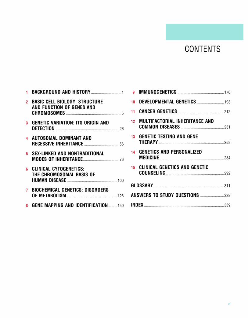
CONTENTS
1 BACKGROUND AND HISTORY ..............................1
2 BASIC CELL BIOLOGY: STRUCTUREAND FUNCTION OF GENES ANDCHROMOSOMES .......................................................5
3 GENETIC VARIATION: ITS ORIGIN ANDDETECTION ...............................................................26
4 AUTOSOMAL DOMINANT ANDRECESSIVE INHERITANCE ...................................56
5 SEX-LINKED AND NONTRADITIONALMODES OF INHERITANCE....................................76
6 CLINICAL CYTOGENETICS:THE CHROMOSOMAL BASIS OFHUMAN DISEASE ..................................................100
7 BIOCHEMICAL GENETICS: DISORDERSOF METABOLISM ..................................................128
8 GENE MAPPING AND IDENTIFICATION .........150
9 IMMUNOGENETICS...............................................176
10 DEVELOPMENTAL GENETICS ...........................193
11 CANCER GENETICS ..............................................212
12 MULTIFACTORIAL INHERITANCE ANDCOMMON DISEASES ...........................................231
13 GENETIC TESTING AND GENETHERAPY .................................................................258
14 GENETICS AND PERSONALIZEDMEDICINE................................................................284
15 CLINICAL GENETICS AND GENETICCOUNSELING .........................................................292
GLOSSARY......................................................................311
ANSWERS TO STUDY QUESTIONS ........................328
INDEX ...............................................................................339
xi

This page intentionally left blank
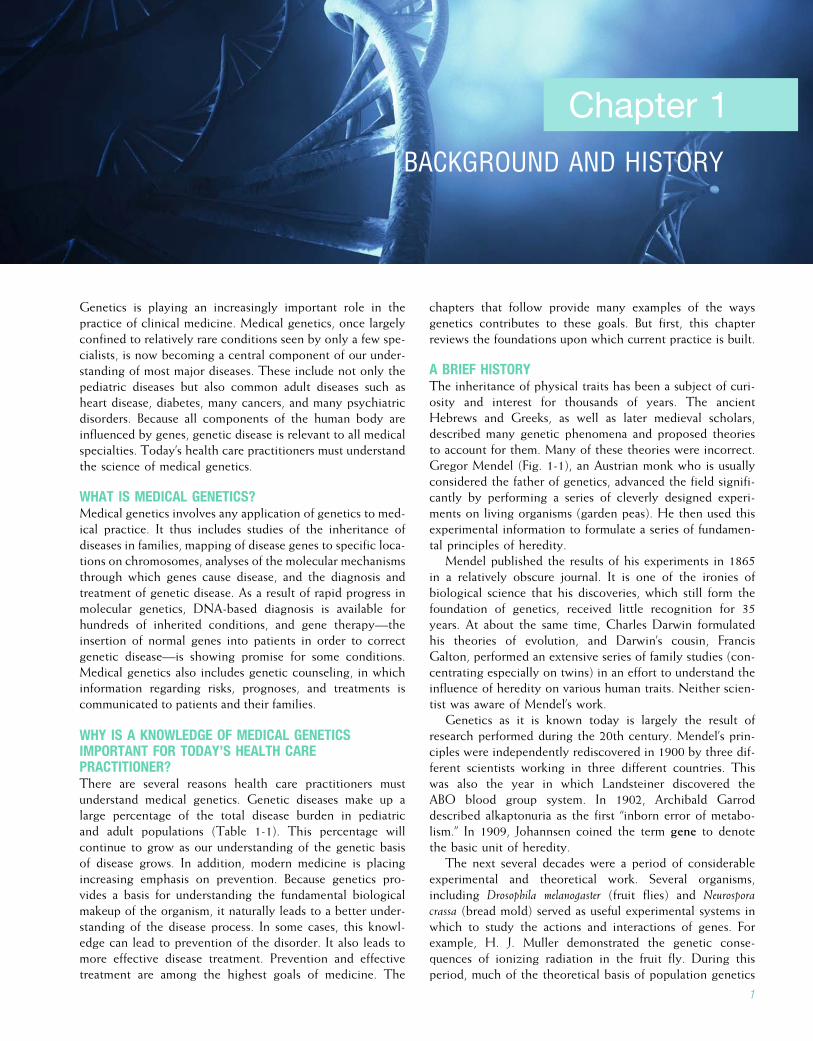
Chapter 1
BACKGROUND AND HISTORY
Genetics is playing an increasingly important role in thepractice of clinical medicine. Medical genetics, once largelyconfined to relatively rare conditions seen by only a few spe-cialists, is now becoming a central component of our under-standing of most major diseases. These include not only thepediatric diseases but also common adult diseases such asheart disease, diabetes, many cancers, and many psychiatricdisorders. Because all components of the human body areinfluenced by genes, genetic disease is relevant to all medicalspecialties. Today’s health care practitioners must understandthe science of medical genetics.
WHAT IS MEDICAL GENETICS?Medical genetics involves any application of genetics to med-ical practice. It thus includes studies of the inheritance ofdiseases in families, mapping of disease genes to specific loca-tions on chromosomes, analyses of the molecular mechanismsthrough which genes cause disease, and the diagnosis andtreatment of genetic disease. As a result of rapid progress inmolecular genetics, DNA-based diagnosis is available forhundreds of inherited conditions, and gene therapy—theinsertion of normal genes into patients in order to correctgenetic disease—is showing promise for some conditions.Medical genetics also includes genetic counseling, in whichinformation regarding risks, prognoses, and treatments iscommunicated to patients and their families.
WHY IS A KNOWLEDGE OF MEDICAL GENETICSIMPORTANT FOR TODAY’S HEALTH CAREPRACTITIONER?There are several reasons health care practitioners mustunderstand medical genetics. Genetic diseases make up alarge percentage of the total disease burden in pediatricand adult populations (Table 1-1). This percentage willcontinue to grow as our understanding of the genetic basisof disease grows. In addition, modern medicine is placingincreasing emphasis on prevention. Because genetics pro-vides a basis for understanding the fundamental biologicalmakeup of the organism, it naturally leads to a better under-standing of the disease process. In some cases, this knowl-edge can lead to prevention of the disorder. It also leads tomore effective disease treatment. Prevention and effectivetreatment are among the highest goals of medicine. The
chapters that follow provide many examples of the waysgenetics contributes to these goals. But first, this chapterreviews the foundations upon which current practice is built.
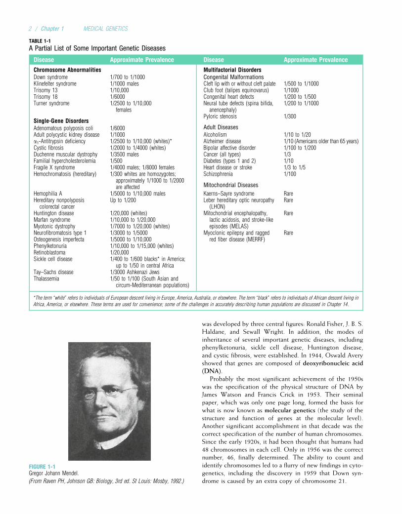
A BRIEF HISTORYThe inheritance of physical traits has been a subject of curi-osity and interest for thousands of years. The ancientHebrews and Greeks, as well as later medieval scholars,described many genetic phenomena and proposed theoriesto account for them. Many of these theories were incorrect.Gregor Mendel (Fig. 1-1), an Austrian monk who is usuallyconsidered the father of genetics, advanced the field signifi-cantly by performing a series of cleverly designed experi-ments on living organisms (garden peas). He then used thisexperimental information to formulate a series of fundamen-tal principles of heredity.
Mendel published the results of his experiments in 1865in a relatively obscure journal. It is one of the ironies ofbiological science that his discoveries, which still form thefoundation of genetics, received little recognition for 35years. At about the same time, Charles Darwin formulatedhis theories of evolution, and Darwin’s cousin, FrancisGalton, performed an extensive series of family studies (con-centrating especially on twins) in an effort to understand theinfluence of heredity on various human traits. Neither scien-tist was aware of Mendel’s work.
Genetics as it is known today is largely the result ofresearch performed during the 20th century. Mendel’s prin-ciples were independently rediscovered in 1900 by three dif-ferent scientists working in three different countries. Thiswas also the year in which Landsteiner discovered theABO blood group system. In 1902, Archibald Garroddescribed alkaptonuria as the first “inborn error of metabo-lism.” In 1909, Johannsen coined the term gene to denotethe basic unit of heredity.
The next several decades were a period of considerableexperimental and theoretical work. Several organisms,including Drosophila melanogaster (fruit flies) and Neurospora
crassa (bread mold) served as useful experimental systems inwhich to study the actions and interactions of genes. Forexample, H. J. Muller demonstrated the genetic conse-quences of ionizing radiation in the fruit fly. During thisperiod, much of the theoretical basis of population genetics
1
was developed by three central figures: Ronald Fisher, J. B. S.Haldane, and Sewall Wright. In addition, the modes ofinheritance of several important genetic diseases, includingphenylketonuria, sickle cell disease, Huntington disease,and cystic fibrosis, were established. In 1944, Oswald Averyshowed that genes are composed of deoxyribonucleic acid(DNA).
Probably the most significant achievement of the 1950swas the specification of the physical structure of DNA byJames Watson and Francis Crick in 1953. Their seminalpaper, which was only one page long, formed the basis forwhat is now known as molecular genetics (the study of thestructure and function of genes at the molecular level).Another significant accomplishment in that decade was thecorrect specification of the number of human chromosomes.Since the early 1920s, it had been thought that humans had48 chromosomes in each cell. Only in 1956 was the correctnumber, 46, finally determined. The ability to count andidentify chromosomes led to a flurry of new findings in cyto-genetics, including the discovery in 1959 that Down syn-drome is caused by an extra copy of chromosome 21.
FIGURE 1-1Gregor Johann Mendel.
(From Raven PH, Johnson GB: Biology, 3rd ed. St Louis: Mosby, 1992.)
TABLE 1-1
A Partial List of Some Important Genetic Diseases
Disease Approximate Prevalence Disease Approximate Prevalence
Chromosome Abnormalities Multifactorial DisordersDown syndrome 1/700 to 1/1000 Congenital MalformationsKlinefelter syndrome 1/1000 males Cleft lip with or without cleft palate 1/500 to 1/1000Trisomy 13 1/10,000 Club foot (talipes equinovarus) 1/1000Trisomy 18 1/6000 Congenital heart defects 1/200 to 1/500Turner syndrome 1/2500 to 1/10,000
femalesNeural tube defects (spina bifida,anencephaly)
1/200 to 1/1000
Single-Gene DisordersPyloric stenosis 1/300
Adenomatous polyposis coli 1/6000 Adult DiseasesAdult polycystic kidney disease 1/1000 Alcoholism 1/10 to 1/20a1-Antitrypsin deficiency 1/2500 to 1/10,000 (whites)* Alzheimer disease 1/10 (Americans older than 65 years)Cystic fibrosis 1/2000 to 1/4000 (whites) Bipolar affective disorder 1/100 to 1/200Duchenne muscular dystrophy 1/3500 males Cancer (all types) 1/3Familial hypercholesterolemia 1/500 Diabetes (types 1 and 2) 1/10Fragile X syndrome 1/4000 males; 1/8000 females Heart disease or stroke 1/3 to 1/5Hemochromatosis (hereditary) 1/300 whites are homozygotes;
approximately 1/1000 to 1/2000are affected
Schizophrenia 1/100
Hemophilia A 1/5000 to 1/10,000 males
Mitochondrial Diseases
Hereditary nonpolyposiscolorectal cancer
Up to 1/200Kaerns–Sayre syndrome Rare
Huntington disease 1/20,000 (whites)
Leber hereditary optic neuropathy(LHON)
Rare
Marfan syndrome 1/10,000 to 1/20,000Mitochondrial encephalopathy,lactic acidosis, and stroke-likeepisodes (MELAS)
Rare
Myotonic dystrophy 1/7000 to 1/20,000 (whites)Myoclonic epilepsy and raggedred fiber disease (MERRF)
RareNeurofibromatosis type 1 1/3000 to 1/5000Osteogenesis imperfecta 1/5000 to 1/10,000Phenylketonuria 1/10,000 to 1/15,000 (whites)Retinoblastoma 1/20,000Sickle cell disease 1/400 to 1/600 blacks* in America;
up to 1/50 in central AfricaTay–Sachs disease 1/3000 Ashkenazi JewsThalassemia 1/50 to 1/100 (South Asian and
circum-Mediterranean populations)
*The term “white” refers to individuals of European descent living in Europe, America, Australia, or elsewhere. The term “black” refers to individuals of African descent living in
Africa, America, or elsewhere. These terms are used for convenience; some of the challenges in accurately describing human populations are discussed in Chapter 14.
2 / Chapter 1 MEDICAL GENETICS
Technological developments since 1960 have broughtabout significant achievements at an ever-increasing rate.The most spectacular advances have occurred in the fieldof molecular genetics. Thousands of genes have beenmapped to specific chromosome locations. The HumanGenome Project, a large collaborative venture begun in1990, provided the complete human DNA sequence in2003 (the term genome refers to all of the DNA in an organ-ism). Important developments in computer technology havehelped to decipher the barrage of data being generated by thisand related projects. In addition to mapping genes, moleculargeneticists have pinpointed the molecular defects underlyinga number of important genetic diseases. This research hascontributed greatly to our understanding of the ways genedefects can cause disease, opening paths to more effectivetreatment and potential cures. The next decade promises tobe a time of great excitement and fulfillment.
TYPES OF GENETIC DISEASESHumans are estimated to have approximately 20,000 to25,000 genes. Alterations in these genes, or in combinationsof them, can produce genetic disorders. These disorders areclassified into several major groups:
• Chromosome disorders, in which entire chromosomes (orlarge segments of them) are missing, duplicated, or other-wise altered. These disorders include diseases such asDown syndrome and Turner syndrome.
• Disorders in which single genes are altered; these areoften termed mendelian conditions, or single-gene disorders.Well-known examples include cystic fibrosis, sickle celldisease, and hemophilia.
• Multifactorial disorders, which result from a combinationof multiple genetic and environmental causes. Many birthdefects, such as cleft lip and cleft palate, as well as manyadult disorders, including heart disease and diabetes,belong in this category.
• Mitochondrial disorders, a relatively small number of dis-eases caused by alterations in the small cytoplasmic mito-chondrial chromosome.
Table 1-1 provides some examples of each of these types ofdiseases.
Of these major classes of diseases, the single-gene disordershave probably received the greatest amount of attention.These disorders are classified according to the way theyare inherited in families: autosomal dominant, autosomalrecessive, or X-linked. These modes of inheritance are dis-cussed extensively in Chapters 4 and 5. The first edition ofMcKusick’s Mendelian Inheritance in Man, published in 1966,
listed only 1,368 autosomal traits and 119 X-linked traits.Today, the online version of McKusick’s compendium listsmore than 19,000 entries, of whichmore than 18,000 are auto-somal, more than 1,000 are X-linked, 57 are Y-linked, and 63are in the mitochondrial genome. DNA variants responsiblefor more than 2,500 of these traits, most of which are inheriteddiseases, have been identified. With continued advances,these numbers are certain to increase.
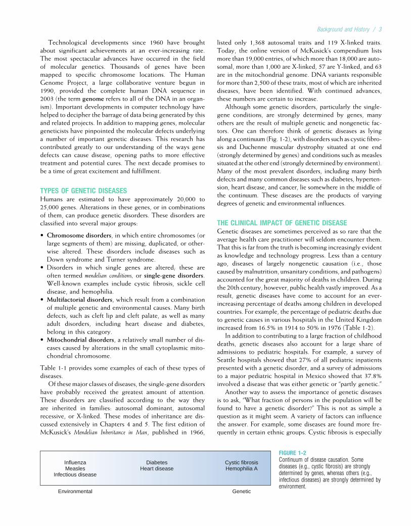
Although some genetic disorders, particularly the single-gene conditions, are strongly determined by genes, manyothers are the result of multiple genetic and nongenetic fac-tors. One can therefore think of genetic diseases as lyingalong a continuum (Fig. 1-2), with disorders such as cystic fibro-sis and Duchenne muscular dystrophy situated at one end(strongly determined by genes) and conditions such as measlessituated at the other end (strongly determined by environment).Many of the most prevalent disorders, including many birthdefects and many common diseases such as diabetes, hyperten-sion, heart disease, and cancer, lie somewhere in the middle ofthe continuum. These diseases are the products of varyingdegrees of genetic and environmental influences.
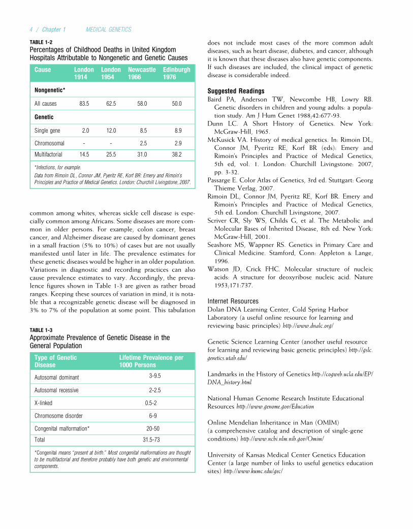
THE CLINICAL IMPACT OF GENETIC DISEASEGenetic diseases are sometimes perceived as so rare that theaverage health care practitioner will seldom encounter them.That this is far from the truth is becoming increasingly evidentas knowledge and technology progress. Less than a centuryago, diseases of largely nongenetic causation (i.e., thosecaused bymalnutrition, unsanitary conditions, and pathogens)accounted for the great majority of deaths in children. Duringthe 20th century, however, public health vastly improved. As aresult, genetic diseases have come to account for an ever-increasing percentage of deaths among children in developedcountries. For example, the percentage of pediatric deaths dueto genetic causes in various hospitals in the United Kingdomincreased from 16.5% in 1914 to 50% in 1976 (Table 1-2).
In addition to contributing to a large fraction of childhooddeaths, genetic diseases also account for a large share ofadmissions to pediatric hospitals. For example, a survey ofSeattle hospitals showed that 27% of all pediatric inpatientspresented with a genetic disorder, and a survey of admissionsto a major pediatric hospital in Mexico showed that 37.8%involved a disease that was either genetic or “partly genetic.”
Another way to assess the importance of genetic diseasesis to ask, “What fraction of persons in the population will befound to have a genetic disorder?” This is not as simple aquestion as it might seem. A variety of factors can influencethe answer. For example, some diseases are found more fre-quently in certain ethnic groups. Cystic fibrosis is especially
Environmental Genetic
InfluenzaMeasles
Infectious disease
DiabetesHeart disease
Cystic fibrosisHemophilia A
FIGURE 1-2Continuum of disease causation. Somediseases (e.g., cystic fibrosis) are stronglydetermined by genes, whereas others (e.g.,infectious diseases) are strongly determined byenvironment.
Background and History / 3
common among whites, whereas sickle cell disease is espe-cially common among Africans. Some diseases are more com-mon in older persons. For example, colon cancer, breastcancer, and Alzheimer disease are caused by dominant genesin a small fraction (5% to 10%) of cases but are not usuallymanifested until later in life. The prevalence estimates forthese genetic diseases would be higher in an older population.Variations in diagnostic and recording practices can alsocause prevalence estimates to vary. Accordingly, the preva-lence figures shown in Table 1-3 are given as rather broadranges. Keeping these sources of variation in mind, it is nota-ble that a recognizable genetic disease will be diagnosed in3% to 7% of the population at some point. This tabulation
does not include most cases of the more common adultdiseases, such as heart disease, diabetes, and cancer, althoughit is known that these diseases also have genetic components.If such diseases are included, the clinical impact of geneticdisease is considerable indeed.
Suggested ReadingsBaird PA, Anderson TW, Newcombe HB, Lowry RB.Genetic disorders in children and young adults: a popula-tion study. Am J Hum Genet 1988;42:677-93.
Dunn LC. A Short History of Genetics. New York:McGraw-Hill, 1965.
McKusick VA. History of medical genetics. In: Rimoin DL,Connor JM, Pyeritz RE, Korf BR (eds): Emery andRimoin’s Principles and Practice of Medical Genetics,5th ed, vol. 1. London: Churchill Livingstone: 2007,pp. 3-32.
Passarge E. Color Atlas of Genetics, 3rd ed. Stuttgart: GeorgThieme Verlag, 2007.
Rimoin DL, Connor JM, Pyeritz RE, Korf BR. Emery andRimoin’s Principles and Practice of Medical Genetics,5th ed. London: Churchill Livingstone, 2007.
Scriver CR, Sly WS, Childs G, et al. The Metabolic andMolecular Bases of Inherited Disease, 8th ed. New York:McGraw-Hill, 2001.
Seashore MS, Wappner RS. Genetics in Primary Care andClinical Medicine. Stamford, Conn: Appleton & Lange,1996.
Watson JD, Crick FHC. Molecular structure of nucleicacids: A structure for deoxyribose nucleic acid. Nature1953;171:737.
Internet ResourcesDolan DNA Learning Center, Cold Spring HarborLaboratory (a useful online resource for learning andreviewing basic principles) http://www.dnalc.org/
Genetic Science Learning Center (another useful resourcefor learning and reviewing basic genetic principles) http://gslc.genetics.utah.edu/
Landmarks in the History of Genetics http://cogweb.ucla.edu/EP/DNA_history.html
National Human Genome Research Institute EducationalResources http://www.genome.gov/Education
Online Mendelian Inheritance in Man (OMIM)(a comprehensive catalog and description of single-geneconditions) http://www.ncbi.nlm.nih.gov/Omim/
University of Kansas Medical Center Genetics EducationCenter (a large number of links to useful genetics educationsites) http://www.kumc.edu/gec/
TABLE 1-3
Approximate Prevalence of Genetic Disease in theGeneral Population
Type of GeneticDisease
Lifetime Prevalence per1000 Persons
Autosomal dominant 3-9.5
Autosomal recessive 2-2.5
X-linked 0.5-2
Chromosome disorder 6-9
Congenital malformation* 20-50
Total 31.5-73
*Congenital means “present at birth.” Most congenital malformations are thought
to be multifactorial and therefore probably have both genetic and environmental
components.
TABLE 1-2
Percentages of Childhood Deaths in United KingdomHospitals Attributable to Nongenetic and Genetic Causes
Cause London1914
London1954
Newcastle1966
Edinburgh1976
Nongenetic*
All causes 83.5 62.5 58.0 50.0
Genetic
Single gene 2.0 12.0 8.5 8.9
Chromosomal - - 2.5 2.9
Multifactorial 14.5 25.5 31.0 38.2
*Infections, for example.
Data from Rimoin DL, Connor JM, Pyeritz RE, Korf BR: Emery and Rimoin’s
Principles and Practice of Medical Genetics. London: Churchill Livingstone, 2007.
4 / Chapter 1 MEDICAL GENETICS
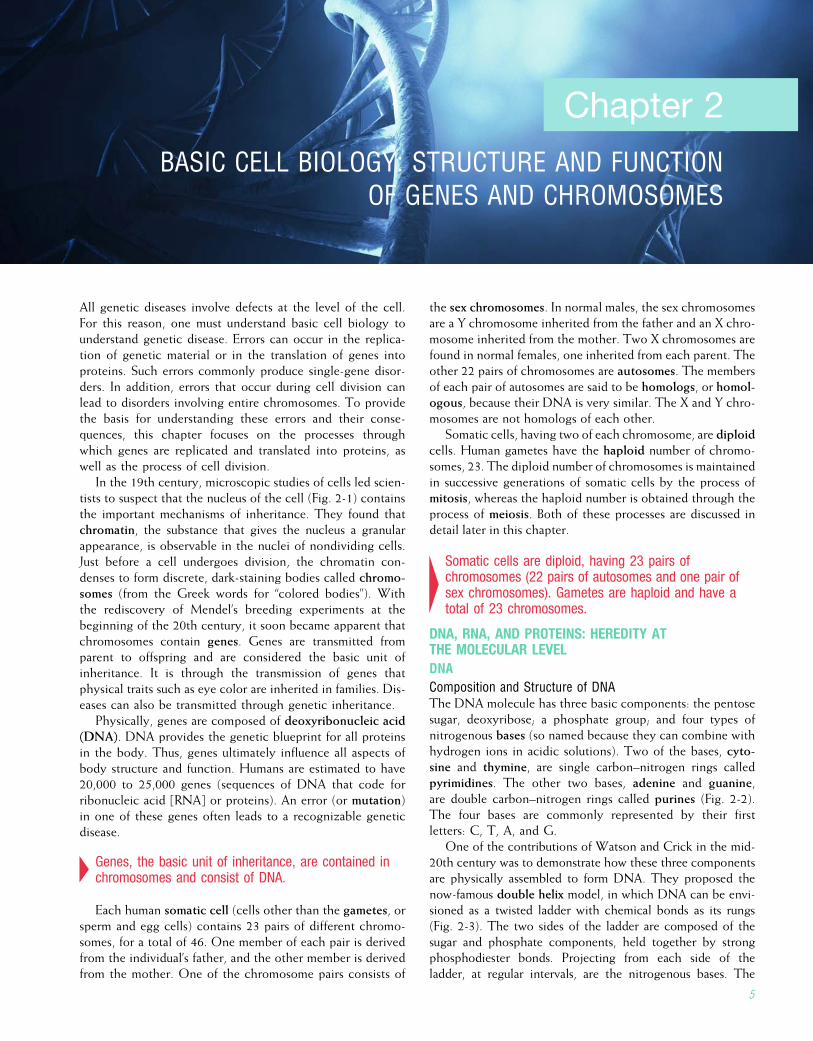
Chapter 2
BASIC CELL BIOLOGY: STRUCTURE AND FUNCTIONOF GENES AND CHROMOSOMES
All genetic diseases involve defects at the level of the cell.For this reason, one must understand basic cell biology tounderstand genetic disease. Errors can occur in the replica-tion of genetic material or in the translation of genes intoproteins. Such errors commonly produce single-gene disor-ders. In addition, errors that occur during cell division canlead to disorders involving entire chromosomes. To providethe basis for understanding these errors and their conse-quences, this chapter focuses on the processes throughwhich genes are replicated and translated into proteins, aswell as the process of cell division.
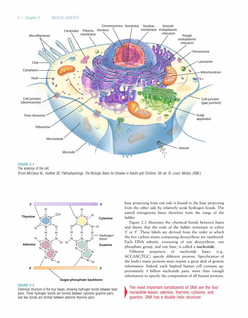
In the 19th century, microscopic studies of cells led scien-tists to suspect that the nucleus of the cell (Fig. 2-1) containsthe important mechanisms of inheritance. They found thatchromatin, the substance that gives the nucleus a granularappearance, is observable in the nuclei of nondividing cells.Just before a cell undergoes division, the chromatin con-denses to form discrete, dark-staining bodies called chromo-somes (from the Greek words for “colored bodies”). Withthe rediscovery of Mendel’s breeding experiments at thebeginning of the 20th century, it soon became apparent thatchromosomes contain genes. Genes are transmitted fromparent to offspring and are considered the basic unit ofinheritance. It is through the transmission of genes thatphysical traits such as eye color are inherited in families. Dis-eases can also be transmitted through genetic inheritance.
Physically, genes are composed of deoxyribonucleic acid(DNA). DNA provides the genetic blueprint for all proteinsin the body. Thus, genes ultimately influence all aspects ofbody structure and function. Humans are estimated to have20,000 to 25,000 genes (sequences of DNA that code forribonucleic acid [RNA] or proteins). An error (or mutation)in one of these genes often leads to a recognizable geneticdisease.
4Genes, the basic unit of inheritance, are contained inchromosomes and consist of DNA.
Each human somatic cell (cells other than the gametes, orsperm and egg cells) contains 23 pairs of different chromo-somes, for a total of 46. One member of each pair is derivedfrom the individual’s father, and the other member is derivedfrom the mother. One of the chromosome pairs consists of
the sex chromosomes. In normal males, the sex chromosomesare a Y chromosome inherited from the father and an X chro-mosome inherited from the mother. Two X chromosomes arefound in normal females, one inherited from each parent. Theother 22 pairs of chromosomes are autosomes. The membersof each pair of autosomes are said to be homologs, or homol-ogous, because their DNA is very similar. The X and Y chro-mosomes are not homologs of each other.
Somatic cells, having two of each chromosome, are diploidcells. Human gametes have the haploid number of chromo-somes, 23. The diploid number of chromosomes is maintainedin successive generations of somatic cells by the process ofmitosis, whereas the haploid number is obtained through theprocess of meiosis. Both of these processes are discussed indetail later in this chapter.
4Somatic cells are diploid, having 23 pairs ofchromosomes (22 pairs of autosomes and one pair ofsex chromosomes). Gametes are haploid and have atotal of 23 chromosomes.
DNA, RNA, AND PROTEINS: HEREDITY ATTHE MOLECULAR LEVELDNA
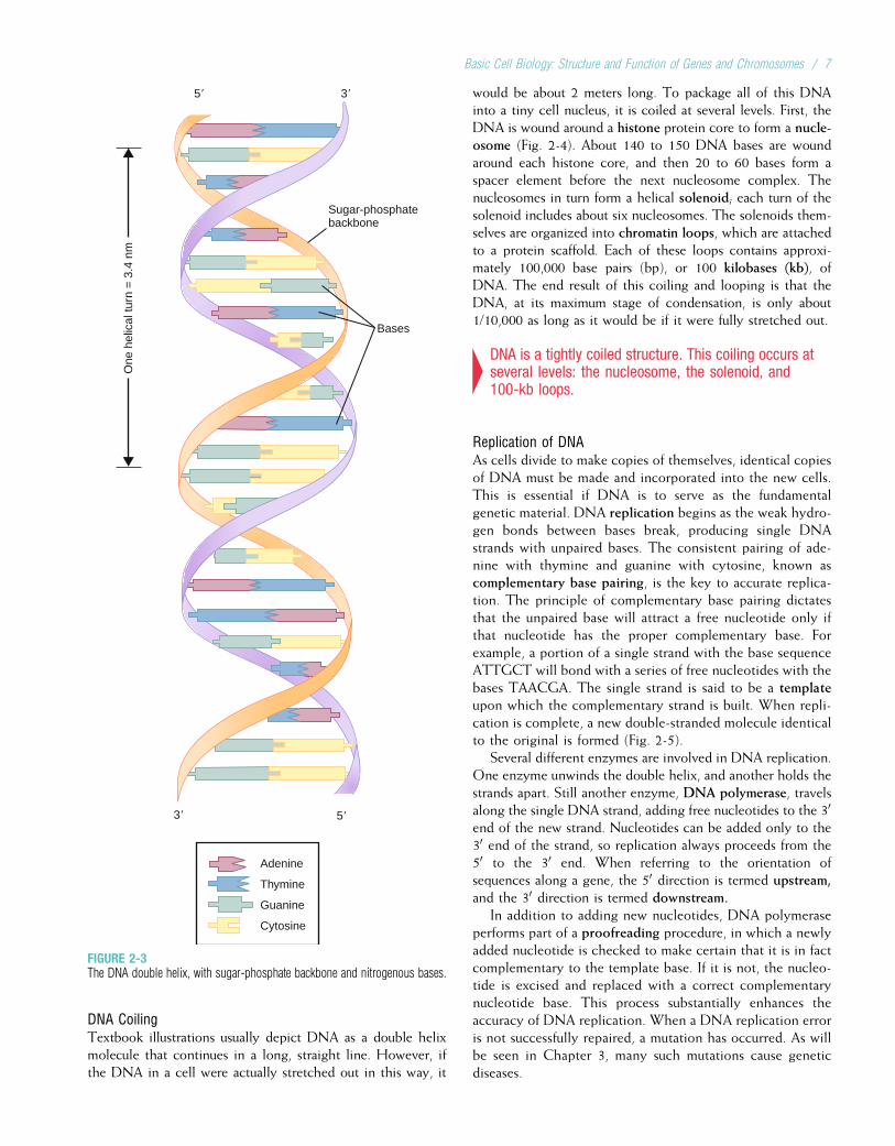
Composition and Structure of DNAThe DNA molecule has three basic components: the pentosesugar, deoxyribose; a phosphate group; and four types ofnitrogenous bases (so named because they can combine withhydrogen ions in acidic solutions). Two of the bases, cyto-sine and thymine, are single carbon–nitrogen rings calledpyrimidines. The other two bases, adenine and guanine,are double carbon–nitrogen rings called purines (Fig. 2-2).The four bases are commonly represented by their firstletters: C, T, A, and G.
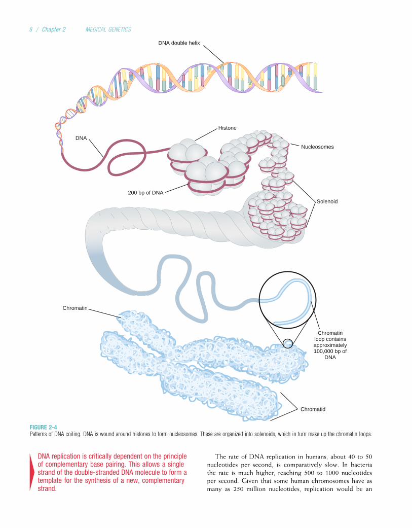
One of the contributions of Watson and Crick in the mid-20th century was to demonstrate how these three componentsare physically assembled to form DNA. They proposed thenow-famous double helix model, in which DNA can be envi-sioned as a twisted ladder with chemical bonds as its rungs(Fig. 2-3). The two sides of the ladder are composed of thesugar and phosphate components, held together by strongphosphodiester bonds. Projecting from each side of theladder, at regular intervals, are the nitrogenous bases. The
5
base projecting from one side is bound to the base projectingfrom the other side by relatively weak hydrogen bonds. Thepaired nitrogenous bases therefore form the rungs of theladder.
Figure 2-2 illustrates the chemical bonds between basesand shows that the ends of the ladder terminate in either30 or 50. These labels are derived from the order in whichthe five carbon atoms composing deoxyribose are numbered.Each DNA subunit, consisting of one deoxyribose, onephosphate group, and one base, is called a nucleotide.
Different sequences of nucleotide bases (e.g.,ACCAAGTGC) specify different proteins. Specification ofthe body’s many proteins must require a great deal of geneticinformation. Indeed, each haploid human cell contains ap-proximately 3 billion nucleotide pairs, more than enoughinformation to specify the composition of all human proteins.
4The most important constituents of DNA are the fournucleotide bases: adenine, thymine, cytosine, andguanine. DNA has a double helix structure.
Hydrogenbond
Guanine
H H
H
H
H
H H H H
H H
C CC
CC
C C C
C CC C
C
C
C
C
CH
CO O O
O
N N
N N HN
N N
N N
N
C
N
N N N N
H H
Sugar-phosphate backbone
Thymine
5939
Cytosine
59 39
Adenine
FIGURE 2-2Chemical structure of the four bases, showing hydrogen bonds between basepairs. Three hydrogen bonds are formed between cytosine–guanine pairs,and two bonds are formed between adenine–thymine pairs.
Ribosome
Roughendoplasmic
reticulum
Smooth endoplasmic
reticulum
Cilia
Cytoplasm
Free ribosome
Microfilaments
Microtubule
Golgiapparatus
MicrovilliVesicle
Vault
Plasmamembrane
NucleolusNucleus
Chromosomes Nuclearmembrane
Peroxisome
Lysosome
Cell junction(gap junction)
Cell junction(desmosome)
Mitochondrion
Centrioles
FIGURE 2-1The anatomy of the cell.
(From McCance KL, Huether SE: Pathophysiology: The Biologic Basis for Disease in Adults and Children, 5th ed. St. Louis: Mosby, 2006.)
6 / Chapter 2 MEDICAL GENETICS
DNA CoilingTextbook illustrations usually depict DNA as a double helixmolecule that continues in a long, straight line. However, ifthe DNA in a cell were actually stretched out in this way, it
would be about 2 meters long. To package all of this DNAinto a tiny cell nucleus, it is coiled at several levels. First, theDNA is wound around a histone protein core to form a nucle-osome (Fig. 2-4). About 140 to 150 DNA bases are woundaround each histone core, and then 20 to 60 bases form aspacer element before the next nucleosome complex. Thenucleosomes in turn form a helical solenoid; each turn of thesolenoid includes about six nucleosomes. The solenoids them-selves are organized into chromatin loops, which are attachedto a protein scaffold. Each of these loops contains approxi-mately 100,000 base pairs (bp), or 100 kilobases (kb), ofDNA. The end result of this coiling and looping is that theDNA, at its maximum stage of condensation, is only about1/10,000 as long as it would be if it were fully stretched out.
4DNA is a tightly coiled structure. This coiling occurs atseveral levels: the nucleosome, the solenoid, and100-kb loops.
Replication of DNAAs cells divide to make copies of themselves, identical copiesof DNA must be made and incorporated into the new cells.This is essential if DNA is to serve as the fundamentalgenetic material. DNA replication begins as the weak hydro-gen bonds between bases break, producing single DNAstrands with unpaired bases. The consistent pairing of ade-nine with thymine and guanine with cytosine, known ascomplementary base pairing, is the key to accurate replica-tion. The principle of complementary base pairing dictatesthat the unpaired base will attract a free nucleotide only ifthat nucleotide has the proper complementary base. Forexample, a portion of a single strand with the base sequenceATTGCT will bond with a series of free nucleotides with thebases TAACGA. The single strand is said to be a templateupon which the complementary strand is built. When repli-cation is complete, a new double-stranded molecule identicalto the original is formed (Fig. 2-5).
Several different enzymes are involved in DNA replication.One enzyme unwinds the double helix, and another holds thestrands apart. Still another enzyme, DNA polymerase, travelsalong the single DNA strand, adding free nucleotides to the 30
end of the new strand. Nucleotides can be added only to the30 end of the strand, so replication always proceeds from the50 to the 30 end. When referring to the orientation ofsequences along a gene, the 50 direction is termed upstream,and the 30 direction is termed downstream.
In addition to adding new nucleotides, DNA polymeraseperforms part of a proofreading procedure, in which a newlyadded nucleotide is checked to make certain that it is in factcomplementary to the template base. If it is not, the nucleo-tide is excised and replaced with a correct complementarynucleotide base. This process substantially enhances theaccuracy of DNA replication. When a DNA replication erroris not successfully repaired, a mutation has occurred. As willbe seen in Chapter 3, many such mutations cause geneticdiseases.
Bases
5�3�
One
hel
ical
turn
= 3
.4 n
m
Adenine
Thymine
Guanine
Cytosine
Sugar-phosphatebackbone
5� 3�
FIGURE 2-3The DNA double helix, with sugar-phosphate backbone and nitrogenous bases.
Basic Cell Biology: Structure and Function of Genes and Chromosomes / 7
4DNA replication is critically dependent on the principleof complementary base pairing. This allows a singlestrand of the double-stranded DNA molecule to form atemplate for the synthesis of a new, complementarystrand.
The rate of DNA replication in humans, about 40 to 50nucleotides per second, is comparatively slow. In bacteriathe rate is much higher, reaching 500 to 1000 nucleotidesper second. Given that some human chromosomes have asmany as 250 million nucleotides, replication would be an
DNA double helix
DNA
Histone
200 bp of DNA
Nucleosomes
Solenoid
Chromatin
Chromatinloop containsapproximately100,000 bp of
DNA
Chromatid
FIGURE 2-4Patterns of DNA coiling. DNA is wound around histones to form nucleosomes. These are organized into solenoids, which in turn make up the chromatin loops.
8 / Chapter 2 MEDICAL GENETICS
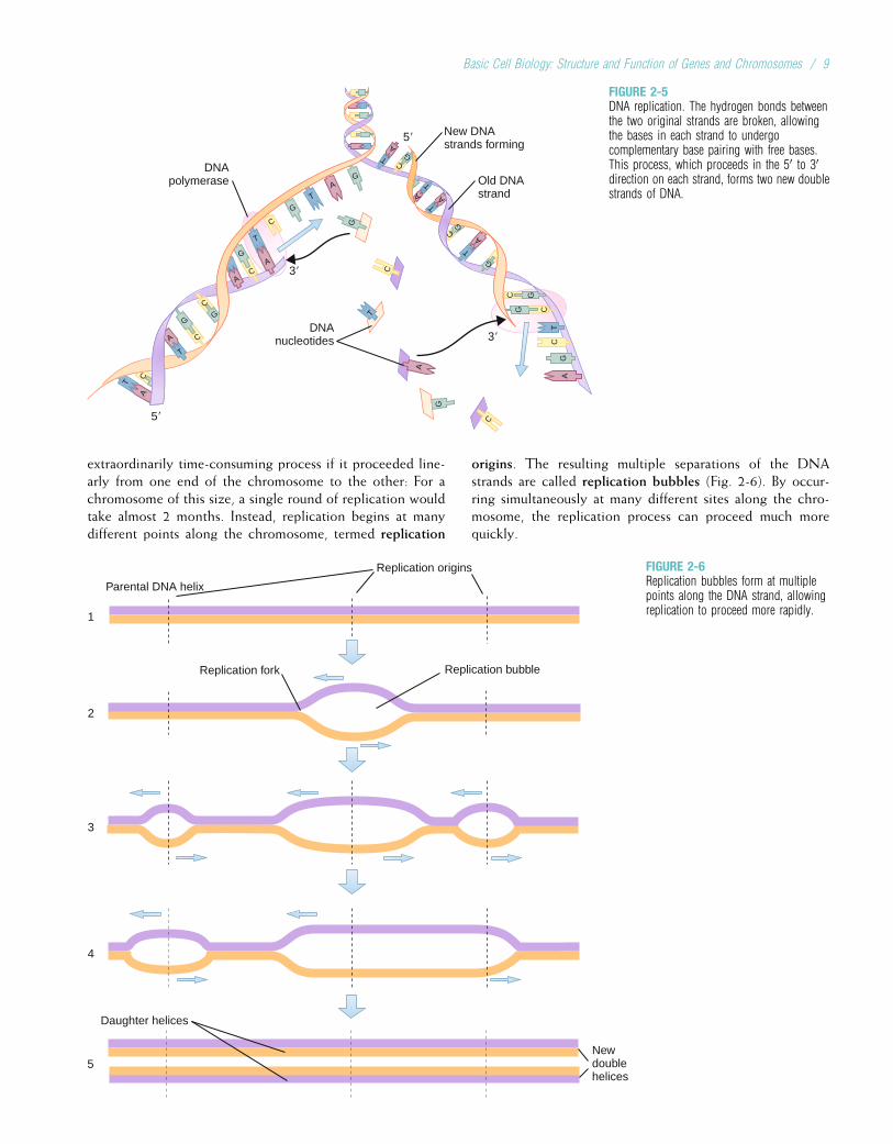
extraordinarily time-consuming process if it proceeded line-arly from one end of the chromosome to the other: For achromosome of this size, a single round of replication wouldtake almost 2 months. Instead, replication begins at manydifferent points along the chromosome, termed replication
origins. The resulting multiple separations of the DNAstrands are called replication bubbles (Fig. 2-6). By occur-ring simultaneously at many different sites along the chro-mosome, the replication process can proceed much morequickly.
5�
3�
3�DNA
nucleotides
DNApolymerase
New DNAstrands forming
Old DNAstrand
5�
A
C
T
T
GG
A
G
G
G
A
AG
G
CC
C
C
CG
G
GC
GC
C
T
A
G
C
C
T
T
T
A
A
T
A
A T
TA
TA
GC
FIGURE 2-5DNA replication. The hydrogen bonds betweenthe two original strands are broken, allowingthe bases in each strand to undergocomplementary base pairing with free bases.This process, which proceeds in the 50 to 30direction on each strand, forms two new doublestrands of DNA.
4
5
3
2
1
Replication origins
Replication fork Replication bubble
Daughter helices
Newdoublehelices
Parental DNA helix
FIGURE 2-6Replication bubbles form at multiplepoints along the DNA strand, allowingreplication to proceed more rapidly.
Basic Cell Biology: Structure and Function of Genes and Chromosomes / 9
4Replication bubbles allow DNA replication to takeplace at multiple locations on the chromosome,greatly speeding the replication process.
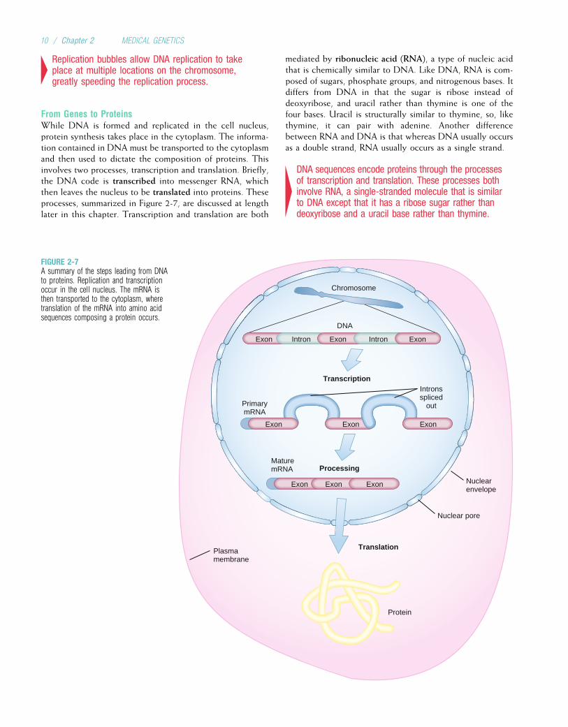
From Genes to ProteinsWhile DNA is formed and replicated in the cell nucleus,protein synthesis takes place in the cytoplasm. The informa-tion contained in DNA must be transported to the cytoplasmand then used to dictate the composition of proteins. Thisinvolves two processes, transcription and translation. Briefly,the DNA code is transcribed into messenger RNA, whichthen leaves the nucleus to be translated into proteins. Theseprocesses, summarized in Figure 2-7, are discussed at lengthlater in this chapter. Transcription and translation are both
mediated by ribonucleic acid (RNA), a type of nucleic acidthat is chemically similar to DNA. Like DNA, RNA is com-posed of sugars, phosphate groups, and nitrogenous bases. Itdiffers from DNA in that the sugar is ribose instead ofdeoxyribose, and uracil rather than thymine is one of thefour bases. Uracil is structurally similar to thymine, so, likethymine, it can pair with adenine. Another differencebetween RNA and DNA is that whereas DNA usually occursas a double strand, RNA usually occurs as a single strand.
4DNA sequences encode proteins through the processesof transcription and translation. These processes bothinvolve RNA, a single-stranded molecule that is similarto DNA except that it has a ribose sugar rather thandeoxyribose and a uracil base rather than thymine.
Intron
DNA
Introns spliced
out
Transcription
Chromosome
Processing
Primary mRNA
Translation
Protein
Plasma membrane
Nuclear pore
Nuclear envelope
Exon
Exon Exon Exon
MaturemRNA
Intron Exon Intron Exon
Exon Exon Exon
FIGURE 2-7A summary of the steps leading from DNAto proteins. Replication and transcriptionoccur in the cell nucleus. The mRNA isthen transported to the cytoplasm, wheretranslation of the mRNA into amino acidsequences composing a protein occurs.
10 / Chapter 2 MEDICAL GENETICS
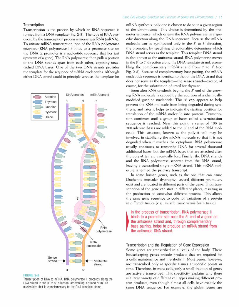
TranscriptionTranscription is the process by which an RNA sequence isformed from a DNA template (Fig. 2-8). The type of RNA pro-duced by the transcription process ismessenger RNA (mRNA).To initiate mRNA transcription, one of the RNA polymeraseenzymes (RNA polymerase II) binds to a promoter site onthe DNA (a promoter is a nucleotide sequence that lies justupstream of a gene). The RNA polymerase then pulls a portionof the DNA strands apart from each other, exposing unat-tached DNA bases. One of the two DNA strands providesthe template for the sequence of mRNA nucleotides. Althougheither DNA strand could in principle serve as the template for
mRNA synthesis, only one is chosen to do so in a given regionof the chromosome. This choice is determined by the pro-moter sequence, which orients the RNA polymerase in a spe-cific direction along the DNA sequence. Because the mRNAmolecule can be synthesized only in the 50 to 30 direction,the promoter, by specifying directionality, determines whichDNA strand serves as the template. This template DNA strandis also known as the antisense strand. RNA polymerase movesin the 30 to 50 direction along the DNA template strand, assem-bling the complementary mRNA strand from 50 to 30 (seeFig. 2-8). Because of complementary base pairing, the mRNAnucleotide sequence is identical to that of the DNA strand thatdoes not serve as the template—the sense strand—except, ofcourse, for the substitution of uracil for thymine.
Soon after RNA synthesis begins, the 50 end of the grow-ing RNA molecule is capped by the addition of a chemicallymodified guanine nucleotide. This 50 cap appears to helpprevent the RNA molecule from being degraded during syn-thesis, and later it helps to indicate the starting position fortranslation of the mRNA molecule into protein. Transcrip-tion continues until a group of bases called a terminationsequence is reached. Near this point, a series of 100 to200 adenine bases are added to the 30 end of the RNA mol-ecule. This structure, known as the poly-A tail, may beinvolved in stabilizing the mRNA molecule so that it is notdegraded when it reaches the cytoplasm. RNA polymeraseusually continues to transcribe DNA for several thousandadditional bases, but the mRNA bases that are attached afterthe poly-A tail are eventually lost. Finally, the DNA strandsand the RNA polymerase separate from the RNA strand,leaving a transcribed single mRNA strand. This mRNA mol-ecule is termed the primary transcript.
In some human genes, such as the one that can causeDuchenne muscular dystrophy, several different promotersexist and are located in different parts of the gene. Thus, tran-scription of the gene can start in different places, resulting inthe production of somewhat different proteins. This allowsthe same gene sequence to code for variations of a proteinin different tissues (e.g., muscle tissue versus brain tissue).
4In the process of transcription, RNA polymerase IIbinds to a promoter site near the 50 end of a gene onthe antisense strand and, through complementarybase pairing, helps to produce an mRNA strand fromthe antisense DNA strand.
Transcription and the Regulation of Gene ExpressionSome genes are transcribed in all cells of the body. Thesehousekeeping genes encode products that are required fora cell’s maintenance and metabolism. Most genes, however,are transcribed only in specific tissues at specific points intime. Therefore, in most cells, only a small fraction of genesare actively transcribed. This specificity explains why thereis a large variety of different cell types making different pro-tein products, even though almost all cells have exactly thesame DNA sequence. For example, the globin genes are
5'3'
3'
5'3'5'
RNAnucleotide
Sensestrand Antisense
strand
RNApolymerase
Adenine
Thymine
Guanine
Cytosine
Uracil
DNA strands mRNA strand
FIGURE 2-8Transcription of DNA to mRNA. RNA polymerase II proceeds along theDNA strand in the 30 to 50 direction, assembling a strand of mRNAnucleotides that is complementary to the DNA template strand.
Basic Cell Biology: Structure and Function of Genes and Chromosomes / 11
transcribed in the progenitors of red blood cells (where theyhelp to form hemoglobin), and the low-density lipoproteinreceptor genes are transcribed in liver cells.
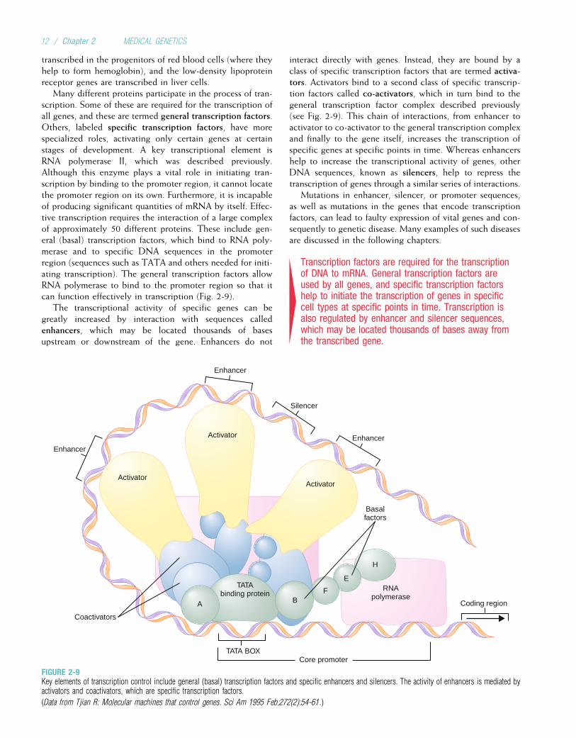
Many different proteins participate in the process of tran-scription. Some of these are required for the transcription ofall genes, and these are termed general transcription factors.Others, labeled specific transcription factors, have morespecialized roles, activating only certain genes at certainstages of development. A key transcriptional element isRNA polymerase II, which was described previously.Although this enzyme plays a vital role in initiating tran-scription by binding to the promoter region, it cannot locatethe promoter region on its own. Furthermore, it is incapableof producing significant quantities of mRNA by itself. Effec-tive transcription requires the interaction of a large complexof approximately 50 different proteins. These include gen-eral (basal) transcription factors, which bind to RNA poly-merase and to specific DNA sequences in the promoterregion (sequences such as TATA and others needed for initi-ating transcription). The general transcription factors allowRNA polymerase to bind to the promoter region so that itcan function effectively in transcription (Fig. 2-9).
The transcriptional activity of specific genes can begreatly increased by interaction with sequences calledenhancers, which may be located thousands of basesupstream or downstream of the gene. Enhancers do not
interact directly with genes. Instead, they are bound by aclass of specific transcription factors that are termed activa-tors. Activators bind to a second class of specific transcrip-tion factors called co-activators, which in turn bind to thegeneral transcription factor complex described previously(see Fig. 2-9). This chain of interactions, from enhancer toactivator to co-activator to the general transcription complexand finally to the gene itself, increases the transcription ofspecific genes at specific points in time. Whereas enhancershelp to increase the transcriptional activity of genes, otherDNA sequences, known as silencers, help to repress thetranscription of genes through a similar series of interactions.
Mutations in enhancer, silencer, or promoter sequences,as well as mutations in the genes that encode transcriptionfactors, can lead to faulty expression of vital genes and con-sequently to genetic disease. Many examples of such diseasesare discussed in the following chapters.
4Transcription factors are required for the transcriptionof DNA to mRNA. General transcription factors areused by all genes, and specific transcription factorshelp to initiate the transcription of genes in specificcell types at specific points in time. Transcription isalso regulated by enhancer and silencer sequences,which may be located thousands of bases away fromthe transcribed gene.
Enhancer
Enhancer
Enhancer
Coding region
Silencer
Activator
Activator
Coactivators
Basalfactors
TATAbinding protein
RNApolymerase
A BF
E
H
TATA BOXCore promoter
Activator
FIGURE 2-9Key elements of transcription control include general (basal) transcription factors and specific enhancers and silencers. The activity of enhancers is mediated byactivators and coactivators, which are specific transcription factors.
(Data from Tjian R: Molecular machines that control genes. Sci Am 1995 Feb;272(2):54-61.)
12 / Chapter 2 MEDICAL GENETICS
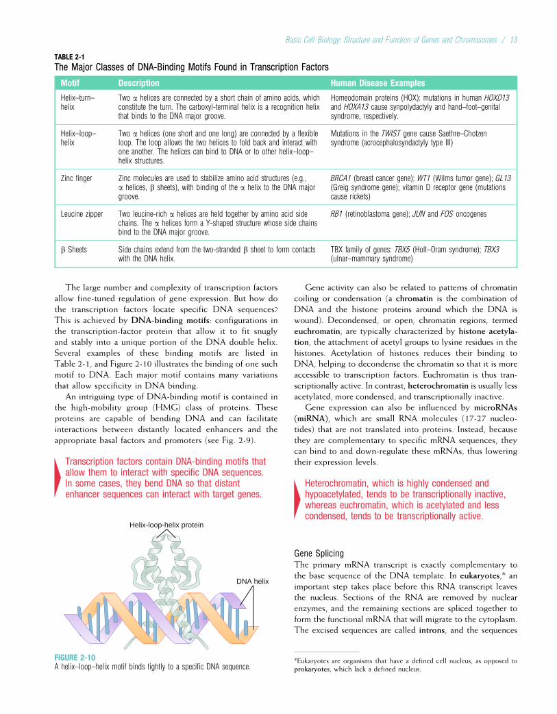
The large number and complexity of transcription factorsallow fine-tuned regulation of gene expression. But how dothe transcription factors locate specific DNA sequences?This is achieved by DNA-binding motifs: configurations inthe transcription-factor protein that allow it to fit snuglyand stably into a unique portion of the DNA double helix.Several examples of these binding motifs are listed inTable 2-1, and Figure 2-10 illustrates the binding of one suchmotif to DNA. Each major motif contains many variationsthat allow specificity in DNA binding.
An intriguing type of DNA-binding motif is contained inthe high-mobility group (HMG) class of proteins. Theseproteins are capable of bending DNA and can facilitateinteractions between distantly located enhancers and theappropriate basal factors and promoters (see Fig. 2-9).
4Transcription factors contain DNA-binding motifs thatallow them to interact with specific DNA sequences.In some cases, they bend DNA so that distantenhancer sequences can interact with target genes.
Gene activity can also be related to patterns of chromatincoiling or condensation (a chromatin is the combination ofDNA and the histone proteins around which the DNA iswound). Decondensed, or open, chromatin regions, termedeuchromatin, are typically characterized by histone acetyla-tion, the attachment of acetyl groups to lysine residues in thehistones. Acetylation of histones reduces their binding toDNA, helping to decondense the chromatin so that it is moreaccessible to transcription factors. Euchromatin is thus tran-scriptionally active. In contrast, heterochromatin is usually lessacetylated, more condensed, and transcriptionally inactive.
Gene expression can also be influenced by microRNAs(miRNA), which are small RNA molecules (17-27 nucleo-tides) that are not translated into proteins. Instead, becausethey are complementary to specific mRNA sequences, theycan bind to and down-regulate these mRNAs, thus loweringtheir expression levels.
4Heterochromatin, which is highly condensed andhypoacetylated, tends to be transcriptionally inactive,whereas euchromatin, which is acetylated and lesscondensed, tends to be transcriptionally active.
Gene SplicingThe primary mRNA transcript is exactly complementary tothe base sequence of the DNA template. In eukaryotes,* animportant step takes place before this RNA transcript leavesthe nucleus. Sections of the RNA are removed by nuclearenzymes, and the remaining sections are spliced together toform the functional mRNA that will migrate to the cytoplasm.The excised sequences are called introns, and the sequences
TABLE 2-1
The Major Classes of DNA-Binding Motifs Found in Transcription Factors
Motif Description Human Disease Examples
Helix–turn–helix
Two a helices are connected by a short chain of amino acids, whichconstitute the turn. The carboxyl-terminal helix is a recognition helixthat binds to the DNA major groove.
Homeodomain proteins (HOX): mutations in human HOXD13and HOXA13 cause synpolydactyly and hand–foot–genitalsyndrome, respectively.
Helix–loop–helix
Two a helices (one short and one long) are connected by a flexibleloop. The loop allows the two helices to fold back and interact withone another. The helices can bind to DNA or to other helix–loop–helix structures.
Mutations in the TWIST gene cause Saethre–Chotzensyndrome (acrocephalosyndactyly type III)
Zinc finger Zinc molecules are used to stabilize amino acid structures (e.g.,a helices, b sheets), with binding of the a helix to the DNA majorgroove.
BRCA1 (breast cancer gene); WT1 (Wilms tumor gene); GL13(Greig syndrome gene); vitamin D receptor gene (mutationscause rickets)
Leucine zipper Two leucine-rich a helices are held together by amino acid sidechains. The a helices form a Y-shaped structure whose side chainsbind to the DNA major groove.
RB1 (retinoblastoma gene); JUN and FOS oncogenes
b Sheets Side chains extend from the two-stranded b sheet to form contactswith the DNA helix.
TBX family of genes: TBX5 (Holt–Oram syndrome); TBX3(ulnar–mammary syndrome)
*Eukaryotes are organisms that have a defined cell nucleus, as opposed toprokaryotes, which lack a defined nucleus.
Helix-loop-helix protein
DNA helix
FIGURE 2-10A helix–loop–helix motif binds tightly to a specific DNA sequence.
Basic Cell Biology: Structure and Function of Genes and Chromosomes / 13
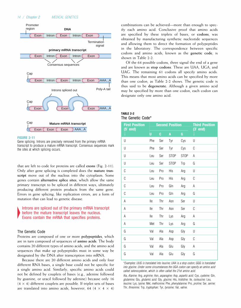
that are left to code for proteins are called exons (Fig. 2-11).Only after gene splicing is completed does the mature tran-script move out of the nucleus into the cytoplasm. Somegenes contain alternative splice sites, which allow the sameprimary transcript to be spliced in different ways, ultimatelyproducing different protein products from the same gene.Errors in gene splicing, like replication errors, are a form ofmutation that can lead to genetic disease.
4Introns are spliced out of the primary mRNA transcriptbefore the mature transcript leaves the nucleus.Exons contain the mRNA that specifies proteins.
The Genetic CodeProteins are composed of one or more polypeptides, whichare in turn composed of sequences of amino acids. The bodycontains 20 different types of amino acids, and the amino acidsequences that make up polypeptides must in some way bedesignated by the DNA after transcription into mRNA.
Because there are 20 different amino acids and only fourdifferent RNA bases, a single base could not be specific fora single amino acid. Similarly, specific amino acids couldnot be defined by couplets of bases (e.g., adenine followedby guanine, or uracil followed by adenine) because only 16(4 � 4) different couplets are possible. If triplet sets of basesare translated into amino acids, however, 64 (4 � 4 � 4)
combinations can be achieved—more than enough to spec-ify each amino acid. Conclusive proof that amino acidsare specified by these triplets of bases, or codons, wasobtained by manufacturing synthetic nucleotide sequencesand allowing them to direct the formation of polypeptidesin the laboratory. The correspondence between specificcodons and amino acids, known as the genetic code, isshown in Table 2-2.
Of the 64 possible codons, three signal the end of a geneand are known as stop codons. These are UAA, UGA, andUAG. The remaining 61 codons all specify amino acids.This means that most amino acids can be specified by morethan one codon, as Table 2-2 shows. The genetic code isthus said to be degenerate. Although a given amino acidmay be specified by more than one codon, each codon candesignate only one amino acid.
TABLE 2-2
The Genetic Code*
First Position(50 end)#
Second Position Third Position(30 end)#U C A G
U Phe Ser Tyr Cys U
U Phe Ser Tyr Cys C
U Leu Ser STOP STOP A
U Leu Ser STOP Trp G
C Leu Pro His Arg U
C Leu Pro His Arg C
C Leu Pro Gln Arg A
C Leu Pro Gln Arg G
A Ile Thr Asn Ser U
A Ile Thr Asn Ser C
A Ile Thr Lys Arg A
A Met Thr Lys Arg G
G Val Ala Asp Gly U
G Val Ala Asp Gly C
G Val Ala Glu Gly A
G Val Ala Glu Gly G
*Examples: UUG is translated into leucine; UAA is a stop codon; GGG is translated
into glycine. Under some circumstances the UGA codon can specify an amino acid
called selenocysteine, which is often called the 21st amino acid.
Ala, Alanine; Arg, arginine; Asn, asparagine; Asp, aspartic acid; Cys, cysteine; Gln,
glutamine; Glu, glutamic acid; Gly, glycine; His, histidine; Ile, isoleucine; Leu,
leucine; Lys, lysine; Met, methionine; Phe, phenylalanine; Pro, proline; Ser, serine;
Thr, threonine; Trp, tryptophan; Tyr, tyrosine; Val, valine.
Mature mRNA transcript
DNA
primary mRNA transcript
Promoter region
Terminationsignal
Consensus sequences
Intron
Intron
Exon
Exon
Exon
Exon
Intron Exon
Intron Exon
Introns spliced out
Cap
Cap
Cap
AAA...A
Poly-A tail
AAA...A
AAA...A
Exon
Exon Exon
Exon Exon Exon
Intron Exon Intron Exon
Exon
FIGURE 2-11Gene splicing. Introns are precisely removed from the primary mRNAtranscript to produce a mature mRNA transcript. Consensus sequences markthe sites at which splicing occurs.
14 / Chapter 2 MEDICAL GENETICS
4Individual amino acids, which compose proteins, areencoded by units of three mRNA bases, termedcodons. There are 64 possible codons and only20 amino acids, so the genetic code is degenerate.
A significant feature of the genetic code is that it is uni-versal: virtually all living organisms use the same DNA codesto specify amino acids. One known exception to this ruleoccurs in mitochondria, cytoplasmic organelles that are thesites of cellular respiration (see Fig. 2-1). The mitochondriahave their own extranuclear DNA molecules. Several codonsof mitochondrial DNA encode different amino acids than dothe same nuclear DNA codons.
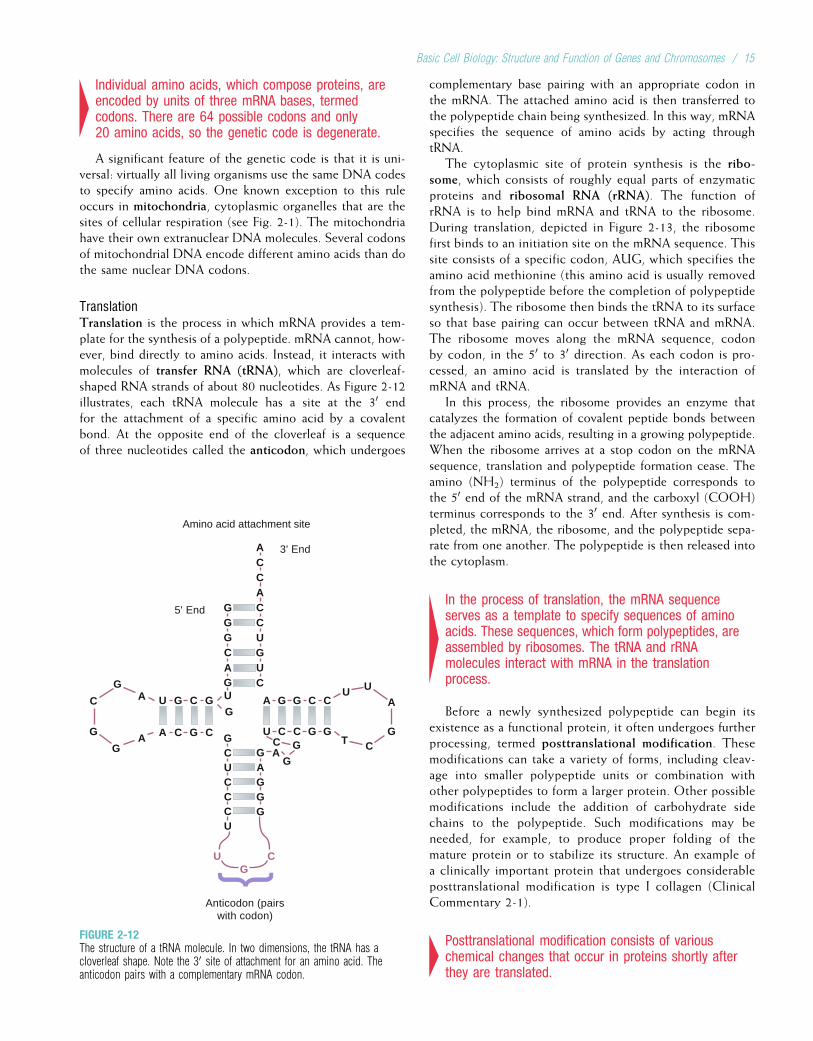
TranslationTranslation is the process in which mRNA provides a tem-plate for the synthesis of a polypeptide. mRNA cannot, how-ever, bind directly to amino acids. Instead, it interacts withmolecules of transfer RNA (tRNA), which are cloverleaf-shaped RNA strands of about 80 nucleotides. As Figure 2-12illustrates, each tRNA molecule has a site at the 30 endfor the attachment of a specific amino acid by a covalentbond. At the opposite end of the cloverleaf is a sequenceof three nucleotides called the anticodon, which undergoes
complementary base pairing with an appropriate codon inthe mRNA. The attached amino acid is then transferred tothe polypeptide chain being synthesized. In this way, mRNAspecifies the sequence of amino acids by acting throughtRNA.
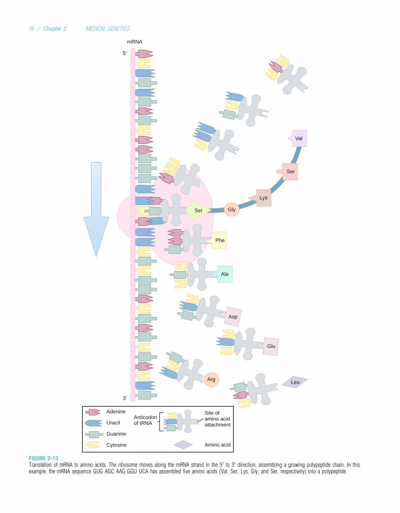
The cytoplasmic site of protein synthesis is the ribo-some, which consists of roughly equal parts of enzymaticproteins and ribosomal RNA (rRNA). The function ofrRNA is to help bind mRNA and tRNA to the ribosome.During translation, depicted in Figure 2-13, the ribosomefirst binds to an initiation site on the mRNA sequence. Thissite consists of a specific codon, AUG, which specifies theamino acid methionine (this amino acid is usually removedfrom the polypeptide before the completion of polypeptidesynthesis). The ribosome then binds the tRNA to its surfaceso that base pairing can occur between tRNA and mRNA.The ribosome moves along the mRNA sequence, codonby codon, in the 50 to 30 direction. As each codon is pro-cessed, an amino acid is translated by the interaction ofmRNA and tRNA.
In this process, the ribosome provides an enzyme thatcatalyzes the formation of covalent peptide bonds betweenthe adjacent amino acids, resulting in a growing polypeptide.When the ribosome arrives at a stop codon on the mRNAsequence, translation and polypeptide formation cease. Theamino (NH2) terminus of the polypeptide corresponds tothe 50 end of the mRNA strand, and the carboxyl (COOH)terminus corresponds to the 30 end. After synthesis is com-pleted, the mRNA, the ribosome, and the polypeptide sepa-rate from one another. The polypeptide is then released intothe cytoplasm.
4In the process of translation, the mRNA sequenceserves as a template to specify sequences of aminoacids. These sequences, which form polypeptides, areassembled by ribosomes. The tRNA and rRNAmolecules interact with mRNA in the translationprocess.
Before a newly synthesized polypeptide can begin itsexistence as a functional protein, it often undergoes furtherprocessing, termed posttranslational modification. Thesemodifications can take a variety of forms, including cleav-age into smaller polypeptide units or combination withother polypeptides to form a larger protein. Other possiblemodifications include the addition of carbohydrate sidechains to the polypeptide. Such modifications may beneeded, for example, to produce proper folding of themature protein or to stabilize its structure. An example ofa clinically important protein that undergoes considerableposttranslational modification is type I collagen (ClinicalCommentary 2-1).
4Posttranslational modification consists of variouschemical changes that occur in proteins shortly afterthey are translated.
5' End
G
ACCACCUGUC
G
U G C G
GGGCAGU
GCUCCC
C
U
U
AGGG
G
A
A
A C G C
G
C
G
G
A
A
G G C C
U CC
C GG
G
G
Anticodon (pairs with codon)
AU U
CTG
3' End
Amino acid attachment site
FIGURE 2-12The structure of a tRNA molecule. In two dimensions, the tRNA has acloverleaf shape. Note the 30 site of attachment for an amino acid. Theanticodon pairs with a complementary mRNA codon.
Basic Cell Biology: Structure and Function of Genes and Chromosomes / 15
5�
3�
mRNA
Arg Leu
Glu
Asp
Ala
Phe
Ser Gly
Lys
Ser
Val
Anticodonof tRNA
Adenine
Uracil
Guanine
Cytosine
Site ofamino acidattachment
Amino acid
FIGURE 2-13Translation of mRNA to amino acids. The ribosome moves along the mRNA strand in the 50 to 30 direction, assembling a growing polypeptide chain. In thisexample, the mRNA sequence GUG AGC AAG GGU UCA has assembled five amino acids (Val, Ser, Lys, Gly, and Ser, respectively) into a polypeptide.
16 / Chapter 2 MEDICAL GENETICS
CLINICAL COMMENTARY 2-1
Osteogenesis Imperfecta, an Inherited Collagen Disorder
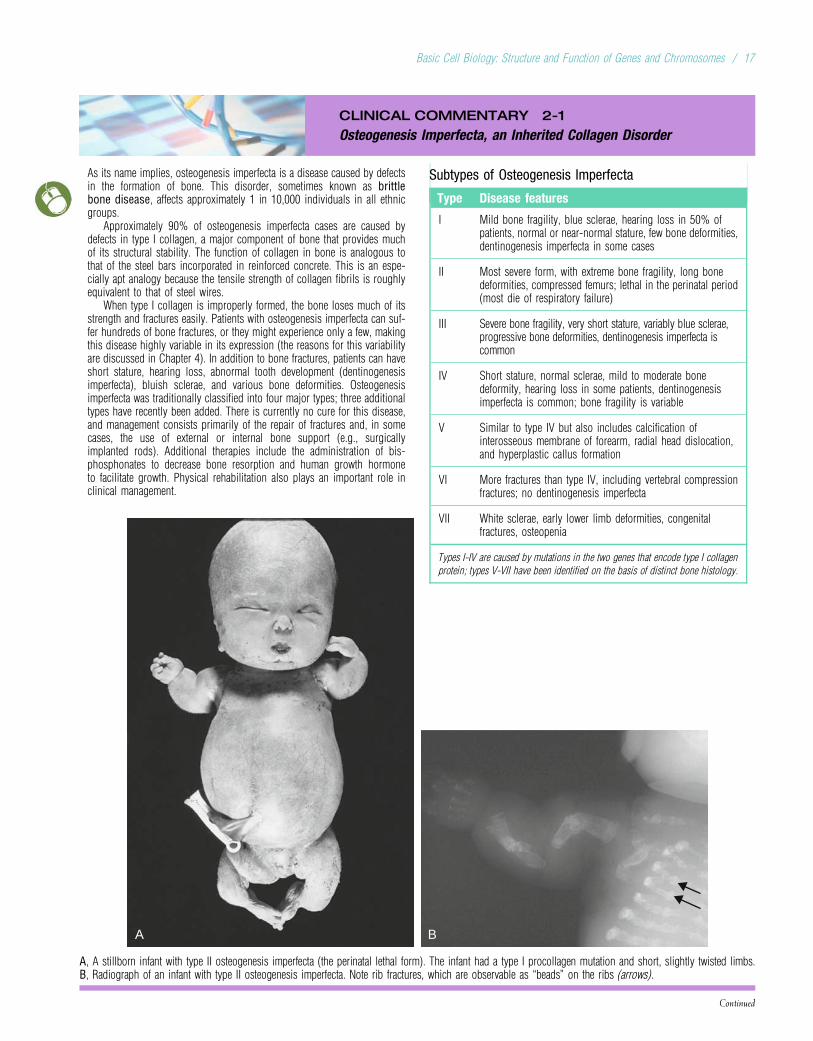
As its name implies, osteogenesis imperfecta is a disease caused by defectsin the formation of bone. This disorder, sometimes known as brittlebone disease, affects approximately 1 in 10,000 individuals in all ethnicgroups.
Approximately 90% of osteogenesis imperfecta cases are caused bydefects in type I collagen, a major component of bone that provides muchof its structural stability. The function of collagen in bone is analogous tothat of the steel bars incorporated in reinforced concrete. This is an espe-cially apt analogy because the tensile strength of collagen fibrils is roughlyequivalent to that of steel wires.
When type I collagen is improperly formed, the bone loses much of itsstrength and fractures easily. Patients with osteogenesis imperfecta can suf-fer hundreds of bone fractures, or they might experience only a few, makingthis disease highly variable in its expression (the reasons for this variabilityare discussed in Chapter 4). In addition to bone fractures, patients can haveshort stature, hearing loss, abnormal tooth development (dentinogenesisimperfecta), bluish sclerae, and various bone deformities. Osteogenesisimperfecta was traditionally classified into four major types; three additionaltypes have recently been added. There is currently no cure for this disease,and management consists primarily of the repair of fractures and, in somecases, the use of external or internal bone support (e.g., surgicallyimplanted rods). Additional therapies include the administration of bis-phosphonates to decrease bone resorption and human growth hormoneto facilitate growth. Physical rehabilitation also plays an important role inclinical management.
Subtypes of Osteogenesis Imperfecta
Type Disease features
I Mild bone fragility, blue sclerae, hearing loss in 50% ofpatients, normal or near-normal stature, few bone deformities,dentinogenesis imperfecta in some cases
II Most severe form, with extreme bone fragility, long bonedeformities, compressed femurs; lethal in the perinatal period(most die of respiratory failure)
III Severe bone fragility, very short stature, variably blue sclerae,progressive bone deformities, dentinogenesis imperfecta iscommon
IV Short stature, normal sclerae, mild to moderate bonedeformity, hearing loss in some patients, dentinogenesisimperfecta is common; bone fragility is variable
V Similar to type IV but also includes calcification ofinterosseous membrane of forearm, radial head dislocation,and hyperplastic callus formation
VI More fractures than type IV, including vertebral compressionfractures; no dentinogenesis imperfecta
VII White sclerae, early lower limb deformities, congenitalfractures, osteopenia
Types I-IV are caused by mutations in the two genes that encode type I collagen
protein; types V-VII have been identified on the basis of distinct bone histology.
Continued
A B
A, A stillborn infant with type II osteogenesis imperfecta (the perinatal lethal form). The infant had a type I procollagen mutation and short, slightly twisted limbs.B, Radiograph of an infant with type II osteogenesis imperfecta. Note rib fractures, which are observable as “beads” on the ribs (arrows).
Basic Cell Biology: Structure and Function of Genes and Chromosomes / 17
CLINICAL COMMENTARY 2-1
Osteogenesis Imperfecta, an Inherited Collagen Disorder—cont’d
Type I collagen is a trimeric protein (i.e., having three subunits) with atriple helix structure. It is formed from a precursor protein, type 1 procollagen.Two of the three subunits of type 1 procollagen, labeled pro-a1(I) chains, are
Triple-helix formation
Glycosylation ofselected hydroxylysines
NH2 COOH
OH
OH
OH
OH OH
Hydroxylation ofselected prolines andlysinesSynthesis of
pro-α chain
OH OH
OH
Nucleus
OH OH
OHOH
OH
OH OH
OH
OH
OH OH
OH
Secretion
Cleavage ofprocollagen
Assembly into fibril
Procollagen molecule
Collagen molecule
Collagen fibril
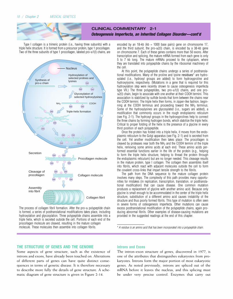
The process of collagen fibril formation. After the pro-a polypeptide chainis formed, a series of posttranslational modifications takes place, includinghydroxylation and glycosylation. Three polypeptide chains assemble into atriple helix, which is secreted outside the cell. Portions of each end of theprocollagen molecule are cleaved, resulting in the mature collagenmolecule. These molecules then assemble into collagen fibrils.
encoded by an 18-kb (kb = 1000 base pairs) gene on chromosome 17,and the third subunit, the pro-a2(I) chain, is encoded by a 38-kb geneon chromosome 7. Each of these genes contains more than 50 exons. Aftertranscription and splicing, the mature mRNA formed from each gene is only5 to 7 kb long. The mature mRNAs proceed to the cytoplasm, wherethey are translated into polypeptide chains by the ribosomal machinery ofthe cell.
At this point, the polypeptide chains undergo a series of posttransla-tional modifications. Many of the proline and lysine residues* are hydro-xylated (i.e., hydroxyl groups are added) to form hydroxyproline andhydroxylysine, respectively. (Mutations in a gene that is required for thishydroxylation step were recently shown to cause osteogenesis imperfectatype VII.) The three polypeptides, two pro-a1(I) chains, and one pro-a2(I) chain, begin to associate with one another at their COOH termini. Thisassociation is stabilized by sulfide bonds that form between the chains nearthe COOH termini. The triple helix then forms, in zipper-like fashion, begin-ning at the COOH terminus and proceeding toward the NH2 terminus.Some of the hydroxylysines are glycosylated (i.e., sugars are added), amodification that commonly occurs in the rough endoplasmic reticulum(see Fig. 2-1). The hydroxyl groups in the hydroxyprolines help to connectthe three chains by forming hydrogen bonds, which stabilize the triple helix.Critical to proper folding of the helix is the presence of a glycine in everythird position of each polypeptide.
Once the protein has folded into a triple helix, it moves from the endo-plasmic reticulum to the Golgi apparatus (see Fig. 2-1) and is secreted fromthe cell. Yet another modification then takes place: The procollagen iscleaved by proteases near both the NH2 and the COOH termini of the triplehelix, removing some amino acids at each end. These amino acids per-formed essential functions earlier in the life of the protein (e.g., helpingto form the triple helix structure, helping to thread the protein throughthe endoplasmic reticulum) but are no longer needed. This cleavage resultsin the mature protein, type I collagen. The collagen then assembles itselfinto fibrils, which react with adjacent molecules outside the cell to formthe covalent cross-links that impart tensile strength to the fibrils.
The path from the DNA sequence to the mature collagen proteininvolves many steps. The complexity of this path provides many opportu-nities for mistakes (in replication, transcription, translation, or posttransla-tional modification) that can cause disease. One common mutationproduces a replacement of glycine with another amino acid. Because onlyglycine is small enough to be accommodated in the center of the triple helixstructure, substitution of a different amino acid causes instability of thestructure and thus poorly formed fibrils. This type of mutation is often seenin severe forms of osteogenesis imperfecta. Other mutations can causeexcess posttranslational modification of the polypeptide chains, again pro-ducing abnormal fibrils. Other examples of disease-causing mutations areprovided in the suggested readings at the end of this chapter.
* A residue is an amino acid that has been incorporated into a polypeptide chain.
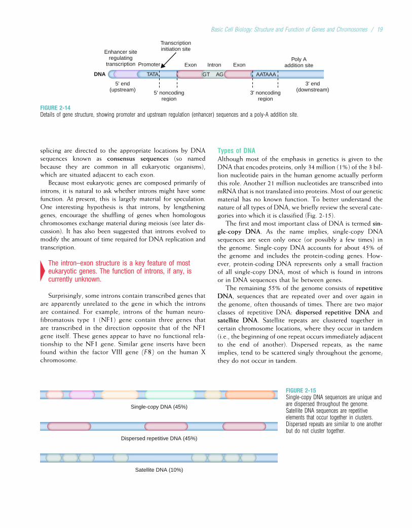
THE STRUCTURE OF GENES AND THE GENOMESome aspects of gene structure, such as the existence ofintrons and exons, have already been touched on. Alterationsof different parts of genes can have quite distinct conse-quences in terms of genetic disease. It is therefore necessaryto describe more fully the details of gene structure. A sche-matic diagram of gene structure is given in Figure 2-14.
Introns and ExonsThe intron–exon structure of genes, discovered in 1977, isone of the attributes that distinguishes eukaryotes from pro-karyotes. Introns form the major portion of most eukaryoticgenes. As noted previously, introns are spliced out of themRNA before it leaves the nucleus, and this splicing mustbe under very precise control. Enzymes that carry out
18 / Chapter 2 MEDICAL GENETICS
splicing are directed to the appropriate locations by DNAsequences known as consensus sequences (so namedbecause they are common in all eukaryotic organisms),which are situated adjacent to each exon.
Because most eukaryotic genes are composed primarily ofintrons, it is natural to ask whether introns might have somefunction. At present, this is largely material for speculation.One interesting hypothesis is that introns, by lengtheninggenes, encourage the shuffling of genes when homologouschromosomes exchange material during meiosis (see later dis-cussion). It has also been suggested that introns evolved tomodify the amount of time required for DNA replication andtranscription.
4The intron–exon structure is a key feature of mosteukaryotic genes. The function of introns, if any, iscurrently unknown.
Surprisingly, some introns contain transcribed genes thatare apparently unrelated to the gene in which the intronsare contained. For example, introns of the human neuro-fibromatosis type 1 (NF1) gene contain three genes thatare transcribed in the direction opposite that of the NF1gene itself. These genes appear to have no functional rela-tionship to the NF1 gene. Similar gene inserts have beenfound within the factor VIII gene (F8) on the human Xchromosome.
Types of DNAAlthough most of the emphasis in genetics is given to theDNA that encodes proteins, only 34 million (1%) of the 3 bil-lion nucleotide pairs in the human genome actually performthis role. Another 21 million nucleotides are transcribed intomRNA that is not translated into proteins. Most of our geneticmaterial has no known function. To better understand thenature of all types of DNA, we briefly review the several cate-gories into which it is classified (Fig. 2-15).
The first and most important class of DNA is termed sin-gle-copy DNA. As the name implies, single-copy DNAsequences are seen only once (or possibly a few times) inthe genome. Single-copy DNA accounts for about 45% ofthe genome and includes the protein-coding genes. How-ever, protein-coding DNA represents only a small fractionof all single-copy DNA, most of which is found in intronsor in DNA sequences that lie between genes.
The remaining 55% of the genome consists of repetitiveDNA, sequences that are repeated over and over again inthe genome, often thousands of times. There are two majorclasses of repetitive DNA: dispersed repetitive DNA andsatellite DNA. Satellite repeats are clustered together incertain chromosome locations, where they occur in tandem(i.e., the beginning of one repeat occurs immediately adjacentto the end of another). Dispersed repeats, as the nameimplies, tend to be scattered singly throughout the genome;they do not occur in tandem.
DNA
Enhancer siteregulating
transcription Promoter
Transcriptioninitiation site
3' end(downstream)
5' end(upstream) 3' noncoding
region5' noncoding
region
Poly Aaddition site
GT AG
Exon Intron Exon
AATAAATATA
FIGURE 2-14Details of gene structure, showing promoter and upstream regulation (enhancer) sequences and a poly-A addition site.
Single-copy DNA (45%)
Dispersed repetitive DNA (45%)
Satellite DNA (10%)
FIGURE 2-15Single-copy DNA sequences are unique andare dispersed throughout the genome.Satellite DNA sequences are repetitiveelements that occur together in clusters.Dispersed repeats are similar to one anotherbut do not cluster together.
Basic Cell Biology: Structure and Function of Genes and Chromosomes / 19
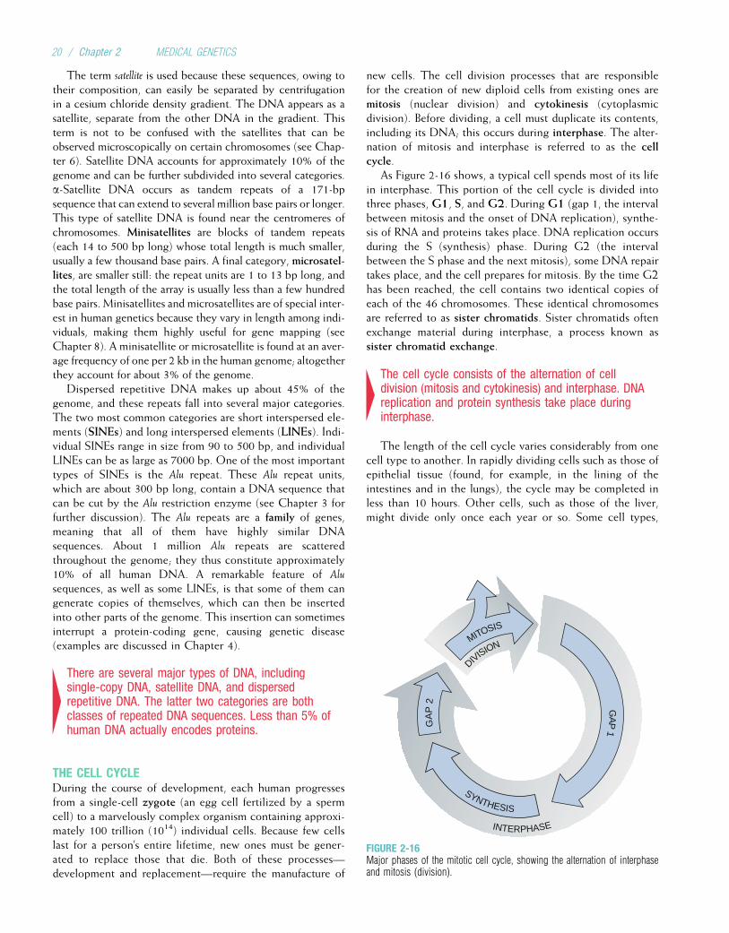
The term satellite is used because these sequences, owing totheir composition, can easily be separated by centrifugationin a cesium chloride density gradient. The DNA appears as asatellite, separate from the other DNA in the gradient. Thisterm is not to be confused with the satellites that can beobserved microscopically on certain chromosomes (see Chap-ter 6). Satellite DNA accounts for approximately 10% of thegenome and can be further subdivided into several categories.a-Satellite DNA occurs as tandem repeats of a 171-bpsequence that can extend to several million base pairs or longer.This type of satellite DNA is found near the centromeres ofchromosomes. Minisatellites are blocks of tandem repeats(each 14 to 500 bp long) whose total length is much smaller,usually a few thousand base pairs. A final category, microsatel-lites, are smaller still: the repeat units are 1 to 13 bp long, andthe total length of the array is usually less than a few hundredbase pairs. Minisatellites and microsatellites are of special inter-est in human genetics because they vary in length among indi-viduals, making them highly useful for gene mapping (seeChapter 8). A minisatellite or microsatellite is found at an aver-age frequency of one per 2 kb in the human genome; altogetherthey account for about 3% of the genome.
Dispersed repetitive DNA makes up about 45% of thegenome, and these repeats fall into several major categories.The two most common categories are short interspersed ele-ments (SINEs) and long interspersed elements (LINEs). Indi-vidual SINEs range in size from 90 to 500 bp, and individualLINEs can be as large as 7000 bp. One of the most importanttypes of SINEs is the Alu repeat. These Alu repeat units,which are about 300 bp long, contain a DNA sequence thatcan be cut by the Alu restriction enzyme (see Chapter 3 forfurther discussion). The Alu repeats are a family of genes,meaning that all of them have highly similar DNAsequences. About 1 million Alu repeats are scatteredthroughout the genome; they thus constitute approximately10% of all human DNA. A remarkable feature of Alu
sequences, as well as some LINEs, is that some of them cangenerate copies of themselves, which can then be insertedinto other parts of the genome. This insertion can sometimesinterrupt a protein-coding gene, causing genetic disease(examples are discussed in Chapter 4).
4There are several major types of DNA, includingsingle-copy DNA, satellite DNA, and dispersedrepetitive DNA. The latter two categories are bothclasses of repeated DNA sequences. Less than 5% ofhuman DNA actually encodes proteins.
THE CELL CYCLEDuring the course of development, each human progressesfrom a single-cell zygote (an egg cell fertilized by a spermcell) to a marvelously complex organism containing approxi-mately 100 trillion (1014) individual cells. Because few cellslast for a person’s entire lifetime, new ones must be gener-ated to replace those that die. Both of these processes—development and replacement—require the manufacture of
new cells. The cell division processes that are responsiblefor the creation of new diploid cells from existing ones aremitosis (nuclear division) and cytokinesis (cytoplasmicdivision). Before dividing, a cell must duplicate its contents,including its DNA; this occurs during interphase. The alter-nation of mitosis and interphase is referred to as the cellcycle.
As Figure 2-16 shows, a typical cell spends most of its lifein interphase. This portion of the cell cycle is divided intothree phases, G1, S, and G2. During G1 (gap 1, the intervalbetween mitosis and the onset of DNA replication), synthe-sis of RNA and proteins takes place. DNA replication occursduring the S (synthesis) phase. During G2 (the intervalbetween the S phase and the next mitosis), some DNA repairtakes place, and the cell prepares for mitosis. By the time G2has been reached, the cell contains two identical copies ofeach of the 46 chromosomes. These identical chromosomesare referred to as sister chromatids. Sister chromatids oftenexchange material during interphase, a process known assister chromatid exchange.
4The cell cycle consists of the alternation of celldivision (mitosis and cytokinesis) and interphase. DNAreplication and protein synthesis take place duringinterphase.
The length of the cell cycle varies considerably from onecell type to another. In rapidly dividing cells such as those ofepithelial tissue (found, for example, in the lining of theintestines and in the lungs), the cycle may be completed inless than 10 hours. Other cells, such as those of the liver,might divide only once each year or so. Some cell types,
MITOSIS
DIVISION
GA
P 2
GA
P 1
INTERPHASE
SYNTHESIS
FIGURE 2-16Major phases of the mitotic cell cycle, showing the alternation of interphaseand mitosis (division).
20 / Chapter 2 MEDICAL GENETICS
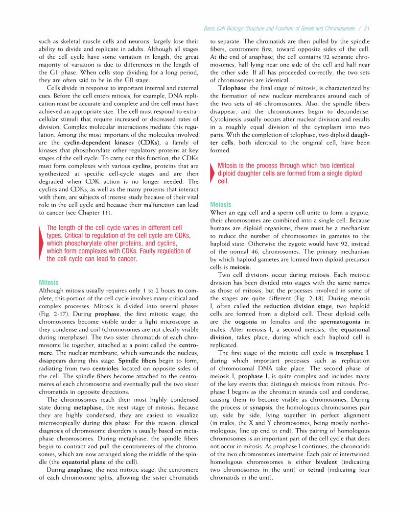
such as skeletal muscle cells and neurons, largely lose theirability to divide and replicate in adults. Although all stagesof the cell cycle have some variation in length, the greatmajority of variation is due to differences in the length ofthe G1 phase. When cells stop dividing for a long period,they are often said to be in the G0 stage.
Cells divide in response to important internal and externalcues. Before the cell enters mitosis, for example, DNA repli-cation must be accurate and complete and the cell must haveachieved an appropriate size. The cell must respond to extra-cellular stimuli that require increased or decreased rates ofdivision. Complex molecular interactions mediate this regu-lation. Among the most important of the molecules involvedare the cyclin-dependent kinases (CDKs), a family ofkinases that phosphorylate other regulatory proteins at keystages of the cell cycle. To carry out this function, the CDKsmust form complexes with various cyclins, proteins that aresynthesized at specific cell-cycle stages and are thendegraded when CDK action is no longer needed. Thecyclins and CDKs, as well as the many proteins that interactwith them, are subjects of intense study because of their vitalrole in the cell cycle and because their malfunction can leadto cancer (see Chapter 11).
4The length of the cell cycle varies in different celltypes. Critical to regulation of the cell cycle are CDKs,which phosphorylate other proteins, and cyclins,which form complexes with CDKs. Faulty regulation ofthe cell cycle can lead to cancer.
MitosisAlthough mitosis usually requires only 1 to 2 hours to com-plete, this portion of the cell cycle involves many critical andcomplex processes. Mitosis is divided into several phases(Fig. 2-17). During prophase, the first mitotic stage, thechromosomes become visible under a light microscope asthey condense and coil (chromosomes are not clearly visibleduring interphase). The two sister chromatids of each chro-mosome lie together, attached at a point called the centro-mere. The nuclear membrane, which surrounds the nucleus,disappears during this stage. Spindle fibers begin to form,radiating from two centrioles located on opposite sides ofthe cell. The spindle fibers become attached to the centro-meres of each chromosome and eventually pull the two sisterchromatids in opposite directions.
The chromosomes reach their most highly condensedstate during metaphase, the next stage of mitosis. Becausethey are highly condensed, they are easiest to visualizemicroscopically during this phase. For this reason, clinicaldiagnosis of chromosome disorders is usually based on meta-phase chromosomes. During metaphase, the spindle fibersbegin to contract and pull the centromeres of the chromo-somes, which are now arranged along the middle of the spin-dle (the equatorial plane of the cell).
During anaphase, the next mitotic stage, the centromereof each chromosome splits, allowing the sister chromatids
to separate. The chromatids are then pulled by the spindlefibers, centromere first, toward opposite sides of the cell.At the end of anaphase, the cell contains 92 separate chro-mosomes, half lying near one side of the cell and half nearthe other side. If all has proceeded correctly, the two setsof chromosomes are identical.
Telophase, the final stage of mitosis, is characterized bythe formation of new nuclear membranes around each ofthe two sets of 46 chromosomes. Also, the spindle fibersdisappear, and the chromosomes begin to decondense.Cytokinesis usually occurs after nuclear division and resultsin a roughly equal division of the cytoplasm into twoparts. With the completion of telophase, two diploid daugh-ter cells, both identical to the original cell, have beenformed.
4Mitosis is the process through which two identicaldiploid daughter cells are formed from a single diploidcell.
MeiosisWhen an egg cell and a sperm cell unite to form a zygote,their chromosomes are combined into a single cell. Becausehumans are diploid organisms, there must be a mechanismto reduce the number of chromosomes in gametes to thehaploid state. Otherwise the zygote would have 92, insteadof the normal 46, chromosomes. The primary mechanismby which haploid gametes are formed from diploid precursorcells is meiosis.
Two cell divisions occur during meiosis. Each meioticdivision has been divided into stages with the same namesas those of mitosis, but the processes involved in some ofthe stages are quite different (Fig. 2-18). During meiosisI, often called the reduction division stage, two haploidcells are formed from a diploid cell. These diploid cellsare the oogonia in females and the spermatogonia inmales. After meiosis I, a second meiosis, the equationaldivision, takes place, during which each haploid cell isreplicated.
The first stage of the meiotic cell cycle is interphase I,during which important processes such as replicationof chromosomal DNA take place. The second phase ofmeiosis I, prophase I, is quite complex and includes manyof the key events that distinguish meiosis from mitosis. Pro-phase I begins as the chromatin strands coil and condense,causing them to become visible as chromosomes. Duringthe process of synapsis, the homologous chromosomes pairup, side by side, lying together in perfect alignment(in males, the X and Y chromosomes, being mostly nonho-mologous, line up end to end). This pairing of homologouschromosomes is an important part of the cell cycle that doesnot occur in mitosis. As prophase I continues, the chromatidsof the two chromosomes intertwine. Each pair of intertwinedhomologous chromosomes is either bivalent (indicatingtwo chromosomes in the unit) or tetrad (indicating fourchromatids in the unit).
Basic Cell Biology: Structure and Function of Genes and Chromosomes / 21
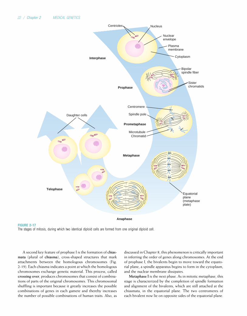
A second key feature of prophase I is the formation of chias-mata (plural of chiasma), cross-shaped structures that markattachments between the homologous chromosomes (Fig.2-19). Each chiasma indicates a point at which the homologouschromosomes exchange genetic material. This process, calledcrossing over, produces chromosomes that consist of combina-tions of parts of the original chromosomes. This chromosomalshuffling is important because it greatly increases the possiblecombinations of genes in each gamete and thereby increasesthe number of possible combinations of human traits. Also, as
discussed in Chapter 8, this phenomenon is critically importantin inferring the order of genes along chromosomes. At the endof prophase I, the bivalents begin to move toward the equato-rial plane, a spindle apparatus begins to form in the cytoplasm,and the nuclear membrane dissipates.
Metaphase I is the next phase. As in mitotic metaphase, thisstage is characterized by the completion of spindle formationand alignment of the bivalents, which are still attached at thechiasmata, in the equatorial plane. The two centromeres ofeach bivalent now lie on opposite sides of the equatorial plane.
Nuclearenvelope
Plasmamembrane
Bipolarspindle fiber
Sisterchromatids
Cytoplasm
Daughter cells
NucleusCentrioles
Centromere
Spindle pole
MicrotubuleChromatid
Prophase
Prometaphase
Metaphase
Interphase
Anaphase
TelophaseEquatorialplane(metaphaseplate)
FIGURE 2-17The stages of mitosis, during which two identical diploid cells are formed from one original diploid cell.
22 / Chapter 2 MEDICAL GENETICS
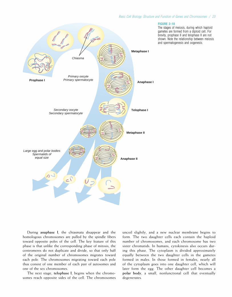
During anaphase I, the chiasmata disappear and thehomologous chromosomes are pulled by the spindle fiberstoward opposite poles of the cell. The key feature of thisphase is that unlike the corresponding phase of mitosis, thecentromeres do not duplicate and divide, so that only halfof the original number of chromosomes migrates towardeach pole. The chromosomes migrating toward each polethus consist of one member of each pair of autosomes andone of the sex chromosomes.
The next stage, telophase I, begins when the chromo-somes reach opposite sides of the cell. The chromosomes
uncoil slightly, and a new nuclear membrane begins toform. The two daughter cells each contain the haploidnumber of chromosomes, and each chromosome has twosister chromatids. In humans, cytokinesis also occurs dur-ing this phase. The cytoplasm is divided approximatelyequally between the two daughter cells in the gametesformed in males. In those formed in females, nearly allof the cytoplasm goes into one daughter cell, which willlater form the egg. The other daughter cell becomes apolar body, a small, nonfunctional cell that eventuallydegenerates.
Chiasma
Primary oocytePrimary spermatocyte
Secondary oocyteSecondary spermatocyte
Large egg and polar bodiesSpermatids of
equal size
Metaphase II
Anaphase II
Telophase I
Prophase I
Metaphase I
Anaphase I
FIGURE 2-18The stages of meiosis, during which haploidgametes are formed from a diploid cell. Forbrevity, prophase II and telophase II are notshown. Note the relationship between meiosisand spermatogenesis and oogenesis.
Basic Cell Biology: Structure and Function of Genes and Chromosomes / 23
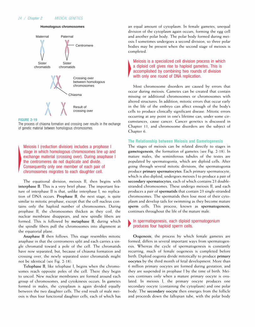
4Meiosis I (reduction division) includes a prophase Istage in which homologous chromosomes line up andexchange material (crossing over). During anaphase Ithe centromeres do not duplicate and divide.Consequently only one member of each pair ofchromosomes migrates to each daughter cell.
The equational division, meiosis II, then begins withinterphase II. This is a very brief phase. The important fea-ture of interphase II is that, unlike interphase I, no replica-tion of DNA occurs. Prophase II, the next stage, is quitesimilar to mitotic prophase, except that the cell nucleus con-tains only the haploid number of chromosomes. Duringprophase II, the chromosomes thicken as they coil, thenuclear membrane disappears, and new spindle fibers areformed. This is followed by metaphase II, during whichthe spindle fibers pull the chromosomes into alignment atthe equatorial plane.
Anaphase II then follows. This stage resembles mitoticanaphase in that the centromeres split and each carries a sin-gle chromatid toward a pole of the cell. The chromatidshave now separated, but, because of chiasma formation andcrossing over, the newly separated sister chromatids mightnot be identical (see Fig. 2-18).
Telophase II, like telophase I, begins when the chromo-somes reach opposite poles of the cell. There they beginto uncoil. New nuclear membranes are formed around eachgroup of chromosomes, and cytokinesis occurs. In gametesformed in males, the cytoplasm is again divided equallybetween the two daughter cells. The end result of male mei-osis is thus four functional daughter cells, each of which has
an equal amount of cytoplasm. In female gametes, unequaldivision of the cytoplasm again occurs, forming the egg celland another polar body. The polar body formed during mei-osis I sometimes undergoes a second division, so three polarbodies may be present when the second stage of meiosis iscompleted.
4Meiosis is a specialized cell division process in whicha diploid cell gives rise to haploid gametes. This isaccomplished by combining two rounds of divisionwith only one round of DNA replication.
Most chromosome disorders are caused by errors thatoccur during meiosis. Gametes can be created that containmissing or additional chromosomes or chromosomes withaltered structures. In addition, mitotic errors that occur earlyin the life of the embryo can affect enough of the body’scells to produce clinically significant disease. Mitotic errorsoccurring at any point in one’s lifetime can, under some cir-cumstances, cause cancer. Cancer genetics is discussed inChapter 11, and chromosome disorders are the subject ofChapter 6.
The Relationship between Meiosis and GametogenesisThe stages of meiosis can be related directly to stages ingametogenesis, the formation of gametes (see Fig. 2-18). Inmature males, the seminiferous tubules of the testes arepopulated by spermatogonia, which are diploid cells. Aftergoing through several mitotic divisions, the spermatogoniaproduce primary spermatocytes. Each primary spermatocyte,which is also diploid, undergoes meiosis I to produce a pair ofsecondary spermatocytes, each of which contains 23 double-stranded chromosomes. These undergo meiosis II, and eachproduces a pair of spermatids that contain 23 single-strandedchromosomes. The spermatids then lose most of their cyto-plasm and develop tails for swimming as they become maturesperm cells. This process, known as spermatogenesis,continues throughout the life of the mature male.
4In spermatogenesis, each diploid spermatogoniumproduces four haploid sperm cells.
Oogenesis, the process by which female gametes areformed, differs in several important ways from spermatogen-esis. Whereas the cycle of spermatogenesis is constantlyrecurring, much of female oogenesis is completed beforebirth. Diploid oogonia divide mitotically to produce primaryoocytes by the third month of fetal development. More than6 million primary oocytes are formed during gestation, andthey are suspended in prophase I by the time of birth. Mei-osis continues only when a mature primary oocyte is ovu-lated. In meiosis I, the primary oocyte produces onesecondary oocyte (containing the cytoplasm) and one polarbody. The secondary oocyte then emerges from the follicleand proceeds down the fallopian tube, with the polar body
Maternal Paternal
Sisterchromatids
Sisterchromatids
Centromere
Crossing overbetween homologouschromosomes
Result ofcrossing over
Chiasma
Homologous chromosomes
FIGURE 2-19The process of chiasma formation and crossing over results in the exchangeof genetic material between homologous chromosomes.
24 / Chapter 2 MEDICAL GENETICS
attached to it. Meiosis II begins only if the secondary oocyteis fertilized by a sperm cell. If this occurs, one haploidmature ovum, containing the cytoplasm, and another hap-loid polar body are produced. The polar bodies eventuallydisintegrate. About 1 hour after fertilization, the nuclei ofthe sperm cell and ovum fuse, forming a diploid zygote.The zygote then begins its development into an embryothrough a series of mitotic divisions.
4In oogenesis, one haploid ovum and three haploidpolar bodies are produced meiotically from a diploidoogonium. In contrast to spermatogenesis, whichcontinues throughout the life of the mature male, thefirst phase of oogenesis is completed before thefemale is born; oogenesis is then halted untilovulation occurs.
Study Questions
1. Consider the following double-stranded DNAsequence:
50-CAG AAG AAA ATT AAC ATG TAA-30
30-GTC TTC TTT TAA TTG TAC ATT-50
If the bottom strand serves as the template, whatis the mRNA sequence produced by transcriptionof this DNA sequence? What is the amino acidsequence produced by translation of the mRNAsequence?
2. Arrange the following terms according to theirhierarchical relationship to one another: genes,chromosomes, exons, codons, nucleotides,genome.
3. Less than 5% of human DNA encodes proteins.Furthermore, in a given cell type only 10% of thecoding DNA actively encodes proteins. Explain thesestatements.
4. What are the major differences between mitosis andmeiosis?
5. The human body contains approximately 1014 cells.Starting with a single-cell zygote, how many mitoticcell divisions, on average, would be required toproduce this number of cells?
6. How many mature sperm cells will be produced by100 primary spermatocytes? How many mature eggcells will be produced by 100 primary oocytes?
Suggested ReadingsAlberts B, Johnson A, Lewis J, et al. Molecular Biology of the
Cell, 4th ed. New York: Garland Science, 2002.Berger SL. Histone modifications in transcriptional regula-
tion. Curr Opin Genet Dev 2002;12:142-48.Byers PH. Osteogenesis imperfecta: Perspectives and oppor-
tunities. Curr Opin Pediatr 2000;12:603-9.Cho KS, Elizondo LI, Boerkoel CF. Advances in chromatin
remodeling and human disease. Current Opin GenetDev 2004;14:308-15.
Cook PR. The organization of replication and transcription.Science 1999;284:1790-5.
Johnson CA. Chromatin modification and disease. J MedGenet 2000;37:905-15.
Lander ES, Linton LM, Birren B, et al. Initial sequencingand analysis of the human genome. Nature 2001;409:860-921.
Lemon B, Tjian R. Orchestrated response: A symphony oftranscription factors for gene control. Genes Dev2000;14: 2551-69.
Lewin B. Genes IX. Boston: Jones and Bartlett, 2008.Mitchison TJ, Salmon ED. Mitosis: A history of division. NatCell Biol 2001;3:E17-21.
Page SL, Hawley RS. Chromosome choreography. Themeiotic ballet. Science 2003;301:785-89.
Rauch F, Glorieux FH. Osteogenesis imperfecta. Lancet2004;363:1377-85.
Internet ResourcesMitosis and meiosis tutorials and animations http://www.biology.arizona.edu/cell_bio/cell_bio.html
Tutorial on DNA structure, replication, transcription, andtranslation http://www.ncc.gmu.edu/dna/
Basic Cell Biology: Structure and Function of Genes and Chromosomes / 25
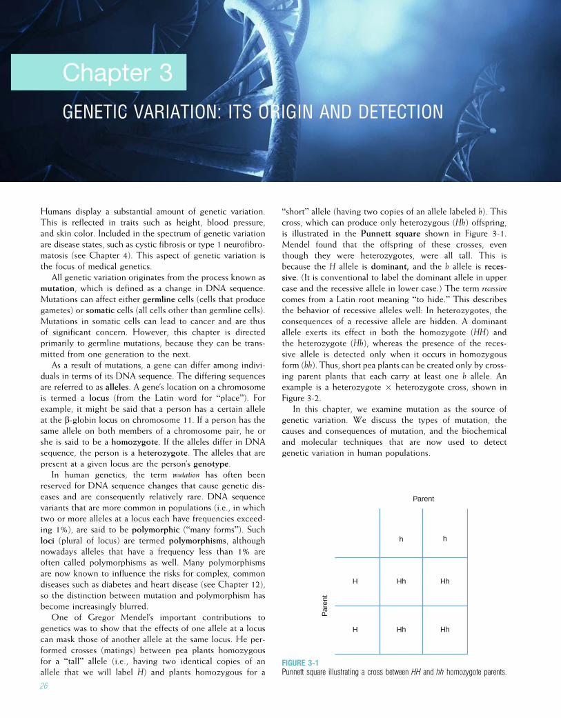
Chapter 3
GENETIC VARIATION: ITS ORIGIN AND DETECTION
Humans display a substantial amount of genetic variation.This is reflected in traits such as height, blood pressure,and skin color. Included in the spectrum of genetic variationare disease states, such as cystic fibrosis or type 1 neurofibro-matosis (see Chapter 4). This aspect of genetic variation isthe focus of medical genetics.
All genetic variation originates from the process known asmutation, which is defined as a change in DNA sequence.Mutations can affect either germline cells (cells that producegametes) or somatic cells (all cells other than germline cells).Mutations in somatic cells can lead to cancer and are thusof significant concern. However, this chapter is directedprimarily to germline mutations, because they can be trans-mitted from one generation to the next.
As a result of mutations, a gene can differ among indivi-duals in terms of its DNA sequence. The differing sequencesare referred to as alleles. A gene’s location on a chromosomeis termed a locus (from the Latin word for “place”). Forexample, it might be said that a person has a certain alleleat the b-globin locus on chromosome 11. If a person has thesame allele on both members of a chromosome pair, he orshe is said to be a homozygote. If the alleles differ in DNAsequence, the person is a heterozygote. The alleles that arepresent at a given locus are the person’s genotype.
In human genetics, the term mutation has often beenreserved for DNA sequence changes that cause genetic dis-eases and are consequently relatively rare. DNA sequencevariants that are more common in populations (i.e., in whichtwo or more alleles at a locus each have frequencies exceed-ing 1%), are said to be polymorphic (“many forms”). Suchloci (plural of locus) are termed polymorphisms, althoughnowadays alleles that have a frequency less than 1% areoften called polymorphisms as well. Many polymorphismsare now known to influence the risks for complex, commondiseases such as diabetes and heart disease (see Chapter 12),so the distinction between mutation and polymorphism hasbecome increasingly blurred.
One of Gregor Mendel’s important contributions togenetics was to show that the effects of one allele at a locuscan mask those of another allele at the same locus. He per-formed crosses (matings) between pea plants homozygousfor a “tall” allele (i.e., having two identical copies of anallele that we will label H) and plants homozygous for a
“short” allele (having two copies of an allele labeled h). Thiscross, which can produce only heterozygous (Hh) offspring,is illustrated in the Punnett square shown in Figure 3-1.Mendel found that the offspring of these crosses, eventhough they were heterozygotes, were all tall. This isbecause the H allele is dominant, and the h allele is reces-sive. (It is conventional to label the dominant allele in uppercase and the recessive allele in lower case.) The term recessivecomes from a Latin root meaning “to hide.” This describesthe behavior of recessive alleles well: In heterozygotes, theconsequences of a recessive allele are hidden. A dominantallele exerts its effect in both the homozygote (HH) andthe heterozygote (Hh), whereas the presence of the reces-sive allele is detected only when it occurs in homozygousform (hh). Thus, short pea plants can be created only by cross-ing parent plants that each carry at least one h allele. Anexample is a heterozygote � heterozygote cross, shown inFigure 3-2.
In this chapter, we examine mutation as the source ofgenetic variation. We discuss the types of mutation, thecauses and consequences of mutation, and the biochemicaland molecular techniques that are now used to detectgenetic variation in human populations.
Parent
Par
ent
h h
H Hh Hh
H Hh Hh
FIGURE 3-1Punnett square illustrating a cross between HH and hh homozygote parents.
26
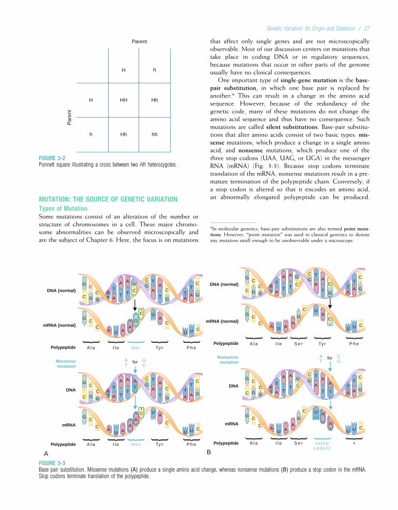
MUTATION: THE SOURCE OF GENETIC VARIATIONTypes of MutationSome mutations consist of an alteration of the number orstructure of chromosomes in a cell. These major chromo-some abnormalities can be observed microscopically andare the subject of Chapter 6. Here, the focus is on mutations
that affect only single genes and are not microscopicallyobservable. Most of our discussion centers on mutations thattake place in coding DNA or in regulatory sequences,because mutations that occur in other parts of the genomeusually have no clinical consequences.
One important type of single-gene mutation is the base-pair substitution, in which one base pair is replaced byanother.* This can result in a change in the amino acidsequence. However, because of the redundancy of thegenetic code, many of these mutations do not change theamino acid sequence and thus have no consequence. Suchmutations are called silent substitutions. Base-pair substitu-tions that alter amino acids consist of two basic types: mis-sense mutations, which produce a change in a single aminoacid, and nonsense mutations, which produce one of thethree stop codons (UAA, UAG, or UGA) in the messengerRNA (mRNA) (Fig. 3-3). Because stop codons terminatetranslation of the mRNA, nonsense mutations result in a pre-mature termination of the polypeptide chain. Conversely, ifa stop codon is altered so that it encodes an amino acid,an abnormally elongated polypeptide can be produced.
*In molecular genetics, base-pair substitutions are also termed point muta-tions. However, “point mutation” was used in classical genetics to denoteany mutation small enough to be unobservable under a microscope.
G G GCC A
T
ATA
T
AG
C
G
C
GG T
A
T
T
AA
T
A
T C
C
GC
CA A
AU U U
CC
GU
AC
GC
CA A
AA
U U U
CC
UAC
C
G GCC A
T
ATA
T
AG
C
G
C
GG T
A
T
T
AA
T
A
T
A
T
C
C
DNA (normal)
mRNA (normal)
Polypeptide
Missensemutation
DNA
mRNA
A l a I l e
forAT
GC
Ty r P h eS e r
Polypeptide A l a I l e Ty r P h eA s n
A
G GGC
C AT
ATA
T
AG
C
G
C
GG T
A
T
T
AA
T
A
T C
C
GC
CA A
AU U U
CC
GU
AC
GC
CA AU U U C
U A
A
C
G GCC A
T
ATA
T
AG
C
GG T
A
T
T
AA
T
A
T
A
T
C
C
DNA (normal)
mRNA (normal)
Polypeptide
Nonsensemutation
DNA
mRNA
A l a I l e
forAT
CG
Ty r P h eS e r
Polypeptide A l a I l e S e r ( s t o pc o d o n )
•
B
AG
C
G
C
FIGURE 3-3Base pair substitution. Missense mutations (A) produce a single amino acid change, whereas nonsense mutations (B) produce a stop codon in the mRNA.Stop codons terminate translation of the polypeptide.
Parent
Par
ent
H h
H HH Hh
h Hh hh
FIGURE 3-2Punnett square illustrating a cross between two Hh heterozygotes.
Genetic Variation: Its Origin and Detection / 27
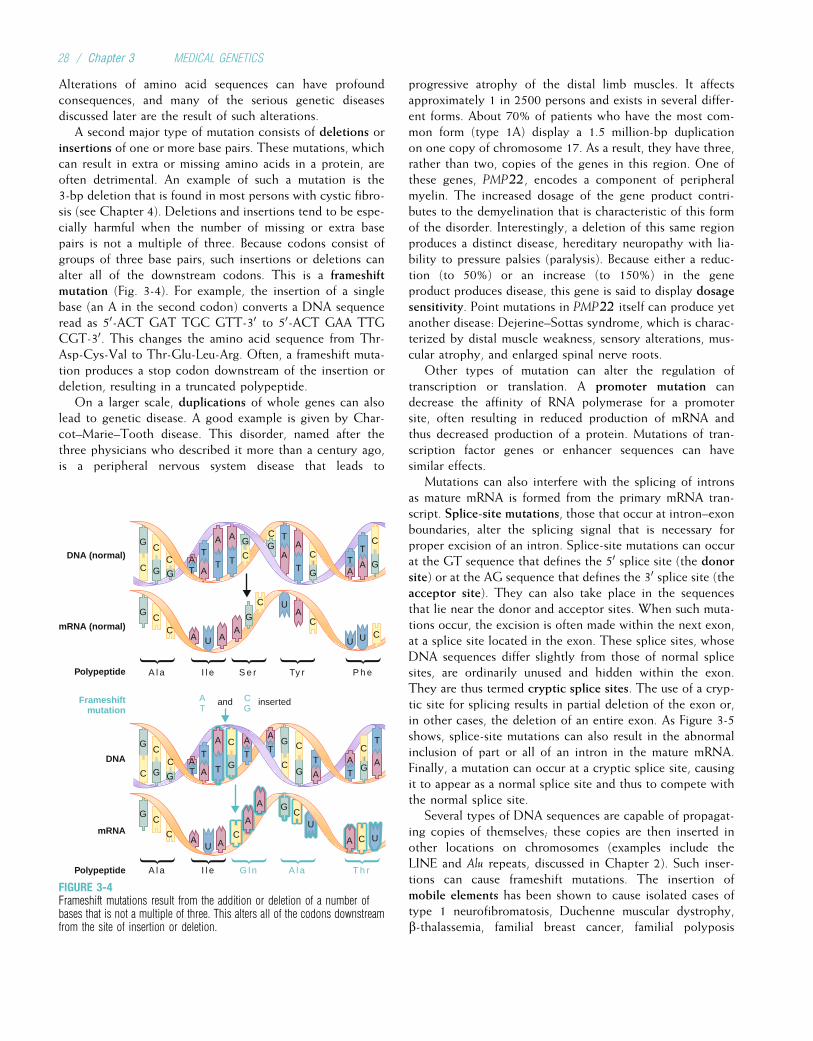
Alterations of amino acid sequences can have profoundconsequences, and many of the serious genetic diseasesdiscussed later are the result of such alterations.
A second major type of mutation consists of deletions orinsertions of one or more base pairs. These mutations, whichcan result in extra or missing amino acids in a protein, areoften detrimental. An example of such a mutation is the3-bp deletion that is found in most persons with cystic fibro-sis (see Chapter 4). Deletions and insertions tend to be espe-cially harmful when the number of missing or extra basepairs is not a multiple of three. Because codons consist ofgroups of three base pairs, such insertions or deletions canalter all of the downstream codons. This is a frameshiftmutation (Fig. 3-4). For example, the insertion of a singlebase (an A in the second codon) converts a DNA sequenceread as 50-ACT GAT TGC GTT-30 to 50-ACT GAA TTGCGT-30. This changes the amino acid sequence from Thr-Asp-Cys-Val to Thr-Glu-Leu-Arg. Often, a frameshift muta-tion produces a stop codon downstream of the insertion ordeletion, resulting in a truncated polypeptide.
On a larger scale, duplications of whole genes can alsolead to genetic disease. A good example is given by Char-cot–Marie–Tooth disease. This disorder, named after thethree physicians who described it more than a century ago,is a peripheral nervous system disease that leads to
progressive atrophy of the distal limb muscles. It affectsapproximately 1 in 2500 persons and exists in several differ-ent forms. About 70% of patients who have the most com-mon form (type 1A) display a 1.5 million-bp duplicationon one copy of chromosome 17. As a result, they have three,rather than two, copies of the genes in this region. One ofthese genes, PMP22, encodes a component of peripheralmyelin. The increased dosage of the gene product contri-butes to the demyelination that is characteristic of this formof the disorder. Interestingly, a deletion of this same regionproduces a distinct disease, hereditary neuropathy with lia-bility to pressure palsies (paralysis). Because either a reduc-tion (to 50%) or an increase (to 150%) in the geneproduct produces disease, this gene is said to display dosagesensitivity. Point mutations in PMP22 itself can produce yetanother disease: Dejerine–Sottas syndrome, which is charac-terized by distal muscle weakness, sensory alterations, mus-cular atrophy, and enlarged spinal nerve roots.
Other types of mutation can alter the regulation oftranscription or translation. A promoter mutation candecrease the affinity of RNA polymerase for a promotersite, often resulting in reduced production of mRNA andthus decreased production of a protein. Mutations of tran-scription factor genes or enhancer sequences can havesimilar effects.
Mutations can also interfere with the splicing of intronsas mature mRNA is formed from the primary mRNA tran-script. Splice-site mutations, those that occur at intron–exonboundaries, alter the splicing signal that is necessary forproper excision of an intron. Splice-site mutations can occurat the GT sequence that defines the 50 splice site (the donorsite) or at the AG sequence that defines the 30 splice site (theacceptor site). They can also take place in the sequencesthat lie near the donor and acceptor sites. When such muta-tions occur, the excision is often made within the next exon,at a splice site located in the exon. These splice sites, whoseDNA sequences differ slightly from those of normal splicesites, are ordinarily unused and hidden within the exon.They are thus termed cryptic splice sites. The use of a cryp-tic site for splicing results in partial deletion of the exon or,in other cases, the deletion of an entire exon. As Figure 3-5shows, splice-site mutations can also result in the abnormalinclusion of part or all of an intron in the mature mRNA.Finally, a mutation can occur at a cryptic splice site, causingit to appear as a normal splice site and thus to compete withthe normal splice site.
Several types of DNA sequences are capable of propagat-ing copies of themselves; these copies are then inserted inother locations on chromosomes (examples include theLINE and Alu repeats, discussed in Chapter 2). Such inser-tions can cause frameshift mutations. The insertion ofmobile elements has been shown to cause isolated cases oftype 1 neurofibromatosis, Duchenne muscular dystrophy,b-thalassemia, familial breast cancer, familial polyposis
G G GCC A
T
ATA
T
AG
C
G
C
GG T
A
T
T
AA
T
A
T C
C
GC
CA A
AU U U
CC
GU
AC
GG
C
CA A
A
A
AU
U
C
C
UC
C
GC
C AT
AAT
T
A
T
AG
C
G
C
G
C
GG T
A
T
A
T
A
T
C
G
C
DNA (normal)
mRNA (normal)
Polypeptide
Frameshiftmutation
DNA
mRNA
A l a I l e
andAT
CG
Ty r P h eS e r
Polypeptide A l a I l e G l n A l a T h r
inserted
FIGURE 3-4Frameshift mutations result from the addition or deletion of a number ofbases that is not a multiple of three. This alters all of the codons downstreamfrom the site of insertion or deletion.
28 / Chapter 3 MEDICAL GENETICS
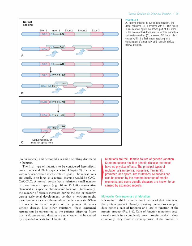
(colon cancer), and hemophilia A and B (clotting disorders)in humans.
The final type of mutation to be considered here affectstandem repeated DNA sequences (see Chapter 2) that occurwithin or near certain disease-related genes. The repeat unitsare usually 3 bp long, so a typical example would be CAG-CAGCAG. A normal person has a relatively small numberof these tandem repeats (e.g., 10 to 30 CAG consecutiveelements) at a specific chromosome location. Occasionally,the number of repeats increases during meiosis or possiblyduring early fetal development, so that a newborn mighthave hundreds or even thousands of tandem repeats. Whenthis occurs in certain regions of the genome, it causesgenetic disease. Like other mutations, these expandedrepeats can be transmitted to the patient’s offspring. Morethan a dozen genetic diseases are now known to be causedby expanded repeats (see Chapter 4).
4Mutations are the ultimate source of genetic variation.Some mutations result in genetic disease, but mosthave no physical effects. The principal types ofmutation are missense, nonsense, frameshift,promoter, and splice-site mutations. Mutations canalso be caused by the random insertion of mobileelements, and some genetic diseases are known to becaused by expanded repeats.
Molecular Consequences of MutationIt is useful to think of mutations in terms of their effects onthe protein product. Broadly speaking, mutations can pro-duce either a gain of function or a loss of function of theprotein product (Fig. 3-6). Gain-of-function mutations occa-sionally result in a completely novel protein product. Morecommonly, they result in overexpression of the product or
A
B
C
Normalsplicing
Exon 1 Intron 1 Exon 2 Intron 2 Exon 3GAG GT... . . .AG
GT.. . . . .AGGAG
GAG GT... . . .AG
GAG ATTGGT..AG
GAG ATTGGT...AG
GAG
GAG
GAG
GTTGGC..AG
GTTGGT..AG
GTTGGT.. .AG
Sequence may ormay not splice here
FIGURE 3-5A, Normal splicing. B, Splice-site mutation. Thedonor sequence, GT, is replaced with AT. This resultsin an incorrect splice that leaves part of the intronin the mature mRNA transcript. In another example ofsplice-site mutation (C), a second GT donor site iscreated within the first intron, resulting in acombination of abnormally and normally splicedmRNA products.
Genetic Variation: Its Origin and Detection / 29
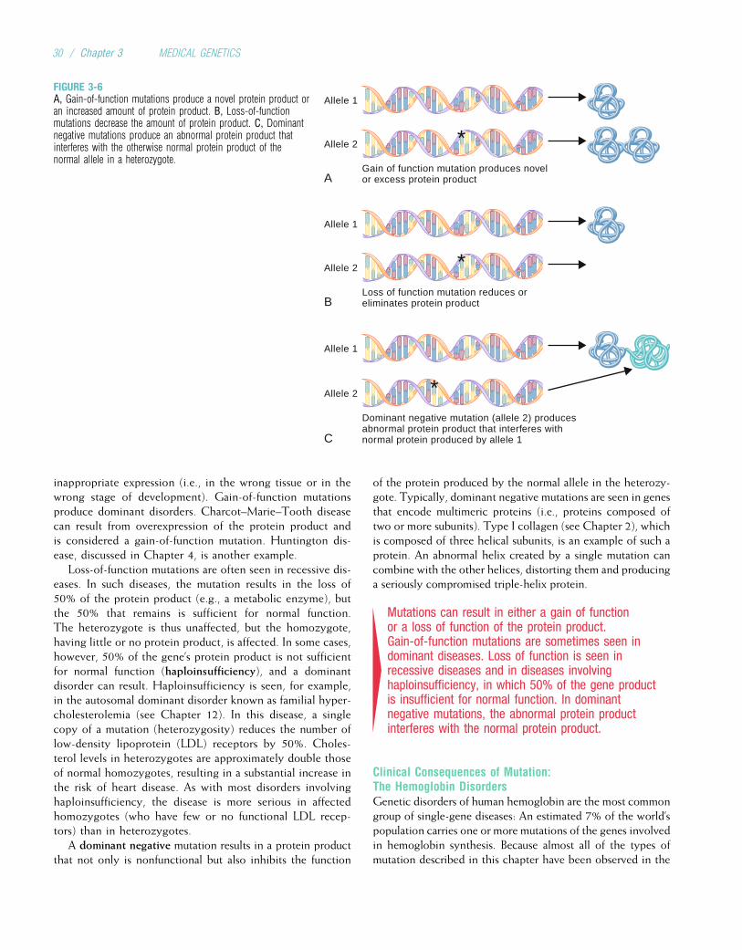
inappropriate expression (i.e., in the wrong tissue or in thewrong stage of development). Gain-of-function mutationsproduce dominant disorders. Charcot–Marie–Tooth diseasecan result from overexpression of the protein product andis considered a gain-of-function mutation. Huntington dis-ease, discussed in Chapter 4, is another example.
Loss-of-function mutations are often seen in recessive dis-eases. In such diseases, the mutation results in the loss of50% of the protein product (e.g., a metabolic enzyme), butthe 50% that remains is sufficient for normal function.The heterozygote is thus unaffected, but the homozygote,having little or no protein product, is affected. In some cases,however, 50% of the gene’s protein product is not sufficientfor normal function (haploinsufficiency), and a dominantdisorder can result. Haploinsufficiency is seen, for example,in the autosomal dominant disorder known as familial hyper-cholesterolemia (see Chapter 12). In this disease, a singlecopy of a mutation (heterozygosity) reduces the number oflow-density lipoprotein (LDL) receptors by 50%. Choles-terol levels in heterozygotes are approximately double thoseof normal homozygotes, resulting in a substantial increase inthe risk of heart disease. As with most disorders involvinghaploinsufficiency, the disease is more serious in affectedhomozygotes (who have few or no functional LDL recep-tors) than in heterozygotes.
A dominant negative mutation results in a protein productthat not only is nonfunctional but also inhibits the function
of the protein produced by the normal allele in the heterozy-gote. Typically, dominant negative mutations are seen in genesthat encode multimeric proteins (i.e., proteins composed oftwo or more subunits). Type I collagen (see Chapter 2), whichis composed of three helical subunits, is an example of such aprotein. An abnormal helix created by a single mutation cancombine with the other helices, distorting them and producinga seriously compromised triple-helix protein.
4Mutations can result in either a gain of functionor a loss of function of the protein product.Gain-of-function mutations are sometimes seen indominant diseases. Loss of function is seen inrecessive diseases and in diseases involvinghaploinsufficiency, in which 50% of the gene productis insufficient for normal function. In dominantnegative mutations, the abnormal protein productinterferes with the normal protein product.
Clinical Consequences of Mutation:The Hemoglobin DisordersGenetic disorders of human hemoglobin are the most commongroup of single-gene diseases: An estimated 7% of the world’spopulation carries one or more mutations of the genes involvedin hemoglobin synthesis. Because almost all of the types ofmutation described in this chapter have been observed in the
Allele 1
Allele 2
A
B
C
Gain of function mutation produces novelor excess protein product
*
Allele 1
Allele 2
Loss of function mutation reduces oreliminates protein product
*
Allele 1
Allele 2
Dominant negative mutation (allele 2) producesabnormal protein product that interferes withnormal protein produced by allele 1
*
FIGURE 3-6A, Gain-of-function mutations produce a novel protein product oran increased amount of protein product. B, Loss-of-functionmutations decrease the amount of protein product. C, Dominantnegative mutations produce an abnormal protein product thatinterferes with the otherwise normal protein product of thenormal allele in a heterozygote.
30 / Chapter 3 MEDICAL GENETICS
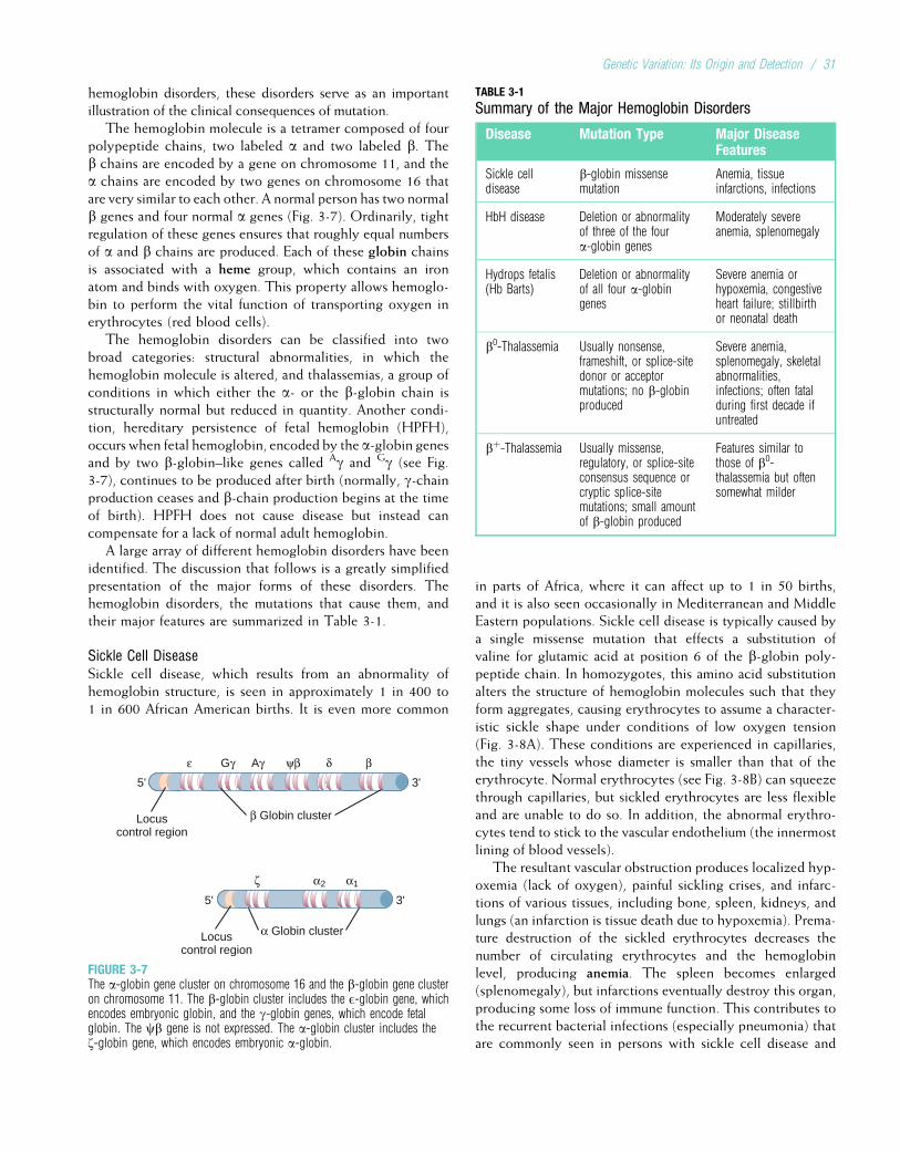
hemoglobin disorders, these disorders serve as an importantillustration of the clinical consequences of mutation.
The hemoglobin molecule is a tetramer composed of fourpolypeptide chains, two labeled a and two labeled b. Theb chains are encoded by a gene on chromosome 11, and thea chains are encoded by two genes on chromosome 16 thatare very similar to each other. A normal person has two normalb genes and four normal a genes (Fig. 3-7). Ordinarily, tightregulation of these genes ensures that roughly equal numbersof a and b chains are produced. Each of these globin chainsis associated with a heme group, which contains an ironatom and binds with oxygen. This property allows hemoglo-bin to perform the vital function of transporting oxygen inerythrocytes (red blood cells).
The hemoglobin disorders can be classified into twobroad categories: structural abnormalities, in which thehemoglobin molecule is altered, and thalassemias, a group ofconditions in which either the a- or the b-globin chain isstructurally normal but reduced in quantity. Another condi-tion, hereditary persistence of fetal hemoglobin (HPFH),occurs when fetal hemoglobin, encoded by the a-globin genesand by two b-globin–like genes called Ag and Gg (see Fig.3-7), continues to be produced after birth (normally, g-chainproduction ceases and b-chain production begins at the timeof birth). HPFH does not cause disease but instead cancompensate for a lack of normal adult hemoglobin.
A large array of different hemoglobin disorders have beenidentified. The discussion that follows is a greatly simplifiedpresentation of the major forms of these disorders. Thehemoglobin disorders, the mutations that cause them, andtheir major features are summarized in Table 3-1.
Sickle Cell DiseaseSickle cell disease, which results from an abnormality ofhemoglobin structure, is seen in approximately 1 in 400 to1 in 600 African American births. It is even more common
in parts of Africa, where it can affect up to 1 in 50 births,and it is also seen occasionally in Mediterranean and MiddleEastern populations. Sickle cell disease is typically caused bya single missense mutation that effects a substitution ofvaline for glutamic acid at position 6 of the b-globin poly-peptide chain. In homozygotes, this amino acid substitutionalters the structure of hemoglobin molecules such that theyform aggregates, causing erythrocytes to assume a character-istic sickle shape under conditions of low oxygen tension(Fig. 3-8A). These conditions are experienced in capillaries,the tiny vessels whose diameter is smaller than that of theerythrocyte. Normal erythrocytes (see Fig. 3-8B) can squeezethrough capillaries, but sickled erythrocytes are less flexibleand are unable to do so. In addition, the abnormal erythro-cytes tend to stick to the vascular endothelium (the innermostlining of blood vessels).
The resultant vascular obstruction produces localized hyp-oxemia (lack of oxygen), painful sickling crises, and infarc-tions of various tissues, including bone, spleen, kidneys, andlungs (an infarction is tissue death due to hypoxemia). Prema-ture destruction of the sickled erythrocytes decreases thenumber of circulating erythrocytes and the hemoglobinlevel, producing anemia. The spleen becomes enlarged(splenomegaly), but infarctions eventually destroy this organ,producing some loss of immune function. This contributes tothe recurrent bacterial infections (especially pneumonia) thatare commonly seen in persons with sickle cell disease and
Locuscontrol region
α Globin cluster
ζ
5' 3'
α2 α1
β Globin clusterLocuscontrol region
5' 3'
ε Gγ Aγ ψβ δ β
FIGURE 3-7The a-globin gene cluster on chromosome 16 and the b-globin gene clusteron chromosome 11. The b-globin cluster includes the E-globin gene, whichencodes embryonic globin, and the g-globin genes, which encode fetalglobin. The cb gene is not expressed. The a-globin cluster includes thez-globin gene, which encodes embryonic a-globin.
TABLE 3-1
Summary of the Major Hemoglobin Disorders
Disease Mutation Type Major DiseaseFeatures
Sickle celldisease
b-globin missensemutation
Anemia, tissueinfarctions, infections
HbH disease Deletion or abnormalityof three of the foura-globin genes
Moderately severeanemia, splenomegaly
Hydrops fetalis(Hb Barts)
Deletion or abnormalityof all four a-globingenes
Severe anemia orhypoxemia, congestiveheart failure; stillbirthor neonatal death
b0-Thalassemia Usually nonsense,frameshift, or splice-sitedonor or acceptormutations; no b-globinproduced
Severe anemia,splenomegaly, skeletalabnormalities,infections; often fatalduring first decade ifuntreated
bþ-Thalassemia Usually missense,regulatory, or splice-siteconsensus sequence orcryptic splice-sitemutations; small amountof b-globin produced
Features similar tothose of b0-thalassemia but oftensomewhat milder
Genetic Variation: Its Origin and Detection / 31
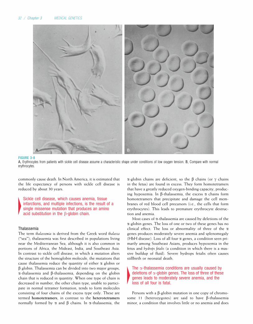
commonly cause death. In North America, it is estimated thatthe life expectancy of persons with sickle cell disease isreduced by about 30 years.
4Sickle cell disease, which causes anemia, tissueinfarctions, and multiple infections, is the result of asingle missense mutation that produces an aminoacid substitution in the b-globin chain.
ThalassemiaThe term thalassemia is derived from the Greek word thalassa
(“sea”); thalassemia was first described in populations livingnear the Mediterranean Sea, although it is also common inportions of Africa, the Mideast, India, and Southeast Asia.In contrast to sickle cell disease, in which a mutation altersthe structure of the hemoglobin molecule, the mutations thatcause thalassemia reduce the quantity of either a globin orb globin. Thalassemia can be divided into two major groups,a-thalassemia and b-thalassemia, depending on the globinchain that is reduced in quantity. When one type of chain isdecreased in number, the other chain type, unable to partici-pate in normal tetramer formation, tends to form moleculesconsisting of four chains of the excess type only. These aretermed homotetramers, in contrast to the heterotetramersnormally formed by a and b chains. In a-thalassemia, the
a-globin chains are deficient, so the b chains (or g chainsin the fetus) are found in excess. They form homotetramersthat have a greatly reduced oxygen-binding capacity, produc-ing hypoxemia. In b-thalassemia, the excess a chains formhomotetramers that precipitate and damage the cell mem-branes of red blood cell precursors (i.e., the cells that formerythrocytes). This leads to premature erythrocyte destruc-tion and anemia.
Most cases of a-thalassemia are caused by deletions of thea-globin genes. The loss of one or two of these genes has noclinical effect. The loss or abnormality of three of the agenes produces moderately severe anemia and splenomegaly(HbH disease). Loss of all four a genes, a condition seen pri-marily among Southeast Asians, produces hypoxemia in thefetus and hydrops fetalis (a condition in which there is a mas-sive buildup of fluid). Severe hydrops fetalis often causesstillbirth or neonatal death.
4The a-thalassemia conditions are usually caused bydeletions of a-globin genes. The loss of three of thesegenes leads to moderately severe anemia, and theloss of all four is fatal.
Persons with a b-globin mutation in one copy of chromo-some 11 (heterozygotes) are said to have b-thalassemiaminor, a condition that involves little or no anemia and does
A B
FIGURE 3-8A, Erythrocytes from patients with sickle cell disease assume a characteristic shape under conditions of low oxygen tension. B, Compare with normalerythrocytes.
32 / Chapter 3 MEDICAL GENETICS
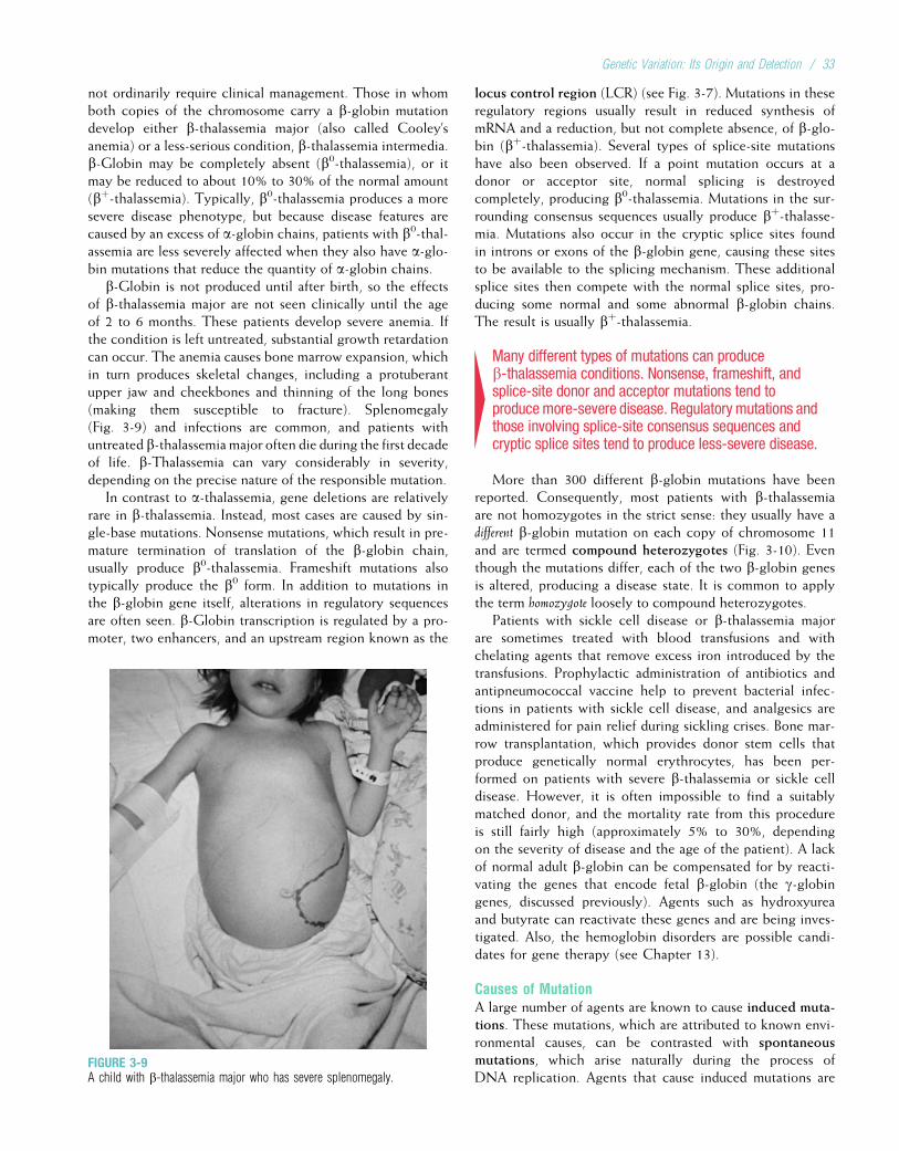
not ordinarily require clinical management. Those in whomboth copies of the chromosome carry a b-globin mutationdevelop either b-thalassemia major (also called Cooley’sanemia) or a less-serious condition, b-thalassemia intermedia.b-Globin may be completely absent (b0-thalassemia), or itmay be reduced to about 10% to 30% of the normal amount(bþ-thalassemia). Typically, b0-thalassemia produces a moresevere disease phenotype, but because disease features arecaused by an excess of a-globin chains, patients with b0-thal-assemia are less severely affected when they also have a-glo-bin mutations that reduce the quantity of a-globin chains.
b-Globin is not produced until after birth, so the effectsof b-thalassemia major are not seen clinically until the ageof 2 to 6 months. These patients develop severe anemia. Ifthe condition is left untreated, substantial growth retardationcan occur. The anemia causes bone marrow expansion, whichin turn produces skeletal changes, including a protuberantupper jaw and cheekbones and thinning of the long bones(making them susceptible to fracture). Splenomegaly(Fig. 3-9) and infections are common, and patients withuntreated b-thalassemia major often die during the first decadeof life. b-Thalassemia can vary considerably in severity,depending on the precise nature of the responsible mutation.
In contrast to a-thalassemia, gene deletions are relativelyrare in b-thalassemia. Instead, most cases are caused by sin-gle-base mutations. Nonsense mutations, which result in pre-mature termination of translation of the b-globin chain,usually produce b0-thalassemia. Frameshift mutations alsotypically produce the b0 form. In addition to mutations inthe b-globin gene itself, alterations in regulatory sequencesare often seen. b-Globin transcription is regulated by a pro-moter, two enhancers, and an upstream region known as the
locus control region (LCR) (see Fig. 3-7). Mutations in theseregulatory regions usually result in reduced synthesis ofmRNA and a reduction, but not complete absence, of b-glo-bin (bþ-thalassemia). Several types of splice-site mutationshave also been observed. If a point mutation occurs at adonor or acceptor site, normal splicing is destroyedcompletely, producing b0-thalassemia. Mutations in the sur-rounding consensus sequences usually produce bþ-thalasse-mia. Mutations also occur in the cryptic splice sites foundin introns or exons of the b-globin gene, causing these sitesto be available to the splicing mechanism. These additionalsplice sites then compete with the normal splice sites, pro-ducing some normal and some abnormal b-globin chains.The result is usually bþ-thalassemia.
4Many different types of mutations can produceb-thalassemia conditions. Nonsense, frameshift, andsplice-site donor and acceptor mutations tend toproducemore-severe disease. Regulatory mutations andthose involving splice-site consensus sequences andcryptic splice sites tend to produce less-severe disease.
More than 300 different b-globin mutations have beenreported. Consequently, most patients with b-thalassemiaare not homozygotes in the strict sense: they usually have adifferent b-globin mutation on each copy of chromosome 11and are termed compound heterozygotes (Fig. 3-10). Eventhough the mutations differ, each of the two b-globin genesis altered, producing a disease state. It is common to applythe term homozygote loosely to compound heterozygotes.
Patients with sickle cell disease or b-thalassemia majorare sometimes treated with blood transfusions and withchelating agents that remove excess iron introduced by thetransfusions. Prophylactic administration of antibiotics andantipneumococcal vaccine help to prevent bacterial infec-tions in patients with sickle cell disease, and analgesics areadministered for pain relief during sickling crises. Bone mar-row transplantation, which provides donor stem cells thatproduce genetically normal erythrocytes, has been per-formed on patients with severe b-thalassemia or sickle celldisease. However, it is often impossible to find a suitablymatched donor, and the mortality rate from this procedureis still fairly high (approximately 5% to 30%, dependingon the severity of disease and the age of the patient). A lackof normal adult b-globin can be compensated for by reacti-vating the genes that encode fetal b-globin (the g-globingenes, discussed previously). Agents such as hydroxyureaand butyrate can reactivate these genes and are being inves-tigated. Also, the hemoglobin disorders are possible candi-dates for gene therapy (see Chapter 13).
Causes of MutationA large number of agents are known to cause induced muta-tions. These mutations, which are attributed to known envi-ronmental causes, can be contrasted with spontaneousmutations, which arise naturally during the process ofDNA replication. Agents that cause induced mutations are
FIGURE 3-9A child with b-thalassemia major who has severe splenomegaly.
Genetic Variation: Its Origin and Detection / 33
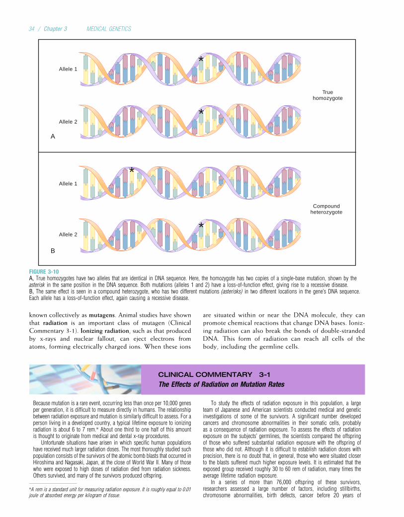
known collectively as mutagens. Animal studies have shownthat radiation is an important class of mutagen (ClinicalCommentary 3-1). Ionizing radiation, such as that producedby x-rays and nuclear fallout, can eject electrons fromatoms, forming electrically charged ions. When these ions
are situated within or near the DNA molecule, they canpromote chemical reactions that change DNA bases. Ioniz-ing radiation can also break the bonds of double-strandedDNA. This form of radiation can reach all cells of thebody, including the germline cells.
CLINICAL COMMENTARY 3-1
The Effects of Radiation on Mutation Rates
Because mutation is a rare event, occurring less than once per 10,000 genesper generation, it is difficult to measure directly in humans. The relationshipbetween radiation exposure and mutation is similarly difficult to assess. For aperson living in a developed country, a typical lifetime exposure to ionizingradiation is about 6 to 7 rem.* About one third to one half of this amountis thought to originate from medical and dental x-ray procedures.
Unfortunate situations have arisen in which specific human populationshave received much larger radiation doses. The most thoroughly studied suchpopulation consists of the survivors of the atomic bomb blasts that occurred inHiroshima and Nagasaki, Japan, at the close of World War II. Many of thosewho were exposed to high doses of radiation died from radiation sickness.Others survived, and many of the survivors produced offspring.
To study the effects of radiation exposure in this population, a largeteam of Japanese and American scientists conducted medical and geneticinvestigations of some of the survivors. A significant number developedcancers and chromosome abnormalities in their somatic cells, probablyas a consequence of radiation exposure. To assess the effects of radiationexposure on the subjects’ germlines, the scientists compared the offspringof those who suffered substantial radiation exposure with the offspring ofthose who did not. Although it is difficult to establish radiation doses withprecision, there is no doubt that, in general, those who were situated closerto the blasts suffered much higher exposure levels. It is estimated that theexposed group received roughly 30 to 60 rem of radiation, many times theaverage lifetime radiation exposure.
In a series of more than 76,000 offspring of these survivors,researchers assessed a large number of factors, including stillbirths,chromosome abnormalities, birth defects, cancer before 20 years of
Allele 1
Allele 2
Truehomozygote
A
*
*
Allele 1
Allele 2
Compoundheterozygote
B
*
*
FIGURE 3-10A, True homozygotes have two alleles that are identical in DNA sequence. Here, the homozygote has two copies of a single-base mutation, shown by theasterisk in the same position in the DNA sequence. Both mutations (alleles 1 and 2) have a loss-of-function effect, giving rise to a recessive disease.B, The same effect is seen in a compound heterozygote, who has two different mutations (asterisks) in two different locations in the gene’s DNA sequence.Each allele has a loss-of-function effect, again causing a recessive disease.
*A rem is a standard unit for measuring radiation exposure. It is roughly equal to 0.01joule of absorbed energy per kilogram of tissue.
34 / Chapter 3 MEDICAL GENETICS
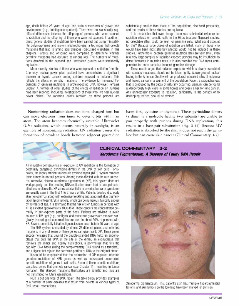
age, death before 26 years of age, and various measures of growth anddevelopment (e.g., intelligence quotient). There were no statistically sig-nificant differences between the offspring of persons who were exposedto radiation and the offspring of those who were not exposed. In addition,direct genetic studies of mutations have been carried out using minisatel-lite polymorphisms and protein electrophoresis, a technique that detectsmutations that lead to amino acid changes (discussed elsewhere in thischapter). Parents and offspring were compared to determine whethergermline mutations had occurred at various loci. The numbers of muta-tions detected in the exposed and unexposed groups were statisticallyequivalent.
More recently, studies of those who were exposed to radiation from theChernobyl nuclear power plant accident have demonstrated a significantincrease in thyroid cancers among children exposed to radiation. Thisreflects the effects of somatic mutations. The evidence for increased fre-quencies of germline mutations in protein-coding DNA, however, remainsunclear. A number of other studies of the effects of radiation on humanshave been reported, including investigations of those who live near nuclearpower plants. The radiation doses received by these persons are
substantially smaller than those of the populations discussed previously,and the results of these studies are equivocal.
It is remarkable that even though there was substantial evidence forradiation effects on somatic cells in the Hiroshima and Nagasaki studies,no detectable effect could be seen for germline cells. What could accountfor this? Because large doses of radiation are lethal, many of those whowould have been most strongly affected would not be included in thesestudies. Furthermore, because germline mutation rates are very small, evenrelatively large samples of radiation-exposed persons may be insufficient todetect increases in mutation rates. It is also possible that DNA repair com-pensated for some radiation-induced germline damage.
These results argue that radiation exposure, which is clearly associatedwith somatic mutations, should not be taken lightly. Above-ground nucleartesting in the American Southwest has produced increased rates of leukemiaand thyroid cancer in a segment of the population. Radon, a radioactive gasthat is produced by the decay of naturally occurring uranium, can be foundat dangerously high levels in some homes and poses a risk for lung cancer.Any unnecessary exposure to radiation, particularly to the gonads or todeveloping fetuses, should be avoided.
Nonionizing radiation does not form charged ions butcan move electrons from inner to outer orbits within anatom. The atom becomes chemically unstable. Ultraviolet(UV) radiation, which occurs naturally in sunlight, is anexample of nonionizing radiation. UV radiation causes theformation of covalent bonds between adjacent pyrimidine
bases (i.e., cytosine or thymine). These pyrimidine dimers(a dimer is a molecule having two subunits) are unable topair properly with purines during DNA replication; thisresults in a base-pair substitution (Fig. 3-11). Because UVradiation is absorbed by the skin, it does not reach the germ-line but can cause skin cancer (Clinical Commentary 3-2).
CLINICAL COMMENTARY 3-2
Xeroderma Pigmentosum: A Disease of Faulty DNA Repair
An inevitable consequence of exposure to UV radiation is the formation ofpotentially dangerous pyrimidine dimers in the DNA of skin cells. Fortu-nately, the highly efficient nucleotide excision repair (NER) system removesthese dimers in normal persons. Among those affected with the rare autoso-mal recessive disease xeroderma pigmentosum (XP), this system does notwork properly, and the resulting DNA replication errors lead to base pair sub-stitutions in skin cells. XP varies substantially in severity, but early symptomsare usually seen in the first 1 to 2 years of life. Patients develop dry, scalyskin (xeroderma) along with extensive freckling and abnormal skin pigmen-tation (pigmentosum). Skin tumors, which can be numerous, typically appearby 10 years of age. It is estimated that the risk of skin tumors in persons withXP is elevated approximately 1000-fold. These cancers are concentrated pri-marily in sun-exposed parts of the body. Patients are advised to avoidsources of UV light (e.g., sunlight), and cancerous growths are removed sur-gically. Neurological abnormalities are seen in about 30% of persons withXP. Severe, potentially lethal malignancies can occur before 20 years of age.
The NER system is encoded by at least 28 different genes, and inheritedmutations in any of seven of these genes can give rise to XP. These genesencode helicases that unwind the double-stranded DNA helix, an endonu-clease that cuts the DNA at the site of the dimer, an exonuclease thatremoves the dimer and nearby nucleotides, a polymerase that fills thegap with DNA bases (using the complementary DNA strand as a template),and a ligase that rejoins the corrected portion of DNA to the original strand.
It should be emphasized that the expression of XP requires inheritedgermline mutations of NER genes as well as subsequent uncorrectedsomatic mutations of genes in skin cells. Some of these somatic mutationscan affect genes that promote cancer (see Chapter 11), resulting in tumorformation. The skin-cell mutations themselves are somatic and thus arenot transmitted to future generations.
NER is but one type of DNA repair. The table below provides examplesof a number of other diseases that result from defects in various types ofDNA repair mechanisms.
Xeroderma pigmentosum. This patient’s skin has multiple hyperpigmentedlesions, and skin tumors on the forehead have been marked for excision.
Continued
Genetic Variation: Its Origin and Detection / 35
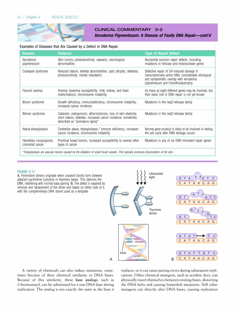
CLINICAL COMMENTARY 3-2
Xeroderma Pigmentosum: A Disease of Faulty DNA Repair—cont’d
Examples of Diseases that Are Caused by a Defect in DNA Repair
Disease Features Type of Repair Defect
Xerodermapigmentosum
Skin tumors, photosensitivity, cataracts, neurologicalabnormalities
Nucleotide excision repair defects, includingmutations in helicase and endonuclease genes
Cockayne syndrome Reduced stature, skeletal abnormalities, optic atrophy, deafness,photosensitivity, mental retardation
Defective repair of UV-induced damage intranscriptionally active DNA; considerable etiologicaland symptomatic overlap with xerodermapigmentosum and trichothiodystrophy
Fanconi anemia Anemia; leukemia susceptibility; limb, kidney, and heartmalformations; chromosome instability
As many as eight different genes may be involved, buttheir exact role in DNA repair is not yet known
Bloom syndrome Growth deficiency, immunodeficiency, chromosome instability,increased cancer incidence
Mutations in the reqQ helicase family
Werner syndrome Cataracts, osteoporosis, atherosclerosis, loss of skin elasticity,short stature, diabetes, increased cancer incidence; sometimesdescribed as “premature aging”
Mutations in the reqQ helicase family
Ataxia-telangiectasia Cerebellar ataxia, telangiectases,* immune deficiency, increasedcancer incidence, chromosome instability
Normal gene product is likely to be involved in haltingthe cell cycle after DNA damage occurs
Hereditary nonpolyposiscolorectal cancer
Proximal bowel tumors, increased susceptibility to several othertypes of cancer
Mutations in any of six DNA mismatch-repair genes
*Telangiectases are vascular lesions caused by the dilatation of small blood vessels. This typically produces discoloration of the skin.
A variety of chemicals can also induce mutations, some-times because of their chemical similarity to DNA bases.Because of this similarity, these base analogs, such as5-bromouracil, can be substituted for a true DNA base duringreplication. The analog is not exactly the same as the base it
replaces, so it can cause pairing errors during subsequent repli-cations. Other chemical mutagens, such as acridine dyes, canphysically insert themselves between existing bases, distortingthe DNA helix and causing frameshift mutations. Still othermutagens can directly alter DNA bases, causing replication
A
A
T
T
T
T
T
T
Kink
Ultravioletlight
A B
Thyminedimer
G T A G T C
C A T A A C A G
C A T A A C A G
C A T A A C A G
C A T A A C A G
C A T A A C A G
G T A T T G T C
G T T C
T TAG T G T C
AG
T T
G T A T T C
T T
FIGURE 3-11A, Pyrimidine dimers originate when covalent bonds form betweenadjacent pyrimidine (cytosine or thymine) bases. This deforms theDNA, interfering with normal base pairing. B, The defect is repaired byremoval and replacement of the dimer and bases on either side of it,with the complementary DNA strand used as a template.
36 / Chapter 3 MEDICAL GENETICS
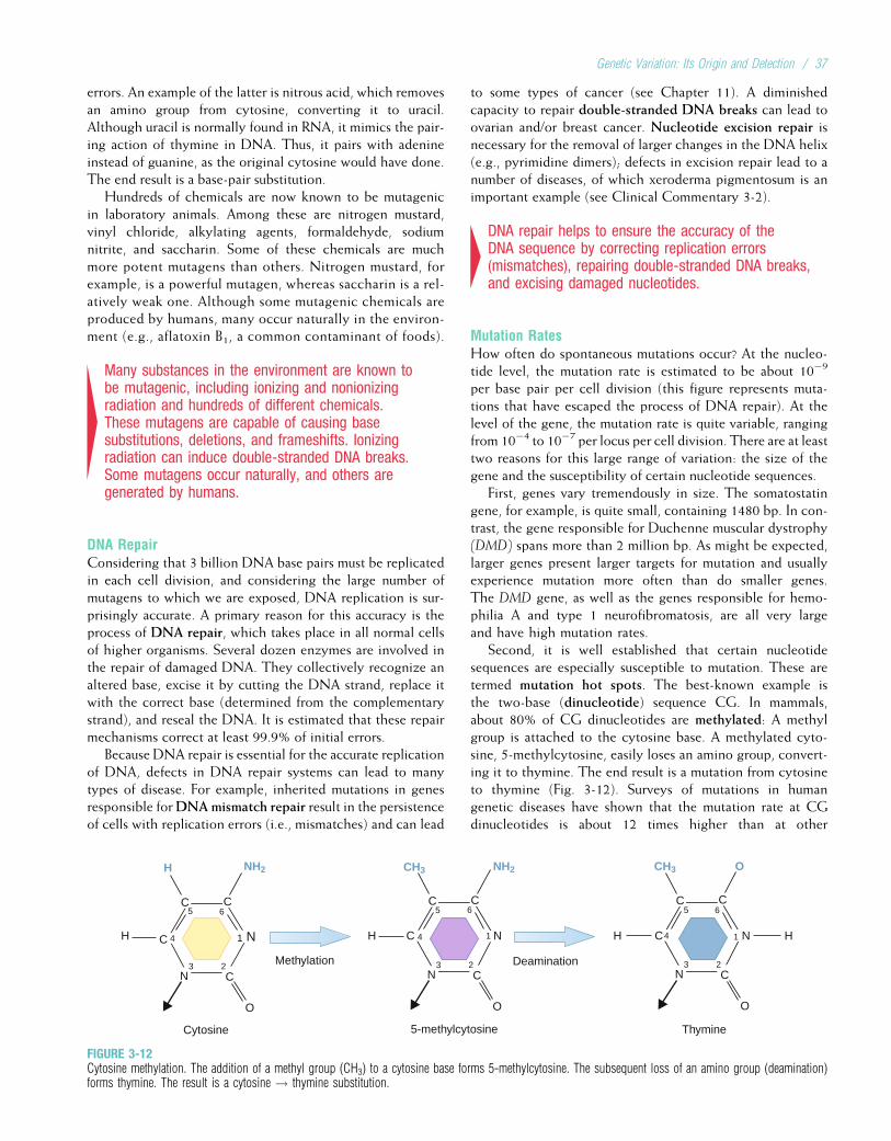
errors. An example of the latter is nitrous acid, which removesan amino group from cytosine, converting it to uracil.Although uracil is normally found in RNA, it mimics the pair-ing action of thymine in DNA. Thus, it pairs with adenineinstead of guanine, as the original cytosine would have done.The end result is a base-pair substitution.
Hundreds of chemicals are now known to be mutagenicin laboratory animals. Among these are nitrogen mustard,vinyl chloride, alkylating agents, formaldehyde, sodiumnitrite, and saccharin. Some of these chemicals are muchmore potent mutagens than others. Nitrogen mustard, forexample, is a powerful mutagen, whereas saccharin is a rel-atively weak one. Although some mutagenic chemicals areproduced by humans, many occur naturally in the environ-ment (e.g., aflatoxin B1, a common contaminant of foods).
4Many substances in the environment are known tobe mutagenic, including ionizing and nonionizingradiation and hundreds of different chemicals.These mutagens are capable of causing basesubstitutions, deletions, and frameshifts. Ionizingradiation can induce double-stranded DNA breaks.Some mutagens occur naturally, and others aregenerated by humans.
DNA RepairConsidering that 3 billion DNA base pairs must be replicatedin each cell division, and considering the large number ofmutagens to which we are exposed, DNA replication is sur-prisingly accurate. A primary reason for this accuracy is theprocess of DNA repair, which takes place in all normal cellsof higher organisms. Several dozen enzymes are involved inthe repair of damaged DNA. They collectively recognize analtered base, excise it by cutting the DNA strand, replace itwith the correct base (determined from the complementarystrand), and reseal the DNA. It is estimated that these repairmechanisms correct at least 99.9% of initial errors.
Because DNA repair is essential for the accurate replicationof DNA, defects in DNA repair systems can lead to manytypes of disease. For example, inherited mutations in genesresponsible forDNAmismatch repair result in the persistenceof cells with replication errors (i.e., mismatches) and can lead
to some types of cancer (see Chapter 11). A diminishedcapacity to repair double-stranded DNA breaks can lead toovarian and/or breast cancer. Nucleotide excision repair isnecessary for the removal of larger changes in the DNA helix(e.g., pyrimidine dimers); defects in excision repair lead to anumber of diseases, of which xeroderma pigmentosum is animportant example (see Clinical Commentary 3-2).
4DNA repair helps to ensure the accuracy of theDNA sequence by correcting replication errors(mismatches), repairing double-stranded DNA breaks,and excising damaged nucleotides.
Mutation RatesHow often do spontaneous mutations occur? At the nucleo-tide level, the mutation rate is estimated to be about 10�9
per base pair per cell division (this figure represents muta-tions that have escaped the process of DNA repair). At thelevel of the gene, the mutation rate is quite variable, rangingfrom 10�4 to 10�7 per locus per cell division. There are at leasttwo reasons for this large range of variation: the size of thegene and the susceptibility of certain nucleotide sequences.
First, genes vary tremendously in size. The somatostatingene, for example, is quite small, containing 1480 bp. In con-trast, the gene responsible for Duchenne muscular dystrophy(DMD) spans more than 2 million bp. As might be expected,larger genes present larger targets for mutation and usuallyexperience mutation more often than do smaller genes.The DMD gene, as well as the genes responsible for hemo-philia A and type 1 neurofibromatosis, are all very largeand have high mutation rates.
Second, it is well established that certain nucleotidesequences are especially susceptible to mutation. These aretermed mutation hot spots. The best-known example isthe two-base (dinucleotide) sequence CG. In mammals,about 80% of CG dinucleotides are methylated: A methylgroup is attached to the cytosine base. A methylated cyto-sine, 5-methylcytosine, easily loses an amino group, convert-ing it to thymine. The end result is a mutation from cytosineto thymine (Fig. 3-12). Surveys of mutations in humangenetic diseases have shown that the mutation rate at CGdinucleotides is about 12 times higher than at other
Methylation
C
C
C
N
N
5
4
2
1H
O
Cytosine
NH2H
Deamination
5-methylcytosine
C
C
C
C
N
N
65
4
3 2
1H
O
NH2CH3
3
6
CH3 O
C C
C
C
C
N
N
65
4
3 2
1H
O
Thymine
H
FIGURE 3-12Cytosine methylation. The addition of a methyl group (CH3) to a cytosine base forms 5-methylcytosine. The subsequent loss of an amino group (deamination)forms thymine. The result is a cytosine ! thymine substitution.
Genetic Variation: Its Origin and Detection / 37
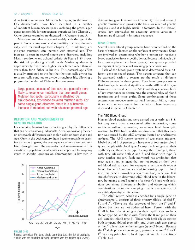
dinucleotide sequences. Mutation hot spots, in the form ofCG dinucleotides, have been identified in a numberof important human disease genes, including the procollagengenes responsible for osteogenesis imperfecta (see Chapter 2).Other disease examples are discussed in Chapters 4 and 5.
Mutation rates also vary considerably with the age of theparent. Some chromosome abnormalities increase dramati-cally with maternal age (see Chapter 6). In addition, sin-gle-gene mutations can increase with paternal age. Thisincrease is seen in several single-gene disorders, includingMarfan syndrome and achondroplasia. As Figure 3-13 shows,the risk of producing a child with Marfan syndrome isapproximately five times higher for a father older than 40years than for a father in his 20s. This paternal age effectis usually attributed to the fact that the stem cells giving riseto sperm cells continue to divide throughout life, allowing aprogressive buildup of DNA replication errors.
4Large genes, because of their size, are generally morelikely to experience mutations than are small genes.Mutation hot spots, particularly methylated CGdinucleotides, experience elevated mutation rates. Forsome single-gene disorders, there is a substantialincrease in mutation risk with advanced paternal age.
DETECTION AND MEASUREMENT OFGENETIC VARIATIONFor centuries, humans have been intrigued by the differencesthat can be seen among individuals. Attention was long focusedon observable differences such as skin color or body shape andsize. Only in the 20th century did it become possible to exam-ine variation in genes, the consequence of mutations accumu-lated through time. The evaluation and measurement of thisvariation in populations and families are important for mappinggenes to specific locations on chromosomes, a key step in
determining gene function (see Chapter 8). The evaluation ofgenetic variation also provides the basis for much of geneticdiagnosis, and it is highly useful in forensics. In this section,several key approaches to detecting genetic variation inhumans are discussed in historical sequence.
Blood GroupsSeveral dozen blood group systems have been defined on thebasis of antigens located on the surfaces of erythrocytes. Someare involved in determining whether a person can receive ablood transfusion from a specific donor. Because individuals dif-fer extensively in terms of blood groups, these systems providedan important early means of assessing genetic variation.
Each of the blood group systems is determined by a dif-ferent gene or set of genes. The various antigens that canbe expressed within a system are the result of differentDNA sequences in these genes. Two blood-group systemsthat have special medical significance—the ABO and Rh sys-tems—are discussed here. The ABO and Rh systems are bothof key importance in determining the compatibility of bloodtransfusions and tissue grafts. Some combinations of thesesystems can produce maternal-fetal incompatibility, some-times with serious results for the fetus. These issues arediscussed in detail in Chapter 9.
The ABO Blood GroupHuman blood transfusions were carried out as early as 1818,but they were often unsuccessful. After transfusion, somerecipients suffered a massive, sometimes fatal, hemolyticreaction. In 1900 Karl Landsteiner discovered that this reac-tion was caused by the ABO antigens located on erythrocytesurfaces. The ABO system consists of two major antigens,labeled A and B. A person can have one of four major bloodtypes: People with blood type A carry the A antigen on theirerythrocytes, those with type B carry the B antigen, thosewith type AB carry both A and B, and those with type Ocarry neither antigen. Each individual has antibodies thatreact against any antigens that are not found on their ownred blood cell surfaces. For example, a person with type Ablood has anti-B antibodies, and transfusing type B bloodinto this person provokes a severe antibody reaction. It isstraightforward to determine ABO blood type in the labora-tory by mixing a small sample of a person’s blood with solu-tions containing different antibodies and observing whichcombinations cause the clumping that is characteristic ofan antibody–antigen interaction.
The ABO system, which is encoded by a single gene onchromosome 9, consists of three primary alleles, labeled IA,IB, and IO. (There are also subtypes of both the IA and IB
alleles, but they are not addressed here.) Persons with theIA allele have the A antigen on their erythrocyte surfaces(blood type A), and those with IB have the B antigen on theircell surfaces (blood type B). Those with both alleles expressboth antigens (blood type AB), and those with two copiesof the IO allele have neither antigen (type O blood). Becausethe IO allele produces no antigen, persons who are IA IO or IB
IO heterozygotes have blood types A and B, respectively(Table 3-2).
6
5
4
3
2Rel
ativ
e in
cide
nce
1
0
Apert syndromeNeurofibromatosisAchondroplasiaMarfan syndrome
<25 25–29 30–34 35–39
Age
40–44 45–49 >49
Population average
FIGURE 3-13Paternal age effect. For some single-gene disorders, the risk of producinga child with the condition (y-axis) increases with the father’s age (x-axis).
38 / Chapter 3 MEDICAL GENETICS
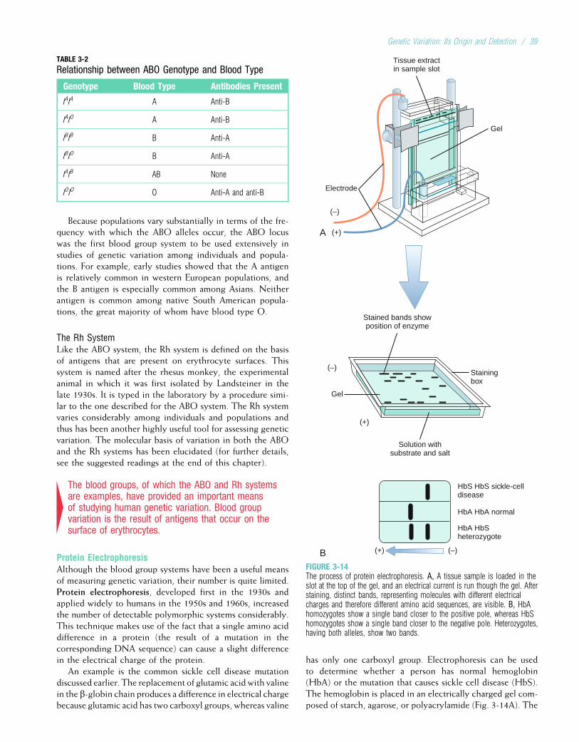
Because populations vary substantially in terms of the fre-quency with which the ABO alleles occur, the ABO locuswas the first blood group system to be used extensively instudies of genetic variation among individuals and popula-tions. For example, early studies showed that the A antigenis relatively common in western European populations, andthe B antigen is especially common among Asians. Neitherantigen is common among native South American popula-tions, the great majority of whom have blood type O.
The Rh SystemLike the ABO system, the Rh system is defined on the basisof antigens that are present on erythrocyte surfaces. Thissystem is named after the rhesus monkey, the experimentalanimal in which it was first isolated by Landsteiner in thelate 1930s. It is typed in the laboratory by a procedure simi-lar to the one described for the ABO system. The Rh systemvaries considerably among individuals and populations andthus has been another highly useful tool for assessing geneticvariation. The molecular basis of variation in both the ABOand the Rh systems has been elucidated (for further details,see the suggested readings at the end of this chapter).
4The blood groups, of which the ABO and Rh systemsare examples, have provided an important meansof studying human genetic variation. Blood groupvariation is the result of antigens that occur on thesurface of erythrocytes.
Protein ElectrophoresisAlthough the blood group systems have been a useful meansof measuring genetic variation, their number is quite limited.Protein electrophoresis, developed first in the 1930s andapplied widely to humans in the 1950s and 1960s, increasedthe number of detectable polymorphic systems considerably.This technique makes use of the fact that a single amino aciddifference in a protein (the result of a mutation in thecorresponding DNA sequence) can cause a slight differencein the electrical charge of the protein.
An example is the common sickle cell disease mutationdiscussed earlier. The replacement of glutamic acid with valinein the b-globin chain produces a difference in electrical chargebecause glutamic acid has two carboxyl groups, whereas valine
has only one carboxyl group. Electrophoresis can be usedto determine whether a person has normal hemoglobin(HbA) or the mutation that causes sickle cell disease (HbS).The hemoglobin is placed in an electrically charged gel com-posed of starch, agarose, or polyacrylamide (Fig. 3-14A). The
B
A
(+) (–)
HbS HbS sickle-celldisease
HbA HbA normal
HbA HbSheterozygote
(+)
(+)
(–)
Solution withsubstrate and salt
Stainingbox
Gel
Stained bands showposition of enzyme
Gel
Tissue extractin sample slot
Electrode
(–)
FIGURE 3-14The process of protein electrophoresis. A, A tissue sample is loaded in theslot at the top of the gel, and an electrical current is run though the gel. Afterstaining, distinct bands, representing molecules with different electricalcharges and therefore different amino acid sequences, are visible. B, HbAhomozygotes show a single band closer to the positive pole, whereas HbShomozygotes show a single band closer to the negative pole. Heterozygotes,having both alleles, show two bands.
TABLE 3-2
Relationship between ABO Genotype and Blood Type
Genotype Blood Type Antibodies Present
IAIA A Anti-B
IAIO A Anti-B
IBIB B Anti-A
IBIO B Anti-A
IAIB AB None
IOIO O Anti-A and anti-B
Genetic Variation: Its Origin and Detection / 39
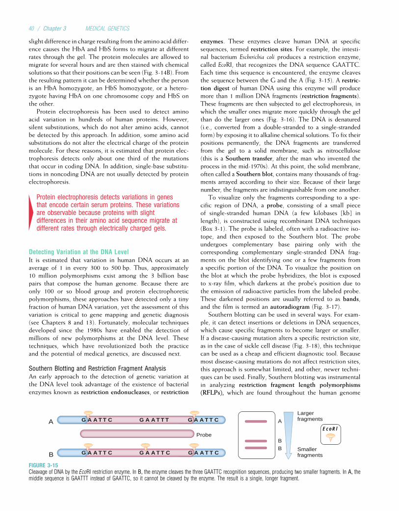
slight difference in charge resulting from the amino acid differ-ence causes the HbA and HbS forms to migrate at differentrates through the gel. The protein molecules are allowed tomigrate for several hours and are then stained with chemicalsolutions so that their positions can be seen (Fig. 3-14B). Fromthe resulting pattern it can be determined whether the personis an HbA homozygote, an HbS homozygote, or a hetero-zygote having HbA on one chromosome copy and HbS onthe other.
Protein electrophoresis has been used to detect aminoacid variation in hundreds of human proteins. However,silent substitutions, which do not alter amino acids, cannotbe detected by this approach. In addition, some amino acidsubstitutions do not alter the electrical charge of the proteinmolecule. For these reasons, it is estimated that protein elec-trophoresis detects only about one third of the mutationsthat occur in coding DNA. In addition, single-base substitu-tions in noncoding DNA are not usually detected by proteinelectrophoresis.
4Protein electrophoresis detects variations in genesthat encode certain serum proteins. These variationsare observable because proteins with slightdifferences in their amino acid sequence migrate atdifferent rates through electrically charged gels.
Detecting Variation at the DNA LevelIt is estimated that variation in human DNA occurs at anaverage of 1 in every 300 to 500 bp. Thus, approximately10 million polymorphisms exist among the 3 billion basepairs that compose the human genome. Because there areonly 100 or so blood group and protein electrophoreticpolymorphisms, these approaches have detected only a tinyfraction of human DNA variation, yet the assessment of thisvariation is critical to gene mapping and genetic diagnosis(see Chapters 8 and 13). Fortunately, molecular techniquesdeveloped since the 1980s have enabled the detection ofmillions of new polymorphisms at the DNA level. Thesetechniques, which have revolutionized both the practiceand the potential of medical genetics, are discussed next.
Southern Blotting and Restriction Fragment AnalysisAn early approach to the detection of genetic variation atthe DNA level took advantage of the existence of bacterialenzymes known as restriction endonucleases, or restriction
enzymes. These enzymes cleave human DNA at specificsequences, termed restriction sites. For example, the intesti-nal bacterium Escherichia coli produces a restriction enzyme,called EcoRI, that recognizes the DNA sequence GAATTC.Each time this sequence is encountered, the enzyme cleavesthe sequence between the G and the A (Fig. 3-15). A restric-tion digest of human DNA using this enzyme will producemore than 1 million DNA fragments (restriction fragments).These fragments are then subjected to gel electrophoresis, inwhich the smaller ones migrate more quickly through the gelthan do the larger ones (Fig. 3-16). The DNA is denatured(i.e., converted from a double-stranded to a single-strandedform) by exposing it to alkaline chemical solutions. To fix theirpositions permanently, the DNA fragments are transferredfrom the gel to a solid membrane, such as nitrocellulose(this is a Southern transfer, after the man who invented theprocess in the mid-1970s). At this point, the solid membrane,often called a Southern blot, contains many thousands of frag-ments arrayed according to their size. Because of their largenumber, the fragments are indistinguishable from one another.
To visualize only the fragments corresponding to a spe-cific region of DNA, a probe, consisting of a small pieceof single-stranded human DNA (a few kilobases [kb] inlength), is constructed using recombinant DNA techniques(Box 3-1). The probe is labeled, often with a radioactive iso-tope, and then exposed to the Southern blot. The probeundergoes complementary base pairing only with thecorresponding complementary single-stranded DNA frag-ments on the blot identifying one or a few fragments froma specific portion of the DNA. To visualize the position onthe blot at which the probe hybridizes, the blot is exposedto x-ray film, which darkens at the probe’s position due tothe emission of radioactive particles from the labeled probe.These darkened positions are usually referred to as bands,and the film is termed an autoradiogram (Fig. 3-17).
Southern blotting can be used in several ways. For exam-ple, it can detect insertions or deletions in DNA sequences,which cause specific fragments to become larger or smaller.If a disease-causing mutation alters a specific restriction site,as in the case of sickle cell disease (Fig. 3-18), this techniquecan be used as a cheap and efficient diagnostic tool. Becausemost disease-causing mutations do not affect restriction sites,this approach is somewhat limited, and other, newer techni-ques can be used. Finally, Southern blotting was instrumentalin analyzing restriction fragment length polymorphisms(RFLPs), which are found throughout the human genome
EcoRl
G A A T T C
Probe
A
B
B
A
B G A A T T CG A A T T C
G A A T T TLargerfragments
Smallerfragments
G A A T T C
G A A T T C
FIGURE 3-15Cleavage of DNA by the EcoRI restriction enzyme. In B, the enzyme cleaves the three GAATTC recognition sequences, producing two smaller fragments. In A, themiddle sequence is GAATTT instead of GAATTC, so it cannot be cleaved by the enzyme. The result is a single, longer fragment.
40 / Chapter 3 MEDICAL GENETICS
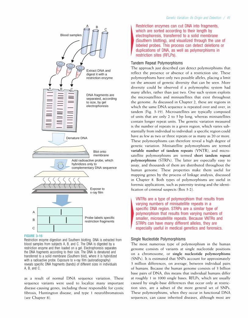
as a result of normal DNA sequence variation. Thesesequence variants were used to localize many importantdisease-causing genes, including those responsible for cysticfibrosis, Huntington disease, and type 1 neurofibromatosis(see Chapter 8).
4Restriction enzymes can cut DNA into fragments,which are sorted according to their length byelectrophoresis, transferred to a solid membrane(Southern blotting), and visualized through the use oflabeled probes. This process can detect deletions orduplications of DNA, as well as polymorphisms inrestriction sites (RFLPs).
Tandem Repeat PolymorphismsThe approach just described can detect polymorphisms thatreflect the presence or absence of a restriction site. Thesepolymorphisms have only two possible alleles, placing a limiton the amount of genetic diversity that can be seen. Morediversity could be observed if a polymorphic system hadmany alleles, rather than just two. One such system exploitsthe microsatellites and minisatellites that exist throughoutthe genome. As discussed in Chapter 2, these are regions inwhich the same DNA sequence is repeated over and over, intandem (Fig. 3-19). Microsatellites are typically composedof units that are only 2 to 5 bp long, whereas minisatellitescontain longer repeat units. The genetic variation measuredis the number of repeats in a given region, which varies sub-stantially from individual to individual: a specific region couldhave as few as two or three repeats or as many as 20 or more.These polymorphisms can therefore reveal a high degree ofgenetic variation. Minisatellite polymorphisms are termedvariable number of tandem repeats (VNTR), and micro-satellite polymorphisms are termed short tandem repeatpolymorphisms (STRPs). The latter are especially easy toassay, and thousands of them are distributed throughout thehuman genome. These properties make them useful formapping genes by the process of linkage analysis, discussedin Chapter 8. Both types of polymorphisms are useful inforensic applications, such as paternity testing and the identi-fication of criminal suspects (Box 3-2).
4VNTRs are a type of polymorphism that results fromvarying numbers of minisatellite repeats in aspecific DNA region. STRPs are a similar type ofpolymorphism that results from varying numbers ofsmaller, microsatellite repeats. Because VNTRs andSTRPs can have many different alleles, they areespecially useful in medical genetics and forensics.
Single Nucleotide PolymorphismsThe most numerous type of polymorphism in the humangenome consists of variants at single nucleotide positionson a chromosome, or single nucleotide polymorphisms(SNPs). It is estimated that SNPs account for approximately3 million differences, on average, between individual pairsof humans. Because the human genome consists of 3 billionbase pairs of DNA, this means that individual humans differat roughly 1 in 1000 single bases. RFLPs, which are usuallycaused by single-base differences that occur only at restric-tion sites, are a subset of the more general set of SNPs.These polymorphisms, when they occur in functional DNAsequences, can cause inherited diseases, although most are
Blood samples
Denature DNA
Blot onto membrane
Expose to x-ray film
A
B
C
A B C
Extract DNA anddigest it with arestriction enzyme
DNA fragments areseparated, accordingto size, by gelelectrophoresis
Add radioactive probe, whichhybridizes only tocomplementary DNA sequence
Probe labels specificrestriction fragments
A BC
FIGURE 3-16Restriction enzyme digestion and Southern blotting. DNA is extracted fromblood samples from subjects A, B, and C. The DNA is digested by arestriction enzyme and then loaded on a gel. Electrophoresis separatesthe DNA fragments according to their size. The DNA is denatured andtransferred to a solid membrane (Southern blot), where it is hybridizedwith a radioactive probe. Exposure to x-ray film (autoradiography)reveals specific DNA fragments (bands) of different sizes in individualsA, B, and C.
Genetic Variation: Its Origin and Detection / 41
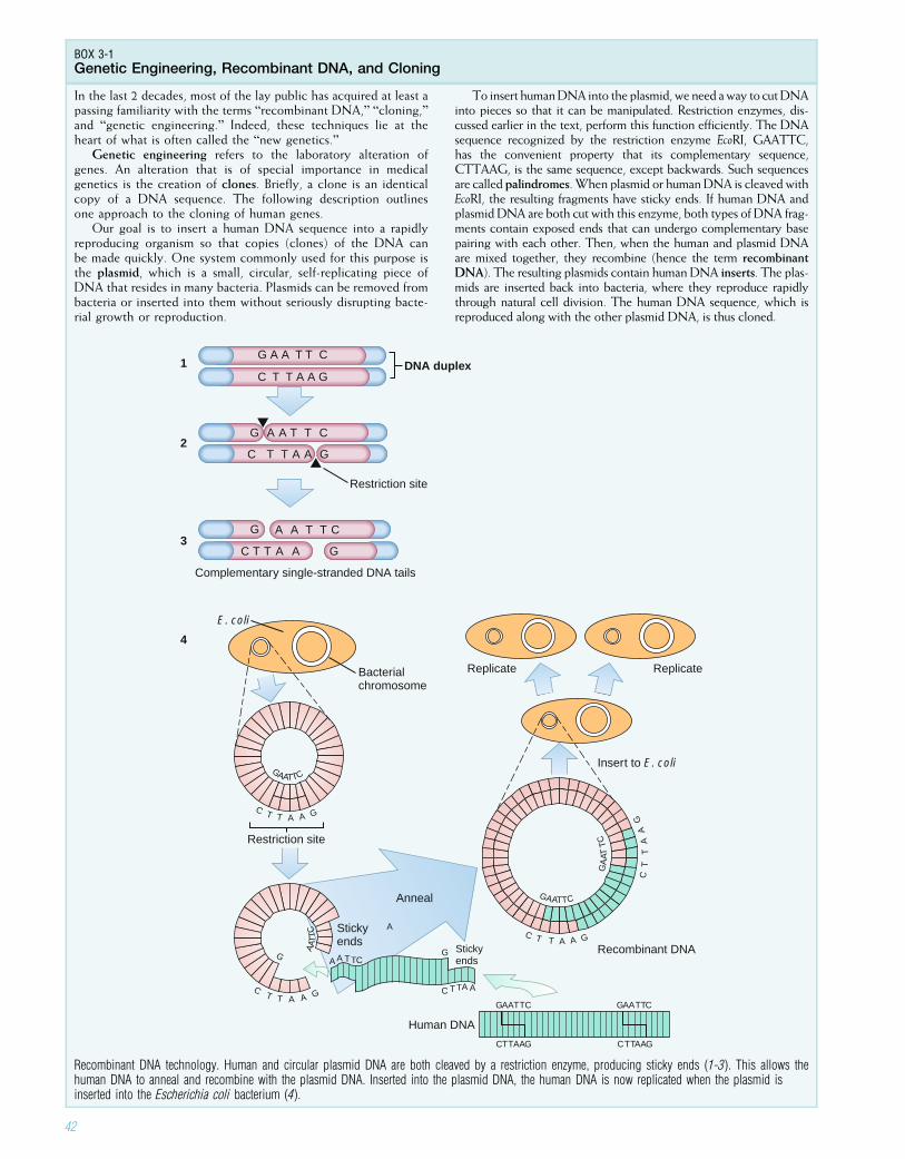
BOX 3-1Genetic Engineering, Recombinant DNA, and Cloning
In the last 2 decades, most of the lay public has acquired at least apassing familiarity with the terms “recombinant DNA,” “cloning,”and “genetic engineering.” Indeed, these techniques lie at theheart of what is often called the “new genetics.”
Genetic engineering refers to the laboratory alteration ofgenes. An alteration that is of special importance in medicalgenetics is the creation of clones. Briefly, a clone is an identicalcopy of a DNA sequence. The following description outlinesone approach to the cloning of human genes.
Our goal is to insert a human DNA sequence into a rapidlyreproducing organism so that copies (clones) of the DNA canbe made quickly. One system commonly used for this purpose isthe plasmid, which is a small, circular, self-replicating piece ofDNA that resides in many bacteria. Plasmids can be removed frombacteria or inserted into them without seriously disrupting bacte-rial growth or reproduction.
To insert humanDNA into the plasmid,we need away to cutDNAinto pieces so that it can be manipulated. Restriction enzymes, dis-cussed earlier in the text, perform this function efficiently. The DNAsequence recognized by the restriction enzyme EcoRI, GAATTC,has the convenient property that its complementary sequence,CTTAAG, is the same sequence, except backwards. Such sequencesare called palindromes.When plasmid or humanDNA is cleaved withEcoRI, the resulting fragments have sticky ends. If human DNA andplasmid DNA are both cut with this enzyme, both types of DNA frag-ments contain exposed ends that can undergo complementary basepairing with each other. Then, when the human and plasmid DNAare mixed together, they recombine (hence the term recombinantDNA). The resulting plasmids contain human DNA inserts. The plas-mids are inserted back into bacteria, where they reproduce rapidlythrough natural cell division. The human DNA sequence, which isreproduced along with the other plasmid DNA, is thus cloned.
Complementary single-stranded DNA tails
Restriction site
Restriction site
Bacterialchromosome
Replicate Replicate
E. coli
DNA duplex
GAATTC
C T T A A G
G
AAT
TC
C T T A A G
Stickyends
Stickyends
Anneal
Human DNA
Recombinant DNA
Insert to E. coli
GA
AT T
C
C T
T
A
A
G
G A A T T C
C T T A A G
G A A T T C
C T T A A G
G A A T T C
C T T A A G
1
2
3
4
A
A A TTC
C TTA A
G
GAATTC
CTTAAG
CTTAAG CTTAAG
CTTAAG
C T T A A G
Recombinant DNA technology. Human and circular plasmid DNA are both cleaved by a restriction enzyme, producing sticky ends (1-3). This allows thehuman DNA to anneal and recombine with the plasmid DNA. Inserted into the plasmid DNA, the human DNA is now replicated when the plasmid isinserted into the Escherichia coli bacterium (4).
42

harmless. Increasingly, they are being detected by microar-ray methods, which are discussed later in this chapter.
Copy Number VariantsAnother type of DNA variation has been shown to occur withsubstantial frequency in the human genome.Copy number var-iants (CNVs), which consist ofDNA segments longer than 1000base pairs, are present in some persons but absent in others.They can also be present in more than one copy in a person.CNVs are estimated to account for at least 4 million base pairsof difference, on average, between individual humans. Thus,they account for slightly more total variation in human DNAsequences than do SNPs. Some CNVs have been shown to beassociated with inherited diseases. Figure 3-20 highlights thedifferences among RFLPs, tandem repeats, SNPs, and CNVs.
4SNPs are the most common type of variation in thehuman genome. CNVs consist of differences in thenumber of repeated DNA sequences longer than1000 bp.
The plasmid is referred to as a vector. Several other types of vec-tors may also be used as cloning vehicles, including bacteriophages(viruses that infect bacteria), cosmids (phage–plasmid hybridscapable of carrying relatively large DNA inserts), yeast artificialchromosomes (YACs; vectors that are inserted into yeast cells andthat behave much like ordinary yeast chromosomes), bacterial arti-ficial chromosomes (BACs), and human artificial chromosomes(see Chapters 8 and 13). Although plasmids and bacteriophagescan accommodate only relatively small inserts (about 10 and20 kb, respectively), cosmids can carry inserts of approximately50 kb, and YACs can carry inserts up to 1000 kb in length.
Cloning can be used to create the thousands of copies of humanDNA needed for Southern blotting and other experimental applica-
tions. In addition, this approach is now used to produce geneticallyengineered therapeutic products, such as insulin, interferon, humangrowth hormone, clotting factor VIII (used in the treatment ofhemophilia A, a coagulation disorder), and tissue plasminogen acti-vator (a blood clot–dissolving protein that helps to prevent heartattacks and strokes). When these genes are cloned into bacteria orother organisms, the organism produces the human gene productalong with its own gene products. In the past, these products wereobtained from donor blood or from other animals. The processesof obtaining and purifying them were slow and costly, and theresulting products sometimes contained contaminants. Geneticallyengineered gene products are rapidly becoming a cheaper, purer,and more efficient alternative.
4.1-kb
3.3-kb
FIGURE 3-17An autoradiogram, showing the positions of a 4.1-kb band and a 3.3-kbband. Each lane represents DNA from a subject in the family whose pedigreeis shown above the autoradiogram.
Glu Glu
Sicklecell
Normal
1.3 kb
Pro
1.1 kb 0.2 kbMst II Mst II Mst II
Mst II Mst II
Glu Glu Pro Val Glu Pro
Pro Glu Glu GluPro ProGlu Glu Glu
CCT CCT CCTGAG GAG GAG GAG...GAG
CCT CCTGAG GAG GAG...GTG
GAG...
CCT GAG GAG...
kb1.31.1
AA AS SS
FIGURE 3-18Cleavage of b-globin DNA by the MstII restriction enzyme. Normal individuals have glutamic acid at position 6 of the b-globin polypeptide. Glutamic acid isencoded by the DNA sequence GAG. The sickle cell mutation results in the sequence GTG at this site instead of GAG, causing valine to be substituted forglutamic acid. The restriction enzyme MstII recognizes the DNA sequence CCTNAGG (the N signifies that the enzyme will recognize any DNA base, including G,in this position). Thus, MstII recognizes and cleaves the DNA sequence of the normal chromosome at this site as well as at the restriction sites on eitherside of it. The sickle cell mutation removes an MstII recognition site, producing a longer, 1.3-kb fragment. The normal DNA sequence includes the restriction site(i.e., the sequence CCTGAG instead of CCTGTG), so a shorter, 1.1-kb fragment is produced. Therefore, on the autoradiogram, sickle cell homozygotes havea single 1.3-kb band, normal homozygotes have a single 1.1-kb band, and heterozygotes have both the 1.1-kb and the 1.3-kb bands. Because shorterfragments migrate farther on a gel, the two fragment sizes can easily be distinguished after hybridization of the blot with a probe containing DNA from theb-globin gene. Note that the banding pattern here, based on DNA sequence differences, resembles the banding pattern shown in Figure 3-14, which is based onhemoglobin amino acid sequences detected by protein electrophoresis.
Genetic Variation: Its Origin and Detection / 43
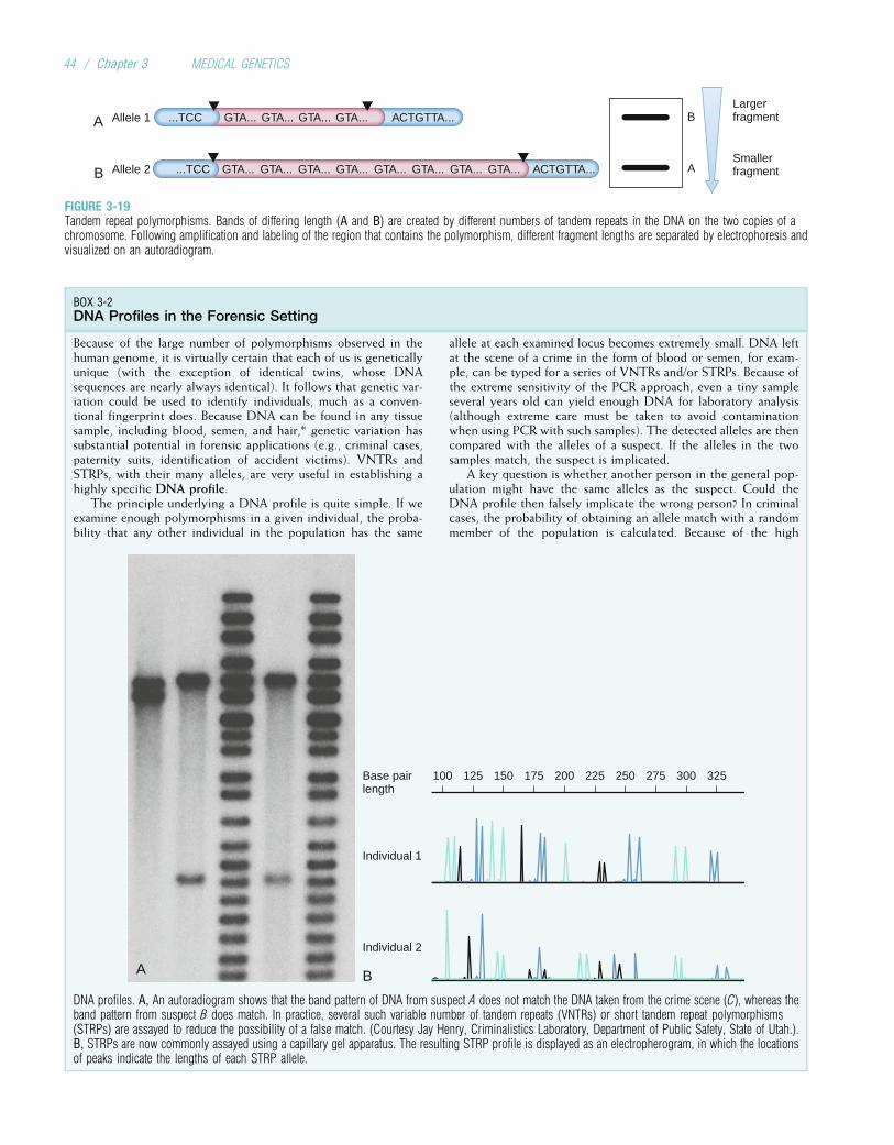
GTA... GTA... GTA... GTA... ACTGTTA...LargerfragmentB
AB
A
Smallerfragment
...TCC
ACTGTTA...GTA... GTA... GTA... GTA... GTA... GTA... GTA... GTA......TCC
Allele 1
Allele 2
FIGURE 3-19Tandem repeat polymorphisms. Bands of differing length (A and B) are created by different numbers of tandem repeats in the DNA on the two copies of achromosome. Following amplification and labeling of the region that contains the polymorphism, different fragment lengths are separated by electrophoresis andvisualized on an autoradiogram.
BOX 3-2DNA Profiles in the Forensic Setting
Because of the large number of polymorphisms observed in thehuman genome, it is virtually certain that each of us is geneticallyunique (with the exception of identical twins, whose DNAsequences are nearly always identical). It follows that genetic var-iation could be used to identify individuals, much as a conven-tional fingerprint does. Because DNA can be found in any tissuesample, including blood, semen, and hair,* genetic variation hassubstantial potential in forensic applications (e.g., criminal cases,paternity suits, identification of accident victims). VNTRs andSTRPs, with their many alleles, are very useful in establishing ahighly specific DNA profile.
The principle underlying a DNA profile is quite simple. If weexamine enough polymorphisms in a given individual, the proba-bility that any other individual in the population has the same
allele at each examined locus becomes extremely small. DNA leftat the scene of a crime in the form of blood or semen, for exam-ple, can be typed for a series of VNTRs and/or STRPs. Because ofthe extreme sensitivity of the PCR approach, even a tiny sampleseveral years old can yield enough DNA for laboratory analysis(although extreme care must be taken to avoid contaminationwhen using PCR with such samples). The detected alleles are thencompared with the alleles of a suspect. If the alleles in the twosamples match, the suspect is implicated.
A key question is whether another person in the general pop-ulation might have the same alleles as the suspect. Could theDNA profile then falsely implicate the wrong person? In criminalcases, the probability of obtaining an allele match with a randommember of the population is calculated. Because of the high
A
100 125 150 175 200 225 250 275 300 325Base pairlength
Individual 1
Individual 2
B
DNA profiles. A, An autoradiogram shows that the band pattern of DNA from suspect A does not match the DNA taken from the crime scene (C), whereas theband pattern from suspect B does match. In practice, several such variable number of tandem repeats (VNTRs) or short tandem repeat polymorphisms(STRPs) are assayed to reduce the possibility of a false match. (Courtesy Jay Henry, Criminalistics Laboratory, Department of Public Safety, State of Utah.).B, STRPs are now commonly assayed using a capillary gel apparatus. The resulting STRP profile is displayed as an electropherogram, in which the locationsof peaks indicate the lengths of each STRP allele.
44 / Chapter 3 MEDICAL GENETICS
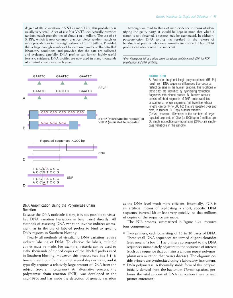
DNA Amplification Using the Polymerase ChainReactionBecause the DNA molecule is tiny, it is not possible to visua-lize DNA variation (variation in base pairs) directly. Allmethods of assessing DNA variation involve indirect assess-ment, as in the use of labeled probes to bind to specificDNA regions in Southern blotting.
Nearly all methods of visualizing DNA variation requireindirect labeling of DNA. To observe the labels, multiplecopies must be made. For example, bacteria can be used tomake thousands of cloned copies of the labeled probes usedin Southern blotting. However, this process (see Box 3-1) istime-consuming, often requiring several days or more, and ittypically requires a relatively large amount of DNA from thesubject (several micrograms). An alternative process, thepolymerase chain reaction (PCR), was developed in themid-1980s and has made the detection of genetic variation
at the DNA level much more efficient. Essentially, PCR isan artificial means of replicating a short, specific DNAsequence (several kb or less) very quickly, so that millionsof copies of the sequence are made.
The PCR process, summarized in Figure 3-21, requiresfour components:
• Two primers, each consisting of 15 to 20 bases of DNA.These small DNA sequences are termed oligonucleotides(oligo means “a few”). The primers correspond to the DNAsequences immediately adjacent to the sequence of interest(such as a sequence that contains a tandem repeat polymor-phism or a mutation that causes disease). The oligonucleo-tide primers are synthesized using a laboratory instrument.
• DNA polymerase. A thermally stable form of this enzyme,initially derived from the bacterium Thermus aquaticus, per-forms the vital process of DNA replication (here termedprimer extension).
degree of allelic variation in VNTRs and STRPs, this probability isusually very small. A set of just four VNTR loci typically providesrandom match probabilities of about 1 in 1 million. The use of 13STRPs, which is now common practice, yields random match ormore probabilities in the neighborhood of 1 in 1 trillion. Providedthat a large enough number of loci are used under well-controlledlaboratory conditions, and provided that the data are collectedand evaluated carefully, DNA profiles can furnish highly usefulforensic evidence. DNA profiles are now used in many thousandsof criminal court cases each year.
Although we tend to think of such evidence in terms of iden-tifying the guilty party, it should be kept in mind that when amatch is not obtained, a suspect may be exonerated. In addition,postconviction DNA testing has resulted in the release ofhundreds of persons who were wrongly imprisoned. Thus, DNAprofiles can also benefit the innocent.
*Even fingerprints left at a crime scene sometimes contain enough DNA for PCR
amplification and DNA profiling.
GAATTC GAATTC GAATTC
GAATTC
Repeated sequences >1000 bp
CAG CAG CAG
CAG CAG CAG
CAG CAG
GACTTCRFLP
STRP (microsatellite repeats) orVNTR (minisatellite repeats)
CNV
SNP
TA
GC
GC
CG
AT
GC
GC
CG
TA
GC
GC
TA
AT
GC
GC
CG
GAATTC
A
B
C
D
FIGURE 3-20A, Restriction fragment length polymorphisms (RFLPs)result from DNA sequence differences that occur atrestriction sites in the human genome. The locations ofthese sites are identified by hybridizing restrictionfragments with cloned probes. B, Tandem repeatsconsist of short segments of DNA (microsatellites)or somewhat longer segments (minisatellites whoselengths can be 14 to 500 bp) that are repeated over andover, in tandem. C, Copy number variants(CNVs) represent differences in the numbers of largerrepeated segments of DNA (>1000 bp to 2 million bp).D, Single nucleotide polymorphisms (SNPs) are single-base variations in the genome.
Genetic Variation: Its Origin and Detection / 45
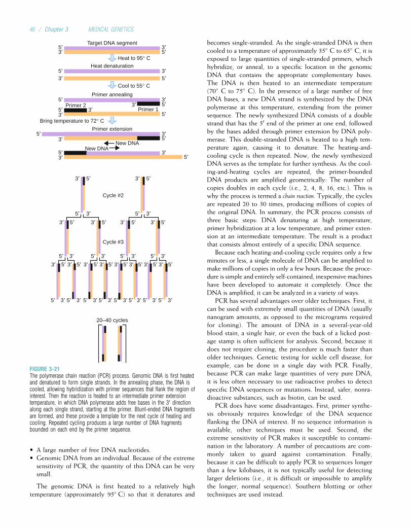
• A large number of free DNA nucleotides.• Genomic DNA from an individual. Because of the extreme
sensitivity of PCR, the quantity of this DNA can be verysmall.
The genomic DNA is first heated to a relatively hightemperature (approximately 95� C) so that it denatures and
becomes single-stranded. As the single-stranded DNA is thencooled to a temperature of approximately 35� C to 65� C, it isexposed to large quantities of single-stranded primers, whichhybridize, or anneal, to a specific location in the genomicDNA that contains the appropriate complementary bases.The DNA is then heated to an intermediate temperature(70� C to 75� C). In the presence of a large number of freeDNA bases, a new DNA strand is synthesized by the DNApolymerase at this temperature, extending from the primersequence. The newly synthesized DNA consists of a doublestrand that has the 50 end of the primer at one end, followedby the bases added through primer extension by DNA poly-merase. This double-stranded DNA is heated to a high tem-perature again, causing it to denature. The heating-and-cooling cycle is then repeated. Now, the newly synthesizedDNA serves as the template for further synthesis. As the cool-ing-and-heating cycles are repeated, the primer-boundedDNA products are amplified geometrically: The number ofcopies doubles in each cycle (i.e., 2, 4, 8, 16, etc.). This iswhy the process is termed a chain reaction. Typically, the cyclesare repeated 20 to 30 times, producing millions of copies ofthe original DNA. In summary, the PCR process consists ofthree basic steps: DNA denaturing at high temperature,primer hybridization at a low temperature, and primer exten-sion at an intermediate temperature. The result is a productthat consists almost entirely of a specific DNA sequence.
Because each heating-and-cooling cycle requires only a fewminutes or less, a single molecule of DNA can be amplified tomake millions of copies in only a few hours. Because the proce-dure is simple and entirely self-contained, inexpensive machineshave been developed to automate it completely. Once theDNA is amplified, it can be analyzed in a variety of ways.
PCR has several advantages over older techniques. First, itcan be used with extremely small quantities of DNA (usuallynanogram amounts, as opposed to the micrograms requiredfor cloning). The amount of DNA in a several-year-oldblood stain, a single hair, or even the back of a licked post-age stamp is often sufficient for analysis. Second, because itdoes not require cloning, the procedure is much faster thanolder techniques. Genetic testing for sickle cell disease, forexample, can be done in a single day with PCR. Finally,because PCR can make large quantities of very pure DNA,it is less often necessary to use radioactive probes to detectspecific DNA sequences or mutations. Instead, safer, nonra-dioactive substances, such as biotin, can be used.
PCR does have some disadvantages. First, primer synthe-sis obviously requires knowledge of the DNA sequenceflanking the DNA of interest. If no sequence information isavailable, other techniques must be used. Second, theextreme sensitivity of PCR makes it susceptible to contami-nation in the laboratory. A number of precautions are com-monly taken to guard against contamination. Finally,because it can be difficult to apply PCR to sequences longerthan a few kilobases, it is not typically useful for detectinglarger deletions (i.e., it is difficult or impossible to amplifythe longer, normal sequence). Southern blotting or othertechniques are used instead.
Target DNA segment
Cycle #2
Cycle #3
20–40 cycles
Heat denaturation
Primer annealing
Primer extension
Primer 2Primer 1
3′
3′
5′
5′
3′
New DNA
3′5′
3′5′
3′5′
5′
5′3′
5′
5′
3′
3′
3′
Heat to 95° C
Cool to 55° C
Bring temperature to 72° C
5′
3′
5′3′
3′3′
3′5′5′ 3′ 5′
3′
5′ 3′3′ 5′
5′3′5′
3′
5′
3′
5′
3′
5′3′5′3′5′3′
5′
3′
5′
5′
3′
5′
3′
3′
5′
3′
5′
5′
3′
3′
5′
5′
3′
3′
5′
3′
5′
5′
5′3′
3′
5′
5′
New DNA
5′
3′
3′
5′
FIGURE 3-21The polymerase chain reaction (PCR) process. Genomic DNA is first heatedand denatured to form single strands. In the annealing phase, the DNA iscooled, allowing hybridization with primer sequences that flank the region ofinterest. Then the reaction is heated to an intermediate primer extensiontemperature, in which DNA polymerase adds free bases in the 30 directionalong each single strand, starting at the primer. Blunt-ended DNA fragmentsare formed, and these provide a template for the next cycle of heating andcooling. Repeated cycling produces a large number of DNA fragmentsbounded on each end by the primer sequence.
46 / Chapter 3 MEDICAL GENETICS
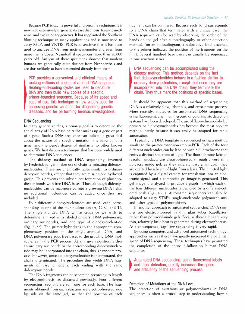
Because PCR is such a powerful and versatile technique, it isnow used extensively in genetic disease diagnosis, forensicmed-icine, and evolutionary genetics. It has supplanted the Southernblotting technique in many applications and is now used toassay RFLPs and VNTRs. PCR is so sensitive that it has beenused to analyze DNA from ancient mummies and even frommore than a dozen Neanderthal specimens more than 30,000years old. Analysis of these specimens showed that modernhumans are genetically quite distinct from Neanderthals andare thus unlikely to have descended directly from them.
4PCR provides a convenient and efficient means ofmaking millions of copies of a short DNA sequence.Heating-and-cooling cycles are used to denatureDNA and then build new copies of a specific,primer-bounded sequence. Because of its speed andease of use, this technique is now widely used forassessing genetic variation, for diagnosing geneticdiseases, and for performing forensic investigations.
DNA SequencingIn many genetic studies, a primary goal is to determine theactual array of DNA base pairs that makes up a gene or partof a gene. Such a DNA sequence can indicate a great dealabout the nature of a specific mutation, the function of agene, and the gene’s degree of similarity to other knowngenes. We first discuss a technique that has been widely usedto determine DNA sequences.
The dideoxy method of DNA sequencing, inventedby Frederick Sanger, makes use of chain-terminating dideoxy-nucleotides. These are chemically quite similar to ordinarydeoxynucleotides, except that they are missing one hydroxylgroup. This prevents the subsequent formation of phospho-diester bonds with free DNA bases. Thus, although dideoxy-nucleotides can be incorporated into a growing DNA helix,no additional nucleotides can be added once they areincluded.
Four different dideoxynucleotides are used, each corre-sponding to one of the four nucleotides (A, C, G, and T).The single-stranded DNA whose sequence we wish todetermine is mixed with labeled primers, DNA polymerase,ordinary nucleotides, and one type of dideoxynucleotide(Fig. 3-22). The primer hybridizes to the appropriate com-plementary position in the single-stranded DNA, andDNA polymerase adds free bases to the growing DNA mol-ecule, as in the PCR process. At any given position, eitheran ordinary nucleotide or the corresponding dideoxynucleo-tide may be incorporated into the chain; this is a random pro-cess. However, once a dideoxynucleotide is incorporated, thechain is terminated. The procedure thus yields DNA frag-ments of varying length, each ending with the samedideoxynucleotide.
The DNA fragments can be separated according to lengthby electrophoresis, as discussed previously. Four differentsequencing reactions are run, one for each base. The frag-ments obtained from each reaction are electrophoresed sideby side on the same gel, so that the position of each
fragment can be compared. Because each band correspondsto a DNA chain that terminates with a unique base, theDNA sequence can be read by observing the order of thebands on the gel after autoradiography or other detectionmethods (on an autoradiogram, a radioactive label attachedto the primer indicates the position of the fragment on thefilm). Several hundred base pairs can usually be sequencedin one reaction series.
4DNA sequencing can be accomplished using thedideoxy method. This method depends on the factthat dideoxynucleotides behave in a fashion similar toordinary deoxynucleotides, except that once they areincorporated into the DNA chain, they terminate thechain. They thus mark the positions of specific bases.
It should be apparent that this method of sequencingDNA is a relatively slow, laborious, and error-prone process.More recently, strategies for automated DNA sequencingusing fluorescent, chemiluminescent, or colorimetric detectionsystems have been developed. The use of fluorochrome-labeledprimers or dideoxynucleotides has become the most popularmethod, partly because it can easily be adapted for rapidautomation.
Typically, a DNA template is sequenced using a methodsimilar to the primer extension step in PCR. Each of the fourdifferent nucleotides can be labeled with a fluorochrome thatemits a distinct spectrum of light. The fluorochrome-labeledreaction products are electrophoresed through a very thinpolyacrylamide gel; as they migrate past a window, theyare excited by a beam of light from a laser. The emitted lightis captured by a digital camera for translation into an elec-tronic signal, and a composite gel image is generated. Thisgel image is analyzed to produce a graph in which each ofthe four different nucleotides is depicted by a different-col-ored peak (Fig. 3-23). Automated sequencers can also beadapted to assay STRPs, single-nucleotide polymorphisms,and other types of polymorphisms.
In another approach to automated sequencing, DNA sam-ples are electrophoresed in thin glass tubes (capillaries)rather than polyacrylamide gels. Because these tubes are verythin, relatively little heat is generated during electrophoresis.As a consequence, capillary sequencing is very rapid.
By using computers and advanced automated technology,approaches such as these have greatly increased the potentialspeed of DNA sequencing. These techniques have permittedthe completion of the entire 3-billion-bp human DNAsequence.
4Automated DNA sequencing, using fluorescent labelsand laser detection, greatly increases the speedand efficiency of the sequencing process.
Detection of Mutations at the DNA LevelThe detection of mutations or polymorphisms in DNAsequences is often a critical step in understanding how a
Genetic Variation: Its Origin and Detection / 47
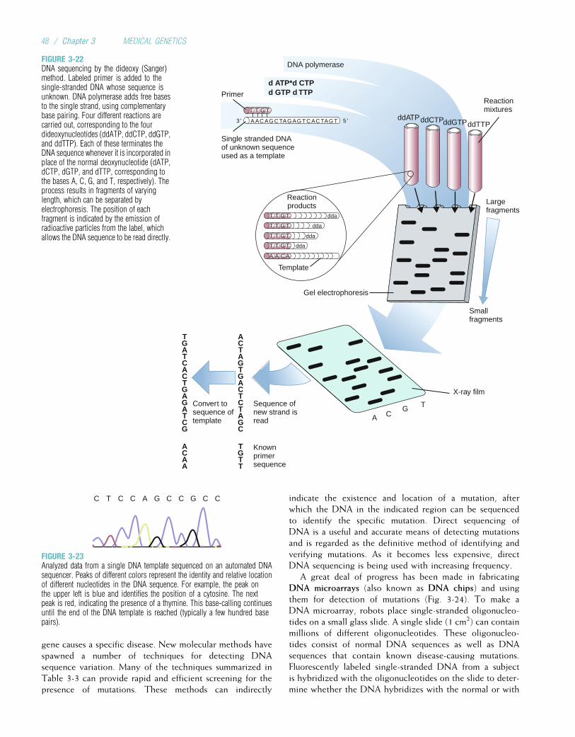
gene causes a specific disease. New molecular methods havespawned a number of techniques for detecting DNAsequence variation. Many of the techniques summarized inTable 3-3 can provide rapid and efficient screening for thepresence of mutations. These methods can indirectly
indicate the existence and location of a mutation, afterwhich the DNA in the indicated region can be sequencedto identify the specific mutation. Direct sequencing ofDNA is a useful and accurate means of detecting mutationsand is regarded as the definitive method of identifying andverifying mutations. As it becomes less expensive, directDNA sequencing is being used with increasing frequency.
A great deal of progress has been made in fabricatingDNA microarrays (also known as DNA chips) and usingthem for detection of mutations (Fig. 3-24). To make aDNA microarray, robots place single-stranded oligonucleo-tides on a small glass slide. A single slide (1 cm2) can containmillions of different oligonucleotides. These oligonucleo-tides consist of normal DNA sequences as well as DNAsequences that contain known disease-causing mutations.Fluorescently labeled single-stranded DNA from a subjectis hybridized with the oligonucleotides on the slide to deter-mine whether the DNA hybridizes with the normal or with
Reactionproducts
Single stranded DNAof unknown sequenceused as a template
PrimerReactionmixtures
Largefragments
Smallfragments
Knownprimersequence
Sequence ofnew strand isread
Convert tosequence oftemplate
X-ray film
Template
Gel electrophoresis
T T G T
A A C A
T T G T
T T G T
T T G T
ddATPddCTPddGTPddTTP
ACTAGTGACTCTAGC
TGATCACTGAGATCG
TGTT
ACAA
A CG T
dda
dda
dda
dda
A AC AG C TAG AG T C AC TAG T 5 '
T T G T
DNA polymerase
d ATP*d CTPd GTP d TTP
3 '
FIGURE 3-22DNA sequencing by the dideoxy (Sanger)method. Labeled primer is added to thesingle-stranded DNA whose sequence isunknown. DNA polymerase adds free basesto the single strand, using complementarybase pairing. Four different reactions arecarried out, corresponding to the fourdideoxynucleotides (ddATP, ddCTP, ddGTP,and ddTTP). Each of these terminates theDNA sequence whenever it is incorporated inplace of the normal deoxynucleotide (dATP,dCTP, dGTP, and dTTP, corresponding tothe bases A, C, G, and T, respectively). Theprocess results in fragments of varyinglength, which can be separated byelectrophoresis. The position of eachfragment is indicated by the emission ofradioactive particles from the label, whichallows the DNA sequence to be read directly.
C T C C A G C C G C C
FIGURE 3-23Analyzed data from a single DNA template sequenced on an automated DNAsequencer. Peaks of different colors represent the identity and relative locationof different nucleotides in the DNA sequence. For example, the peak onthe upper left is blue and identifies the position of a cytosine. The nextpeak is red, indicating the presence of a thymine. This base-calling continuesuntil the end of the DNA template is reached (typically a few hundred basepairs).
48 / Chapter 3 MEDICAL GENETICS
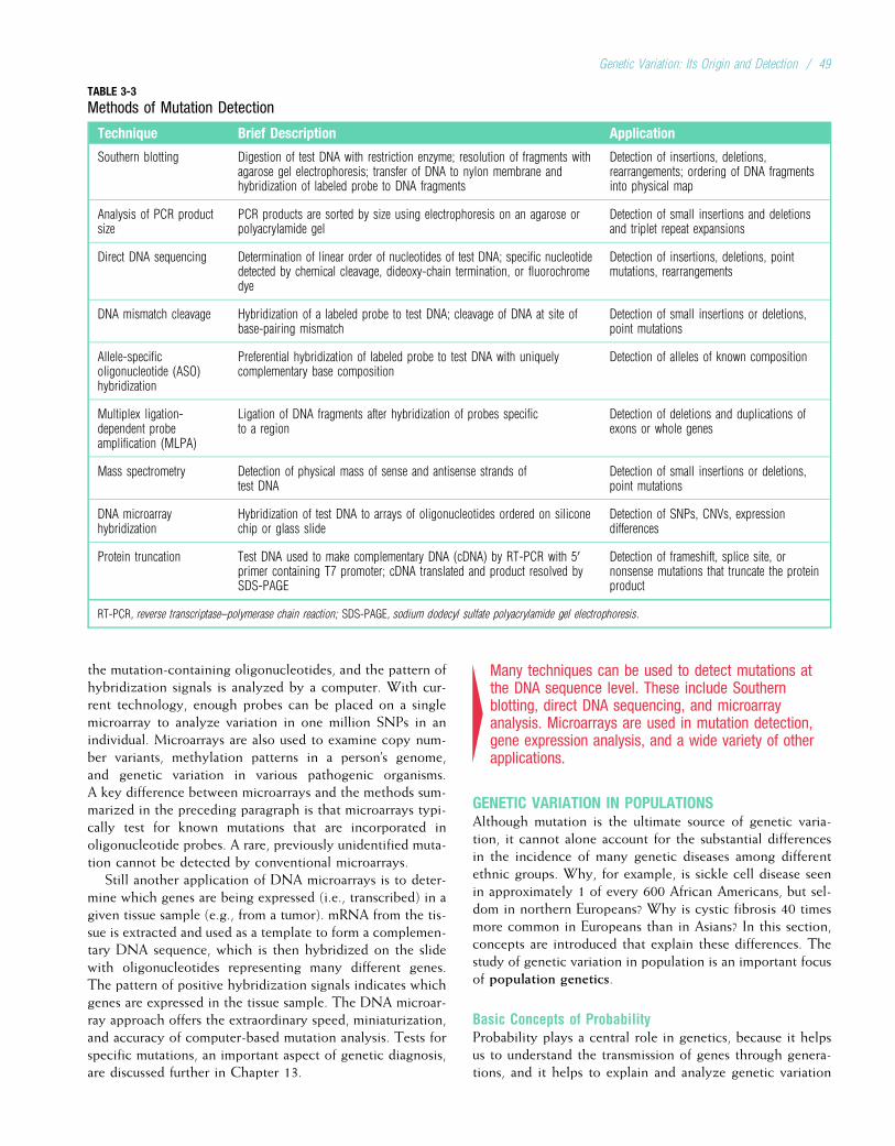
the mutation-containing oligonucleotides, and the pattern ofhybridization signals is analyzed by a computer. With cur-rent technology, enough probes can be placed on a singlemicroarray to analyze variation in one million SNPs in anindividual. Microarrays are also used to examine copy num-ber variants, methylation patterns in a person’s genome,and genetic variation in various pathogenic organisms.A key difference between microarrays and the methods sum-marized in the preceding paragraph is that microarrays typi-cally test for known mutations that are incorporated inoligonucleotide probes. A rare, previously unidentified muta-tion cannot be detected by conventional microarrays.
Still another application of DNA microarrays is to deter-mine which genes are being expressed (i.e., transcribed) in agiven tissue sample (e.g., from a tumor). mRNA from the tis-sue is extracted and used as a template to form a complemen-tary DNA sequence, which is then hybridized on the slidewith oligonucleotides representing many different genes.The pattern of positive hybridization signals indicates whichgenes are expressed in the tissue sample. The DNA microar-ray approach offers the extraordinary speed, miniaturization,and accuracy of computer-based mutation analysis. Tests forspecific mutations, an important aspect of genetic diagnosis,are discussed further in Chapter 13.
4Many techniques can be used to detect mutations atthe DNA sequence level. These include Southernblotting, direct DNA sequencing, and microarrayanalysis. Microarrays are used in mutation detection,gene expression analysis, and a wide variety of otherapplications.
GENETIC VARIATION IN POPULATIONSAlthough mutation is the ultimate source of genetic varia-tion, it cannot alone account for the substantial differencesin the incidence of many genetic diseases among differentethnic groups. Why, for example, is sickle cell disease seenin approximately 1 of every 600 African Americans, but sel-dom in northern Europeans? Why is cystic fibrosis 40 timesmore common in Europeans than in Asians? In this section,concepts are introduced that explain these differences. Thestudy of genetic variation in population is an important focusof population genetics.
Basic Concepts of ProbabilityProbability plays a central role in genetics, because it helpsus to understand the transmission of genes through genera-tions, and it helps to explain and analyze genetic variation
TABLE 3-3
Methods of Mutation Detection
Technique Brief Description Application
Southern blotting Digestion of test DNA with restriction enzyme; resolution of fragments withagarose gel electrophoresis; transfer of DNA to nylon membrane andhybridization of labeled probe to DNA fragments
Detection of insertions, deletions,rearrangements; ordering of DNA fragmentsinto physical map
Analysis of PCR productsize
PCR products are sorted by size using electrophoresis on an agarose orpolyacrylamide gel
Detection of small insertions and deletionsand triplet repeat expansions
Direct DNA sequencing Determination of linear order of nucleotides of test DNA; specific nucleotidedetected by chemical cleavage, dideoxy-chain termination, or fluorochromedye
Detection of insertions, deletions, pointmutations, rearrangements
DNA mismatch cleavage Hybridization of a labeled probe to test DNA; cleavage of DNA at site ofbase-pairing mismatch
Detection of small insertions or deletions,point mutations
Allele-specificoligonucleotide (ASO)hybridization
Preferential hybridization of labeled probe to test DNA with uniquelycomplementary base composition
Detection of alleles of known composition
Multiplex ligation-dependent probeamplification (MLPA)
Ligation of DNA fragments after hybridization of probes specificto a region
Detection of deletions and duplications ofexons or whole genes
Mass spectrometry Detection of physical mass of sense and antisense strands oftest DNA
Detection of small insertions or deletions,point mutations
DNA microarrayhybridization
Hybridization of test DNA to arrays of oligonucleotides ordered on siliconechip or glass slide
Detection of SNPs, CNVs, expressiondifferences
Protein truncation Test DNA used to make complementary DNA (cDNA) by RT-PCR with 50primer containing T7 promoter; cDNA translated and product resolved bySDS-PAGE
Detection of frameshift, splice site, ornonsense mutations that truncate the proteinproduct
RT-PCR, reverse transcriptase–polymerase chain reaction; SDS-PAGE, sodium dodecyl sulfate polyacrylamide gel electrophoresis.
Genetic Variation: Its Origin and Detection / 49
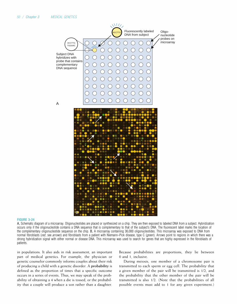
in populations. It also aids in risk assessment, an importantpart of medical genetics. For example, the physician orgenetic counselor commonly informs couples about their riskof producing a child with a genetic disorder. A probability isdefined as the proportion of times that a specific outcomeoccurs in a series of events. Thus, we may speak of the prob-ability of obtaining a 4 when a die is tossed, or the probabil-ity that a couple will produce a son rather than a daughter.
Because probabilities are proportions, they lie between0 and 1, inclusive.
During meiosis, one member of a chromosome pair istransmitted to each sperm or egg cell. The probability thata given member of the pair will be transmitted is 1/2, andthe probability that the other member of the pair will betransmitted is also 1/2. (Note that the probabilities of allpossible events must add to 1 for any given experiment.)
ACCTTG Fluorescently labeledDNA from subject
Oligo-nucleotideprobes onmicroarrayACCTTG
TGGAAC
Subject DNAhybridizes withprobe that containscomplementaryDNA sequence
A
B
FIGURE 3-24A, Schematic diagram of a microarray. Oligonucleotides are placed or synthesized on a chip. They are then exposed to labeled DNA from a subject. Hybridizationoccurs only if the oligonucleotide contains a DNA sequence that is complementary to that of the subject’s DNA. The fluorescent label marks the location ofthe complementary oligonucleotide sequence on the chip. B, A microarray containing 36,000 oligonucleotides. This microarray was exposed to DNA fromnormal fibroblasts (red, see arrows) and fibroblasts from a patient with Niemann–Pick disease, type C (green). Arrows point to regions in which there was astrong hybridization signal with either normal or disease DNA. This microarray was used to search for genes that are highly expressed in the fibroblasts ofpatients.
50 / Chapter 3 MEDICAL GENETICS
Because this situation is directly analogous to coin tossing,in which the probabilities of obtaining heads or tailsare each 1/2, we will use coin tossing as our illustrativeexample.
When a coin is tossed repeatedly, the outcome of eachtoss has no effect on subsequent outcomes. Each event (toss)is said to be independent. Even if we have obtained 10 headsin a row, the probability of obtaining heads or tails on thenext toss remains 1/2. Similarly, the probability that a parentwill transmit one of the two alleles at a locus is independentfrom one reproductive event to the next.
The independence principle allows us to deduce twofundamental concepts of probability, the multiplication ruleand the addition rule. The multiplication rule states that iftwo trials are independent, then the probability of obtaininga given outcome in both trials is the product of the probabil-ities of each outcome. For example, we may wish to knowthe probability of obtaining heads on both tosses of a faircoin. Because the tosses are independent events, this proba-bility is given by the product of the probabilities of obtain-ing heads in each individual toss: 1/2 � 1/2 ¼ 1/4.Similarly, the probability of obtaining two tails in a row is1/2 � 1/2 ¼ 1/4.
The multiplication rule can be extended for any numberof trials. Suppose a couple wants to know the probabilitythat all three of their planned children will be girls. Becausethe probability of producing a girl is approximately 1/2, andbecause reproductive events are independent of one another,the probability of producing three girls is 1/2 � 1/2 � 1/2 ¼1/8. However, if the couple has already produced two girlsand then wants to know the probability of producing a thirdgirl, it is simply 1/2. This is because the previous two eventsare no longer probabilities; they have actually occurred.Because of independence, these past events have no effecton the outcome of the third event.
The addition rule states that if we want to know the prob-ability of either one outcome or another, we can simply addthe respective probabilities together. For example, the prob-ability of getting two heads in a row is 1/2 � 1/2, or 1/4, andthe probability of getting two tails in a row is the same. Theprobability of getting either two heads or two tails in a totalof two tosses is the sum of the probabilities: 1/4 þ 1/4 ¼ 1/2.As another example, imagine that a couple plans to havethree children, and they have a strong aversion to havingthree children all of the same sex. They can be reassuredsomewhat by knowing that the probability of producingthree girls or three boys is only 1/8 þ 1/8, or 1/4. The prob-ability that they will have some combination of boys andgirls is 3/4, because the sum of the probabilities of all possi-ble outcomes must add to 1.
4Basic probability enables us to understand andestimate genetic risks and to understand geneticvariation among populations. The multiplication rule isused to estimate the probability that two events willoccur together. The addition rule is used to estimatethe probability that one event or another will occur.
Gene and Genotype FrequenciesThe prevalence of many genetic diseases varies considerablyfrom one population to another. The concepts of genotypefrequency and gene frequency help us to measure andunderstand population variation in the incidence of geneticdisease.
Imagine that we have typed 200 persons in a populationfor the MN blood group. This blood group, which isencoded by a locus on chromosome 2, has two major alleles,labeled M and N. In the MN system, the effects of bothalleles can be observed in the heterozygote. M and N aretherefore said to be codominant: The heterozygote can bedistinguished from both homozygotes. Any individual inthe population can have one of three possible genotypes(recall that the genotype is one’s genetic makeup at a locus):He or she could be homozygous for M (genotype MM), het-erozygous (MN), or homozygous for N (NN). After typingeach person in our sample, we find the following distribu-tion of genotypes: MM, 64; MN, 120; NN, 16. The genotypefrequency is obtained simply by dividing each genotypecount by the total number of subjects. The frequency ofMM is 64/200, or 0.32; the frequency of MN is 120/200, or0.60; and the frequency of NN is 16/200, or 0.08. The sumof these frequencies must equal 1.
The gene frequency for each allele, M and N, can beobtained here by the process of gene counting. Each MM
homozygote has two M alleles, and each heterozygote hasone M allele. Similarly, NN homozygotes have two N alleles,and heterozygotes have one N allele. In the exampledescribed, there are
ð64� 2Þ þ 120 ¼ 248M alleles
ð16� 2Þ þ 120 ¼ 152N alleles
In total, there are 400 alleles at the MN locus (i.e., twice thenumber of subjects, because each subject has two alleles). Toobtain the frequency of M, we divide the number of M
alleles by the total number of alleles at that locus: 248/400 ¼0.62. Likewise, the frequency of N is 152/400, or 0.38. Thesum of the two frequencies must equal 1.
4Gene and genotype frequencies specify theproportions of each allele and each genotype,respectively, in a population. Under simple conditionsthese frequencies can be estimated by directcounting.
The Hardy–Weinberg PrincipleThe example given for the MN locus presents an ideal situa-tion for gene frequency estimation, because, owing to co-dominance, the three genotypes can easily be distinguishedand counted. What happens when one of the homozygotesis indistinguishable from the heterozygote (i.e., when thereis dominance)? Here the basic concepts of probability canbe used to specify a predictable relationship between genefrequencies and genotype frequencies.
Genetic Variation: Its Origin and Detection / 51
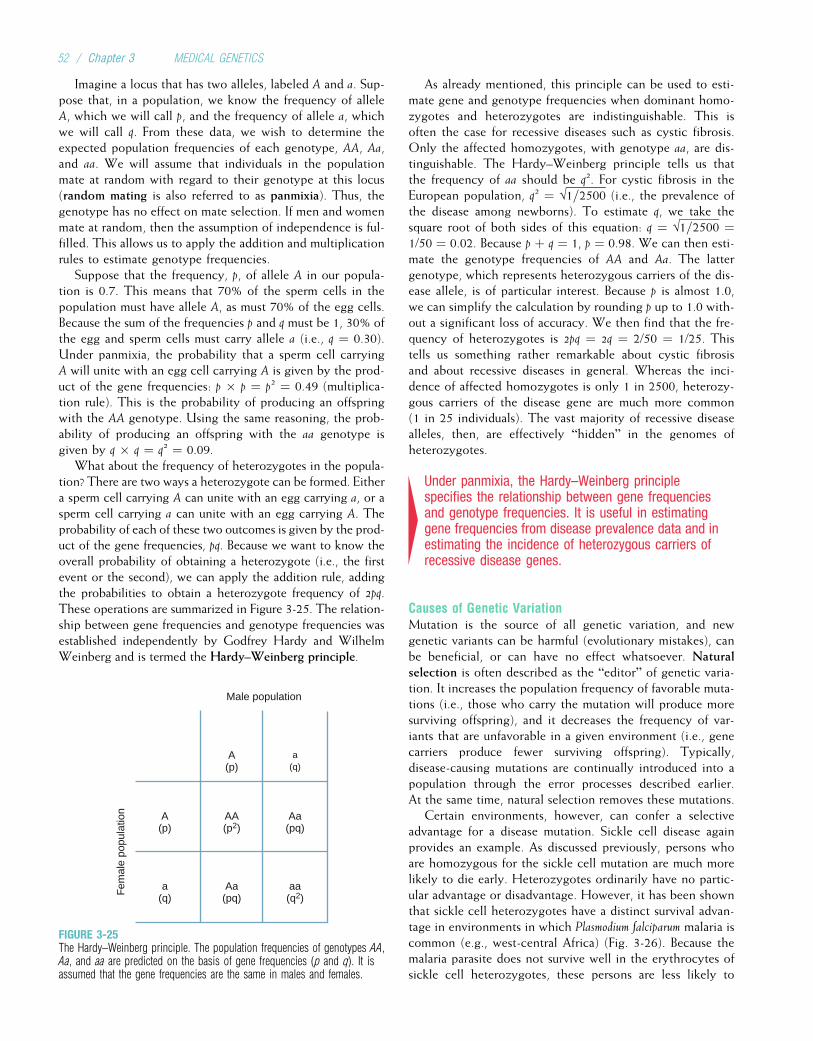
Imagine a locus that has two alleles, labeled A and a. Sup-pose that, in a population, we know the frequency of alleleA, which we will call p, and the frequency of allele a, whichwe will call q. From these data, we wish to determine theexpected population frequencies of each genotype, AA, Aa,and aa. We will assume that individuals in the populationmate at random with regard to their genotype at this locus(random mating is also referred to as panmixia). Thus, thegenotype has no effect on mate selection. If men and womenmate at random, then the assumption of independence is ful-filled. This allows us to apply the addition and multiplicationrules to estimate genotype frequencies.
Suppose that the frequency, p, of allele A in our popula-tion is 0.7. This means that 70% of the sperm cells in thepopulation must have allele A, as must 70% of the egg cells.Because the sum of the frequencies p and q must be 1, 30% ofthe egg and sperm cells must carry allele a (i.e., q ¼ 0.30).Under panmixia, the probability that a sperm cell carryingA will unite with an egg cell carrying A is given by the prod-uct of the gene frequencies: p � p ¼ p2 ¼ 0.49 (multiplica-tion rule). This is the probability of producing an offspringwith the AA genotype. Using the same reasoning, the prob-ability of producing an offspring with the aa genotype isgiven by q � q ¼ q2 ¼ 0.09.
What about the frequency of heterozygotes in the popula-tion? There are two ways a heterozygote can be formed. Eithera sperm cell carrying A can unite with an egg carrying a, or asperm cell carrying a can unite with an egg carrying A. Theprobability of each of these two outcomes is given by the prod-uct of the gene frequencies, pq. Because we want to know theoverall probability of obtaining a heterozygote (i.e., the firstevent or the second), we can apply the addition rule, addingthe probabilities to obtain a heterozygote frequency of 2pq.These operations are summarized in Figure 3-25. The relation-ship between gene frequencies and genotype frequencies wasestablished independently by Godfrey Hardy and WilhelmWeinberg and is termed the Hardy–Weinberg principle.
As already mentioned, this principle can be used to esti-mate gene and genotype frequencies when dominant homo-zygotes and heterozygotes are indistinguishable. This isoften the case for recessive diseases such as cystic fibrosis.Only the affected homozygotes, with genotype aa, are dis-tinguishable. The Hardy–Weinberg principle tells us thatthe frequency of aa should be q2. For cystic fibrosis in theEuropean population, q2 ¼ √1=2500 (i.e., the prevalence ofthe disease among newborns). To estimate q, we take thesquare root of both sides of this equation: q ¼ √1=2500 ¼1/50 ¼ 0.02. Because p þ q ¼ 1, p ¼ 0.98. We can then esti-mate the genotype frequencies of AA and Aa. The lattergenotype, which represents heterozygous carriers of the dis-ease allele, is of particular interest. Because p is almost 1.0,we can simplify the calculation by rounding p up to 1.0 with-out a significant loss of accuracy. We then find that the fre-quency of heterozygotes is 2pq ¼ 2q ¼ 2/50 ¼ 1/25. Thistells us something rather remarkable about cystic fibrosisand about recessive diseases in general. Whereas the inci-dence of affected homozygotes is only 1 in 2500, heterozy-gous carriers of the disease gene are much more common(1 in 25 individuals). The vast majority of recessive diseasealleles, then, are effectively “hidden” in the genomes ofheterozygotes.
4Under panmixia, the Hardy–Weinberg principlespecifies the relationship between gene frequenciesand genotype frequencies. It is useful in estimatinggene frequencies from disease prevalence data and inestimating the incidence of heterozygous carriers ofrecessive disease genes.
Causes of Genetic VariationMutation is the source of all genetic variation, and newgenetic variants can be harmful (evolutionary mistakes), canbe beneficial, or can have no effect whatsoever. Naturalselection is often described as the “editor” of genetic varia-tion. It increases the population frequency of favorable muta-tions (i.e., those who carry the mutation will produce moresurviving offspring), and it decreases the frequency of var-iants that are unfavorable in a given environment (i.e., genecarriers produce fewer surviving offspring). Typically,disease-causing mutations are continually introduced into apopulation through the error processes described earlier.At the same time, natural selection removes these mutations.
Certain environments, however, can confer a selectiveadvantage for a disease mutation. Sickle cell disease againprovides an example. As discussed previously, persons whoare homozygous for the sickle cell mutation are much morelikely to die early. Heterozygotes ordinarily have no partic-ular advantage or disadvantage. However, it has been shownthat sickle cell heterozygotes have a distinct survival advan-tage in environments in which Plasmodium falciparum malaria iscommon (e.g., west-central Africa) (Fig. 3-26). Because themalaria parasite does not survive well in the erythrocytes ofsickle cell heterozygotes, these persons are less likely to
Male population
Fem
ale
popu
latio
n
A(p)
a(q)
A(p)
AA(p2)
Aa(pq)
a(q)
Aa(pq)
aa(q2)
FIGURE 3-25The Hardy–Weinberg principle. The population frequencies of genotypes AA,Aa, and aa are predicted on the basis of gene frequencies (p and q). It isassumed that the gene frequencies are the same in males and females.
52 / Chapter 3 MEDICAL GENETICS
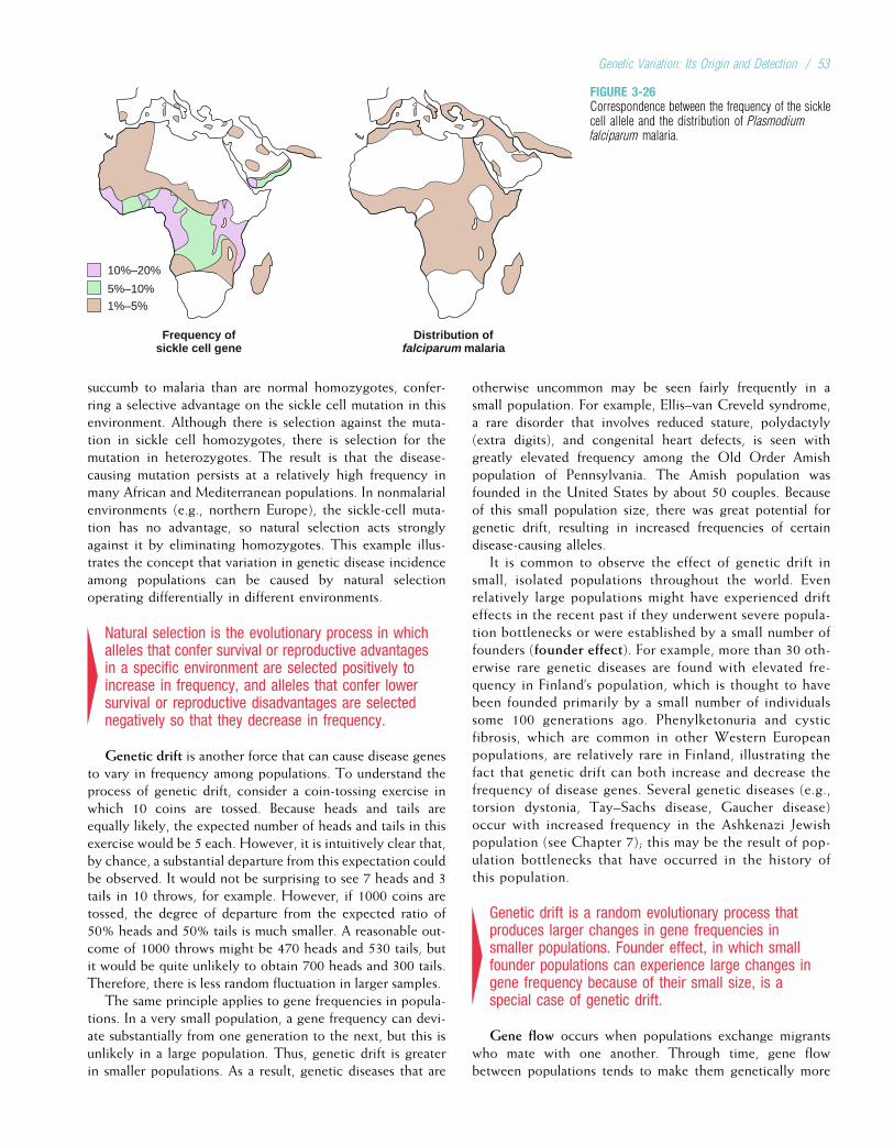
succumb to malaria than are normal homozygotes, confer-ring a selective advantage on the sickle cell mutation in thisenvironment. Although there is selection against the muta-tion in sickle cell homozygotes, there is selection for themutation in heterozygotes. The result is that the disease-causing mutation persists at a relatively high frequency inmany African and Mediterranean populations. In nonmalarialenvironments (e.g., northern Europe), the sickle-cell muta-tion has no advantage, so natural selection acts stronglyagainst it by eliminating homozygotes. This example illus-trates the concept that variation in genetic disease incidenceamong populations can be caused by natural selectionoperating differentially in different environments.
4Natural selection is the evolutionary process in whichalleles that confer survival or reproductive advantagesin a specific environment are selected positively toincrease in frequency, and alleles that confer lowersurvival or reproductive disadvantages are selectednegatively so that they decrease in frequency.
Genetic drift is another force that can cause disease genesto vary in frequency among populations. To understand theprocess of genetic drift, consider a coin-tossing exercise inwhich 10 coins are tossed. Because heads and tails areequally likely, the expected number of heads and tails in thisexercise would be 5 each. However, it is intuitively clear that,by chance, a substantial departure from this expectation couldbe observed. It would not be surprising to see 7 heads and 3tails in 10 throws, for example. However, if 1000 coins aretossed, the degree of departure from the expected ratio of50% heads and 50% tails is much smaller. A reasonable out-come of 1000 throws might be 470 heads and 530 tails, butit would be quite unlikely to obtain 700 heads and 300 tails.Therefore, there is less random fluctuation in larger samples.
The same principle applies to gene frequencies in popula-tions. In a very small population, a gene frequency can devi-ate substantially from one generation to the next, but this isunlikely in a large population. Thus, genetic drift is greaterin smaller populations. As a result, genetic diseases that are
otherwise uncommon may be seen fairly frequently in asmall population. For example, Ellis–van Creveld syndrome,a rare disorder that involves reduced stature, polydactyly(extra digits), and congenital heart defects, is seen withgreatly elevated frequency among the Old Order Amishpopulation of Pennsylvania. The Amish population wasfounded in the United States by about 50 couples. Becauseof this small population size, there was great potential forgenetic drift, resulting in increased frequencies of certaindisease-causing alleles.
It is common to observe the effect of genetic drift insmall, isolated populations throughout the world. Evenrelatively large populations might have experienced drifteffects in the recent past if they underwent severe popula-tion bottlenecks or were established by a small number offounders (founder effect). For example, more than 30 oth-erwise rare genetic diseases are found with elevated fre-quency in Finland’s population, which is thought to havebeen founded primarily by a small number of individualssome 100 generations ago. Phenylketonuria and cysticfibrosis, which are common in other Western Europeanpopulations, are relatively rare in Finland, illustrating thefact that genetic drift can both increase and decrease thefrequency of disease genes. Several genetic diseases (e.g.,torsion dystonia, Tay–Sachs disease, Gaucher disease)occur with increased frequency in the Ashkenazi Jewishpopulation (see Chapter 7); this may be the result of pop-ulation bottlenecks that have occurred in the history ofthis population.
4Genetic drift is a random evolutionary process thatproduces larger changes in gene frequencies insmaller populations. Founder effect, in which smallfounder populations can experience large changes ingene frequency because of their small size, is aspecial case of genetic drift.
Gene flow occurs when populations exchange migrantswho mate with one another. Through time, gene flowbetween populations tends to make them genetically more
10%–20%
5%–10%
1%–5%
Frequency ofsickle cell gene
Distribution offalciparum malaria
FIGURE 3-26Correspondence between the frequency of the sicklecell allele and the distribution of Plasmodiumfalciparum malaria.
Genetic Variation: Its Origin and Detection / 53
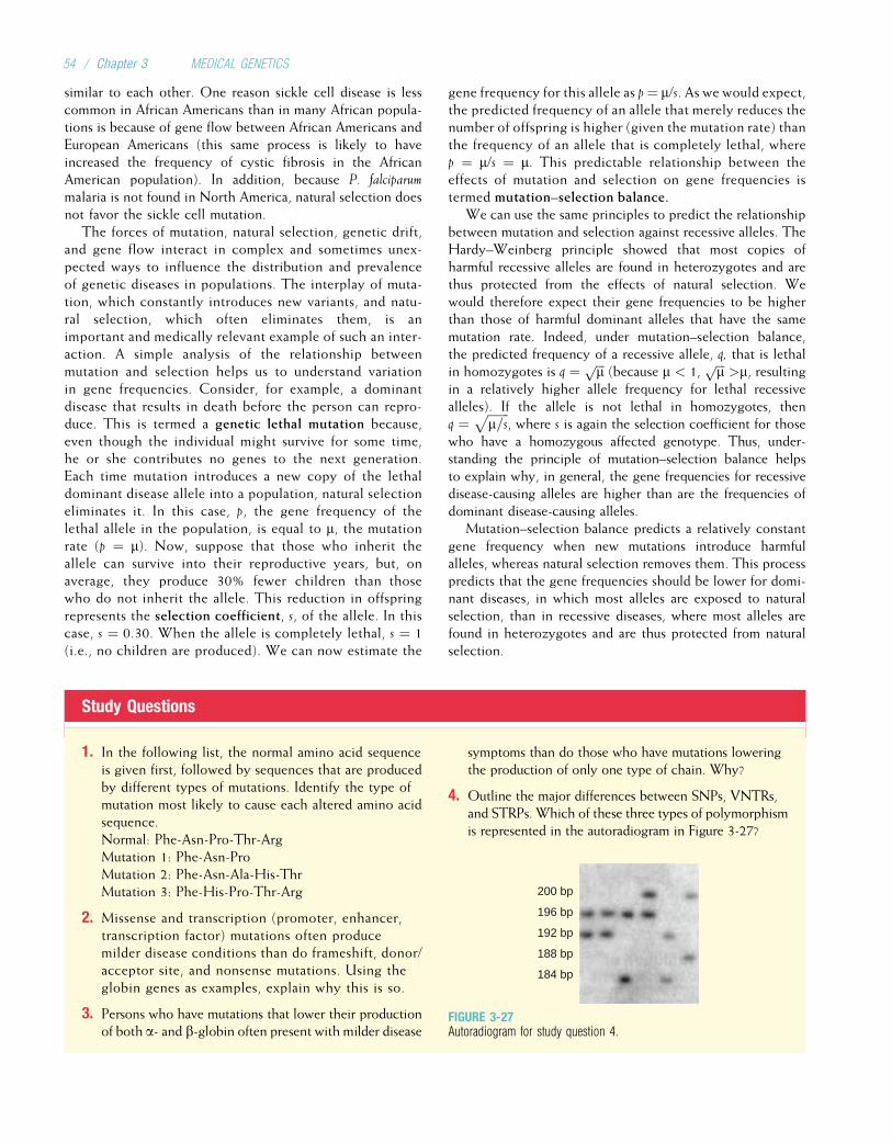
similar to each other. One reason sickle cell disease is lesscommon in African Americans than in many African popula-tions is because of gene flow between African Americans andEuropean Americans (this same process is likely to haveincreased the frequency of cystic fibrosis in the AfricanAmerican population). In addition, because P. falciparum
malaria is not found in North America, natural selection doesnot favor the sickle cell mutation.
The forces of mutation, natural selection, genetic drift,and gene flow interact in complex and sometimes unex-pected ways to influence the distribution and prevalenceof genetic diseases in populations. The interplay of muta-tion, which constantly introduces new variants, and natu-ral selection, which often eliminates them, is animportant and medically relevant example of such an inter-action. A simple analysis of the relationship betweenmutation and selection helps us to understand variationin gene frequencies. Consider, for example, a dominantdisease that results in death before the person can repro-duce. This is termed a genetic lethal mutation because,even though the individual might survive for some time,he or she contributes no genes to the next generation.Each time mutation introduces a new copy of the lethaldominant disease allele into a population, natural selectioneliminates it. In this case, p, the gene frequency of thelethal allele in the population, is equal to m, the mutationrate (p ¼ m). Now, suppose that those who inherit theallele can survive into their reproductive years, but, onaverage, they produce 30% fewer children than thosewho do not inherit the allele. This reduction in offspringrepresents the selection coefficient, s, of the allele. In thiscase, s ¼ 0.30. When the allele is completely lethal, s ¼ 1(i.e., no children are produced). We can now estimate the
gene frequency for this allele as p¼ m/s. As we would expect,the predicted frequency of an allele that merely reduces thenumber of offspring is higher (given the mutation rate) thanthe frequency of an allele that is completely lethal, wherep ¼ m/s ¼ m. This predictable relationship between theeffects of mutation and selection on gene frequencies istermed mutation–selection balance.
We can use the same principles to predict the relationshipbetween mutation and selection against recessive alleles. TheHardy–Weinberg principle showed that most copies ofharmful recessive alleles are found in heterozygotes and arethus protected from the effects of natural selection. Wewould therefore expect their gene frequencies to be higherthan those of harmful dominant alleles that have the samemutation rate. Indeed, under mutation–selection balance,the predicted frequency of a recessive allele, q, that is lethalin homozygotes is q ¼ ffiffiffi
mp
(because m < 1,ffiffiffim
p>m, resulting
in a relatively higher allele frequency for lethal recessivealleles). If the allele is not lethal in homozygotes, thenq ¼ ffiffiffiffiffiffiffi
m=sp
, where s is again the selection coefficient for thosewho have a homozygous affected genotype. Thus, under-standing the principle of mutation–selection balance helpsto explain why, in general, the gene frequencies for recessivedisease-causing alleles are higher than are the frequencies ofdominant disease-causing alleles.
Mutation–selection balance predicts a relatively constantgene frequency when new mutations introduce harmfulalleles, whereas natural selection removes them. This processpredicts that the gene frequencies should be lower for domi-nant diseases, in which most alleles are exposed to naturalselection, than in recessive diseases, where most alleles arefound in heterozygotes and are thus protected from naturalselection.
Study Questions
1. In the following list, the normal amino acid sequenceis given first, followed by sequences that are producedby different types of mutations. Identify the type ofmutation most likely to cause each altered amino acidsequence.Normal: Phe-Asn-Pro-Thr-ArgMutation 1: Phe-Asn-ProMutation 2: Phe-Asn-Ala-His-ThrMutation 3: Phe-His-Pro-Thr-Arg
2. Missense and transcription (promoter, enhancer,transcription factor) mutations often producemilder disease conditions than do frameshift, donor/acceptor site, and nonsense mutations. Using theglobin genes as examples, explain why this is so.
3. Persons who have mutations that lower their productionof both a- and b-globin often present with milder disease
symptoms than do those who have mutations loweringthe production of only one type of chain. Why?
4. Outline the major differences between SNPs, VNTRs,and STRPs.Which of these three types of polymorphismis represented in the autoradiogram in Figure 3-27?
200 bp
196 bp
192 bp
188 bp
184 bp
FIGURE 3-27Autoradiogram for study question 4.
54 / Chapter 3 MEDICAL GENETICS
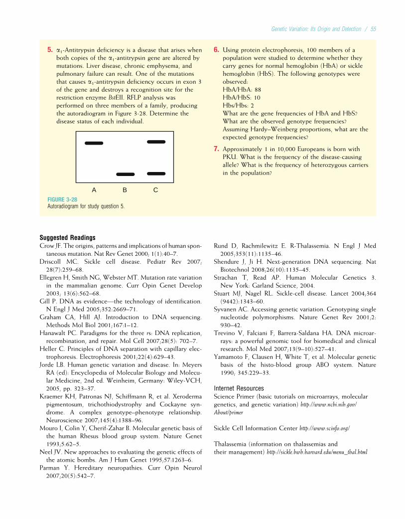
5. a1-Antitrypsin deficiency is a disease that arises whenboth copies of the a1-antitrypsin gene are altered bymutations. Liver disease, chronic emphysema, andpulmonary failure can result. One of the mutationsthat causes a1-antitrypsin deficiency occurs in exon 3of the gene and destroys a recognition site for therestriction enzyme BstEII. RFLP analysis wasperformed on three members of a family, producingthe autoradiogram in Figure 3-28. Determine thedisease status of each individual.
A B CFIGURE 3-28Autoradiogram for study question 5.
6. Using protein electrophoresis, 100 members of apopulation were studied to determine whether theycarry genes for normal hemoglobin (HbA) or sicklehemoglobin (HbS). The following genotypes wereobserved:HbA/HbA: 88HbA/HbS: 10Hbs/Hbs: 2What are the gene frequencies of HbA and HbS?What are the observed genotype frequencies?Assuming Hardy–Weinberg proportions, what are theexpected genotype frequencies?
7. Approximately 1 in 10,000 Europeans is born withPKU. What is the frequency of the disease-causingallele? What is the frequency of heterozygous carriersin the population?
Suggested ReadingsCrow JF. The origins, patterns and implications of human spon-
taneous mutation. Nat Rev Genet 2000; 1(1):40–7.Driscoll MC. Sickle cell disease. Pediatr Rev 2007;
28(7):259–68.Ellegren H, Smith NG, Webster MT. Mutation rate variation
in the mammalian genome. Curr Opin Genet Develop2003; 13(6):562–68.
Gill P. DNA as evidence—the technology of identification.N Engl J Med 2005;352:2669–71.
Graham CA, Hill AJ. Introduction to DNA sequencing.Methods Mol Biol 2001;167:1–12.
Hanawalt PC. Paradigms for the three rs: DNA replication,recombination, and repair. Mol Cell 2007;28(5): 702–7.
Heller C. Principles of DNA separation with capillary elec-trophoresis. Electrophoresis 2001;22(4):629–43.
Jorde LB. Human genetic variation and disease. In: MeyersRA (ed): Encyclopedia of Molecular Biology and Molecu-lar Medicine, 2nd ed. Weinheim, Germany: Wiley-VCH,2005, pp. 323–37.
Kraemer KH, Patronas NJ, Schiffmann R, et al. Xerodermapigmentosum, trichothiodystrophy and Cockayne syn-drome. A complex genotype–phenotype relationship.Neuroscience 2007;145(4):1388–96.
Mouro I, Colin Y, Cherif-Zahar B. Molecular genetic basis ofthe human Rhesus blood group system. Nature Genet1993;5:62–5.
Neel JV. New approaches to evaluating the genetic effects ofthe atomic bombs. Am J Hum Genet 1995;57:1263–6.
Parman Y. Hereditary neuropathies. Curr Opin Neurol2007;20(5):542–7.
Rund D, Rachmilewitz E. R-Thalassemia. N Engl J Med2005;353(11):1135–46.
Shendure J, Ji H. Next-generation DNA sequencing. NatBiotechnol 2008;26(10):1135–45.
Strachan T, Read AP. Human Molecular Genetics 3.New York: Garland Science, 2004.
Stuart MJ, Nagel RL. Sickle-cell disease. Lancet 2004;364(9442):1343–60.
Syvanen AC. Accessing genetic variation. Genotyping singlenucleotide polymorphisms. Nature Genet Rev 2001;2:930–42.
Trevino V, Falciani F, Barrera-Saldana HA. DNA microar-rays: a powerful genomic tool for biomedical and clinicalresearch. Mol Med 2007;13(9–10):527–41.
Yamamoto F, Clausen H, White T, et al. Molecular geneticbasis of the histo-blood group ABO system. Nature1990; 345:229–33.
Internet ResourcesScience Primer (basic tutorials on microarrays, moleculargenetics, and genetic variation) http://www.ncbi.nih.gov/About/primer
Sickle Cell Information Center http://www.scinfo.org/
Thalassemia (information on thalassemias andtheir management) http://sickle.bwh.harvard.edu/menu_thal.html
Genetic Variation: Its Origin and Detection / 55
Chapter 4
AUTOSOMAL DOMINANT AND RECESSIVE INHERITANCE
Many important and well-understood genetic diseases arethe result of a mutation in a single gene. The 2009 on-lineedition of McKusick’s Mendelian Inheritance in Man (http://www.ncbi.nlm.nih.gov/Omim/) lists more than 19,000 knownsingle-gene, or monogenic, traits defined thus far in humans.Of these, more than 18,000 are located on autosomes, nomore than 1000 are located on the X chromosome, and 57are located on the Y chromosome. Monogenic traits havebeen the focus of the research done thus far in medical genet-ics. In many cases, these genes have been mapped to specificchromosome locations, cloned, and sequenced. This researchhas led to new and exciting insights not only in genetics butalso in the basic pathophysiology of disease.
In this chapter we focus on single-gene disorders causedby mutations on the autosomes. (Single-gene disorderscaused by mutations on the sex chromosomes are the subjectof Chapter 5.) We discuss the patterns of inheritance of thesediseases in families, as well as factors that complicate thesepatterns. When appropriate, the molecular mechanism thatcauses genetic disease is addressed. We also discuss the risksof transmitting single-gene diseases to one’s offspring, becausethis is usually an important concern for at-risk couples.
BASIC CONCEPTS OF FORMAL GENETICSGregor Mendel’s ContributionsMonogenic traits are also known as mendelian traits, afterGregor Mendel, the 19th-century Austrian monk whodeduced several important genetic principles from his well-designed experiments with garden peas. Mendel studiedseven traits in the pea, each of which is determined by asingle gene. These traits included attributes such as height(tall versus short plants) and seed shape (rounded versuswrinkled). The variation in each of these traits is caused bythe presence of different alleles at individual loci.
Two central principles emerged from Mendel’s work. Thefirst is the principle of segregation, which states that sexu-ally reproducing organisms possess genes that occur in pairsand that only one member of this pair is transmitted to theoffspring (i.e., it segregates). The prevalent thinking duringMendel’s time was that hereditary factors from the two par-ents are blended in the offspring. In contrast, the principleof segregation states that genes remain intact and distinct.
An allele for “rounded” seed shape can be transmitted to anoffspring in the next generation, which can, in turn, transmitthe same allele to its own offspring. If, instead of remainingdistinct, genes were somehow blended in offspring, it wouldbe impossible to trace genetic inheritance from one genera-tion to the next. Thus, the principle of segregation was akey development in modern genetics.
Mendel’s principle of independent assortment was hissecond great contribution to genetics. This principle statesthat genes at different loci are transmitted independently.Consider the two loci mentioned previously. One locuscan have either the “rounded” or the “wrinkled” allele, andthe other can have either the “tall” or the “short” allele. Ina reproductive event, a parent transmits one allele from eachlocus to its offspring. The principle of independent assort-ment dictates that the transmission of a specific allele atone locus (“rounded” or “wrinkled”) has no effect on whichallele is transmitted at the other locus (“tall” or “short”).
The principle of segregation describes the behavior ofchromosomes in meiosis. The genes on chromosomes seg-regate during meiosis, and they are transmitted as distinctentities from one generation to the next. When Mendelperformed his critical experiments, he had no direct knowl-edge of chromosomes, meiosis, or genes (indeed, the last termwas not coined until 1909, long after Mendel’s death).Although his work was published in 1865 and cited occasion-ally, its fundamental significance was unrecognized for severaldecades. Yet Mendel’s research, which was eventually repli-cated by other researchers at the turn of the 20th century,forms the foundation of much of modern genetics.
4Mendel’s key contributions were the principles ofsegregation and independent assortment.
The Concept of PhenotypeThe term genotype has been defined as an individual’s geneticconstitution at a locus. The phenotype is what is actuallyobserved physically or clinically. Genotypes do not uniquelycorrespond to phenotypes. Individuals with two differentgenotypes, a dominant homozygote and a heterozygote,can have the same phenotype. An example is cystic fibrosis(Clinical Commentary 4-1), an autosomal recessive condition
56
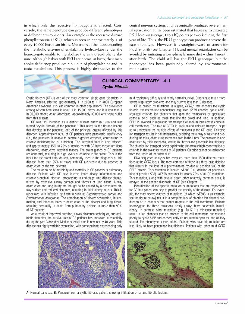
in which only the recessive homozygote is affected. Con-versely, the same genotype can produce different phenotypesin different environments. An example is the recessive diseasephenylketonuria (PKU), which is seen in approximately 1 ofevery 10,000 European births. Mutations at the locus encodingthe metabolic enzyme phenylalanine hydroxylase render thehomozygote unable to metabolize the amino acid phenylala-nine. Although babies with PKU are normal at birth, their met-abolic deficiency produces a buildup of phenylalanine and itstoxic metabolites. This process is highly destructive to the
central nervous system, and it eventually produces severe men-tal retardation. It has been estimated that babies with untreatedPKU lose, on average, 1 to 2 IQ points per week during the firstyear of life. Thus, the PKU genotype can produce a severe dis-ease phenotype. However, it is straightforward to screen forPKU at birth (see Chapter 13), and mental retardation can beavoided by initiating a low-phenylalanine diet within 1 monthafter birth. The child still has the PKU genotype, but thephenotype has been profoundly altered by environmentalmodification.
CLINICAL COMMENTARY 4-1
Cystic Fibrosis
Cystic fibrosis (CF) is one of the most common single-gene disorders inNorth America, affecting approximately 1 in 2000 to 1 in 4000 EuropeanAmerican newborns. It is less common in other populations. The prevalenceamong African Americans is about 1 in 15,000 births, and it is less than 1in 30,000 among Asian Americans. Approximately 30,000 Americans sufferfrom this disease.
CF was first identified as a distinct disease entity in 1938 and wastermed “cystic fibrosis of the pancreas.” This refers to the fibrotic lesionsthat develop in the pancreas, one of the principal organs affected by thisdisorder. Approximately 85% of CF patients have pancreatic insufficiency(i.e., the pancreas is unable to secrete digestive enzymes, contributing tochronic malabsorption of nutrients). The intestinal tract is also affected,and approximately 15% to 20% of newborns with CF have meconium ileus(thickened, obstructive intestinal matter). The sweat glands of CF patientsare abnormal, resulting in high levels of chloride in the sweat. This is thebasis for the sweat chloride test, commonly used in the diagnosis of thisdisease. More than 95% of males with CF are sterile due to absence orobstruction of the vas deferens.
The major cause of morbidity and mortality in CF patients is pulmonarydisease. Patients with CF have intense lower airway inflammation andchronic bronchial infection, progressing to end-stage lung disease charac-terized by extensive airway damage and fibrosis of lung tissue. Airwayobstruction and lung injury are thought to be caused by a dehydrated air-way surface and reduced clearance, resulting in thick airway mucus. This isassociated with infection by bacteria such as Staphylococcus aureus andPseudomonas aeruginosa. The combination of airway obstruction, inflam-mation, and infection leads to destruction of the airways and lung tissue,resulting eventually in death from pulmonary disease in more than 90%of CF patients.
As a result of improved nutrition, airway clearance techniques, and anti-biotic therapies, the survival rate of CF patients has improved substantiallyduring the past 3 decades. Median survival time is now nearly 40 years. Thisdisease has highly variable expression, with some patients experiencing only
mild respiratory difficulty and nearly normal survival. Others have much moresevere respiratory problems and may survive less than 2 decades.
CF is caused by mutations in a gene, CFTR,* that encodes the cysticfibrosis transmembrane conductance regulator. CFTR encodes cyclic AMP-regulated chloride ion channels that span the membranes of specializedepithelial cells, such as those that line the bowel and lung. In addition,CFTR is involved in regulating the transport of sodium ions across epithelialcell membranes. The role of CFTR in sodium and chloride transport helpsus to understand the multiple effects of mutations at the CF locus. Defectiveion transport results in salt imbalances, depleting the airway of water and pro-ducing the thick, obstructive secretions seen in the lungs. The pancreas is alsoobstructed by thick secretions, leading to fibrosis and pancreatic insufficiency.The chloride ion transport defect explains the abnormally high concentration ofchloride in the sweat secretions of CF patients: Chloride cannot be reabsorbedfrom the lumen of the sweat duct.
DNA sequence analysis has revealed more than 1500 different muta-tions at the CFTR locus. The most common of these is a three-base deletionthat results in the loss of a phenylalanine residue at position 508 of theCFTR protein. This mutation is labeled DF508 (i.e., deletion of phenylala-nine at position 508). DF508 accounts for nearly 70% of all CF mutations.This mutation, along with several dozen other relatively common ones, isassayed in the genetic diagnosis of CF (see Chapter 13).
Identification of the specific mutation or mutations that are responsiblefor CF in a patient can help to predict the severity of the disease. For exam-ple, the most severe classes of mutations (of which DF508 is an example;see the figure below) result in a complete lack of chloride ion channel pro-duction or in channels that cannot migrate to the cell membrane. Patientshomozygous for these mutations nearly always have pancreatic insuffi-ciency. In contrast, other mutations (e.g., R117H, a missense mutation)result in ion channels that do proceed to the cell membrane but respondpoorly to cyclic AMP and consequently do not remain open as long as theyshould. The phenotype is thus milder: Patients who have this mutation areless likely to have pancreatic insufficiency. Patients with other mild CFTR
A B
A, Normal pancreas. B, Pancreas from a cystic fibrosis patient, showing infiltration of fat and fibrotic lesions.
Continued
Autosomal Dominant and Recessive Inheritance / 57
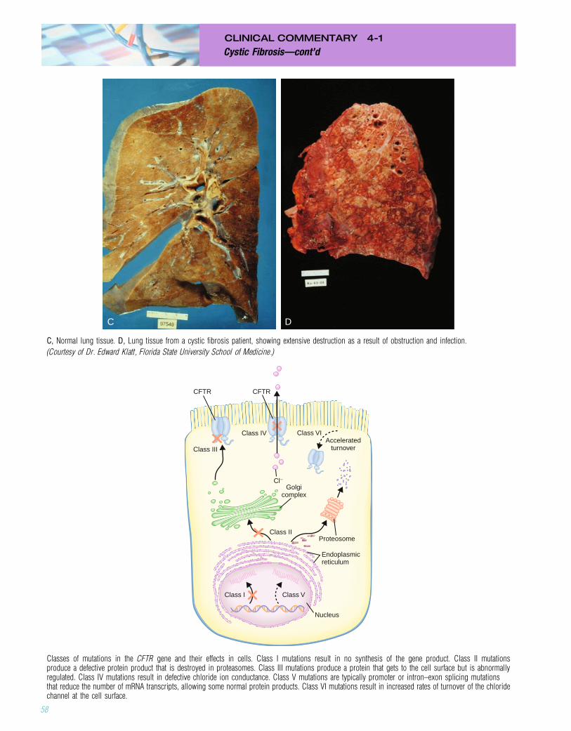
CLINICAL COMMENTARY 4-1
Cystic Fibrosis—cont’d
C D
C, Normal lung tissue. D, Lung tissue from a cystic fibrosis patient, showing extensive destruction as a result of obstruction and infection.
(Courtesy of Dr. Edward Klatt, Florida State University School of Medicine.)
Class III
Class I Class V
Class II
Class IV Class VIAccelerated
turnover
Golgicomplex
Endoplasmicreticulum
Nucleus
Proteosome
CFTR CFTR
Cl−
Classes of mutations in the CFTR gene and their effects in cells. Class I mutations result in no synthesis of the gene product. Class II mutationsproduce a defective protein product that is destroyed in proteasomes. Class III mutations produce a protein that gets to the cell surface but is abnormallyregulated. Class IV mutations result in defective chloride ion conductance. Class V mutations are typically promoter or intron–exon splicing mutationsthat reduce the number of mRNA transcripts, allowing some normal protein products. Class VI mutations result in increased rates of turnover of the chloridechannel at the cell surface.
58
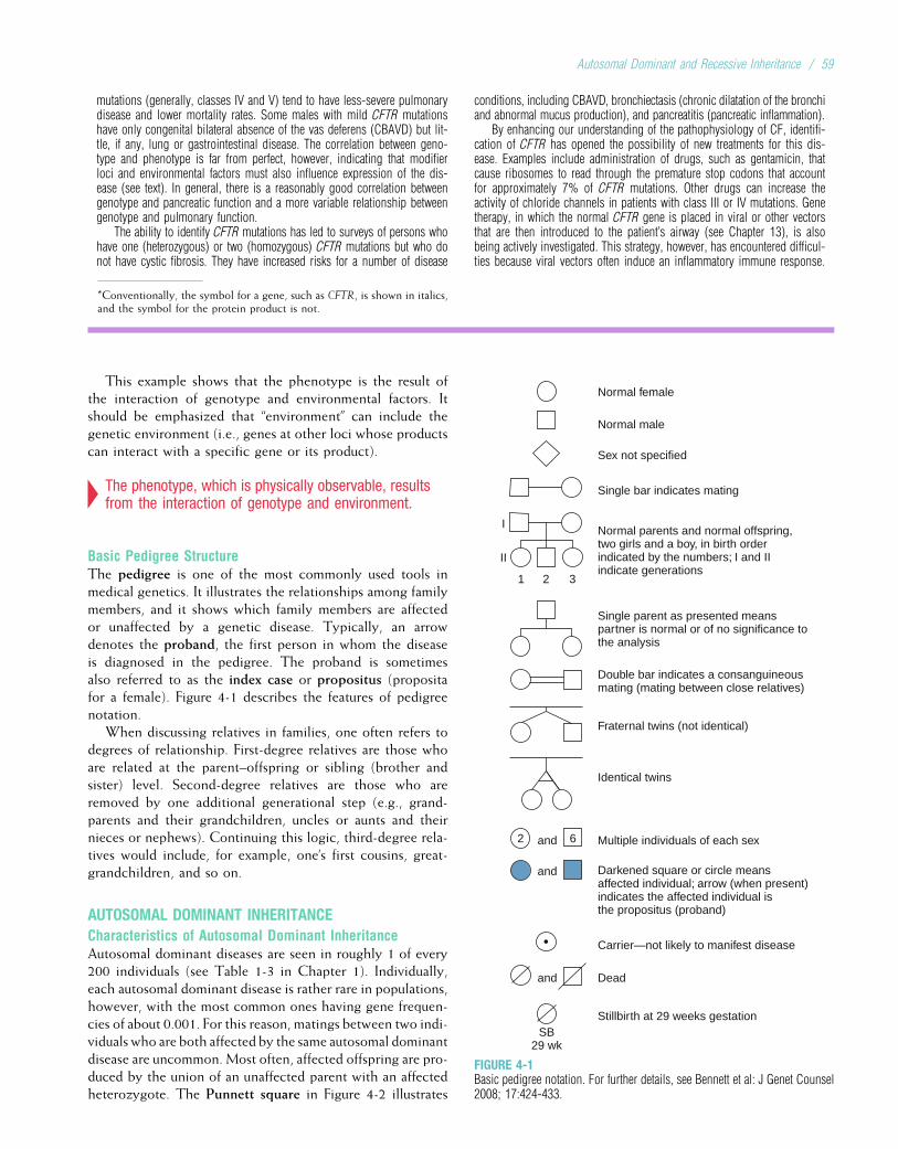
mutations (generally, classes IV and V) tend to have less-severe pulmonarydisease and lower mortality rates. Some males with mild CFTR mutationshave only congenital bilateral absence of the vas deferens (CBAVD) but lit-tle, if any, lung or gastrointestinal disease. The correlation between geno-type and phenotype is far from perfect, however, indicating that modifierloci and environmental factors must also influence expression of the dis-ease (see text). In general, there is a reasonably good correlation betweengenotype and pancreatic function and a more variable relationship betweengenotype and pulmonary function.
The ability to identify CFTR mutations has led to surveys of persons whohave one (heterozygous) or two (homozygous) CFTR mutations but who donot have cystic fibrosis. They have increased risks for a number of disease
conditions, including CBAVD, bronchiectasis (chronic dilatation of the bronchiand abnormal mucus production), and pancreatitis (pancreatic inflammation).
By enhancing our understanding of the pathophysiology of CF, identifi-cation of CFTR has opened the possibility of new treatments for this dis-ease. Examples include administration of drugs, such as gentamicin, thatcause ribosomes to read through the premature stop codons that accountfor approximately 7% of CFTR mutations. Other drugs can increase theactivity of chloride channels in patients with class III or IV mutations. Genetherapy, in which the normal CFTR gene is placed in viral or other vectorsthat are then introduced to the patient’s airway (see Chapter 13), is alsobeing actively investigated. This strategy, however, has encountered difficul-ties because viral vectors often induce an inflammatory immune response.
This example shows that the phenotype is the result ofthe interaction of genotype and environmental factors. Itshould be emphasized that “environment” can include thegenetic environment (i.e., genes at other loci whose productscan interact with a specific gene or its product).
4The phenotype, which is physically observable, resultsfrom the interaction of genotype and environment.
Basic Pedigree StructureThe pedigree is one of the most commonly used tools inmedical genetics. It illustrates the relationships among familymembers, and it shows which family members are affectedor unaffected by a genetic disease. Typically, an arrowdenotes the proband, the first person in whom the diseaseis diagnosed in the pedigree. The proband is sometimesalso referred to as the index case or propositus (propositafor a female). Figure 4-1 describes the features of pedigreenotation.
When discussing relatives in families, one often refers todegrees of relationship. First-degree relatives are those whoare related at the parent–offspring or sibling (brother andsister) level. Second-degree relatives are those who areremoved by one additional generational step (e.g., grand-parents and their grandchildren, uncles or aunts and theirnieces or nephews). Continuing this logic, third-degree rela-tives would include, for example, one’s first cousins, great-grandchildren, and so on.
AUTOSOMAL DOMINANT INHERITANCECharacteristics of Autosomal Dominant InheritanceAutosomal dominant diseases are seen in roughly 1 of every200 individuals (see Table 1-3 in Chapter 1). Individually,each autosomal dominant disease is rather rare in populations,however, with the most common ones having gene frequen-cies of about 0.001. For this reason, matings between two indi-viduals who are both affected by the same autosomal dominantdisease are uncommon. Most often, affected offspring are pro-duced by the union of an unaffected parent with an affectedheterozygote. The Punnett square in Figure 4-2 illustrates
*Conventionally, the symbol for a gene, such as CFTR, is shown in italics,and the symbol for the protein product is not.
Carrier—not likely to manifest disease
Multiple individuals of each sex
Identical twins
Sex not specified
Single bar indicates mating
Normal male
Normal female
Fraternal twins (not identical)
Double bar indicates a consanguineousmating (mating between close relatives)
Single parent as presented meanspartner is normal or of no significance tothe analysis
Normal parents and normal offspring,two girls and a boy, in birth orderindicated by the numbers; I and IIindicate generations
Darkened square or circle meansaffected individual; arrow (when present)indicates the affected individual isthe propositus (proband)
and
and
2
2 31
II
I
6and
Dead
Stillbirth at 29 weeks gestationSB
29 wk
FIGURE 4-1Basic pedigree notation. For further details, see Bennett et al: J Genet Counsel2008; 17:424-433.
Autosomal Dominant and Recessive Inheritance / 59
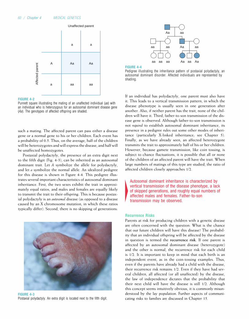
such a mating. The affected parent can pass either a diseasegene or a normal gene to his or her children. Each event hasa probability of 0.5. Thus, on the average, half of the childrenwill be heterozygotes and will express the disease, and half willbe unaffected homozygotes.
Postaxial polydactyly, the presence of an extra digit nextto the fifth digit (Fig. 4-3), can be inherited as an autosomaldominant trait. Let A symbolize the allele for polydactyly,and let a symbolize the normal allele. An idealized pedigreefor this disease is shown in Figure 4-4. This pedigree illus-trates several important characteristics of autosomal dominantinheritance. First, the two sexes exhibit the trait in approxi-mately equal ratios, and males and females are equally likelyto transmit the trait to their offspring. This is because postax-ial polydactyly is an autosomal disease (as opposed to a diseasecaused by an X chromosome mutation, in which these ratiostypically differ). Second, there is no skipping of generations:
If an individual has polydactyly, one parent must also haveit. This leads to a vertical transmission pattern, in which thedisease phenotype is usually seen in one generation afteranother. Also, if neither parent has the trait, none of the chil-dren will have it. Third, father-to-son transmission of the dis-ease gene is observed. Although father-to-son transmission isnot required to establish autosomal dominant inheritance, itspresence in a pedigree rules out some other modes of inheri-tance (particularly X-linked inheritance; see Chapter 5).Finally, as we have already seen, an affected heterozygotetransmits the trait to approximately half of his or her children.However, because gamete transmission, like coin tossing, issubject to chance fluctuations, it is possible that all or noneof the children of an affected parent will have the trait. Whenlarge numbers of matings of this type are studied, the ratio ofaffected children closely approaches 1/2.
4Autosomal dominant inheritance is characterized byvertical transmission of the disease phenotype, a lackof skipped generations, and roughly equal numbers ofaffected males and females. Father-to-sontransmission may be observed.
Recurrence RisksParents at risk for producing children with a genetic diseaseare often concerned with the question: What is the chancethat our future children will have this disease? The probabil-ity that an individual offspring will be affected by the diseasein question is termed the recurrence risk. If one parent isaffected by an autosomal dominant disease (heterozygote)and the other is normal, the recurrence risk for each childis 1/2. It is important to keep in mind that each birth is anindependent event, as in the coin-tossing examples. Thus,even if the parents have already had a child with the disease,their recurrence risk remains 1/2. Even if they have had sev-eral children, all affected (or all unaffected) by the disease,the law of independence dictates that the probability thattheir next child will have the disease is still 1/2. Althoughthis concept seems intuitively obvious, it is commonly misun-derstood by the lay population. Further aspects of communi-cating risks to families are discussed in Chapter 15.
aaaa
AaAa
aAffe
cted
par
ent A
aa
Unaffected parent
FIGURE 4-2Punnett square illustrating the mating of an unaffected individual (aa) withan individual who is heterozygous for an autosomal dominant disease gene(Aa). The genotypes of affected offspring are shaded.
FIGURE 4-3Postaxial polydactyly. An extra digit is located next to the fifth digit.
Aaaa aaaa
aaAa
aa aaaa aa AaAa
FIGURE 4-4Pedigree illustrating the inheritance pattern of postaxial polydactyly, anautosomal dominant disorder. Affected individuals are represented byshading.
60 / Chapter 4 MEDICAL GENETICS
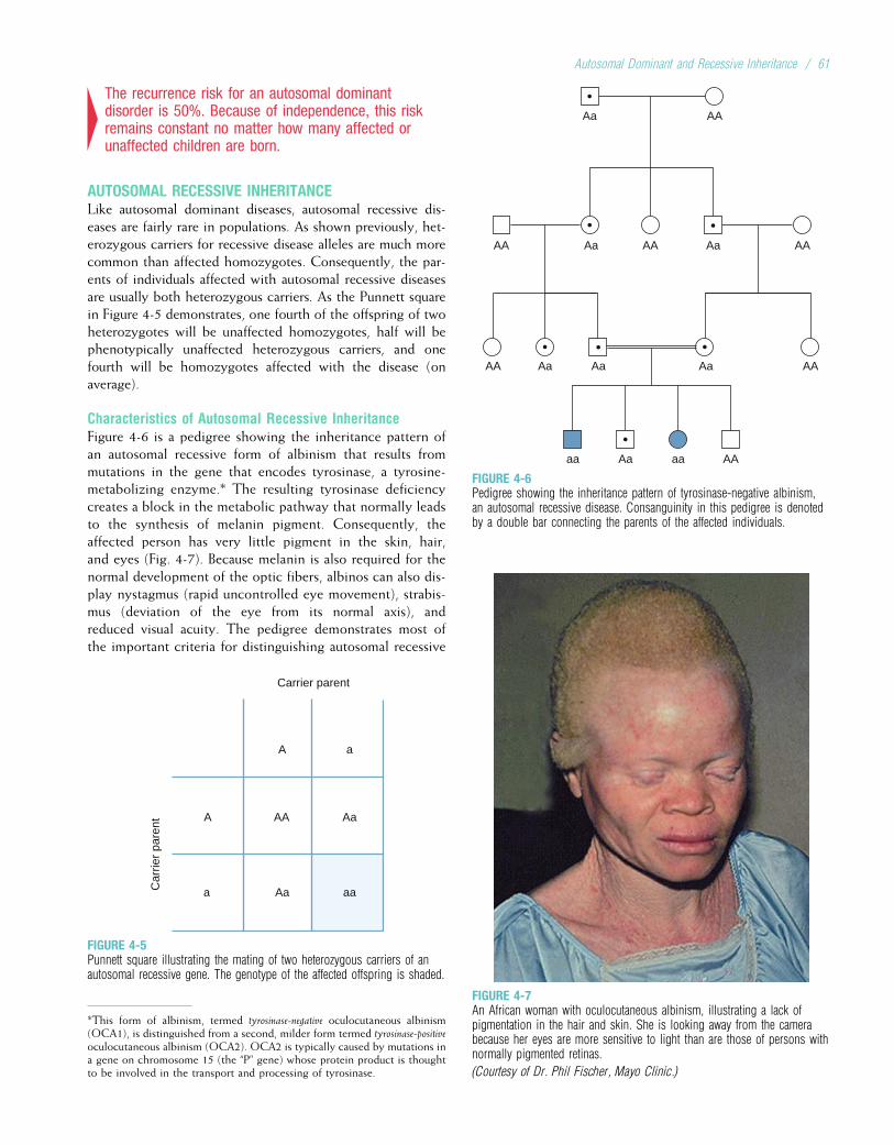
4The recurrence risk for an autosomal dominantdisorder is 50%. Because of independence, this riskremains constant no matter how many affected orunaffected children are born.
AUTOSOMAL RECESSIVE INHERITANCELike autosomal dominant diseases, autosomal recessive dis-eases are fairly rare in populations. As shown previously, het-erozygous carriers for recessive disease alleles are much morecommon than affected homozygotes. Consequently, the par-ents of individuals affected with autosomal recessive diseasesare usually both heterozygous carriers. As the Punnett squarein Figure 4-5 demonstrates, one fourth of the offspring of twoheterozygotes will be unaffected homozygotes, half will bephenotypically unaffected heterozygous carriers, and onefourth will be homozygotes affected with the disease (onaverage).
Characteristics of Autosomal Recessive InheritanceFigure 4-6 is a pedigree showing the inheritance pattern ofan autosomal recessive form of albinism that results frommutations in the gene that encodes tyrosinase, a tyrosine-metabolizing enzyme.* The resulting tyrosinase deficiencycreates a block in the metabolic pathway that normally leadsto the synthesis of melanin pigment. Consequently, theaffected person has very little pigment in the skin, hair,and eyes (Fig. 4-7). Because melanin is also required for thenormal development of the optic fibers, albinos can also dis-play nystagmus (rapid uncontrolled eye movement), strabis-mus (deviation of the eye from its normal axis), andreduced visual acuity. The pedigree demonstrates most ofthe important criteria for distinguishing autosomal recessive
aaAa
AaAA
aCar
rier
pare
nt
A
aA
Carrier parent
FIGURE 4-5Punnett square illustrating the mating of two heterozygous carriers of anautosomal recessive gene. The genotype of the affected offspring is shaded.
AAaaaa Aa
AA Aa Aa AAAa
AAAAAA AaAa
AAAa
FIGURE 4-6Pedigree showing the inheritance pattern of tyrosinase-negative albinism,an autosomal recessive disease. Consanguinity in this pedigree is denotedby a double bar connecting the parents of the affected individuals.
*This form of albinism, termed tyrosinase-negative oculocutaneous albinism(OCA1), is distinguished from a second, milder form termed tyrosinase-positiveoculocutaneous albinism (OCA2). OCA2 is typically caused by mutations ina gene on chromosome 15 (the “P” gene) whose protein product is thoughtto be involved in the transport and processing of tyrosinase.
FIGURE 4-7An African woman with oculocutaneous albinism, illustrating a lack ofpigmentation in the hair and skin. She is looking away from the camerabecause her eyes are more sensitive to light than are those of persons withnormally pigmented retinas.
(Courtesy of Dr. Phil Fischer, Mayo Clinic.)
Autosomal Dominant and Recessive Inheritance / 61
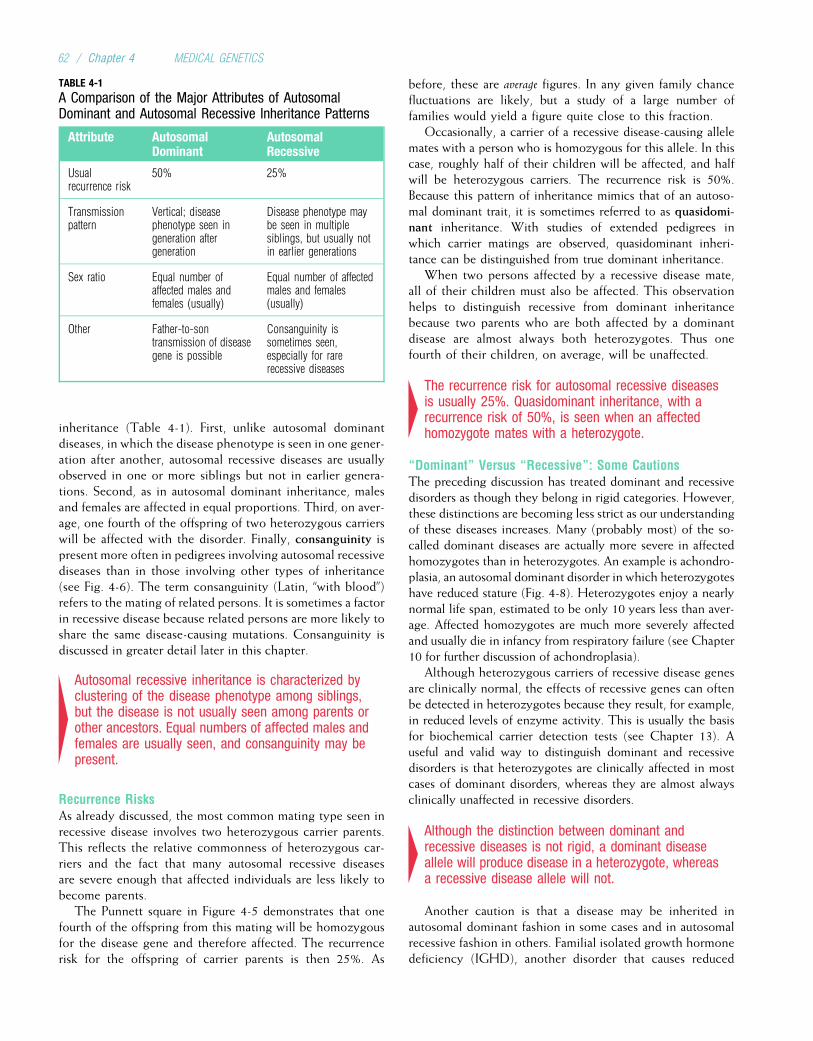
inheritance (Table 4-1). First, unlike autosomal dominantdiseases, in which the disease phenotype is seen in one gener-ation after another, autosomal recessive diseases are usuallyobserved in one or more siblings but not in earlier genera-tions. Second, as in autosomal dominant inheritance, malesand females are affected in equal proportions. Third, on aver-age, one fourth of the offspring of two heterozygous carrierswill be affected with the disorder. Finally, consanguinity ispresent more often in pedigrees involving autosomal recessivediseases than in those involving other types of inheritance(see Fig. 4-6). The term consanguinity (Latin, “with blood”)refers to the mating of related persons. It is sometimes a factorin recessive disease because related persons are more likely toshare the same disease-causing mutations. Consanguinity isdiscussed in greater detail later in this chapter.
4Autosomal recessive inheritance is characterized byclustering of the disease phenotype among siblings,but the disease is not usually seen among parents orother ancestors. Equal numbers of affected males andfemales are usually seen, and consanguinity may bepresent.
Recurrence RisksAs already discussed, the most common mating type seen inrecessive disease involves two heterozygous carrier parents.This reflects the relative commonness of heterozygous car-riers and the fact that many autosomal recessive diseasesare severe enough that affected individuals are less likely tobecome parents.
The Punnett square in Figure 4-5 demonstrates that onefourth of the offspring from this mating will be homozygousfor the disease gene and therefore affected. The recurrencerisk for the offspring of carrier parents is then 25%. As
before, these are average figures. In any given family chancefluctuations are likely, but a study of a large number offamilies would yield a figure quite close to this fraction.
Occasionally, a carrier of a recessive disease-causing allelemates with a person who is homozygous for this allele. In thiscase, roughly half of their children will be affected, and halfwill be heterozygous carriers. The recurrence risk is 50%.Because this pattern of inheritance mimics that of an autoso-mal dominant trait, it is sometimes referred to as quasidomi-nant inheritance. With studies of extended pedigrees inwhich carrier matings are observed, quasidominant inheri-tance can be distinguished from true dominant inheritance.
When two persons affected by a recessive disease mate,all of their children must also be affected. This observationhelps to distinguish recessive from dominant inheritancebecause two parents who are both affected by a dominantdisease are almost always both heterozygotes. Thus onefourth of their children, on average, will be unaffected.
4The recurrence risk for autosomal recessive diseasesis usually 25%. Quasidominant inheritance, with arecurrence risk of 50%, is seen when an affectedhomozygote mates with a heterozygote.
“Dominant” Versus “Recessive”: Some CautionsThe preceding discussion has treated dominant and recessivedisorders as though they belong in rigid categories. However,these distinctions are becoming less strict as our understandingof these diseases increases. Many (probably most) of the so-called dominant diseases are actually more severe in affectedhomozygotes than in heterozygotes. An example is achondro-plasia, an autosomal dominant disorder in which heterozygoteshave reduced stature (Fig. 4-8). Heterozygotes enjoy a nearlynormal life span, estimated to be only 10 years less than aver-age. Affected homozygotes are much more severely affectedand usually die in infancy from respiratory failure (see Chapter10 for further discussion of achondroplasia).
Although heterozygous carriers of recessive disease genesare clinically normal, the effects of recessive genes can oftenbe detected in heterozygotes because they result, for example,in reduced levels of enzyme activity. This is usually the basisfor biochemical carrier detection tests (see Chapter 13). Auseful and valid way to distinguish dominant and recessivedisorders is that heterozygotes are clinically affected in mostcases of dominant disorders, whereas they are almost alwaysclinically unaffected in recessive disorders.
4Although the distinction between dominant andrecessive diseases is not rigid, a dominant diseaseallele will produce disease in a heterozygote, whereasa recessive disease allele will not.
Another caution is that a disease may be inherited inautosomal dominant fashion in some cases and in autosomalrecessive fashion in others. Familial isolated growth hormonedeficiency (IGHD), another disorder that causes reduced
TABLE 4-1
A Comparison of the Major Attributes of AutosomalDominant and Autosomal Recessive Inheritance Patterns
Attribute AutosomalDominant
AutosomalRecessive
Usualrecurrence risk
50% 25%
Transmissionpattern
Vertical; diseasephenotype seen ingeneration aftergeneration
Disease phenotype maybe seen in multiplesiblings, but usually notin earlier generations
Sex ratio Equal number ofaffected males andfemales (usually)
Equal number of affectedmales and females(usually)
Other Father-to-sontransmission of diseasegene is possible
Consanguinity issometimes seen,especially for rarerecessive diseases
62 / Chapter 4 MEDICAL GENETICS
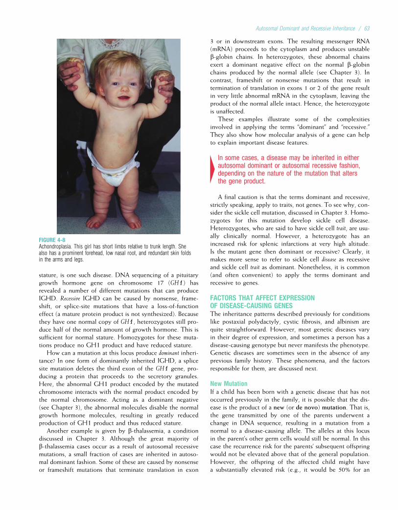
stature, is one such disease. DNA sequencing of a pituitarygrowth hormone gene on chromosome 17 (GH1) hasrevealed a number of different mutations that can produceIGHD. Recessive IGHD can be caused by nonsense, frame-shift, or splice-site mutations that have a loss-of-functioneffect (a mature protein product is not synthesized). Becausethey have one normal copy of GH1, heterozygotes still pro-duce half of the normal amount of growth hormone. This issufficient for normal stature. Homozygotes for these muta-tions produce no GH1 product and have reduced stature.
How can a mutation at this locus produce dominant inheri-tance? In one form of dominantly inherited IGHD, a splicesite mutation deletes the third exon of the GH1 gene, pro-ducing a protein that proceeds to the secretory granules.Here, the abnormal GH1 product encoded by the mutatedchromosome interacts with the normal product encoded bythe normal chromosome. Acting as a dominant negative(see Chapter 3), the abnormal molecules disable the normalgrowth hormone molecules, resulting in greatly reducedproduction of GH1 product and thus reduced stature.
Another example is given by b-thalassemia, a conditiondiscussed in Chapter 3. Although the great majority ofb-thalassemia cases occur as a result of autosomal recessivemutations, a small fraction of cases are inherited in autoso-mal dominant fashion. Some of these are caused by nonsenseor frameshift mutations that terminate translation in exon
3 or in downstream exons. The resulting messenger RNA(mRNA) proceeds to the cytoplasm and produces unstableb-globin chains. In heterozygotes, these abnormal chainsexert a dominant negative effect on the normal b-globinchains produced by the normal allele (see Chapter 3). Incontrast, frameshift or nonsense mutations that result intermination of translation in exons 1 or 2 of the gene resultin very little abnormal mRNA in the cytoplasm, leaving theproduct of the normal allele intact. Hence, the heterozygoteis unaffected.
These examples illustrate some of the complexitiesinvolved in applying the terms “dominant” and “recessive.”They also show how molecular analysis of a gene can helpto explain important disease features.
4In some cases, a disease may be inherited in eitherautosomal dominant or autosomal recessive fashion,depending on the nature of the mutation that altersthe gene product.
A final caution is that the terms dominant and recessive,strictly speaking, apply to traits, not genes. To see why, con-sider the sickle cell mutation, discussed in Chapter 3. Homo-zygotes for this mutation develop sickle cell disease.Heterozygotes, who are said to have sickle cell trait, are usu-ally clinically normal. However, a heterozygote has anincreased risk for splenic infarctions at very high altitude.Is the mutant gene then dominant or recessive? Clearly, itmakes more sense to refer to sickle cell disease as recessiveand sickle cell trait as dominant. Nonetheless, it is common(and often convenient) to apply the terms dominant andrecessive to genes.
FACTORS THAT AFFECT EXPRESSIONOF DISEASE-CAUSING GENESThe inheritance patterns described previously for conditionslike postaxial polydactyly, cystic fibrosis, and albinism arequite straightforward. However, most genetic diseases varyin their degree of expression, and sometimes a person has adisease-causing genotype but never manifests the phenotype.Genetic diseases are sometimes seen in the absence of anyprevious family history. These phenomena, and the factorsresponsible for them, are discussed next.
New MutationIf a child has been born with a genetic disease that has notoccurred previously in the family, it is possible that the dis-ease is the product of a new (or de novo) mutation. That is,the gene transmitted by one of the parents underwent achange in DNA sequence, resulting in a mutation from anormal to a disease-causing allele. The alleles at this locusin the parent’s other germ cells would still be normal. In thiscase the recurrence risk for the parents’ subsequent offspringwould not be elevated above that of the general population.However, the offspring of the affected child might havea substantially elevated risk (e.g., it would be 50% for an
FIGURE 4-8Achondroplasia. This girl has short limbs relative to trunk length. Shealso has a prominent forehead, low nasal root, and redundant skin foldsin the arms and legs.
Autosomal Dominant and Recessive Inheritance / 63
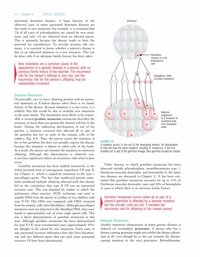
autosomal dominant disease). A large fraction of theobserved cases of many autosomal dominant diseases arethe result of new mutations. For example, it is estimated that7/8 of all cases of achondroplasia are caused by new muta-tions, and only 1/8 are inherited from an affected parent.This is primarily because the disease tends to limit thepotential for reproduction. To provide accurate risk esti-mates, it is essential to know whether a patient’s disease isdue to an inherited mutation or a new mutation. This canbe done only if an adequate family history has been taken.
4New mutations are a common cause of theappearance of a genetic disease in a person with noprevious family history of the disorder. The recurrencerisk for the person’s siblings is very low, but therecurrence risk for the person’s offspring may besubstantially increased.
Germline MosaicismOccasionally, two or more offspring present with an autoso-mal dominant or X-linked disease when there is no familyhistory of the disease. Because mutation is a rare event, it isunlikely that this would be due to multiple new mutationsin the same family. The mechanism most likely to be respon-sible is termed germline mosaicism (mosaicism describes thepresence of more than one genetically distinct cell line in thebody). During the embryonic development of one of theparents, a mutation occurred that affected all or part ofthe germline but few or none of the somatic cells of theembryo (Fig. 4-9). Thus, the parent carries the mutation inhis or her germline but does not actually express the diseasebecause the mutation is absent in other cells of the body.As a result, the parent can transmit the mutation to numerousoffspring. Although this phenomenon is relatively rare,it can have significant effects on recurrence risks when it doesoccur.
Germline mosaicism has been studied extensively in thelethal perinatal form of osteogenesis imperfecta (OI type II;see Chapter 2), which is caused by mutations in the type 1procollagen genes. The fact that unaffected parents some-times produced multiple offspring affected with this diseaseled to the conclusion that type II OI was an autosomalrecessive trait. This was disputed by studies in which thepolymerase chain reaction (PCR) technique was used toamplify DNA from the sperm of a father of two children withtype II OI. This DNA was compared with DNA extractedfrom his somatic cells (skin fibroblasts). Although procollagenmutations were not detected in the fibroblast DNA, they werefound in approximately one of every eight sperm cells. Thiswas a direct demonstration of germline mosaicism in thisman. Although germline mosaicism has been demonstratedfor type II OI, most noninherited cases (approximately 95%)are thought to be caused by new mutations. Some cases oftrue autosomal recessive inheritance have also been documen-ted, and two different genes that can each cause autosomalrecessive OI have been documented.
Other diseases in which germline mosaicism has beenobserved include achondroplasia, neurofibromatosis type 1,Duchenne muscular dystrophy, and hemophilia A (the lattertwo diseases are discussed in Chapter 5). It has been esti-mated that germline mosaicism accounts for up to 15% ofDuchenne muscular dystrophy cases and 20% of hemophiliaA cases in which there is no previous family history.
4Germline mosaicism occurs when all or part of aparent’s germline is affected by a disease mutationbut the somatic cells are not. It elevates therecurrence risk for offspring of the mosaic parent.
Reduced PenetranceAnother important characteristic of many genetic diseases isreduced (or incomplete) penetrance: A person who has adisease-causing genotype might not exhibit the disease pheno-type at all, even though he or she can transmit the disease-causing mutation to the next generation. Retinoblastoma,
*
*
****
*
* *
**
*
**
*
Mutationoccurs in oneembryoniccell
Embryo
Matureorganismis a mosaic ofmutated andnon-mutatedcells
Daughter cellscontain mutation
FIGURE 4-9A mutation occurs in one cell of the developing embryo. All descendantsof that cell have the same mutation, resulting in mosaicism. If the firstmutated cell is part of the germline lineage, then germline mosaicism results.
64 / Chapter 4 MEDICAL GENETICS
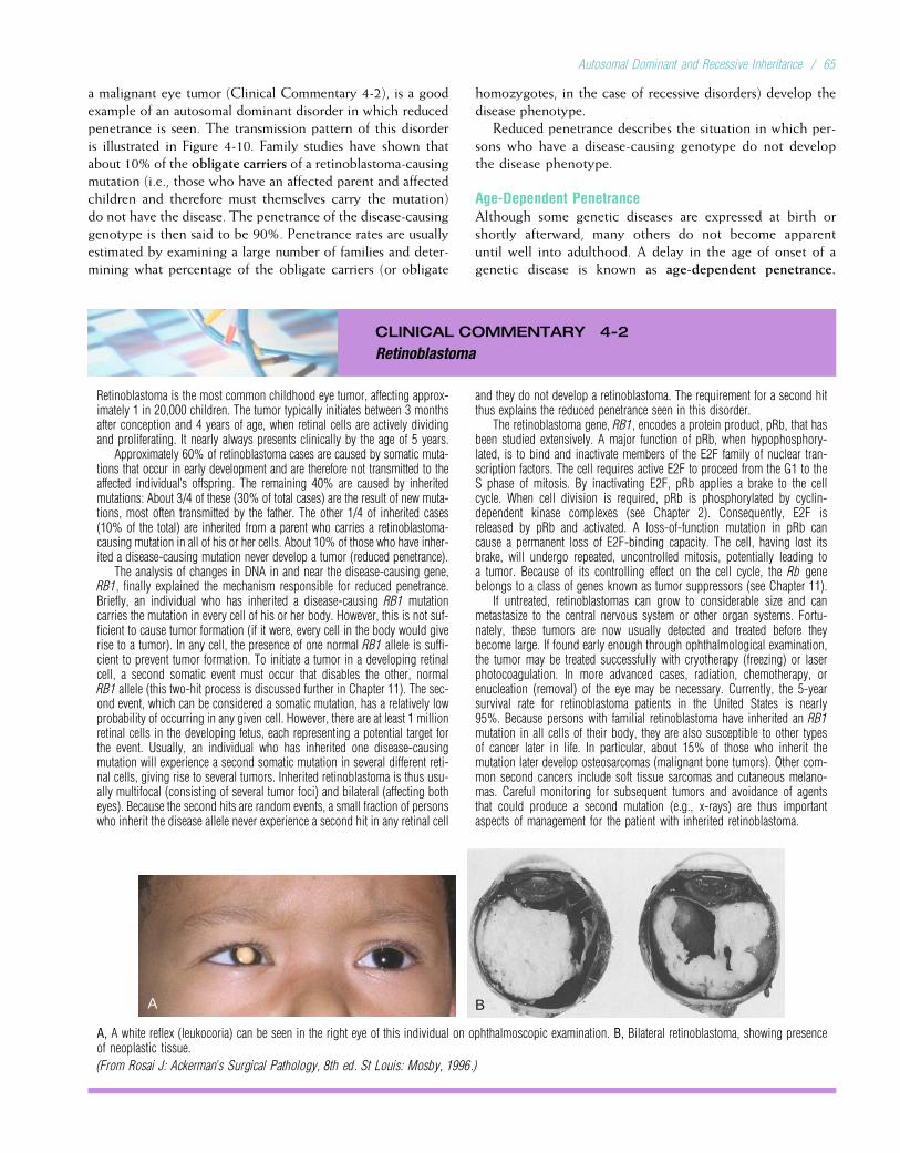
a malignant eye tumor (Clinical Commentary 4-2), is a goodexample of an autosomal dominant disorder in which reducedpenetrance is seen. The transmission pattern of this disorderis illustrated in Figure 4-10. Family studies have shown thatabout 10% of the obligate carriers of a retinoblastoma-causingmutation (i.e., those who have an affected parent and affectedchildren and therefore must themselves carry the mutation)do not have the disease. The penetrance of the disease-causinggenotype is then said to be 90%. Penetrance rates are usuallyestimated by examining a large number of families and deter-mining what percentage of the obligate carriers (or obligate
homozygotes, in the case of recessive disorders) develop thedisease phenotype.
Reduced penetrance describes the situation in which per-sons who have a disease-causing genotype do not developthe disease phenotype.
Age-Dependent PenetranceAlthough some genetic diseases are expressed at birth orshortly afterward, many others do not become apparentuntil well into adulthood. A delay in the age of onset of agenetic disease is known as age-dependent penetrance.
CLINICAL COMMENTARY 4-2
Retinoblastoma
Retinoblastoma is the most common childhood eye tumor, affecting approx-imately 1 in 20,000 children. The tumor typically initiates between 3 monthsafter conception and 4 years of age, when retinal cells are actively dividingand proliferating. It nearly always presents clinically by the age of 5 years.
Approximately 60% of retinoblastoma cases are caused by somatic muta-tions that occur in early development and are therefore not transmitted to theaffected individual’s offspring. The remaining 40% are caused by inheritedmutations: About 3/4 of these (30% of total cases) are the result of new muta-tions, most often transmitted by the father. The other 1/4 of inherited cases(10% of the total) are inherited from a parent who carries a retinoblastoma-causing mutation in all of his or her cells. About 10% of those who have inher-ited a disease-causing mutation never develop a tumor (reduced penetrance).
The analysis of changes in DNA in and near the disease-causing gene,RB1, finally explained the mechanism responsible for reduced penetrance.Briefly, an individual who has inherited a disease-causing RB1 mutationcarries the mutation in every cell of his or her body. However, this is not suf-ficient to cause tumor formation (if it were, every cell in the body would giverise to a tumor). In any cell, the presence of one normal RB1 allele is suffi-cient to prevent tumor formation. To initiate a tumor in a developing retinalcell, a second somatic event must occur that disables the other, normalRB1 allele (this two-hit process is discussed further in Chapter 11). The sec-ond event, which can be considered a somatic mutation, has a relatively lowprobability of occurring in any given cell. However, there are at least 1 millionretinal cells in the developing fetus, each representing a potential target forthe event. Usually, an individual who has inherited one disease-causingmutation will experience a second somatic mutation in several different reti-nal cells, giving rise to several tumors. Inherited retinoblastoma is thus usu-ally multifocal (consisting of several tumor foci) and bilateral (affecting botheyes). Because the second hits are random events, a small fraction of personswho inherit the disease allele never experience a second hit in any retinal cell
and they do not develop a retinoblastoma. The requirement for a second hitthus explains the reduced penetrance seen in this disorder.
The retinoblastoma gene, RB1, encodes a protein product, pRb, that hasbeen studied extensively. A major function of pRb, when hypophosphory-lated, is to bind and inactivate members of the E2F family of nuclear tran-scription factors. The cell requires active E2F to proceed from the G1 to theS phase of mitosis. By inactivating E2F, pRb applies a brake to the cellcycle. When cell division is required, pRb is phosphorylated by cyclin-dependent kinase complexes (see Chapter 2). Consequently, E2F isreleased by pRb and activated. A loss-of-function mutation in pRb cancause a permanent loss of E2F-binding capacity. The cell, having lost itsbrake, will undergo repeated, uncontrolled mitosis, potentially leading toa tumor. Because of its controlling effect on the cell cycle, the Rb genebelongs to a class of genes known as tumor suppressors (see Chapter 11).
If untreated, retinoblastomas can grow to considerable size and canmetastasize to the central nervous system or other organ systems. Fortu-nately, these tumors are now usually detected and treated before theybecome large. If found early enough through ophthalmological examination,the tumor may be treated successfully with cryotherapy (freezing) or laserphotocoagulation. In more advanced cases, radiation, chemotherapy, orenucleation (removal) of the eye may be necessary. Currently, the 5-yearsurvival rate for retinoblastoma patients in the United States is nearly95%. Because persons with familial retinoblastoma have inherited an RB1mutation in all cells of their body, they are also susceptible to other typesof cancer later in life. In particular, about 15% of those who inherit themutation later develop osteosarcomas (malignant bone tumors). Other com-mon second cancers include soft tissue sarcomas and cutaneous melano-mas. Careful monitoring for subsequent tumors and avoidance of agentsthat could produce a second mutation (e.g., x-rays) are thus importantaspects of management for the patient with inherited retinoblastoma.
A B
A, A white reflex (leukocoria) can be seen in the right eye of this individual on ophthalmoscopic examination. B, Bilateral retinoblastoma, showing presenceof neoplastic tissue.
(From Rosai J: Ackerman’s Surgical Pathology, 8th ed. St Louis: Mosby, 1996.)
Autosomal Dominant and Recessive Inheritance / 65
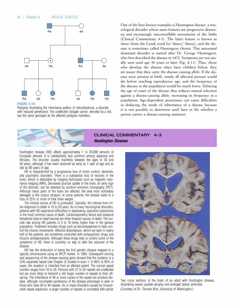
One of the best-known examples is Huntington disease, a neu-rological disorder whose main features are progressive demen-tia and increasingly uncontrollable movements of the limbs(Clinical Commentary 4-3). The latter feature is known aschorea (from the Greek word for “dance,” khoreia), and the dis-ease is sometimes called Huntington chorea. This autosomaldominant disorder is named after Dr. George Huntington,who first described the disease in 1872. Symptoms are not usu-ally seen until age 30 years or later (Fig. 4-11). Thus, thosewho develop the disease often have children before theyare aware that they carry the disease-causing allele. If the dis-ease were present at birth, nearly all affected persons woulddie before reaching reproductive age, and the frequency ofthe disease in the population would be much lower. Delayingthe age of onset of the disease thus reduces natural selectionagainst a disease-causing allele, increasing its frequency in apopulation. Age-dependent penetrance can cause difficultiesin deducing the mode of inheritance of a disease becauseit is not possible to determine until later in life whether aperson carries a disease-causing mutation.
CLINICAL COMMENTARY 4-3
Huntington Disease
Huntington disease (HD) affects approximately 1 in 20,000 persons ofEuropean descent. It is substantially less common among Japanese andAfricans. The disorder usually manifests between the ages of 30 and50 years, although it has been observed as early as 1 year of age and aslate as 80 years of age.
HD is characterized by a progressive loss of motor control, dementia,and psychiatric disorders. There is a substantial loss of neurons in thebrain, which is detectable by imaging techniques such as magnetic reso-nance imaging (MRI). Decreased glucose uptake in the brain, an early signof the disorder, can be detected by positron-emission tomography (PET).Although many parts of the brain are affected, the area most noticeablydamaged is the corpus striatum. In some patients, the disease leads to aloss of 25% or more of total brain weight.
The clinical course of HD is protracted. Typically, the interval from ini-tial diagnosis to death is 15 to 20 years. As in many neurological disorders,patients with HD experience difficulties in swallowing; aspiration pneumoniais the most common cause of death. Cardiorespiratory failure and subduralhematoma (due to head trauma) are other frequent causes of death. The sui-cide rate among HD patients is 5 to 10 times higher than in the generalpopulation. Treatment includes drugs such as benzodiazepines to help con-trol the choreic movements. Affective disturbances, which are seen in nearlyhalf of the patients, are sometimes controlled with antipsychotic drugs andtricyclic antidepressants. Although these drugs help to control some of thesymptoms of HD, there is currently no way to alter the outcome of thedisease.
HD has the distinction of being the first genetic disease mapped to aspecific chromosome using an RFLP marker, in 1983. Subsequent cloningand sequencing of the disease-causing gene showed that the mutation is aCAG expanded repeat (see Chapter 3) located in exon 1. In 90% to 95% ofcases, the mutation is inherited from an affected parent. The normal repeatnumber ranges from 10 to 26. Persons with 27 to 35 repeats are unaffectedbut are more likely to transmit a still larger number of repeats to their off-spring. The inheritance of 36 or more copies of the repeat can produce dis-ease, although incomplete penetrance of the disease phenotype is seen inthose who have 36 to 40 repeats. As in many disorders caused by trinucle-otide repeat expansion, a larger number of repeats is correlated with earlier
Two cross sections of the brain of an adult with Huntington disease,illustrating severe caudate atrophy and enlarged lateral ventricles.
(Courtesy of Dr. Thomas Bird, University of Washington.)
aaAa Aa aa Aa Aa
aaaa AaAa
aaAa
FIGURE 4-10Pedigree illustrating the inheritance pattern of retinoblastoma, a disorderwith reduced penetrance. The unaffected obligate carrier, denoted by a dot,has the same genotype as the affected pedigree members.
66 / Chapter 4 MEDICAL GENETICS
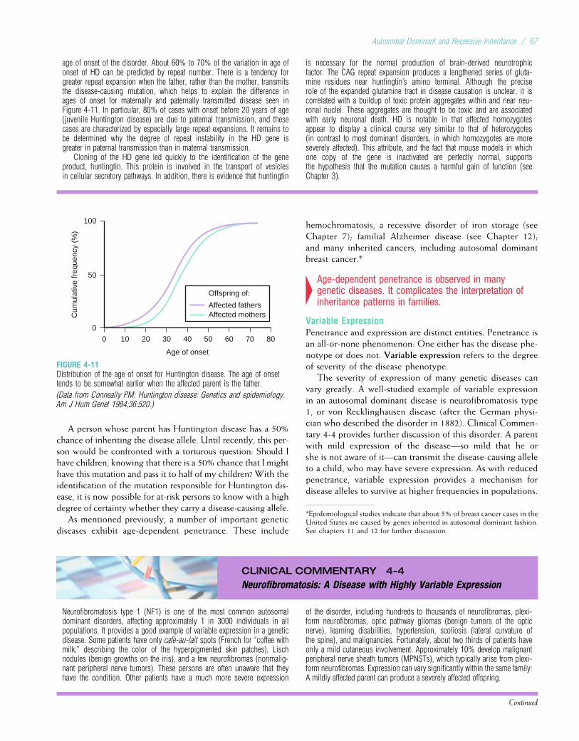
age of onset of the disorder. About 60% to 70% of the variation in age ofonset of HD can be predicted by repeat number. There is a tendency forgreater repeat expansion when the father, rather than the mother, transmitsthe disease-causing mutation, which helps to explain the difference inages of onset for maternally and paternally transmitted disease seen inFigure 4-11. In particular, 80% of cases with onset before 20 years of age(juvenile Huntington disease) are due to paternal transmission, and thesecases are characterized by especially large repeat expansions. It remains tobe determined why the degree of repeat instability in the HD gene isgreater in paternal transmission than in maternal transmission.
Cloning of the HD gene led quickly to the identification of the geneproduct, huntingtin. This protein is involved in the transport of vesiclesin cellular secretory pathways. In addition, there is evidence that huntingtin
is necessary for the normal production of brain-derived neurotrophicfactor. The CAG repeat expansion produces a lengthened series of gluta-mine residues near huntingtin’s amino terminal. Although the preciserole of the expanded glutamine tract in disease causation is unclear, it iscorrelated with a buildup of toxic protein aggregates within and near neu-ronal nuclei. These aggregates are thought to be toxic and are associatedwith early neuronal death. HD is notable in that affected homozygotesappear to display a clinical course very similar to that of heterozygotes(in contrast to most dominant disorders, in which homozygotes are moreseverely affected). This attribute, and the fact that mouse models in whichone copy of the gene is inactivated are perfectly normal, supportsthe hypothesis that the mutation causes a harmful gain of function (seeChapter 3).
A person whose parent has Huntington disease has a 50%chance of inheriting the disease allele. Until recently, this per-son would be confronted with a torturous question: Should Ihave children, knowing that there is a 50% chance that I mighthave this mutation and pass it to half of my children? With theidentification of the mutation responsible for Huntington dis-ease, it is now possible for at-risk persons to know with a highdegree of certainty whether they carry a disease-causing allele.
As mentioned previously, a number of important geneticdiseases exhibit age-dependent penetrance. These include
hemochromatosis, a recessive disorder of iron storage (seeChapter 7); familial Alzheimer disease (see Chapter 12);and many inherited cancers, including autosomal dominantbreast cancer.*
4Age-dependent penetrance is observed in manygenetic diseases. It complicates the interpretation ofinheritance patterns in families.
Variable ExpressionPenetrance and expression are distinct entities. Penetrance isan all-or-none phenomenon: One either has the disease phe-notype or does not. Variable expression refers to the degreeof severity of the disease phenotype.
The severity of expression of many genetic diseases canvary greatly. A well-studied example of variable expressionin an autosomal dominant disease is neurofibromatosis type1, or von Recklinghausen disease (after the German physi-cian who described the disorder in 1882). Clinical Commen-tary 4-4 provides further discussion of this disorder. A parentwith mild expression of the disease—so mild that he orshe is not aware of it—can transmit the disease-causing alleleto a child, who may have severe expression. As with reducedpenetrance, variable expression provides a mechanism fordisease alleles to survive at higher frequencies in populations.
CLINICAL COMMENTARY 4-4
Neurofibromatosis: A Disease with Highly Variable Expression
Neurofibromatosis type 1 (NF1) is one of the most common autosomaldominant disorders, affecting approximately 1 in 3000 individuals in allpopulations. It provides a good example of variable expression in a geneticdisease. Some patients have only cafe-au-lait spots (French for “coffee withmilk,” describing the color of the hyperpigmented skin patches), Lischnodules (benign growths on the iris), and a few neurofibromas (nonmalig-nant peripheral nerve tumors). These persons are often unaware that theyhave the condition. Other patients have a much more severe expression
of the disorder, including hundreds to thousands of neurofibromas, plexi-form neurofibromas, optic pathway gliomas (benign tumors of the opticnerve), learning disabilities, hypertension, scoliosis (lateral curvature ofthe spine), and malignancies. Fortunately, about two thirds of patients haveonly a mild cutaneous involvement. Approximately 10% develop malignantperipheral nerve sheath tumors (MPNSTs), which typically arise from plexi-form neurofibromas. Expression can vary significantly within the same family:A mildly affected parent can produce a severely affected offspring.
Continued
Offspring of:
Affected fathersAffected mothers
0 10 20 30
Age of onset
40 50 60 70 80
Cum
ulat
ive
freq
uenc
y (%
)
100
50
0
FIGURE 4-11Distribution of the age of onset for Huntington disease. The age of onsettends to be somewhat earlier when the affected parent is the father.
(Data from Conneally PM: Huntington disease: Genetics and epidemiology.Am J Hum Genet 1984;36:520.)
*Epidemiological studies indicate that about 5% of breast cancer cases in theUnited States are caused by genes inherited in autosomal dominant fashion.See chapters 11 and 12 for further discussion.
Autosomal Dominant and Recessive Inheritance / 67
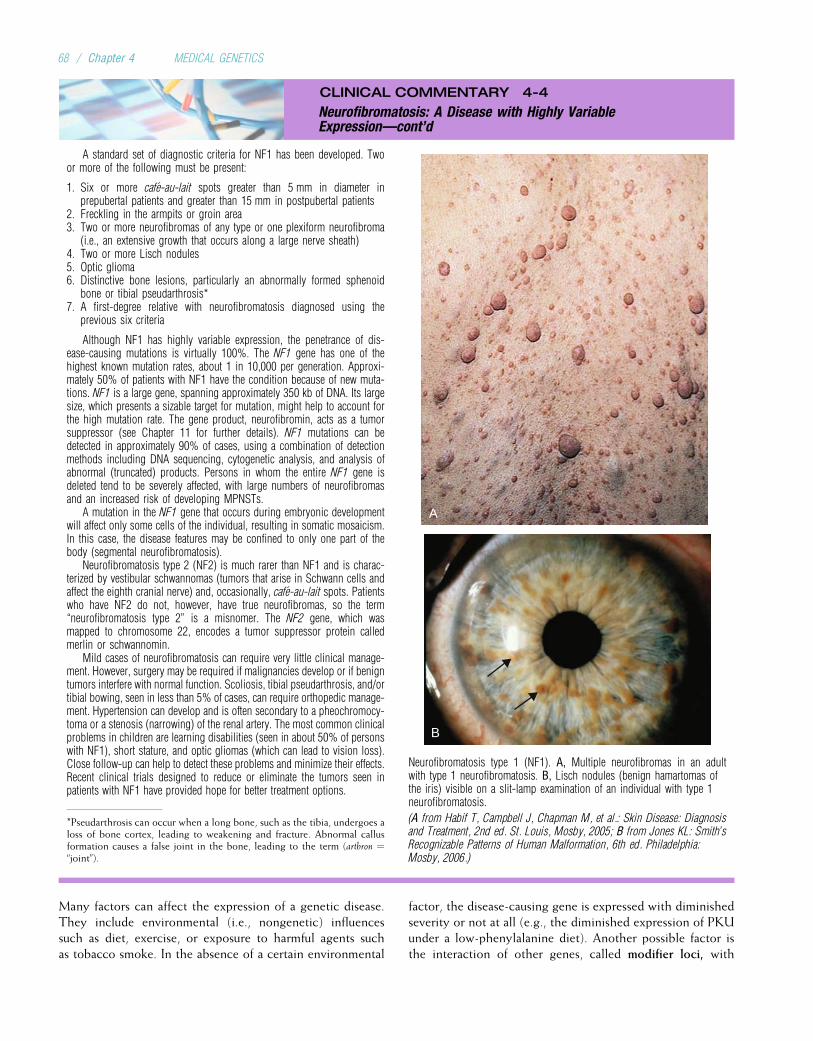
CLINICAL COMMENTARY 4-4
Neurofibromatosis: A Disease with Highly VariableExpression—cont’d
A standard set of diagnostic criteria for NF1 has been developed. Twoor more of the following must be present:
1. Six or more cafe-au-lait spots greater than 5 mm in diameter inprepubertal patients and greater than 15 mm in postpubertal patients
2. Freckling in the armpits or groin area3. Two or more neurofibromas of any type or one plexiform neurofibroma
(i.e., an extensive growth that occurs along a large nerve sheath)4. Two or more Lisch nodules5. Optic glioma6. Distinctive bone lesions, particularly an abnormally formed sphenoid
bone or tibial pseudarthrosis*7. A first-degree relative with neurofibromatosis diagnosed using the
previous six criteria
Although NF1 has highly variable expression, the penetrance of dis-ease-causing mutations is virtually 100%. The NF1 gene has one of thehighest known mutation rates, about 1 in 10,000 per generation. Approxi-mately 50% of patients with NF1 have the condition because of new muta-tions. NF1 is a large gene, spanning approximately 350 kb of DNA. Its largesize, which presents a sizable target for mutation, might help to account forthe high mutation rate. The gene product, neurofibromin, acts as a tumorsuppressor (see Chapter 11 for further details). NF1 mutations can bedetected in approximately 90% of cases, using a combination of detectionmethods including DNA sequencing, cytogenetic analysis, and analysis ofabnormal (truncated) products. Persons in whom the entire NF1 gene isdeleted tend to be severely affected, with large numbers of neurofibromasand an increased risk of developing MPNSTs.
A mutation in the NF1 gene that occurs during embryonic developmentwill affect only some cells of the individual, resulting in somatic mosaicism.In this case, the disease features may be confined to only one part of thebody (segmental neurofibromatosis).
Neurofibromatosis type 2 (NF2) is much rarer than NF1 and is charac-terized by vestibular schwannomas (tumors that arise in Schwann cells andaffect the eighth cranial nerve) and, occasionally, cafe-au-lait spots. Patientswho have NF2 do not, however, have true neurofibromas, so the term“neurofibromatosis type 2” is a misnomer. The NF2 gene, which wasmapped to chromosome 22, encodes a tumor suppressor protein calledmerlin or schwannomin.
Mild cases of neurofibromatosis can require very little clinical manage-ment. However, surgery may be required if malignancies develop or if benigntumors interfere with normal function. Scoliosis, tibial pseudarthrosis, and/ortibial bowing, seen in less than 5% of cases, can require orthopedic manage-ment. Hypertension can develop and is often secondary to a pheochromocy-toma or a stenosis (narrowing) of the renal artery. The most common clinicalproblems in children are learning disabilities (seen in about 50% of personswith NF1), short stature, and optic gliomas (which can lead to vision loss).Close follow-up can help to detect these problems and minimize their effects.Recent clinical trials designed to reduce or eliminate the tumors seen inpatients with NF1 have provided hope for better treatment options.
A
B
Neurofibromatosis type 1 (NF1). A, Multiple neurofibromas in an adultwith type 1 neurofibromatosis. B, Lisch nodules (benign hamartomas ofthe iris) visible on a slit-lamp examination of an individual with type 1neurofibromatosis.
(A from Habif T, Campbell J, Chapman M, et al.: Skin Disease: Diagnosisand Treatment, 2nd ed. St. Louis, Mosby, 2005; B from Jones KL: Smith’sRecognizable Patterns of Human Malformation, 6th ed. Philadelphia:Mosby, 2006.)
*Pseudarthrosis can occur when a long bone, such as the tibia, undergoes aloss of bone cortex, leading to weakening and fracture. Abnormal callusformation causes a false joint in the bone, leading to the term (arthron ¼“joint”).
Many factors can affect the expression of a genetic disease.They include environmental (i.e., nongenetic) influencessuch as diet, exercise, or exposure to harmful agents suchas tobacco smoke. In the absence of a certain environmental
factor, the disease-causing gene is expressed with diminishedseverity or not at all (e.g., the diminished expression of PKUunder a low-phenylalanine diet). Another possible factor isthe interaction of other genes, called modifier loci, with
68 / Chapter 4 MEDICAL GENETICS
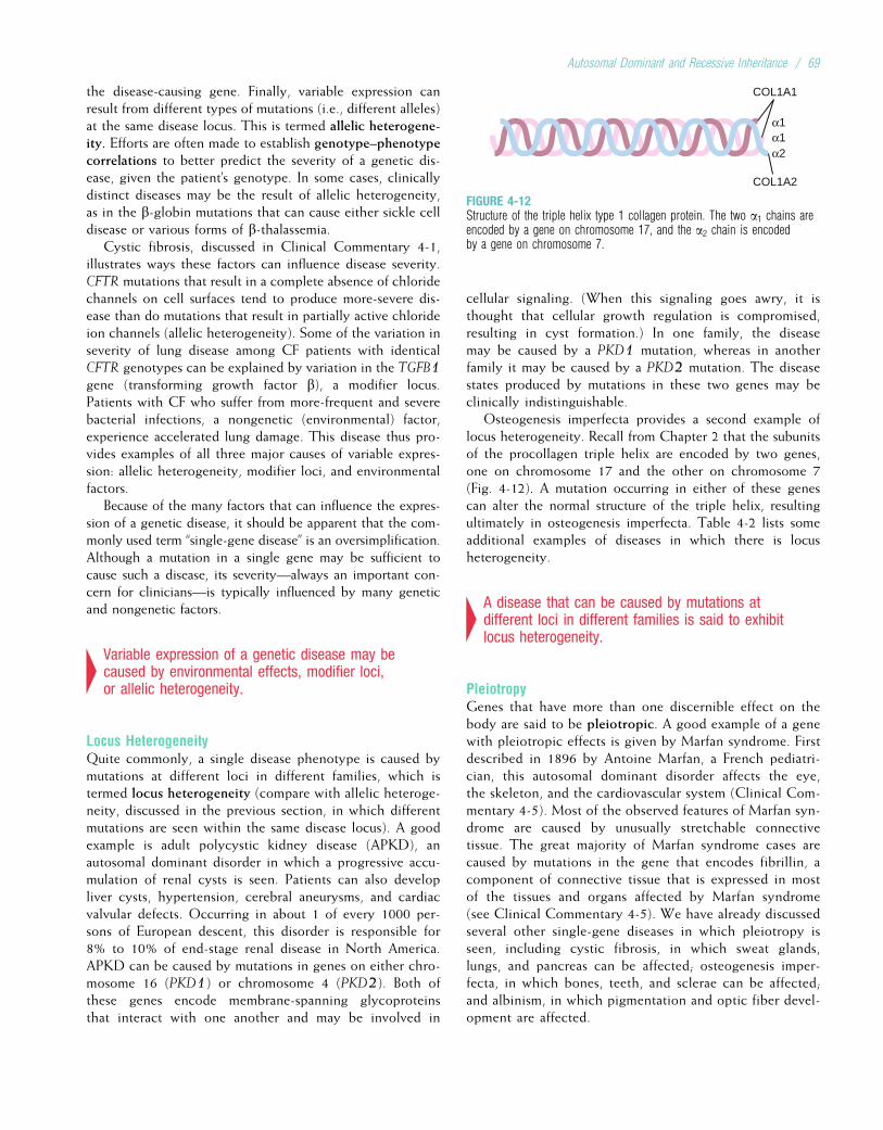
the disease-causing gene. Finally, variable expression canresult from different types of mutations (i.e., different alleles)at the same disease locus. This is termed allelic heterogene-ity. Efforts are often made to establish genotype–phenotypecorrelations to better predict the severity of a genetic dis-ease, given the patient’s genotype. In some cases, clinicallydistinct diseases may be the result of allelic heterogeneity,as in the b-globin mutations that can cause either sickle celldisease or various forms of b-thalassemia.
Cystic fibrosis, discussed in Clinical Commentary 4-1,illustrates ways these factors can influence disease severity.CFTR mutations that result in a complete absence of chloridechannels on cell surfaces tend to produce more-severe dis-ease than do mutations that result in partially active chlorideion channels (allelic heterogeneity). Some of the variation inseverity of lung disease among CF patients with identicalCFTR genotypes can be explained by variation in the TGFB1gene (transforming growth factor b), a modifier locus.Patients with CF who suffer from more-frequent and severebacterial infections, a nongenetic (environmental) factor,experience accelerated lung damage. This disease thus pro-vides examples of all three major causes of variable expres-sion: allelic heterogeneity, modifier loci, and environmentalfactors.
Because of the many factors that can influence the expres-sion of a genetic disease, it should be apparent that the com-monly used term “single-gene disease” is an oversimplification.Although a mutation in a single gene may be sufficient tocause such a disease, its severity—always an important con-cern for clinicians—is typically influenced by many geneticand nongenetic factors.
4Variable expression of a genetic disease may becaused by environmental effects, modifier loci,or allelic heterogeneity.
Locus HeterogeneityQuite commonly, a single disease phenotype is caused bymutations at different loci in different families, which istermed locus heterogeneity (compare with allelic heteroge-neity, discussed in the previous section, in which differentmutations are seen within the same disease locus). A goodexample is adult polycystic kidney disease (APKD), anautosomal dominant disorder in which a progressive accu-mulation of renal cysts is seen. Patients can also developliver cysts, hypertension, cerebral aneurysms, and cardiacvalvular defects. Occurring in about 1 of every 1000 per-sons of European descent, this disorder is responsible for8% to 10% of end-stage renal disease in North America.APKD can be caused by mutations in genes on either chro-mosome 16 (PKD1) or chromosome 4 (PKD2). Both ofthese genes encode membrane-spanning glycoproteinsthat interact with one another and may be involved in
cellular signaling. (When this signaling goes awry, it isthought that cellular growth regulation is compromised,resulting in cyst formation.) In one family, the diseasemay be caused by a PKD1 mutation, whereas in anotherfamily it may be caused by a PKD2 mutation. The diseasestates produced by mutations in these two genes may beclinically indistinguishable.
Osteogenesis imperfecta provides a second example oflocus heterogeneity. Recall from Chapter 2 that the subunitsof the procollagen triple helix are encoded by two genes,one on chromosome 17 and the other on chromosome 7(Fig. 4-12). A mutation occurring in either of these genescan alter the normal structure of the triple helix, resultingultimately in osteogenesis imperfecta. Table 4-2 lists someadditional examples of diseases in which there is locusheterogeneity.
4A disease that can be caused by mutations atdifferent loci in different families is said to exhibitlocus heterogeneity.
PleiotropyGenes that have more than one discernible effect on thebody are said to be pleiotropic. A good example of a genewith pleiotropic effects is given by Marfan syndrome. Firstdescribed in 1896 by Antoine Marfan, a French pediatri-cian, this autosomal dominant disorder affects the eye,the skeleton, and the cardiovascular system (Clinical Com-mentary 4-5). Most of the observed features of Marfan syn-drome are caused by unusually stretchable connectivetissue. The great majority of Marfan syndrome cases arecaused by mutations in the gene that encodes fibrillin, acomponent of connective tissue that is expressed in mostof the tissues and organs affected by Marfan syndrome(see Clinical Commentary 4-5). We have already discussedseveral other single-gene diseases in which pleiotropy isseen, including cystic fibrosis, in which sweat glands,lungs, and pancreas can be affected; osteogenesis imper-fecta, in which bones, teeth, and sclerae can be affected;and albinism, in which pigmentation and optic fiber devel-opment are affected.
α1α1α2
COL1A1
COL1A2
FIGURE 4-12Structure of the triple helix type 1 collagen protein. The two a1 chains areencoded by a gene on chromosome 17, and the a2 chain is encodedby a gene on chromosome 7.
Autosomal Dominant and Recessive Inheritance / 69
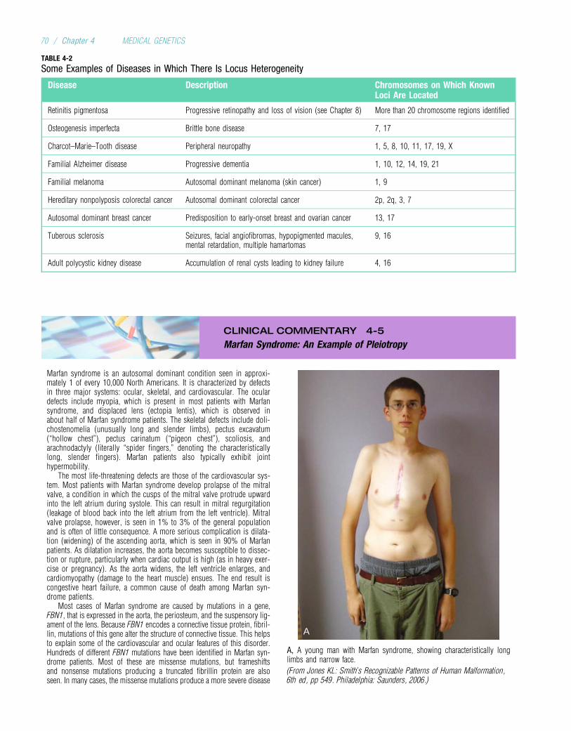
CLINICAL COMMENTARY 4-5
Marfan Syndrome: An Example of Pleiotropy
Marfan syndrome is an autosomal dominant condition seen in approxi-mately 1 of every 10,000 North Americans. It is characterized by defectsin three major systems: ocular, skeletal, and cardiovascular. The oculardefects include myopia, which is present in most patients with Marfansyndrome, and displaced lens (ectopia lentis), which is observed inabout half of Marfan syndrome patients. The skeletal defects include doli-chostenomelia (unusually long and slender limbs), pectus excavatum(“hollow chest”), pectus carinatum (“pigeon chest”), scoliosis, andarachnodactyly (literally “spider fingers,” denoting the characteristicallylong, slender fingers). Marfan patients also typically exhibit jointhypermobility.
The most life-threatening defects are those of the cardiovascular sys-tem. Most patients with Marfan syndrome develop prolapse of the mitralvalve, a condition in which the cusps of the mitral valve protrude upwardinto the left atrium during systole. This can result in mitral regurgitation(leakage of blood back into the left atrium from the left ventricle). Mitralvalve prolapse, however, is seen in 1% to 3% of the general populationand is often of little consequence. A more serious complication is dilata-tion (widening) of the ascending aorta, which is seen in 90% of Marfanpatients. As dilatation increases, the aorta becomes susceptible to dissec-tion or rupture, particularly when cardiac output is high (as in heavy exer-cise or pregnancy). As the aorta widens, the left ventricle enlarges, andcardiomyopathy (damage to the heart muscle) ensues. The end result iscongestive heart failure, a common cause of death among Marfan syn-drome patients.
Most cases of Marfan syndrome are caused by mutations in a gene,FBN1, that is expressed in the aorta, the periosteum, and the suspensory lig-ament of the lens. Because FBN1 encodes a connective tissue protein, fibril-lin, mutations of this gene alter the structure of connective tissue. This helpsto explain some of the cardiovascular and ocular features of this disorder.Hundreds of different FBN1 mutations have been identified in Marfan syn-drome patients. Most of these are missense mutations, but frameshiftsand nonsense mutations producing a truncated fibrillin protein are alsoseen. In many cases, the missense mutations produce a more severe disease
A
A, A young man with Marfan syndrome, showing characteristically longlimbs and narrow face.
(From Jones KL: Smith’s Recognizable Patterns of Human Malformation,6th ed, pp 549. Philadelphia: Saunders, 2006.)
TABLE 4-2
Some Examples of Diseases in Which There Is Locus Heterogeneity
Disease Description Chromosomes on Which KnownLoci Are Located
Retinitis pigmentosa Progressive retinopathy and loss of vision (see Chapter 8) More than 20 chromosome regions identified
Osteogenesis imperfecta Brittle bone disease 7, 17
Charcot–Marie–Tooth disease Peripheral neuropathy 1, 5, 8, 10, 11, 17, 19, X
Familial Alzheimer disease Progressive dementia 1, 10, 12, 14, 19, 21
Familial melanoma Autosomal dominant melanoma (skin cancer) 1, 9
Hereditary nonpolyposis colorectal cancer Autosomal dominant colorectal cancer 2p, 2q, 3, 7
Autosomal dominant breast cancer Predisposition to early-onset breast and ovarian cancer 13, 17
Tuberous sclerosis Seizures, facial angiofibromas, hypopigmented macules,mental retardation, multiple hamartomas
9, 16
Adult polycystic kidney disease Accumulation of renal cysts leading to kidney failure 4, 16
70 / Chapter 4 MEDICAL GENETICS
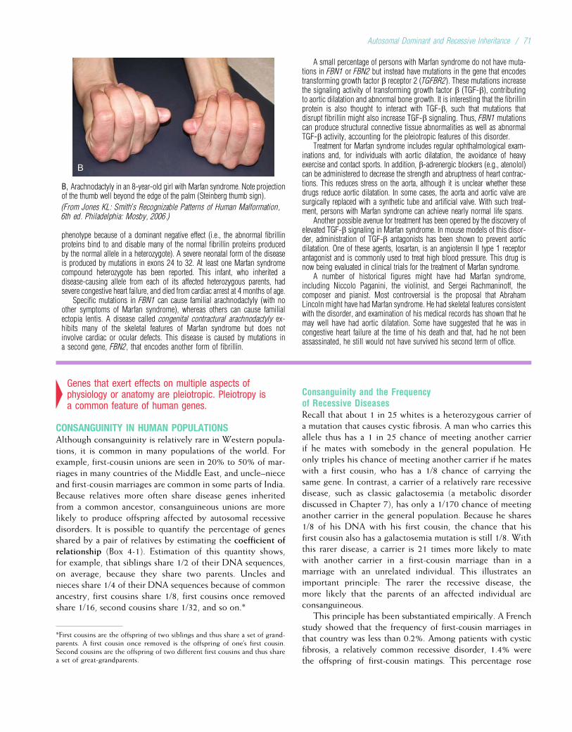
B
B, Arachnodactyly in an 8-year-old girl with Marfan syndrome. Note projectionof the thumb well beyond the edge of the palm (Steinberg thumb sign).
(From Jones KL: Smith’s Recognizable Patterns of Human Malformation,6th ed. Philadelphia: Mosby, 2006.)
phenotype because of a dominant negative effect (i.e., the abnormal fibrillinproteins bind to and disable many of the normal fibrillin proteins producedby the normal allele in a heterozygote). A severe neonatal form of the diseaseis produced by mutations in exons 24 to 32. At least one Marfan syndromecompound heterozygote has been reported. This infant, who inherited adisease-causing allele from each of its affected heterozygous parents, hadsevere congestive heart failure, and died from cardiac arrest at 4 months of age.
Specific mutations in FBN1 can cause familial arachnodactyly (with noother symptoms of Marfan syndrome), whereas others can cause familialectopia lentis. A disease called congenital contractural arachnodactyly ex-hibits many of the skeletal features of Marfan syndrome but does notinvolve cardiac or ocular defects. This disease is caused by mutations ina second gene, FBN2, that encodes another form of fibrillin.
A small percentage of persons with Marfan syndrome do not have muta-tions in FBN1 or FBN2 but instead have mutations in the gene that encodestransforming growth factor b receptor 2 (TGFBR2). These mutations increasethe signaling activity of transforming growth factor b (TGF-b), contributingto aortic dilatation and abnormal bone growth. It is interesting that the fibrillinprotein is also thought to interact with TGF-b, such that mutations thatdisrupt fibrillin might also increase TGF-b signaling. Thus, FBN1 mutationscan produce structural connective tissue abnormalities as well as abnormalTGF-b activity, accounting for the pleiotropic features of this disorder.
Treatment for Marfan syndrome includes regular ophthalmological exam-inations and, for individuals with aortic dilatation, the avoidance of heavyexercise and contact sports. In addition, b-adrenergic blockers (e.g., atenolol)can be administered to decrease the strength and abruptness of heart contrac-tions. This reduces stress on the aorta, although it is unclear whether thesedrugs reduce aortic dilatation. In some cases, the aorta and aortic valve aresurgically replaced with a synthetic tube and artificial valve. With such treat-ment, persons with Marfan syndrome can achieve nearly normal life spans.
Another possible avenue for treatment has been opened by the discovery ofelevated TGF-b signaling in Marfan syndrome. In mouse models of this disor-der, administration of TGF-b antagonists has been shown to prevent aorticdilatation. One of these agents, losartan, is an angiotensin II type 1 receptorantagonist and is commonly used to treat high blood pressure. This drug isnow being evaluated in clinical trials for the treatment of Marfan syndrome.
A number of historical figures might have had Marfan syndrome,including Niccolo Paganini, the violinist, and Sergei Rachmaninoff, thecomposer and pianist. Most controversial is the proposal that AbrahamLincoln might have had Marfan syndrome. He had skeletal features consistentwith the disorder, and examination of his medical records has shown that hemay well have had aortic dilatation. Some have suggested that he was incongestive heart failure at the time of his death and that, had he not beenassassinated, he still would not have survived his second term of office.
*First cousins are the offspring of two siblings and thus share a set of grand-parents. A first cousin once removed is the offspring of one’s first cousin.Second cousins are the offspring of two different first cousins and thus sharea set of great-grandparents.
4Genes that exert effects on multiple aspects ofphysiology or anatomy are pleiotropic. Pleiotropy isa common feature of human genes.
CONSANGUINITY IN HUMAN POPULATIONSAlthough consanguinity is relatively rare in Western popula-tions, it is common in many populations of the world. Forexample, first-cousin unions are seen in 20% to 50% of mar-riages in many countries of the Middle East, and uncle–nieceand first-cousin marriages are common in some parts of India.Because relatives more often share disease genes inheritedfrom a common ancestor, consanguineous unions are morelikely to produce offspring affected by autosomal recessivedisorders. It is possible to quantify the percentage of genesshared by a pair of relatives by estimating the coefficient ofrelationship (Box 4-1). Estimation of this quantity shows,for example, that siblings share 1/2 of their DNA sequences,on average, because they share two parents. Uncles andnieces share 1/4 of their DNA sequences because of commonancestry, first cousins share 1/8, first cousins once removedshare 1/16, second cousins share 1/32, and so on.*
Consanguinity and the Frequencyof Recessive DiseasesRecall that about 1 in 25 whites is a heterozygous carrier ofa mutation that causes cystic fibrosis. A man who carries thisallele thus has a 1 in 25 chance of meeting another carrierif he mates with somebody in the general population. Heonly triples his chance of meeting another carrier if he mateswith a first cousin, who has a 1/8 chance of carrying thesame gene. In contrast, a carrier of a relatively rare recessivedisease, such as classic galactosemia (a metabolic disorderdiscussed in Chapter 7), has only a 1/170 chance of meetinganother carrier in the general population. Because he shares1/8 of his DNA with his first cousin, the chance that hisfirst cousin also has a galactosemia mutation is still 1/8. Withthis rarer disease, a carrier is 21 times more likely to matewith another carrier in a first-cousin marriage than in amarriage with an unrelated individual. This illustrates animportant principle: The rarer the recessive disease, themore likely that the parents of an affected individual areconsanguineous.
This principle has been substantiated empirically. A Frenchstudy showed that the frequency of first-cousin marriages inthat country was less than 0.2%. Among patients with cysticfibrosis, a relatively common recessive disorder, 1.4% werethe offspring of first-cousin matings. This percentage rose
Autosomal Dominant and Recessive Inheritance / 71
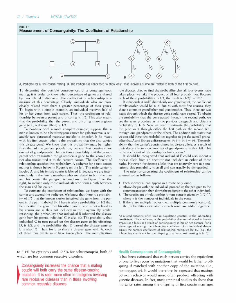
to 7.1% for cystinosis and 12.5% for achromatopsia, both ofwhich are less-common recessive disorders.
4Consanguinity increases the chance that a matingcouple will both carry the same disease-causingmutation. It is seen more often in pedigrees involvingrare recessive diseases than in those involvingcommon recessive diseases.
Health Consequences of ConsanguinityIt has been estimated that each person carries the equivalentof one to five recessive mutations that would be lethal to off-spring if matched with another copy of the mutation (i.e.,homozygosity). It would therefore be expected that matingsbetween relatives would more often produce offspring withgenetic diseases. In fact, most empirical studies do show thatmortality rates among the offspring of first-cousin marriages
BOX 4-1Measurement of Consanguinity: The Coefficient of Relationship
A EA E A E
D D
FFC
A B
C
B
DB
B
12
12
12
12
12
12
12
12
A, Pedigree for a first-cousin mating. B, The Pedigree is condensed to show only those individuals who are related to both of the first cousins.
To determine the possible consequences of a consanguineousmating, it is useful to know what percentage of genes are sharedby two related individuals. The coefficient of relationship is ameasure of this percentage. Clearly, individuals who are moreclosely related must share a greater percentage of their genes.To begin with a simple example, an individual receives half ofhis or her genes from each parent. Thus, the coefficient of rela-tionship between a parent and offspring is 1/2. This also meansthat the probability that the parent and offspring share a givengene (e.g., a disease allele) is 1/2.
To continue with a more complex example, suppose that aman is known to be a heterozygous carrier for galactosemia, a rel-atively rare autosomal recessive metabolic disorder. If he mateswith his first cousin, what is the probability that she also carriesthis disease gene? We know that this probability must be higherthan that of the general population, because first cousins shareone set of grandparents. There is thus a possibility that the grand-parent who transmitted the galactosemia gene to the known car-rier also transmitted it to the carrier’s cousin. The coefficient ofrelationship specifies this probability. A pedigree for a first-cousinmating is shown below in Figure A on the left. The male carrier islabeled A, and his female cousin is labeled E. Because we are inter-ested only in the family members who are related to both the manand his cousin, the pedigree is condensed, in Figure B on theright, to include only those individuals who form a path betweenthe man and his cousin.
To estimate the coefficient of relationship, we begin with thecarrier and ascend the pedigree. We know that there is a probabil-ity of 1/2 that the known carrier inherited the gene from the par-ent in the path (labeled B). There is also a probability of 1/2 thathe inherited the gene from his other parent, who is not related tohis cousin and is thus not included in the diagram. By similarreasoning, the probability that individual B inherited the diseasegene from his parent, individual C, is also 1/2. The probability thatindividual C in turn passed on the disease gene to his offspring,D, is 1/2, and the probability that D passed the disease gene toE is also 1/2. Thus, for E to share a disease gene with A, eachof these four events must have taken place. The multiplication
rule dictates that, to find the probability that all four events havetaken place, we take the product of all four probabilities. Becauseeach of these probabilities is 1/2, the result is (1/2)4 ¼ 1/16.
If individuals A and E shared only one grandparent, the coefficientof relationship would be 1/16. But, as with most first cousins, theyshare a common grandfather and grandmother. Thus, there are twopaths through which the disease gene could have passed. To obtainthe probability that the gene passed through the second path, weuse the same procedure as in the previous paragraph and obtain aprobability of 1/16. Now we need to estimate the probability thatthe gene went through either the first path or the second (i.e.,through one grandparent or the other). The addition rule states thatwe can add these two probabilities together to get the overall proba-bility that A and E share a disease gene: 1/16þ 1/16¼ 1/8. The prob-ability that the carrier’s cousin shares his disease allele, as a result oftheir descent from a common set of grandparents, is thus 1/8. Thisis the coefficient of relationship for first cousins.*
It should be recognized that individual E could also inherit adisease allele from an ancestor not included in either of thesepaths. However, for disease alleles that are relatively rare in popu-lations, this probability is small and can usually be disregarded.
The rules for calculating the coefficient of relationship can besummarized as follows:
1. Each individual can appear in a route only once.2. Always begin with one individual, proceed up the pedigree to the
common ancestor, then down the pedigree to the other individual.3. The coefficient of relationship for one route is given by (1/2)n-1,
where n is the number of individuals in the route.4. If there are multiple routes (i.e., multiple common ancestors),
the probabilities estimated for each route are added together.
*A related quantity, often used in population genetics, is the inbreedingcoefficient. This coefficient is the probability that an individual is homo-zygous at a locus as a result of consanguinity in his or her parents. For agiven type of mating, the inbreeding coefficient of an individual alwaysequals the parents’ coefficient of relationship multiplied by 1/2 (e.g., theinbreeding coefficient for the offspring of a first-cousin mating is 1/16).
72 / Chapter 4 MEDICAL GENETICS
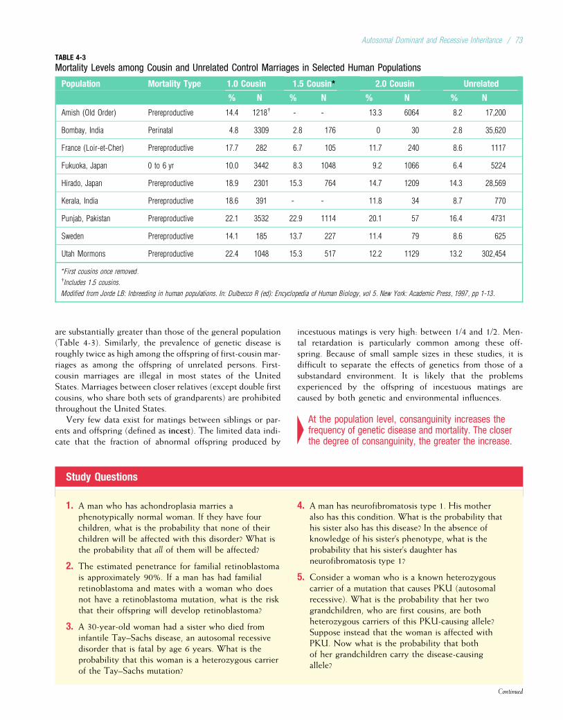
are substantially greater than those of the general population(Table 4-3). Similarly, the prevalence of genetic disease isroughly twice as high among the offspring of first-cousin mar-riages as among the offspring of unrelated persons. First-cousin marriages are illegal in most states of the UnitedStates. Marriages between closer relatives (except double firstcousins, who share both sets of grandparents) are prohibitedthroughout the United States.
Very few data exist for matings between siblings or par-ents and offspring (defined as incest). The limited data indi-cate that the fraction of abnormal offspring produced by
incestuous matings is very high: between 1/4 and 1/2. Men-tal retardation is particularly common among these off-spring. Because of small sample sizes in these studies, it isdifficult to separate the effects of genetics from those of asubstandard environment. It is likely that the problemsexperienced by the offspring of incestuous matings arecaused by both genetic and environmental influences.
4At the population level, consanguinity increases thefrequency of genetic disease and mortality. The closerthe degree of consanguinity, the greater the increase.
Study Questions
1. A man who has achondroplasia marries aphenotypically normal woman. If they have fourchildren, what is the probability that none of theirchildren will be affected with this disorder? What isthe probability that all of them will be affected?
2. The estimated penetrance for familial retinoblastomais approximately 90%. If a man has had familialretinoblastoma and mates with a woman who doesnot have a retinoblastoma mutation, what is the riskthat their offspring will develop retinoblastoma?
3. A 30-year-old woman had a sister who died frominfantile Tay–Sachs disease, an autosomal recessivedisorder that is fatal by age 6 years. What is theprobability that this woman is a heterozygous carrierof the Tay–Sachs mutation?
4. A man has neurofibromatosis type 1. His motheralso has this condition. What is the probability thathis sister also has this disease? In the absence ofknowledge of his sister’s phenotype, what is theprobability that his sister’s daughter hasneurofibromatosis type 1?
5. Consider a woman who is a known heterozygouscarrier of a mutation that causes PKU (autosomalrecessive). What is the probability that her twograndchildren, who are first cousins, are bothheterozygous carriers of this PKU-causing allele?Suppose instead that the woman is affected withPKU. Now what is the probability that bothof her grandchildren carry the disease-causingallele?
Continued
TABLE 4-3
Mortality Levels among Cousin and Unrelated Control Marriages in Selected Human Populations
Population Mortality Type 1.0 Cousin 1.5 Cousin* 2.0 Cousin Unrelated
% N % N % N % N
Amish (Old Order) Prereproductive 14.4 1218{ - - 13.3 6064 8.2 17,200
Bombay, India Perinatal 4.8 3309 2.8 176 0 30 2.8 35,620
France (Loir-et-Cher) Prereproductive 17.7 282 6.7 105 11.7 240 8.6 1117
Fukuoka, Japan 0 to 6 yr 10.0 3442 8.3 1048 9.2 1066 6.4 5224
Hirado, Japan Prereproductive 18.9 2301 15.3 764 14.7 1209 14.3 28,569
Kerala, India Prereproductive 18.6 391 - - 11.8 34 8.7 770
Punjab, Pakistan Prereproductive 22.1 3532 22.9 1114 20.1 57 16.4 4731
Sweden Prereproductive 14.1 185 13.7 227 11.4 79 8.6 625
Utah Mormons Prereproductive 22.4 1048 15.3 517 12.2 1129 13.2 302,454
*First cousins once removed.{Includes 1.5 cousins.
Modified from Jorde LB: Inbreeding in human populations. In: Dulbecco R (ed): Encyclopedia of Human Biology, vol 5. New York: Academic Press, 1997, pp 1-13.
Autosomal Dominant and Recessive Inheritance / 73
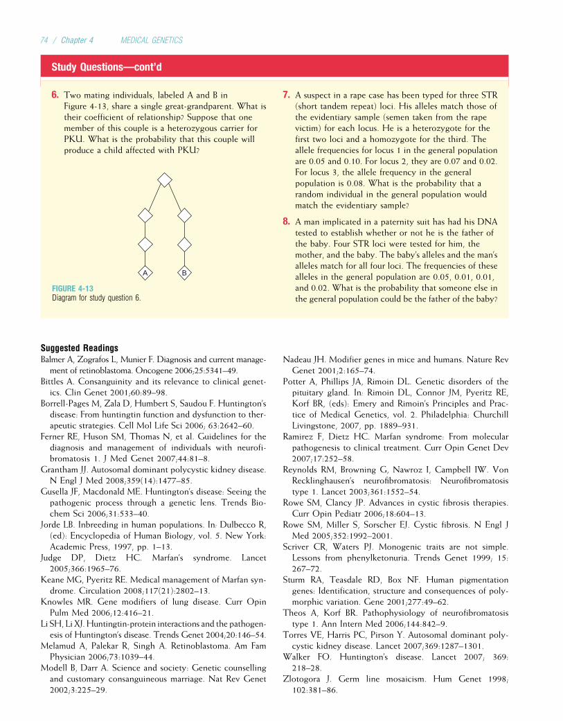
Study Questions—cont’d
6. Two mating individuals, labeled A and B inFigure 4-13, share a single great-grandparent. What istheir coefficient of relationship? Suppose that onemember of this couple is a heterozygous carrier forPKU. What is the probability that this couple willproduce a child affected with PKU?
7. A suspect in a rape case has been typed for three STR(short tandem repeat) loci. His alleles match those ofthe evidentiary sample (semen taken from the rapevictim) for each locus. He is a heterozygote for thefirst two loci and a homozygote for the third. Theallele frequencies for locus 1 in the general populationare 0.05 and 0.10. For locus 2, they are 0.07 and 0.02.For locus 3, the allele frequency in the generalpopulation is 0.08. What is the probability that arandom individual in the general population wouldmatch the evidentiary sample?
8. A man implicated in a paternity suit has had his DNAtested to establish whether or not he is the father ofthe baby. Four STR loci were tested for him, themother, and the baby. The baby’s alleles and the man’salleles match for all four loci. The frequencies of thesealleles in the general population are 0.05, 0.01, 0.01,and 0.02. What is the probability that someone else inthe general population could be the father of the baby?
Suggested ReadingsBalmer A, Zografos L, Munier F. Diagnosis and current manage-
ment of retinoblastoma. Oncogene 2006;25:5341–49.Bittles A. Consanguinity and its relevance to clinical genet-
ics. Clin Genet 2001;60:89–98.Borrell-Pages M, Zala D, Humbert S, Saudou F. Huntington’s
disease: From huntingtin function and dysfunction to ther-apeutic strategies. Cell Mol Life Sci 2006; 63:2642–60.
Ferner RE, Huson SM, Thomas N, et al. Guidelines for thediagnosis and management of individuals with neurofi-bromatosis 1. J Med Genet 2007;44:81–8.
Grantham JJ. Autosomal dominant polycystic kidney disease.N Engl J Med 2008;359(14):1477–85.
Gusella JF, Macdonald ME. Huntington’s disease: Seeing thepathogenic process through a genetic lens. Trends Bio-chem Sci 2006;31:533–40.
Jorde LB. Inbreeding in human populations. In: Dulbecco R,(ed): Encyclopedia of Human Biology, vol. 5. New York:Academic Press, 1997, pp. 1–13.
Judge DP, Dietz HC. Marfan’s syndrome. Lancet2005;366:1965–76.
Keane MG, Pyeritz RE. Medical management of Marfan syn-drome. Circulation 2008;117(21):2802–13.
Knowles MR. Gene modifiers of lung disease. Curr OpinPulm Med 2006;12:416–21.
Li SH, Li XJ. Huntingtin-protein interactions and the pathogen-esis of Huntington’s disease. Trends Genet 2004;20:146–54.
Melamud A, Palekar R, Singh A. Retinoblastoma. Am FamPhysician 2006;73:1039–44.
Modell B, Darr A. Science and society: Genetic counsellingand customary consanguineous marriage. Nat Rev Genet2002;3:225–29.
Nadeau JH. Modifier genes in mice and humans. Nature RevGenet 2001;2:165–74.
Potter A, Phillips JA, Rimoin DL. Genetic disorders of thepituitary gland. In: Rimoin DL, Connor JM, Pyeritz RE,Korf BR, (eds): Emery and Rimoin’s Principles and Prac-tice of Medical Genetics, vol. 2. Philadelphia: ChurchillLivingstone, 2007, pp. 1889–931.
Ramirez F, Dietz HC. Marfan syndrome: From molecularpathogenesis to clinical treatment. Curr Opin Genet Dev2007;17:252–58.
Reynolds RM, Browning G, Nawroz I, Campbell IW. VonRecklinghausen’s neurofibromatosis: Neurofibromatosistype 1. Lancet 2003;361:1552–54.
Rowe SM, Clancy JP. Advances in cystic fibrosis therapies.Curr Opin Pediatr 2006;18:604–13.
Rowe SM, Miller S, Sorscher EJ. Cystic fibrosis. N Engl JMed 2005;352:1992–2001.
Scriver CR, Waters PJ. Monogenic traits are not simple.Lessons from phenylketonuria. Trends Genet 1999; 15:267–72.
Sturm RA, Teasdale RD, Box NF. Human pigmentationgenes: Identification, structure and consequences of poly-morphic variation. Gene 2001;277:49–62.
Theos A, Korf BR. Pathophysiology of neurofibromatosistype 1. Ann Intern Med 2006;144:842–9.
Torres VE, Harris PC, Pirson Y. Autosomal dominant poly-cystic kidney disease. Lancet 2007;369:1287–1301.
Walker FO. Huntington’s disease. Lancet 2007; 369:218–28.
Zlotogora J. Germ line mosaicism. Hum Genet 1998;102:381–86.
A B
FIGURE 4-13Diagram for study question 6.
74 / Chapter 4 MEDICAL GENETICS
Internet ResourcesCystic Fibrosis Mutation Database (also contains links to otheruseful cystic fibrosis websites) http://www.genet.sickkids.on.ca/cftr/
Eye Cancer Network: Retinoblastoma (descriptions,photographs, and useful links) http://www.eyecancer.com/Patient/Condition.aspx?nID=53&Category=Retinal+Tumors&Condition=
Retinoblastoma
National Center for Biotechnology Information: Genes andDisease (brief summaries of many of the genetic diseasesdiscussed in this text) http://www.ncbi.nlm.nih.gov/books/bv.fcgi?call=bv.View..ShowTOC&rid=gnd.TOC&depth=2
National Institute of Neurological Diseases and Stroke:Huntington Disease Information Page http://www.ninds.nih.gov/
disorders/huntington/huntington.htm
National Marfan Foundation (basic information aboutMarfan syndrome, with links to other sites) http://www.marfan.org/
National Neurofibromatosis Foundation (many useful linksto online resources) http://www.ctf.org/
Autosomal Dominant and Recessive Inheritance / 75
Chapter 5
SEX-LINKED AND NONTRADITIONALMODES OF INHERITANCE
The previous chapter dealt with genes located on the 22autosomes; their mode of inheritance was elucidated byGregor Mendel. In this chapter we discuss disease-causingmutations that are inherited in ways that were unknown toMendel and are thus sometimes termed nonmendelian.
The first mutations to be discussed are DNA variants ofthe sex chromosomes (X and Y), known as sex-linked muta-tions. The human X chromosome is a large chromosome,containing about 5% of the nuclear genome’s DNA (approx-imately 155 million base pairs [155 megabases, 155 Mb]).Almost 1100 genes have been localized to the X chromo-some. Diseases caused by genes on this chromosome are saidto be X-linked. In contrast to the X chromosome, the Ychromosome is quite small (60 Mb) and contains only afew dozen genes.
The next group of disease-causing mutations is located inthe mitochondrial genome, which is inherited only fromone’s mother. Mitochondrial diseases thus display a uniquepattern of inheritance in families. Extensive analyses haverevealed a growing number of disease-causing mutations inthe mitochondrial genome.
Finally, we discuss two processes that have been eluci-dated only in the past 2 to 3 decades: anticipation andimprinting. Anticipation refers to earlier age-of-onset ofsome genetic diseases in more recent generations of families.Imprinting refers to the fact that some genes are expressedonly on paternally transmitted chromosomes and others areexpressed only on maternally transmitted chromosomes.Our understanding of both of these recently discovered pro-cesses has been greatly enhanced by detailed molecular ana-lyses of humans and model organisms.
X INACTIVATIONThe X chromosome contains many important protein-codinggenes, and it has long been known that human females havetwo X chromosomes and males have only one. Thus, femaleshave two copies of each X-linked gene, and males have onlyone copy. Yet males and females do not differ in terms of theamounts of protein products (e.g., enzyme levels) encodedby most of these genes. What could account for this?
In the early 1960s Mary Lyon hypothesized that oneX chromosome in each somatic cell of the female is inactivated.
This would result in dosage compensation, an equalizationof the amount of X-linked gene products in males andfemales. The Lyon hypothesis stated that X inactivationoccurs early in female embryonic development and that theX chromosome contributed by the father is inactivated insome cells, whereas in other cells the X chromosome con-tributed by the mother is inactivated. In each cell, one ofthe two X chromosomes is chosen at random for inactiva-tion, so the maternally and paternally derived X chromo-somes are each inactivated in about half of the embryo’scells. Thus, inactivation, like gamete transmission, is anal-ogous to a coin-tossing experiment. Once an X chromo-some is inactivated in a cell, it will remain inactive in alldescendants of that cell. X inactivation is therefore a ran-domly determined, but fixed (or permanent), process. As aresult of X inactivation, all normal females have two dis-tinct populations of cells: One population has an activepaternally derived X chromosome, and the other has anactive maternally derived X chromosome. (Fig. 5-1 pro-vides a summary of this process.) Because they have twopopulations of cells, females are mosaics (see Chapter 4)for X chromosome activity. Males, having only one copyof the X chromosome, are not mosaics but are hemizy-gous for the X chromosome (hemi means “half”).
4The Lyon hypothesis states that one X chromosome ineach cell is randomly inactivated early in theembryonic development of females. This ensures thatfemales, who have two copies of the X chromosome,will produce X-linked gene products in quantitiesroughly similar to those produced in males (dosagecompensation).
The Lyon hypothesis relied on several pieces of evidence,most of which were derived from animal studies. First, it wasknown that females are typically mosaics for some X-linkedtraits and males are not. For example, female mice that areheterozygous for certain X-linked coat-color genes exhibita dappled coloring of their fur, whereas male mice do not.A similar example is given by the calico cat. These femalecats have alternating black and orange patches of fur thatcorrespond to two populations of cells: one that contains
76
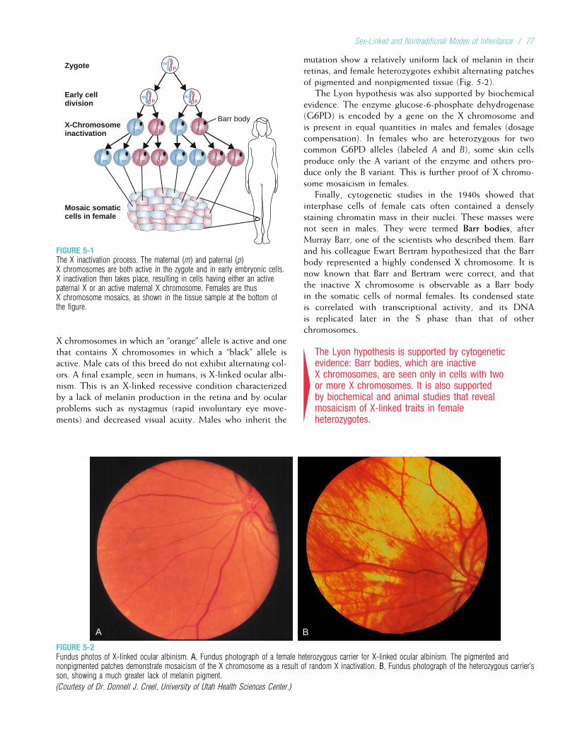
X chromosomes in which an “orange” allele is active and onethat contains X chromosomes in which a “black” allele isactive. Male cats of this breed do not exhibit alternating col-ors. A final example, seen in humans, is X-linked ocular albi-nism. This is an X-linked recessive condition characterizedby a lack of melanin production in the retina and by ocularproblems such as nystagmus (rapid involuntary eye move-ments) and decreased visual acuity. Males who inherit the
mutation show a relatively uniform lack of melanin in theirretinas, and female heterozygotes exhibit alternating patchesof pigmented and nonpigmented tissue (Fig. 5-2).
The Lyon hypothesis was also supported by biochemicalevidence. The enzyme glucose-6-phosphate dehydrogenase(G6PD) is encoded by a gene on the X chromosome andis present in equal quantities in males and females (dosagecompensation). In females who are heterozygous for twocommon G6PD alleles (labeled A and B), some skin cellsproduce only the A variant of the enzyme and others pro-duce only the B variant. This is further proof of X chromo-some mosaicism in females.
Finally, cytogenetic studies in the 1940s showed thatinterphase cells of female cats often contained a denselystaining chromatin mass in their nuclei. These masses werenot seen in males. They were termed Barr bodies, afterMurray Barr, one of the scientists who described them. Barrand his colleague Ewart Bertram hypothesized that the Barrbody represented a highly condensed X chromosome. It isnow known that Barr and Bertram were correct, and thatthe inactive X chromosome is observable as a Barr bodyin the somatic cells of normal females. Its condensed stateis correlated with transcriptional activity, and its DNAis replicated later in the S phase than that of otherchromosomes.
4The Lyon hypothesis is supported by cytogeneticevidence: Barr bodies, which are inactiveX chromosomes, are seen only in cells with twoor more X chromosomes. It is also supportedby biochemical and animal studies that revealmosaicism of X-linked traits in femaleheterozygotes.
A B
FIGURE 5-2Fundus photos of X-linked ocular albinism. A, Fundus photograph of a female heterozygous carrier for X-linked ocular albinism. The pigmented andnonpigmented patches demonstrate mosaicism of the X chromosome as a result of random X inactivation. B, Fundus photograph of the heterozygous carrier’sson, showing a much greater lack of melanin pigment.
(Courtesy of Dr. Donnell J. Creel, University of Utah Health Sciences Center.)
Barr body
m
m
m
m
m
m m m m
Zygote
Early celldivision
X-Chromosomeinactivation
Mosaic somaticcells in female
p pp p
p
pp
p
p
FIGURE 5-1The X inactivation process. The maternal (m) and paternal (p)X chromosomes are both active in the zygote and in early embryonic cells.X inactivation then takes place, resulting in cells having either an activepaternal X or an active maternal X chromosome. Females are thusX chromosome mosaics, as shown in the tissue sample at the bottom ofthe figure.
Sex-Linked and Nontraditional Modes of Inheritance / 77
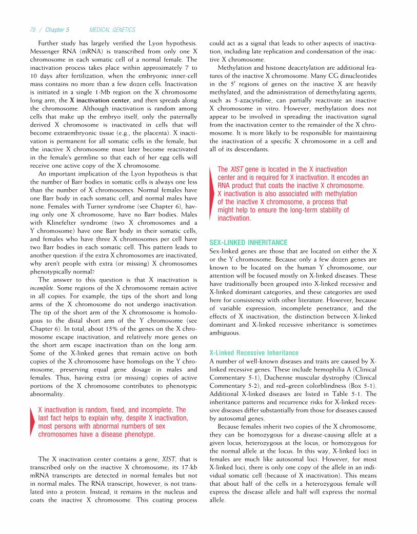
Further study has largely verified the Lyon hypothesis.Messenger RNA (mRNA) is transcribed from only one Xchromosome in each somatic cell of a normal female. Theinactivation process takes place within approximately 7 to10 days after fertilization, when the embryonic inner-cellmass contains no more than a few dozen cells. Inactivationis initiated in a single 1-Mb region on the X chromosomelong arm, the X inactivation center, and then spreads alongthe chromosome. Although inactivation is random amongcells that make up the embryo itself, only the paternallyderived X chromosome is inactivated in cells that willbecome extraembryonic tissue (e.g., the placenta). X inacti-vation is permanent for all somatic cells in the female, butthe inactive X chromosome must later become reactivatedin the female’s germline so that each of her egg cells willreceive one active copy of the X chromosome.
An important implication of the Lyon hypothesis is thatthe number of Barr bodies in somatic cells is always one lessthan the number of X chromosomes. Normal females haveone Barr body in each somatic cell, and normal males havenone. Females with Turner syndrome (see Chapter 6), hav-ing only one X chromosome, have no Barr bodies. Maleswith Klinefelter syndrome (two X chromosomes and aY chromosome) have one Barr body in their somatic cells,and females who have three X chromosomes per cell havetwo Barr bodies in each somatic cell. This pattern leads toanother question: if the extra X chromosomes are inactivated,why aren’t people with extra (or missing) X chromosomesphenotypically normal?
The answer to this question is that X inactivation isincomplete. Some regions of the X chromosome remain activein all copies. For example, the tips of the short and longarms of the X chromosome do not undergo inactivation.The tip of the short arm of the X chromosome is homolo-gous to the distal short arm of the Y chromosome (seeChapter 6). In total, about 15% of the genes on the X chro-mosome escape inactivation, and relatively more genes onthe short arm escape inactivation than on the long arm.Some of the X-linked genes that remain active on bothcopies of the X chromosome have homologs on the Y chro-mosome, preserving equal gene dosage in males andfemales. Thus, having extra (or missing) copies of activeportions of the X chromosome contributes to phenotypicabnormality.
4X inactivation is random, fixed, and incomplete. Thelast fact helps to explain why, despite X inactivation,most persons with abnormal numbers of sexchromosomes have a disease phenotype.
The X inactivation center contains a gene, XIST, that istranscribed only on the inactive X chromosome; its 17-kbmRNA transcripts are detected in normal females but notin normal males. The RNA transcript, however, is not trans-lated into a protein. Instead, it remains in the nucleus andcoats the inactive X chromosome. This coating process
could act as a signal that leads to other aspects of inactiva-tion, including late replication and condensation of the inac-tive X chromosome.
Methylation and histone deacetylation are additional fea-tures of the inactive X chromosome. Many CG dinucleotidesin the 50 regions of genes on the inactive X are heavilymethylated, and the administration of demethylating agents,such as 5-azacytidine, can partially reactivate an inactiveX chromosome in vitro. However, methylation does notappear to be involved in spreading the inactivation signalfrom the inactivation center to the remainder of the X chro-mosome. It is more likely to be responsible for maintainingthe inactivation of a specific X chromosome in a cell andall of its descendants.
4The XIST gene is located in the X inactivationcenter and is required for X inactivation. It encodes anRNA product that coats the inactive X chromosome.X inactivation is also associated with methylationof the inactive X chromosome, a process thatmight help to ensure the long-term stability ofinactivation.
SEX-LINKED INHERITANCESex-linked genes are those that are located on either the Xor the Y chromosome. Because only a few dozen genes areknown to be located on the human Y chromosome, ourattention will be focused mostly on X-linked diseases. Thesehave traditionally been grouped into X-linked recessive andX-linked dominant categories, and these categories are usedhere for consistency with other literature. However, becauseof variable expression, incomplete penetrance, and theeffects of X inactivation, the distinction between X-linkeddominant and X-linked recessive inheritance is sometimesambiguous.
X-Linked Recessive InheritanceA number of well-known diseases and traits are caused by X-linked recessive genes. These include hemophilia A (ClinicalCommentary 5-1), Duchenne muscular dystrophy (ClinicalCommentary 5-2), and red–green colorblindness (Box 5-1).Additional X-linked diseases are listed in Table 5-1. Theinheritance patterns and recurrence risks for X-linked reces-sive diseases differ substantially from those for diseases causedby autosomal genes.
Because females inherit two copies of the X chromosome,they can be homozygous for a disease-causing allele at agiven locus, heterozygous at the locus, or homozygous forthe normal allele at the locus. In this way, X-linked loci infemales are much like autosomal loci. However, for mostX-linked loci, there is only one copy of the allele in an indi-vidual somatic cell (because of X inactivation). This meansthat about half of the cells in a heterozygous female willexpress the disease allele and half will express the normalallele.
78 / Chapter 5 MEDICAL GENETICS
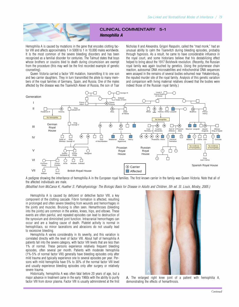
CLINICAL COMMENTARY 5-1
Hemophilia A
Hemophilia A is caused by mutations in the gene that encodes clotting fac-tor VIII and affects approximately 1 in 5000 to 1 in 10,000 males worldwide.It is the most common of the severe bleeding disorders and has beenrecognized as a familial disorder for centuries. The Talmud states that boyswhose brothers or cousins bled to death during circumcision are exemptfrom the procedure (this may well be the first recorded example of geneticcounseling).
Queen Victoria carried a factor VIII mutation, transmitting it to one sonand two carrier daughters. They in turn transmitted the allele to many mem-bers of the royal families of Germany, Spain, and Russia. One of the malesaffected by the disease was the Tsarevitch Alexei of Russia, the son of Tsar
Nicholas II and Alexandra. Grigori Rasputin, called the “mad monk,” had anunusual ability to calm the Tsarevitch during bleeding episodes, probablythrough hypnosis. As a result, he came to have considerable influence inthe royal court, and some historians believe that his destabilizing effecthelped to bring about the 1917 Bolshevik revolution. (Recently, the Russianroyal family was again touched by genetics. Using the polymerase chainreaction, autosomal DNA microsatellites and mitochondrial DNA sequenceswere assayed in the remains of several bodies exhumed near Yekaterinburg,the reputed murder site of the royal family. Analysis of this genetic variationand comparison with living maternal relatives showed that the bodies wereindeed those of the Russian royal family.)
Generation
I
IV
V
VI
VIIWilliam
PrincessDiana
PrinceCharles
Anne
QueenElizabeth II
PrincePhilip
Margaret
GermanRoyalHouse
No hemophilia
KingGeorge V
Victoria Alice Dukeof Hesse
Frederick III
KingEdward VII
PrinceAlbert
QueenVictoria
EdwardDuke of Kent
George III
Earlof Mountbatten
Waldemar PrinceSigismond
PrussianRoyalHouse
Irene
Andrew
British Royal House
Edward
Henry
II
III
No hemophilia
No evidenceof hemophilia
No evidenceof hemophilia
Alfred Helena Arthur Leopold Beatrice PrinceHenry
Louis IIGrand Duke
of Hesse
RussianRoyalHouse
Spanish Royal HouseCarrierAffected
KingJuan Carlos
Anastasia Alexis ViscountTremation
JaimeAlfonso Gonzalo
CzarNicholas II
CzarinaAlexandra
Earl ofAthlone
PrincessAlice
Maurice Leopold QueenEugenie
AlfonsoKing of Spain
? ? ? ?
?
? ?
Henry JuanDuke ofWindsor
KingGeorge VI
A pedigree showing the inheritance of hemophilia A in the European royal families. The first known carrier in the family was Queen Victoria. Note that all ofthe affected individuals are male.
(Modified from McCance K, Huether S: Pathophysiology: The Biologic Basis for Disease in Adults and Children, 5th ed. St. Louis, Mosby, 2005.)
Hemophilia A is caused by deficient or defective factor VIII, a keycomponent of the clotting cascade. Fibrin formation is affected, resultingin prolonged and often severe bleeding from wounds and hemorrhages inthe joints and muscles. Bruising is often seen. Hemarthroses (bleedinginto the joints) are common in the ankles, knees, hips, and elbows. Theseevents are often painful, and repeated episodes can lead to destruction ofthe synovium and diminished joint function. Intracranial hemorrhages canoccur and are a leading cause of death. Platelet activity is normal inhemophiliacs, so minor lacerations and abrasions do not usually leadto excessive bleeding.
Hemophilia A varies considerably in its severity, and this variation iscorrelated directly with the level of factor VIII. About half of hemophilia Apatients fall into the severe category, with factor VIII levels that are less than1% of normal. These persons experience relatively frequent bleedingepisodes, often several per month. Patients with moderate hemophilia(1%-5% of normal factor VIII) generally have bleeding episodes only aftermild trauma and typically experience one to several episodes per year. Per-sons with mild hemophilia have 5% to 30% of the normal factor VIII leveland usually experience bleeding episodes only after surgery or relativelysevere trauma.
Historically, hemophilia A was often fatal before 20 years of age, but amajor advance in treatment came in the early 1960s with the ability to purifyfactor VIII from donor plasma. Factor VIII is usually administered at the first
A
A, The enlarged right knee joint of a patient with hemophilia A,demonstrating the effects of hemarthrosis.
Continued
Sex-Linked and Nontraditional Modes of Inheritance / 79
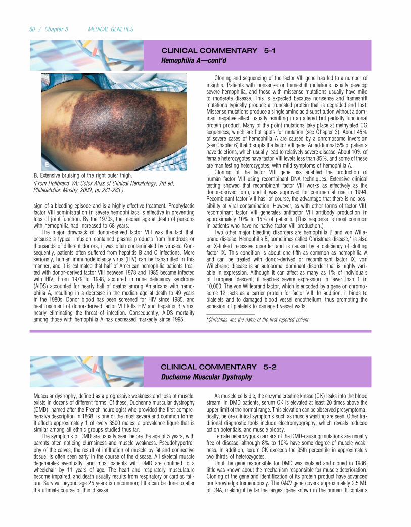
CLINICAL COMMENTARY 5-1
Hemophilia A—cont’d
B
B, Extensive bruising of the right outer thigh.
(From Hoffbrand VA: Color Atlas of Clinical Hematology, 3rd ed,Philadelphia: Mosby, 2000, pp 281-283.)
sign of a bleeding episode and is a highly effective treatment. Prophylacticfactor VIII administration in severe hemophiliacs is effective in preventingloss of joint function. By the 1970s, the median age at death of personswith hemophilia had increased to 68 years.
The major drawback of donor-derived factor VIII was the fact that,because a typical infusion contained plasma products from hundreds orthousands of different donors, it was often contaminated by viruses. Con-sequently, patients often suffered from hepatitis B and C infections. Moreseriously, human immunodeficiency virus (HIV) can be transmitted in thismanner, and it is estimated that half of American hemophilia patients trea-ted with donor-derived factor VIII between 1978 and 1985 became infectedwith HIV. From 1979 to 1998, acquired immune deficiency syndrome(AIDS) accounted for nearly half of deaths among Americans with hemo-philia A, resulting in a decrease in the median age at death to 49 yearsin the 1980s. Donor blood has been screened for HIV since 1985, andheat treatment of donor-derived factor VIII kills HIV and hepatitis B virus,nearly eliminating the threat of infection. Consequently, AIDS mortalityamong those with hemophilia A has decreased markedly since 1995.
Cloning and sequencing of the factor VIII gene has led to a number ofinsights. Patients with nonsense or frameshift mutations usually developsevere hemophilia, and those with missense mutations usually have mildto moderate disease. This is expected because nonsense and frameshiftmutations typically produce a truncated protein that is degraded and lost.Missense mutations produce a single amino acid substitution without a dom-inant negative effect, usually resulting in an altered but partially functionalprotein product. Many of the point mutations take place at methylated CGsequences, which are hot spots for mutation (see Chapter 3). About 45%of severe cases of hemophilia A are caused by a chromosome inversion(see Chapter 6) that disrupts the factor VIII gene. An additional 5% of patientshave deletions, which usually lead to relatively severe disease. About 10% offemale heterozygotes have factor VIII levels less than 35%, and some of theseare manifesting heterozygotes, with mild symptoms of hemophilia A.
Cloning of the factor VIII gene has enabled the production ofhuman factor VIII using recombinant DNA techniques. Extensive clinicaltesting showed that recombinant factor VIII works as effectively as thedonor-derived form, and it was approved for commercial use in 1994.Recombinant factor VIII has, of course, the advantage that there is no pos-sibility of viral contamination. However, as with other forms of factor VIII,recombinant factor VIII generates antifactor VIII antibody production inapproximately 10% to 15% of patients. (This response is most commonin patients who have no native factor VIII production.)
Two other major bleeding disorders are hemophilia B and von Wille-brand disease. Hemophilia B, sometimes called Christmas disease,* is alsoan X-linked recessive disorder and is caused by a deficiency of clottingfactor IX. This condition is about one fifth as common as hemophilia Aand can be treated with donor-derived or recombinant factor IX. vonWillebrand disease is an autosomal dominant disorder that is highly vari-able in expression. Although it can affect as many as 1% of individualsof European descent, it reaches severe expression in fewer than 1 in10,000. The von Willebrand factor, which is encoded by a gene on chromo-some 12, acts as a carrier protein for factor VIII. In addition, it binds toplatelets and to damaged blood vessel endothelium, thus promoting theadhesion of platelets to damaged vessel walls.
*Christmas was the name of the first reported patient.
CLINICAL COMMENTARY 5-2
Duchenne Muscular Dystrophy
Muscular dystrophy, defined as a progressive weakness and loss of muscle,exists in dozens of different forms. Of these, Duchenne muscular dystrophy(DMD), named after the French neurologist who provided the first compre-hensive description in 1868, is one of the most severe and common forms.It affects approximately 1 of every 3500 males, a prevalence figure that issimilar among all ethnic groups studied thus far.
The symptoms of DMD are usually seen before the age of 5 years, withparents often noticing clumsiness and muscle weakness. Pseudohypertro-phy of the calves, the result of infiltration of muscle by fat and connectivetissue, is often seen early in the course of the disease. All skeletal muscledegenerates eventually, and most patients with DMD are confined to awheelchair by 11 years of age. The heart and respiratory musculaturebecome impaired, and death usually results from respiratory or cardiac fail-ure. Survival beyond age 25 years is uncommon; little can be done to alterthe ultimate course of this disease.
As muscle cells die, the enzyme creatine kinase (CK) leaks into the bloodstream. In DMD patients, serum CK is elevated at least 20 times above theupper limit of the normal range. This elevation can be observed presymptoma-tically, before clinical symptoms such as muscle wasting are seen. Other tra-ditional diagnostic tools include electromyography, which reveals reducedaction potentials, and muscle biopsy.
Female heterozygous carriers of the DMD-causing mutations are usuallyfree of disease, although 8% to 10% have some degree of muscle weak-ness. In addition, serum CK exceeds the 95th percentile in approximatelytwo thirds of heterozygotes.
Until the gene responsible for DMD was isolated and cloned in 1986,little was known about the mechanism responsible for muscle deterioration.Cloning of the gene and identification of its protein product have advancedour knowledge tremendously. The DMD gene covers approximately 2.5 Mbof DNA, making it by far the largest gene known in the human. It contains
80 / Chapter 5 MEDICAL GENETICS
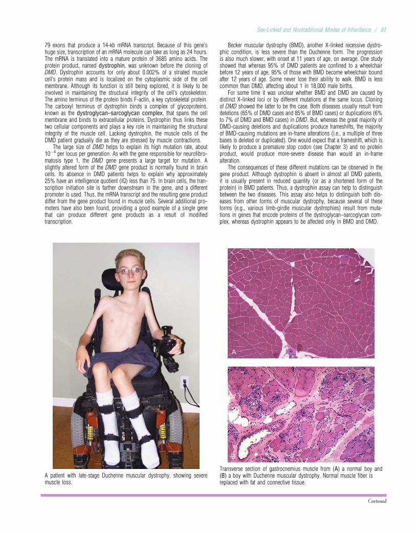
79 exons that produce a 14-kb mRNA transcript. Because of this gene’shuge size, transcription of an mRNA molecule can take as long as 24 hours.The mRNA is translated into a mature protein of 3685 amino acids. Theprotein product, named dystrophin, was unknown before the cloning ofDMD. Dystrophin accounts for only about 0.002% of a striated musclecell’s protein mass and is localized on the cytoplasmic side of the cellmembrane. Although its function is still being explored, it is likely to beinvolved in maintaining the structural integrity of the cell’s cytoskeleton.The amino terminus of the protein binds F-actin, a key cytoskeletal protein.The carboxyl terminus of dystrophin binds a complex of glycoproteins,known as the dystroglycan–sarcoglycan complex, that spans the cellmembrane and binds to extracellular proteins. Dystrophin thus links thesetwo cellular components and plays a key role in maintaining the structuralintegrity of the muscle cell. Lacking dystrophin, the muscle cells of theDMD patient gradually die as they are stressed by muscle contractions.
The large size of DMD helps to explain its high mutation rate, about10�4 per locus per generation. As with the gene responsible for neurofibro-matosis type 1, the DMD gene presents a large target for mutation. Aslightly altered form of the DMD gene product is normally found in braincells. Its absence in DMD patients helps to explain why approximately25% have an intelligence quotient (IQ) less than 75. In brain cells, the tran-scription initiation site is farther downstream in the gene, and a differentpromoter is used. Thus, the mRNA transcript and the resulting gene productdiffer from the gene product found in muscle cells. Several additional pro-moters have also been found, providing a good example of a single genethat can produce different gene products as a result of modifiedtranscription.
A patient with late-stage Duchenne muscular dystrophy, showing severemuscle loss.
Becker muscular dystrophy (BMD), another X-linked recessive dystro-phic condition, is less severe than the Duchenne form. The progressionis also much slower, with onset at 11 years of age, on average. One studyshowed that whereas 95% of DMD patients are confined to a wheelchairbefore 12 years of age, 95% of those with BMD become wheelchair boundafter 12 years of age. Some never lose their ability to walk. BMD is lesscommon than DMD, affecting about 1 in 18,000 male births.
For some time it was unclear whether BMD and DMD are caused bydistinct X-linked loci or by different mutations at the same locus. Cloningof DMD showed the latter to be the case. Both diseases usually result fromdeletions (65% of DMD cases and 85% of BMD cases) or duplications (6%to 7% of DMD and BMD cases) in DMD. But, whereas the great majority ofDMD-causing deletions and duplications produce frameshifts, the majorityof BMD-causing mutations are in-frame alterations (i.e., a multiple of threebases is deleted or duplicated). One would expect that a frameshift, which islikely to produce a premature stop codon (see Chapter 3) and no proteinproduct, would produce more-severe disease than would an in-framealteration.
The consequences of these different mutations can be observed in thegene product. Although dystrophin is absent in almost all DMD patients,it is usually present in reduced quantity (or as a shortened form of theprotein) in BMD patients. Thus, a dystrophin assay can help to distinguishbetween the two diseases. This assay also helps to distinguish both dis-eases from other forms of muscular dystrophy, because several of theseforms (e.g., various limb-girdle muscular dystrophies) result from muta-tions in genes that encode proteins of the dystroglycan–sarcoglycan com-plex, whereas dystrophin appears to be affected only in BMD and DMD.
A
B
Transverse section of gastrocnemius muscle from (A) a normal boy and(B) a boy with Duchenne muscular dystrophy. Normal muscle fiber isreplaced with fat and connective tissue.
Continued
Sex-Linked and Nontraditional Modes of Inheritance / 81
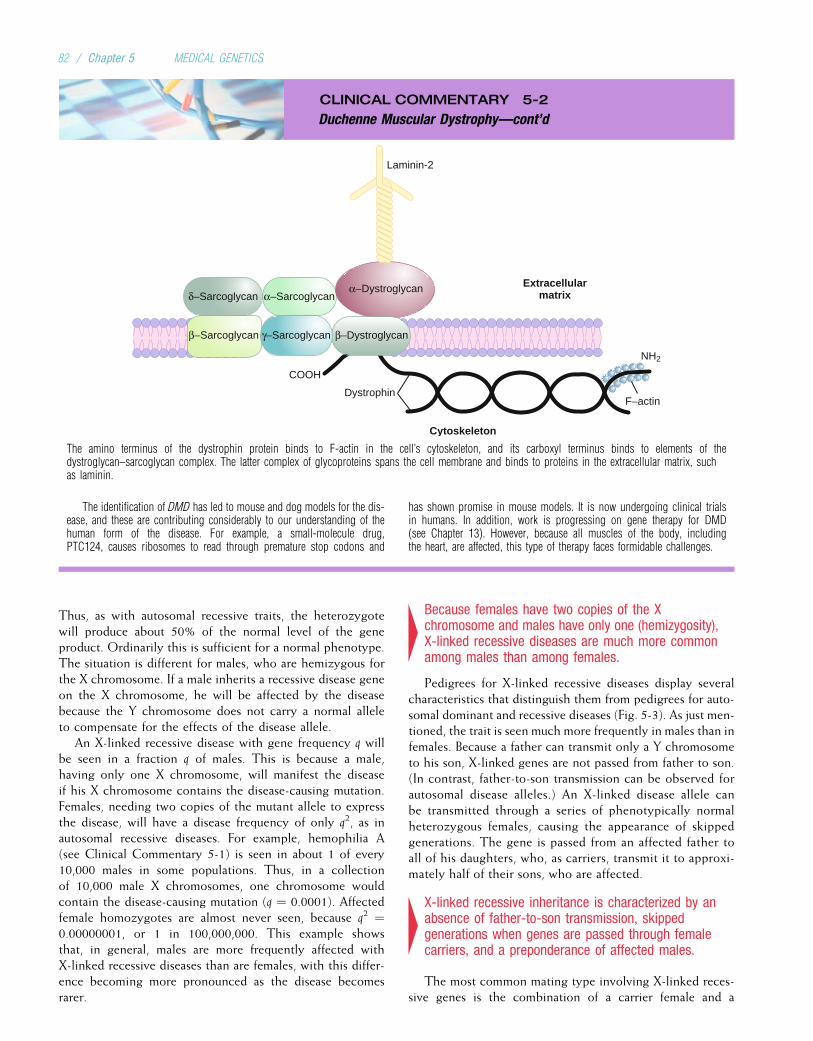
CLINICAL COMMENTARY 5-2
Duchenne Muscular Dystrophy—cont’d
Cytoskeleton
F–actin
NH2
Dystrophin
COOH
α–Dystroglycan
β–Sarcoglycan
δ–Sarcoglycan
γ–Sarcoglycan
α–Sarcoglycan
Laminin-2
Extracellularmatrix
β–Dystroglycan
The amino terminus of the dystrophin protein binds to F-actin in the cell’s cytoskeleton, and its carboxyl terminus binds to elements of thedystroglycan–sarcoglycan complex. The latter complex of glycoproteins spans the cell membrane and binds to proteins in the extracellular matrix, suchas laminin.
The identification of DMD has led to mouse and dog models for the dis-ease, and these are contributing considerably to our understanding of thehuman form of the disease. For example, a small-molecule drug,PTC124, causes ribosomes to read through premature stop codons and
has shown promise in mouse models. It is now undergoing clinical trialsin humans. In addition, work is progressing on gene therapy for DMD(see Chapter 13). However, because all muscles of the body, includingthe heart, are affected, this type of therapy faces formidable challenges.
Thus, as with autosomal recessive traits, the heterozygotewill produce about 50% of the normal level of the geneproduct. Ordinarily this is sufficient for a normal phenotype.The situation is different for males, who are hemizygous forthe X chromosome. If a male inherits a recessive disease geneon the X chromosome, he will be affected by the diseasebecause the Y chromosome does not carry a normal alleleto compensate for the effects of the disease allele.
An X-linked recessive disease with gene frequency q willbe seen in a fraction q of males. This is because a male,having only one X chromosome, will manifest the diseaseif his X chromosome contains the disease-causing mutation.Females, needing two copies of the mutant allele to expressthe disease, will have a disease frequency of only q2, as inautosomal recessive diseases. For example, hemophilia A(see Clinical Commentary 5-1) is seen in about 1 of every10,000 males in some populations. Thus, in a collectionof 10,000 male X chromosomes, one chromosome wouldcontain the disease-causing mutation (q ¼ 0.0001). Affectedfemale homozygotes are almost never seen, because q2 ¼0.00000001, or 1 in 100,000,000. This example showsthat, in general, males are more frequently affected withX-linked recessive diseases than are females, with this differ-ence becoming more pronounced as the disease becomesrarer.
4Because females have two copies of the Xchromosome and males have only one (hemizygosity),X-linked recessive diseases are much more commonamong males than among females.
Pedigrees for X-linked recessive diseases display severalcharacteristics that distinguish them from pedigrees for auto-somal dominant and recessive diseases (Fig. 5-3). As just men-tioned, the trait is seen much more frequently in males than infemales. Because a father can transmit only a Y chromosometo his son, X-linked genes are not passed from father to son.(In contrast, father-to-son transmission can be observed forautosomal disease alleles.) An X-linked disease allele canbe transmitted through a series of phenotypically normalheterozygous females, causing the appearance of skippedgenerations. The gene is passed from an affected father toall of his daughters, who, as carriers, transmit it to approxi-mately half of their sons, who are affected.
4X-linked recessive inheritance is characterized by anabsence of father-to-son transmission, skippedgenerations when genes are passed through femalecarriers, and a preponderance of affected males.
The most common mating type involving X-linked reces-sive genes is the combination of a carrier female and a
82 / Chapter 5 MEDICAL GENETICS
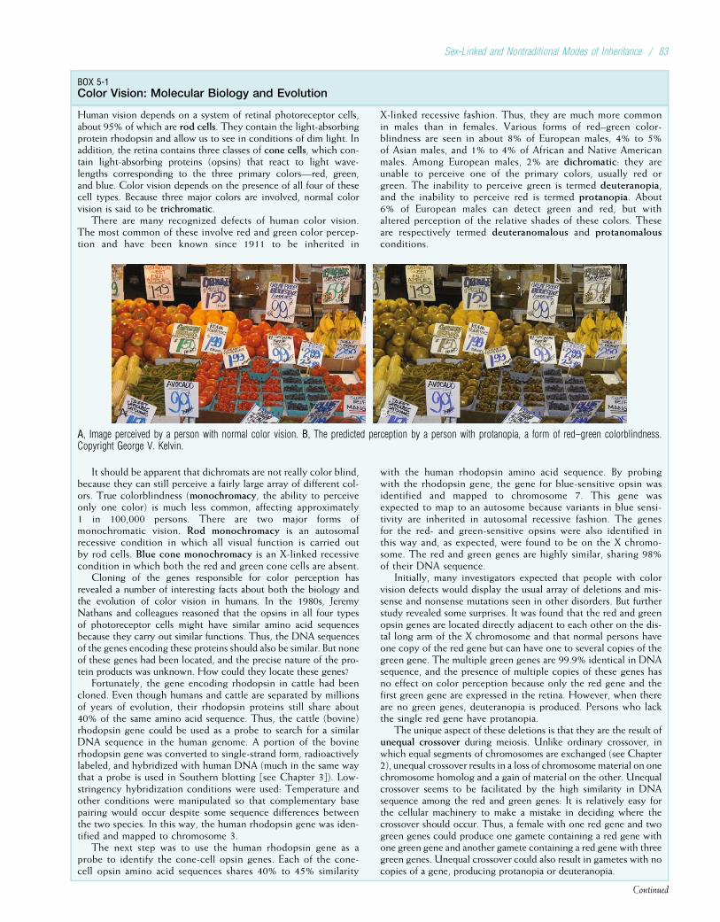
BOX 5-1Color Vision: Molecular Biology and Evolution
Human vision depends on a system of retinal photoreceptor cells,about 95% of which are rod cells. They contain the light-absorbingprotein rhodopsin and allow us to see in conditions of dim light. Inaddition, the retina contains three classes of cone cells, which con-tain light-absorbing proteins (opsins) that react to light wave-lengths corresponding to the three primary colors—red, green,and blue. Color vision depends on the presence of all four of thesecell types. Because three major colors are involved, normal colorvision is said to be trichromatic.
There are many recognized defects of human color vision.The most common of these involve red and green color percep-tion and have been known since 1911 to be inherited in
X-linked recessive fashion. Thus, they are much more commonin males than in females. Various forms of red–green color-blindness are seen in about 8% of European males, 4% to 5%of Asian males, and 1% to 4% of African and Native Americanmales. Among European males, 2% are dichromatic: they areunable to perceive one of the primary colors, usually red orgreen. The inability to perceive green is termed deuteranopia,and the inability to perceive red is termed protanopia. About6% of European males can detect green and red, but withaltered perception of the relative shades of these colors. Theseare respectively termed deuteranomalous and protanomalousconditions.
A B
A, Image perceived by a person with normal color vision. B, The predicted perception by a person with protanopia, a form of red–green colorblindness.Copyright George V. Kelvin.
It should be apparent that dichromats are not really color blind,because they can still perceive a fairly large array of different col-ors. True colorblindness (monochromacy, the ability to perceiveonly one color) is much less common, affecting approximately1 in 100,000 persons. There are two major forms ofmonochromatic vision. Rod monochromacy is an autosomalrecessive condition in which all visual function is carried outby rod cells. Blue cone monochromacy is an X-linked recessivecondition in which both the red and green cone cells are absent.
Cloning of the genes responsible for color perception hasrevealed a number of interesting facts about both the biology andthe evolution of color vision in humans. In the 1980s, JeremyNathans and colleagues reasoned that the opsins in all four typesof photoreceptor cells might have similar amino acid sequencesbecause they carry out similar functions. Thus, the DNA sequencesof the genes encoding these proteins should also be similar. But noneof these genes had been located, and the precise nature of the pro-tein products was unknown. How could they locate these genes?
Fortunately, the gene encoding rhodopsin in cattle had beencloned. Even though humans and cattle are separated by millionsof years of evolution, their rhodopsin proteins still share about40% of the same amino acid sequence. Thus, the cattle (bovine)rhodopsin gene could be used as a probe to search for a similarDNA sequence in the human genome. A portion of the bovinerhodopsin gene was converted to single-strand form, radioactivelylabeled, and hybridized with human DNA (much in the same waythat a probe is used in Southern blotting [see Chapter 3]). Low-stringency hybridization conditions were used: Temperature andother conditions were manipulated so that complementary basepairing would occur despite some sequence differences betweenthe two species. In this way, the human rhodopsin gene was iden-tified and mapped to chromosome 3.
The next step was to use the human rhodopsin gene as aprobe to identify the cone-cell opsin genes. Each of the cone-cell opsin amino acid sequences shares 40% to 45% similarity
with the human rhodopsin amino acid sequence. By probingwith the rhodopsin gene, the gene for blue-sensitive opsin wasidentified and mapped to chromosome 7. This gene wasexpected to map to an autosome because variants in blue sensi-tivity are inherited in autosomal recessive fashion. The genesfor the red- and green-sensitive opsins were also identified inthis way and, as expected, were found to be on the X chromo-some. The red and green genes are highly similar, sharing 98%of their DNA sequence.
Initially, many investigators expected that people with colorvision defects would display the usual array of deletions and mis-sense and nonsense mutations seen in other disorders. But furtherstudy revealed some surprises. It was found that the red and greenopsin genes are located directly adjacent to each other on the dis-tal long arm of the X chromosome and that normal persons haveone copy of the red gene but can have one to several copies of thegreen gene. The multiple green genes are 99.9% identical in DNAsequence, and the presence of multiple copies of these genes hasno effect on color perception because only the red gene and thefirst green gene are expressed in the retina. However, when thereare no green genes, deuteranopia is produced. Persons who lackthe single red gene have protanopia.
The unique aspect of these deletions is that they are the result ofunequal crossover during meiosis. Unlike ordinary crossover, inwhich equal segments of chromosomes are exchanged (see Chapter2), unequal crossover results in a loss of chromosomematerial on onechromosome homolog and a gain of material on the other. Unequalcrossover seems to be facilitated by the high similarity in DNAsequence among the red and green genes: It is relatively easy forthe cellular machinery to make a mistake in deciding where thecrossover should occur. Thus, a female with one red gene and twogreen genes could produce one gamete containing a red gene withone green gene and another gamete containing a red gene with threegreen genes. Unequal crossover could also result in gametes with nocopies of a gene, producing protanopia or deuteranopia.
Continued
Sex-Linked and Nontraditional Modes of Inheritance / 83
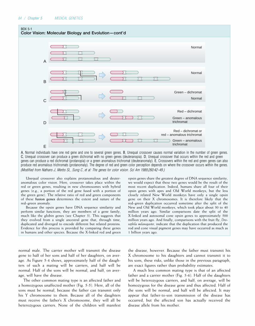
normal male. The carrier mother will transmit the diseasegene to half of her sons and half of her daughters, on aver-age. As Figure 5-4 shows, approximately half of the daugh-ters of such a mating will be carriers, and half will benormal. Half of the sons will be normal, and half, on aver-age, will have the disease.
The other common mating type is an affected father anda homozygous unaffected mother (Fig. 5-5). Here, all of thesons must be normal, because the father can transmit onlyhis Y chromosome to them. Because all of the daughtersmust receive the father’s X chromosome, they will all beheterozygous carriers. None of the children will manifest
the disease, however. Because the father must transmit hisX chromosome to his daughters and cannot transmit it tohis sons, these risks, unlike those in the previous paragraph,are exact figures rather than probability estimates.
A much less common mating type is that of an affectedfather and a carrier mother (Fig. 5-6). Half of the daughterswill be heterozygous carriers, and half, on average, will behomozygous for the disease gene and thus affected. Half ofthe sons will be normal, and half will be affected. It mayappear that father-to-son transmission of the disease hasoccurred, but the affected son has actually received thedisease allele from his mother.
BOX 5-1Color Vision: Molecular Biology and Evolution—cont’d
B
C
D
E
A
Normal
Normal
Normal
Green – dichromat
Red – dichromat
Red – dichromat orred – anomalous trichromat
Green – anomaloustrichromat
Green – anomaloustrichromat
A, Normal individuals have one red gene and one to several green genes. B, Unequal crossover causes normal variation in the number of green genes.C, Unequal crossover can produce a green dichromat with no green genes (deuteranopia). D, Unequal crossover that occurs within the red and greengenes can produce a red dichromat (protanopia) or a green anomalous trichromat (deuteranomaly). E, Crossovers within the red and green genes can alsoproduce red anomalous trichromats (protanomaly). The degree of red and green color perception depends on where the crossover occurs within the genes.
(Modified from Nathans J, Merbs SL, Sung C, et al: The genes for color vision. Sci Am 1989;260:42–49.)
Unequal crossover also explains protanomalous and deuter-anomalous color vision. Here, crossover takes place within thered or green genes, resulting in new chromosomes with hybridgenes (e.g., a portion of the red gene fused with a portion ofthe green gene). The relative ratio of red and green componentsof these fusion genes determines the extent and nature of thered–green anomaly.
Because the opsin genes have DNA sequence similarity andperform similar functions, they are members of a gene family,much like the globin genes (see Chapter 3). This suggests thatthey evolved from a single ancestral gene that, through time,duplicated and diverged to encode different but related proteins.Evidence for this process is provided by comparing these genesin humans and other species. Because the X-linked red and green
opsin genes share the greatest degree of DNA sequence similarity,we would expect that these two genes would be the result of themost recent duplication. Indeed, humans share all four of theiropsin genes with apes and Old World monkeys, but the lessclosely related New World monkeys have only a single opsingene on their X chromosomes. It is therefore likely that thered–green duplication occurred sometime after the split of theNew and Old World monkeys, which took place about 30 to 40million years ago. Similar comparisons date the split of theX-linked and autosomal cone opsin genes to approximately 500million years ago. And finally, comparisons with the fruit fly, Dro-sophila melanogaster, indicate that the duplication that produced therod and cone visual pigment genes may have occurred as much as1 billion years ago.
84 / Chapter 5 MEDICAL GENETICS
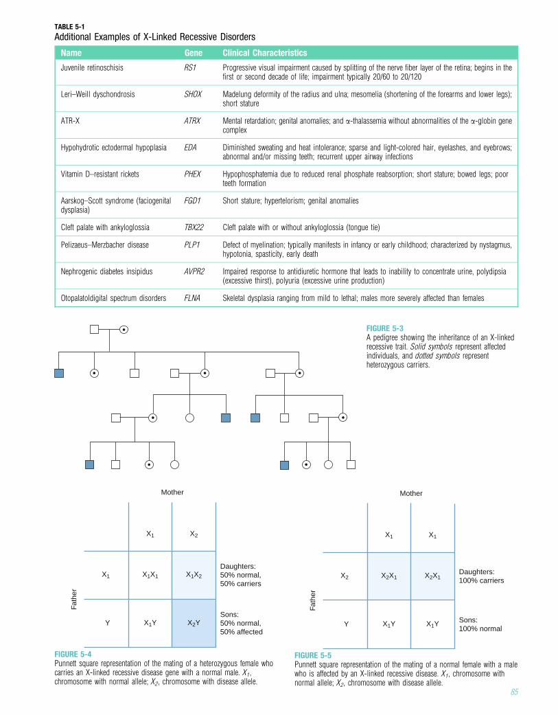
TABLE 5-1
Additional Examples of X-Linked Recessive Disorders
Name Gene Clinical Characteristics
Juvenile retinoschisis RS1 Progressive visual impairment caused by splitting of the nerve fiber layer of the retina; begins in thefirst or second decade of life; impairment typically 20/60 to 20/120
Leri–Weill dyschondrosis SHOX Madelung deformity of the radius and ulna; mesomelia (shortening of the forearms and lower legs);short stature
ATR-X ATRX Mental retardation; genital anomalies; and a-thalassemia without abnormalities of the a-globin genecomplex
Hypohydrotic ectodermal hypoplasia EDA Diminished sweating and heat intolerance; sparse and light-colored hair, eyelashes, and eyebrows;abnormal and/or missing teeth; recurrent upper airway infections
Vitamin D–resistant rickets PHEX Hypophosphatemia due to reduced renal phosphate reabsorption; short stature; bowed legs; poorteeth formation
Aarskog–Scott syndrome (faciogenitaldysplasia)
FGD1 Short stature; hypertelorism; genital anomalies
Cleft palate with ankyloglossia TBX22 Cleft palate with or without ankyloglossia (tongue tie)
Pelizaeus–Merzbacher disease PLP1 Defect of myelination; typically manifests in infancy or early childhood; characterized by nystagmus,hypotonia, spasticity, early death
Nephrogenic diabetes insipidus AVPR2 Impaired response to antidiuretic hormone that leads to inability to concentrate urine, polydipsia(excessive thirst), polyuria (excessive urine production)
Otopalatoldigital spectrum disorders FLNA Skeletal dysplasia ranging from mild to lethal; males more severely affected than females
FIGURE 5-3A pedigree showing the inheritance of an X-linkedrecessive trait. Solid symbols represent affectedindividuals, and dotted symbols representheterozygous carriers.
Fath
er
Sons:100% normal
Daughters:100% carriers
Mother
X1
X2X1X2 X2X1
X1
X1YY X1Y
FIGURE 5-5Punnett square representation of the mating of a normal female with a malewho is affected by an X-linked recessive disease. X1, chromosome withnormal allele; X2, chromosome with disease allele.
Fath
er
Sons:50% normal,50% affected
Daughters:50% normal,50% carriers
Mother
X1
X1X1X1 X1X2
X2
X1YY X2Y
FIGURE 5-4Punnett square representation of the mating of a heterozygous female whocarries an X-linked recessive disease gene with a normal male. X1,chromosome with normal allele; X2, chromosome with disease allele.
85
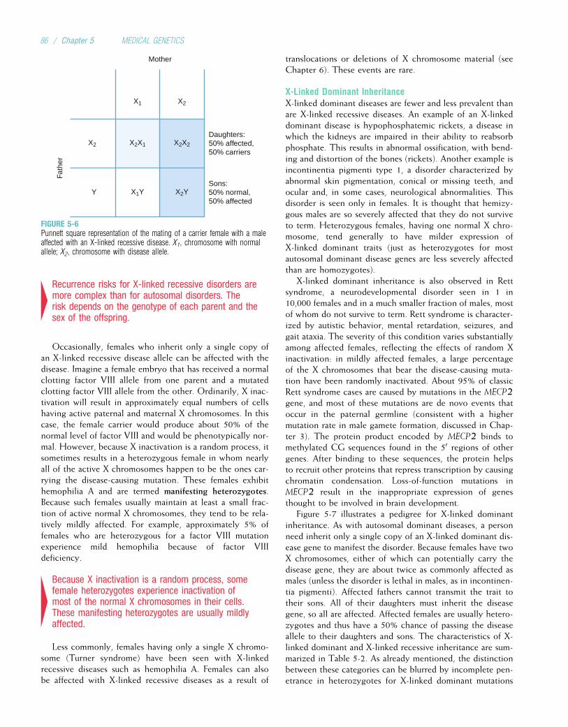
4Recurrence risks for X-linked recessive disorders aremore complex than for autosomal disorders. Therisk depends on the genotype of each parent and thesex of the offspring.
Occasionally, females who inherit only a single copy ofan X-linked recessive disease allele can be affected with thedisease. Imagine a female embryo that has received a normalclotting factor VIII allele from one parent and a mutatedclotting factor VIII allele from the other. Ordinarily, X inac-tivation will result in approximately equal numbers of cellshaving active paternal and maternal X chromosomes. In thiscase, the female carrier would produce about 50% of thenormal level of factor VIII and would be phenotypically nor-mal. However, because X inactivation is a random process, itsometimes results in a heterozygous female in whom nearlyall of the active X chromosomes happen to be the ones car-rying the disease-causing mutation. These females exhibithemophilia A and are termed manifesting heterozygotes.Because such females usually maintain at least a small frac-tion of active normal X chromosomes, they tend to be rela-tively mildly affected. For example, approximately 5% offemales who are heterozygous for a factor VIII mutationexperience mild hemophilia because of factor VIIIdeficiency.
4Because X inactivation is a random process, somefemale heterozygotes experience inactivation ofmost of the normal X chromosomes in their cells.These manifesting heterozygotes are usually mildlyaffected.
Less commonly, females having only a single X chromo-some (Turner syndrome) have been seen with X-linkedrecessive diseases such as hemophilia A. Females can alsobe affected with X-linked recessive diseases as a result of
translocations or deletions of X chromosome material (seeChapter 6). These events are rare.
X-Linked Dominant InheritanceX-linked dominant diseases are fewer and less prevalent thanare X-linked recessive diseases. An example of an X-linkeddominant disease is hypophosphatemic rickets, a disease inwhich the kidneys are impaired in their ability to reabsorbphosphate. This results in abnormal ossification, with bend-ing and distortion of the bones (rickets). Another example isincontinentia pigmenti type 1, a disorder characterized byabnormal skin pigmentation, conical or missing teeth, andocular and, in some cases, neurological abnormalities. Thisdisorder is seen only in females. It is thought that hemizy-gous males are so severely affected that they do not surviveto term. Heterozygous females, having one normal X chro-mosome, tend generally to have milder expression ofX-linked dominant traits (just as heterozygotes for mostautosomal dominant disease genes are less severely affectedthan are homozygotes).
X-linked dominant inheritance is also observed in Rettsyndrome, a neurodevelopmental disorder seen in 1 in10,000 females and in a much smaller fraction of males, mostof whom do not survive to term. Rett syndrome is character-ized by autistic behavior, mental retardation, seizures, andgait ataxia. The severity of this condition varies substantiallyamong affected females, reflecting the effects of random Xinactivation: in mildly affected females, a large percentageof the X chromosomes that bear the disease-causing muta-tion have been randomly inactivated. About 95% of classicRett syndrome cases are caused by mutations in the MECP2gene, and most of these mutations are de novo events thatoccur in the paternal germline (consistent with a highermutation rate in male gamete formation, discussed in Chap-ter 3). The protein product encoded by MECP2 binds tomethylated CG sequences found in the 50 regions of othergenes. After binding to these sequences, the protein helpsto recruit other proteins that repress transcription by causingchromatin condensation. Loss-of-function mutations inMECP2 result in the inappropriate expression of genesthought to be involved in brain development.
Figure 5-7 illustrates a pedigree for X-linked dominantinheritance. As with autosomal dominant diseases, a personneed inherit only a single copy of an X-linked dominant dis-ease gene to manifest the disorder. Because females have twoX chromosomes, either of which can potentially carry thedisease gene, they are about twice as commonly affected asmales (unless the disorder is lethal in males, as in incontinen-tia pigmenti). Affected fathers cannot transmit the trait totheir sons. All of their daughters must inherit the diseasegene, so all are affected. Affected females are usually hetero-zygotes and thus have a 50% chance of passing the diseaseallele to their daughters and sons. The characteristics of X-linked dominant and X-linked recessive inheritance are sum-marized in Table 5-2. As already mentioned, the distinctionbetween these categories can be blurred by incomplete pen-etrance in heterozygotes for X-linked dominant mutations
Fath
er
Sons:50% normal,50% affected
Daughters:50% affected,50% carriers
Mother
X1
X2X1X2 X2X2
X2
X1YY X2Y
FIGURE 5-6Punnett square representation of the mating of a carrier female with a maleaffected with an X-linked recessive disease. X1, chromosome with normalallele; X2, chromosome with disease allele.
86 / Chapter 5 MEDICAL GENETICS
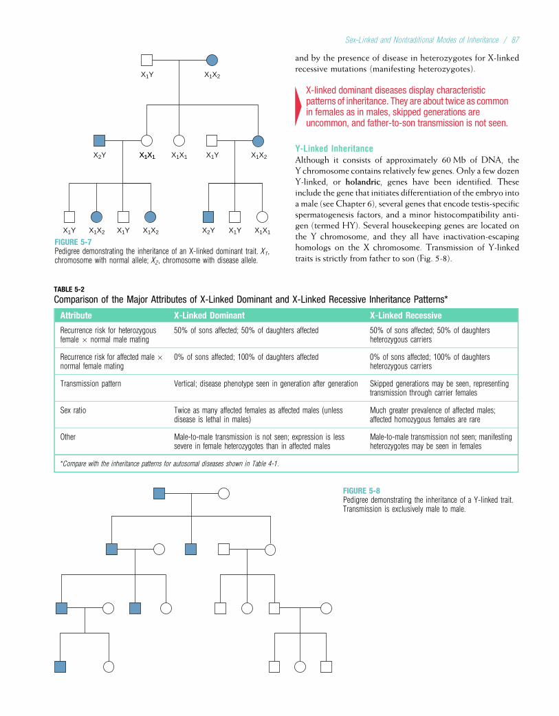
and by the presence of disease in heterozygotes for X-linkedrecessive mutations (manifesting heterozygotes).
4X-linked dominant diseases display characteristicpatterns of inheritance. They are about twice as commonin females as in males, skipped generations areuncommon, and father-to-son transmission is not seen.
Y-Linked InheritanceAlthough it consists of approximately 60 Mb of DNA, theY chromosome contains relatively few genes. Only a few dozenY-linked, or holandric, genes have been identified. Theseinclude the gene that initiates differentiation of the embryo intoa male (see Chapter 6), several genes that encode testis-specificspermatogenesis factors, and a minor histocompatibility anti-gen (termed HY). Several housekeeping genes are located onthe Y chromosome, and they all have inactivation-escapinghomologs on the X chromosome. Transmission of Y-linkedtraits is strictly from father to son (Fig. 5-8).
X1X1
X1Y X1X2
X1X1 X1X1X2Y X1Y X1X2
X1Y X1X2 X1Y X1X2 X1X1X2Y X1Y
FIGURE 5-7Pedigree demonstrating the inheritance of an X-linked dominant trait. X1,chromosome with normal allele; X2, chromosome with disease allele.
TABLE 5-2
Comparison of the Major Attributes of X-Linked Dominant and X-Linked Recessive Inheritance Patterns*
Attribute X-Linked Dominant X-Linked Recessive
Recurrence risk for heterozygousfemale � normal male mating
50% of sons affected; 50% of daughters affected 50% of sons affected; 50% of daughtersheterozygous carriers
Recurrence risk for affected male �normal female mating
0% of sons affected; 100% of daughters affected 0% of sons affected; 100% of daughtersheterozygous carriers
Transmission pattern Vertical; disease phenotype seen in generation after generation Skipped generations may be seen, representingtransmission through carrier females
Sex ratio Twice as many affected females as affected males (unlessdisease is lethal in males)
Much greater prevalence of affected males;affected homozygous females are rare
Other Male-to-male transmission is not seen; expression is lesssevere in female heterozygotes than in affected males
Male-to-male transmission not seen; manifestingheterozygotes may be seen in females
*Compare with the inheritance patterns for autosomal diseases shown in Table 4-1.
FIGURE 5-8Pedigree demonstrating the inheritance of a Y-linked trait.Transmission is exclusively male to male.
Sex-Linked and Nontraditional Modes of Inheritance / 87
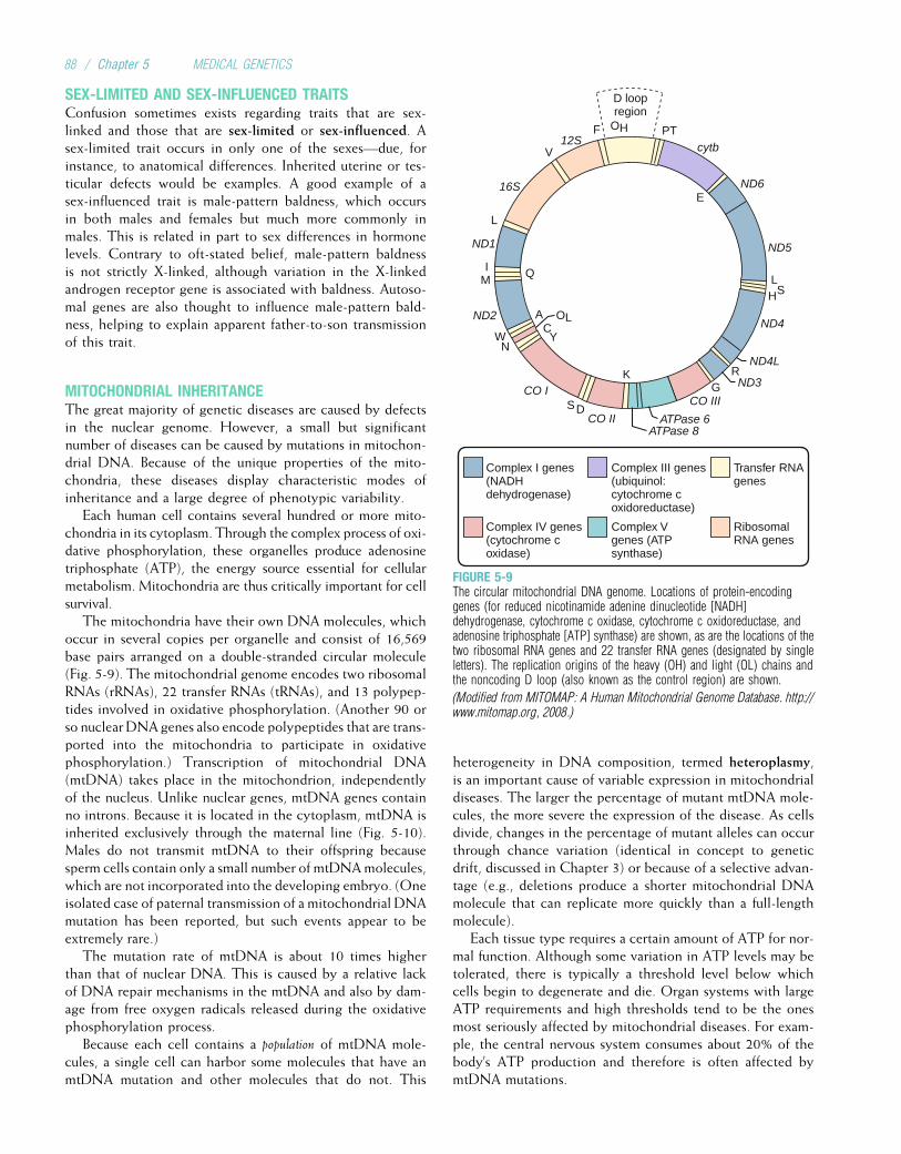
SEX-LIMITED AND SEX-INFLUENCED TRAITSConfusion sometimes exists regarding traits that are sex-linked and those that are sex-limited or sex-influenced. Asex-limited trait occurs in only one of the sexes—due, forinstance, to anatomical differences. Inherited uterine or tes-ticular defects would be examples. A good example of asex-influenced trait is male-pattern baldness, which occursin both males and females but much more commonly inmales. This is related in part to sex differences in hormonelevels. Contrary to oft-stated belief, male-pattern baldnessis not strictly X-linked, although variation in the X-linkedandrogen receptor gene is associated with baldness. Autoso-mal genes are also thought to influence male-pattern bald-ness, helping to explain apparent father-to-son transmissionof this trait.
MITOCHONDRIAL INHERITANCEThe great majority of genetic diseases are caused by defectsin the nuclear genome. However, a small but significantnumber of diseases can be caused by mutations in mitochon-drial DNA. Because of the unique properties of the mito-chondria, these diseases display characteristic modes ofinheritance and a large degree of phenotypic variability.
Each human cell contains several hundred or more mito-chondria in its cytoplasm. Through the complex process of oxi-dative phosphorylation, these organelles produce adenosinetriphosphate (ATP), the energy source essential for cellularmetabolism. Mitochondria are thus critically important for cellsurvival.
The mitochondria have their own DNA molecules, whichoccur in several copies per organelle and consist of 16,569base pairs arranged on a double-stranded circular molecule(Fig. 5-9). The mitochondrial genome encodes two ribosomalRNAs (rRNAs), 22 transfer RNAs (tRNAs), and 13 polypep-tides involved in oxidative phosphorylation. (Another 90 orso nuclear DNA genes also encode polypeptides that are trans-ported into the mitochondria to participate in oxidativephosphorylation.) Transcription of mitochondrial DNA(mtDNA) takes place in the mitochondrion, independentlyof the nucleus. Unlike nuclear genes, mtDNA genes containno introns. Because it is located in the cytoplasm, mtDNA isinherited exclusively through the maternal line (Fig. 5-10).Males do not transmit mtDNA to their offspring becausesperm cells contain only a small number of mtDNAmolecules,which are not incorporated into the developing embryo. (Oneisolated case of paternal transmission of a mitochondrial DNAmutation has been reported, but such events appear to beextremely rare.)
The mutation rate of mtDNA is about 10 times higherthan that of nuclear DNA. This is caused by a relative lackof DNA repair mechanisms in the mtDNA and also by dam-age from free oxygen radicals released during the oxidativephosphorylation process.
Because each cell contains a population of mtDNA mole-cules, a single cell can harbor some molecules that have anmtDNA mutation and other molecules that do not. This
heterogeneity in DNA composition, termed heteroplasmy,is an important cause of variable expression in mitochondrialdiseases. The larger the percentage of mutant mtDNA mole-cules, the more severe the expression of the disease. As cellsdivide, changes in the percentage of mutant alleles can occurthrough chance variation (identical in concept to geneticdrift, discussed in Chapter 3) or because of a selective advan-tage (e.g., deletions produce a shorter mitochondrial DNAmolecule that can replicate more quickly than a full-lengthmolecule).
Each tissue type requires a certain amount of ATP for nor-mal function. Although some variation in ATP levels may betolerated, there is typically a threshold level below whichcells begin to degenerate and die. Organ systems with largeATP requirements and high thresholds tend to be the onesmost seriously affected by mitochondrial diseases. For exam-ple, the central nervous system consumes about 20% of thebody’s ATP production and therefore is often affected bymtDNA mutations.
PTF
V
L
IM
Q
K
AC
WN
SD
Y
12S
16S
ND1
ND2
CO I
CO II
CO III
OH
OL
D loopregion
cytb
ND6
ND5
E
Complex I genes(NADHdehydrogenase)
Complex IV genes(cytochrome coxidase)
Transfer RNAgenes
ATPase 6ATPase 8
RibosomalRNA genes
Complex III genes(ubiquinol:cytochrome coxidoreductase)
Complex Vgenes (ATPsynthase)
LH
RG
S
ND4
ND4L
ND3
FIGURE 5-9The circular mitochondrial DNA genome. Locations of protein-encodinggenes (for reduced nicotinamide adenine dinucleotide [NADH]dehydrogenase, cytochrome c oxidase, cytochrome c oxidoreductase, andadenosine triphosphate [ATP] synthase) are shown, as are the locations of thetwo ribosomal RNA genes and 22 transfer RNA genes (designated by singleletters). The replication origins of the heavy (OH) and light (OL) chains andthe noncoding D loop (also known as the control region) are shown.
(Modified from MITOMAP: A Human Mitochondrial Genome Database. http://www.mitomap.org, 2008.)
88 / Chapter 5 MEDICAL GENETICS
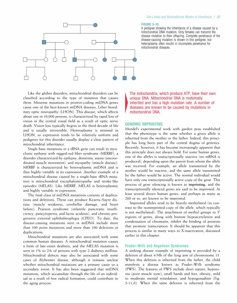
Like the globin disorders, mitochondrial disorders can beclassified according to the type of mutation that causesthem. Missense mutations in protein-coding mtDNA genescause one of the best-known mtDNA diseases, Leber hered-itary optic neuropathy (LHON). This disease, which affectsabout one in 10,000 persons, is characterized by rapid loss ofvision in the central visual field as a result of optic nervedeath. Vision loss typically begins in the third decade of lifeand is usually irreversible. Heteroplasmy is minimal inLHON, so expression tends to be relatively uniform andpedigrees for this disorder usually display a clear pattern ofmitochondrial inheritance.
Single-base mutations in a tRNA gene can result in myo-clonic epilepsy with ragged-red fiber syndrome (MERRF), adisorder characterized by epilepsy, dementia, ataxia (uncoor-dinated muscle movement), and myopathy (muscle disease).MERRF is characterized by heteroplasmic mtDNA and isthus highly variable in its expression. Another example of amitochondrial disease caused by a single-base tRNA muta-tion is mitochondrial encephalomyopathy and stroke-likeepisodes (MELAS). Like MERRF, MELAS is heteroplasmicand highly variable in expression.
The final class of mtDNA mutations consists of duplica-tions and deletions. These can produce Kearns–Sayre dis-ease (muscle weakness, cerebellar damage, and heartfailure), Pearson syndrome (infantile pancreatic insuffi-ciency, pancytopenia, and lactic acidosis), and chronic pro-gressive external ophthalmoplegia (CPEO). To date, thedisease-causing mutations seen in mtDNA include morethan 100 point mutations and more than 100 deletions orduplications.
Mitochondrial mutations are also associated with somecommon human diseases. A mitochondrial mutation causesa form of late-onset deafness, and the MELAS mutation isseen in 1% to 2% of persons with type 2 diabetes mellitus.Mitochondrial defects may also be associated with somecases of Alzheimer disease, although it remains unclearwhether mitochondrial mutations are a primary cause or asecondary event. It has also been suggested that mtDNAmutations, which accumulate through the life of an individ-ual as a result of free radical formation, could contribute tothe aging process.
4The mitochondria, which produce ATP, have their ownunique DNA. Mitochondrial DNA is maternallyinherited and has a high mutation rate. A number ofdiseases are known to be caused by mutations inmitochondrial DNA.
GENOMIC IMPRINTINGMendel’s experimental work with garden peas establishedthat the phenotype is the same whether a given allele isinherited from the mother or the father. Indeed, this princi-ple has long been part of the central dogma of genetics.Recently, however, it has become increasingly apparent thatthis principle does not always hold. For some human genes,one of the alleles is transcriptionally inactive (no mRNA isproduced), depending upon the parent from whom the allelewas received. For example, an allele transmitted by themother would be inactive, and the same allele transmittedby the father would be active. The normal individual wouldhave only one transcriptionally active copy of the gene. Thisprocess of gene silencing is known as imprinting, and thetranscriptionally silenced genes are said to be imprinted. Atleast several dozen human genes, and perhaps as many as200 or so, are known to be imprinted.
Imprinted alleles tend to be heavily methylated (in con-trast to the nonimprinted copy of the allele, which typicallyis not methylated). The attachment of methyl groups to 50
regions of genes, along with histone hypoacetylation andcondensation of chromatin, inhibit the binding of proteinsthat promote transcription. It should be apparent that thisprocess is similar in many ways to X-inactivation, discussedearlier in this chapter.
Prader–Willi and Angelman SyndromesA striking disease example of imprinting is provided by adeletion of about 4 Mb of the long arm of chromosome 15.When this deletion is inherited from the father, the childmanifests a disease known as Prader–Willi syndrome(PWS). The features of PWS include short stature, hypoto-nia (poor muscle tone), small hands and feet, obesity, mildto moderate mental retardation, and hypogonadism (Fig.5-11,A). When the same deletion is inherited from the
FIGURE 5-10A pedigree showing the inheritance of a disease caused by amitochondrial DNA mutation. Only females can transmit thedisease mutation to their offspring. Complete penetrance of thedisease-causing mutation is shown in this pedigree, butheteroplasmy often results in incomplete penetrance formitochondrial diseases.
Sex-Linked and Nontraditional Modes of Inheritance / 89
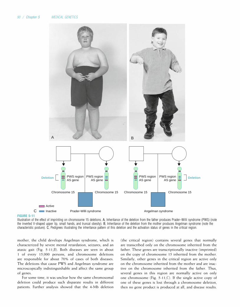
mother, the child develops Angelman syndrome, which ischaracterized by severe mental retardation, seizures, and anataxic gait (Fig. 5-11,B). Both diseases are seen in about1 of every 15,000 persons, and chromosome deletionsare responsible for about 70% of cases of both diseases.The deletions that cause PWS and Angelman syndrome aremicroscopically indistinguishable and affect the same groupof genes.
For some time, it was unclear how the same chromosomaldeletion could produce such disparate results in differentpatients. Further analysis showed that the 4-Mb deletion
(the critical region) contains several genes that normallyare transcribed only on the chromosome inherited from thefather. These genes are transcriptionally inactive (imprinted)on the copy of chromosome 15 inherited from the mother.Similarly, other genes in the critical region are active onlyon the chromosome inherited from the mother and are inac-tive on the chromosome inherited from the father. Thus,several genes in this region are normally active on onlyone chromosome (Fig. 5-11,C). If the single active copy ofone of these genes is lost through a chromosome deletion,then no gene product is produced at all, and disease results.
A B
CActive
Inactive
Chromosome 15 Chromosome 15 Chromosome 15 Chromosome 15
Prader-Willi syndrome Angelman syndrome
Deletion DeletionPWS regionAS gene
PWS regionAS gene
PWS regionAS gene
PWS regionAS gene
FIGURE 5-11Illustration of the effect of imprinting on chromosome 15 deletions. A, Inheritance of the deletion from the father produces Prader–Willi syndrome (PWS) (notethe inverted V-shaped upper lip, small hands, and truncal obesity). B, Inheritance of the deletion from the mother produces Angelman syndrome (note thecharacteristic posture). C, Pedigrees illustrating the inheritance pattern of this deletion and the activation status of genes in the critical region.
90 / Chapter 5 MEDICAL GENETICS
Molecular analysis using many of the tools and tech-niques outlined in Chapter 3 (microsatellite polymorphisms,cloning, and DNA sequencing) has identified several specificgenes in the critical region of chromosome 15. The generesponsible for Angelman syndrome encodes a proteininvolved in ubiquitin-mediated protein degradation duringbrain development (consistent with the mental retardationand ataxia observed in this disorder). In brain tissue, thisgene is active only on the chromosome inherited from themother; thus, a maternally transmitted deletion removesthe single active copy of this gene. Several genes in the crit-ical region are involved in PWS, and they are transcribedonly on the chromosome transmitted by the father.
Several mechanisms in addition to chromosome deletionscan cause PWS and Angelman syndrome. One of these is uni-parental disomy, a condition in which the person inherits twocopies of a chromosome from one parent and none from theother (see Chapter 6 for further discussion). When two copiesof the maternal chromosome 15 are inherited, PWS resultsbecause no active paternal genes are present in the criticalregion. Conversely, disomy of the paternal chromosome 15produces Angelman syndrome. Point mutations in the identi-fied Angelman syndrome gene can also produce disease.Finally, about 1% of cases of PWS result from a small deletionof the region that contains an imprinting control center onchromosome 15. This is the DNA sequence that apparentlyhelps to set and reset the imprint itself. Box 5-2 presents clin-ical issues of PWS from the perspective of a patient’s family.
Beckwith–Wiedemann SyndromeA second example of imprinting in the human genome isgiven by Beckwith–Wiedemann syndrome, an overgrowth
condition accompanied by an increased predisposition tocancer. Beckwith–Wiedemann syndrome is usually recogniz-able at birth because of large size for gestational age, neona-tal hypoglycemia, a large tongue, creases on the ear lobe,and omphalocele (an abdominal wall defect). Some childrenwith Beckwith–Wiedemann syndrome also develop asym-metrical overgrowth of a limb or one side of the face ortrunk (i.e., hemihyperplasia). Children with Beckwith–Wie-demann syndrome have an increased risk for developingWilms tumor (a kidney cancer) and hepatoblastoma (a livercancer). Both of these tumors can be treated effectively ifthey are detected early, so screening at regular intervals isan important part of management (see Chapter 15).
As with Angelman syndrome, a minority of Beckwith–Wiedemann syndrome cases (about 20% to 30%) are causedby the inheritance of two copies of a chromosome from thefather and no copy of the chromosome from the mother(uniparental disomy, in this case affecting chromosome 11).Several genes on the short arm of chromosome 11 areimprinted on either the paternal or maternal chromosome(Fig. 5-12). These genes are found in two separate, differen-tially methylated regions (DMRs). In DMR1, the gene thatencodes insulin-like growth factor 2 (IGF2) is normally inac-tive on the maternally transmitted chromosome and activeon the paternally transmitted chromosome. Normally, then,a person has only one active copy of IGF2. When two copiesof the paternal chromosome are inherited (i.e., paternal unipa-rental disomy) or there is loss of imprinting on the maternalcopy of IGF2, an active IGF2 gene is present in double dose.This results in increased levels of IGF2 during fetal develop-ment, contributing to the overgrowth features of Beckwith–Wiedemann syndrome. (Note that, in contrast to PWS and
BOX 5-2A Mother’s Perspective of Prader–Willi syndrome
We have a 3½-year-old son, John, who has Prader–Willi syn-drome. Months before John was born, we were concerned abouthis well-being because he wasn’t as active in utero as his older sib-lings had been. At the first sight of John, the doctors suspectedthat things “weren’t quite right.” John opened his eyes but madeno other movements. He couldn’t adequately suck, he requiredsupplemental oxygen, and he was “puffy.” He remained hospita-lized for nearly 3 weeks. The next 3 years were filled with visitsto occupational therapists, physical therapists, home health careaides, early childhood service providers, and speech therapists.
From the day John was born, we searched diligently for a diag-nosis. His father insisted that we need only love and help him.However, I wanted specifics on how to help him and knowledgefrom other parents who might have traveled a similar path. Afterextensive testing and three “chromosome checks,” John’s problemwas diagnosed as Prader–Willi syndrome (PWS). We were glad tobe provided with some direction and decided that we would dealwith further challenges as they came upon us. We used what welearned about PWS to get started helping John reach his poten-tial. We were not going to worry about all the potential problemsJohn could have because of his PWS.
John attends a special education preschool at the local elemen-tary school 4 days a week. The bus ride takes about 5 minutes, but
it is long enough for John to very much anticipate it each day.If he is ill, we have to tell him that the bus is broken. He attendsa Sunday school class with children of a similar age. He misbe-haves by saying “hi” and “bye” very loudly to each participant.He receives speech therapy once a week, and I spend at least30 minutes each day with John practicing speech, cognitive, andplay skills. John has not yet experienced the feeding difficultiescommonly observed in children with PWS. However, excessiveeating and weight gain are more common in older children withPWS.
Compared with other 3-year-old children, John struggles withspeech and motor developmental milestones. Yet, he loves toplay with his siblings and their friends and to look at books. Infact, we struggle to keep people from doing too many things forJohn because they might prevent him from attaining the samegoal independently. We feel very privileged to have him in ourfamily.
Our expectations for John are that he achieves everything thatis possible for him plus a little bit more. Indeed, some of his careproviders are already impressed with his capabilities. I hope thathis success is partly a result of the care and support that we havegiven to him. Moreover, I hope that John continues to overcomethe daily challenges that face him.
Sex-Linked and Nontraditional Modes of Inheritance / 91
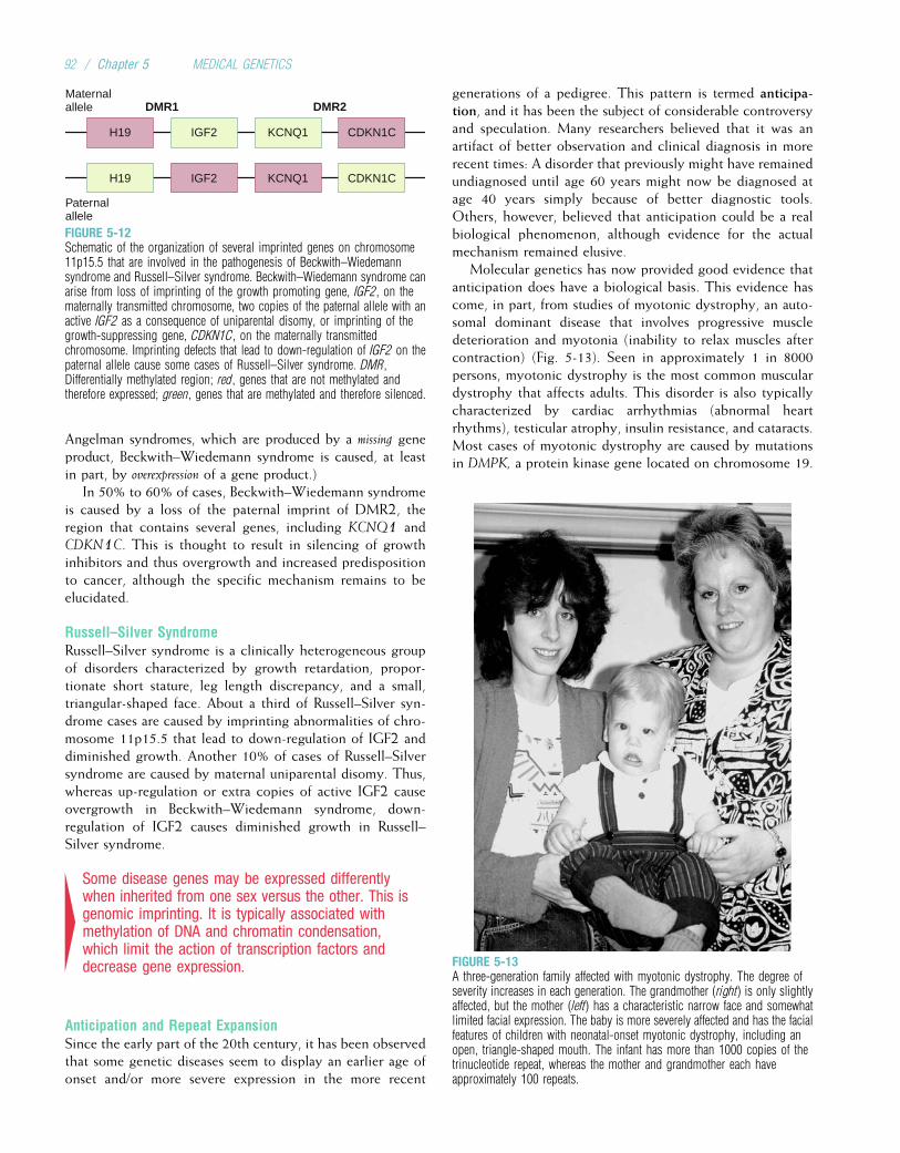
Angelman syndromes, which are produced by a missing geneproduct, Beckwith–Wiedemann syndrome is caused, at leastin part, by overexpression of a gene product.)
In 50% to 60% of cases, Beckwith–Wiedemann syndromeis caused by a loss of the paternal imprint of DMR2, theregion that contains several genes, including KCNQ1 andCDKN1C. This is thought to result in silencing of growthinhibitors and thus overgrowth and increased predispositionto cancer, although the specific mechanism remains to beelucidated.
Russell–Silver SyndromeRussell–Silver syndrome is a clinically heterogeneous groupof disorders characterized by growth retardation, propor-tionate short stature, leg length discrepancy, and a small,triangular-shaped face. About a third of Russell–Silver syn-drome cases are caused by imprinting abnormalities of chro-mosome 11p15.5 that lead to down-regulation of IGF2 anddiminished growth. Another 10% of cases of Russell–Silversyndrome are caused by maternal uniparental disomy. Thus,whereas up-regulation or extra copies of active IGF2 causeovergrowth in Beckwith–Wiedemann syndrome, down-regulation of IGF2 causes diminished growth in Russell–Silver syndrome.
4Some disease genes may be expressed differentlywhen inherited from one sex versus the other. This isgenomic imprinting. It is typically associated withmethylation of DNA and chromatin condensation,which limit the action of transcription factors anddecrease gene expression.
Anticipation and Repeat ExpansionSince the early part of the 20th century, it has been observedthat some genetic diseases seem to display an earlier age ofonset and/or more severe expression in the more recent
generations of a pedigree. This pattern is termed anticipa-tion, and it has been the subject of considerable controversyand speculation. Many researchers believed that it was anartifact of better observation and clinical diagnosis in morerecent times: A disorder that previously might have remainedundiagnosed until age 60 years might now be diagnosed atage 40 years simply because of better diagnostic tools.Others, however, believed that anticipation could be a realbiological phenomenon, although evidence for the actualmechanism remained elusive.
Molecular genetics has now provided good evidence thatanticipation does have a biological basis. This evidence hascome, in part, from studies of myotonic dystrophy, an auto-somal dominant disease that involves progressive muscledeterioration and myotonia (inability to relax muscles aftercontraction) (Fig. 5-13). Seen in approximately 1 in 8000persons, myotonic dystrophy is the most common musculardystrophy that affects adults. This disorder is also typicallycharacterized by cardiac arrhythmias (abnormal heartrhythms), testicular atrophy, insulin resistance, and cataracts.Most cases of myotonic dystrophy are caused by mutationsin DMPK, a protein kinase gene located on chromosome 19.
Maternalallele
H19
H19
IGF2
IGF2
KCNQ1
KCNQ1
Paternalallele
DMR1 DMR2
CDKN1C
CDKN1C
FIGURE 5-12Schematic of the organization of several imprinted genes on chromosome11p15.5 that are involved in the pathogenesis of Beckwith–Wiedemannsyndrome and Russell–Silver syndrome. Beckwith–Wiedemann syndrome canarise from loss of imprinting of the growth promoting gene, IGF2, on thematernally transmitted chromosome, two copies of the paternal allele with anactive IGF2 as a consequence of uniparental disomy, or imprinting of thegrowth-suppressing gene, CDKN1C, on the maternally transmittedchromosome. Imprinting defects that lead to down-regulation of IGF2 on thepaternal allele cause some cases of Russell–Silver syndrome. DMR,Differentially methylated region; red, genes that are not methylated andtherefore expressed; green, genes that are methylated and therefore silenced.
FIGURE 5-13A three-generation family affected with myotonic dystrophy. The degree ofseverity increases in each generation. The grandmother (right) is only slightlyaffected, but the mother (left) has a characteristic narrow face and somewhatlimited facial expression. The baby is more severely affected and has the facialfeatures of children with neonatal-onset myotonic dystrophy, including anopen, triangle-shaped mouth. The infant has more than 1000 copies of thetrinucleotide repeat, whereas the mother and grandmother each haveapproximately 100 repeats.
92 / Chapter 5 MEDICAL GENETICS
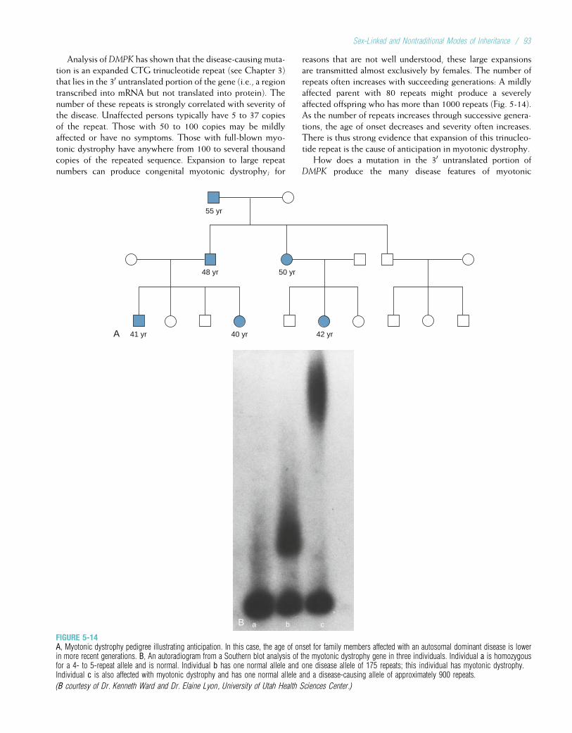
Analysis ofDMPK has shown that the disease-causing muta-tion is an expanded CTG trinucleotide repeat (see Chapter 3)that lies in the 30 untranslated portion of the gene (i.e., a regiontranscribed into mRNA but not translated into protein). Thenumber of these repeats is strongly correlated with severity ofthe disease. Unaffected persons typically have 5 to 37 copiesof the repeat. Those with 50 to 100 copies may be mildlyaffected or have no symptoms. Those with full-blown myo-tonic dystrophy have anywhere from 100 to several thousandcopies of the repeated sequence. Expansion to large repeatnumbers can produce congenital myotonic dystrophy; for
reasons that are not well understood, these large expansionsare transmitted almost exclusively by females. The number ofrepeats often increases with succeeding generations: A mildlyaffected parent with 80 repeats might produce a severelyaffected offspring who has more than 1000 repeats (Fig. 5-14).As the number of repeats increases through successive genera-tions, the age of onset decreases and severity often increases.There is thus strong evidence that expansion of this trinucleo-tide repeat is the cause of anticipation in myotonic dystrophy.
How does a mutation in the 30 untranslated portion ofDMPK produce the many disease features of myotonic
A
55 yr
48 yr 50 yr
41 yr 40 yr 42 yr
B a b c
FIGURE 5-14A, Myotonic dystrophy pedigree illustrating anticipation. In this case, the age of onset for family members affected with an autosomal dominant disease is lowerin more recent generations. B, An autoradiogram from a Southern blot analysis of the myotonic dystrophy gene in three individuals. Individual a is homozygousfor a 4- to 5-repeat allele and is normal. Individual b has one normal allele and one disease allele of 175 repeats; this individual has myotonic dystrophy.Individual c is also affected with myotonic dystrophy and has one normal allele and a disease-causing allele of approximately 900 repeats.
(B courtesy of Dr. Kenneth Ward and Dr. Elaine Lyon, University of Utah Health Sciences Center.)
Sex-Linked and Nontraditional Modes of Inheritance / 93
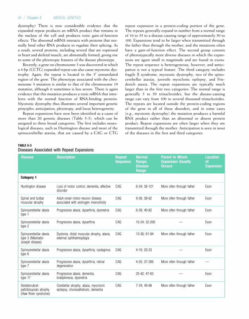
dystrophy? There is now considerable evidence that theexpanded repeat produces an mRNA product that remains inthe nucleus of the cell and produces toxic gain-of-functioneffects. The abnormal mRNA interacts with proteins that nor-mally bind other RNA products to regulate their splicing. Asa result, several proteins, including several that are expressedin heart and skeletal muscle, are abnormally formed, giving riseto some of the pleiotropic features of the disease phenotype.
Recently, a gene on chromosome 3 was discovered in whicha 4-bp (CCTG) expanded repeat can also cause myotonic dys-trophy. Again, the repeat is located in the 30 untranslatedregion of the gene. The phenotype associated with the chro-mosome 3 mutation is similar to that of the chromosome 19mutation, although it sometimes is less severe. There is againevidence that this mutation produces a toxic mRNA that inter-feres with the normal function of RNA-binding proteins.Myotonic dystrophy thus illustrates several important geneticprinciples: anticipation, pleiotropy, and locus heterogeneity.
Repeat expansions have now been identified as a cause ofmore than 20 genetic diseases (Table 5-3), which can beassigned to three broad categories. The first includes neuro-logical diseases, such as Huntington disease and most of thespinocerebellar ataxias, that are caused by a CAG or CTG
repeat expansion in a protein-coding portion of the gene.The repeats generally expand in number from a normal rangeof 10 to 35 to a disease-causing range of approximately 50 to100. Expansions tend to be larger when transmitted throughthe father than through the mother, and the mutations oftenhave a gain-of-function effect. The second group consistsof phenotypically more diverse diseases in which the expan-sions are again small in magnitude and are found in exons.The repeat sequence is heterogeneous, however, and antici-pation is not a typical feature. The third category includesfragile X syndrome, myotonic dystrophy, two of the spino-cerebellar ataxias, juvenile myoclonic epilepsy, and Frie-dreich ataxia. The repeat expansions are typically muchlarger than in the first two categories: The normal range isgenerally 5 to 50 trinucleotides, but the disease-causingrange can vary from 100 to several thousand trinucleotides.The repeats are located outside the protein-coding regionsof the gene in all of these disorders, and in some cases(e.g., myotonic dystrophy) the mutation produces a harmfulRNA product rather than an abnormal or absent proteinproduct. Repeat expansions are often larger when they aretransmitted through the mother. Anticipation is seen in mostof the diseases in the first and third categories.
TABLE 5-3
Diseases Associated with Repeat Expansions
Disease Description RepeatSequence
NormalRange;DiseaseRange
Parent in WhomExpansion UsuallyOccurs
LocationofExpansion
Category 1
Huntington disease Loss of motor control, dementia, affectivedisorder
CAG 6-34; 36-121 More often through father Exon
Spinal and bulbarmuscular atrophy
Adult-onset motor-neuron diseaseassociated with androgen insensitivity
CAG 9-36; 38-62 More often through father Exon
Spinocerebellar ataxiatype 1
Progressive ataxia, dysarthria, dysmetria CAG 6-39; 40-82 More often through father Exon
Spinocerebellar ataxiatype 2
Progressive ataxia, dysarthria CAG 15-24; 32-200 — Exon
Spinocerebellar ataxiatype 3 (Machado-Joseph disease)
Dystonia, distal muscular atrophy, ataxia,external ophthalmoplegia
CAG 13-36; 61-84 More often through father Exon
Spinocerebellar ataxiatype 6
Progressive ataxia, dysarthria, nystagmus CAG 4-19; 20-33 — Exon
Spinocerebellar ataxiatype 7
Progressive ataxia, dysarthria, retinaldegeneration
CAG 4-35; 37-306 More often through father —
Spinocerebellar ataxiatype 17
Progressive ataxia, dementia,bradykinesia, dysmetria
CAG 25-42; 47-63 — Exon
Dentatorubral-pallidoluysian atrophy(Haw River syndrome)
Cerebellar atrophy, ataxia, myoclonicepilepsy, choreoathetosis, dementia
CAG 7-34; 49-88 More often through father Exon
94 / Chapter 5 MEDICAL GENETICS
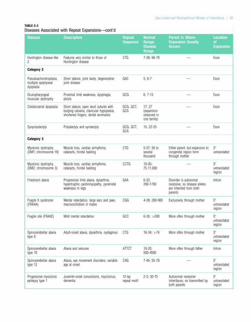
TABLE 5-3
Diseases Associated with Repeat Expansions—cont’d
Disease Description RepeatSequence
NormalRange;DiseaseRange
Parent in WhomExpansion UsuallyOccurs
LocationofExpansion
Huntington disease-like2
Features very similar to those ofHuntington disease
CTG 7-28; 66-78 — Exon
Category 2
Pseudoachondroplasia,multiple epiphysealdysplasia
Short stature, joint laxity, degenerativejoint disease
GAC 5; 6-7 — Exon
Oculopharyngealmuscular dystrophy
Proximal limb weakness, dysphagia,ptosis
GCG 6; 7-13 — Exon
Cleidocranial dysplasia Short stature, open skull sutures withbulging calvaria, clavicular hypoplasia,shortened fingers, dental anomalies
GCG, GCT,GCA
17; 27(expansionobserved inone family)
— Exon
Synpolydactyly Polydactyly and syndactyly GCG, GCT,GCA
15; 22-25 — Exon
Category 3
Myotonic dystrophy(DM1; chromosome 19)
Muscle loss, cardiac arrhythmia,cataracts, frontal balding
CTG 5-37; 50 toseveralthousand
Either parent, but expansion tocongenital region formthrough mother
30untranslated
Myotonic dystrophy(DM2; chromosome 3)
Muscle loss, cardiac arrhythmia,cataracts, frontal balding
CCTG 10-26;75-11,000
— 30untranslatedregion
Friedreich ataxia Progressive limb ataxia, dysarthria,hypertrophic cardiomyopathy, pyramidalweakness in legs
GAA 6-32;200-1700
Disorder is autosomalrecessive, so disease allelesare inherited from bothparents
Intron
Fragile X syndrome(FRAXA)
Mental retardation, large ears and jaws,macroorchidism in males
CGG 4-39; 200-900 Exclusively through mother 50untranslatedregion
Fragile site (FRAXE) Mild mental retardation GCC 6-35; >200 More often through mother 50untranslatedregion
Spinocerebellar ataxiatype 8
Adult-onset ataxia, dysarthria, nystagmus CTG 16-34; >74 More often through mother 30untranslatedregion
Spinocerebellar ataxiatype 10
Ataxia and seizures ATTCT 10-20;500-4500
More often through father Intron
Spinocerebellar ataxiatype 12
Ataxia, eye movement disorders; variableage at onset
CAG 7-45; 55-78 — 50untranslatedregion
Progressive myoclonicepilepsy type 1
Juvenile-onset convulsions, myoclonus,dementia
12-bprepeat motif
2-3; 30-75 Autosomal recessiveinheritance, so transmitted byboth parents
50untranslatedregion
Sex-Linked and Nontraditional Modes of Inheritance / 95
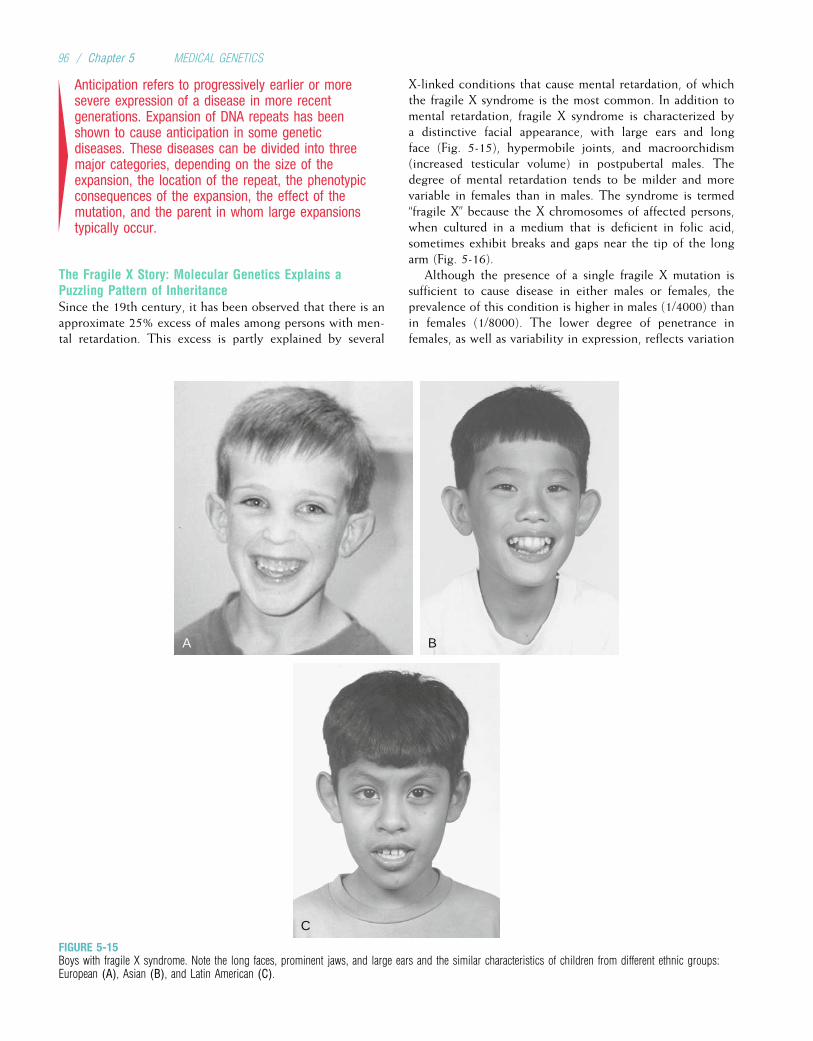
4Anticipation refers to progressively earlier or moresevere expression of a disease in more recentgenerations. Expansion of DNA repeats has beenshown to cause anticipation in some geneticdiseases. These diseases can be divided into threemajor categories, depending on the size of theexpansion, the location of the repeat, the phenotypicconsequences of the expansion, the effect of themutation, and the parent in whom large expansionstypically occur.
The Fragile X Story: Molecular Genetics Explains aPuzzling Pattern of InheritanceSince the 19th century, it has been observed that there is anapproximate 25% excess of males among persons with men-tal retardation. This excess is partly explained by several
X-linked conditions that cause mental retardation, of whichthe fragile X syndrome is the most common. In addition tomental retardation, fragile X syndrome is characterized bya distinctive facial appearance, with large ears and longface (Fig. 5-15), hypermobile joints, and macroorchidism(increased testicular volume) in postpubertal males. Thedegree of mental retardation tends to be milder and morevariable in females than in males. The syndrome is termed“fragile X” because the X chromosomes of affected persons,when cultured in a medium that is deficient in folic acid,sometimes exhibit breaks and gaps near the tip of the longarm (Fig. 5-16).
Although the presence of a single fragile X mutation issufficient to cause disease in either males or females, theprevalence of this condition is higher in males (1/4000) thanin females (1/8000). The lower degree of penetrance infemales, as well as variability in expression, reflects variation
A B
C
FIGURE 5-15Boys with fragile X syndrome. Note the long faces, prominent jaws, and large ears and the similar characteristics of children from different ethnic groups:European (A), Asian (B), and Latin American (C).
96 / Chapter 5 MEDICAL GENETICS
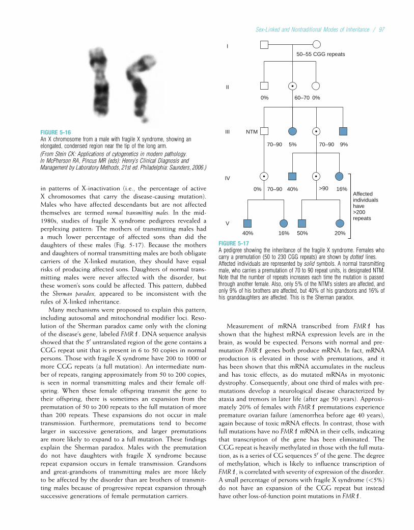
in patterns of X-inactivation (i.e., the percentage of activeX chromosomes that carry the disease-causing mutation).Males who have affected descendants but are not affectedthemselves are termed normal transmitting males. In the mid-1980s, studies of fragile X syndrome pedigrees revealed aperplexing pattern: The mothers of transmitting males hada much lower percentage of affected sons than did thedaughters of these males (Fig. 5-17). Because the mothersand daughters of normal transmitting males are both obligatecarriers of the X-linked mutation, they should have equalrisks of producing affected sons. Daughters of normal trans-mitting males were never affected with the disorder, butthese women’s sons could be affected. This pattern, dubbedthe Sherman paradox, appeared to be inconsistent with therules of X-linked inheritance.
Many mechanisms were proposed to explain this pattern,including autosomal and mitochondrial modifier loci. Reso-lution of the Sherman paradox came only with the cloningof the disease’s gene, labeled FMR1. DNA sequence analysisshowed that the 50 untranslated region of the gene contains aCGG repeat unit that is present in 6 to 50 copies in normalpersons. Those with fragile X syndrome have 200 to 1000 ormore CGG repeats (a full mutation). An intermediate num-ber of repeats, ranging approximately from 50 to 200 copies,is seen in normal transmitting males and their female off-spring. When these female offspring transmit the gene totheir offspring, there is sometimes an expansion from thepremutation of 50 to 200 repeats to the full mutation of morethan 200 repeats. These expansions do not occur in maletransmission. Furthermore, premutations tend to becomelarger in successive generations, and larger premutationsare more likely to expand to a full mutation. These findingsexplain the Sherman paradox. Males with the premutationdo not have daughters with fragile X syndrome becauserepeat expansion occurs in female transmission. Grandsonsand great-grandsons of transmitting males are more likelyto be affected by the disorder than are brothers of transmit-ting males because of progressive repeat expansion throughsuccessive generations of female permutation carriers.
Measurement of mRNA transcribed from FMR1 hasshown that the highest mRNA expression levels are in thebrain, as would be expected. Persons with normal and pre-mutation FMR1 genes both produce mRNA. In fact, mRNAproduction is elevated in those with premutations, and ithas been shown that this mRNA accumulates in the nucleusand has toxic effects, as do mutated mRNAs in myotonicdystrophy. Consequently, about one third of males with pre-mutations develop a neurological disease characterized byataxia and tremors in later life (after age 50 years). Approxi-mately 20% of females with FMR1 premutations experiencepremature ovarian failure (amenorrhea before age 40 years),again because of toxic mRNA effects. In contrast, those withfull mutations have no FMR1 mRNA in their cells, indicatingthat transcription of the gene has been eliminated. TheCGG repeat is heavily methylated in those with the full muta-tion, as is a series of CG sequences 50 of the gene. The degreeof methylation, which is likely to influence transcription ofFMR1, is correlated with severity of expression of the disorder.A small percentage of persons with fragile X syndrome (<5%)do not have an expansion of the CGG repeat but insteadhave other loss-of-function point mutations in FMR1.
FIGURE 5-16An X chromosome from a male with fragile X syndrome, showing anelongated, condensed region near the tip of the long arm.
(From Stein CK: Applications of cytogenetics in modern pathology.In McPherson RA, Pincus MR (eds): Henry’s Clinical Diagnosis andManagement by Laboratory Methods, 21st ed. Philadelphia: Saunders, 2006.)
I
II
III
IV
V
50–55 CGG repeats
60–70
70–90
NTM
70–90
>9070–90
0%
5% 9%
40%
40% 50%16% 20%
0%
0%Affectedindividualshave>200repeats
16%
FIGURE 5-17A pedigree showing the inheritance of the fragile X syndrome. Females whocarry a premutation (50 to 230 CGG repeats) are shown by dotted lines.Affected individuals are represented by solid symbols. A normal transmittingmale, who carries a premutation of 70 to 90 repeat units, is designated NTM.Note that the number of repeats increases each time the mutation is passedthrough another female. Also, only 5% of the NTM’s sisters are affected, andonly 9% of his brothers are affected, but 40% of his grandsons and 16% ofhis granddaughters are affected. This is the Sherman paradox.
Sex-Linked and Nontraditional Modes of Inheritance / 97
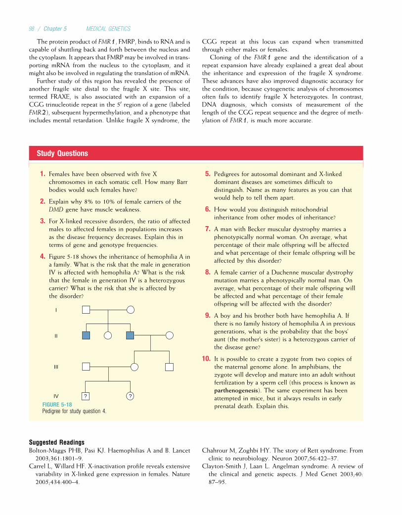
The protein product of FMR1, FMRP, binds to RNA and iscapable of shuttling back and forth between the nucleus andthe cytoplasm. It appears that FMRP may be involved in trans-porting mRNA from the nucleus to the cytoplasm, and itmight also be involved in regulating the translation of mRNA.
Further study of this region has revealed the presence ofanother fragile site distal to the fragile X site. This site,termed FRAXE, is also associated with an expansion of aCGG trinucleotide repeat in the 50 region of a gene (labeledFMR2), subsequent hypermethylation, and a phenotype thatincludes mental retardation. Unlike fragile X syndrome, the
CGG repeat at this locus can expand when transmittedthrough either males or females.
Cloning of the FMR1 gene and the identification of arepeat expansion have already explained a great deal aboutthe inheritance and expression of the fragile X syndrome.These advances have also improved diagnostic accuracy forthe condition, because cytogenetic analysis of chromosomesoften fails to identify fragile X heterozygotes. In contrast,DNA diagnosis, which consists of measurement of thelength of the CGG repeat sequence and the degree of meth-ylation of FMR1, is much more accurate.
Study Questions
1. Females have been observed with five Xchromosomes in each somatic cell. How many Barrbodies would such females have?
2. Explain why 8% to 10% of female carriers of theDMD gene have muscle weakness.
3. For X-linked recessive disorders, the ratio of affectedmales to affected females in populations increasesas the disease frequency decreases. Explain this interms of gene and genotype frequencies.
4. Figure 5-18 shows the inheritance of hemophilia A ina family. What is the risk that the male in generationIV is affected with hemophilia A? What is the riskthat the female in generation IV is a heterozygouscarrier? What is the risk that she is affected bythe disorder?
I
II
III
IV ? ?
FIGURE 5-18Pedigree for study question 4.
5. Pedigrees for autosomal dominant and X-linkeddominant diseases are sometimes difficult todistinguish. Name as many features as you can thatwould help to tell them apart.
6. How would you distinguish mitochondrialinheritance from other modes of inheritance?
7. A man with Becker muscular dystrophy marries aphenotypically normal woman. On average, whatpercentage of their male offspring will be affectedand what percentage of their female offspring will beaffected by this disorder?
8. A female carrier of a Duchenne muscular dystrophymutation marries a phenotypically normal man. Onaverage, what percentage of their male offspring willbe affected and what percentage of their femaleoffspring will be affected with the disorder?
9. A boy and his brother both have hemophilia A. Ifthere is no family history of hemophilia A in previousgenerations, what is the probability that the boys’aunt (the mother’s sister) is a heterozygous carrier ofthe disease gene?
10. It is possible to create a zygote from two copies ofthe maternal genome alone. In amphibians, thezygote will develop and mature into an adult withoutfertilization by a sperm cell (this process is known asparthenogenesis). The same experiment has beenattempted in mice, but it always results in earlyprenatal death. Explain this.
Suggested ReadingsBolton-Maggs PHB, Pasi KJ. Haemophilias A and B. Lancet
2003;361:1801–9.Carrel L, Willard HF. X-inactivation profile reveals extensive
variability in X-linked gene expression in females. Nature2005;434:400–4.
Chahrour M, Zoghbi HY. The story of Rett syndrome: Fromclinic to neurobiology. Neuron 2007;56:422–37.
Clayton-Smith J, Laan L. Angelman syndrome: A review ofthe clinical and genetic aspects. J Med Genet 2003;40:87–95.
98 / Chapter 5 MEDICAL GENETICS
Clerc P, Avner P. Random X-chromosome inactivation:Skewing lessons for mice and men. Curr Opin GenetDev 2006;16:246–53.
Dalkilic I, Kunkel LM. Muscular dystrophies: Genes to path-ogenesis. Curr Opin Genet Dev 2003;13:231–38.
Deeb SS. The molecular basis of variation in human colorvision. Clin Genet 2005;67:369–77.
Emery AE. The muscular dystrophies. Lancet 2002;359:687–95.
Garber K, Smith KT, Reines D, Warren ST. Transcription,translation and fragile X syndrome. Curr Opin GenetDev 2006;16:270–75.
Garber KB, Visootsak J, Warren ST. Fragile X syndrome. EurJ Hum Genet 2008;16:666–72.
Graw J, Brackmann HH, Oldenburg J, et al. Haemophilia A:From mutation analysis to new therapies. Nat Rev Genet2005;6:488–501.
Horsthemke B, Buiting K. Imprinting defects on humanchromosome 15. Cytogenet Genome Res 2006;113:292–9.
Jiang Y-H, Bressler J, Beaudet AL. Epigenetics and humandisease. Annu Rev Genomics Hum Genet 2004;5:479–510.
Orr HT, Zoghbi HY. Trinucleotide repeat disorders. AnnuRev Neurosci 2007;30:575–621.
Ranum LP, Cooper TA. RNA-mediated neuromuscular disor-ders. Annu Rev Neurosci 2006;29:259–77.
Ross MT, Grafham DV, Coffey AJ, et al. The DNA sequenceof the human X chromosome. Nature 2005; 434:325–37.
Schapira AHV. Mitochondrial disease. Lancet 2006; 368:70–82.
Straub T, Becker PB. Dosage compensation: The beginningand end of generalization. Nat Rev Genet 2007;8: 47–57.
Terracciano A, Chiurazzi P, Neri G. Fragile X syndrome. Am JMed Genet C Semin Med Genet 2005;137:32–7.
Wallace DC. A mitochondrial paradigm of metabolic anddegenerative diseases, aging, and cancer: A dawn for evo-lutionary medicine. Annu Rev Genet 2005;39: 359–407.
Wattendorf DJ, Muenke M. Prader–Willi syndrome. AmFam Physician 2005;72:827–30.
Weksberg R, Shuman C, Smith AC. Beckwith–Wiedemannsyndrome. Am J Med Genet C Semin Med Genet2005;137:12–23.
Wutz A, Gribnau J. X inactivation Xplained. Curr OpinGenet Dev 2007;17:387–93.
Internet ResourcesMITOMAP (extensive information about the mitochondrialgenome and its role in disease) http://www.mitomap.org/
Muscular Dystrophy Association (information aboutvarious types of muscular dystrophy with links to otherwebsites) http://www.mdausa.org/
National Hemophilia Foundation (information abouthemophilia and links to other websites) http://www.hemophilia.org/home.htm
Sex-Linked and Nontraditional Modes of Inheritance / 99
Chapter 6
CLINICAL CYTOGENETICS: THE CHROMOSOMALBASIS OF HUMAN DISEASE
The previous two chapters have dealt with single-gene dis-eases. We turn now to diseases caused by alterations in thenumber or structure of chromosomes. The study of chromo-somes and their abnormalities is called cytogenetics.
Chromosome abnormalities are responsible for a signifi-cant fraction of genetic diseases, occurring in approximately1 of every 150 live births. They are the leading known causeof mental retardation and pregnancy loss. Chromosomeabnormalities are seen in 50% of first-trimester and 20% ofsecond-trimester spontaneous abortions. Thus, they are animportant cause of morbidity and mortality.
As in other areas of medical genetics, advances in moleculargenetics have contributedmany new insights in the field of cyto-genetics. For example, molecular techniques have permitted theidentification of chromosome abnormalities, such as deletions,that affect very small regions. In some cases, specific genes thatcontribute to cytogenetic syndromes are being pinpointed. Inaddition, the ability to identify DNA polymorphisms in parentsand offspring has enabled researchers to specify whether anabnormal chromosome is derived from the mother or from thefather. This has increased our understanding of the biologicalbasis of meiotic errors and chromosome abnormalities.
In this chapter we discuss abnormalities of chromosomenumber and structure. The genetic basis of sex determinationis reviewed, the role of chromosome alterations in cancer isexamined, and several diseases caused by chromosomalinstability are discussed. Emphasis is given to the new contri-butions of molecular genetics to cytogenetics.
CYTOGENETIC TECHNOLOGY AND NOMENCLATUREAlthough it was possible to visualize chromosomes undermicroscopes as early as the mid-1800s, it was quite difficultto observe individual chromosomes. It was thus hard tocount the number of chromosomes in a cell or to examinestructural abnormalities. Beginning in the 1950s, severaltechniques were developed that improved our ability toobserve chromosomes. These included the use of spindlepoisons, such as colchicine and colcemid, that arrest dividingsomatic cells in metaphase, when chromosomes are maxi-mally condensed and easiest to see; the use of a hypotonic(low-salt) solution, which causes swelling of cells, ruptureof the nucleus, and better separation of individual
chromosomes; and the use of staining materials that areabsorbed differently by different parts of chromosomes, thusproducing the characteristic light and dark bands that helpto identify individual chromosomes.
4Our ability to study chromosomes has been improvedby the visualization of chromosomes in metaphase, byhypotonic solutions that cause nuclear swelling, andby staining techniques that label chromosome bands.
Chromosomes are analyzed by collecting a living tissue(usually blood), culturing the tissue for the appropriateamount of time (usually 48 to 72 hours for peripheral lym-phocytes), adding colcemid to produce metaphase arrest,harvesting the cells, placing the cell sediment on a slide, rup-turing the cell nucleus with a hypotonic saline solution,staining with a designated nuclear stain, and photographingthe metaphase spreads of chromosomes on the slide. Theimages of the 22 pairs of autosomes are arranged accordingto length, with the sex chromosomes in the right-hand cor-ner. This ordered display of chromosomes is termed a karyo-gram or karyotype (Fig. 6-1). (The term karyotype refers tothe number and type of chromosomes present in an individ-ual, and karyogram is now often used to designate the printeddisplay of chromosomes.) Currently, computerized imageanalyzers are usually used to display chromosomes.
After sorting by size, chromosomes are further classifiedaccording to the position of the centromere. If the centro-mere occurs near the middle of the chromosome, the chromo-some is said to be metacentric (Fig. 6-2). An acrocentricchromosome has its centromere near the tip, and submeta-centric chromosomes have centromeres somewhere betweenthe middle and the tip. The tip of each chromosome is thetelomere. The short arm of a chromosome is labeled p (forpetite), and the long arm is labeled q. In metacentric chromo-somes, where the arms are of roughly equal length, thep and q arms are designated by convention.
4A karyotype, or karyogram, is a display ofchromosomes ordered according to length. Dependingon the position of the centromere, a chromosome maybe acrocentric, submetacentric, or metacentric.
100
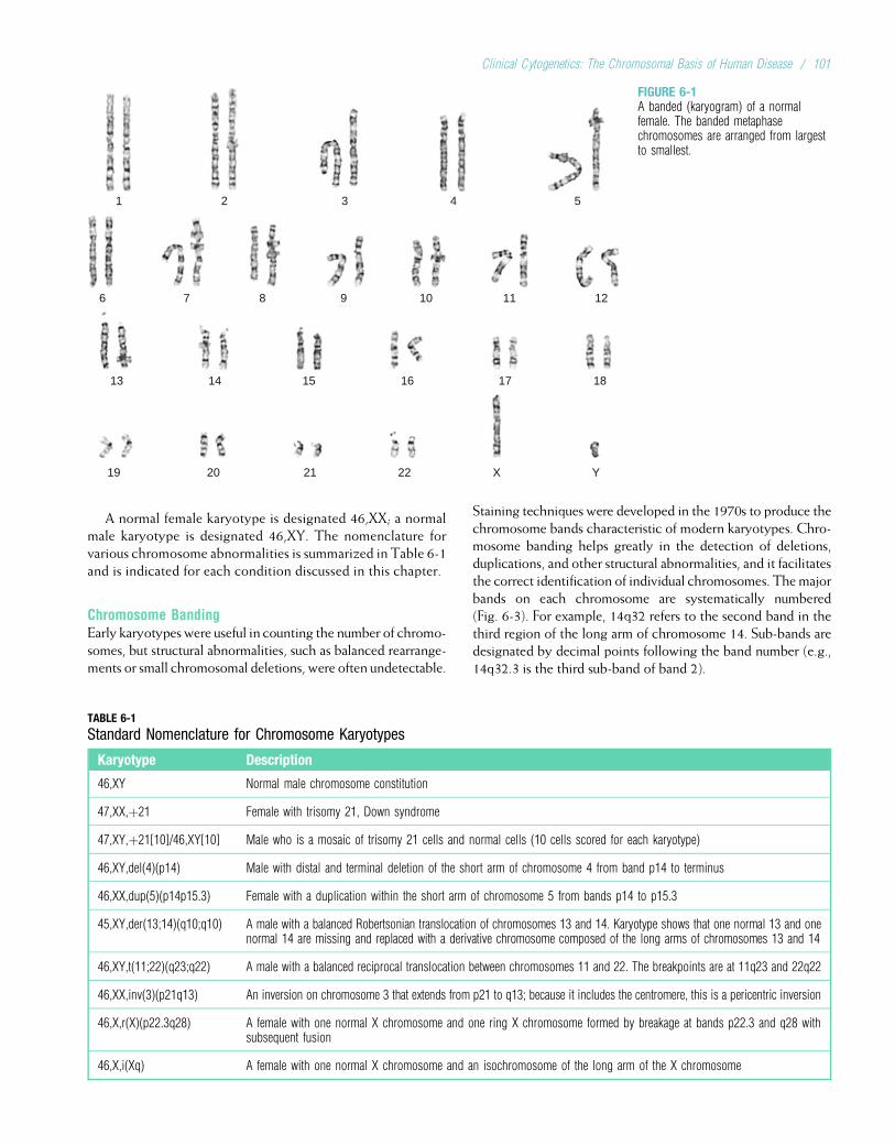
A normal female karyotype is designated 46,XX; a normalmale karyotype is designated 46,XY. The nomenclature forvarious chromosome abnormalities is summarized in Table 6-1and is indicated for each condition discussed in this chapter.
Chromosome BandingEarly karyotypes were useful in counting the number of chromo-somes, but structural abnormalities, such as balanced rearrange-ments or small chromosomal deletions, were often undetectable.
Staining techniques were developed in the 1970s to produce thechromosome bands characteristic of modern karyotypes. Chro-mosome banding helps greatly in the detection of deletions,duplications, and other structural abnormalities, and it facilitatesthe correct identification of individual chromosomes. Themajorbands on each chromosome are systematically numbered(Fig. 6-3). For example, 14q32 refers to the second band in thethird region of the long arm of chromosome 14. Sub-bands aredesignated by decimal points following the band number (e.g.,14q32.3 is the third sub-band of band 2).
1 2 3 4 5
1211109876
13
19
14
20
15
21
16
22
17 18
X Y
FIGURE 6-1A banded (karyogram) of a normalfemale. The banded metaphasechromosomes are arranged from largestto smallest.
TABLE 6-1
Standard Nomenclature for Chromosome Karyotypes
Karyotype Description
46,XY Normal male chromosome constitution
47,XX,þ21 Female with trisomy 21, Down syndrome
47,XY,þ21[10]/46,XY[10] Male who is a mosaic of trisomy 21 cells and normal cells (10 cells scored for each karyotype)
46,XY,del(4)(p14) Male with distal and terminal deletion of the short arm of chromosome 4 from band p14 to terminus
46,XX,dup(5)(p14p15.3) Female with a duplication within the short arm of chromosome 5 from bands p14 to p15.3
45,XY,der(13;14)(q10;q10) A male with a balanced Robertsonian translocation of chromosomes 13 and 14. Karyotype shows that one normal 13 and onenormal 14 are missing and replaced with a derivative chromosome composed of the long arms of chromosomes 13 and 14
46,XY,t(11;22)(q23;q22) A male with a balanced reciprocal translocation between chromosomes 11 and 22. The breakpoints are at 11q23 and 22q22
46,XX,inv(3)(p21q13) An inversion on chromosome 3 that extends from p21 to q13; because it includes the centromere, this is a pericentric inversion
46,X,r(X)(p22.3q28) A female with one normal X chromosome and one ring X chromosome formed by breakage at bands p22.3 and q28 withsubsequent fusion
46,X,i(Xq) A female with one normal X chromosome and an isochromosome of the long arm of the X chromosome
Clinical Cytogenetics: The Chromosomal Basis of Human Disease / 101
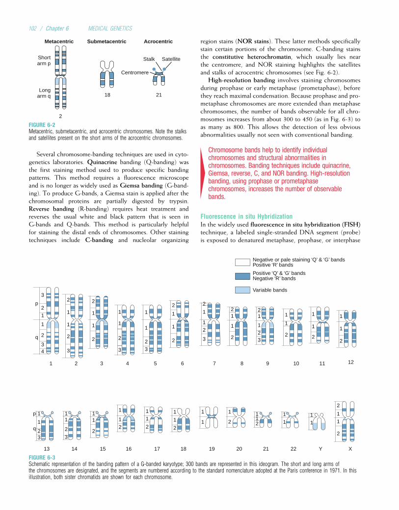
Several chromosome-banding techniques are used in cyto-genetics laboratories. Quinacrine banding (Q-banding) wasthe first staining method used to produce specific bandingpatterns. This method requires a fluorescence microscopeand is no longer as widely used as Giemsa banding (G-band-ing). To produce G-bands, a Giemsa stain is applied after thechromosomal proteins are partially digested by trypsin.Reverse banding (R-banding) requires heat treatment andreverses the usual white and black pattern that is seen inG-bands and Q-bands. This method is particularly helpfulfor staining the distal ends of chromosomes. Other stainingtechniques include C-banding and nucleolar organizing
region stains (NOR stains). These latter methods specificallystain certain portions of the chromosome. C-banding stainsthe constitutive heterochromatin, which usually lies nearthe centromere, and NOR staining highlights the satellitesand stalks of acrocentric chromosomes (see Fig. 6-2).
High-resolution banding involves staining chromosomesduring prophase or early metaphase (prometaphase), beforethey reach maximal condensation. Because prophase and pro-metaphase chromosomes are more extended than metaphasechromosomes, the number of bands observable for all chro-mosomes increases from about 300 to 450 (as in Fig. 6-3) toas many as 800. This allows the detection of less obviousabnormalities usually not seen with conventional banding.
4Chromosome bands help to identify individualchromosomes and structural abnormalities inchromosomes. Banding techniques include quinacrine,Giemsa, reverse, C, and NOR banding. High-resolutionbanding, using prophase or prometaphasechromosomes, increases the number of observablebands.
Fluorescence in situ HybridizationIn the widely used fluorescence in situ hybridization (FISH)technique, a labeled single-stranded DNA segment (probe)is exposed to denatured metaphase, prophase, or interphase
Negative or pale staining ‘Q’ & ‘G’ bandsPositive ‘R’ bands
Positive ‘Q’ & ‘G’ bandsNegative ‘R’ bands
Variable bands
1 2 3 4 5 6 7 8 9 10 11
13 14 15 16 17 18 19 20 21 22 Y X
1
1
2
1
1
2
1
1
2
21
1
2
21
1
2 3
2
1
1
2
2
1
1
2
2
1
1
2
1
1
2
3
1
1
2
3
3
2
1
p
q
p
q
1
2
3
2
3
34
1
1
2
32
1
1
3
2
11
2
11
2
1
1
2
11
2
1
1
1
1
2
1
211
1
1
1
11
1
2
2
12
FIGURE 6-3Schematic representation of the banding pattern of a G-banded karyotype; 300 bands are represented in this ideogram. The short and long arms ofthe chromosomes are designated, and the segments are numbered according to the standard nomenclature adopted at the Paris conference in 1971. In thisillustration, both sister chromatids are shown for each chromosome.
AcrocentricSubmetacentric
Shortarm p
Longarm q
Metacentric
Centromere
SatelliteStalk
2118
2
FIGURE 6-2Metacentric, submetacentric, and acrocentric chromosomes. Note the stalksand satellites present on the short arms of the acrocentric chromosomes.
102 / Chapter 6 MEDICAL GENETICS
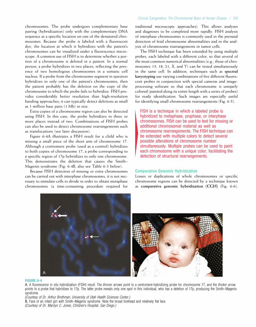
chromosomes. The probe undergoes complementary basepairing (hybridization) only with the complementary DNAsequence at a specific location on one of the denatured chro-mosomes. Because the probe is labeled with a fluorescentdye, the location at which it hybridizes with the patient’schromosomes can be visualized under a fluorescence micro-scope. A common use of FISH is to determine whether a por-tion of a chromosome is deleted in a patient. In a normalperson, a probe hybridizes in two places, reflecting the pres-ence of two homologous chromosomes in a somatic cellnucleus. If a probe from the chromosome segment in questionhybridizes to only one of the patient’s chromosomes, thenthe patient probably has the deletion on the copy of thechromosome to which the probe fails to hybridize. FISH pro-vides considerably better resolution than high-resolutionbanding approaches; it can typically detect deletions as smallas 1 million base pairs (1 Mb) in size.
Extra copies of a chromosome region can also be detectedusing FISH. In this case, the probe hybridizes in three ormore places instead of two. Combinations of FISH probescan also be used to detect chromosome rearrangements suchas translocations (see later discussion).
Figure 6-4A illustrates a FISH result for a child who ismissing a small piece of the short arm of chromosome 17.Although a centromere probe (used as a control) hybridizesto both copies of chromosome 17, a probe corresponding toa specific region of 17p hybridizes to only one chromosome.This demonstrates the deletion that causes the Smith–Magenis syndrome (Fig. 6-4B; also see Table 6-3 below).
Because FISH detection of missing or extra chromosomescan be carried out with interphase chromosomes, it is not nec-essary to stimulate cells to divide in order to obtain metaphasechromosomes (a time-consuming procedure required for
traditional microscopic approaches). This allows analysesand diagnoses to be completed more rapidly. FISH analysisof interphase chromosomes is commonly used in the prenataldetection of fetal chromosome abnormalities and in the anal-ysis of chromosome rearrangements in tumor cells.
The FISH technique has been extended by using multipleprobes, each labeled with a different color, so that several ofthe most common numerical abnormalities (e.g., those of chro-mosomes 13, 18, 21, X, and Y) can be tested simultaneouslyin the same cell. In addition, techniques such as spectralkaryotyping use varying combinations of five different fluores-cent probes in conjunction with special cameras and image-processing software so that each chromosome is uniquelycolored (painted along its entire length with a series of probes)for ready identification. Such images are especially usefulfor identifying small chromosome rearrangements (Fig. 6-5).
4FISH is a technique in which a labeled probe ishybridized to metaphase, prophase, or interphasechromosomes. FISH can be used to test for missing oradditional chromosomal material as well aschromosome rearrangements. The FISH technique canbe extended with multiple colors to detect severalpossible alterations of chromosome numbersimultaneously. Multiple probes can be used to painteach chromosome with a unique color, facilitating thedetection of structural rearrangements.
Comparative Genomic HybridizationLosses or duplications of whole chromosomes or specificchromosome regions can be detected by a technique knownas comparative genomic hybridization (CGH) (Fig. 6-6).
A B
FIGURE 6-4A, A fluorescence in situ hybridization (FISH) result. The thinner arrows point to a centromere-hybridizing probe for chromosome 17, and the thicker arrowpoints to a probe that hybridizes to 17p. The latter probe reveals only one spot in this individual, who has a deletion of 17p, producing the Smith–Magenissyndrome.(Courtesy of Dr. Arthur Brothman, University of Utah Health Sciences Center.)B, Face of an infant girl with Smith–Magenis syndrome. Note the broad forehead and relatively flat face.(Courtesy of Dr. Marilyn C. Jones, Children’s Hospital, San Diego.)
Clinical Cytogenetics: The Chromosomal Basis of Human Disease / 103
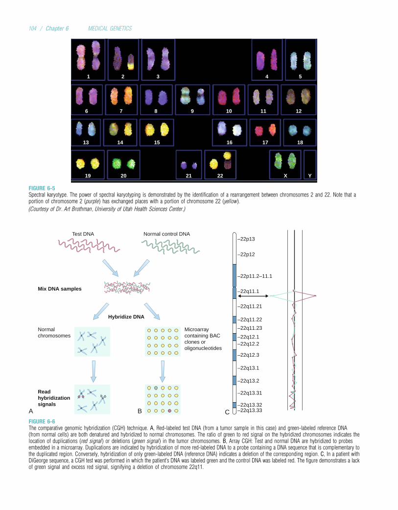
1
6
13
19 20 21 22 X Y
14 15 16 17 18
7 8 9 10 11 12
2 3 54
FIGURE 6-5Spectral karyotype. The power of spectral karyotyping is demonstrated by the identification of a rearrangement between chromosomes 2 and 22. Note that aportion of chromosome 2 (purple) has exchanged places with a portion of chromosome 22 (yellow).
(Courtesy of Dr. Art Brothman, University of Utah Health Sciences Center.)
Test DNA
A B
Normalchromosomes
Microarraycontaining BACclones oroligonucleotides
Mix DNA samples
Hybridize DNA
Readhybridizationsignals
Normal control DNA
C
–22p13
–22p12
–22p11.2–11.1
–22q11.1
–22q11.21
–22q11.22
–22q11.23
–22q12.1–22q12.2
–22q12.3
–22q13.1
–22q13.2
–22q13.31
–22q13.32–22q13.33
FIGURE 6-6The comparative genomic hybridization (CGH) technique. A, Red-labeled test DNA (from a tumor sample in this case) and green-labeled reference DNA(from normal cells) are both denatured and hybridized to normal chromosomes. The ratio of green to red signal on the hybridized chromosomes indicates thelocation of duplications (red signal) or deletions (green signal) in the tumor chromosomes. B, Array CGH: Test and normal DNA are hybridized to probesembedded in a microarray. Duplications are indicated by hybridization of more red-labeled DNA to a probe containing a DNA sequence that is complementary tothe duplicated region. Conversely, hybridization of only green-labeled DNA (reference DNA) indicates a deletion of the corresponding region. C, In a patient withDiGeorge sequence, a CGH test was performed in which the patient’s DNA was labeled green and the control DNA was labeled red. The figure demonstrates a lackof green signal and excess red signal, signifying a deletion of chromosome 22q11.
104 / Chapter 6 MEDICAL GENETICS
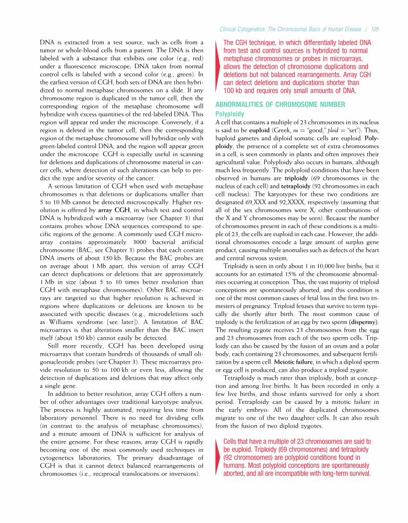
DNA is extracted from a test source, such as cells from atumor or whole-blood cells from a patient. The DNA is thenlabeled with a substance that exhibits one color (e.g., red)under a fluorescence microscope; DNA taken from normalcontrol cells is labeled with a second color (e.g., green). Inthe earliest version of CGH, both sets of DNA are then hybri-dized to normal metaphase chromosomes on a slide. If anychromosome region is duplicated in the tumor cell, then thecorresponding region of the metaphase chromosome willhybridize with excess quantities of the red-labeled DNA. Thisregion will appear red under the microscope. Conversely, if aregion is deleted in the tumor cell, then the correspondingregion of the metaphase chromosome will hybridize only withgreen-labeled control DNA, and the region will appear greenunder the microscope. CGH is especially useful in scanningfor deletions and duplications of chromosome material in can-cer cells, where detection of such alterations can help to pre-dict the type and/or severity of the cancer.
A serious limitation of CGH when used with metaphasechromosomes is that deletions or duplications smaller than5 to 10 Mb cannot be detected microscopically. Higher res-olution is offered by array CGH, in which test and controlDNA is hybridized with a microarray (see Chapter 3) thatcontains probes whose DNA sequences correspond to spe-cific regions of the genome. A commonly used CGH micro-array contains approximately 3000 bacterial artificialchromosome (BAC, see Chapter 3) probes that each containDNA inserts of about 150 kb. Because the BAC probes areon average about 1 Mb apart, this version of array CGHcan detect duplications or deletions that are approximately1 Mb in size (about 5 to 10 times better resolution thanCGH with metaphase chromosomes). Other BAC microar-rays are targeted so that higher resolution is achieved inregions where duplications or deletions are known to beassociated with specific diseases (e.g., microdeletions suchas Williams syndrome [see later]). A limitation of BACmicroarrays is that alterations smaller than the BAC insertitself (about 150 kb) cannot easily be detected.
Still more recently, CGH has been developed usingmicroarrays that contain hundreds of thousands of small oli-gonucleotide probes (see Chapter 3). These microarrays pro-vide resolution to 50 to 100 kb or even less, allowing thedetection of duplications and deletions that may affect onlya single gene.
In addition to better resolution, array CGH offers a num-ber of other advantages over traditional karyotype analysis.The process is highly automated, requiring less time fromlaboratory personnel. There is no need for dividing cells(in contrast to the analysis of metaphase chromosomes),and a minute amount of DNA is sufficient for analysis ofthe entire genome. For these reasons, array CGH is rapidlybecoming one of the most commonly used techniques incytogenetics laboratories. The primary disadvantage ofCGH is that it cannot detect balanced rearrangements ofchromosomes (i.e., reciprocal translocations or inversions).
4The CGH technique, in which differentially labeled DNAfrom test and control sources is hybridized to normalmetaphase chromosomes or probes in microarrays,allows the detection of chromosome duplications anddeletions but not balanced rearrangements. Array CGHcan detect deletions and duplications shorter than100 kb and requires only small amounts of DNA.
ABNORMALITIES OF CHROMOSOME NUMBERPolyploidyA cell that contains a multiple of 23 chromosomes in its nucleusis said to be euploid (Greek, eu ¼ “good,” ploid ¼ “set”). Thus,haploid gametes and diploid somatic cells are euploid. Poly-ploidy, the presence of a complete set of extra chromosomesin a cell, is seen commonly in plants and often improves theiragricultural value. Polyploidy also occurs in humans, althoughmuch less frequently. The polyploid conditions that have beenobserved in humans are triploidy (69 chromosomes in thenucleus of each cell) and tetraploidy (92 chromosomes in eachcell nucleus). The karyotypes for these two conditions aredesignated 69,XXX and 92,XXXX, respectively (assuming thatall of the sex chromosomes were X; other combinations ofthe X and Y chromosomes may be seen). Because the numberof chromosomes present in each of these conditions is a multi-ple of 23, the cells are euploid in each case. However, the addi-tional chromosomes encode a large amount of surplus geneproduct, causing multiple anomalies such as defects of the heartand central nervous system.
Triploidy is seen in only about 1 in 10,000 live births, but itaccounts for an estimated 15% of the chromosome abnormal-ities occurring at conception. Thus, the vast majority of triploidconceptions are spontaneously aborted, and this condition isone of the most common causes of fetal loss in the first two tri-mesters of pregnancy. Triploid fetuses that survive to term typi-cally die shortly after birth. The most common cause oftriploidy is the fertilization of an egg by two sperm (dispermy).The resulting zygote receives 23 chromosomes from the eggand 23 chromosomes from each of the two sperm cells. Trip-loidy can also be caused by the fusion of an ovum and a polarbody, each containing 23 chromosomes, and subsequent fertili-zation by a sperm cell.Meiotic failure, in which a diploid spermor egg cell is produced, can also produce a triploid zygote.
Tetraploidy is much rarer than triploidy, both at concep-tion and among live births. It has been recorded in only afew live births, and those infants survived for only a shortperiod. Tetraploidy can be caused by a mitotic failure inthe early embryo: All of the duplicated chromosomesmigrate to one of the two daughter cells. It can also resultfrom the fusion of two diploid zygotes.
4Cells that have a multiple of 23 chromosomes are said tobe euploid. Triploidy (69 chromosomes) and tetraploidy(92 chromosomes) are polyploid conditions found inhumans. Most polyploid conceptions are spontaneouslyaborted, and all are incompatible with long-term survival.
Clinical Cytogenetics: The Chromosomal Basis of Human Disease / 105
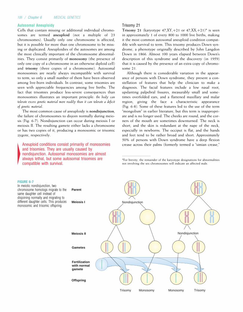
Autosomal AneuploidyCells that contain missing or additional individual chromo-somes are termed aneuploid (not a multiple of 23chromosomes). Usually only one chromosome is affected,but it is possible for more than one chromosome to be miss-ing or duplicated. Aneuploidies of the autosomes are amongthe most clinically important of the chromosome abnormal-ities. They consist primarily of monosomy (the presence ofonly one copy of a chromosome in an otherwise diploid cell)and trisomy (three copies of a chromosome). Autosomalmonosomies are nearly always incompatible with survivalto term, so only a small number of them have been observedamong live-born individuals. In contrast, some trisomies areseen with appreciable frequencies among live births. Thefact that trisomies produce less-severe consequences thanmonosomies illustrates an important principle: the body cantolerate excess genetic material more readily than it can tolerate a deficit
of genetic material.The most common cause of aneuploidy is nondisjunction,
the failure of chromosomes to disjoin normally during meio-sis (Fig. 6-7). Nondisjunction can occur during meiosis I ormeiosis II. The resulting gamete either lacks a chromosomeor has two copies of it, producing a monosomic or trisomiczygote, respectively.
4Aneuploid conditions consist primarily of monosomiesand trisomies. They are usually caused bynondisjunction. Autosomal monosomies are almostalways lethal, but some autosomal trisomies arecompatible with survival.
Trisomy 21Trisomy 21 (karyotype 47,XY,þ21 or 47,XX,þ21)* is seenin approximately 1 of every 800 to 1000 live births, makingit the most common autosomal aneuploid condition compat-ible with survival to term. This trisomy produces Down syn-drome, a phenotype originally described by John LangdonDown in 1866. Almost 100 years elapsed between Down’sdescription of this syndrome and the discovery (in 1959)that it is caused by the presence of an extra copy of chromo-some 21.
Although there is considerable variation in the appear-ance of persons with Down syndrome, they present a con-stellation of features that help the clinician to make adiagnosis. The facial features include a low nasal root,upslanting palpebral fissures, measurably small and some-times overfolded ears, and a flattened maxillary and malarregion, giving the face a characteristic appearance(Fig. 6-8). Some of these features led to the use of the term“mongolism” in earlier literature, but this term is inappropri-ate and is no longer used. The cheeks are round, and the cor-ners of the mouth are sometimes downturned. The neck isshort, and the skin is redundant at the nape of the neck,especially in newborns. The occiput is flat, and the handsand feet tend to be rather broad and short. Approximately50% of persons with Down syndrome have a deep flexioncrease across their palms (formerly termed a “simian crease,”
Nondisjunction
Nondisjunction
Meiosis I
Meiosis II
Fertilization with normal gamete
Trisomy Monosomy Monosomy Trisomy
Offspring
Parent
Gametes
FIGURE 6-7In meiotic nondisjunction, twochromosome homologs migrate to thesame daughter cell instead ofdisjoining normally and migrating todifferent daughter cells. This producesmonosomic and trisomic offspring.
*For brevity, the remainder of the karyotype designations for abnormalitiesnot involving the sex chromosomes will indicate an affected male.
106 / Chapter 6 MEDICAL GENETICS
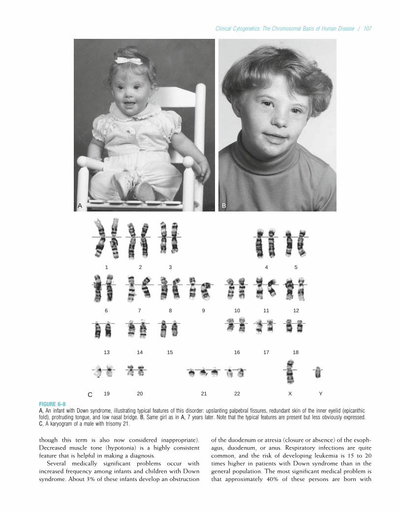
though this term is also now considered inappropriate).Decreased muscle tone (hypotonia) is a highly consistentfeature that is helpful in making a diagnosis.
Several medically significant problems occur withincreased frequency among infants and children with Downsyndrome. About 3% of these infants develop an obstruction
of the duodenum or atresia (closure or absence) of the esoph-agus, duodenum, or anus. Respiratory infections are quitecommon, and the risk of developing leukemia is 15 to 20times higher in patients with Down syndrome than in thegeneral population. The most significant medical problem isthat approximately 40% of these persons are born with
A B
C
1
6
13
19 20 21 22 X Y
14 15 16 17 18
7 8 9 10 11 12
2 3 54
FIGURE 6-8A, An infant with Down syndrome, illustrating typical features of this disorder: upslanting palpebral fissures, redundant skin of the inner eyelid (epicanthicfold), protruding tongue, and low nasal bridge. B, Same girl as in A, 7 years later. Note that the typical features are present but less obviously expressed.C, A karyogram of a male with trisomy 21.
Clinical Cytogenetics: The Chromosomal Basis of Human Disease / 107
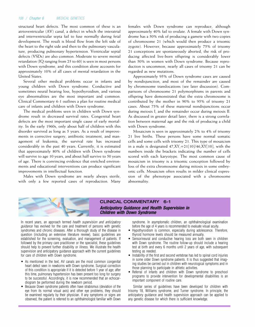
structural heart defects. The most common of these is anatrioventricular (AV) canal, a defect in which the interatrialand interventricular septa fail to fuse normally during fetaldevelopment. The result is blood flow from the left side ofthe heart to the right side and then to the pulmonary vascula-ture, producing pulmonary hypertension. Ventricular septaldefects (VSDs) are also common. Moderate to severe mentalretardation (IQ ranging from 25 to 60) is seen in most personswith Down syndrome, and this condition alone accounts forapproximately 10% of all cases of mental retardation in theUnited States.
Several other medical problems occur in infants andyoung children with Down syndrome. Conductive andsometimes neural hearing loss, hypothyroidism, and variouseye abnormalities are the most important and common.Clinical Commentary 6-1 outlines a plan for routine medicalcare of infants and children with Down syndrome.
The medical problems seen in children with Down syn-drome result in decreased survival rates. Congenital heartdefects are the most important single cause of early mortal-ity. In the early 1960s, only about half of children with thisdisorder survived as long as 5 years. As a result of improve-ments in corrective surgery, antibiotic treatment, and man-agement of leukemia, the survival rate has increasedconsiderably in the past 40 years. Currently, it is estimatedthat approximately 80% of children with Down syndromewill survive to age 10 years, and about half survive to 50 yearsof age. There is convincing evidence that enriched environ-ments and educational interventions can produce significantimprovements in intellectual function.
Males with Down syndrome are nearly always sterile,with only a few reported cases of reproduction. Many
females with Down syndrome can reproduce, althoughapproximately 40% fail to ovulate. A female with Down syn-drome has a 50% risk of producing a gamete with two copiesof chromosome 21 (which would then produce a trisomiczygote). However, because approximately 75% of trisomy21 conceptions are spontaneously aborted, the risk of pro-ducing affected live-born offspring is considerably lowerthan 50% in women with Down syndrome. Because repro-duction is uncommon, nearly all cases of trisomy 21 can beregarded as new mutations.
Approximately 95% of Down syndrome cases are causedby nondisjunction, and most of the remainder are causedby chromosome translocations (see later discussion). Com-parisons of chromosome 21 polymorphisms in parents andoffspring have demonstrated that the extra chromosome iscontributed by the mother in 90% to 95% of trisomy 21cases. About 75% of these maternal nondisjunctions occurduring meiosis I, and the remainder occur during meiosis II.As discussed in greater detail later, there is a strong correla-tion between maternal age and the risk of producing a childwith Down syndrome.
Mosaicism is seen in approximately 2% to 4% of trisomy21 live births. These persons have some normal somaticcells and some cells with trisomy 21. This type of mosaicismin a male is designated 47,XY,þ21[10]/46,XY[10], with thenumbers inside the brackets indicating the number of cellsscored with each karyotype. The most common cause ofmosaicism in trisomy is a trisomic conception followed byloss of the extra chromosome during mitosis in some embry-onic cells. Mosaicism often results in milder clinical expres-sion of the phenotype associated with a chromosomeabnormality.
CLINICAL COMMENTARY 6-1
Anticipatory Guidance and Health Supervision inChildren with Down Syndrome
In recent years, an approach termed health supervision and anticipatoryguidance has evolved for the care and treatment of persons with geneticsyndromes and chronic diseases. After a thorough study of the disease inquestion (including an extensive literature review), basic guidelines areestablished for the screening, evaluation, and management of patients. Iffollowed by the primary care practitioner or the specialist, these guidelinesshould help to prevent further disability or illness. We illustrate the healthsupervision and anticipatory guidance approach with the current guidelinesfor care of children with Down syndrome.
• As mentioned in the text, AV canals are the most common congenitalheart defect seen in newborns with Down syndrome. Surgical correctionof this condition is appropriate if it is detected before 1 year of age; afterthis time, pulmonary hypertension has been present too long for surgeryto be successful. Accordingly, it is now recommended that an echocar-diogram be performed during the newborn period.
• Because Down syndrome patients often have strabismus (deviation of theeye from its normal visual axis) and other eye problems, they shouldbe examined regularly by their physician. If any symptoms or signs areobserved, the patient is referred to an ophthalmologist familiar with Down
syndrome. In asymptomatic children, an ophthalmological examinationbefore the age of 4 years is recommended to evaluate visual acuity.
• Hypothyroidism is common, especially during adolescence. Therefore,thyroid hormone levels should be measured annually.
• Sensorineural and conductive hearing loss are both seen in childrenwith Down syndrome. The routine follow-up should include a hearingtest at birth and every 6 months until 2 years of age, with subsequenttesting as needed.
• Instability of the first and second vertebrae has led to spinal cord injuriesin some older Down syndrome patients. It is thus suggested that imag-ing studies be carried out in children with neurological symptoms and inthose planning to participate in athletic activities.
• Referral of infants and children with Down syndrome to preschoolprograms to provide intervention for developmental disabilities is animportant component of routine care.
Similar series of guidelines have been developed for children withtrisomy 18, Williams syndrome, and Turner syndrome. In principle, theanticipatory guidance and health supervision approach can be applied toany genetic disease for which there is sufficient knowledge.
108 / Chapter 6 MEDICAL GENETICS
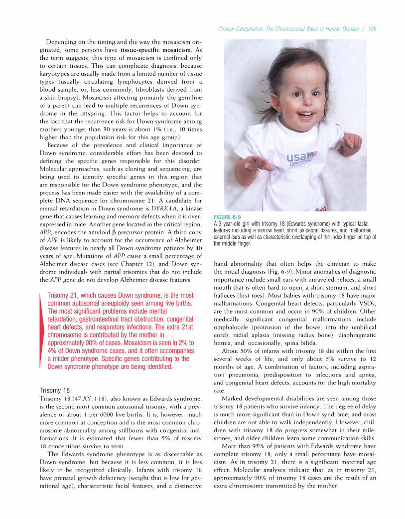
Depending on the timing and the way the mosaicism ori-ginated, some persons have tissue-specific mosaicism. Asthe term suggests, this type of mosaicism is confined onlyto certain tissues. This can complicate diagnosis, becausekaryotypes are usually made from a limited number of tissuetypes (usually circulating lymphocytes derived from ablood sample, or, less commonly, fibroblasts derived froma skin biopsy). Mosaicism affecting primarily the germlineof a parent can lead to multiple recurrences of Down syn-drome in the offspring. This factor helps to account forthe fact that the recurrence risk for Down syndrome amongmothers younger than 30 years is about 1% (i.e., 10 timeshigher than the population risk for this age group).
Because of the prevalence and clinical importance ofDown syndrome, considerable effort has been devoted todefining the specific genes responsible for this disorder.Molecular approaches, such as cloning and sequencing, arebeing used to identify specific genes in this region thatare responsible for the Down syndrome phenotype, and theprocess has been made easier with the availability of a com-plete DNA sequence for chromosome 21. A candidate formental retardation in Down syndrome is DYRK1A, a kinasegene that causes learning and memory defects when it is over-expressed in mice. Another gene located in the critical region,APP, encodes the amyloid b precursor protein. A third copyof APP is likely to account for the occurrence of Alzheimerdisease features in nearly all Down syndrome patients by 40years of age. Mutations of APP cause a small percentage ofAlzheimer disease cases (see Chapter 12), and Down syn-drome individuals with partial trisomies that do not includethe APP gene do not develop Alzheimer disease features.
4Trisomy 21, which causes Down syndrome, is the mostcommon autosomal aneuploidy seen among live births.The most significant problems include mentalretardation, gastrointestinal tract obstruction, congenitalheart defects, and respiratory infections. The extra 21stchromosome is contributed by the mother inapproximately 90% of cases. Mosaicism is seen in 2% to4% of Down syndrome cases, and it often accompaniesa milder phenotype. Specific genes contributing to theDown syndrome phenotype are being identified.
Trisomy 18Trisomy 18 (47,XY,þ18), also known as Edwards syndrome,is the second most common autosomal trisomy, with a prev-alence of about 1 per 6000 live births. It is, however, muchmore common at conception and is the most common chro-mosome abnormality among stillborns with congenital mal-formations. It is estimated that fewer than 5% of trisomy18 conceptions survive to term.
The Edwards syndrome phenotype is as discernable asDown syndrome, but because it is less common, it is lesslikely to be recognized clinically. Infants with trisomy 18have prenatal growth deficiency (weight that is low for ges-tational age), characteristic facial features, and a distinctive
hand abnormality that often helps the clinician to makethe initial diagnosis (Fig. 6-9). Minor anomalies of diagnosticimportance include small ears with unraveled helices, a smallmouth that is often hard to open, a short sternum, and shorthalluces (first toes). Most babies with trisomy 18 have majormalformations. Congenital heart defects, particularly VSDs,are the most common and occur in 90% of children. Othermedically significant congenital malformations includeomphalocele (protrusion of the bowel into the umbilicalcord), radial aplasia (missing radius bone), diaphragmatichernia, and, occasionally, spina bifida.
About 50% of infants with trisomy 18 die within the firstseveral weeks of life, and only about 5% survive to 12months of age. A combination of factors, including aspira-tion pneumonia, predisposition to infections and apnea,and congenital heart defects, accounts for the high mortalityrate.
Marked developmental disabilities are seen among thosetrisomy 18 patients who survive infancy. The degree of delayis much more significant than in Down syndrome, and mostchildren are not able to walk independently. However, chil-dren with trisomy 18 do progress somewhat in their mile-stones, and older children learn some communication skills.
More than 95% of patients with Edwards syndrome havecomplete trisomy 18; only a small percentage have mosai-cism. As in trisomy 21, there is a significant maternal ageeffect. Molecular analyses indicate that, as in trisomy 21,approximately 90% of trisomy 18 cases are the result of anextra chromosome transmitted by the mother.
FIGURE 6-9A 3-year-old girl with trisomy 18 (Edwards syndrome) with typical facialfeatures including a narrow head, short palpebral fissures, and malformedexternal ears as well as characteristic overlapping of the index finger on top ofthe middle finger.
Clinical Cytogenetics: The Chromosomal Basis of Human Disease / 109
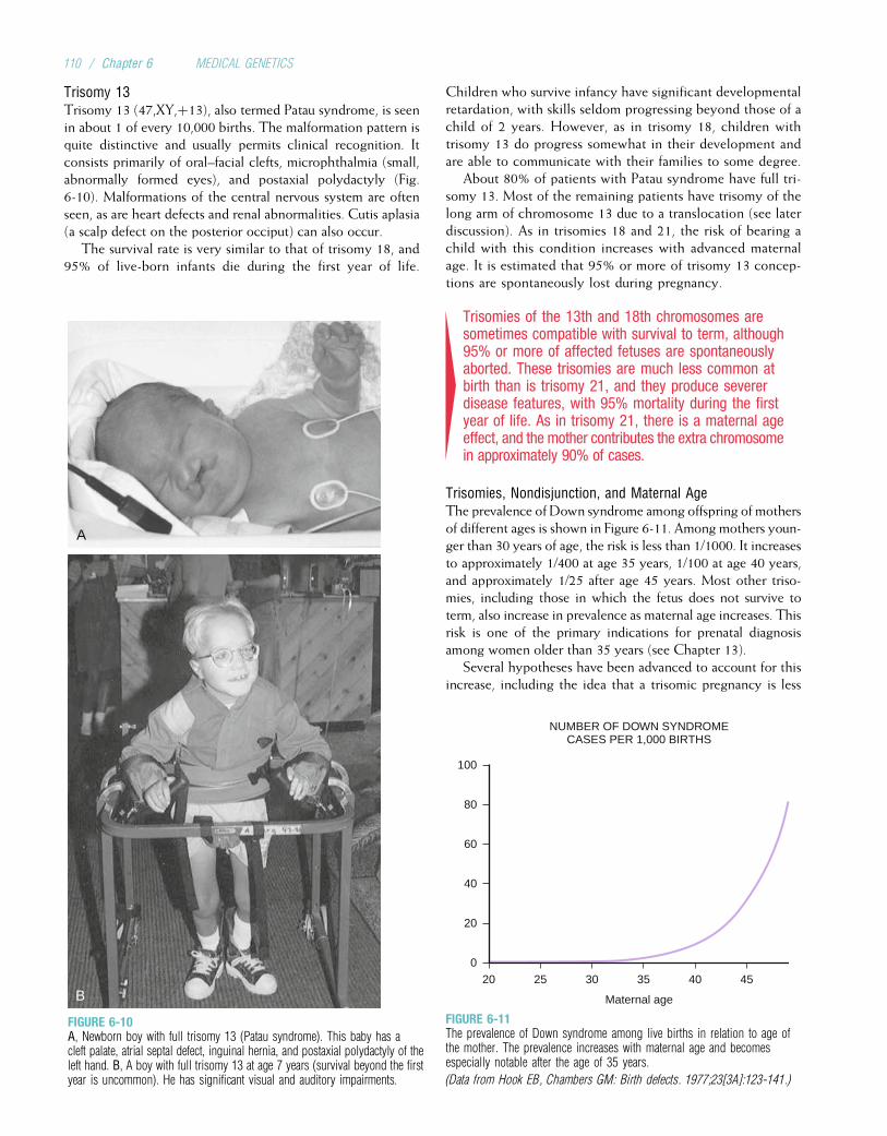
Trisomy 13Trisomy 13 (47,XY,þ13), also termed Patau syndrome, is seenin about 1 of every 10,000 births. The malformation pattern isquite distinctive and usually permits clinical recognition. Itconsists primarily of oral–facial clefts, microphthalmia (small,abnormally formed eyes), and postaxial polydactyly (Fig.6-10). Malformations of the central nervous system are oftenseen, as are heart defects and renal abnormalities. Cutis aplasia(a scalp defect on the posterior occiput) can also occur.
The survival rate is very similar to that of trisomy 18, and95% of live-born infants die during the first year of life.
Children who survive infancy have significant developmentalretardation, with skills seldom progressing beyond those of achild of 2 years. However, as in trisomy 18, children withtrisomy 13 do progress somewhat in their development andare able to communicate with their families to some degree.
About 80% of patients with Patau syndrome have full tri-somy 13. Most of the remaining patients have trisomy of thelong arm of chromosome 13 due to a translocation (see laterdiscussion). As in trisomies 18 and 21, the risk of bearing achild with this condition increases with advanced maternalage. It is estimated that 95% or more of trisomy 13 concep-tions are spontaneously lost during pregnancy.
4Trisomies of the 13th and 18th chromosomes aresometimes compatible with survival to term, although95% or more of affected fetuses are spontaneouslyaborted. These trisomies are much less common atbirth than is trisomy 21, and they produce severerdisease features, with 95% mortality during the firstyear of life. As in trisomy 21, there is a maternal ageeffect, and the mother contributes the extra chromosomein approximately 90% of cases.
Trisomies, Nondisjunction, and Maternal AgeThe prevalence of Down syndrome among offspring of mothersof different ages is shown in Figure 6-11. Among mothers youn-ger than 30 years of age, the risk is less than 1/1000. It increasesto approximately 1/400 at age 35 years, 1/100 at age 40 years,and approximately 1/25 after age 45 years. Most other triso-mies, including those in which the fetus does not survive toterm, also increase in prevalence as maternal age increases. Thisrisk is one of the primary indications for prenatal diagnosisamong women older than 35 years (see Chapter 13).
Several hypotheses have been advanced to account for thisincrease, including the idea that a trisomic pregnancy is less
A
B
FIGURE 6-10A, Newborn boy with full trisomy 13 (Patau syndrome). This baby has acleft palate, atrial septal defect, inguinal hernia, and postaxial polydactyly of theleft hand. B, A boy with full trisomy 13 at age 7 years (survival beyond the firstyear is uncommon). He has significant visual and auditory impairments.
0
NUMBER OF DOWN SYNDROMECASES PER 1,000 BIRTHS
Maternal age
20 25 30 35 40 45
20
40
60
80
100
FIGURE 6-11The prevalence of Down syndrome among live births in relation to age ofthe mother. The prevalence increases with maternal age and becomesespecially notable after the age of 35 years.
(Data from Hook EB, Chambers GM: Birth defects. 1977;23[3A]:123-141.)
110 / Chapter 6 MEDICAL GENETICS
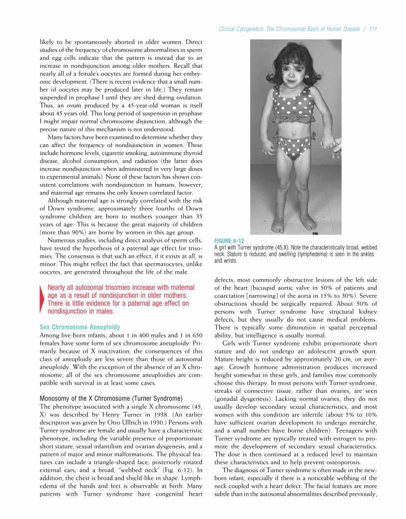
likely to be spontaneously aborted in older women. Directstudies of the frequency of chromosome abnormalities in spermand egg cells indicate that the pattern is instead due to anincrease in nondisjunction among older mothers. Recall thatnearly all of a female’s oocytes are formed during her embry-onic development. (There is recent evidence that a small num-ber of oocytes may be produced later in life.) They remainsuspended in prophase I until they are shed during ovulation.Thus, an ovum produced by a 45-year-old woman is itselfabout 45 years old. This long period of suspension in prophaseI might impair normal chromosome disjunction, although theprecise nature of this mechanism is not understood.
Many factors have been examined to determine whether theycan affect the frequency of nondisjunction in women. Theseinclude hormone levels, cigarette smoking, autoimmune thyroiddisease, alcohol consumption, and radiation (the latter doesincrease nondisjunction when administered in very large dosesto experimental animals). None of these factors has shown con-sistent correlations with nondisjunction in humans, however,and maternal age remains the only known correlated factor.
Although maternal age is strongly correlated with the riskof Down syndrome, approximately three fourths of Downsyndrome children are born to mothers younger than 35years of age. This is because the great majority of children(more than 90%) are borne by women in this age group.
Numerous studies, including direct analysis of sperm cells,have tested the hypothesis of a paternal age effect for triso-mies. The consensus is that such an effect, if it exists at all, isminor. This might reflect the fact that spermatocytes, unlikeoocytes, are generated throughout the life of the male.
4Nearly all autosomal trisomies increase with maternalage as a result of nondisjunction in older mothers.There is little evidence for a paternal age effect onnondisjunction in males.
Sex Chromosome AneuploidyAmong live-born infants, about 1 in 400 males and 1 in 650females have some form of sex chromosome aneuploidy. Pri-marily because of X inactivation, the consequences of thisclass of aneuploidy are less severe than those of autosomalaneuploidy. With the exception of the absence of an X chro-mosome, all of the sex chromosome aneuploidies are com-patible with survival in at least some cases.
Monosomy of the X Chromosome (Turner Syndrome)The phenotype associated with a single X chromosome (45,X) was described by Henry Turner in 1938. (An earlierdescription was given by Otto Ullrich in 1930.) Persons withTurner syndrome are female and usually have a characteristicphenotype, including the variable presence of proportionateshort stature, sexual infantilism and ovarian dysgenesis, and apattern of major and minor malformations. The physical fea-tures can include a triangle-shaped face; posteriorly rotatedexternal ears; and a broad, “webbed neck” (Fig. 6-12). Inaddition, the chest is broad and shield-like in shape. Lymph-edema of the hands and feet is observable at birth. Manypatients with Turner syndrome have congenital heart
defects, most commonly obstructive lesions of the left sideof the heart (bicuspid aortic valve in 50% of patients andcoarctation [narrowing] of the aorta in 15% to 30%). Severeobstructions should be surgically repaired. About 50% ofpersons with Turner syndrome have structural kidneydefects, but they usually do not cause medical problems.There is typically some diminution in spatial perceptualability, but intelligence is usually normal.
Girls with Turner syndrome exhibit proportionate shortstature and do not undergo an adolescent growth spurt.Mature height is reduced by approximately 20 cm, on aver-age. Growth hormone administration produces increasedheight somewhat in these girls, and families now commonlychoose this therapy. In most persons with Turner syndrome,streaks of connective tissue, rather than ovaries, are seen(gonadal dysgenesis). Lacking normal ovaries, they do notusually develop secondary sexual characteristics, and mostwomen with this condition are infertile (about 5% to 10%have sufficient ovarian development to undergo menarche,and a small number have borne children). Teenagers withTurner syndrome are typically treated with estrogen to pro-mote the development of secondary sexual characteristics.The dose is then continued at a reduced level to maintainthese characteristics and to help prevent osteoporosis.
The diagnosis of Turner syndrome is often made in the new-born infant, especially if there is a noticeable webbing of theneck coupled with a heart defect. The facial features are moresubtle than in the autosomal abnormalities described previously,
FIGURE 6-12A girl with Turner syndrome (45,X). Note the characteristically broad, webbedneck. Stature is reduced, and swelling (lymphedema) is seen in the anklesand wrists.
Clinical Cytogenetics: The Chromosomal Basis of Human Disease / 111
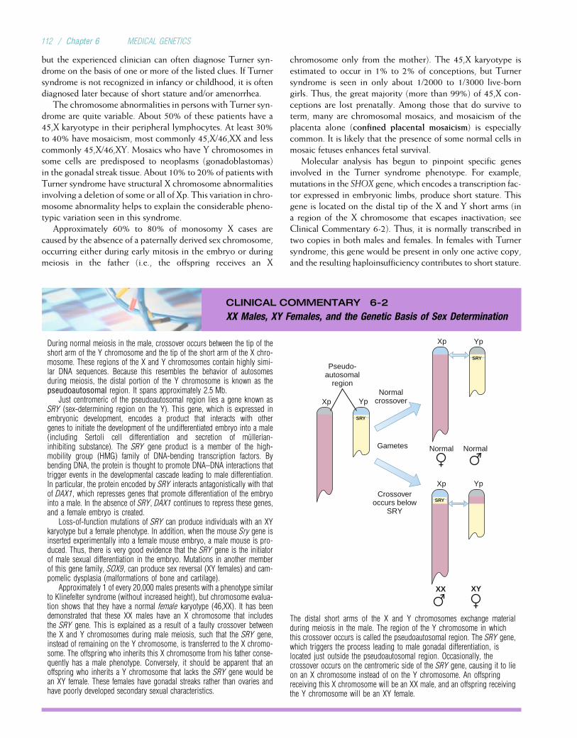
but the experienced clinician can often diagnose Turner syn-drome on the basis of one or more of the listed clues. If Turnersyndrome is not recognized in infancy or childhood, it is oftendiagnosed later because of short stature and/or amenorrhea.
The chromosome abnormalities in persons with Turner syn-drome are quite variable. About 50% of these patients have a45,X karyotype in their peripheral lymphocytes. At least 30%to 40% have mosaicism, most commonly 45,X/46,XX and lesscommonly 45,X/46,XY. Mosaics who have Y chromosomes insome cells are predisposed to neoplasms (gonadoblastomas)in the gonadal streak tissue. About 10% to 20% of patients withTurner syndrome have structural X chromosome abnormalitiesinvolving a deletion of some or all of Xp. This variation in chro-mosome abnormality helps to explain the considerable pheno-typic variation seen in this syndrome.
Approximately 60% to 80% of monosomy X cases arecaused by the absence of a paternally derived sex chromosome,occurring either during early mitosis in the embryo or duringmeiosis in the father (i.e., the offspring receives an X
chromosome only from the mother). The 45,X karyotype isestimated to occur in 1% to 2% of conceptions, but Turnersyndrome is seen in only about 1/2000 to 1/3000 live-borngirls. Thus, the great majority (more than 99%) of 45,X con-ceptions are lost prenatally. Among those that do survive toterm, many are chromosomal mosaics, and mosaicism of theplacenta alone (confined placental mosaicism) is especiallycommon. It is likely that the presence of some normal cells inmosaic fetuses enhances fetal survival.
Molecular analysis has begun to pinpoint specific genesinvolved in the Turner syndrome phenotype. For example,mutations in the SHOX gene, which encodes a transcription fac-tor expressed in embryonic limbs, produce short stature. Thisgene is located on the distal tip of the X and Y short arms (ina region of the X chromosome that escapes inactivation; seeClinical Commentary 6-2). Thus, it is normally transcribed intwo copies in both males and females. In females with Turnersyndrome, this gene would be present in only one active copy,and the resulting haploinsufficiency contributes to short stature.
CLINICAL COMMENTARY 6-2
XX Males, XY Females, and the Genetic Basis of Sex Determination
During normal meiosis in the male, crossover occurs between the tip of theshort arm of the Y chromosome and the tip of the short arm of the X chro-mosome. These regions of the X and Y chromosomes contain highly simi-lar DNA sequences. Because this resembles the behavior of autosomesduring meiosis, the distal portion of the Y chromosome is known as thepseudoautosomal region. It spans approximately 2.5 Mb.
Just centromeric of the pseudoautosomal region lies a gene known asSRY (sex-determining region on the Y). This gene, which is expressed inembryonic development, encodes a product that interacts with othergenes to initiate the development of the undifferentiated embryo into a male(including Sertoli cell differentiation and secretion of mullerian-inhibiting substance). The SRY gene product is a member of the high-mobility group (HMG) family of DNA-bending transcription factors. Bybending DNA, the protein is thought to promote DNA–DNA interactions thattrigger events in the developmental cascade leading to male differentiation.In particular, the protein encoded by SRY interacts antagonistically with thatof DAX1, which represses genes that promote differentiation of the embryointo a male. In the absence of SRY, DAX1 continues to repress these genes,and a female embryo is created.
Loss-of-function mutations of SRY can produce individuals with an XYkaryotype but a female phenotype. In addition, when the mouse Sry gene isinserted experimentally into a female mouse embryo, a male mouse is pro-duced. Thus, there is very good evidence that the SRY gene is the initiatorof male sexual differentiation in the embryo. Mutations in another memberof this gene family, SOX9, can produce sex reversal (XY females) and cam-pomelic dysplasia (malformations of bone and cartilage).
Approximately 1 of every 20,000 males presents with a phenotype similarto Klinefelter syndrome (without increased height), but chromosome evalua-tion shows that they have a normal female karyotype (46,XX). It has beendemonstrated that these XX males have an X chromosome that includesthe SRY gene. This is explained as a result of a faulty crossover betweenthe X and Y chromosomes during male meiosis, such that the SRY gene,instead of remaining on the Y chromosome, is transferred to the X chromo-some. The offspring who inherits this X chromosome from his father conse-quently has a male phenotype. Conversely, it should be apparent that anoffspring who inherits a Y chromosome that lacks the SRY gene would bean XY female. These females have gonadal streaks rather than ovaries andhave poorly developed secondary sexual characteristics.
Xp
XX XY
SRY
SRY
SRY
Yp
Normal Normal
Xp Yp
Crossoveroccurs below
SRY
Gametes
Normalcrossover
Pseudo-autosomal
region
Xp Yp
The distal short arms of the X and Y chromosomes exchange materialduring meiosis in the male. The region of the Y chromosome in whichthis crossover occurs is called the pseudoautosomal region. The SRY gene,which triggers the process leading to male gonadal differentiation, islocated just outside the pseudoautosomal region. Occasionally, thecrossover occurs on the centromeric side of the SRY gene, causing it to lieon an X chromosome instead of on the Y chromosome. An offspringreceiving this X chromosome will be an XX male, and an offspring receivingthe Y chromosome will be an XY female.
112 / Chapter 6 MEDICAL GENETICS
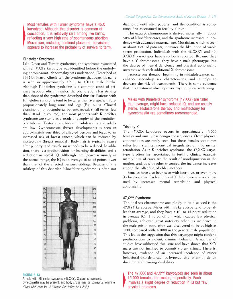
4Most females with Turner syndrome have a 45,Xkaryotype. Although this disorder is common atconception, it is relatively rare among live births,reflecting a very high rate of spontaneous abortion.Mosaicism, including confined placental mosaicism,appears to increase the probability of survival to term.
Klinefelter SyndromeLike Down and Turner syndromes, the syndrome associatedwith a 47,XXY karyotype was identified before the underly-ing chromosomal abnormality was understood. Described in1942 by Harry Klinefelter, the syndrome that bears his nameis seen in approximately 1/500 to 1/1000 male births.Although Klinefelter syndrome is a common cause of pri-mary hypogonadism in males, the phenotype is less strikingthan those of the syndromes described thus far. Patients withKlinefelter syndrome tend to be taller than average, with dis-proportionately long arms and legs (Fig. 6-13). Clinicalexamination of postpubertal patients reveals small testes (lessthan 10 mL in volume), and most patients with Klinefeltersyndrome are sterile as a result of atrophy of the seminifer-ous tubules. Testosterone levels in adolescents and adultsare low. Gynecomastia (breast development) is seen inapproximately one third of affected persons and leads to anincreased risk of breast cancer, which can be reduced bymastectomy (breast removal). Body hair is typically sparseafter puberty, and muscle mass tends to be reduced. In addi-tion, there is a predisposition for learning disabilities and areduction in verbal IQ. Although intelligence is usually inthe normal range, the IQ is on average 10 to 15 points lowerthan that of the affected person’s siblings. Because of thesubtlety of this disorder, Klinefelter syndrome is often not
diagnosed until after puberty, and the condition is some-times first ascertained in fertility clinics.
The extra X chromosome is derived maternally in about50% of Klinefelter cases, and the syndrome increases in inci-dence with advanced maternal age. Mosaicism, which is seenin about 15% of patients, increases the likelihood of viablesperm production. Individuals with the 48,XXXY and 49,XXXXY karyotypes have also been reported. Because theyhave a Y chromosome, they have a male phenotype, butthe degree of mental deficiency and physical abnormalityincreases with each additional X chromosome.
Testosterone therapy, beginning in midadolescence, canenhance secondary sex characteristics, and it helps todecrease the risk of osteoporosis. There is some evidencethat this treatment also improves psychological well-being.
4Males with Klinefelter syndrome (47,XXY) are tallerthan average, might have reduced IQ, and are usuallysterile. Testosterone therapy and mastectomy forgynecomastia are sometimes recommended.
Trisomy XThe 47,XXX karyotype occurs in approximately 1/1000females and usually has benign consequences. Overt physicalabnormalities are rarely seen, but these females sometimessuffer from sterility, menstrual irregularity, or mild mentalretardation. As in Klinefelter syndrome, the 47,XXX karyo-type is often first ascertained in fertility clinics. Approxi-mately 90% of cases are the result of nondisjunction in themother, and, as with other trisomies, the incidence increasesamong the offspring of older mothers.
Females have also been seen with four, five, or even moreX chromosomes. Each additional X chromosome is accompa-nied by increased mental retardation and physicalabnormality.
47,XYY SyndromeThe final sex chromosome aneuploidy to be discussed is the47,XYY karyotype. Males with this karyotype tend to be tal-ler than average, and they have a 10- to 15-point reductionin average IQ. This condition, which causes few physicalproblems, achieved great notoriety when its incidence inthe male prison population was discovered to be as high as1/30, compared with 1/1000 in the general male population.This led to the suggestion that this karyotype might confer apredisposition to violent, criminal behavior. A number ofstudies have addressed this issue and have shown that XYYmales are not inclined to commit violent crimes. There is,however, evidence of an increased incidence of minorbehavioral disorders, such as hyperactivity, attention deficitdisorder, and learning disabilities.
4The 47,XXX and 47,XYY karyotypes are seen in about1/1000 females and males, respectively. Eachinvolves a slight degree of reduction in IQ but fewphysical problems.
FIGURE 6-13A male with Klinefelter syndrome (47,XXY). Stature is increased,gynecomastia may be present, and body shape may be somewhat feminine.
(From McKusick VA: J Chronic Dis 1960; 12-1-202.)
Clinical Cytogenetics: The Chromosomal Basis of Human Disease / 113
CHROMOSOME ABNORMALITIES ANDPREGNANCY LOSSFor a long time it was difficult to detect the early stages ofpregnancy with certainty. It was thus possible for a womanto become pregnant but to lose the embryo before knowingof the pregnancy. Sensitive urinary assays of chorionic gonad-otropin levels, which increase when the embryo implants inthe uterine wall, have allowed researchers to pinpoint accu-rately the occurrence of pregnancy at this early stage. Fol-low-up of women in whom implantation was verified in thisway has shown that about one third of pregnancies are lostafter implantation (the number lost before implantation isunknown). Therefore, spontaneous pregnancy loss is com-mon in humans.
As mentioned earlier, chromosome abnormalities are theleading known cause of pregnancy loss. It is estimated that aminimum of 10% to 20% of conceptions have a chromosomeabnormality, and at least 95% of these chromosomally abnor-mal conceptions are lost before term. Karyotype studies ofmiscarriages indicate that about 50% of the chromosomeabnormalities are trisomies, 20% are monosomies, 15% aretriploids, and the remainder consists of tetraploids and struc-tural abnormalities. Some chromosome abnormalities that arecommon at conception seldom or never survive to term. Forexample, trisomy 16 is thought to be the most common tri-somy at conception, but it is never seen in live births.
It is possible to study chromosome abnormalities directlyin sperm and egg cells. Oocytes are typically obtained fromunused material in in vitro fertilization studies. Karyotypesobtained from these cells indicate that 20% to 25% ofoocytes have missing or extra chromosomes. Human spermcells can be studied by FISH analysis or after fusing themwith hamster oocytes so that their DNA begins mitosis andcondenses, allowing easier visualization. The frequency ofaneuploidy in these sperm cells is about 3% to 4%. Struc-tural abnormalities (see later discussion) are seen in about1% of oocytes and 5% of sperm cells, and the incidenceincreases with advanced paternal age. Undoubtedly, thishigh rate of chromosome abnormality contributes impor-tantly to later pregnancy loss.
These approaches, while informative, can involve somebiases. For example, mothers in whom in vitro fertilizationis performed are not a representative sample of the popula-tion. In addition, their oocytes have been stimulated artifi-cially, and only those oocytes that could not be fertilizedby sperm cells are studied. Thus, the oocytes themselvesmight not be a representative sample. The sperm cells stud-ied in human–hamster hybrids represent only those that arecapable of penetrating the hamster oocyte and again mightnot be a representative sample.
FISH analysis of aneuploidy can evaluate thousands ofcells fairly rapidly, giving it an important advantage overthe human–hamster technique. In general, the FISH studieshave yielded results similar to those of the human–hamsterstudies, showing that on average, the frequency of disomyis approximately 0.15% for each autosome and 0.26% forthe sex chromosomes. These studies have also confirmed a
tendency for elevated frequencies of nondisjunction of thesex chromosomes and some of the acrocentric chromosomes,including chromosome 21, in sperm cells.
4Pregnancy loss is common in humans, withapproximately one third of pregnancies lostspontaneously after implantation. Chromosomeabnormalities, which have been studied in sperm andegg cells and in miscarriages and stillbirths, are animportant cause of pregnancy loss.
ABNORMALITIES OF CHROMOSOME STRUCTUREIn addition to the loss or gain of whole chromosomes, partsof chromosomes can be lost or duplicated as gametes areformed, and the arrangement of portions of chromosomescan be altered. Structural chromosome abnormalities maybe unbalanced (the rearrangement causes a gain or loss ofchromosomal material) or balanced (the rearrangement doesnot produce a loss or gain of chromosome material). Unlikeaneuploidy and polyploidy, balanced structural abnormalitiesoften do not produce serious health consequences. Never-theless, abnormalities of chromosome structure, especiallythose that are unbalanced, can produce serious disease inindividuals or their offspring.
Alterations of chromosome structure can occur whenhomologous chromosomes line up improperly during meiosis(e.g., unequal crossover, as described in Chapter 5). In addi-tion, chromosome breakage can occur during meiosis ormitosis. Mechanisms exist to repair these breaks, and usuallythe break is repaired perfectly with no damage to the daugh-ter cell. Sometimes, however, the breaks remain, or they healin a fashion that alters the structure of the chromosome. Thelikelihood of chromosome breakage may be increased in thepresence of certain harmful agents, called clastogens. Clasto-gens identified in experimental systems include ionizing radi-ation, some viral infections, and some chemicals.
TranslocationsA translocation is the interchange of genetic materialbetween nonhomologous chromosomes. Balanced transloca-tions represent one of the most common chromosomal aber-rations in humans, occurring in 1 of every 500 to 1000individuals (Table 6-2). There are two basic types of translo-cations, reciprocal and Robertsonian.
Reciprocal TranslocationsReciprocal translocations happen when breaks occur in twodifferent chromosomes and the material is mutuallyexchanged. The resulting chromosomes are called derivativechromosomes. The carrier of a reciprocal translocation isusually unaffected because he or she has a normal comple-ment of genetic material. However, the carrier’s offspringcan be normal, can carry the translocation, or can haveduplications or deletions of genetic material.
An example of a reciprocal translocation between chromo-somes 3 and 6 is shown in Figure 6-14. The distal half of the
114 / Chapter 6 MEDICAL GENETICS
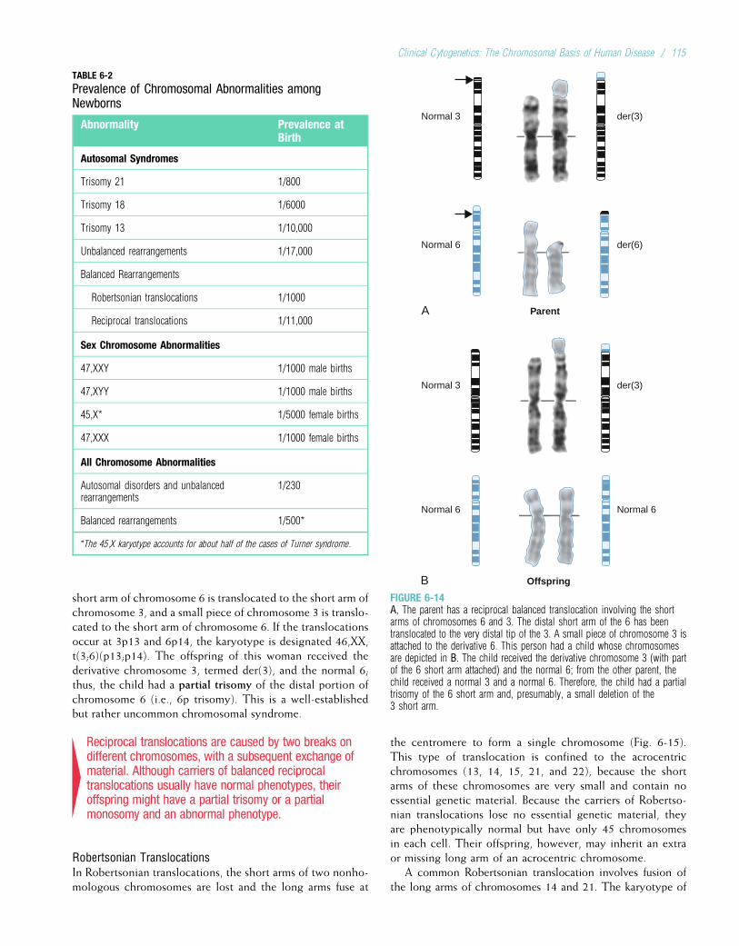
short arm of chromosome 6 is translocated to the short arm ofchromosome 3, and a small piece of chromosome 3 is translo-cated to the short arm of chromosome 6. If the translocationsoccur at 3p13 and 6p14, the karyotype is designated 46,XX,t(3;6)(p13;p14). The offspring of this woman received thederivative chromosome 3, termed der(3), and the normal 6;thus, the child had a partial trisomy of the distal portion ofchromosome 6 (i.e., 6p trisomy). This is a well-establishedbut rather uncommon chromosomal syndrome.
4Reciprocal translocations are caused by two breaks ondifferent chromosomes, with a subsequent exchange ofmaterial. Although carriers of balanced reciprocaltranslocations usually have normal phenotypes, theiroffspring might have a partial trisomy or a partialmonosomy and an abnormal phenotype.
Robertsonian TranslocationsIn Robertsonian translocations, the short arms of two nonho-mologous chromosomes are lost and the long arms fuse at
the centromere to form a single chromosome (Fig. 6-15).This type of translocation is confined to the acrocentricchromosomes (13, 14, 15, 21, and 22), because the shortarms of these chromosomes are very small and contain noessential genetic material. Because the carriers of Robertso-nian translocations lose no essential genetic material, theyare phenotypically normal but have only 45 chromosomesin each cell. Their offspring, however, may inherit an extraor missing long arm of an acrocentric chromosome.
A common Robertsonian translocation involves fusion ofthe long arms of chromosomes 14 and 21. The karyotype of
TABLE 6-2
Prevalence of Chromosomal Abnormalities amongNewborns
Abnormality Prevalence atBirth
Autosomal Syndromes
Trisomy 21 1/800
Trisomy 18 1/6000
Trisomy 13 1/10,000
Unbalanced rearrangements 1/17,000
Balanced Rearrangements
Robertsonian translocations 1/1000
Reciprocal translocations 1/11,000
Sex Chromosome Abnormalities
47,XXY 1/1000 male births
47,XYY 1/1000 male births
45,X* 1/5000 female births
47,XXX 1/1000 female births
All Chromosome Abnormalities
Autosomal disorders and unbalancedrearrangements
1/230
Balanced rearrangements 1/500*
*The 45,X karyotype accounts for about half of the cases of Turner syndrome.
B
Normal 6
Offspring
Normal 6
der(3)Normal 3
Parent
der(6)Normal 6
A
Normal 3 der(3)
FIGURE 6-14A, The parent has a reciprocal balanced translocation involving the shortarms of chromosomes 6 and 3. The distal short arm of the 6 has beentranslocated to the very distal tip of the 3. A small piece of chromosome 3 isattached to the derivative 6. This person had a child whose chromosomesare depicted in B. The child received the derivative chromosome 3 (with partof the 6 short arm attached) and the normal 6; from the other parent, thechild received a normal 3 and a normal 6. Therefore, the child had a partialtrisomy of the 6 short arm and, presumably, a small deletion of the3 short arm.
Clinical Cytogenetics: The Chromosomal Basis of Human Disease / 115
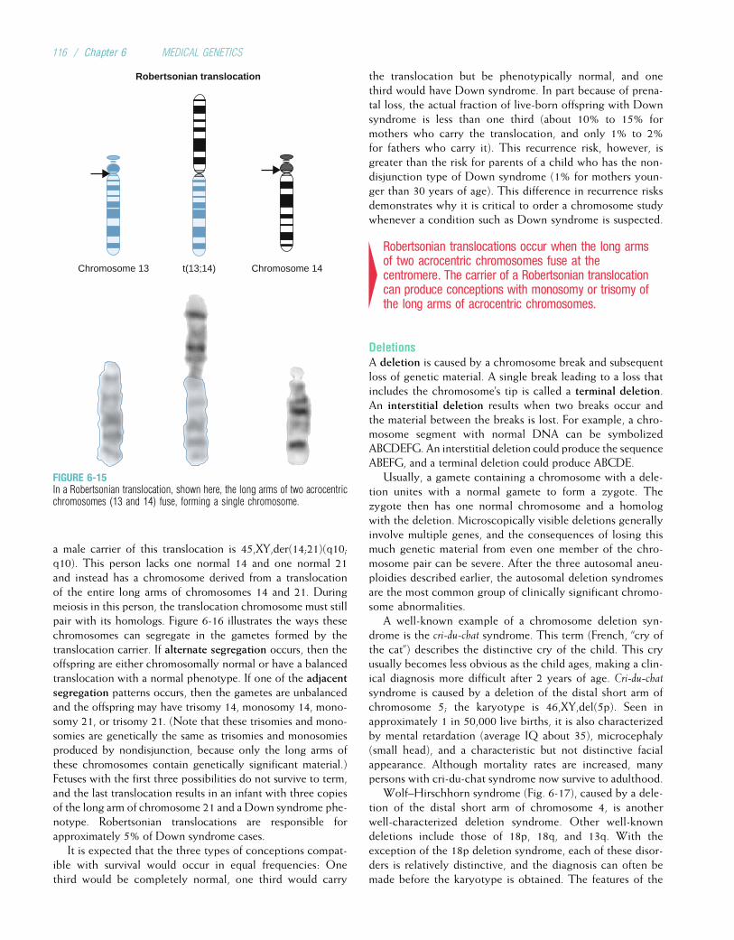
a male carrier of this translocation is 45,XY,der(14;21)(q10;q10). This person lacks one normal 14 and one normal 21and instead has a chromosome derived from a translocationof the entire long arms of chromosomes 14 and 21. Duringmeiosis in this person, the translocation chromosome must stillpair with its homologs. Figure 6-16 illustrates the ways thesechromosomes can segregate in the gametes formed by thetranslocation carrier. If alternate segregation occurs, then theoffspring are either chromosomally normal or have a balancedtranslocation with a normal phenotype. If one of the adjacentsegregation patterns occurs, then the gametes are unbalancedand the offspring may have trisomy 14, monosomy 14, mono-somy 21, or trisomy 21. (Note that these trisomies and mono-somies are genetically the same as trisomies and monosomiesproduced by nondisjunction, because only the long arms ofthese chromosomes contain genetically significant material.)Fetuses with the first three possibilities do not survive to term,and the last translocation results in an infant with three copiesof the long arm of chromosome 21 and a Down syndrome phe-notype. Robertsonian translocations are responsible forapproximately 5% of Down syndrome cases.
It is expected that the three types of conceptions compat-ible with survival would occur in equal frequencies: Onethird would be completely normal, one third would carry
the translocation but be phenotypically normal, and onethird would have Down syndrome. In part because of prena-tal loss, the actual fraction of live-born offspring with Downsyndrome is less than one third (about 10% to 15% formothers who carry the translocation, and only 1% to 2%for fathers who carry it). This recurrence risk, however, isgreater than the risk for parents of a child who has the non-disjunction type of Down syndrome (1% for mothers youn-ger than 30 years of age). This difference in recurrence risksdemonstrates why it is critical to order a chromosome studywhenever a condition such as Down syndrome is suspected.
4Robertsonian translocations occur when the long armsof two acrocentric chromosomes fuse at thecentromere. The carrier of a Robertsonian translocationcan produce conceptions with monosomy or trisomy ofthe long arms of acrocentric chromosomes.
DeletionsA deletion is caused by a chromosome break and subsequentloss of genetic material. A single break leading to a loss thatincludes the chromosome’s tip is called a terminal deletion.An interstitial deletion results when two breaks occur andthe material between the breaks is lost. For example, a chro-mosome segment with normal DNA can be symbolizedABCDEFG. An interstitial deletion could produce the sequenceABEFG, and a terminal deletion could produce ABCDE.
Usually, a gamete containing a chromosome with a dele-tion unites with a normal gamete to form a zygote. Thezygote then has one normal chromosome and a homologwith the deletion. Microscopically visible deletions generallyinvolve multiple genes, and the consequences of losing thismuch genetic material from even one member of the chro-mosome pair can be severe. After the three autosomal aneu-ploidies described earlier, the autosomal deletion syndromesare the most common group of clinically significant chromo-some abnormalities.
A well-known example of a chromosome deletion syn-drome is the cri-du-chat syndrome. This term (French, “cry ofthe cat”) describes the distinctive cry of the child. This cryusually becomes less obvious as the child ages, making a clin-ical diagnosis more difficult after 2 years of age. Cri-du-chatsyndrome is caused by a deletion of the distal short arm ofchromosome 5; the karyotype is 46,XY,del(5p). Seen inapproximately 1 in 50,000 live births, it is also characterizedby mental retardation (average IQ about 35), microcephaly(small head), and a characteristic but not distinctive facialappearance. Although mortality rates are increased, manypersons with cri-du-chat syndrome now survive to adulthood.
Wolf–Hirschhorn syndrome (Fig. 6-17), caused by a dele-tion of the distal short arm of chromosome 4, is anotherwell-characterized deletion syndrome. Other well-knowndeletions include those of 18p, 18q, and 13q. With theexception of the 18p deletion syndrome, each of these disor-ders is relatively distinctive, and the diagnosis can often bemade before the karyotype is obtained. The features of the
Chromosome 14t(13;14)Chromosome 13
Robertsonian translocation
FIGURE 6-15In a Robertsonian translocation, shown here, the long arms of two acrocentricchromosomes (13 and 14) fuse, forming a single chromosome.
116 / Chapter 6 MEDICAL GENETICS
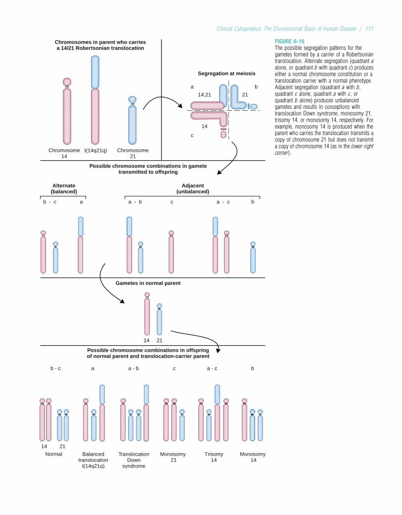
Chromosomes in parent who carriesa 14/21 Robertsonian translocation
Possible chromosome combinations in gametetransmitted to offspring
Gametes in normal parent
Possible chromosome combinations in offspringof normal parent and translocation-carrier parent
Segregation at meiosis
a
14;21
14
c
21
b
Chromosome14
t(14q21q) Chromosome21
Alternate(balanced)
ab - c a - b c a - c b
14 21
Adjacent(unbalanced)
14 21
Normal Monosomy 21
Trisomy14
Monosomy 14
Balancedtranslocation
t(14q21q)
TranslocationDown
syndrome
b - c a a - b c a - c b
FIGURE 6-16The possible segregation patterns for thegametes formed by a carrier of a Robertsoniantranslocation. Alternate segregation (quadrant aalone, or quadrant b with quadrant c) produceseither a normal chromosome constitution or atranslocation carrier with a normal phenotype.Adjacent segregation (quadrant a with b,quadrant c alone, quadrant a with c, orquadrant b alone) produces unbalancedgametes and results in conceptions withtranslocation Down syndrome, monosomy 21,trisomy 14, or monosomy 14, respectively. Forexample, monosomy 14 is produced when theparent who carries the translocation transmits acopy of chromosome 21 but does not transmita copy of chromosome 14 (as in the lower rightcorner).
Clinical Cytogenetics: The Chromosomal Basis of Human Disease / 117
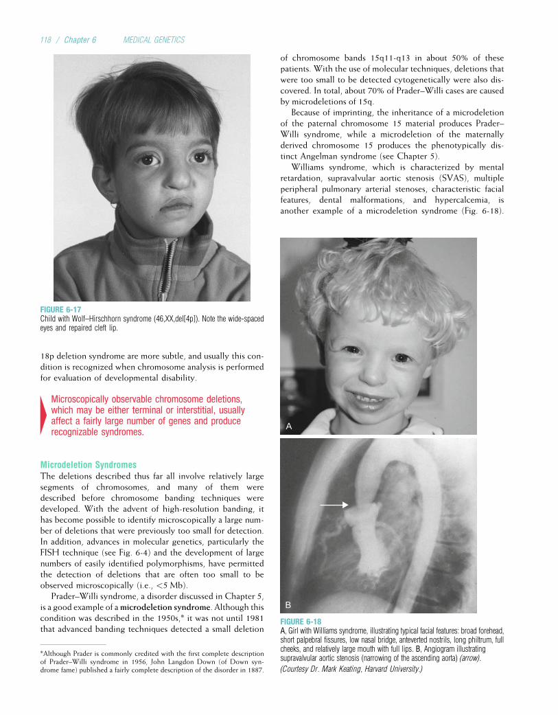
18p deletion syndrome are more subtle, and usually this con-dition is recognized when chromosome analysis is performedfor evaluation of developmental disability.
4Microscopically observable chromosome deletions,which may be either terminal or interstitial, usuallyaffect a fairly large number of genes and producerecognizable syndromes.
Microdeletion SyndromesThe deletions described thus far all involve relatively largesegments of chromosomes, and many of them weredescribed before chromosome banding techniques weredeveloped. With the advent of high-resolution banding, ithas become possible to identify microscopically a large num-ber of deletions that were previously too small for detection.In addition, advances in molecular genetics, particularly theFISH technique (see Fig. 6-4) and the development of largenumbers of easily identified polymorphisms, have permittedthe detection of deletions that are often too small to beobserved microscopically (i.e., <5 Mb).
Prader–Willi syndrome, a disorder discussed in Chapter 5,is a good example of amicrodeletion syndrome. Although thiscondition was described in the 1950s,* it was not until 1981that advanced banding techniques detected a small deletion
of chromosome bands 15q11-q13 in about 50% of thesepatients. With the use of molecular techniques, deletions thatwere too small to be detected cytogenetically were also dis-covered. In total, about 70% of Prader–Willi cases are causedby microdeletions of 15q.
Because of imprinting, the inheritance of a microdeletionof the paternal chromosome 15 material produces Prader–Willi syndrome, while a microdeletion of the maternallyderived chromosome 15 produces the phenotypically dis-tinct Angelman syndrome (see Chapter 5).
Williams syndrome, which is characterized by mentalretardation, supravalvular aortic stenosis (SVAS), multipleperipheral pulmonary arterial stenoses, characteristic facialfeatures, dental malformations, and hypercalcemia, isanother example of a microdeletion syndrome (Fig. 6-18).
FIGURE 6-17Child with Wolf–Hirschhorn syndrome (46,XX,del[4p]). Note the wide-spacedeyes and repaired cleft lip.
A
B
FIGURE 6-18A, Girl with Williams syndrome, illustrating typical facial features: broad forehead,short palpebral fissures, low nasal bridge, anteverted nostrils, long philtrum, fullcheeks, and relatively large mouth with full lips. B, Angiogram illustratingsupravalvular aortic stenosis (narrowing of the ascending aorta) (arrow).
(Courtesy Dr. Mark Keating, Harvard University.)
*Although Prader is commonly credited with the first complete descriptionof Prader–Willi syndrome in 1956, John Langdon Down (of Down syn-drome fame) published a fairly complete description of the disorder in 1887.
118 / Chapter 6 MEDICAL GENETICS
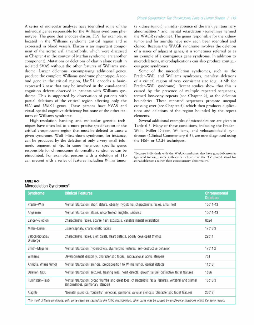
A series of molecular analyses have identified some of theindividual genes responsible for the Williams syndrome phe-notype. The gene that encodes elastin, ELN, for example, islocated in the Williams syndrome critical region and isexpressed in blood vessels. Elastin is an important compo-nent of the aortic wall (microfibrils, which were discussedin Chapter 4 in the context of Marfan syndrome, are anothercomponent). Mutations or deletions of elastin alone result inisolated SVAS without the other features of Williams syn-drome. Larger deletions, encompassing additional genes,produce the complete Williams syndrome phenotype. A sec-ond gene in the critical region, LIMK1, encodes a brain-expressed kinase that may be involved in the visual–spatialcognition defects observed in patients with Williams syn-drome. This is supported by observation of patients withpartial deletions of the critical region affecting only theELN and LIMK1 genes. These persons have SVAS andvisual–spatial cognitive deficiency but none of the other fea-tures of Williams syndrome.
High-resolution banding and molecular genetic tech-niques have often led to a more precise specification of thecritical chromosome region that must be deleted to cause agiven syndrome. Wolf–Hirschhorn syndrome, for instance,can be produced by the deletion of only a very small telo-meric segment of 4p. In some instances, specific genesresponsible for chromosome abnormality syndromes can bepinpointed. For example, persons with a deletion of 11pcan present with a series of features including Wilms tumor
(a kidney tumor), aniridia (absence of the iris), genitourinaryabnormalities,* and mental retardation (sometimes termedthe WAGR syndrome). The genes responsible for the kidneytumor and for aniridia have now each been identified andcloned. Because the WAGR syndrome involves the deletionof a series of adjacent genes, it is sometimes referred to asan example of a contiguous gene syndrome. In addition tomicrodeletions, microduplications can also produce contigu-ous gene syndromes.
Some of the microdeletion syndromes, such as thePrader–Willi and Williams syndromes, manifest deletionsof a critical region of very consistent size (e.g., 4 Mb forPrader–Willi syndrome). Recent studies show that this iscaused by the presence of multiple repeated sequences,termed low-copy repeats (see Chapter 2), at the deletionboundaries. These repeated sequences promote unequalcrossing over (see Chapter 5), which then produces duplica-tions and deletions of the region bounded by the repeatelements.
Several additional examples of microdeletions are given inTable 6-3. Many of these conditions, including the Prader–Willi, Miller–Dieker, Williams, and velocardiofacial syn-dromes (Clinical Commentary 6-3), are now diagnosed usingthe FISH or CGH techniques.
TABLE 6-3
Microdeletion Syndromes*
Syndrome Clinical Features ChromosomalDeletion
Prader–Willi Mental retardation, short stature, obesity, hypotonia, characteristic facies, small feet 15q11-13
Angelman Mental retardation, ataxia, uncontrolled laughter, seizures 15q11-13
Langer–Giedion Characteristic facies, sparse hair, exostosis, variable mental retardation 8q24
Miller–Dieker Lissencephaly, characteristic facies 17p13.3
Velocardiofacial/DiGeorge
Characteristic facies, cleft palate, heart defects, poorly developed thymus 22q11
Smith–Magenis Mental retardation, hyperactivity, dysmorphic features, self-destructive behavior 17p11.2
Williams Developmental disability, characteristic facies, supravalvular aortic stenosis 7q1
Aniridia, Wilms tumor Mental retardation, aniridia, predisposition to Wilms tumor, genital defects 11p13
Deletion 1p36 Mental retardation, seizures, hearing loss, heart defects, growth failure, distinctive facial features 1p36
Rubinstein–Taybi Mental retardation, broad thumbs and great toes, characteristic facial features, vertebral and sternalabnormalities, pulmonary stenosis
16p13.3
Alagille Neonatal jaundice, “butterfly” vertebrae, pulmonic valvular stenosis, characteristic facial features 20p12
*For most of these conditions, only some cases are caused by the listed microdeletion; other cases may be caused by single-gene mutations within the same region.
*Because individuals with the WAGR syndrome also have gonadoblastomas(gonadal tumors), some authorities believe that the "G" should stand forgonadoblastoma rather than genitourinary abnormality.
Clinical Cytogenetics: The Chromosomal Basis of Human Disease / 119
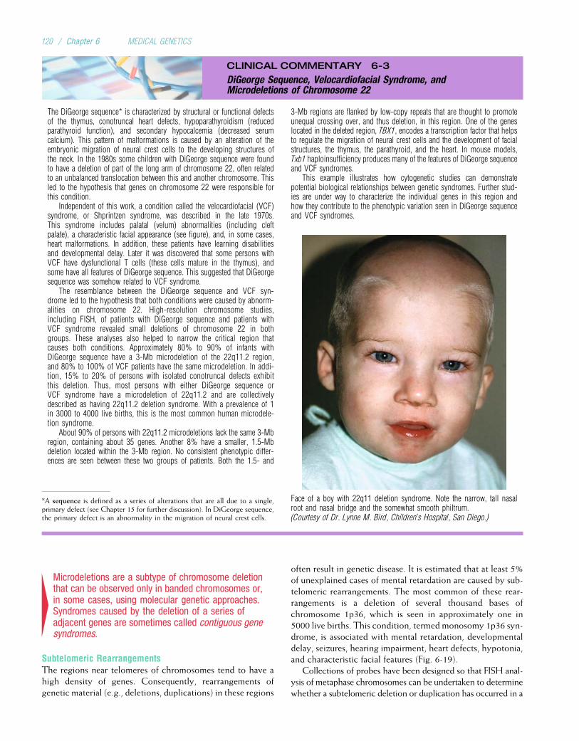
CLINICAL COMMENTARY 6-3
DiGeorge Sequence, Velocardiofacial Syndrome, andMicrodeletions of Chromosome 22
The DiGeorge sequence* is characterized by structural or functional defectsof the thymus, conotruncal heart defects, hypoparathyroidism (reducedparathyroid function), and secondary hypocalcemia (decreased serumcalcium). This pattern of malformations is caused by an alteration of theembryonic migration of neural crest cells to the developing structures ofthe neck. In the 1980s some children with DiGeorge sequence were foundto have a deletion of part of the long arm of chromosome 22, often relatedto an unbalanced translocation between this and another chromosome. Thisled to the hypothesis that genes on chromosome 22 were responsible forthis condition.
Independent of this work, a condition called the velocardiofacial (VCF)syndrome, or Shprintzen syndrome, was described in the late 1970s.This syndrome includes palatal (velum) abnormalities (including cleftpalate), a characteristic facial appearance (see figure), and, in some cases,heart malformations. In addition, these patients have learning disabilitiesand developmental delay. Later it was discovered that some persons withVCF have dysfunctional T cells (these cells mature in the thymus), andsome have all features of DiGeorge sequence. This suggested that DiGeorgesequence was somehow related to VCF syndrome.
The resemblance between the DiGeorge sequence and VCF syn-drome led to the hypothesis that both conditions were caused by abnorm-alities on chromosome 22. High-resolution chromosome studies,including FISH, of patients with DiGeorge sequence and patients withVCF syndrome revealed small deletions of chromosome 22 in bothgroups. These analyses also helped to narrow the critical region thatcauses both conditions. Approximately 80% to 90% of infants withDiGeorge sequence have a 3-Mb microdeletion of the 22q11.2 region,and 80% to 100% of VCF patients have the same microdeletion. In addi-tion, 15% to 20% of persons with isolated conotruncal defects exhibitthis deletion. Thus, most persons with either DiGeorge sequence orVCF syndrome have a microdeletion of 22q11.2 and are collectivelydescribed as having 22q11.2 deletion syndrome. With a prevalence of 1in 3000 to 4000 live births, this is the most common human microdele-tion syndrome.
About 90% of persons with 22q11.2 microdeletions lack the same 3-Mbregion, containing about 35 genes. Another 8% have a smaller, 1.5-Mbdeletion located within the 3-Mb region. No consistent phenotypic differ-ences are seen between these two groups of patients. Both the 1.5- and
3-Mb regions are flanked by low-copy repeats that are thought to promoteunequal crossing over, and thus deletion, in this region. One of the geneslocated in the deleted region, TBX1, encodes a transcription factor that helpsto regulate the migration of neural crest cells and the development of facialstructures, the thymus, the parathyroid, and the heart. In mouse models,Txb1 haploinsufficiency produces many of the features of DiGeorge sequenceand VCF syndromes.
This example illustrates how cytogenetic studies can demonstratepotential biological relationships between genetic syndromes. Further stud-ies are under way to characterize the individual genes in this region andhow they contribute to the phenotypic variation seen in DiGeorge sequenceand VCF syndromes.
Face of a boy with 22q11 deletion syndrome. Note the narrow, tall nasalroot and nasal bridge and the somewhat smooth philtrum.(Courtesy of Dr. Lynne M. Bird, Children’s Hospital, San Diego.)
4Microdeletions are a subtype of chromosome deletionthat can be observed only in banded chromosomes or,in some cases, using molecular genetic approaches.Syndromes caused by the deletion of a series ofadjacent genes are sometimes called contiguous genesyndromes.
Subtelomeric RearrangementsThe regions near telomeres of chromosomes tend to have ahigh density of genes. Consequently, rearrangements ofgenetic material (e.g., deletions, duplications) in these regions
often result in genetic disease. It is estimated that at least 5%of unexplained cases of mental retardation are caused by sub-telomeric rearrangements. The most common of these rear-rangements is a deletion of several thousand bases ofchromosome 1p36, which is seen in approximately one in5000 live births. This condition, termed monosomy 1p36 syn-drome, is associated with mental retardation, developmentaldelay, seizures, hearing impairment, heart defects, hypotonia,and characteristic facial features (Fig. 6-19).
Collections of probes have been designed so that FISH anal-ysis of metaphase chromosomes can be undertaken to determinewhether a subtelomeric deletion or duplication has occurred in a
*A sequence is defined as a series of alterations that are all due to a single,primary defect (see Chapter 15 for further discussion). In DiGeorge sequence,the primary defect is an abnormality in the migration of neural crest cells.
120 / Chapter 6 MEDICAL GENETICS
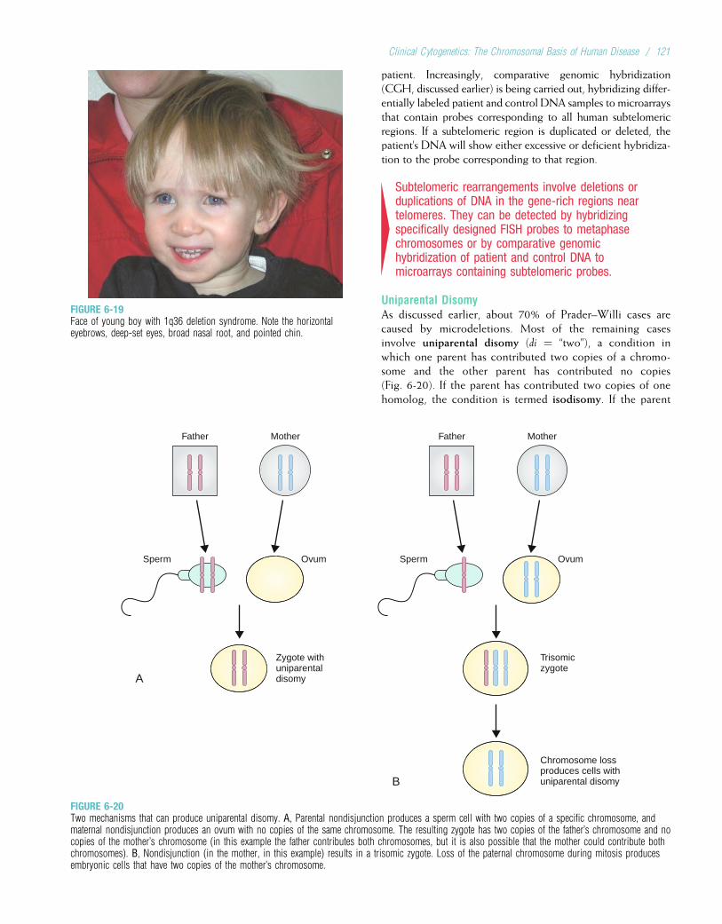
patient. Increasingly, comparative genomic hybridization(CGH, discussed earlier) is being carried out, hybridizing differ-entially labeled patient and control DNA samples to microarraysthat contain probes corresponding to all human subtelomericregions. If a subtelomeric region is duplicated or deleted, thepatient’s DNA will show either excessive or deficient hybridiza-tion to the probe corresponding to that region.
4Subtelomeric rearrangements involve deletions orduplications of DNA in the gene-rich regions neartelomeres. They can be detected by hybridizingspecifically designed FISH probes to metaphasechromosomes or by comparative genomichybridization of patient and control DNA tomicroarrays containing subtelomeric probes.
Uniparental DisomyAs discussed earlier, about 70% of Prader–Willi cases arecaused by microdeletions. Most of the remaining casesinvolve uniparental disomy (di ¼ “two”), a condition inwhich one parent has contributed two copies of a chromo-some and the other parent has contributed no copies(Fig. 6-20). If the parent has contributed two copies of onehomolog, the condition is termed isodisomy. If the parent
FIGURE 6-19Face of young boy with 1q36 deletion syndrome. Note the horizontaleyebrows, deep-set eyes, broad nasal root, and pointed chin.
A
B
Father Mother
Ovum
Zygote withuniparentaldisomy
Sperm
Father Mother
Ovum
Trisomiczygote
Chromosome lossproduces cells withuniparental disomy
Sperm
FIGURE 6-20Two mechanisms that can produce uniparental disomy. A, Parental nondisjunction produces a sperm cell with two copies of a specific chromosome, andmaternal nondisjunction produces an ovum with no copies of the same chromosome. The resulting zygote has two copies of the father’s chromosome and nocopies of the mother’s chromosome (in this example the father contributes both chromosomes, but it is also possible that the mother could contribute bothchromosomes). B, Nondisjunction (in the mother, in this example) results in a trisomic zygote. Loss of the paternal chromosome during mitosis producesembryonic cells that have two copies of the mother’s chromosome.
Clinical Cytogenetics: The Chromosomal Basis of Human Disease / 121
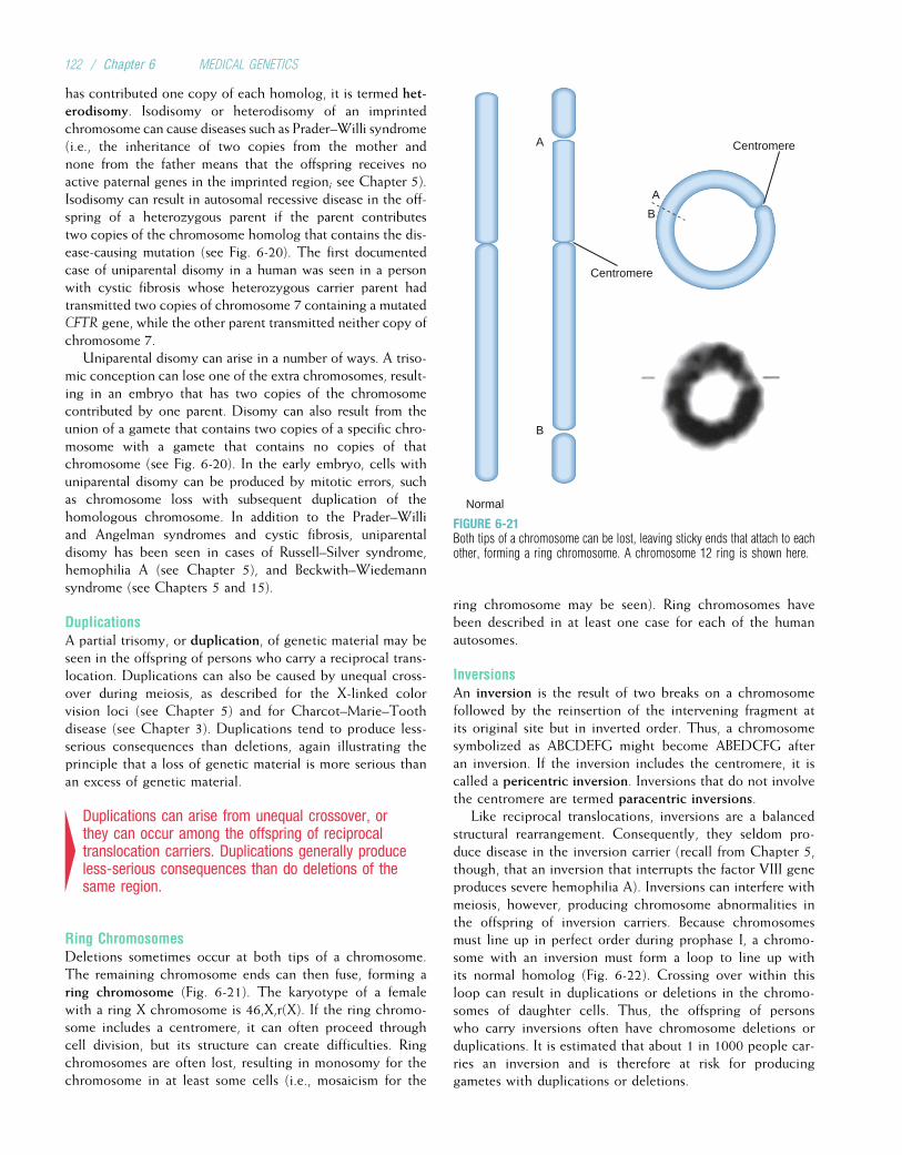
has contributed one copy of each homolog, it is termed het-erodisomy. Isodisomy or heterodisomy of an imprintedchromosome can cause diseases such as Prader–Willi syndrome(i.e., the inheritance of two copies from the mother andnone from the father means that the offspring receives noactive paternal genes in the imprinted region; see Chapter 5).Isodisomy can result in autosomal recessive disease in the off-spring of a heterozygous parent if the parent contributestwo copies of the chromosome homolog that contains the dis-ease-causing mutation (see Fig. 6-20). The first documentedcase of uniparental disomy in a human was seen in a personwith cystic fibrosis whose heterozygous carrier parent hadtransmitted two copies of chromosome 7 containing a mutatedCFTR gene, while the other parent transmitted neither copy ofchromosome 7.
Uniparental disomy can arise in a number of ways. A triso-mic conception can lose one of the extra chromosomes, result-ing in an embryo that has two copies of the chromosomecontributed by one parent. Disomy can also result from theunion of a gamete that contains two copies of a specific chro-mosome with a gamete that contains no copies of thatchromosome (see Fig. 6-20). In the early embryo, cells withuniparental disomy can be produced by mitotic errors, suchas chromosome loss with subsequent duplication of thehomologous chromosome. In addition to the Prader–Williand Angelman syndromes and cystic fibrosis, uniparentaldisomy has been seen in cases of Russell–Silver syndrome,hemophilia A (see Chapter 5), and Beckwith–Wiedemannsyndrome (see Chapters 5 and 15).
DuplicationsA partial trisomy, or duplication, of genetic material may beseen in the offspring of persons who carry a reciprocal trans-location. Duplications can also be caused by unequal cross-over during meiosis, as described for the X-linked colorvision loci (see Chapter 5) and for Charcot–Marie–Toothdisease (see Chapter 3). Duplications tend to produce less-serious consequences than deletions, again illustrating theprinciple that a loss of genetic material is more serious thanan excess of genetic material.
4Duplications can arise from unequal crossover, orthey can occur among the offspring of reciprocaltranslocation carriers. Duplications generally produceless-serious consequences than do deletions of thesame region.
Ring ChromosomesDeletions sometimes occur at both tips of a chromosome.The remaining chromosome ends can then fuse, forming aring chromosome (Fig. 6-21). The karyotype of a femalewith a ring X chromosome is 46,X,r(X). If the ring chromo-some includes a centromere, it can often proceed throughcell division, but its structure can create difficulties. Ringchromosomes are often lost, resulting in monosomy for thechromosome in at least some cells (i.e., mosaicism for the
ring chromosome may be seen). Ring chromosomes havebeen described in at least one case for each of the humanautosomes.
InversionsAn inversion is the result of two breaks on a chromosomefollowed by the reinsertion of the intervening fragment atits original site but in inverted order. Thus, a chromosomesymbolized as ABCDEFG might become ABEDCFG afteran inversion. If the inversion includes the centromere, it iscalled a pericentric inversion. Inversions that do not involvethe centromere are termed paracentric inversions.
Like reciprocal translocations, inversions are a balancedstructural rearrangement. Consequently, they seldom pro-duce disease in the inversion carrier (recall from Chapter 5,though, that an inversion that interrupts the factor VIII geneproduces severe hemophilia A). Inversions can interfere withmeiosis, however, producing chromosome abnormalities inthe offspring of inversion carriers. Because chromosomesmust line up in perfect order during prophase I, a chromo-some with an inversion must form a loop to line up withits normal homolog (Fig. 6-22). Crossing over within thisloop can result in duplications or deletions in the chromo-somes of daughter cells. Thus, the offspring of personswho carry inversions often have chromosome deletions orduplications. It is estimated that about 1 in 1000 people car-ries an inversion and is therefore at risk for producinggametes with duplications or deletions.
B
Normal
A
Centromere
A
B
Centromere
FIGURE 6-21Both tips of a chromosome can be lost, leaving sticky ends that attach to eachother, forming a ring chromosome. A chromosome 12 ring is shown here.
122 / Chapter 6 MEDICAL GENETICS
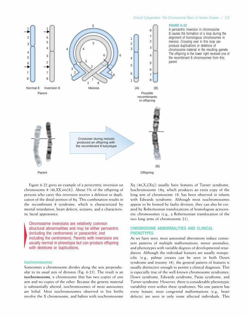
Figure 6-22 gives an example of a pericentric inversion onchromosome 8 (46,XX,inv[8]). About 5% of the offspring ofpersons who carry this inversion receive a deletion or dupli-cation of the distal portion of 8q. This combination results inthe recombinant 8 syndrome, which is characterized bymental retardation, heart defects, seizures, and a characteris-tic facial appearance.
4Chromosome inversions are relatively commonstructural abnormalities and may be either pericentric(including the centromere) or paracentric (notincluding the centromere). Parents with inversions areusually normal in phenotype but can produce offspringwith deletions or duplications.
IsochromosomesSometimes a chromosome divides along the axis perpendic-ular to its usual axis of division (Fig. 6-23). The result is anisochromosome, a chromosome that has two copies of onearm and no copies of the other. Because the genetic materialis substantially altered, isochromosomes of most autosomesare lethal. Most isochromosomes observed in live birthsinvolve the X chromosome, and babies with isochromosome
Xq (46,X,i[Xq]) usually have features of Turner syndrome.Isochromosome 18q, which produces an extra copy of thelong arm of chromosome 18, has been observed in infantswith Edwards syndrome. Although most isochromosomesappear to be formed by faulty division, they can also be cre-ated by Robertsonian translocations of homologous acrocen-tric chromosomes (e.g., a Robertsonian translocation of thetwo long arms of chromosome 21).
CHROMOSOME ABNORMALITIES AND CLINICALPHENOTYPESAs we have seen, most autosomal aberrations induce consis-tent patterns of multiple malformations, minor anomalies,and phenotypes with variable degrees of developmental retar-dation. Although the individual features are usually nonspe-cific (e.g., palmar creases can be seen in both Downsyndrome and trisomy 18), the general pattern of features isusually distinctive enough to permit a clinical diagnosis. Thisis especially true of the well-known chromosome syndromes:Down syndrome, Edwards syndrome, Patau syndrome, andTurner syndrome. However, there is considerable phenotypicvariability even within these syndromes. No one patient hasevery feature; most congenital malformations (e.g., heartdefects) are seen in only some affected individuals. This
(B)
OffspringParent
8
7
2
3
4
5
6
7
8
4
5
6
1
1
2
3
6 6
87
5
5
44
2
3
32
87
1
Crossover during meiosisproduced an offspring with
the recombinant 8 karyotype
1
2
3
4
5
6
7
8
7
8
1
6
5
4
3
2
Normal 8 Inversion 8
Parent
Meiosis
Possiblerecombinantsin offspring
(A) (B)
1
FIGURE 6-22A pericentric inversion in chromosome8 causes the formation of a loop during thealignment of homologous chromosomes inmeiosis. Crossing over in this loop canproduce duplications or deletions ofchromosome material in the resulting gamete.The offspring in the lower right received one ofthe recombinant 8 chromosomes from thisparent.
Clinical Cytogenetics: The Chromosomal Basis of Human Disease / 123
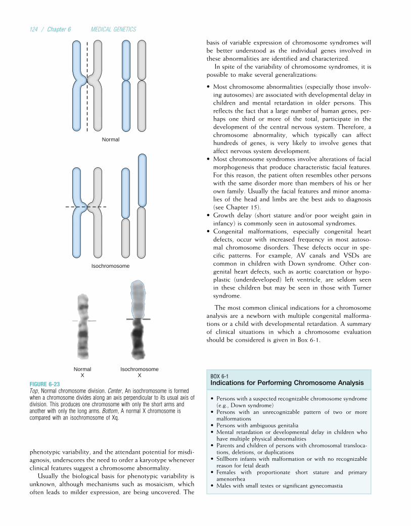
phenotypic variability, and the attendant potential for misdi-agnosis, underscores the need to order a karyotype wheneverclinical features suggest a chromosome abnormality.
Usually the biological basis for phenotypic variability isunknown, although mechanisms such as mosaicism, whichoften leads to milder expression, are being uncovered. The
basis of variable expression of chromosome syndromes willbe better understood as the individual genes involved inthese abnormalities are identified and characterized.
In spite of the variability of chromosome syndromes, it ispossible to make several generalizations:
• Most chromosome abnormalities (especially those involv-ing autosomes) are associated with developmental delay inchildren and mental retardation in older persons. Thisreflects the fact that a large number of human genes, per-haps one third or more of the total, participate in thedevelopment of the central nervous system. Therefore, achromosome abnormality, which typically can affecthundreds of genes, is very likely to involve genes thataffect nervous system development.
• Most chromosome syndromes involve alterations of facialmorphogenesis that produce characteristic facial features.For this reason, the patient often resembles other personswith the same disorder more than members of his or herown family. Usually the facial features and minor anoma-lies of the head and limbs are the best aids to diagnosis(see Chapter 15).
• Growth delay (short stature and/or poor weight gain ininfancy) is commonly seen in autosomal syndromes.
• Congenital malformations, especially congenital heartdefects, occur with increased frequency in most autoso-mal chromosome disorders. These defects occur in spe-cific patterns. For example, AV canals and VSDs arecommon in children with Down syndrome. Other con-genital heart defects, such as aortic coarctation or hypo-plastic (underdeveloped) left ventricle, are seldom seenin these children but may be seen in those with Turnersyndrome.
The most common clinical indications for a chromosomeanalysis are a newborn with multiple congenital malforma-tions or a child with developmental retardation. A summaryof clinical situations in which a chromosome evaluationshould be considered is given in Box 6-1.
IsochromosomeX
NormalX
Isochromosome
Normal
FIGURE 6-23Top, Normal chromosome division. Center, An isochromosome is formedwhen a chromosome divides along an axis perpendicular to its usual axis ofdivision. This produces one chromosome with only the short arms andanother with only the long arms. Bottom, A normal X chromosome iscompared with an isochromosome of Xq.
BOX 6-1Indications for Performing Chromosome Analysis
• Persons with a suspected recognizable chromosome syndrome(e.g., Down syndrome)
• Persons with an unrecognizable pattern of two or moremalformations
• Persons with ambiguous genitalia• Mental retardation or developmental delay in children whohave multiple physical abnormalities
• Parents and children of persons with chromosomal transloca-tions, deletions, or duplications
• Stillborn infants with malformation or with no recognizablereason for fetal death
• Females with proportionate short stature and primaryamenorrhea
• Males with small testes or significant gynecomastia
124 / Chapter 6 MEDICAL GENETICS
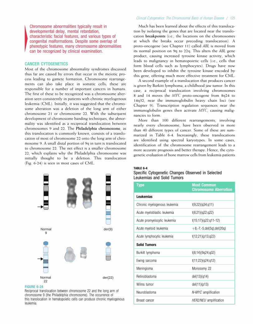
4Chromosome abnormalities typically result indevelopmental delay, mental retardation,characteristic facial features, and various types ofcongenital malformations. Despite some overlap ofphenotypic features, many chromosome abnormalitiescan be recognized by clinical examination.
CANCER CYTOGENETICSMost of the chromosome abnormality syndromes discussedthus far are caused by errors that occur in the meiotic pro-cess leading to gamete formation. Chromosome rearrange-ments can also take place in somatic cells; these areresponsible for a number of important cancers in humans.The first of these to be recognized was a chromosome alter-ation seen consistently in patients with chronic myelogenousleukemia (CML). Initially, it was suggested that the chromo-some alteration was a deletion of the long arm of eitherchromosome 21 or chromosome 22. With the subsequentdevelopment of chromosome banding techniques, the abnor-mality was identified as a reciprocal translocation betweenchromosomes 9 and 22. The Philadelphia chromosome, asthis translocation is commonly known, consists of a translo-cation of most of chromosome 22 onto the long arm of chro-mosome 9. A small distal portion of 9q in turn is translocatedto chromosome 22. The net effect is a smaller chromosome22, which explains why the Philadelphia chromosome wasinitially thought to be a deletion. This translocation(Fig. 6-24) is seen in most cases of CML.
Much has been learned about the effects of this transloca-tion by isolating the genes that are located near the translo-cation breakpoints (i.e., the locations on the chromosomesat which the breaks occur preceding translocation). Aproto-oncogene (see Chapter 11) called ABL is moved fromits normal position on 9q to 22q. This alters the ABL geneproduct, causing increased tyrosine kinase activity, whichleads to malignancy in hematopoietic cells (i.e., cells thatform blood cells such as lymphocytes). Drugs have nowbeen developed to inhibit the tyrosine kinase encoded bythis gene, offering much more effective treatment for CML.
A second example of a translocation that produces canceris given by Burkitt lymphoma, a childhood jaw tumor. In thiscase, a reciprocal translocation involving chromosomes8 and 14 moves the MYC proto-oncogene from 8q24 to14q32, near the immunoglobulin heavy chain loci (seeChapter 9). Transcription regulation sequences near theimmunoglobulin genes then activate MYC, causing malig-nancies to form.
More than 100 different rearrangements, involvingnearly every chromosome, have been observed in morethan 40 different types of cancer. Some of these are sum-marized in Table 6-4. Increasingly, these translocationsare identified using spectral karyotypes. In some cases,identification of the chromosome rearrangement leads to amore accurate prognosis and better therapy. Hence, the cyto-genetic evaluation of bone marrow cells from leukemia patients
der(22)Normal22
der(9)Normal9
FIGURE 6-24Reciprocal translocation between chromosome 22 and the long arm ofchromosome 9 (the Philadelphia chromosome). The occurrence ofthis translocation in hematopoietic cells can produce chronic myelogenousleukemia.
TABLE 6-4
Specific Cytogenetic Changes Observed in SelectedLeukemias and Solid Tumors
Type Most CommonChromosome Aberration
Leukemias
Chronic myelogenous leukemia t(9;22)(q34;q11)
Acute myeloblastic leukemia t(8;21)(q22;q22)
Acute promyelocytic leukemia t(15;17)(q22;q11-12)
Acute myeloid leukemia þ8,-7,-5,del(5q),del(20q)
Acute lymphocytic leukemia t(12;21)(p13;q22)
Solid Tumors
Burkitt lymphoma t(8;14)(9q24;q32)
Ewing sarcoma t(11;22)(q24;q12)
Meningioma Monosomy 22
Retinoblastoma del(13)(q14)
Wilms tumor del(11)(p13)
Neuroblastoma N-MYC amplification
Breast cancer HER2/NEU amplification
Clinical Cytogenetics: The Chromosomal Basis of Human Disease / 125
is a routine part of diagnosis. Furthermore, identification andcharacterization of the genes that are altered in translocationsyndromes are leading to a better understanding of carcinogen-esis in general.
4Balanced translocations in somatic cells cansometimes cause malignancies by interrupting oraltering genes or their regulatory sequences.
CHROMOSOME INSTABILITY SYNDROMESSeveral autosomal recessive disease conditions exhibit anincreased incidence of chromosome breaks under specificlaboratory conditions. These conditions, which are termedchromosome instability syndromes, include ataxia–telangiectasia,
Bloom syndrome, Fanconi anemia, and xeroderma pigmentosum(see Chapter 2). Among patients with Fanconi anemia, the fre-quency of breaks can be increased further if the chromosomesare exposed to certain alkylating agents. Patients with Bloom syn-drome also have a high incidence of somatic cell sister chromatidexchange (exchange of chromosome material between sisterchromatids; see Chapter 2). Each of these syndromes is asso-ciated with a significant increase in cancer risk. All are thoughtto be the result of faulty DNA replication or repair, as discussedin Chapter 2.
4The chromosome instability syndromes all involveincreased frequencies of chromosome breakage andan increased risk of malignancy. All are associatedwith defects in DNA replication or repair.
Study Questions
1. Distinguish among haploidy, diploidy, polyploidy,euploidy, and aneuploidy.
2. Explain the uses and relative advantages of FISH,spectral karyotyping, and comparative genomichybridization (CGH).
3. Describe three ways in which triploidy could arise.
4. Studies of karyotypes obtained by prenatal diagnosisat 10 weeks’ gestation (chorionic villus sampling; seeChapter 13) reveal prevalence rates of chromosomeabnormalities that differ from those obtained inkaryotypes at 16 weeks’ gestation (amniocentesis; seeChapter 13). Explain this.
5. Even though conditions such as Down syndrome andEdwards syndrome can usually be diagnosedaccurately by clinical examination alone, a karyotypeis always recommended. Why?
6. Rank the following, from lowest to highest, in termsof the risk of producing a child with Downsyndrome:45-year-old woman with no previous family historyof Down syndrome
25-year-old woman who has had one previous childwith Down syndrome25-year-old male carrier of a 21/14 Robertsoniantranslocation25-year-old female carrier of a 21/14 Robertsoniantranslocation
7. Females with the 49,XXXXX karyotype have beenreported. Explain how this karyotype could occur.
8. A man with hemophilia A and a normal womanproduce a child with Turner syndrome (45,X). Thechild has normal factor VIII activity. In which parentdid the meiotic error occur?
9. A cytogenetics laboratory reports a karyotype of 46,XY,del(8)(p11) for one patient and a karyotype of 46,XY,dup(8)(p11) for another patient. Based on thisinformation alone, which patient is expected to bemore severely affected?
10. Why do translocations in somatic cells sometimeslead to cancer?
Suggested ReadingsAlbertson DG, Collins C, McCormick F, Gray JW. Chromo-
some aberrations in solid tumors. Nat Genet 2003;34(4):369–76.
American Academy of Pediatrics. Committee on Genetics.Health supervision for children with Down syndrome.Pediatrics 2001;107(2):442–9.
Antonarakis SE, Epstein CJ. The challenge of Down syn-drome. Trends Mol Med 2006;12(10):473–9.
Aradhya S, Cherry AM. Array-based comparative genomichybridization. Clinical contexts for targeted and whole-genome designs. Genet Med 2007;9(9):553–9.
Battaglia A. Del 1p36 syndrome: A newly emerging clinicalentity. Brain and Development 2005;27(5):358–61.
Blaschke RJ, Rappold G. The pseudoautosomal regions,SHOX and disease. Curr Opin Genet Dev 2006;16(3):233–9.
126 / Chapter 6 MEDICAL GENETICS
Carey JC. Trisomy 18 and 13 syndromes. In Cassidy SB,Allanson JE (eds): Management of Genetic Syndromes.Hoboken, NJ: Wiley–Liss, 2005, pp. 555–68.
Emanuel BS, Saitta SC. From microscopes to microarrays.Dissecting recurrent chromosomal rearrangements. NatRev Genet 2007;8(11):869–83.
Fleming A, Vilain E. The endless quest for sex determinationgenes. Clin Genet 2005;67(1):15–25.
Frohling S, Dohner H. Chromosomal abnormalities in can-cer. N Engl J Med 2008;359(7):722–34.
Gartler SM. The chromosome number in humans. A briefhistory. Nat Rev Genet 2006;7(8):655–60.
Gotz MJ, Johnstone EC, Ratcliffe SG. Criminality and antiso-cial behaviour in unselected men with sex chromosomeabnormalities. Psychol Med 1999;29(4):953–62.
Graham GE, Allanson JE, Gerritsen JA. Sex chromosomeabnormalities. In: Rimoin DL, Connor JM, Pyeritz RE,Korf BR (eds): Emery and Rimoin’s Principles and Prac-tice of Medical Genetics, 5th ed. London: ChurchillLivingstone; 2007, pp. 1038–57.
Hall H, Hunt P, Hassold T. Meiosis and sex chromosomeaneuploidy: How meiotic errors cause aneuploidy; howaneuploidy causes meiotic errors. Curr Opin Genet Dev2006;16(3):323–9.
Kobrynski LJ, Sullivan KE. Velocardiofacial syndrome,DiGeorge syndrome: The chromosome 22q11.2 deletionsyndromes. Lancet 2007;370(9596):1443–52.
Lanfranco F, Kamischke A, Zitzmann M, Nieschlag E. Kline-felter’s syndrome. Lancet 2004;364(9430):273–83.
Lee C, Iafrate AJ, Brothman AR. Copy number variations andclinical cytogenetic diagnosis of constitutional disorders.Nat Genet 2007;39(Suppl 7):S48–S54.
Loscalzo ML. Turner syndrome. Pediatr Rev 2008;29(7):219–27.
Patterson D, Costa AC. Down syndrome and genetics—acase of linked histories. Nat Rev Genet 2005;6(2): 137–47.
Pober BR, Morris CA. Diagnosis and management of medicalproblems in adults with Williams–Beuren syndrome. Am JMed Genet C Semin Med Genet 2007;145(3):280–90.
Robinson WP. Mechanisms leading to uniparental disomy andtheir clinical consequences. Bioessays 2000;22(5):452–9.
Roizen NJ, Patterson D. Down’s syndrome. Lancet 2003;361(9365):1281–9.
Schinzel A. Catalogue of Unbalanced Chromosome Aberra-tions. Berlin: Walter de Gruyter, 2001.
Shaffer LG, Tommerup N (eds): An International System forHuman Cytogenetic Nomenclature. Basel: S. Karger,2005.
Speicher MR, Carter NP. The new cytogenetics. Blurring theboundaries with molecular biology. Nat Rev Genet 2005;6(10):782–92.
Spinner NB, Saitta SC, Emanuel BS. Deletions and other struc-tural abnormalities of the autosomes. In: Rimoin DL, ConnorJM, Pyeritz RE, Korf BR (eds): Emery and Rimoin’s Principlesand Practice of Medical Genetics, 5th ed. London: ChurchillLivingstone, 2007, pp. 1058–82.
Stankiewicz P, Beaudet AL. Use of array CGH in the evalu-ation of dysmorphology, malformations, developmentaldelay, and idiopathic mental retardation. Curr OpinGenet Dev 2007;17(3):182–92.
Sybert VP, McCauley E. Turner’s syndrome. N Engl J Med2004;351(12):1227–38.
Tolmie JL, MacFayden U. Clinical genetics of common auto-somal trisomies. In: Rimoin DL, Connor JM, Pyeritz RE,Korf BR (eds): Emery and Rimoin’s Principles and Practiceof Medical Genetics, 5th ed. London: Churchill Living-stone, 2007, pp. 1015–37.
Wattendorf DJ, Muenke M. Klinefelter syndrome. Am FamPhysician 2005;72(11):2259–62.
Internet ResourcesEuropean Cytogenetics Association (a series of URLs forvarious cytogenetics websites) http://www.biologia.uniba.it/eca/
Mitelman Database of Chromosome Aberrations in Cancerhttp://cgap.nci.nih.gov/Chromosomes/Mitelman
National Association for Down Syndrome (contains manyURLs for Down syndrome websites) http://www.nads.org/
Support Organization for Trisomy 18, 13, and RelatedDisorders (S.O.F.T.) http://www.trisomy.org/
Clinical Cytogenetics: The Chromosomal Basis of Human Disease / 127
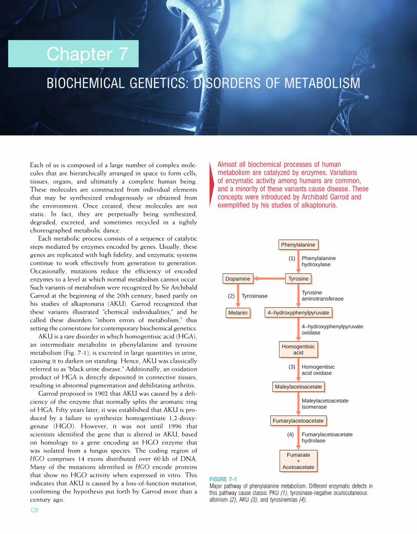
Chapter 7
BIOCHEMICAL GENETICS: DISORDERS OF METABOLISM
Each of us is composed of a large number of complex mole-cules that are hierarchically arranged in space to form cells,tissues, organs, and ultimately a complete human being.These molecules are constructed from individual elementsthat may be synthesized endogenously or obtained fromthe environment. Once created, these molecules are notstatic. In fact, they are perpetually being synthesized,degraded, excreted, and sometimes recycled in a tightlychoreographed metabolic dance.
Each metabolic process consists of a sequence of catalyticsteps mediated by enzymes encoded by genes. Usually, thesegenes are replicated with high fidelity, and enzymatic systemscontinue to work effectively from generation to generation.Occasionally, mutations reduce the efficiency of encodedenzymes to a level at which normal metabolism cannot occur.Such variants of metabolism were recognized by Sir ArchibaldGarrod at the beginning of the 20th century, based partly onhis studies of alkaptonuria (AKU). Garrod recognized thatthese variants illustrated “chemical individualities,” and hecalled these disorders “inborn errors of metabolism,” thussetting the cornerstone for contemporary biochemical genetics.
AKU is a rare disorder in which homogentisic acid (HGA),an intermediate metabolite in phenylalanine and tyrosinemetabolism (Fig. 7-1), is excreted in large quantities in urine,causing it to darken on standing. Hence, AKU was classicallyreferred to as “black urine disease.” Additionally, an oxidationproduct of HGA is directly deposited in connective tissues,resulting in abnormal pigmentation and debilitating arthritis.
Garrod proposed in 1902 that AKU was caused by a defi-ciency of the enzyme that normally splits the aromatic ringof HGA. Fifty years later, it was established that AKU is pro-duced by a failure to synthesize homogentisate 1,2-dioxy-genase (HGO). However, it was not until 1996 thatscientists identified the gene that is altered in AKU, basedon homology to a gene encoding an HGO enzyme thatwas isolated from a fungus species. The coding region ofHGO comprises 14 exons distributed over 60 kb of DNA.Many of the mutations identified in HGO encode proteinsthat show no HGO activity when expressed in vitro. Thisindicates that AKU is caused by a loss-of-function mutation,confirming the hypothesis put forth by Garrod more than acentury ago.
4Almost all biochemical processes of humanmetabolism are catalyzed by enzymes. Variationsof enzymatic activity among humans are common,and a minority of these variants cause disease. Theseconcepts were introduced by Archibald Garrod andexemplified by his studies of alkaptonuria.
Phenylalanine
(1) Phenylalaninehydroxylase
Tyrosine
Melanin
(2) Tyrosinase
Dopamine
Tyrosineaminotransferase
4–hydroxyphenylpyruvate
4–hydroxyphenylpyruvateoxidase
Homogentisicacid
(3) Homogentisic acid oxidase
Maleylacetoacetate
Maleylacetoacetateisomerase
Fumarylacetoacetate
(4) Fumarylacetoacetatehydrolase
Fumarate+
Acetoacetate
FIGURE 7-1Major pathway of phenylalanine metabolism. Different enzymatic defects inthis pathway cause classic PKU (1), tyrosinase-negative oculocutaneousalbinism (2), AKU (3), and tyrosinemias (4).
128
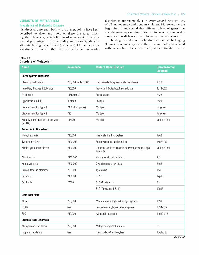
VARIANTS OF METABOLISMPrevalence of Metabolic DiseaseHundreds of different inborn errors of metabolism have beendescribed to date, and most of these are rare. Takentogether, however, metabolic disorders account for a sub-stantial percentage of the morbidity and mortality directlyattributable to genetic disease (Table 7-1). One survey con-servatively estimated that the incidence of metabolic
disorders is approximately 1 in every 2500 births, or 10%of all monogenic conditions in children. Moreover, we arebeginning to understand that different alleles of genes thatencode enzymes can alter one’s risk for many common dis-eases, such as diabetes, heart disease, stroke, and cancer.
The diagnosis of a metabolic disorder can be challenging(Clinical Commentary 7-1); thus, the morbidity associatedwith metabolic defects is probably underestimated. In the
TABLE 7-1
Disorders of Metabolism
Name Prevalence Mutant Gene Product ChromosomalLocation
Carbohydrate Disorders
Classic galactosemia 1/35,000 to 1/60,000 Galactose-1-phosphate uridyl transferase 9p13
Hereditary fructose intolerance 1/20,000 Fructose 1,6-bisphosphate aldolase 9q13-q32
Fructosuria �1/100,000 Fructokinase 2p23
Hypolactasia (adult) Common Lactase 2q21
Diabetes mellitus type 1 1/400 (Europeans) Multiple Polygenic
Diabetes mellitus type 2 1/20 Multiple Polygenic
Maturity-onset diabetes of the young(MODY)
�1/400 Multiple Multiple loci
Amino Acid Disorders
Phenylketonuria 1/10,000 Phenylalanine hydroxylase 12q24
Tyrosinemia (type 1) 1/100,000 Fumarylacetoacetate hydrolase 15q23-25
Maple syrup urine disease 1/180,000 Branched-chain a-ketoacid dehydrogenase (multiplesubunits)
Multiple loci
Alkaptonuria 1/250,000 Homogentisic acid oxidase 3q2
Homocystinuria 1/340,000 Cystathionine b-synthase 21q2
Oculocutaneous albinism 1/35,000 Tyrosinase 11q
Cystinosis 1/100,000 CTNS 17p13
Cystinuria 1/7000 SLC3A1 (type 1) 2p
SLC7A9 (types II & III) 19q13
Lipid Disorders
MCAD 1/20,000 Medium-chain acyl-CoA dehydrogenase 1p31
LCAD Rare Long-chain acyl-CoA dehydrogenase 2q34-q35
SLO 1/10,000 D7-sterol reductase 11q12-q13
Organic Acid Disorders
Methylmalonic acidemia 1/20,000 Methylmalonyl-CoA mutase 6p
Propionic acidemia Rare Propionyl-CoA carboxylase 13q32; 3q
Continued
Biochemical Genetics: Disorders of Metabolism / 129
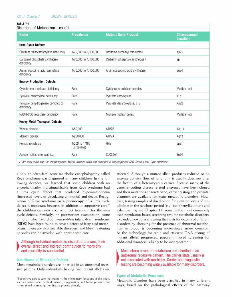
1970s, an often fatal acute metabolic encephalopathy calledReye syndrome was diagnosed in many children. In the fol-lowing decades, we learned that some children with anencephalopathy indistinguishable from Reye syndrome hada urea cycle defect that produced hyperammonemia(increased levels of circulating ammonia) and death. Recog-nition of Reye syndrome as a phenocopy of a urea cycledefect is important because, in addition to supportive care,*the children can now receive direct treatment for the ureacycle defects. Similarly, on postmortem examination, somechildren who have died from sudden infant death syndrome(SIDS) have been found to have a defect of fatty acid metab-olism. These are also treatable disorders, and life-threateningepisodes can be avoided with appropriate care.
4Although individual metabolic disorders are rare, theiroverall direct and indirect contribution to morbidityand mortality is substantial.
Inheritance of Metabolic DefectsMost metabolic disorders are inherited in an autosomal reces-sive pattern: Only individuals having two mutant alleles are
affected. Although a mutant allele produces reduced or noenzyme activity (loss of function), it usually does not alterthe health of a heterozygous carrier. Because many of thegenes encoding disease-related enzymes have been clonedand their mutations characterized, carrier testing and prenataldiagnosis are available for many metabolic disorders. How-ever, testing samples of dried blood for elevated levels of me-tabolites in the newborn period (e.g., for phenylketonuria andgalactosemia; see Chapter 13) remains the most commonlyused population-based screening test for metabolic disorders.Expanded newborn screening that tests for dozens of differentdisorders by checking for the presence of abnormal metabo-lites in blood is becoming increasingly more common.As the technology for rapid and efficient DNA testing ofmutant alleles progresses, population-based screening foradditional disorders is likely to be incorporated.
4Most inborn errors of metabolism are inherited in anautosomal recessive pattern. The carrier state usually isnot associated with morbidity. Carrier and diagnostictesting are becomingwidely available for many disorders.
Types of Metabolic ProcessesMetabolic disorders have been classified in many differentways, based on the pathological effects of the pathway
TABLE 7-1
Disorders of Metabolism—cont’d
Name Prevalence Mutant Gene Product ChromosomalLocation
Urea Cycle Defects
Ornithine transcarbamylase deficiency 1/70,000 to 1/100,000 Ornithine carbamyl transferase Xp21
Carbamyl phosphate synthetasedeficiency
1/70,000 to 1/100,000 Carbamyl phosphate synthetase I 2p
Argininosuccinic acid synthetasedeficiency
1/70,000 to 1/100,000 Argininosuccinic acid synthetase 9q34
Energy Production Defects
Cytochrome c oxidase deficiency Rare Cytochrome oxidase peptides Multiple loci
Pyruvate carboxylase deficiency Rare Pyruvate carboxylase 11q
Pyruvate dehydrogenase complex (E1)deficiency
Rare Pyruvate decarboxylase, E1a Xp22
NADH-CoQ reductase deficiency Rare Multiple nuclear genes Multiple loci
Heavy Metal Transport Defects
Wilson disease 1/50,000 ATP7B 13q14
Menkes disease 1/250,000 ATP7A Xq13
Hemochromatosis 1/200 to 1/400(Europeans)
HFE 6p21
Acrodermatitis enteropathica Rare SLC39A4 8q24
LCAD, long-chain acyl-CoA dehydrogenase; MCAD, medium-chain acyl-coenzyme A dehydrogenase; SLO, Smith–Lemli–Opitz syndrome.
*Supportive care is care that supports the elementary functions of the body,such as maintenance of fluid balance, oxygenation, and blood pressure, butis not aimed at treating the disease process directly.
130 / Chapter 7 MEDICAL GENETICS
CLINICAL COMMENTARY 7-1
Diagnosis of a Metabolic Disorder
The presentations of persons with inborn errors of metabolism are highlyvariable. During gestation, the maternal–placental unit usually providesessential nutrients and prevents the accumulation of toxic substrates. Thus,a fetus is infrequently symptomatic. However, after birth, persons with met-abolic disorders can present at ages ranging from the first 24 hours of lifeto adulthood. The presentation may be precipitous and characterized bydramatic alterations in homeostasis and even death. In contrast, the disor-der may be insidious, with only subtle changes in function over long per-iods. For most metabolic disorders, the presymptomatic period and onset ofsymptoms lie somewhere between these two extremes. The following caseillustrates this point.
Anthony is a 9-month-old Latin American boy who comes to the emer-gency department accompanied by his parents. His parents complain thathe has been irritable and vomiting for the last 36 hours, and over the past12 hours he has become increasingly sleepy. They sought medical attentionbecause it was difficult to awaken Anthony to breast-feed him. Anthony’smedical history is unremarkable. He has a healthy 8-year-old sister andhad a brother who died in his crib at 7 months of age. An investigationof the brother’s death and an autopsy were performed. The findings wereconsistent with sudden infant death syndrome (SIDS).
Anthony is hospitalized and is noted to be hypoglycemic (low serum glu-cose level), slightly acidemic (serum pH <7.4), and hyperammonemic (ele-vated plasma ammonia). Intravenous infusion of glucose transientlyimproves his level of alertness, but he becomes comatose and dies 5 dayslater. An autopsy reveals marked cerebral edema (swelling of the brain) andfatty infiltration of the liver consistent with a diagnosis of Reye syndrome.Anthony’s mother is concerned that the boys’ deaths are related to each other,especially since she is pregnant again. She is counseled that the causes ofdeath are unrelated and neither disorder is likely to recur in her family.
One year later her 6-month-old daughter, Maria, is hospitalized for thethird time because of lethargy and weakness. Laboratory studies reveal
moderate hypoglycemia, hyperammonemia, and ketonuria (ketones inthe urine). Additional studies, including measurement of urine organicacids,* serum amino acids, and free and esterified plasma carnitines, sug-gest that Maria has a defect of fatty acid oxidation. Therapy is initiatedwith intravenous glucose, oral carnitine, and the restriction of fats to nomore than 20% of her caloric requirements. More specific biochemicaland molecular studies confirm that Maria has medium-chain acyl-CoAdehydrogenase (MCAD) deficiency. Molecular studies from preserved tis-sues that had been collected at autopsy from Maria’s deceased brothersindicate that they also had MCAD deficiency. Maria’s asymptomatic oldersister is similarly affected. Both girls are healthy 2 years later, eating alow-fat diet and using a carnitine supplement. They have a new babybrother who underwent prenatal testing for MCAD and was found to beunaffected.
The disparate presentations of MCAD deficiency in this family (suddendeath, acute illness, chronic illness, and asymptomatic) illustrate the pheno-typic variability often observed in persons with inborn errors of metabolism,even those sharing an identical mutation. Thus, there might not be a dis-ease-specific pattern of symptoms and findings. Often it is the heightenedindex of suspicion of care providers that leads to the testing necessary toidentify a metabolic disorder. Supportive therapy can be lifesaving andshould be initiated before making a diagnosis. Nevertheless, it is imperativethat prudent attempts be made to make a specific diagnosis, because it canhave important implications for the family (e.g., prenatal testing, presymp-tomatic therapy). The treatment of MCAD deficiency is completely effectivein preventing early death from the toxic effects of accumulated fatty acidintermediates.
blocked (e.g., absence of end product, accumulation ofsubstrate); different functional classes of proteins (e.g.,receptors, hormones); associated cofactors (e.g., metals, vita-mins); and pathways affected (e.g., glycolysis, citric acidcycle). Each of these has advantages and disadvantages,and none of them encompasses all metabolic disorders.However, the classification that most completely integratesour knowledge of cell biology, physiology, and pathologywith metabolic disorders categorizes defects of metabolismby the types of processes that are disturbed.
DEFECTS OF METABOLIC PROCESSESAlmost all biochemical reactions in the human body are con-trolled by enzymes, which act as catalysts. The catalyticproperties of enzymes typically increase reaction rates bymore than a million-fold. These reactions mediate the syn-thesis, transfer, use, and degradation of biomolecules to buildand maintain the internal structures of cells, tissues, andorgans. Biomolecules can be categorized into four primarygroups: nucleic acids, proteins, carbohydrates, and lipids.The major metabolic pathways that metabolize these mole-cules include glycolysis, citric acid cycle, pentose phosphateshunt, gluconeogenesis, glycogen and fatty acid synthesisand storage, degradative pathways, energy production, and
transport systems. We now discuss how defects in each ofthese metabolic pathways can cause human disease.
Carbohydrate MetabolismBecause of the many different applications that they serve inall organisms, carbohydrates are the most abundant organicsubstance on Earth. Carbohydrates function as substrates forenergy production and storage, as intermediates of metabolicpathways, and as the structural framework of DNA and RNA.Consequently, carbohydrates account for a major portion ofthe human diet and are metabolized into three principalmonosaccharides: glucose, galactose, and fructose. Galactoseand fructose are converted to glucose before glycolysis. Thefailure to effectively use these sugars accounts for the majorityof the inborn errors of human carbohydrate metabolism.
GalactoseThe most common monogenic disorder of carbohydratemetabolism, transferase deficiency galactosemia (classicgalactosemia) affects 1 in every 30,000 newborns. It is mostcommonly caused by mutations in the gene encoding galac-tose-1-phosphate uridyl transferase (GAL-1-P uridyl trans-ferase) (Fig. 7-2). This gene is composed of 11 exonsdistributed across 4 kb of DNA, and approximately 70% of
*Organic acids are carbon-based acids that are products of intermediate metabolism
and normally do not accumulate in plasma or urine beyond the buffering capacities of
these fluids.
Biochemical Genetics: Disorders of Metabolism / 131
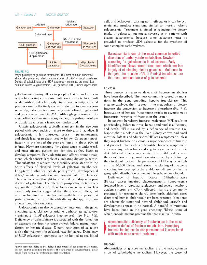
galactosemia-causing alleles in people of Western Europeanorigin have a single missense mutation in exon 6. As a resultof diminished GAL-1-P uridyl transferase activity, affectedpersons cannot effectively convert galactose to glucose; con-sequently, galactose is alternatively metabolized to galactitoland galactonate (see Fig. 7-2). Although galactose and itsmetabolites accumulate in many tissues, the pathophysiologyof classic galactosemia is not well understood.
Classic galactosemia typically manifests in the newbornperiod with poor sucking, failure to thrive, and jaundice. Ifgalactosemia is left untreated, sepsis, hyperammonemia,and shock leading to death usually follow. Cataracts (opaci-fication of the lens of the eye) are found in about 10% ofinfants. Newborn screening for galactosemia is widespread,and most affected persons are identified as they begin todevelop symptoms. Early identification affords prompt treat-ment, which consists largely of eliminating dietary galactose.This substantially reduces the morbidity associated with theacute effects of elevated levels of galactose metabolites.Long-term disabilities include poor growth, developmentaldelay,* mental retardation, and ovarian failure in females.These sequelae are thought to be caused by endogenous pro-duction of galactose. The effects of prospective dietary ther-apy on the prevalence of these long-term sequelae are lessclear. Early studies suggested that there was no effect, butas more longitudinal data become available, it appears thatpatients treated early in life with dietary therapy may havea better cognitive outcome.
Galactosemia can also be caused by mutations in the genesencoding galactokinase or uridine diphosphate galactose-4-epimerase (UDP-galactose-4-epimerase) (see Fig. 7-2).Deficiency of galactokinase is associated with the formationof cataracts but does not cause growth failure, mental retar-dation, or hepatic disease. Dietary restriction of galactoseis also the treatment for galactokinase deficiency. Deficiencyof UDP-galactose-4-epimerase can be limited to red blood
cells and leukocytes, causing no ill effects, or it can be sys-temic and produce symptoms similar to those of classicgalactosemia. Treatment is aimed at reducing the dietaryintake of galactose, but not as severely as in patients withclassic galactosemia, because some galactose must beprovided to produce UDP-galactose for the synthesis ofsome complex carbohydrates.
4Galactosemia is one of the most common inheriteddisorders of carbohydrate metabolism. Newbornscreening for galactosemia is widespread. Earlyidentification allows prompt treatment, which consistslargely of eliminating dietary galactose. Mutations inthe gene that encodes GAL-1-P uridyl transferase arethe most common cause of galactosemia.
FructoseThree autosomal recessive defects of fructose metabolismhave been described. The most common is caused by muta-tions in the gene encoding hepatic fructokinase. Thisenzyme catalyzes the first step in the metabolism of dietaryfructose, the conversion to fructose-1-phosphate (Fig. 7-3).Inactivation of hepatic fructokinase results in asymptomaticfructosuria (presence of fructose in the urine).
In contrast, hereditary fructose intolerance (HFI) results inpoor feeding, failure to thrive, hepatic and renal insufficiency,and death. HFI is caused by a deficiency of fructose 1,6-bisphosphate aldolase in the liver, kidney cortex, and smallintestine. Infants and adults with HFI are asymptomatic unlessthey ingest fructose or sucrose (a sugar composed of fructoseand glucose). Infants who are breast-fed become symptomaticafter weaning, when fruits and vegetables are added to theirdiet. Affected infants may survive into childhood becausethey avoid foods they consider noxious, thereby self-limitingtheir intake of fructose. The prevalence of HFI may be as highas 1 in 20,000 births, and, since the cloning of the geneencoding fructose-1-phosphate aldolase, differences in thegeographic distribution of mutant alleles have been found.
Deficiency of hepatic fructose 1,6-bisphosphatase(FBPase) causes impaired gluconeogenesis, hypoglycemia(reduced level of circulating glucose), and severe metabolicacidemia (serum pH <7.4). Affected infants are commonlypresented for treatment shortly after birth, although casesdiagnosed later in childhood have been reported. If patientsare adequately supported beyond childhood, growth anddevelopment appear to be normal. A handful of mutationshave been found in the gene encoding FBPase, some ofwhich encode mutant proteins that are inactive in vitro.
4Asymptomatic deficiency of fructokinase is the mostcommon defect of fructose metabolism. Hereditaryfructose intolerance is less prevalent but is associatedwith much more severe problems.
GlucoseAbnormalities of glucose metabolism are the most commonerrors of carbohydrate metabolism. However, the causes of
UDP-galactose4 epimerase
OxidaseGalactonate Galactose Galactitol
Galactokinase
GlycolipidsGlycoproteins
Aldolasereductase
Galactose1-phosphate
GAL-1-P uridyltransferase Glucose 1-
phosphate
UDP-galactose
UDP-glucose +
FIGURE 7-2Major pathways of galactose metabolism. The most common enzymaticabnormality producing galactosemia is a defect of GAL-1-P uridyl transferase.Defects of galactokinase or of UDP-galactose 4-epimerase are much lesscommon causes of galactosemia. GAL, galactose; UDP, uridine diphosphate.
*Developmental delay is the delayed attainment of age-appropriate motor,speech, and/or cognitive milestones; the outcomes of developmental delayrange from normal to profound mental retardation.
132 / Chapter 7 MEDICAL GENETICS
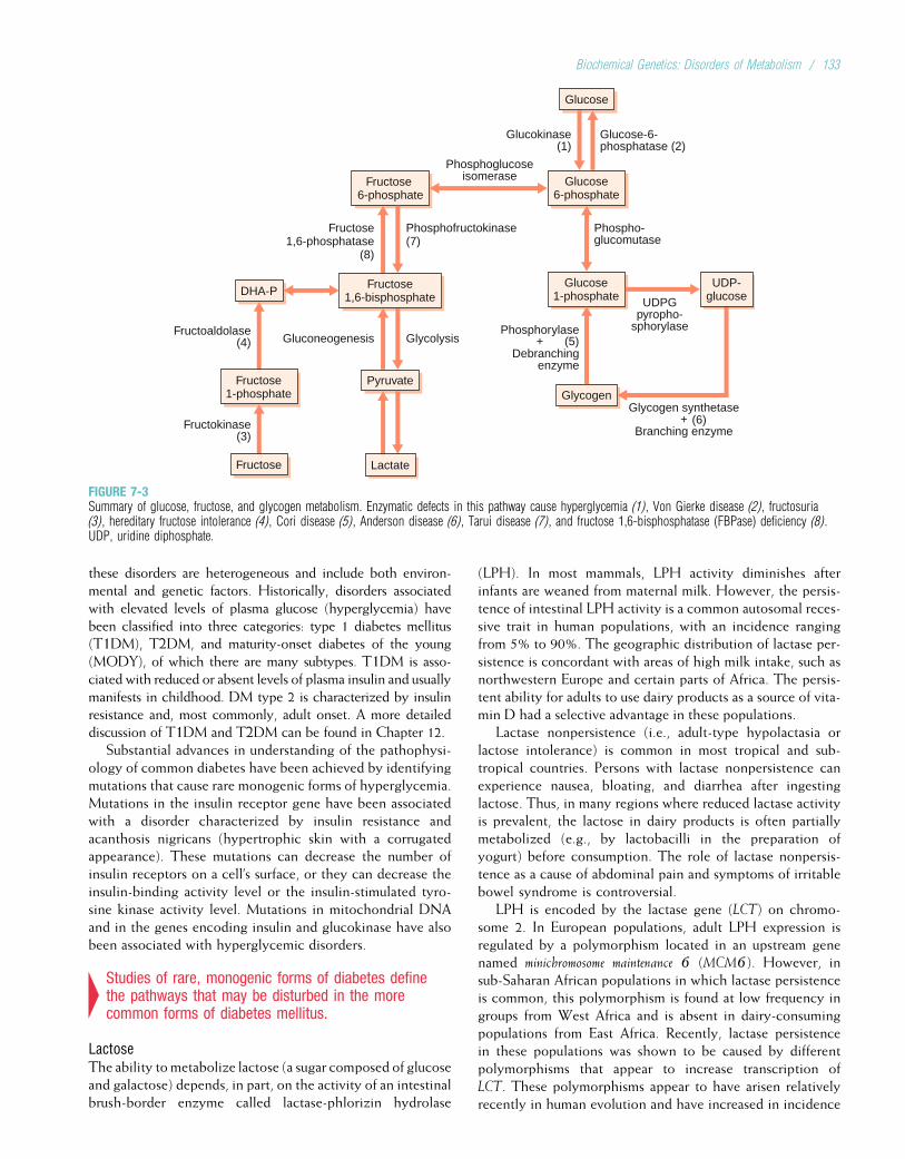
these disorders are heterogeneous and include both environ-mental and genetic factors. Historically, disorders associatedwith elevated levels of plasma glucose (hyperglycemia) havebeen classified into three categories: type 1 diabetes mellitus(T1DM), T2DM, and maturity-onset diabetes of the young(MODY), of which there are many subtypes. T1DM is asso-ciated with reduced or absent levels of plasma insulin and usuallymanifests in childhood. DM type 2 is characterized by insulinresistance and, most commonly, adult onset. A more detaileddiscussion of T1DM and T2DM can be found in Chapter 12.
Substantial advances in understanding of the pathophysi-ology of common diabetes have been achieved by identifyingmutations that cause rare monogenic forms of hyperglycemia.Mutations in the insulin receptor gene have been associatedwith a disorder characterized by insulin resistance andacanthosis nigricans (hypertrophic skin with a corrugatedappearance). These mutations can decrease the number ofinsulin receptors on a cell’s surface, or they can decrease theinsulin-binding activity level or the insulin-stimulated tyro-sine kinase activity level. Mutations in mitochondrial DNAand in the genes encoding insulin and glucokinase have alsobeen associated with hyperglycemic disorders.
4Studies of rare, monogenic forms of diabetes definethe pathways that may be disturbed in the morecommon forms of diabetes mellitus.
LactoseThe ability to metabolize lactose (a sugar composed of glucoseand galactose) depends, in part, on the activity of an intestinalbrush-border enzyme called lactase-phlorizin hydrolase
(LPH). In most mammals, LPH activity diminishes afterinfants are weaned from maternal milk. However, the persis-tence of intestinal LPH activity is a common autosomal reces-sive trait in human populations, with an incidence rangingfrom 5% to 90%. The geographic distribution of lactase per-sistence is concordant with areas of high milk intake, such asnorthwestern Europe and certain parts of Africa. The persis-tent ability for adults to use dairy products as a source of vita-min D had a selective advantage in these populations.
Lactase nonpersistence (i.e., adult-type hypolactasia orlactose intolerance) is common in most tropical and sub-tropical countries. Persons with lactase nonpersistence canexperience nausea, bloating, and diarrhea after ingestinglactose. Thus, in many regions where reduced lactase activityis prevalent, the lactose in dairy products is often partiallymetabolized (e.g., by lactobacilli in the preparation ofyogurt) before consumption. The role of lactase nonpersis-tence as a cause of abdominal pain and symptoms of irritablebowel syndrome is controversial.
LPH is encoded by the lactase gene (LCT) on chromo-some 2. In European populations, adult LPH expression isregulated by a polymorphism located in an upstream genenamed minichromosome maintenance 6 (MCM6). However, insub-Saharan African populations in which lactase persistenceis common, this polymorphism is found at low frequency ingroups from West Africa and is absent in dairy-consumingpopulations from East Africa. Recently, lactase persistencein these populations was shown to be caused by differentpolymorphisms that appear to increase transcription ofLCT. These polymorphisms appear to have arisen relativelyrecently in human evolution and have increased in incidence
Phosphofructokinase(7)
Fructose 6-phosphate
Phospho-glucomutase
UDPGpyropho-
sphorylase
Glucose-6-phosphatase (2)
Glucose
Glucose6-phosphate
Glycogen
Glucose1-phosphate
Glucokinase(1)
Phosphoglucoseisomerase
UDP-glucose
Fructose1,6-phosphatase
(8)
GlycolysisGluconeogenesis
DHA-P
Fructoaldolase(4)
Fructokinase(3)
Phosphorylase
Debranchingenzyme
(5)+
Glycogen synthetase+
Branching enzyme(6)
Fructose1,6-bisphosphate
Pyruvate
Lactate
Fructose1-phosphate
Fructose
FIGURE 7-3Summary of glucose, fructose, and glycogen metabolism. Enzymatic defects in this pathway cause hyperglycemia (1), Von Gierke disease (2), fructosuria(3), hereditary fructose intolerance (4), Cori disease (5), Anderson disease (6), Tarui disease (7), and fructose 1,6-bisphosphatase (FBPase) deficiency (8).UDP, uridine diphosphate.
Biochemical Genetics: Disorders of Metabolism / 133
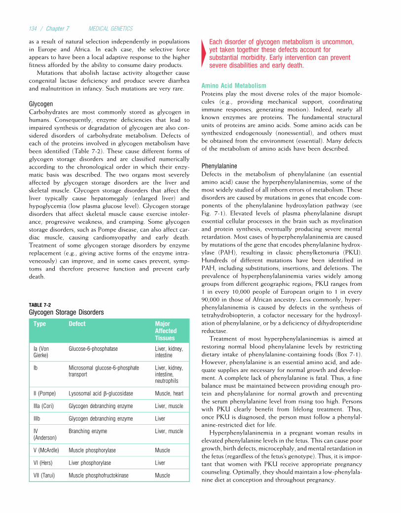
as a result of natural selection independently in populationsin Europe and Africa. In each case, the selective forceappears to have been a local adaptive response to the higherfitness afforded by the ability to consume dairy products.
Mutations that abolish lactase activity altogether causecongenital lactase deficiency and produce severe diarrheaand malnutrition in infancy. Such mutations are very rare.
GlycogenCarbohydrates are most commonly stored as glycogen inhumans. Consequently, enzyme deficiencies that lead toimpaired synthesis or degradation of glycogen are also con-sidered disorders of carbohydrate metabolism. Defects ofeach of the proteins involved in glycogen metabolism havebeen identified (Table 7-2). These cause different forms ofglycogen storage disorders and are classified numericallyaccording to the chronological order in which their enzy-matic basis was described. The two organs most severelyaffected by glycogen storage disorders are the liver andskeletal muscle. Glycogen storage disorders that affect theliver typically cause hepatomegaly (enlarged liver) andhypoglycemia (low plasma glucose level). Glycogen storagedisorders that affect skeletal muscle cause exercise intoler-ance, progressive weakness, and cramping. Some glycogenstorage disorders, such as Pompe disease, can also affect car-diac muscle, causing cardiomyopathy and early death.Treatment of some glycogen storage disorders by enzymereplacement (e.g., giving active forms of the enzyme intra-veneously) can improve, and in some cases prevent, symp-toms and therefore preserve function and prevent earlydeath.
4Each disorder of glycogen metabolism is uncommon,yet taken together these defects account forsubstantial morbidity. Early intervention can preventsevere disabilities and early death.
Amino Acid MetabolismProteins play the most diverse roles of the major biomole-cules (e.g., providing mechanical support, coordinatingimmune responses, generating motion). Indeed, nearly allknown enzymes are proteins. The fundamental structuralunits of proteins are amino acids. Some amino acids can besynthesized endogenously (nonessential), and others mustbe obtained from the environment (essential). Many defectsof the metabolism of amino acids have been described.
PhenylalanineDefects in the metabolism of phenylalanine (an essentialamino acid) cause the hyperphenylalaninemias, some of themost widely studied of all inborn errors of metabolism. Thesedisorders are caused by mutations in genes that encode com-ponents of the phenylalanine hydroxylation pathway (seeFig. 7-1). Elevated levels of plasma phenylalanine disruptessential cellular processes in the brain such as myelinationand protein synthesis, eventually producing severe mentalretardation. Most cases of hyperphenylalaninemia are causedby mutations of the gene that encodes phenylalanine hydrox-ylase (PAH), resulting in classic phenylketonuria (PKU).Hundreds of different mutations have been identified inPAH, including substitutions, insertions, and deletions. Theprevalence of hyperphenylalaninemia varies widely amonggroups from different geographic regions; PKU ranges from1 in every 10,000 people of European origin to 1 in every90,000 in those of African ancestry. Less commonly, hyper-phenylalaninemia is caused by defects in the synthesis oftetrahydrobiopterin, a cofactor necessary for the hydroxyl-ation of phenylalanine, or by a deficiency of dihydropteridinereductase.
Treatment of most hyperphenylalaninemias is aimed atrestoring normal blood phenylalanine levels by restrictingdietary intake of phenylalanine-containing foods (Box 7-1).However, phenylalanine is an essential amino acid, and ade-quate supplies are necessary for normal growth and develop-ment. A complete lack of phenylalanine is fatal. Thus, a finebalance must be maintained between providing enough pro-tein and phenylalanine for normal growth and preventingthe serum phenylalanine level from rising too high. Personswith PKU clearly benefit from lifelong treatment. Thus,once PKU is diagnosed, the person must follow a phenylal-anine-restricted diet for life.
Hyperphenylalaninemia in a pregnant woman results inelevated phenylalanine levels in the fetus. This can cause poorgrowth, birth defects, microcephaly, andmental retardation inthe fetus (regardless of the fetus’s genotype). Thus, it is impor-tant that women with PKU receive appropriate pregnancycounseling. Optimally, they should maintain a low-phenylala-nine diet at conception and throughout pregnancy.
TABLE 7-2
Glycogen Storage Disorders
Type Defect MajorAffectedTissues
Ia (VonGierke)
Glucose-6-phosphatase Liver, kidney,intestine
Ib Microsomal glucose-6-phosphatetransport
Liver, kidney,intestine,neutrophils
II (Pompe) Lysosomal acid b-glucosidase Muscle, heart
IIIa (Cori) Glycogen debranching enzyme Liver, muscle
IIIb Glycogen debranching enzyme Liver
IV(Anderson)
Branching enzyme Liver, muscle
V (McArdle) Muscle phosphorylase Muscle
VI (Hers) Liver phosphorylase Liver
VII (Tarui) Muscle phosphofructokinase Muscle
134 / Chapter 7 MEDICAL GENETICS
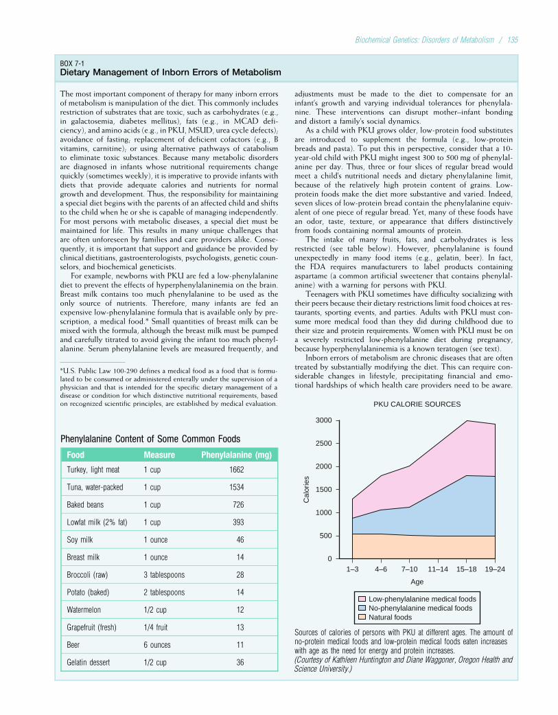
BOX 7-1Dietary Management of Inborn Errors of Metabolism
The most important component of therapy for many inborn errorsof metabolism is manipulation of the diet. This commonly includesrestriction of substrates that are toxic, such as carbohydrates (e.g.,in galactosemia, diabetes mellitus), fats (e.g., in MCAD defi-ciency), and amino acids (e.g., in PKU,MSUD, urea cycle defects);avoidance of fasting; replacement of deficient cofactors (e.g., Bvitamins, carnitine); or using alternative pathways of catabolismto eliminate toxic substances. Because many metabolic disordersare diagnosed in infants whose nutritional requirements changequickly (sometimes weekly), it is imperative to provide infants withdiets that provide adequate calories and nutrients for normalgrowth and development. Thus, the responsibility for maintaininga special diet begins with the parents of an affected child and shiftsto the child when he or she is capable of managing independently.For most persons with metabolic diseases, a special diet must bemaintained for life. This results in many unique challenges thatare often unforeseen by families and care providers alike. Conse-quently, it is important that support and guidance be provided byclinical dietitians, gastroenterologists, psychologists, genetic coun-selors, and biochemical geneticists.
For example, newborns with PKU are fed a low-phenylalaninediet to prevent the effects of hyperphenylalaninemia on the brain.Breast milk contains too much phenylalanine to be used as theonly source of nutrients. Therefore, many infants are fed anexpensive low-phenylalanine formula that is available only by pre-scription, a medical food.* Small quantities of breast milk can bemixed with the formula, although the breast milk must be pumpedand carefully titrated to avoid giving the infant too much phenyl-alanine. Serum phenylalanine levels are measured frequently, and
adjustments must be made to the diet to compensate for aninfant’s growth and varying individual tolerances for phenylala-nine. These interventions can disrupt mother–infant bondingand distort a family’s social dynamics.
As a child with PKU grows older, low-protein food substitutesare introduced to supplement the formula (e.g., low-proteinbreads and pasta). To put this in perspective, consider that a 10-year-old child with PKU might ingest 300 to 500 mg of phenylal-anine per day. Thus, three or four slices of regular bread wouldmeet a child’s nutritional needs and dietary phenylalanine limit,because of the relatively high protein content of grains. Low-protein foods make the diet more substantive and varied. Indeed,seven slices of low-protein bread contain the phenylalanine equiv-alent of one piece of regular bread. Yet, many of these foods havean odor, taste, texture, or appearance that differs distinctivelyfrom foods containing normal amounts of protein.
The intake of many fruits, fats, and carbohydrates is lessrestricted (see table below). However, phenylalanine is foundunexpectedly in many food items (e.g., gelatin, beer). In fact,the FDA requires manufacturers to label products containingaspartame (a common artificial sweetener that contains phenylal-anine) with a warning for persons with PKU.
Teenagers with PKU sometimes have difficulty socializing withtheir peers because their dietary restrictions limit food choices at res-taurants, sporting events, and parties. Adults with PKU must con-sume more medical food than they did during childhood due totheir size and protein requirements. Women with PKU must be ona severely restricted low-phenylalanine diet during pregnancy,because hyperphenylalaninemia is a known teratogen (see text).
Inborn errors of metabolism are chronic diseases that are oftentreated by substantially modifying the diet. This can require con-siderable changes in lifestyle, precipitating financial and emo-tional hardships of which health care providers need to be aware.
PKU CALORIE SOURCES
Low-phenylalanine medical foodsNo-phenylalanine medical foodsNatural foods
Cal
orie
s
3000
2500
2000
1500
1000
500
01–3
Age
4–6 7–10 11–14 15–18 19–24
Sources of calories of persons with PKU at different ages. The amount ofno-protein medical foods and low-protein medical foods eaten increaseswith age as the need for energy and protein increases.(Courtesy of Kathleen Huntington and Diane Waggoner, Oregon Health andScience University.)
*U.S. Public Law 100-290 defines a medical food as a food that is formu-lated to be consumed or administered enterally under the supervision of aphysician and that is intended for the specific dietary management of adisease or condition for which distinctive nutritional requirements, basedon recognized scientific principles, are established by medical evaluation.
Phenylalanine Content of Some Common Foods
Food Measure Phenylalanine (mg)
Turkey, light meat 1 cup 1662
Tuna, water-packed 1 cup 1534
Baked beans 1 cup 726
Lowfat milk (2% fat) 1 cup 393
Soy milk 1 ounce 46
Breast milk 1 ounce 14
Broccoli (raw) 3 tablespoons 28
Potato (baked) 2 tablespoons 14
Watermelon 1/2 cup 12
Grapefruit (fresh) 1/4 fruit 13
Beer 6 ounces 11
Gelatin dessert 1/2 cup 36
Biochemical Genetics: Disorders of Metabolism / 135
4Hyperphenylalaninemias are the most commondefects of amino acid metabolism. Classic PKU iscaused by mutations in the gene that encodesphenylalanine hydroxylase. Hyperphenylalaninemiasare treated by restricting dietary intake ofphenylalanine-containing foods.
TyrosineThe amino acid tyrosine is the starting point of the syntheticpathways leading to catecholamines, thyroid hormones, andmelanin pigments, and it is incorporated into many proteins.Elevated levels of serum tyrosine can be acquired (e.g.,severe hepatocellular dysfunction), or they can result froman inborn error of catabolism, of which there are several.Hereditary tyrosinemia type 1 (HT1) is the most commonmetabolic defect and is caused by a deficiency of fumarylace-toacetate hydrolase (FAH), which catalyzes the last step intyrosine catabolism (see Fig. 7-1). Accumulation of FAH’ssubstrate, fumarylacetoacetate, and its precursor, maleylace-toacetate, is thought to be mutagenic and toxic to the liver.Consequently, HT1 is characterized by dysfunction of therenal tubules, acute episodes of peripheral neuropathy, pro-gressive liver disease leading to cirrhosis, and a high riskfor developing liver cancer (hepatocellular carcinoma).
Management of HT1 includes supportive care, dietaryrestriction of phenylalanine and tyrosine, and administrationof 2-(nitro-4-trifluoro-methylbenzoyl)-1,3-cyclohexanedione(NTBC) or nitisinone, an inhibitor of an enzyme upstream ofFAH (4-hydroxyphenylpyruvate dioxygenase). Use of NTBC,combined with a low-tyrosine diet, has produced markedimprovement in children with HT1. The long-term effects ofNTBC are still unclear, but children who have been treatedfor more than 15 years appear to be doing well. Liver transplan-tation can be curative but is typically reserved for persons whofail to respond to NTBC or who develop a malignancy. In amouse model of HT1, gene therapy (see Chapter 13) has beenused to repopulate the liver with cells that exhibit stable long-term expression of FAH (FAHþ hepatocytes). Because someFAH-hepatocytes remain in these livers, it is unclear whetherthe risk of hepatocellular carcinoma is reduced. Gene therapyfor HT1 in humans might someday replace life-long dietaryand pharmacologic treatment.
The gene that encodes FAH has been cloned, and muta-tions have been identified in many families. A splice-sitemutation is quite common in French-Canadians, and its highfrequency is probably the result of founder effect (see Chap-ter 3). This mutation results in an in-frame deletion of anexon, which removes a series of critically important aminoacids from FAH. Missense and nonsense mutations of FAHhave also been found in persons with HT1.
Tyrosinemia type 2 (oculocutaneous tyrosinemia) is causedby a deficiency of tyrosine aminotransferase. It is character-ized by corneal erosions, thickening of the skin on the palmsand the soles, and variable mental retardation. Tyrosinemiatype 3 is associated with reduced activity of 4-hydroxyphenyl-pyruvate dioxygenase and neurological dysfunction. Only afew affected persons have been reported.
4Deficiency of FAH causes HT1. Accumulation of thesubstrates of FAH leads to neurological, kidney, andliver dysfunction. Although liver transplantation hasbeen the cornerstone of treatment for HT1, the use ofdrugs that block the production of FAH has provedeffective.
Branched-Chain Amino AcidsApproximately 40% of the preformed amino acids requiredby mammals are branched-chain amino acids (BCAAs) suchas valine, leucine, and isoleucine. BCAAs can be used as asource of energy through an oxidative pathway that uses ana-ketoacid as an intermediate. Decarboxylation of a-keto-acids is mediated by a multimeric enzyme complex calledbranched-chain a-ketoacid dehydrogenase (BCKAD). TheBCKAD complex is composed of at least four catalytic com-ponents and two regulatory enzymes, which are encoded bysix genes. A deficiency of any one of these six componentsproduces a disorder known as maple syrup urine disease(MSUD), so named because the urine of affected personshas an odor reminiscent of maple syrup.
The prevalence of MSUD in the general population is low,but MSUD is relatively common in the Mennonite commu-nity of Lancaster County, Pennsylvania, where approximately1 in every 7 persons is a heterozygous carrier. All of these car-riers have the same disease-causing mutation of E1a, one ofthe loci encoding a catalytic component of BCKAD, and allare descendants of a couple who emigrated from Europe inthe 18th century. This is another example of founder effectin a small, relatively isolated population (see Chapter 3).
Untreated patients with MSUD accumulate BCAAs andtheir associated ketoacids, leading to progressive neurodegen-eration and death in the first few months of life. Treatment ofMSUD consists of dietary restriction of BCAAs to the minimumrequired for normal growth. Despite treatment, episodic deteri-oration is common, and supportive care is required during thesecrises. Because increasing BCKAD activity by only a few per-centage points can alter the course of disease substantially, ther-apy with thiamine, a cofactor of BCKAD, is used to treat thesepatients. Gene therapy for MSUD is also being investigated.
4Maple syrup urine disease is caused by defects inbranched-chain a-ketoacid dehydrogenase.Accumulation of BCAAs causes progressiveneurodegeneration and death. Treatment consists ofrestricting dietary intake of BCAAs to a minimal level.
Early detection of amino acid defects, coupled withprompt intervention, can prevent severe physical impairmentand death. Moderate increases of enzyme activity can dra-matically alter the course of some aminoacidopathies,making them good candidates for somatic cell gene therapy.
Lipid MetabolismLipids (Greek, lipos, “fat”) are a heterogeneous group of biomole-cules that are insoluble in water and highly soluble in organicsolvents (e.g., chloroform). They provide the backbone for
136 / Chapter 7 MEDICAL GENETICS
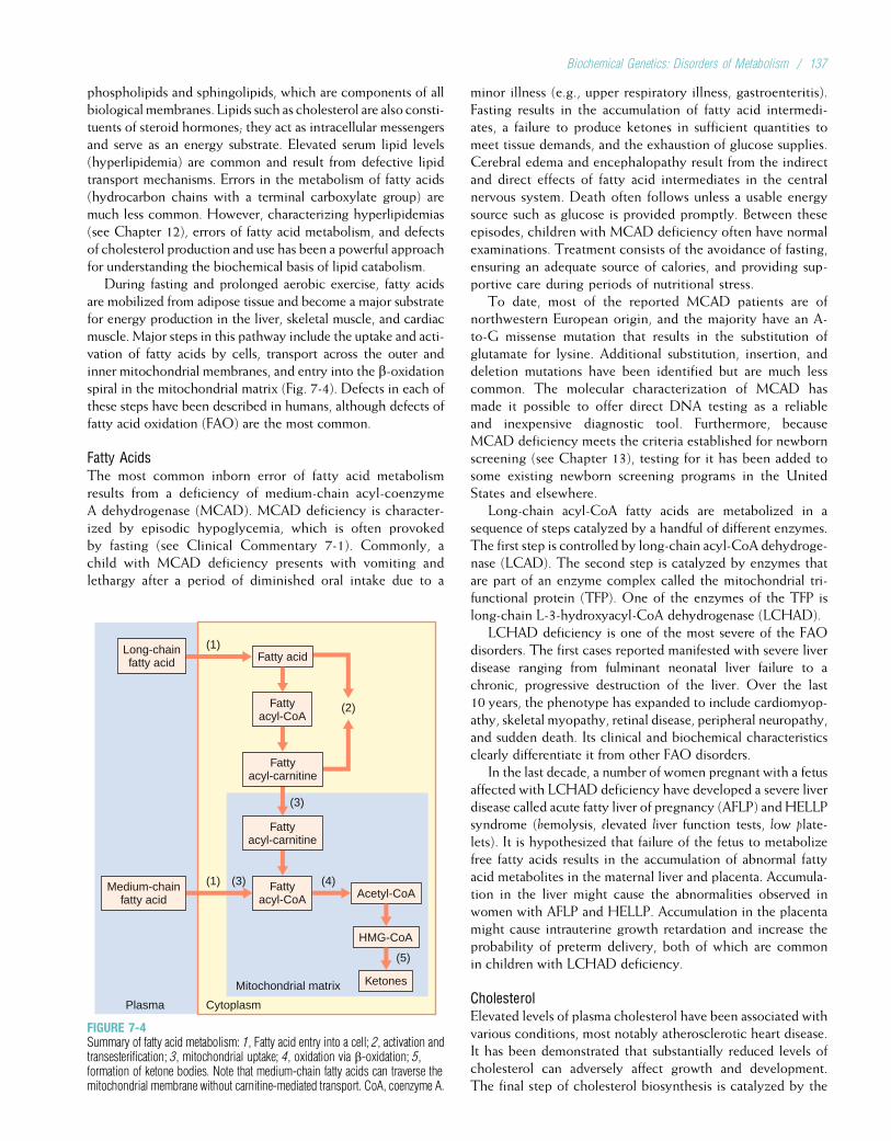
phospholipids and sphingolipids, which are components of allbiological membranes. Lipids such as cholesterol are also consti-tuents of steroid hormones; they act as intracellular messengersand serve as an energy substrate. Elevated serum lipid levels(hyperlipidemia) are common and result from defective lipidtransport mechanisms. Errors in the metabolism of fatty acids(hydrocarbon chains with a terminal carboxylate group) aremuch less common. However, characterizing hyperlipidemias(see Chapter 12), errors of fatty acid metabolism, and defectsof cholesterol production and use has been a powerful approachfor understanding the biochemical basis of lipid catabolism.
During fasting and prolonged aerobic exercise, fatty acidsare mobilized from adipose tissue and become a major substratefor energy production in the liver, skeletal muscle, and cardiacmuscle. Major steps in this pathway include the uptake and acti-vation of fatty acids by cells, transport across the outer andinner mitochondrial membranes, and entry into the b-oxidationspiral in the mitochondrial matrix (Fig. 7-4). Defects in each ofthese steps have been described in humans, although defects offatty acid oxidation (FAO) are the most common.
Fatty AcidsThe most common inborn error of fatty acid metabolismresults from a deficiency of medium-chain acyl-coenzymeA dehydrogenase (MCAD). MCAD deficiency is character-ized by episodic hypoglycemia, which is often provokedby fasting (see Clinical Commentary 7-1). Commonly, achild with MCAD deficiency presents with vomiting andlethargy after a period of diminished oral intake due to a
minor illness (e.g., upper respiratory illness, gastroenteritis).Fasting results in the accumulation of fatty acid intermedi-ates, a failure to produce ketones in sufficient quantities tomeet tissue demands, and the exhaustion of glucose supplies.Cerebral edema and encephalopathy result from the indirectand direct effects of fatty acid intermediates in the centralnervous system. Death often follows unless a usable energysource such as glucose is provided promptly. Between theseepisodes, children with MCAD deficiency often have normalexaminations. Treatment consists of the avoidance of fasting,ensuring an adequate source of calories, and providing sup-portive care during periods of nutritional stress.
To date, most of the reported MCAD patients are ofnorthwestern European origin, and the majority have an A-to-G missense mutation that results in the substitution ofglutamate for lysine. Additional substitution, insertion, anddeletion mutations have been identified but are much lesscommon. The molecular characterization of MCAD hasmade it possible to offer direct DNA testing as a reliableand inexpensive diagnostic tool. Furthermore, becauseMCAD deficiency meets the criteria established for newbornscreening (see Chapter 13), testing for it has been added tosome existing newborn screening programs in the UnitedStates and elsewhere.
Long-chain acyl-CoA fatty acids are metabolized in asequence of steps catalyzed by a handful of different enzymes.The first step is controlled by long-chain acyl-CoA dehydroge-nase (LCAD). The second step is catalyzed by enzymes thatare part of an enzyme complex called the mitochondrial tri-functional protein (TFP). One of the enzymes of the TFP islong-chain L-3-hydroxyacyl-CoA dehydrogenase (LCHAD).
LCHAD deficiency is one of the most severe of the FAOdisorders. The first cases reported manifested with severe liverdisease ranging from fulminant neonatal liver failure to achronic, progressive destruction of the liver. Over the last10 years, the phenotype has expanded to include cardiomyop-athy, skeletal myopathy, retinal disease, peripheral neuropathy,and sudden death. Its clinical and biochemical characteristicsclearly differentiate it from other FAO disorders.
In the last decade, a number of women pregnant with a fetusaffected with LCHAD deficiency have developed a severe liverdisease called acute fatty liver of pregnancy (AFLP) andHELLPsyndrome (hemolysis, elevated liver function tests, low plate-lets). It is hypothesized that failure of the fetus to metabolizefree fatty acids results in the accumulation of abnormal fattyacid metabolites in the maternal liver and placenta. Accumula-tion in the liver might cause the abnormalities observed inwomen with AFLP and HELLP. Accumulation in the placentamight cause intrauterine growth retardation and increase theprobability of preterm delivery, both of which are commonin children with LCHAD deficiency.
CholesterolElevated levels of plasma cholesterol have been associated withvarious conditions, most notably atherosclerotic heart disease.It has been demonstrated that substantially reduced levels ofcholesterol can adversely affect growth and development.The final step of cholesterol biosynthesis is catalyzed by the
Fattyacyl-CoA
Fatty acid(1)
(2)
Fattyacyl-carnitine
(3)
Fattyacyl-carnitine
(3)(1) (4)Fattyacyl-CoA
Plasma Cytoplasm
Mitochondrial matrix
Acetyl-CoA
(5)
HMG-CoA
Ketones
Long-chainfatty acid
Medium-chainfatty acid
FIGURE 7-4Summary of fatty acid metabolism: 1, Fatty acid entry into a cell; 2, activation andtransesterification; 3, mitochondrial uptake; 4, oxidation via b-oxidation; 5,formation of ketone bodies. Note that medium-chain fatty acids can traverse themitochondrial membrane without carnitine-mediated transport. CoA, coenzyme A.
Biochemical Genetics: Disorders of Metabolism / 137
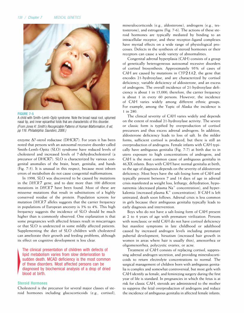
enzyme D7-sterol reductase (DHCR7). For years it has beennoted that persons with an autosomal recessive disorder calledSmith–Lemli–Opitz (SLO) syndrome have reduced levels ofcholesterol and increased levels of 7-dehydrocholesterol (aprecursor of DHCR7). SLO is characterized by various con-genital anomalies of the brain, heart, genitalia, and hands(Fig. 7-5). It is unusual in this respect, because most inbornerrors of metabolism do not cause congenital malformations.
In 1998, SLO was discovered to be caused by mutationsin the DHCR7 gene, and to date more than 100 differentmutations in DHCR7 have been found. Most of these aremissense mutations that result in substitutions of a highlyconserved residue of the protein. Population screens formutation DHCR7 alleles suggests that the carrier frequencyin populations of European ancestry is 3% to 4%. This highfrequency suggests the incidence of SLO should be muchhigher than is commonly observed. One explanation is thatsome pregnancies with affected fetuses result in miscarriagesor that SLO is undetected in some mildly affected patients.Supplementing the diet of SLO children with cholesterolcan ameliorate their growth and feeding problems, althoughits effect on cognitive development is less clear.
4The clinical presentation of children with defects oflipid metabolism varies from slow deterioration tosudden death. MCAD deficiency is the most commonof these disorders. Most affected persons can bediagnosed by biochemical analysis of a drop of driedblood at birth.
Steroid HormonesCholesterol is the precursor for several major classes of ste-roid hormones including glucocorticoids (e.g., cortisol),
mineralocorticoids (e.g., aldosterone), androgens (e.g., tes-tosterone), and estrogens (Fig. 7-6). The actions of these ste-roid hormones are typically mediated by binding to anintracellular receptor, and these receptor–ligand complexeshave myriad effects on a wide range of physiological pro-cesses. Defects in the synthesis of steroid hormones or theirreceptors can cause a wide variety of abnormalities.
Congenital adrenal hyperplasia (CAH) consists of a groupof genetically heterogeneous autosomal recessive disordersof cortisol biosynthesis. Approximately 95% of cases ofCAH are caused by mutations in CYP21A2, the gene thatencodes 21-hydroxylase, and are characterized by cortisoldeficiency, variable deficiency of aldosterone, and an excessof androgens. The overall incidence of 21-hydroxylase defi-ciency is about 1 in 15,000; therefore, the carrier frequencyis about 1 in every 60 persons. However, the incidenceof CAH varies widely among different ethnic groups.For example, among the Yupic of Alaska the incidence is1 in 280.
The clinical severity of CAH varies widely and dependson the extent of residual 21-hydroxylase activity. The severeor classic form is typified by overproduction of cortisolprecursors and thus excess adrenal androgens. In addition,aldosterone deficiency leads to loss of salt. In the milderforms, sufficient cortisol is produced, but there is still anoverproduction of androgens. Female infants with CAH typi-cally have ambiguous genitalia (Fig. 7-7) at birth due to inutero exposure to high concentrations of androgens, andCAH is the most common cause of ambiguous genitalia in46,XX infants. Boys with CAH have normal genitalia at birth,so the age of diagnosis depends on the severity of aldosteronedeficiency. Most boys have the salt-losing form of CAH andtypically present between 7 and 14 days of age in adrenalcrisis manifested as weight loss, lethargy, dehydration, hypo-natremia (decreased plasma Naþ concentration), and hyper-kalemia (increased plasma Kþ concentration). If CAH is leftuntreated, death soon follows. Adrenal crisis is less commonin girls because their ambiguous genitalia typically leads toearly diagnosis and intervention.
Boys who do not have a salt-losing form of CAH presentat 2 to 4 years of age with premature virilization. Personswith nonclassic or mild CAH do not have cortisol deficiencybut manifest symptoms in late childhood or adulthoodcaused by increased androgen levels including prematurepubertal development, hirsutism (increased hair growth inwomen in areas where hair is usually thin), amenorrhea oroligomenorrhea, polycystic ovaries, or acne.
Treatment of CAH consists of replacing cortisol, suppres-sing adrenal androgen secretion, and providing mineralocorti-coids to return electrolyte concentrations to normal. Thesurgical management of children born with ambiguous genita-lia is complex and somewhat controversial, but most girls withCAH identify as female, and feminizing surgery during the firstyear of life is standard. In pregnancies in which the fetus is atrisk for classic CAH, steroids are administered to the motherto suppress the fetal overproduction of androgens and reducethe incidence of ambiguous genitalia in affected female infants.
FIGURE 7-5A child with Smith–Lemli–Opitz syndrome. Note the broad nasal root, upturnednasal tip, and inner epicanthal folds that are characteristic of this disorder.
(From Jones K: Smith’s Recognizable Patterns of Human Malformation, 6 ed,pp 116. Philadelphia: Saunders, 2006.)
138 / Chapter 7 MEDICAL GENETICS
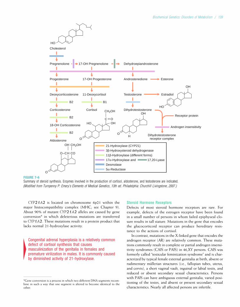
CYP21A2 is located on chromosome 6p21 within themajor histocompatibility complex (MHC; see Chapter 9).About 90% of mutant CYP21A2 alleles are caused by geneconversion* in which deleterious mutations are transferredto CYP1A2. These mutations result in a protein product thatlacks normal 21-hydroxylase activity.
4Congenital adrenal hyperplasia is a relatively commondefect of cortisol synthesis that causesmasculinization of the genitalia in females andpremature virilization in males. It is commonly causedby diminished activity of 21-hydroxylase.
Steroid Hormone ReceptorsDefects of most steroid hormone receptors are rare. Forexample, defects of the estrogen receptor have been foundin a small number of persons in whom failed epiphyseal clo-sure results in tall stature. Mutations in the gene that encodesthe glucocorticoid receptor can produce hereditary resis-tance to the actions of cortisol.
In contrast, mutations in the X-linked gene that encodes theandrogen receptor (AR) are relatively common. These muta-tions commonly result in complete or partial androgen insensi-tivity syndromes (CAIS or PAIS) in 46,XY persons. CAIS wasformerly called “testicular feminization syndrome” and is char-acterized by typical female external genitalia at birth, absent orrudimentary mullerian structures (i.e., fallopian tubes, uterus,and cervix), a short vaginal vault, inguinal or labial testis, andreduced or absent secondary sexual characteristics. Personswith PAIS can have ambiguous external genitalia, varied posi-tioning of the testes, and absent or present secondary sexualcharacteristics. Nearly all affected persons are infertile.
Cholesterol
HO
B2 B1
Androgen insensitivity
B2
B2
O–CH
OH CH2OH
O
C O
O
HO OH
O
OH
HO
OH
Pregnenolone 17-OH Pregnenolone Dehydroepiandrosterone
Progesterone 17-OH Progesterone Androstenedione
Deoxycorticosterone 11-Deoxycortisol
21-Hydroxylase (CYP21)
3β-Hydroxysteroid dehydrogenase
11β-Hydroxylase (different forms)
17α-Hydroxylase and 17,20-Lyase
Desmolase
5α-Reductase
Testosterone
Esterone
Estradiol
Receptor protein
Corticosterone Cortisol Dihydrotestosterone
Dihydrotestosteronereceptor complex
18-OH Corticosterone
Aldosterone
CO
H
CH2OH
FIGURE 7-6Summary of steroid synthesis. Enzymes involved in the production of cortisol, aldosterone, and testosterone are indicated.
(Modified from Turnpenny P: Emery ’s Elements of Medical Genetics, 13th ed. Philadelphia: Churchill Livingstone, 2007.)
*Gene conversion is a process in which two different DNA segments recom-bine in such a way that one segment is altered to become identical to theother.
Biochemical Genetics: Disorders of Metabolism / 139
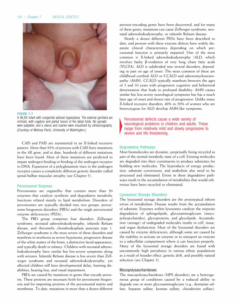
CAIS and PAIS are transmitted in an X-linked recessivepattern. More than 95% of persons with CAIS have mutationsin the AR gene, and to date, hundreds of different mutationshave been found. Most of these mutations are predicted toimpair androgen binding or binding of the androgen receptorto DNA. Expansion of a polyglutamine tract in the androgenreceptor causes a completely different genetic disorder calledspinal bulbar muscular atrophy (see Chapter 5).
Peroxisomal EnzymesPeroxisomes are organelles that contain more than 50enzymes that catalyze synthetic and degradative metabolicfunctions related mainly to lipid metabolism. Disorders ofperoxisomes are typically divided into two groups: peroxi-some biogenesis disorders (PBDs) and the single peroxisomalenzyme deficiencies (PEDs).
The PBD group comprises four disorders: Zellwegersyndrome, neonatal adrenoleukodystrophy, infantile Refsumdisease, and rhizomelic chondrodysplasia punctata type 1.Zellweger syndrome is the most severe of these disorders andmanifests in newborns as severe hypotonia, progressive diseaseof the white matter of the brain, a distinctive facial appearance,and typically death in infancy. Children with neonatal adreno-leukodystrophy have similar but less-severe symptoms alongwith seizures. Infantile Refsum disease is less severe than Zell-weger syndrome and neonatal adrenoleukodystrophy, yetaffected children still have developmental delay, learning dis-abilities, hearing loss, and visual impairment.
PBDs are caused by mutations in genes that encode perox-ins. These proteins are necessary both for peroxisome biogen-esis and for importing proteins of the peroxisomal matrix andmembrane. To date, mutations in more than a dozen different
peroxin-encoding genes have been discovered, and for manyof these genes, mutations can cause Zellweger syndrome, neo-natal adrenoleukodystrophy, or infantile Refsum disease.
Nearly a dozen different PEDs have been described todate, and persons with these enzyme defects have widely dis-parate clinical characteristics depending on which per-oxisomal function is primarily impaired. One of the mostcommon is X-linked adrenoleukodystrophy (ALD, whichinvolves faulty b-oxidation of very long chain fatty acids(VLCFA). ALD is subdivided into several disorders, depend-ing in part on age of onset. The most common of these arechildhood cerebral ALD or CCALD and adrenomyeloneuro-pathy (AMN). CCALD typically manifests between the agesof 3 and 10 years with progressive cognitive and behavioraldeterioration that leads to profound disability. AMN causessimilar but less-severe neurological symptoms but has a muchlater age of onset and slower rate of progression. Unlike manyX-linked recessive disorders, 40% to 50% of women who areheterozygous for ALD develop AMN-like symptoms.
4Peroxisomal defects cause a wide variety ofneurological problems in children and adults. Theserange from relatively mild and slowly progressive tosevere and life threatening.
Degradative PathwaysMost biomolecules are dynamic, perpetually being recycled aspart of the normal metabolic state of a cell. Existing moleculesare degraded into their constituents to produce substrates forbuilding new molecules. The byproducts of energy produc-tion, substrate conversions, and anabolism also need to beprocessed and eliminated. Errors in these degradative path-ways result in the accumulation of metabolites that would oth-erwise have been recycled or eliminated.
Lysosomal Storage DisordersThe lysosomal storage disorders are the prototypical inbornerrors of metabolism: Disease results from the accumulationof substrate. Enzymes within lysosomes catalyze the stepwisedegradation of sphingolipids, glycosaminoglycans (muco-polysaccharides), glycoproteins, and glycolipids. Accumula-tion (storage) of undegraded molecules results in cell, tissue,and organ dysfunction. Most of the lysosomal disorders arecaused by enzyme deficiencies, although some are caused bythe inability to activate an enzyme or to transport an enzymeto a subcellular compartment where it can function properly.Many of the lysosomal storage disorders are found withuncommonly high prevalence in various ethnic populationsas a result of founder effect, genetic drift, and possibly naturalselection (see Chapter 3).
MucopolysaccharidosesThe mucopolysaccharidoses (MPS disorders) are a heteroge-neous group of conditions caused by a reduced ability todegrade one or more glycosaminoglycans (e.g., dermatan sul-fate, heparan sulfate, keratan sulfate, chondroitin sulfate).
FIGURE 7-7A 46,XX infant with congenital adrenal hyperplasia. The external genitalia arevirilized, with rugation and partial fusion of the labial folds. No gonadswere palpable, and a uterus and ovaries were visualized by ultrasonography.
(Courtesy of Melissa Parisi, University of Washington.)
140 / Chapter 7 MEDICAL GENETICS
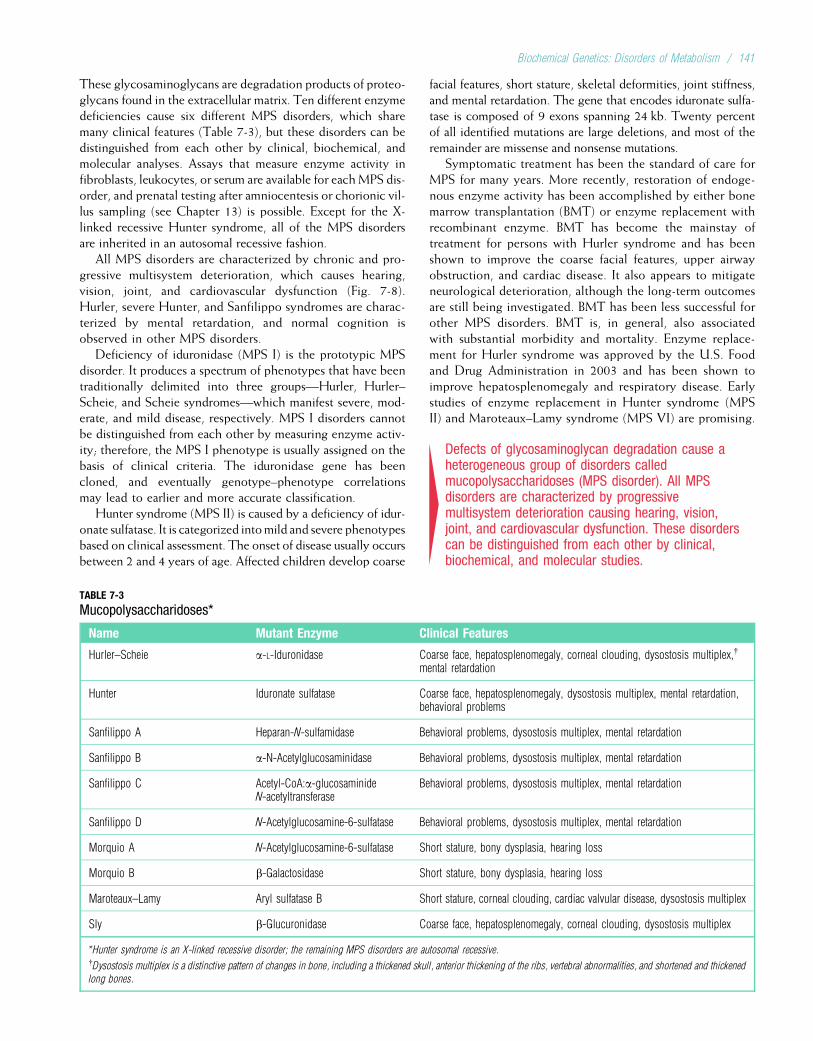
These glycosaminoglycans are degradation products of proteo-glycans found in the extracellular matrix. Ten different enzymedeficiencies cause six different MPS disorders, which sharemany clinical features (Table 7-3), but these disorders can bedistinguished from each other by clinical, biochemical, andmolecular analyses. Assays that measure enzyme activity infibroblasts, leukocytes, or serum are available for eachMPS dis-order, and prenatal testing after amniocentesis or chorionic vil-lus sampling (see Chapter 13) is possible. Except for the X-linked recessive Hunter syndrome, all of the MPS disordersare inherited in an autosomal recessive fashion.
All MPS disorders are characterized by chronic and pro-gressive multisystem deterioration, which causes hearing,vision, joint, and cardiovascular dysfunction (Fig. 7-8).Hurler, severe Hunter, and Sanfilippo syndromes are charac-terized by mental retardation, and normal cognition isobserved in other MPS disorders.
Deficiency of iduronidase (MPS I) is the prototypic MPSdisorder. It produces a spectrum of phenotypes that have beentraditionally delimited into three groups—Hurler, Hurler–Scheie, and Scheie syndromes—which manifest severe, mod-erate, and mild disease, respectively. MPS I disorders cannotbe distinguished from each other by measuring enzyme activ-ity; therefore, the MPS I phenotype is usually assigned on thebasis of clinical criteria. The iduronidase gene has beencloned, and eventually genotype–phenotype correlationsmay lead to earlier and more accurate classification.
Hunter syndrome (MPS II) is caused by a deficiency of idur-onate sulfatase. It is categorized intomild and severe phenotypesbased on clinical assessment. The onset of disease usually occursbetween 2 and 4 years of age. Affected children develop coarse
facial features, short stature, skeletal deformities, joint stiffness,and mental retardation. The gene that encodes iduronate sulfa-tase is composed of 9 exons spanning 24 kb. Twenty percentof all identified mutations are large deletions, and most of theremainder are missense and nonsense mutations.
Symptomatic treatment has been the standard of care forMPS for many years. More recently, restoration of endoge-nous enzyme activity has been accomplished by either bonemarrow transplantation (BMT) or enzyme replacement withrecombinant enzyme. BMT has become the mainstay oftreatment for persons with Hurler syndrome and has beenshown to improve the coarse facial features, upper airwayobstruction, and cardiac disease. It also appears to mitigateneurological deterioration, although the long-term outcomesare still being investigated. BMT has been less successful forother MPS disorders. BMT is, in general, also associatedwith substantial morbidity and mortality. Enzyme replace-ment for Hurler syndrome was approved by the U.S. Foodand Drug Administration in 2003 and has been shown toimprove hepatosplenomegaly and respiratory disease. Earlystudies of enzyme replacement in Hunter syndrome (MPSII) and Maroteaux–Lamy syndrome (MPS VI) are promising.
4Defects of glycosaminoglycan degradation cause aheterogeneous group of disorders calledmucopolysaccharidoses (MPS disorder). All MPSdisorders are characterized by progressivemultisystem deterioration causing hearing, vision,joint, and cardiovascular dysfunction. These disorderscan be distinguished from each other by clinical,biochemical, and molecular studies.
TABLE 7-3
Mucopolysaccharidoses*
Name Mutant Enzyme Clinical Features
Hurler–Scheie a-L-Iduronidase Coarse face, hepatosplenomegaly, corneal clouding, dysostosis multiplex,{
mental retardation
Hunter Iduronate sulfatase Coarse face, hepatosplenomegaly, dysostosis multiplex, mental retardation,behavioral problems
Sanfilippo A Heparan-N-sulfamidase Behavioral problems, dysostosis multiplex, mental retardation
Sanfilippo B a-N-Acetylglucosaminidase Behavioral problems, dysostosis multiplex, mental retardation
Sanfilippo C Acetyl-CoA:a-glucosaminideN-acetyltransferase
Behavioral problems, dysostosis multiplex, mental retardation
Sanfilippo D N-Acetylglucosamine-6-sulfatase Behavioral problems, dysostosis multiplex, mental retardation
Morquio A N-Acetylglucosamine-6-sulfatase Short stature, bony dysplasia, hearing loss
Morquio B b-Galactosidase Short stature, bony dysplasia, hearing loss
Maroteaux–Lamy Aryl sulfatase B Short stature, corneal clouding, cardiac valvular disease, dysostosis multiplex
Sly b-Glucuronidase Coarse face, hepatosplenomegaly, corneal clouding, dysostosis multiplex
*Hunter syndrome is an X-linked recessive disorder; the remaining MPS disorders are autosomal recessive.{Dysostosis multiplex is a distinctive pattern of changes in bone, including a thickened skull, anterior thickening of the ribs, vertebral abnormalities, and shortened and thickened
long bones.
Biochemical Genetics: Disorders of Metabolism / 141
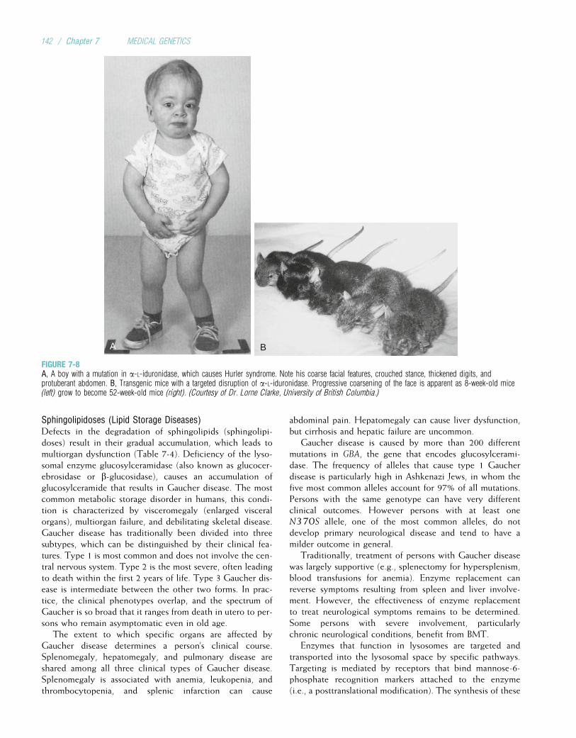
Sphingolipidoses (Lipid Storage Diseases)Defects in the degradation of sphingolipids (sphingolipi-doses) result in their gradual accumulation, which leads tomultiorgan dysfunction (Table 7-4). Deficiency of the lyso-somal enzyme glucosylceramidase (also known as glucocer-ebrosidase or b-glucosidase), causes an accumulation ofglucosylceramide that results in Gaucher disease. The mostcommon metabolic storage disorder in humans, this condi-tion is characterized by visceromegaly (enlarged visceralorgans), multiorgan failure, and debilitating skeletal disease.Gaucher disease has traditionally been divided into threesubtypes, which can be distinguished by their clinical fea-tures. Type 1 is most common and does not involve the cen-tral nervous system. Type 2 is the most severe, often leadingto death within the first 2 years of life. Type 3 Gaucher dis-ease is intermediate between the other two forms. In prac-tice, the clinical phenotypes overlap, and the spectrum ofGaucher is so broad that it ranges from death in utero to per-sons who remain asymptomatic even in old age.
The extent to which specific organs are affected byGaucher disease determines a person’s clinical course.Splenomegaly, hepatomegaly, and pulmonary disease areshared among all three clinical types of Gaucher disease.Splenomegaly is associated with anemia, leukopenia, andthrombocytopenia, and splenic infarction can cause
abdominal pain. Hepatomegaly can cause liver dysfunction,but cirrhosis and hepatic failure are uncommon.
Gaucher disease is caused by more than 200 differentmutations in GBA, the gene that encodes glucosylcerami-dase. The frequency of alleles that cause type 1 Gaucherdisease is particularly high in Ashkenazi Jews, in whom thefive most common alleles account for 97% of all mutations.Persons with the same genotype can have very differentclinical outcomes. However persons with at least oneN370S allele, one of the most common alleles, do notdevelop primary neurological disease and tend to have amilder outcome in general.
Traditionally, treatment of persons with Gaucher diseasewas largely supportive (e.g., splenectomy for hypersplenism,blood transfusions for anemia). Enzyme replacement canreverse symptoms resulting from spleen and liver involve-ment. However, the effectiveness of enzyme replacementto treat neurological symptoms remains to be determined.Some persons with severe involvement, particularlychronic neurological conditions, benefit from BMT.
Enzymes that function in lysosomes are targeted andtransported into the lysosomal space by specific pathways.Targeting is mediated by receptors that bind mannose-6-phosphate recognition markers attached to the enzyme(i.e., a posttranslational modification). The synthesis of these
A B
FIGURE 7-8A, A boy with a mutation in a-L-iduronidase, which causes Hurler syndrome. Note his coarse facial features, crouched stance, thickened digits, andprotuberant abdomen. B, Transgenic mice with a targeted disruption of a-L-iduronidase. Progressive coarsening of the face is apparent as 8-week-old mice(left) grow to become 52-week-old mice (right). (Courtesy of Dr. Lorne Clarke, University of British Columbia.)
142 / Chapter 7 MEDICAL GENETICS
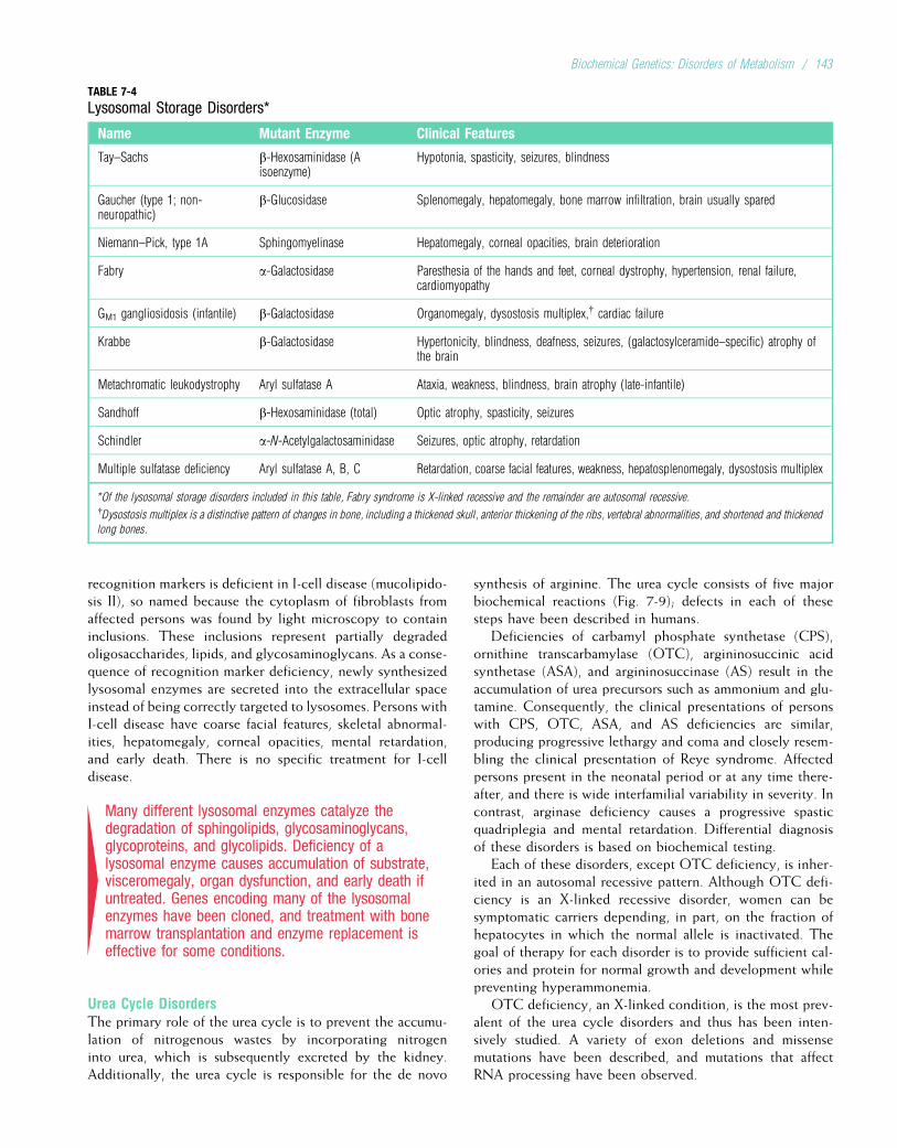
recognition markers is deficient in I-cell disease (mucolipido-sis II), so named because the cytoplasm of fibroblasts fromaffected persons was found by light microscopy to containinclusions. These inclusions represent partially degradedoligosaccharides, lipids, and glycosaminoglycans. As a conse-quence of recognition marker deficiency, newly synthesizedlysosomal enzymes are secreted into the extracellular spaceinstead of being correctly targeted to lysosomes. Persons withI-cell disease have coarse facial features, skeletal abnormal-ities, hepatomegaly, corneal opacities, mental retardation,and early death. There is no specific treatment for I-celldisease.
4Many different lysosomal enzymes catalyze thedegradation of sphingolipids, glycosaminoglycans,glycoproteins, and glycolipids. Deficiency of alysosomal enzyme causes accumulation of substrate,visceromegaly, organ dysfunction, and early death ifuntreated. Genes encoding many of the lysosomalenzymes have been cloned, and treatment with bonemarrow transplantation and enzyme replacement iseffective for some conditions.
Urea Cycle DisordersThe primary role of the urea cycle is to prevent the accumu-lation of nitrogenous wastes by incorporating nitrogeninto urea, which is subsequently excreted by the kidney.Additionally, the urea cycle is responsible for the de novo
synthesis of arginine. The urea cycle consists of five majorbiochemical reactions (Fig. 7-9); defects in each of thesesteps have been described in humans.
Deficiencies of carbamyl phosphate synthetase (CPS),ornithine transcarbamylase (OTC), argininosuccinic acidsynthetase (ASA), and argininosuccinase (AS) result in theaccumulation of urea precursors such as ammonium and glu-tamine. Consequently, the clinical presentations of personswith CPS, OTC, ASA, and AS deficiencies are similar,producing progressive lethargy and coma and closely resem-bling the clinical presentation of Reye syndrome. Affectedpersons present in the neonatal period or at any time there-after, and there is wide interfamilial variability in severity. Incontrast, arginase deficiency causes a progressive spasticquadriplegia and mental retardation. Differential diagnosisof these disorders is based on biochemical testing.
Each of these disorders, except OTC deficiency, is inher-ited in an autosomal recessive pattern. Although OTC defi-ciency is an X-linked recessive disorder, women can besymptomatic carriers depending, in part, on the fraction ofhepatocytes in which the normal allele is inactivated. Thegoal of therapy for each disorder is to provide sufficient cal-ories and protein for normal growth and development whilepreventing hyperammonemia.
OTC deficiency, an X-linked condition, is the most prev-alent of the urea cycle disorders and thus has been inten-sively studied. A variety of exon deletions and missensemutations have been described, and mutations that affectRNA processing have been observed.
TABLE 7-4
Lysosomal Storage Disorders*
Name Mutant Enzyme Clinical Features
Tay–Sachs b-Hexosaminidase (Aisoenzyme)
Hypotonia, spasticity, seizures, blindness
Gaucher (type 1; non-neuropathic)
b-Glucosidase Splenomegaly, hepatomegaly, bone marrow infiltration, brain usually spared
Niemann–Pick, type 1A Sphingomyelinase Hepatomegaly, corneal opacities, brain deterioration
Fabry a-Galactosidase Paresthesia of the hands and feet, corneal dystrophy, hypertension, renal failure,cardiomyopathy
GM1 gangliosidosis (infantile) b-Galactosidase Organomegaly, dysostosis multiplex,{ cardiac failure
Krabbe b-Galactosidase Hypertonicity, blindness, deafness, seizures, (galactosylceramide–specific) atrophy ofthe brain
Metachromatic leukodystrophy Aryl sulfatase A Ataxia, weakness, blindness, brain atrophy (late-infantile)
Sandhoff b-Hexosaminidase (total) Optic atrophy, spasticity, seizures
Schindler a-N-Acetylgalactosaminidase Seizures, optic atrophy, retardation
Multiple sulfatase deficiency Aryl sulfatase A, B, C Retardation, coarse facial features, weakness, hepatosplenomegaly, dysostosis multiplex
*Of the lysosomal storage disorders included in this table, Fabry syndrome is X-linked recessive and the remainder are autosomal recessive.{Dysostosis multiplex is a distinctive pattern of changes in bone, including a thickened skull, anterior thickening of the ribs, vertebral abnormalities, and shortened and thickened
long bones.
Biochemical Genetics: Disorders of Metabolism / 143
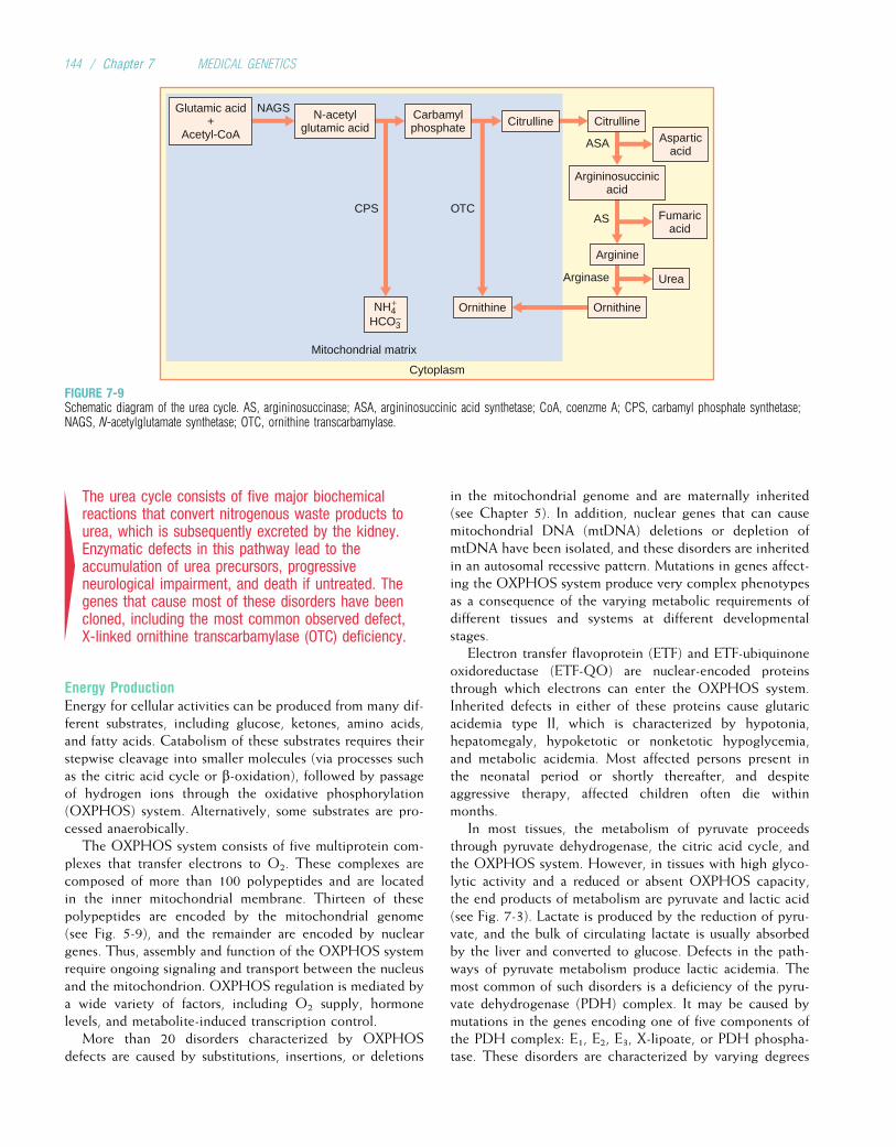
4The urea cycle consists of five major biochemicalreactions that convert nitrogenous waste products tourea, which is subsequently excreted by the kidney.Enzymatic defects in this pathway lead to theaccumulation of urea precursors, progressiveneurological impairment, and death if untreated. Thegenes that cause most of these disorders have beencloned, including the most common observed defect,X-linked ornithine transcarbamylase (OTC) deficiency.
Energy ProductionEnergy for cellular activities can be produced from many dif-ferent substrates, including glucose, ketones, amino acids,and fatty acids. Catabolism of these substrates requires theirstepwise cleavage into smaller molecules (via processes suchas the citric acid cycle or b-oxidation), followed by passageof hydrogen ions through the oxidative phosphorylation(OXPHOS) system. Alternatively, some substrates are pro-cessed anaerobically.
The OXPHOS system consists of five multiprotein com-plexes that transfer electrons to O2. These complexes arecomposed of more than 100 polypeptides and are locatedin the inner mitochondrial membrane. Thirteen of thesepolypeptides are encoded by the mitochondrial genome(see Fig. 5-9), and the remainder are encoded by nucleargenes. Thus, assembly and function of the OXPHOS systemrequire ongoing signaling and transport between the nucleusand the mitochondrion. OXPHOS regulation is mediated bya wide variety of factors, including O2 supply, hormonelevels, and metabolite-induced transcription control.
More than 20 disorders characterized by OXPHOSdefects are caused by substitutions, insertions, or deletions
in the mitochondrial genome and are maternally inherited(see Chapter 5). In addition, nuclear genes that can causemitochondrial DNA (mtDNA) deletions or depletion ofmtDNA have been isolated, and these disorders are inheritedin an autosomal recessive pattern. Mutations in genes affect-ing the OXPHOS system produce very complex phenotypesas a consequence of the varying metabolic requirements ofdifferent tissues and systems at different developmentalstages.
Electron transfer flavoprotein (ETF) and ETF-ubiquinoneoxidoreductase (ETF-QO) are nuclear-encoded proteinsthrough which electrons can enter the OXPHOS system.Inherited defects in either of these proteins cause glutaricacidemia type II, which is characterized by hypotonia,hepatomegaly, hypoketotic or nonketotic hypoglycemia,and metabolic acidemia. Most affected persons present inthe neonatal period or shortly thereafter, and despiteaggressive therapy, affected children often die withinmonths.
In most tissues, the metabolism of pyruvate proceedsthrough pyruvate dehydrogenase, the citric acid cycle, andthe OXPHOS system. However, in tissues with high glyco-lytic activity and a reduced or absent OXPHOS capacity,the end products of metabolism are pyruvate and lactic acid(see Fig. 7-3). Lactate is produced by the reduction of pyru-vate, and the bulk of circulating lactate is usually absorbedby the liver and converted to glucose. Defects in the path-ways of pyruvate metabolism produce lactic acidemia. Themost common of such disorders is a deficiency of the pyru-vate dehydrogenase (PDH) complex. It may be caused bymutations in the genes encoding one of five components ofthe PDH complex: E1, E2, E3, X-lipoate, or PDH phospha-tase. These disorders are characterized by varying degrees
OTC
Arginine
Arginase Urea
OrnithineOrnithine
Cytoplasm
Glutamic acid+
Acetyl-CoA
NAGS N-acetylglutamic acid
CPS
Carbamylphosphate Citrulline Citrulline
Asparticacid
ASA
Argininosuccinicacid
AS Fumaricacid
_HCO3
+NH4
Mitochondrial matrix
FIGURE 7-9Schematic diagram of the urea cycle. AS, argininosuccinase; ASA, argininosuccinic acid synthetase; CoA, coenzme A; CPS, carbamyl phosphate synthetase;NAGS, N-acetylglutamate synthetase; OTC, ornithine transcarbamylase.
144 / Chapter 7 MEDICAL GENETICS
of lactic acidemia, developmental delay, and abnormalities ofthe central nervous system. It has been suggested that thefacial features of some children with PDH deficiency resem-ble those of children with fetal alcohol syndrome (see Clini-cal Commentary 15-5 in Chapter 15). It has also beenproposed that acetaldehyde from the circulation of motherswith alcoholism inhibits PDH in the fetus, creating a pheno-copy of PDH deficiency.
4The phenotype produced by defects of energymetabolism is complex because of the varyingoxidative demands of different tissues and organs.Once the condition is diagnosed, the goal of treatmentis to use alternative pathways of energy production.
Transport SystemsThe efficient movement of molecules between compartments(e.g., organelles, cells, the environment), and hence across abarrier, often requires a macromolecule that connects thecompartments and mediates transport through the barrier.Abnormalities of these transport systems have myriad effects,depending on whether altered barrier integrity or the accu-mulation of substrate has a greater impact on normalphysiology.
CystineCystine is the disulfide derivative of the amino acid cysteine.Abnormal cystine transport can produce two diseases: cystin-uria and cystinosis. Both disorders are inherited in an autoso-mal recessive fashion.
Abnormal cystine transport between cells and the extra-cellular environment causes cystinuria, one of the most com-mon inherited disorders of metabolism. Although cystinuriaproduces substantial morbidity, early death is uncommon.Cystinuria is a genetically heterogeneous disorder causedby a defect of dibasic amino acid transport affecting theepithelial cells of the gastrointestinal tract and renal tubules.As a result, cystine, lysine, arginine, and ornithine areexcreted in urine in quantities higher than normal. Cystineis the most insoluble of the amino acids; therefore, elevatedurinary cystine predisposes to the formation of renal calculi(kidney stones). Complications of chronic nephrolithiasis(presence of kidney stones) include infection, hypertension,and renal failure. Treatment of cystinuria consists largely ofrendering cystine more soluble. This is accomplished byadministering pharmacological amounts of water (4-6 L/day), alkalinizing the urine, and using chelating agents suchas penicillamine.
Based on amino acid excretion studies, cystinuria has beendivided into three phenotypes. Type I cystinuria has beenassociated with missense, nonsense, and deletion mutationsin a gene termed soluble carrier family 3, member 1 amino acidtransporter (SLC3A1). Types II and III cystinuria are causedby mutations in a gene called SLC7A9. SLC3A1 andSLC7A9 encode the heavy and light subunits of the amino
acid transporter b0,þ located on the brush-border plasmamembrane of epithelial cells in the proximal tubules of thekidney. In vitro studies of mutant SLC3A1 and SLC7A9 pro-tein have demonstrated a marked reduction in transportactivity, providing direct evidence for the role of these pro-teins in cystinuria.
Cystinosis is a rare disorder caused by a diminished abil-ity to transport cystine across the lysosomal membrane. Thisproduces an accumulation of cystine crystals in the lyso-somes of most tissues. Affected persons are normal at birthbut develop electrolyte disturbances, corneal crystals, rick-ets, and poor growth by the age of 1 year. Renal glomerulardamage is severe enough to necessitate dialysis or transplan-tation within the first decade of life. Transplanted kidneysfunction normally, but chronic complications such as diabe-tes mellitus, pancreatic insufficiency, hypogonadism, myopa-thy, and blindness occur. Until recently, treatment waslargely supportive, including renal transplantation. However,cysteine-depleting agents such as cysteamine have provedsuccessful in slowing renal deterioration and improvinggrowth. A novel gene encoding an integral lysosomal mem-brane protein was recently found to be mutated in personswith cystinosis.
4The phenotype of transport defects is partlycontingent on the degree of barrier disruption as wellas the compartments through which normal traffic iscompromised. Abnormal cystine transport betweencells and the extracellular environment causescystinuria, renal disease, and hypertension. Cystinosisis produced by a defect of cystine efflux from thelysosome; it leads to severe chronic disabilities and, ifuntreated, death.
Heavy MetalsMany of the enzymes controlling metabolic processesrequire additional factors (cofactors) to function properlyand efficiently. These cofactors are commonly trace ele-ments such as ions of heavy metals (metals that are moredense than those in the first two groups of the periodictable). At least 12 trace elements are essential to humans.For example, a zinc ion acts as a cofactor in carbonic anhy-drase, placing a hydroxide ion next to carbon dioxide tofacilitate the formation of bicarbonate. Although an ade-quate supply of trace elements is critical for normal metabo-lism, excessive amounts of circulating and/or stored heavymetals are highly toxic. Accordingly, a complex series oftransport and storage proteins precisely control heavy metalhomeostasis. Abnormalities of these proteins cause progres-sive dysfunction of various organs, often leading to prema-ture death if untreated. Human disorders that disrupt thenormal homeostasis of copper (Wilson disease, Menkes dis-ease, occipital horn syndrome), iron (hereditary hemochro-matosis; Clinical Commentary 7-2), and zinc (hereditaryacrodermatitis enteropathica) have been described.
Biochemical Genetics: Disorders of Metabolism / 145
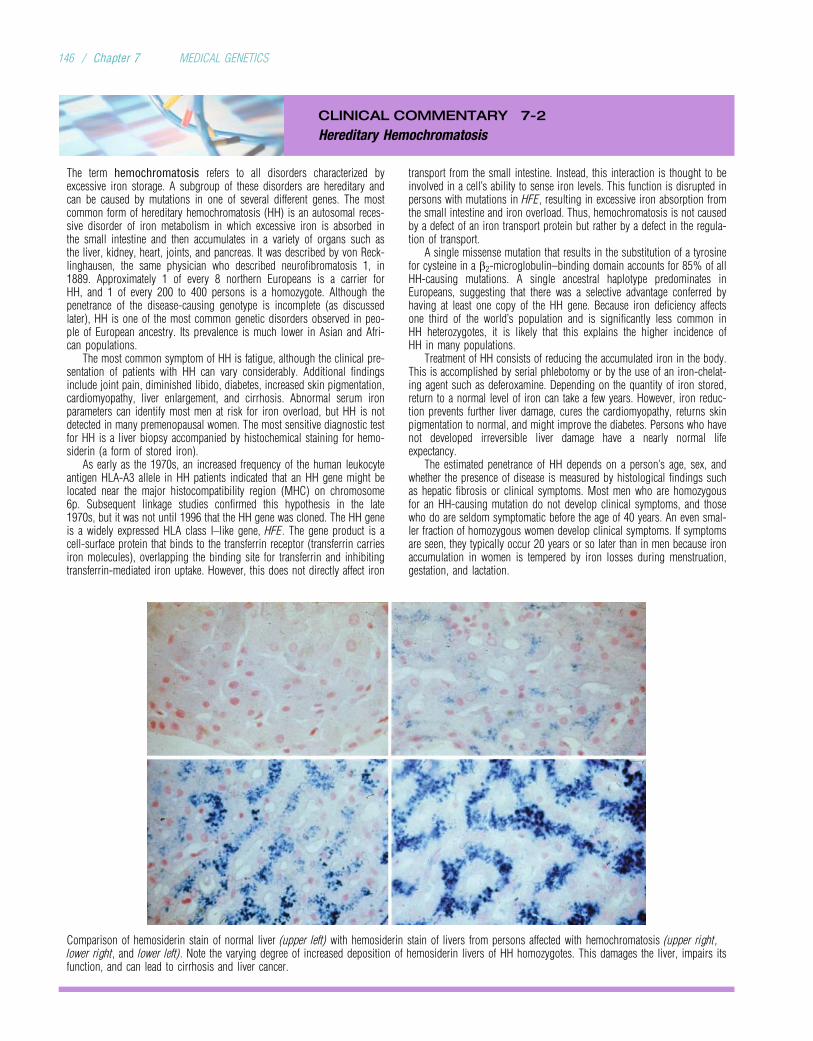
CLINICAL COMMENTARY 7-2
Hereditary Hemochromatosis
The term hemochromatosis refers to all disorders characterized byexcessive iron storage. A subgroup of these disorders are hereditary andcan be caused by mutations in one of several different genes. The mostcommon form of hereditary hemochromatosis (HH) is an autosomal reces-sive disorder of iron metabolism in which excessive iron is absorbed inthe small intestine and then accumulates in a variety of organs such asthe liver, kidney, heart, joints, and pancreas. It was described by von Reck-linghausen, the same physician who described neurofibromatosis 1, in1889. Approximately 1 of every 8 northern Europeans is a carrier forHH, and 1 of every 200 to 400 persons is a homozygote. Although thepenetrance of the disease-causing genotype is incomplete (as discussedlater), HH is one of the most common genetic disorders observed in peo-ple of European ancestry. Its prevalence is much lower in Asian and Afri-can populations.
The most common symptom of HH is fatigue, although the clinical pre-sentation of patients with HH can vary considerably. Additional findingsinclude joint pain, diminished libido, diabetes, increased skin pigmentation,cardiomyopathy, liver enlargement, and cirrhosis. Abnormal serum ironparameters can identify most men at risk for iron overload, but HH is notdetected in many premenopausal women. The most sensitive diagnostic testfor HH is a liver biopsy accompanied by histochemical staining for hemo-siderin (a form of stored iron).
As early as the 1970s, an increased frequency of the human leukocyteantigen HLA-A3 allele in HH patients indicated that an HH gene might belocated near the major histocompatibility region (MHC) on chromosome6p. Subsequent linkage studies confirmed this hypothesis in the late1970s, but it was not until 1996 that the HH gene was cloned. The HH geneis a widely expressed HLA class I–like gene, HFE. The gene product is acell-surface protein that binds to the transferrin receptor (transferrin carriesiron molecules), overlapping the binding site for transferrin and inhibitingtransferrin-mediated iron uptake. However, this does not directly affect iron
transport from the small intestine. Instead, this interaction is thought to beinvolved in a cell’s ability to sense iron levels. This function is disrupted inpersons with mutations in HFE, resulting in excessive iron absorption fromthe small intestine and iron overload. Thus, hemochromatosis is not causedby a defect of an iron transport protein but rather by a defect in the regula-tion of transport.
A single missense mutation that results in the substitution of a tyrosinefor cysteine in a b2-microglobulin–binding domain accounts for 85% of allHH-causing mutations. A single ancestral haplotype predominates inEuropeans, suggesting that there was a selective advantage conferred byhaving at least one copy of the HH gene. Because iron deficiency affectsone third of the world’s population and is significantly less common inHH heterozygotes, it is likely that this explains the higher incidence ofHH in many populations.
Treatment of HH consists of reducing the accumulated iron in the body.This is accomplished by serial phlebotomy or by the use of an iron-chelat-ing agent such as deferoxamine. Depending on the quantity of iron stored,return to a normal level of iron can take a few years. However, iron reduc-tion prevents further liver damage, cures the cardiomyopathy, returns skinpigmentation to normal, and might improve the diabetes. Persons who havenot developed irreversible liver damage have a nearly normal lifeexpectancy.
The estimated penetrance of HH depends on a person’s age, sex, andwhether the presence of disease is measured by histological findings suchas hepatic fibrosis or clinical symptoms. Most men who are homozygousfor an HH-causing mutation do not develop clinical symptoms, and thosewho do are seldom symptomatic before the age of 40 years. An even smal-ler fraction of homozygous women develop clinical symptoms. If symptomsare seen, they typically occur 20 years or so later than in men because ironaccumulation in women is tempered by iron losses during menstruation,gestation, and lactation.
Comparison of hemosiderin stain of normal liver (upper left) with hemosiderin stain of livers from persons affected with hemochromatosis (upper right,lower right, and lower left). Note the varying degree of increased deposition of hemosiderin livers of HH homozygotes. This damages the liver, impairs itsfunction, and can lead to cirrhosis and liver cancer.
146 / Chapter 7 MEDICAL GENETICS
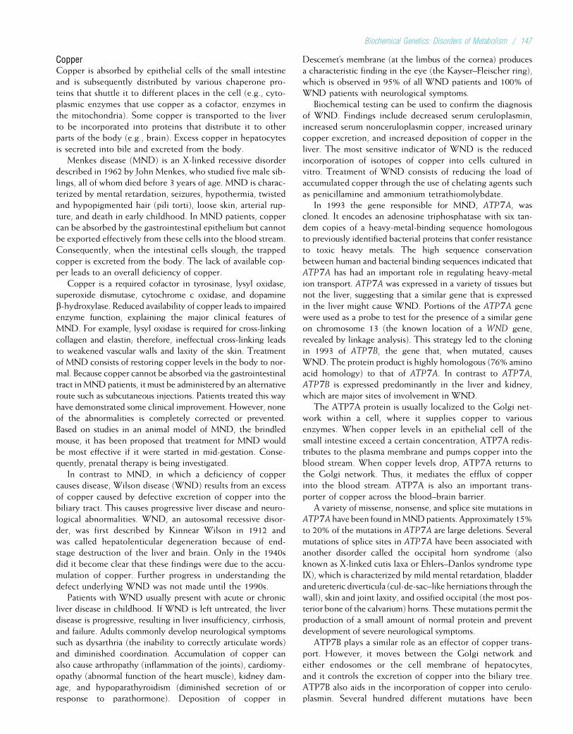
CopperCopper is absorbed by epithelial cells of the small intestineand is subsequently distributed by various chaperone pro-teins that shuttle it to different places in the cell (e.g., cyto-plasmic enzymes that use copper as a cofactor, enzymes inthe mitochondria). Some copper is transported to the liverto be incorporated into proteins that distribute it to otherparts of the body (e.g., brain). Excess copper in hepatocytesis secreted into bile and excreted from the body.
Menkes disease (MND) is an X-linked recessive disorderdescribed in 1962 by John Menkes, who studied five male sib-lings, all of whom died before 3 years of age. MND is charac-terized by mental retardation, seizures, hypothermia, twistedand hypopigmented hair (pili torti), loose skin, arterial rup-ture, and death in early childhood. In MND patients, coppercan be absorbed by the gastrointestinal epithelium but cannotbe exported effectively from these cells into the blood stream.Consequently, when the intestinal cells slough, the trappedcopper is excreted from the body. The lack of available cop-per leads to an overall deficiency of copper.
Copper is a required cofactor in tyrosinase, lysyl oxidase,superoxide dismutase, cytochrome c oxidase, and dopamineb-hydroxylase. Reduced availability of copper leads to impairedenzyme function, explaining the major clinical features ofMND. For example, lysyl oxidase is required for cross-linkingcollagen and elastin; therefore, ineffectual cross-linking leadsto weakened vascular walls and laxity of the skin. Treatmentof MND consists of restoring copper levels in the body to nor-mal. Because copper cannot be absorbed via the gastrointestinaltract inMND patients, it must be administered by an alternativeroute such as subcutaneous injections. Patients treated this wayhave demonstrated some clinical improvement. However, noneof the abnormalities is completely corrected or prevented.Based on studies in an animal model of MND, the brindledmouse, it has been proposed that treatment for MND wouldbe most effective if it were started in mid-gestation. Conse-quently, prenatal therapy is being investigated.
In contrast to MND, in which a deficiency of coppercauses disease, Wilson disease (WND) results from an excessof copper caused by defective excretion of copper into thebiliary tract. This causes progressive liver disease and neuro-logical abnormalities. WND, an autosomal recessive disor-der, was first described by Kinnear Wilson in 1912 andwas called hepatolenticular degeneration because of end-stage destruction of the liver and brain. Only in the 1940sdid it become clear that these findings were due to the accu-mulation of copper. Further progress in understanding thedefect underlying WND was not made until the 1990s.
Patients with WND usually present with acute or chronicliver disease in childhood. If WND is left untreated, the liverdisease is progressive, resulting in liver insufficiency, cirrhosis,and failure. Adults commonly develop neurological symptomssuch as dysarthria (the inability to correctly articulate words)and diminished coordination. Accumulation of copper canalso cause arthropathy (inflammation of the joints), cardiomy-opathy (abnormal function of the heart muscle), kidney dam-age, and hypoparathyroidism (diminished secretion of orresponse to parathormone). Deposition of copper in
Descemet’s membrane (at the limbus of the cornea) producesa characteristic finding in the eye (the Kayser–Fleischer ring),which is observed in 95% of all WND patients and 100% ofWND patients with neurological symptoms.
Biochemical testing can be used to confirm the diagnosisof WND. Findings include decreased serum ceruloplasmin,increased serum nonceruloplasmin copper, increased urinarycopper excretion, and increased deposition of copper in theliver. The most sensitive indicator of WND is the reducedincorporation of isotopes of copper into cells cultured invitro. Treatment of WND consists of reducing the load ofaccumulated copper through the use of chelating agents suchas penicillamine and ammonium tetrathiomolybdate.
In 1993 the gene responsible for MND, ATP7A, wascloned. It encodes an adenosine triphosphatase with six tan-dem copies of a heavy-metal-binding sequence homologousto previously identified bacterial proteins that confer resistanceto toxic heavy metals. The high sequence conservationbetween human and bacterial binding sequences indicated thatATP7A has had an important role in regulating heavy-metalion transport. ATP7A was expressed in a variety of tissues butnot the liver, suggesting that a similar gene that is expressedin the liver might cause WND. Portions of the ATP7A genewere used as a probe to test for the presence of a similar geneon chromosome 13 (the known location of a WND gene,revealed by linkage analysis). This strategy led to the cloningin 1993 of ATP7B, the gene that, when mutated, causesWND. The protein product is highly homologous (76% aminoacid homology) to that of ATP7A. In contrast to ATP7A,ATP7B is expressed predominantly in the liver and kidney,which are major sites of involvement in WND.
The ATP7A protein is usually localized to the Golgi net-work within a cell, where it supplies copper to variousenzymes. When copper levels in an epithelial cell of thesmall intestine exceed a certain concentration, ATP7A redis-tributes to the plasma membrane and pumps copper into theblood stream. When copper levels drop, ATP7A returns tothe Golgi network. Thus, it mediates the efflux of copperinto the blood stream. ATP7A is also an important trans-porter of copper across the blood–brain barrier.
A variety of missense, nonsense, and splice site mutations inATP7A have been found inMNDpatients. Approximately 15%to 20% of the mutations in ATP7A are large deletions. Severalmutations of splice sites in ATP7A have been associated withanother disorder called the occipital horn syndrome (alsoknown as X-linked cutis laxa or Ehlers–Danlos syndrome typeIX), which is characterized by mild mental retardation, bladderand ureteric diverticula (cul-de-sac–like herniations through thewall), skin and joint laxity, and ossified occipital (the most pos-terior bone of the calvarium) horns. These mutations permit theproduction of a small amount of normal protein and preventdevelopment of severe neurological symptoms.
ATP7B plays a similar role as an effector of copper trans-port. However, it moves between the Golgi network andeither endosomes or the cell membrane of hepatocytes,and it controls the excretion of copper into the biliary tree.ATP7B also aids in the incorporation of copper into cerulo-plasmin. Several hundred different mutations have been
Biochemical Genetics: Disorders of Metabolism / 147
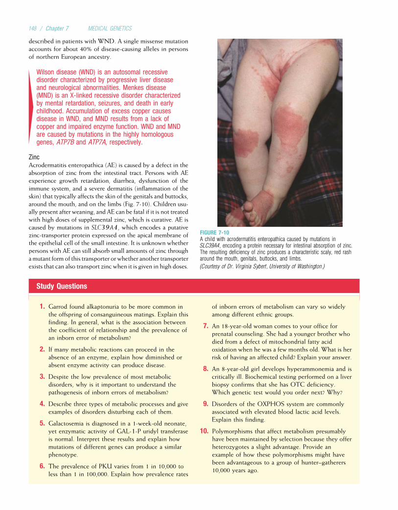
described in patients with WND. A single missense mutationaccounts for about 40% of disease-causing alleles in personsof northern European ancestry.
4Wilson disease (WND) is an autosomal recessivedisorder characterized by progressive liver diseaseand neurological abnormalities. Menkes disease(MND) is an X-linked recessive disorder characterizedby mental retardation, seizures, and death in earlychildhood. Accumulation of excess copper causesdisease in WND, and MND results from a lack ofcopper and impaired enzyme function. WND and MNDare caused by mutations in the highly homologousgenes, ATP7B and ATP7A, respectively.
ZincAcrodermatitis enteropathica (AE) is caused by a defect in theabsorption of zinc from the intestinal tract. Persons with AEexperience growth retardation, diarrhea, dysfunction of theimmune system, and a severe dermatitis (inflammation of theskin) that typically affects the skin of the genitals and buttocks,around the mouth, and on the limbs (Fig. 7-10). Children usu-ally present after weaning, and AE can be fatal if it is not treatedwith high doses of supplemental zinc, which is curative. AE iscaused by mutations in SLC39A4, which encodes a putativezinc-transporter protein expressed on the apical membrane ofthe epithelial cell of the small intestine. It is unknown whetherpersons with AE can still absorb small amounts of zinc throughamutant form of this transporter orwhether another transporterexists that can also transport zinc when it is given in high doses.
Study Questions
1. Garrod found alkaptonuria to be more common inthe offspring of consanguineous matings. Explain thisfinding. In general, what is the association betweenthe coefficient of relationship and the prevalence ofan inborn error of metabolism?
2. If many metabolic reactions can proceed in theabsence of an enzyme, explain how diminished orabsent enzyme activity can produce disease.
3. Despite the low prevalence of most metabolicdisorders, why is it important to understand thepathogenesis of inborn errors of metabolism?
4. Describe three types of metabolic processes and giveexamples of disorders disturbing each of them.
5. Galactosemia is diagnosed in a 1-week-old neonate,yet enzymatic activity of GAL-1-P uridyl transferaseis normal. Interpret these results and explain howmutations of different genes can produce a similarphenotype.
6. The prevalence of PKU varies from 1 in 10,000 toless than 1 in 100,000. Explain how prevalence rates
of inborn errors of metabolism can vary so widelyamong different ethnic groups.
7. An 18-year-old woman comes to your office forprenatal counseling. She had a younger brother whodied from a defect of mitochondrial fatty acidoxidation when he was a few months old. What is herrisk of having an affected child? Explain your answer.
8. An 8-year-old girl develops hyperammonemia and iscritically ill. Biochemical testing performed on a liverbiopsy confirms that she has OTC deficiency.Which genetic test would you order next? Why?
9. Disorders of the OXPHOS system are commonlyassociated with elevated blood lactic acid levels.Explain this finding.
10. Polymorphisms that affect metabolism presumablyhave been maintained by selection because they offerheterozygotes a slight advantage. Provide anexample of how these polymorphisms might havebeen advantageous to a group of hunter–gatherers10,000 years ago.
FIGURE 7-10A child with acrodermatitis enteropathica caused by mutations inSLC39A4, encoding a protein necessary for intestinal absorption of zinc.The resulting deficiency of zinc produces a characteristic scaly, red rasharound the mouth, genitals, buttocks, and limbs.
(Courtesy of Dr. Virginia Sybert, University of Washington.)
148 / Chapter 7 MEDICAL GENETICS
Suggested ReadingsAdams PC, Barton JC. Haemochromatosis. Lancet
2007;370:1855–60.Allen KJ, Gurrin LC, Constantine CC, et al. Iron-overload-
related disease in HFE hereditary hemochromatosis. NEngl J Med 2008;358(3):221–30.
Andrews NC. Metal transporters and disease. Curr OpinChem Biol 2002;6:181–6.
Beutler E. Hemochromatosis: Genetics and pathophysiology.Ann Rev Med 2006;57:331–47.
Bosch AM. Classical galactosemia revisited. J Inherit MetabDis 2006;29:516–25.
Burrow TA, Hopkin RJ, Leslie ND, et al. Enzyme reconstitu-tion/replacement therapy for lysosomal storage diseases.Curr Opin Pediatr 2007;19:628–35.
de Bie P, Muller P, Wijmenga C, Klomp LWJ. Molecularpathogenesis of Wilson and Menkes disease: Correlationof mutations with molecular defects and disease pheno-types. J Med Genet 2007;44:673–88.
James PM, Levy HL. The clinical aspects of newborn screen-ing: Importance of newborn screening follow-up. MentRetard Dev Disabil Res Rev 2006;12:246–54.
Merke DP, Bornstein SR. Congenital adrenal hyperplasia.Lancet 2005;365:2125–36.
Nassogne MC, Heron B, Touaita G, et al. Urea cycledefects: Management and outcome. J Inherit Metab Dis2005;28:407–14.
Rinaldo P, Matern D. Fatty acid oxidation disorders. AnnuRev Physiol 2002;64:477–502.
Schulze A, Lindner M, Kohlmuller D, et al. Expanded new-born screening for inborn errors of metabolism by electro-spray ionization-tandem mass spectrometry: Results,outcome, and implications. Pediatrics 2003; 111:1399–406.
Scott CR. The genetic tyrosinemias. Am J Med Genet CSemin Med Genet 2006;142:121–26.
Scriver CR, Sly WS, Childs B, et al. The Metabolic andMolecular Bases of Inherited Disease. New York:McGraw–Hill, 2001.
Scriver CR. The PAH gene, phenylkentonuria, and a para-digm shift. Hum Mutation 2007;28:831–45.
Tishkoff SA, Reed FA, Ranciaro A, et al. Convergent adapta-tion of human lactase persistence in Africa and Europe.Nat Genet 2006;39:31–40.
Wappner R, Cho S, Kronmal RA, et al. Management of phe-nylketonuria for optimal outcome: A review of guidelinesfor phenylketonuria management and a report of surveysof parents, patients, and clinic directors. Pediatrics1999;104:e68.
Yu H, Patel SB. Recent insights into Smith–Lemli–Opitz syn-drome. Clin Genet 2005;68:383–91.
Biochemical Genetics: Disorders of Metabolism / 149
Chapter 8
GENE MAPPING AND IDENTIFICATION
The identification of mutations that cause disease is a centralfocus of medical genetics. This process often begins bymapping mutations that occur in affected persons to specificlocations on chromosomes. With the completion of theHuman Genome Project (see Box 8-2 later in this chapter),the locations of virtually every human gene in the genomeare now known. The availability of these data, along withdramatic advances in molecular genetic technology andimportant developments in the statistical analysis of geneticdata, has greatly expedited the mapping of disease-causingmutations. However, the specific genetic alterations respon-sible for most inherited disease phenotypes remain unknown(i.e., which gene or genes contribute to which disease).Much research is now devoted to discovering these geneticmutations and their consequences. As this work continues,our understanding of the biological basis of genetic diseasewill surely progress.
Mapping disease-causing genes is an important step in theunderstanding, diagnosis, and eventual treatment of agenetic disease. When a disease-causing gene’s location hasbeen pinpointed, it is often possible to provide a more accu-rate prognosis for persons at risk for a genetic disease. Animportant next step is often the cloning of the gene (as dis-cussed in Chapter 3, cloning refers to inserting a gene into avector so that copies, or clones, can be made). Once a genehas been cloned, its DNA sequence and protein product canbe studied directly. This can contribute to our understandingof the actual cause of the disease. Furthermore, gene cloningcan open the way to the manufacture of a normal gene prod-uct through recombinant DNA techniques, permitting moreeffective treatment of a genetic disease. For example, recom-binant clotting factor VIII is used to treat hemophilia A, asdiscussed in Chapter 5, and recombinant insulin is used inthe treatment of type 1 diabetes. Gene therapy—modifyinggenes of persons with a genetic disease—also becomes apossibility. Thus, gene mapping contributes directly to manyof the primary goals of medical genetics.
This chapter discusses the approaches commonly used ingene mapping and identification. Two major types of genemapping can be distinguished. In gene mapping, the fre-quency of meiotic crossovers between loci is used to esti-mate distances between loci. Physical mapping involvesusing cytogenetic, molecular, and computational methods
to determine the actual physical locations of disease-causingmutations on chromosomes. Later sections in this chapterdescribe how these mapping techniques lead to the charac-terization of disease-causing genes and thus to more accurateprediction of disease risk in families.
GENE MAPPINGLinkage AnalysisOne of Gregor Mendel’s laws, the principle of independentassortment, states that an individual’s genes will be transmit-ted to the next generation independently of one another (seeChapter 4). Mendel was not aware that genes are located onchromosomes and that genes located near one another onthe same chromosome are transmitted together rather thanindependently. Thus, the principle of independent assort-ment holds true for most pairs of loci, but not for those thatoccupy the same region of a chromosome. Such loci are saidto be linked.
Figure 8-1 depicts two loci, A and B, that are located closetogether on the same chromosome. A third locus, C, islocated on another chromosome. In the individual in ourexample, each of these loci has two alleles, designated 1and 2. A and B are linked, so A1 and B1 are inheritedtogether. Because A and C are on different chromosomesand thus unlinked, their alleles do follow the principle ofindependent assortment. Hence, if the process of meiosisplaces A1 in a gamete, the probability that C1 will be foundin the same gamete is 50%.
Recall from Chapter 2 that homologous chromosomessometimes exchange portions of their DNA during prophase I(this is known as crossing over or crossover). The averagechromosome experiences one to three crossover events dur-ing meiosis. As a result of crossover, new combinations ofalleles can be formed on a chromosome. Consider againthe linked loci, A and B, in Figure 8-1. Alleles A1 and B1are located close together on one chromosome, and allelesA2 and B2 are located on the homologous chromosome.The combination of alleles on each chromosome is ahaplotype (from “haploid genotype”). The two haplotypes ofthis individual are denoted A1B1/A2B2. As Figure 8-2A shows,in the absence of crossover A1B1 will be found in one gameteand A2B2 in the other. But when there is a crossover, new
150
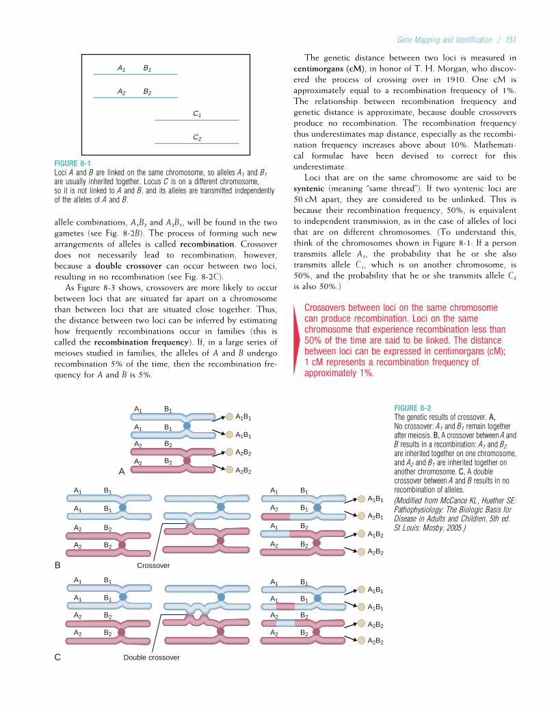
allele combinations, A1B2 and A2B1, will be found in the twogametes (see Fig. 8-2B). The process of forming such newarrangements of alleles is called recombination. Crossoverdoes not necessarily lead to recombination, however,because a double crossover can occur between two loci,resulting in no recombination (see Fig. 8-2C).
As Figure 8-3 shows, crossovers are more likely to occurbetween loci that are situated far apart on a chromosomethan between loci that are situated close together. Thus,the distance between two loci can be inferred by estimatinghow frequently recombinations occur in families (this iscalled the recombination frequency). If, in a large series ofmeioses studied in families, the alleles of A and B undergorecombination 5% of the time, then the recombination fre-quency for A and B is 5%.
The genetic distance between two loci is measured incentimorgans (cM), in honor of T. H. Morgan, who discov-ered the process of crossing over in 1910. One cM isapproximately equal to a recombination frequency of 1%.The relationship between recombination frequency andgenetic distance is approximate, because double crossoversproduce no recombination. The recombination frequencythus underestimates map distance, especially as the recombi-nation frequency increases above about 10%. Mathemati-cal formulae have been devised to correct for thisunderestimate.
Loci that are on the same chromosome are said to besyntenic (meaning “same thread”). If two syntenic loci are50 cM apart, they are considered to be unlinked. This isbecause their recombination frequency, 50%, is equivalentto independent transmission, as in the case of alleles of locithat are on different chromosomes. (To understand this,think of the chromosomes shown in Figure 8-1: If a persontransmits allele A1, the probability that he or she alsotransmits allele C1, which is on another chromosome, is50%, and the probability that he or she transmits allele C2is also 50%.)
4Crossovers between loci on the same chromosomecan produce recombination. Loci on the samechromosome that experience recombination less than50% of the time are said to be linked. The distancebetween loci can be expressed in centimorgans (cM);1 cM represents a recombination frequency ofapproximately 1%.
A1 B1
A2 B2
C1
C2
FIGURE 8-1Loci A and B are linked on the same chromosome, so alleles A1 and B1are usually inherited together. Locus C is on a different chromosome,so it is not linked to A and B, and its alleles are transmitted independentlyof the alleles of A and B.
A
Crossover
Double crossover
B
C
A1
A1
A2
A2
B1
B1
B2
B2
A1B1
A1B1
A2B2
A2B2
A1 B1 A1 B1
A1 B1
A1 B1
A1 B1
A1 B1
A2 B2
A2 B2
A2 B2
A2 B2
A2 B1
A1 B2
A2 B2
A1 B1
A2 B2
A2 B2
A1B1
A2B1
A1B2
A2B2
A1B1
A1B1
A2B2
A2B2
FIGURE 8-2The genetic results of crossover. A,No crossover: A1 and B1 remain togetherafter meiosis. B, A crossover between A andB results in a recombination: A1 and B2are inherited together on one chromosome,and A2 and B1 are inherited together onanother chromosome. C, A doublecrossover between A and B results in norecombination of alleles.
(Modified from McCance KL, Huether SE:Pathophysiology: The Biologic Basis forDisease in Adults and Children, 5th ed.St Louis: Mosby, 2005.)
Gene Mapping and Identification / 151
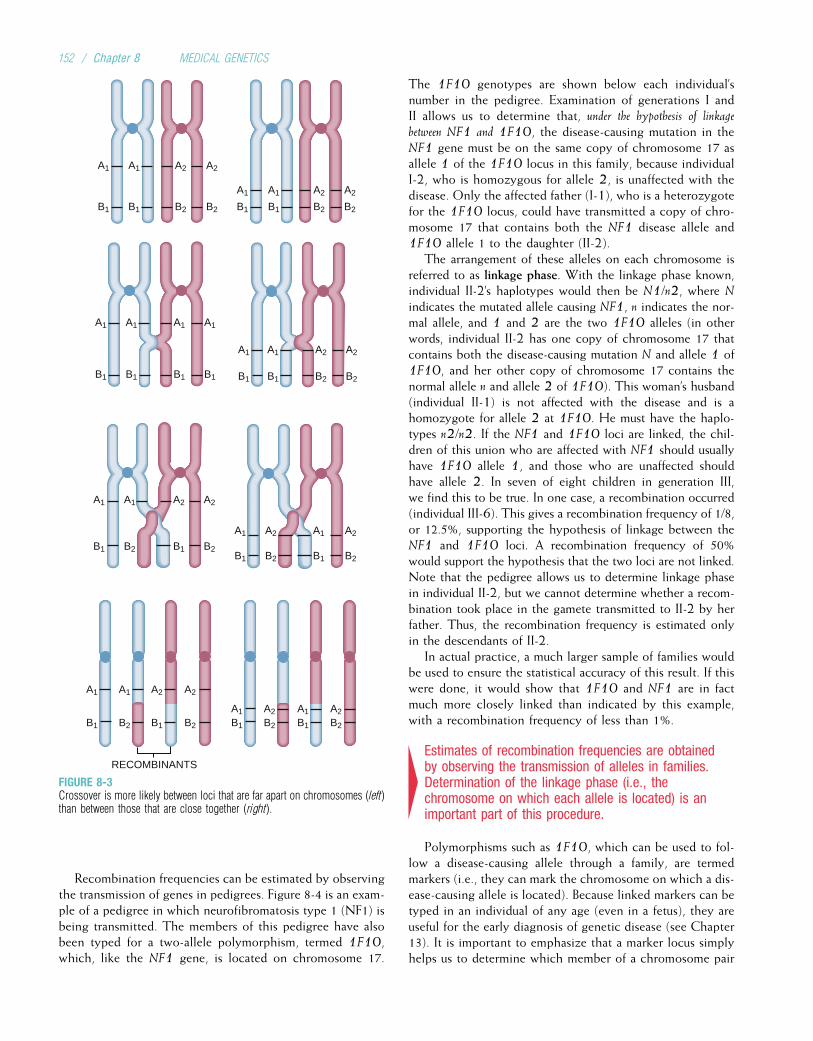
Recombination frequencies can be estimated by observingthe transmission of genes in pedigrees. Figure 8-4 is an exam-ple of a pedigree in which neurofibromatosis type 1 (NF1) isbeing transmitted. The members of this pedigree have alsobeen typed for a two-allele polymorphism, termed 1F10,which, like the NF1 gene, is located on chromosome 17.
The 1F10 genotypes are shown below each individual’snumber in the pedigree. Examination of generations I andII allows us to determine that, under the hypothesis of linkage
between NF1 and 1F10, the disease-causing mutation in theNF1 gene must be on the same copy of chromosome 17 asallele 1 of the 1F10 locus in this family, because individualI-2, who is homozygous for allele 2, is unaffected with thedisease. Only the affected father (I-1), who is a heterozygotefor the 1F10 locus, could have transmitted a copy of chro-mosome 17 that contains both the NF1 disease allele and1F10 allele 1 to the daughter (II-2).
The arrangement of these alleles on each chromosome isreferred to as linkage phase. With the linkage phase known,individual II-2’s haplotypes would then be N1/n2, where N
indicates the mutated allele causing NF1, n indicates the nor-mal allele, and 1 and 2 are the two 1F10 alleles (in otherwords, individual II-2 has one copy of chromosome 17 thatcontains both the disease-causing mutation N and allele 1 of1F10, and her other copy of chromosome 17 contains thenormal allele n and allele 2 of 1F10). This woman’s husband(individual II-1) is not affected with the disease and is ahomozygote for allele 2 at 1F10. He must have the haplo-types n2/n2. If the NF1 and 1F10 loci are linked, the chil-dren of this union who are affected with NF1 should usuallyhave 1F10 allele 1, and those who are unaffected shouldhave allele 2. In seven of eight children in generation III,we find this to be true. In one case, a recombination occurred(individual III-6). This gives a recombination frequency of 1/8,or 12.5%, supporting the hypothesis of linkage between theNF1 and 1F10 loci. A recombination frequency of 50%would support the hypothesis that the two loci are not linked.Note that the pedigree allows us to determine linkage phasein individual II-2, but we cannot determine whether a recom-bination took place in the gamete transmitted to II-2 by herfather. Thus, the recombination frequency is estimated onlyin the descendants of II-2.
In actual practice, a much larger sample of families wouldbe used to ensure the statistical accuracy of this result. If thiswere done, it would show that 1F10 and NF1 are in factmuch more closely linked than indicated by this example,with a recombination frequency of less than 1%.
4Estimates of recombination frequencies are obtainedby observing the transmission of alleles in families.Determination of the linkage phase (i.e., thechromosome on which each allele is located) is animportant part of this procedure.
Polymorphisms such as 1F10, which can be used to fol-low a disease-causing allele through a family, are termedmarkers (i.e., they can mark the chromosome on which a dis-ease-causing allele is located). Because linked markers can betyped in an individual of any age (even in a fetus), they areuseful for the early diagnosis of genetic disease (see Chapter13). It is important to emphasize that a marker locus simplyhelps us to determine which member of a chromosome pair
RECOMBINANTS
A1
B1
A1
B1
A1 A1 A2 A2
B1
A1 A1 A2
B1 B1B2
A2
B2
B2 B1 B2
A1
B1
A1
B1
A1
B1
A1
B1
A1
B1
A1
B1
A2
B2
A1
B1
A2
B2
A2
B2
A1
B1
A2
B2
A1
B1
A2
B2
A2
B2
A1
B1
A1
B1
A2
B2
A2
B2
A1
B1
A2
B2
A2
B2
FIGURE 8-3Crossover is more likely between loci that are far apart on chromosomes (left)than between those that are close together (right).
152 / Chapter 8 MEDICAL GENETICS
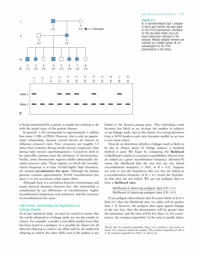
is being transmitted by a parent; it usually has nothing to dowith the actual cause of the genetic disease.
In general, 1 cM corresponds to approximately 1 millionbase pairs (1 Mb) of DNA. However, this is only an approx-imate relationship, because several factors are known toinfluence crossover rates. First, crossovers are roughly 1.5times more common during female meiosis (oogenesis) thanduring male meiosis (spermatogenesis). Crossovers tend tobe especially common near the telomeres of chromosomes.Finally, some chromosome regions exhibit substantially ele-vated crossover rates. These regions, in which the recombi-nation frequency is at least 10-fold higher than elsewhere,are termed recombination hot spots. Although the humangenome contains approximately 50,000 recombination hotspots, it is not yet known what causes them.
Although there is a correlation between centimorgans andactual physical distances between loci, this relationship iscomplicated by sex differences in recombination, higherrecombination frequencies near telomeres, and the existenceof recombination hot spots.
LOD Scores: Determining the Significance ofLinkage ResultsAs in any statistical study, we must be careful to ensure thatthe results obtained in a linkage study are not due simply tochance. For example, consider a two-allele marker locus thathas been typed in a pedigree. It is possible by chance for allaffected offspring to inherit one allele and for all unaffectedoffspring to inherit the other allele even if the marker is not
linked to the disease-causing gene. This misleading resultbecomes less likely as we increase the number of subjectsin our linkage study (just as the chance of a strong deviationfrom a 50/50 heads-to-tails ratio becomes smaller as we tossa coin many times).
How do we determine whether a linkage result is likely tobe due to chance alone? In linkage analysis, a standardmethod is used. We begin by comparing the likelihood(a likelihood is similar in concept to a probability) that two lociare linked at a given recombination frequency (denoted y)versus the likelihood that the two loci are not linked(recombination frequency ¼ 50%, or y ¼ 0.5). Supposewe wish to test the hypothesis that two loci are linked ata recombination frequency of y ¼ 0.1 versus the hypothe-sis that they are not linked. We use our pedigree data toform a likelihood ratio:
likelihood of observing pedigree data if y ¼ 0:1
likelihood of observing pedigree data if y ¼ 0:5
If our pedigree data indicate that y is more likely to be 0.1than 0.5, then the likelihood ratio (or odds) will be greaterthan 1. If, however, the pedigree data argue against linkageof the two loci, then the denominator will be greater thanthe numerator, and the ratio will be less than 1.0. For conve-nience, the common logarithm* of the ratio is usually taken;
A
Genotype2
2,2
12,2
21,2
11,2
11,2
21,2
32,2
41,2
52,2
62,2
71,2
82,2
I
II
III
B
Allele 1
I – 1
I – 2
II –
1
II –
2
III –
1
III –
2
III –
3
III –
4
III –
5
III –
6
III –
7
III –
8
Allele 2
FIGURE 8-4A, A neurofibromatosis type 1 pedigreein which each member has been typedfor the 1F10 polymorphism. Genotypesfor this two-allele marker locus areshown below each individual in thepedigree. Affected pedigree members areindicated by a shaded symbol. B, Anautoradiogram for the 1F10polymorphism in this family.
*Recall that the common logarithm (log10) of a number is the power towhich 10 is raised to obtain the number. The common logarithm of 100 is2, the common logarithm of 1000 is 3, and so on.
Gene Mapping and Identification / 153
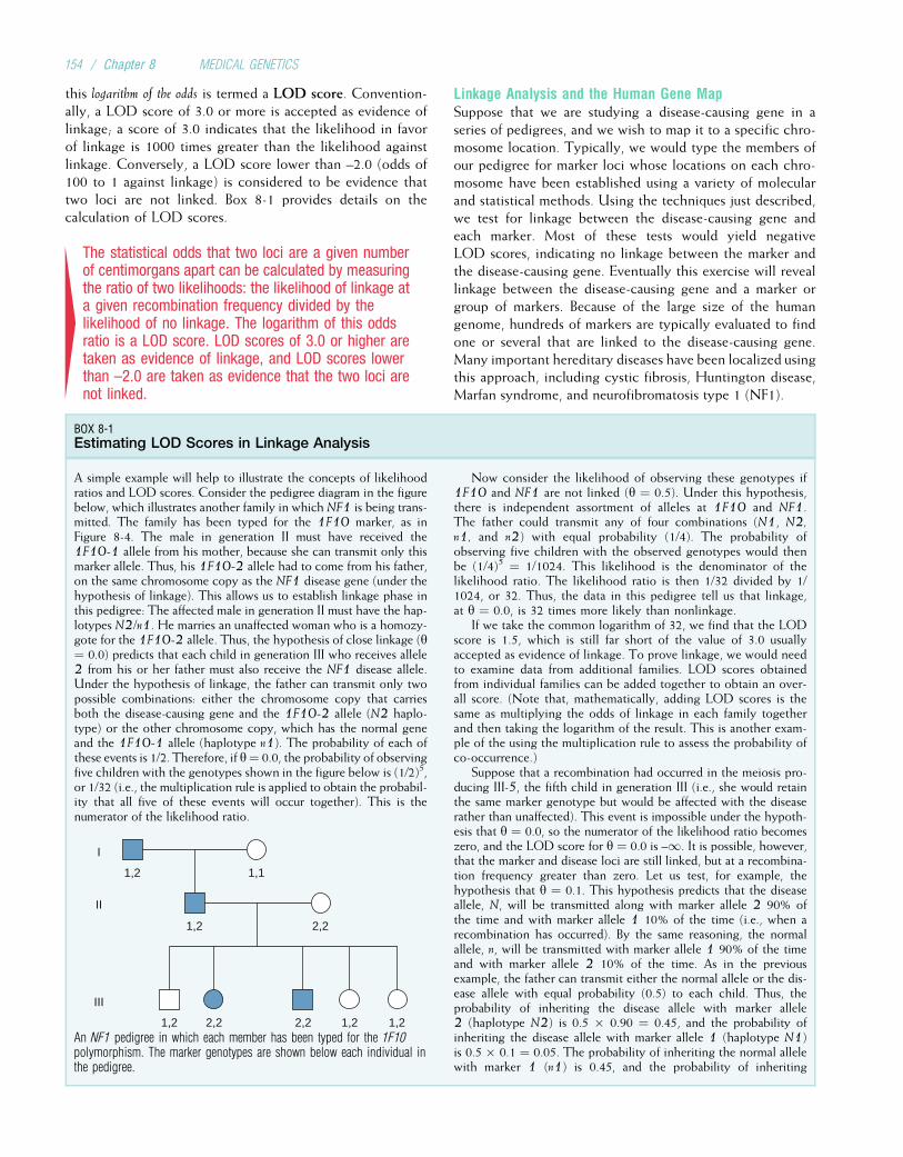
this logarithm of the odds is termed a LOD score. Convention-ally, a LOD score of 3.0 or more is accepted as evidence oflinkage; a score of 3.0 indicates that the likelihood in favorof linkage is 1000 times greater than the likelihood againstlinkage. Conversely, a LOD score lower than –2.0 (odds of100 to 1 against linkage) is considered to be evidence thattwo loci are not linked. Box 8-1 provides details on thecalculation of LOD scores.
4The statistical odds that two loci are a given numberof centimorgans apart can be calculated by measuringthe ratio of two likelihoods: the likelihood of linkage ata given recombination frequency divided by thelikelihood of no linkage. The logarithm of this oddsratio is a LOD score. LOD scores of 3.0 or higher aretaken as evidence of linkage, and LOD scores lowerthan –2.0 are taken as evidence that the two loci arenot linked.
Linkage Analysis and the Human Gene MapSuppose that we are studying a disease-causing gene in aseries of pedigrees, and we wish to map it to a specific chro-mosome location. Typically, we would type the members ofour pedigree for marker loci whose locations on each chro-mosome have been established using a variety of molecularand statistical methods. Using the techniques just described,we test for linkage between the disease-causing gene andeach marker. Most of these tests would yield negativeLOD scores, indicating no linkage between the marker andthe disease-causing gene. Eventually this exercise will reveallinkage between the disease-causing gene and a marker orgroup of markers. Because of the large size of the humangenome, hundreds of markers are typically evaluated to findone or several that are linked to the disease-causing gene.Many important hereditary diseases have been localized usingthis approach, including cystic fibrosis, Huntington disease,Marfan syndrome, and neurofibromatosis type 1 (NF1).
BOX 8-1Estimating LOD Scores in Linkage Analysis
A simple example will help to illustrate the concepts of likelihoodratios and LOD scores. Consider the pedigree diagram in the figurebelow, which illustrates another family in which NF1 is being trans-mitted. The family has been typed for the 1F10 marker, as inFigure 8-4. The male in generation II must have received the1F10-1 allele from his mother, because she can transmit only thismarker allele. Thus, his 1F10-2 allele had to come from his father,on the same chromosome copy as the NF1 disease gene (under thehypothesis of linkage). This allows us to establish linkage phase inthis pedigree: The affected male in generation II must have the hap-lotypes N2/n1. He marries an unaffected woman who is a homozy-gote for the 1F10-2 allele. Thus, the hypothesis of close linkage (y¼ 0.0) predicts that each child in generation III who receives allele2 from his or her father must also receive the NF1 disease allele.Under the hypothesis of linkage, the father can transmit only twopossible combinations: either the chromosome copy that carriesboth the disease-causing gene and the 1F10-2 allele (N2 haplo-type) or the other chromosome copy, which has the normal geneand the 1F10-1 allele (haplotype n1). The probability of each ofthese events is 1/2. Therefore, if y¼ 0.0, the probability of observingfive children with the genotypes shown in the figure below is (1/2)5,or 1/32 (i.e., the multiplication rule is applied to obtain the probabil-ity that all five of these events will occur together). This is thenumerator of the likelihood ratio.
1,2 1,1
I
II
III
2,2
2,2
1,2
1,21,21,2 2,2An NF1 pedigree in which each member has been typed for the 1F10polymorphism. The marker genotypes are shown below each individual inthe pedigree.
Now consider the likelihood of observing these genotypes if1F10 and NF1 are not linked (y ¼ 0.5). Under this hypothesis,there is independent assortment of alleles at 1F10 and NF1.The father could transmit any of four combinations (N1, N2,n1, and n2) with equal probability (1/4). The probability ofobserving five children with the observed genotypes would thenbe (1/4)5 ¼ 1/1024. This likelihood is the denominator of thelikelihood ratio. The likelihood ratio is then 1/32 divided by 1/1024, or 32. Thus, the data in this pedigree tell us that linkage,at y ¼ 0.0, is 32 times more likely than nonlinkage.
If we take the common logarithm of 32, we find that the LODscore is 1.5, which is still far short of the value of 3.0 usuallyaccepted as evidence of linkage. To prove linkage, we would needto examine data from additional families. LOD scores obtainedfrom individual families can be added together to obtain an over-all score. (Note that, mathematically, adding LOD scores is thesame as multiplying the odds of linkage in each family togetherand then taking the logarithm of the result. This is another exam-ple of the using the multiplication rule to assess the probability ofco-occurrence.)
Suppose that a recombination had occurred in the meiosis pro-ducing III-5, the fifth child in generation III (i.e., she would retainthe same marker genotype but would be affected with the diseaserather than unaffected). This event is impossible under the hypoth-esis that y ¼ 0.0, so the numerator of the likelihood ratio becomeszero, and the LOD score for y ¼ 0.0 is –1. It is possible, however,that the marker and disease loci are still linked, but at a recombina-tion frequency greater than zero. Let us test, for example, thehypothesis that y ¼ 0.1. This hypothesis predicts that the diseaseallele, N, will be transmitted along with marker allele 2 90% ofthe time and with marker allele 1 10% of the time (i.e., when arecombination has occurred). By the same reasoning, the normalallele, n, will be transmitted with marker allele 1 90% of the timeand with marker allele 2 10% of the time. As in the previousexample, the father can transmit either the normal allele or the dis-ease allele with equal probability (0.5) to each child. Thus, theprobability of inheriting the disease allele with marker allele2 (haplotype N2) is 0.5 � 0.90 ¼ 0.45, and the probability ofinheriting the disease allele with marker allele 1 (haplotype N1)is 0.5 � 0.1 ¼ 0.05. The probability of inheriting the normal allelewith marker 1 (n1) is 0.45, and the probability of inheriting
154 / Chapter 8 MEDICAL GENETICS
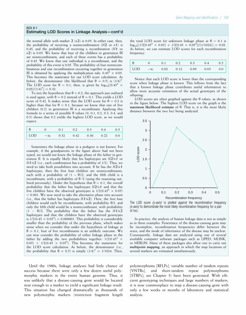
Until the 1980s, linkage analyses had little chance ofsuccess because there were only a few dozen useful poly-morphic markers in the entire human genome. Thus, itwas unlikely that a disease-causing gene would be locatednear enough to a marker to yield a significant linkage result.This situation has changed dramatically as thousands ofnew polymorphic markers (restriction fragment length
polymorphisms [RFLPs], variable number of tandem repeats[VNTRs], and short-tandem repeat polymorphisms[STRPs]; see Chapter 3) have been generated. With effi-cient genotyping techniques and large numbers of markers,it is now commonplace to map a disease-causing gene withonly a few weeks or months of laboratory and statisticalanalysis.
BOX 8-1Estimating LOD Scores in Linkage Analysis—cont’d
the normal allele with marker 2 (n2) is 0.05. In either case, then,the probability of receiving a nonrecombination (N2 or n1) is0.45, and the probability of receiving a recombination (N1 orn2) is 0.05. We know that four of the children in generation IIIare nonrecombinants, and each of these events has a probabilityof 0.45. We know that one individual is a recombinant, and theprobability of this event is 0.05. The probability of four nonrecom-binations and one recombination occurring together in generationIII is obtained by applying the multiplication rule: 0.454 � 0.05.This becomes the numerator for our LOD score calculation. Asbefore, the denominator (the likelihood that y ¼ 0.5) is (1/4)5.The LOD score for y ¼ 0.1, then, is given by log10[(0.45
4 �0.05)/(1/4)5] ¼ 0.32.
To test the hypothesis that y ¼ 0.2, the approach just outlinedis used again, with y ¼ 0.2 instead of y ¼ 0.1. This yields a LODscore of 0.42. It makes sense that the LOD score for y ¼ 0.2 ishigher than that for y ¼ 0.1, because we know that one of fivechildren (0.2) in generation III is a recombinant. Applying thisformula to a series of possible y values (0, 0.1, 0.2, 0.3, 0.4, and0.5) shows that 0.2 yields the highest LOD score, as we wouldexpect:
y 0 0.1 0.2 0.3 0.4 0.5
LOD �1 0.32 0.42 0.36 0.22 0.0
Sometimes the linkage phase in a pedigree is not known. Forexample, if the grandparents in the figure above had not beentyped, we would not know the linkage phase of the father in gen-eration II. It is equally likely that his haplotypes are N2/n1 orN1/n2 (i.e., each combination has a probability of 1/2). Thus, weneed to take both possibilities into account. If he has the N2/n1haplotypes, then the first four children are nonrecombinants,each with a probability of (1 – y)/2, and the fifth child is arecombinant, with a probability of y /2 (using the reasoning out-lined previously). Under the hypothesis that y ¼ 0.1, the overallprobability that the father has haplotypes N2/n1 and that thefive children have the observed genotypes is 1/2(0.454 � 0.05)¼ 0.001. We now need to take the alternative phase into account(i.e., that the father has haplotypes N1/n2). Here, the first fourchildren would each be recombinants, with probability y/2, andonly the fifth child would be a nonrecombinant, with probability(1 � y)/2. The probability that the father has the N1/n2haplotypes and that the children have the observed genotypesis 1/2(0.45 � 0.054) ¼ 0.000001. This probability is considerablysmaller than the probability of the previous phase, which makessense when we consider that under the hypothesis of linkage aty ¼ 0.1, four of five recombinants is an unlikely outcome. Wecan now consider the probability of either linkage phase in thefather by adding the two probabilities together: 1/2(0.454 �0.05) þ 1/2(0.45 � 0.054). This becomes the numerator forthe LOD score calculation. As before, the denominator (i.e.,the probability that y ¼ 0.5) is simply (1/4)5 ¼ 1/1024. Then,
the total LOD score for unknown linkage phase at y ¼ 0.1 islog10[(1/2[0.45
4 � 0.05] þ 1/2[0.45 � 0.054])/(1/1024)] ¼ 0.02.As before, we can estimate LOD scores for each recombinationfrequency:
y 0 0.1 0.2 0.3 0.4 0.5
LOD –1 0.02 0.12 0.09 0.03 0.0
Notice that each LOD score is lower than the correspondingscore when linkage phase is known. This follows from the factthat a known linkage phase contributes useful information toallow more accurate estimation of the actual genotypes of theoffspring.
LOD scores are often graphed against the y values, as shownin the figure below. The highest LOD score on the graph is themaximum likelihood estimate of y. That is, it is the most likelydistance between the two loci being analyzed.
LOD
sco
re
3.5
3
2.5
2
1.5
1
0.5
00
Recombination frequency
0.1 0.2 0.3 0.4 0.5
The LOD score (y-axis) is plotted against the recombination frequency(x-axis) to demonstrate the most likely recombination frequency for a pairof loci.
In practice, the analysis of human linkage data is not as simpleas in these examples. Penetrance of the disease-causing gene maybe incomplete, recombination frequencies differ between thesexes, and the mode of inheritance of the disease may be unclear.Consequently, linkage data are analyzed using one of severalavailable computer software packages such as LIPED, MLINK,or MERLIN. Many of these packages also allow one to carry outmultipoint mapping, an approach in which the map locations ofseveral markers are estimated simultaneously.
Gene Mapping and Identification / 155
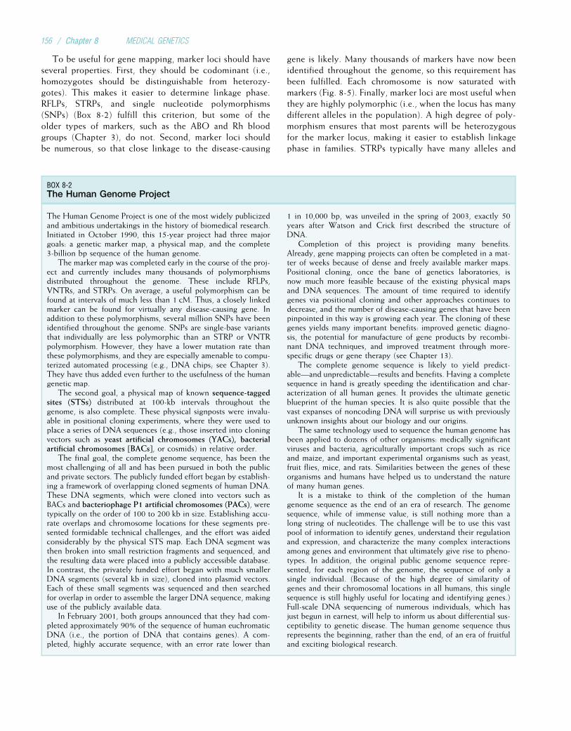
To be useful for gene mapping, marker loci should haveseveral properties. First, they should be codominant (i.e.,homozygotes should be distinguishable from heterozy-gotes). This makes it easier to determine linkage phase.RFLPs, STRPs, and single nucleotide polymorphisms(SNPs) (Box 8-2) fulfill this criterion, but some of theolder types of markers, such as the ABO and Rh bloodgroups (Chapter 3), do not. Second, marker loci shouldbe numerous, so that close linkage to the disease-causing
gene is likely. Many thousands of markers have now beenidentified throughout the genome, so this requirement hasbeen fulfilled. Each chromosome is now saturated withmarkers (Fig. 8-5). Finally, marker loci are most useful whenthey are highly polymorphic (i.e., when the locus has manydifferent alleles in the population). A high degree of poly-morphism ensures that most parents will be heterozygousfor the marker locus, making it easier to establish linkagephase in families. STRPs typically have many alleles and
BOX 8-2The Human Genome Project
The Human Genome Project is one of the most widely publicizedand ambitious undertakings in the history of biomedical research.Initiated in October 1990, this 15-year project had three majorgoals: a genetic marker map, a physical map, and the complete3-billion bp sequence of the human genome.
The marker map was completed early in the course of the proj-ect and currently includes many thousands of polymorphismsdistributed throughout the genome. These include RFLPs,VNTRs, and STRPs. On average, a useful polymorphism can befound at intervals of much less than 1 cM. Thus, a closely linkedmarker can be found for virtually any disease-causing gene. Inaddition to these polymorphisms, several million SNPs have beenidentified throughout the genome. SNPs are single-base variantsthat individually are less polymorphic than an STRP or VNTRpolymorphism. However, they have a lower mutation rate thanthese polymorphisms, and they are especially amenable to compu-terized automated processing (e.g., DNA chips; see Chapter 3).They have thus added even further to the usefulness of the humangenetic map.
The second goal, a physical map of known sequence-taggedsites (STSs) distributed at 100-kb intervals throughout thegenome, is also complete. These physical signposts were invalu-able in positional cloning experiments, where they were used toplace a series of DNA sequences (e.g., those inserted into cloningvectors such as yeast artificial chromosomes (YACs), bacterialartificial chromosomes [BACs], or cosmids) in relative order.
The final goal, the complete genome sequence, has been themost challenging of all and has been pursued in both the publicand private sectors. The publicly funded effort began by establish-ing a framework of overlapping cloned segments of human DNA.These DNA segments, which were cloned into vectors such asBACs and bacteriophage P1 artificial chromosomes (PACs), weretypically on the order of 100 to 200 kb in size. Establishing accu-rate overlaps and chromosome locations for these segments pre-sented formidable technical challenges, and the effort was aidedconsiderably by the physical STS map. Each DNA segment wasthen broken into small restriction fragments and sequenced, andthe resulting data were placed into a publicly accessible database.In contrast, the privately funded effort began with much smallerDNA segments (several kb in size), cloned into plasmid vectors.Each of these small segments was sequenced and then searchedfor overlap in order to assemble the larger DNA sequence, makinguse of the publicly available data.
In February 2001, both groups announced that they had com-pleted approximately 90% of the sequence of human euchromaticDNA (i.e., the portion of DNA that contains genes). A com-pleted, highly accurate sequence, with an error rate lower than
1 in 10,000 bp, was unveiled in the spring of 2003, exactly 50years after Watson and Crick first described the structure ofDNA.
Completion of this project is providing many benefits.Already, gene mapping projects can often be completed in a mat-ter of weeks because of dense and freely available marker maps.Positional cloning, once the bane of genetics laboratories, isnow much more feasible because of the existing physical mapsand DNA sequences. The amount of time required to identifygenes via positional cloning and other approaches continues todecrease, and the number of disease-causing genes that have beenpinpointed in this way is growing each year. The cloning of thesegenes yields many important benefits: improved genetic diagno-sis, the potential for manufacture of gene products by recombi-nant DNA techniques, and improved treatment through more-specific drugs or gene therapy (see Chapter 13).
The complete genome sequence is likely to yield predict-able—and unpredictable—results and benefits. Having a completesequence in hand is greatly speeding the identification and char-acterization of all human genes. It provides the ultimate geneticblueprint of the human species. It is also quite possible that thevast expanses of noncoding DNA will surprise us with previouslyunknown insights about our biology and our origins.
The same technology used to sequence the human genome hasbeen applied to dozens of other organisms: medically significantviruses and bacteria, agriculturally important crops such as riceand maize, and important experimental organisms such as yeast,fruit flies, mice, and rats. Similarities between the genes of theseorganisms and humans have helped us to understand the natureof many human genes.
It is a mistake to think of the completion of the humangenome sequence as the end of an era of research. The genomesequence, while of immense value, is still nothing more than along string of nucleotides. The challenge will be to use this vastpool of information to identify genes, understand their regulationand expression, and characterize the many complex interactionsamong genes and environment that ultimately give rise to pheno-types. In addition, the original public genome sequence repre-sented, for each region of the genome, the sequence of only asingle individual. (Because of the high degree of similarity ofgenes and their chromosomal locations in all humans, this singlesequence is still highly useful for locating and identifying genes.)Full-scale DNA sequencing of numerous individuals, which hasjust begun in earnest, will help to inform us about differential sus-ceptibility to genetic disease. The human genome sequence thusrepresents the beginning, rather than the end, of an era of fruitfuland exciting biological research.
156 / Chapter 8 MEDICAL GENETICS
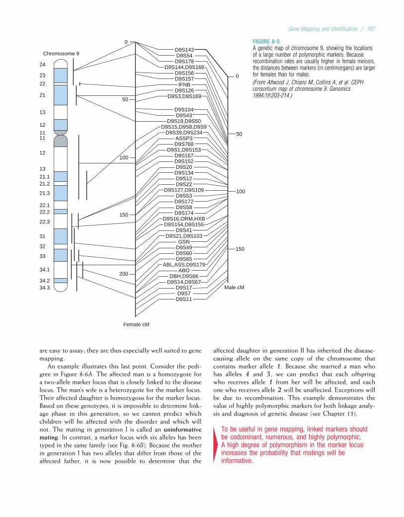
are easy to assay; they are thus especially well suited to genemapping.
An example illustrates this last point. Consider the pedi-gree in Figure 8-6A. The affected man is a homozygote fora two-allele marker locus that is closely linked to the diseaselocus. The man’s wife is a heterozygote for the marker locus.Their affected daughter is homozygous for the marker locus.Based on these genotypes, it is impossible to determine link-age phase in this generation, so we cannot predict whichchildren will be affected with the disorder and which willnot. The mating in generation I is called an uninformativemating. In contrast, a marker locus with six alleles has beentyped in the same family (see Fig. 8-6B). Because the motherin generation I has two alleles that differ from those of theaffected father, it is now possible to determine that the
affected daughter in generation II has inherited the disease-causing allele on the same copy of the chromosome thatcontains marker allele 1. Because she married a man whohas alleles 4 and 5, we can predict that each offspringwho receives allele 1 from her will be affected, and eachone who receives allele 2 will be unaffected. Exceptions willbe due to recombination. This example demonstrates thevalue of highly polymorphic markers for both linkage analy-sis and diagnosis of genetic disease (see Chapter 13).
4To be useful in gene mapping, linked markers shouldbe codominant, numerous, and highly polymorphic.A high degree of polymorphism in the marker locusincreases the probability that matings will beinformative.
0
50
100
150
200
0
50
100
150
Female cM
D9S143D9S54D9S178
D9S144,D9S168D9S156D9S157
IFNBD9S126
D9S3,D9S169
D9S104D9S43
D9S19,D9S50D9S15,D9S8,D9S9
D9S39,D9S234ASSP3D9S768
D9S1,D9S153D9S167D9S152D9S20D9S134D9S12D9S22
D9S127,D9S109D9S53D9S172D9S58D9S174
D9S16,ORM,HXBD9S154,D9S155
D9S41D9S21,D9S103
GSND9S49D9S60D9S65
ABL,ASS,D9S179ABO
DBH,D9S66D9S14,D9S67
D9S17D9S7D9S11
Male cM
22.122.2
22.3
31
12
13
21.121.2
21.3
32
33
34.1
34.234.3
Chromosome 9
24
23
22
21
13
12
1111
FIGURE 8-5A genetic map of chromosome 9, showing the locationsof a large number of polymorphic markers. Becauserecombination rates are usually higher in female meiosis,the distances between markers (in centimorgans) are largerfor females than for males.
(From Attwood J, Chiano M, Collins A, et al: CEPHconsortium map of chromosome 9. Genomics1994;19:203-214.)
Gene Mapping and Identification / 157
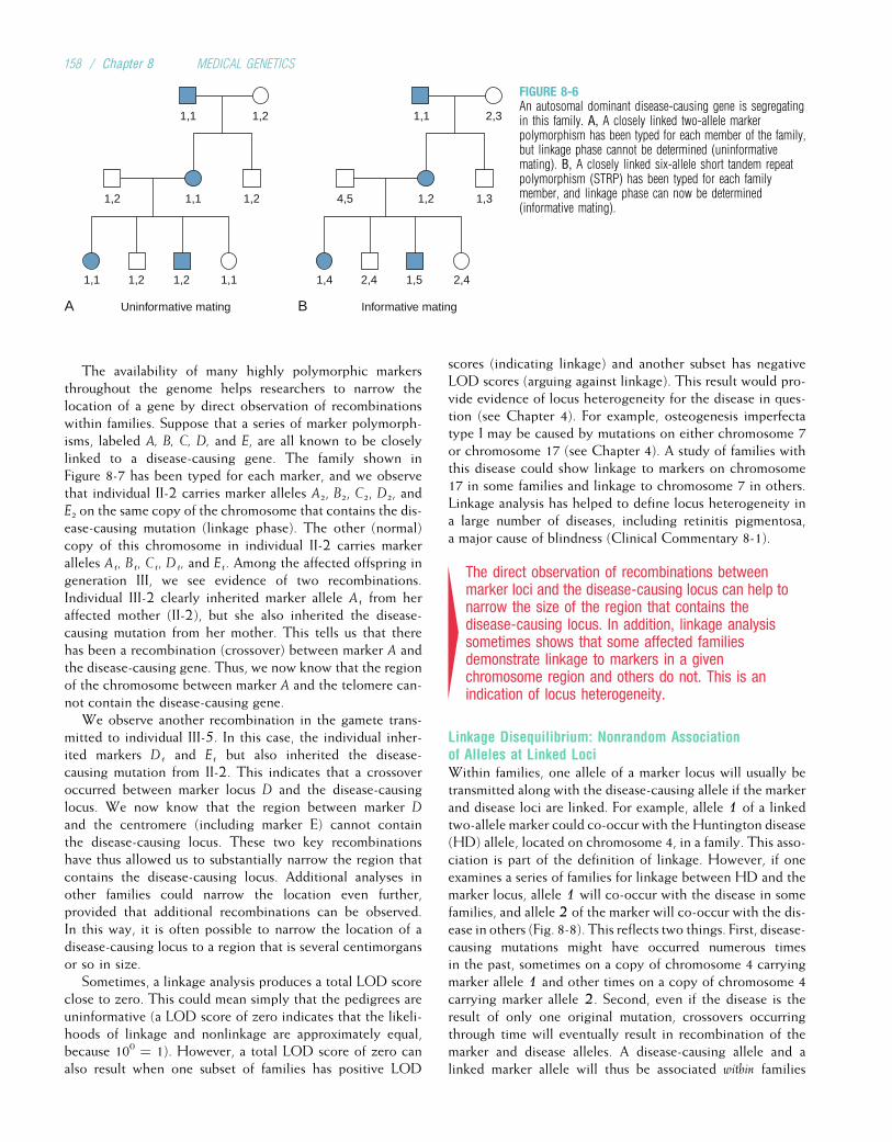
The availability of many highly polymorphic markersthroughout the genome helps researchers to narrow thelocation of a gene by direct observation of recombinationswithin families. Suppose that a series of marker polymorph-isms, labeled A, B, C, D, and E, are all known to be closelylinked to a disease-causing gene. The family shown inFigure 8-7 has been typed for each marker, and we observethat individual II-2 carries marker alleles A2, B2, C2, D2, andE2 on the same copy of the chromosome that contains the dis-ease-causing mutation (linkage phase). The other (normal)copy of this chromosome in individual II-2 carries markeralleles A1, B1, C1, D1, and E1. Among the affected offspring ingeneration III, we see evidence of two recombinations.Individual III-2 clearly inherited marker allele A1 from heraffected mother (II-2), but she also inherited the disease-causing mutation from her mother. This tells us that therehas been a recombination (crossover) between marker A andthe disease-causing gene. Thus, we now know that the regionof the chromosome between marker A and the telomere can-not contain the disease-causing gene.
We observe another recombination in the gamete trans-mitted to individual III-5. In this case, the individual inher-ited markers D1 and E1 but also inherited the disease-causing mutation from II-2. This indicates that a crossoveroccurred between marker locus D and the disease-causinglocus. We now know that the region between marker D
and the centromere (including marker E) cannot containthe disease-causing locus. These two key recombinationshave thus allowed us to substantially narrow the region thatcontains the disease-causing locus. Additional analyses inother families could narrow the location even further,provided that additional recombinations can be observed.In this way, it is often possible to narrow the location of adisease-causing locus to a region that is several centimorgansor so in size.
Sometimes, a linkage analysis produces a total LOD scoreclose to zero. This could mean simply that the pedigrees areuninformative (a LOD score of zero indicates that the likeli-hoods of linkage and nonlinkage are approximately equal,because 100 ¼ 1). However, a total LOD score of zero canalso result when one subset of families has positive LOD
scores (indicating linkage) and another subset has negativeLOD scores (arguing against linkage). This result would pro-vide evidence of locus heterogeneity for the disease in ques-tion (see Chapter 4). For example, osteogenesis imperfectatype I may be caused by mutations on either chromosome 7or chromosome 17 (see Chapter 4). A study of families withthis disease could show linkage to markers on chromosome17 in some families and linkage to chromosome 7 in others.Linkage analysis has helped to define locus heterogeneity ina large number of diseases, including retinitis pigmentosa,a major cause of blindness (Clinical Commentary 8-1).
4The direct observation of recombinations betweenmarker loci and the disease-causing locus can help tonarrow the size of the region that contains thedisease-causing locus. In addition, linkage analysissometimes shows that some affected familiesdemonstrate linkage to markers in a givenchromosome region and others do not. This is anindication of locus heterogeneity.
Linkage Disequilibrium: Nonrandom Associationof Alleles at Linked LociWithin families, one allele of a marker locus will usually betransmitted along with the disease-causing allele if the markerand disease loci are linked. For example, allele 1 of a linkedtwo-allele marker could co-occur with the Huntington disease(HD) allele, located on chromosome 4, in a family. This asso-ciation is part of the definition of linkage. However, if oneexamines a series of families for linkage between HD and themarker locus, allele 1 will co-occur with the disease in somefamilies, and allele 2 of the marker will co-occur with the dis-ease in others (Fig. 8-8). This reflects two things. First, disease-causing mutations might have occurred numerous timesin the past, sometimes on a copy of chromosome 4 carryingmarker allele 1 and other times on a copy of chromosome 4carrying marker allele 2. Second, even if the disease is theresult of only one original mutation, crossovers occurringthrough time will eventually result in recombination of themarker and disease alleles. A disease-causing allele and alinked marker allele will thus be associated within families
A
1,1 1,2
1,2 1,1 1,2
1,1 1,21,2 1,1 1,4 1,52,4 2,4
4,5 1,2 1,3
1,1 2,3
BUninformative mating Informative mating
FIGURE 8-6An autosomal dominant disease-causing gene is segregatingin this family. A, A closely linked two-allele markerpolymorphism has been typed for each member of the family,but linkage phase cannot be determined (uninformativemating). B, A closely linked six-allele short tandem repeatpolymorphism (STRP) has been typed for each familymember, and linkage phase can now be determined(informative mating).
158 / Chapter 8 MEDICAL GENETICS
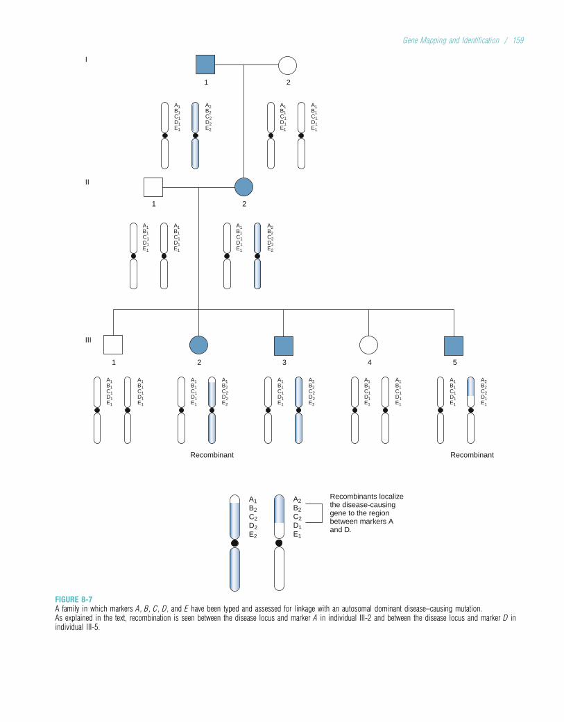
I
1
A1B1C1D1E1
A2B2C2D2E2
A1B1C1D1E1
A1B1C1D1E1
2
1
II
III
1 5
Recombinant
Recombinants localizethe disease-causinggene to the regionbetween markers Aand D.
Recombinant
432
2
A1B1C1D1E1
A1B1C1D1E1
A1B1C1D1E1
A2B2C2D2E2
A1B1C1D1E1
A1B1C1D1E1
A1B1C1D1E1
A1B2C2D2E2
A1B1C1D1E1
A2B2C2D2E2
A1B1C1D1E1
A1B1C1D1E1
A1B1C1D1E1
A2B2C2D1E1
A1B2C2D2E2
A2B2C2D1E1
FIGURE 8-7A family in which markers A, B, C, D, and E have been typed and assessed for linkage with an autosomal dominant disease–causing mutation.As explained in the text, recombination is seen between the disease locus and marker A in individual III-2 and between the disease locus and marker D inindividual III-5.
Gene Mapping and Identification / 159
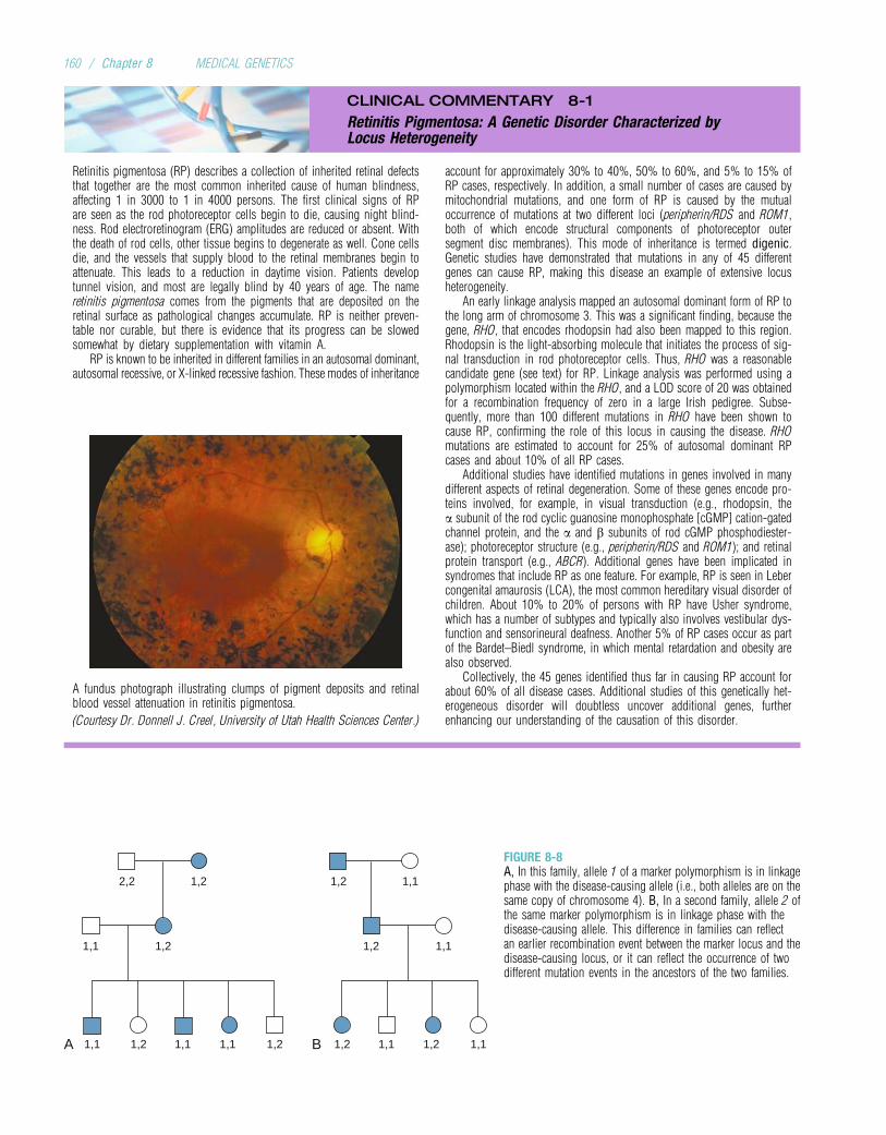
CLINICAL COMMENTARY 8-1
Retinitis Pigmentosa: A Genetic Disorder Characterized byLocus Heterogeneity
Retinitis pigmentosa (RP) describes a collection of inherited retinal defectsthat together are the most common inherited cause of human blindness,affecting 1 in 3000 to 1 in 4000 persons. The first clinical signs of RPare seen as the rod photoreceptor cells begin to die, causing night blind-ness. Rod electroretinogram (ERG) amplitudes are reduced or absent. Withthe death of rod cells, other tissue begins to degenerate as well. Cone cellsdie, and the vessels that supply blood to the retinal membranes begin toattenuate. This leads to a reduction in daytime vision. Patients developtunnel vision, and most are legally blind by 40 years of age. The nameretinitis pigmentosa comes from the pigments that are deposited on theretinal surface as pathological changes accumulate. RP is neither preven-table nor curable, but there is evidence that its progress can be slowedsomewhat by dietary supplementation with vitamin A.
RP is known to be inherited in different families in an autosomal dominant,autosomal recessive, or X-linked recessive fashion. These modes of inheritance
A fundus photograph illustrating clumps of pigment deposits and retinalblood vessel attenuation in retinitis pigmentosa.
(Courtesy Dr. Donnell J. Creel, University of Utah Health Sciences Center.)
account for approximately 30% to 40%, 50% to 60%, and 5% to 15% ofRP cases, respectively. In addition, a small number of cases are caused bymitochondrial mutations, and one form of RP is caused by the mutualoccurrence of mutations at two different loci (peripherin/RDS and ROM1,both of which encode structural components of photoreceptor outersegment disc membranes). This mode of inheritance is termed digenic.Genetic studies have demonstrated that mutations in any of 45 differentgenes can cause RP, making this disease an example of extensive locusheterogeneity.
An early linkage analysis mapped an autosomal dominant form of RP tothe long arm of chromosome 3. This was a significant finding, because thegene, RHO, that encodes rhodopsin had also been mapped to this region.Rhodopsin is the light-absorbing molecule that initiates the process of sig-nal transduction in rod photoreceptor cells. Thus, RHO was a reasonablecandidate gene (see text) for RP. Linkage analysis was performed using apolymorphism located within the RHO, and a LOD score of 20 was obtainedfor a recombination frequency of zero in a large Irish pedigree. Subse-quently, more than 100 different mutations in RHO have been shown tocause RP, confirming the role of this locus in causing the disease. RHOmutations are estimated to account for 25% of autosomal dominant RPcases and about 10% of all RP cases.
Additional studies have identified mutations in genes involved in manydifferent aspects of retinal degeneration. Some of these genes encode pro-teins involved, for example, in visual transduction (e.g., rhodopsin, thea subunit of the rod cyclic guanosine monophosphate [cGMP] cation-gatedchannel protein, and the a and b subunits of rod cGMP phosphodiester-ase); photoreceptor structure (e.g., peripherin/RDS and ROM1); and retinalprotein transport (e.g., ABCR). Additional genes have been implicated insyndromes that include RP as one feature. For example, RP is seen in Lebercongenital amaurosis (LCA), the most common hereditary visual disorder ofchildren. About 10% to 20% of persons with RP have Usher syndrome,which has a number of subtypes and typically also involves vestibular dys-function and sensorineural deafness. Another 5% of RP cases occur as partof the Bardet–Biedl syndrome, in which mental retardation and obesity arealso observed.
Collectively, the 45 genes identified thus far in causing RP account forabout 60% of all disease cases. Additional studies of this genetically het-erogeneous disorder will doubtless uncover additional genes, furtherenhancing our understanding of the causation of this disorder.
A
2,2 1,2
1,1 1,2 1,2 1,1
1,2 1,11,11,1 1,2 1,2 1,21,1 1,1
1,2 1,1
B
FIGURE 8-8A, In this family, allele 1 of a marker polymorphism is in linkagephase with the disease-causing allele (i.e., both alleles are on thesame copy of chromosome 4). B, In a second family, allele 2 ofthe same marker polymorphism is in linkage phase with thedisease-causing allele. This difference in families can reflectan earlier recombination event between the marker locus and thedisease-causing locus, or it can reflect the occurrence of twodifferent mutation events in the ancestors of the two families.
160 / Chapter 8 MEDICAL GENETICS
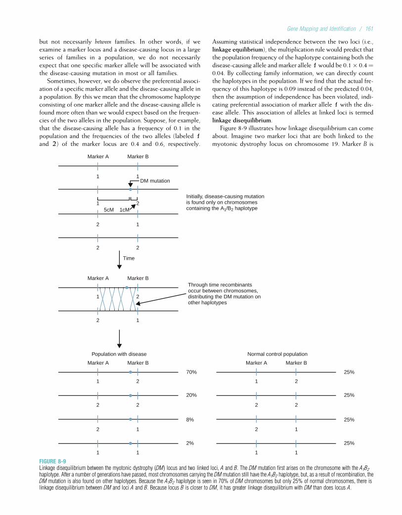
but not necessarily between families. In other words, if weexamine a marker locus and a disease-causing locus in a largeseries of families in a population, we do not necessarilyexpect that one specific marker allele will be associated withthe disease-causing mutation in most or all families.
Sometimes, however, we do observe the preferential associ-ation of a specific marker allele and the disease-causing allele ina population. By this we mean that the chromosome haplotypeconsisting of one marker allele and the disease-causing allele isfound more often than we would expect based on the frequen-cies of the two alleles in the population. Suppose, for example,that the disease-causing allele has a frequency of 0.1 in thepopulation and the frequencies of the two alleles (labeled 1and 2) of the marker locus are 0.4 and 0.6, respectively.
Assuming statistical independence between the two loci (i.e.,linkage equilibrium), the multiplication rule would predict thatthe population frequency of the haplotype containing both thedisease-causing allele and marker allele 1would be 0.1� 0.4¼0.04. By collecting family information, we can directly countthe haplotypes in the population. If we find that the actual fre-quency of this haplotype is 0.09 instead of the predicted 0.04,then the assumption of independence has been violated, indi-cating preferential association of marker allele 1 with the dis-ease allele. This association of alleles at linked loci is termedlinkage disequilibrium.
Figure 8-9 illustrates how linkage disequilibrium can comeabout. Imagine two marker loci that are both linked to themyotonic dystrophy locus on chromosome 19. Marker B is
1
Marker A Marker B
Marker A Marker B
Through time recombinantsoccur between chromosomes,distributing the DM mutation onother haplotypes
Initially, disease-causing mutationis found only on chromosomescontaining the A1/B2 haplotype
DM mutation
5cM 1cM
70%
20%
8%
2%
25%
1 2
2 2
2 1
1 1
1 2
2 2
2 1
1 1
2
25%
25%
25%
Marker A Marker B
Time
1 2
2 1
Population with disease
Marker A Marker B
Normal control population
2 1
2 2
1 1
FIGURE 8-9Linkage disequilibrium between the myotonic dystrophy (DM) locus and two linked loci, A and B. The DM mutation first arises on the chromosome with the A1B2haplotype. After a number of generations have passed, most chromosomes carrying the DMmutation still have the A1B2 haplotype, but, as a result of recombination, theDM mutation is also found on other haplotypes. Because the A1B2 haplotype is seen in 70% of DM chromosomes but only 25% of normal chromosomes, there islinkage disequilibrium between DM and loci A and B. Because locus B is closer to DM, it has greater linkage disequilibrium with DM than does locus A.
Gene Mapping and Identification / 161
closely linked, at less than 1 cM away. Marker A is lessclosely linked, at about 5 cM away. Because each of thesemarker loci has two alleles (denoted 1 and 2), there are fourpossible combinations of marker alleles at the two loci, asshown in Figure 8-9. When a new myotonic dystrophymutation first occurs in a population, it can be found on onlyone copy of a chromosome, in this case the one with theA1B2 marker combination. As the disease-causing mutation(allele) is passed through multiple generations, crossoverswill occur between it and the two markers. Because the dis-ease locus is more closely linked to marker B than markerA, fewer crossovers will occur between the disease-causingallele and marker B. As a result, the disease-causing allele isfound on a B2-containing chromosome 90% of the time,and it is found on an A1-containing chromosome 72% ofthe time. The degree of linkage disequilibrium is strongerbetween marker B and the disease-causing allele thanbetween marker A and the disease-causing allele. Notice alsothat both the A1 and the B2 alleles are still positively asso-ciated with the disease-causing allele, because each markerallele has a much lower frequency (50%) in the populationof individuals who lack the disease-causing allele (seeFig. 8-9). If enough generations elapse, recombination wouldeventually eliminate the allelic associations completely, andthe loci would be in linkage equilibrium.
Because linkage disequilibrium is a function of the dis-tance between loci, it can be used to help infer the orderof genes on chromosomes. Linkage disequilibrium providesan advantage over linkage analysis in that it reflects theaction of recombinations that have occurred during dozensor hundreds of past generations (i.e., the number of genera-tions that have elapsed since the disease-causing mutationfirst occurred in a population). Linkage analysis, in contrast,is limited to the recombinations that can be directlyobserved in only the past several generations. Consequently,there are seldom enough recombinants in a series of familiesto map a gene to a region smaller than several centimorgansusing linkage analysis, whereas linkage disequilibrium analy-sis can sometimes map a gene to an interval of 0.1 cM orless. However, linkage disequilibrium can be influenced byevolutionary forces, such as natural selection or genetic drift,that have acted during the history of a population. For exam-ple, some loci in the major histocompatibility complex onchromosome 6 (see Chapter 9) are in disequilibrium, possi-bly because certain allelic combinations confer a selectiveadvantage for immunity to some diseases.
4Linkage disequilibrium is the nonrandom associationof alleles at linked loci. Linkage disequilibriumbetween loci diminishes through time as a result ofrecombination. It can be used to infer the order ofgenes on chromosomes.
Linkage versus Association in PopulationsThe phenomena of linkage and association are sometimes con-fused. Linkage refers to the positions of loci on chromosomes.
When two loci are linked, specific combinations of alleles atthese loci will be transmitted together within families becausethey are located together on the same chromosome. But, asthe HD example cited previously showed, the specific combi-nations of alleles transmitted together can vary from one familyto another. Association, on the other hand, refers to a statisti-cal relationship between two traits in the general population. Thetwo traits occur together in the same individual more oftenthan would be expected by chance alone.
As just discussed, the alleles of two linked loci may beassociated in a population (linkage disequilibrium, which is aform of association). In this case, a population associationcan lead to mapping of a disease-causing gene. An exampleis given by hereditary hemochromatosis, an autosomal reces-sive disorder discussed in Chapter 7. One association studyshowed that 78% of patients with hemochromatosis hadthe A3 allele of the human leukocyte antigen (HLA) A locus(see Chapter 9 for further discussion of the HLA system),whereas only 27% of unaffected subjects (controls) had thisallele. This strong statistical association prompted linkageanalyses using HLA polymorphisms and led to the mappingof the major hemochromatosis-causing gene to a region ofseveral centimorgans on chromosome 6. The large size ofthis region made it very difficult to pinpoint a specific gene.Linkage disequilibrium analysis was subsequently used to nar-row the region to approximately 250 kb, prompting the readyidentification of an HLA-like gene (HFE) in which a singlemutation is responsible for the great majority of cases ofhereditary hemochromatosis. The association between thishemochromatosis-causing gene and the linked HLA-A locusis probably the result of a recent hemochromatosis-causingmutation that occurred on a chromosome copy that containedthe HLA-A3 allele. Because the mutation occurred only 50 to100 generations ago, linkage disequilibrium is still seenbetween the HLA-A3 allele and the major hemochromatosis-causing mutation.
Population associations can also result from a causal rela-tionship between an allele and a disease condition. An exam-ple of such an association involves ankylosing spondylitis, adisease that primarily affects the sacroiliac joint (Fig. 8-10).Inflammation of the ligaments leads to their ossificationand eventually to fusion of the joints (ankylosis). The HLA-
B27 allele is found in about 90% of European Americanswho have ankylosing spondylitis but in only 5% to 10% ofthe general European American population. Because the pop-ulation incidence of ankylosing spondylitis is quite low(<1%), most people who have the HLA-B27 allele do notdevelop ankylosing spondylitis. However, those who havethe HLA-B27 allele are about 90 times more likely todevelop the disease than are those who do not have it (i.e.,9% of persons with HLA-B27 shown in Table 8-1 haveankylosing spondylitis, and only about 0.1% of those with-out HLA-B27 have the disease). Because of this strong asso-ciation, a test for HLA-B27 is sometimes included as part ofthe diagnosis of ankylosing spondylitis. Because ankylosingspondylitis is thought to be an autoimmune disorder, the
162 / Chapter 8 MEDICAL GENETICS
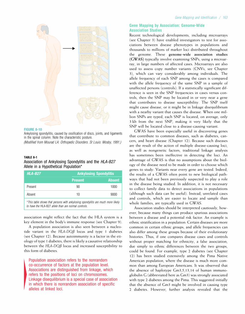
association might reflect the fact that the HLA system is akey element in the body’s immune response (see Chapter 9).
A population association is also seen between a nucleo-tide variant in the HLA-DQb locus and type 1 diabetes(see Chapter 12). Because autoimmunity is a factor in the eti-ology of type 1 diabetes, there is likely a causative relationshipbetween the HLA-DQb locus and increased susceptibility tothis form of diabetes.
4Population association refers to the nonrandomco-occurrence of factors at the population level.Associations are distinguished from linkage, whichrefers to the positions of loci on chromosomes.Linkage disequilibrium is a special case of associationin which there is nonrandom association of specificalleles at linked loci.
Gene Mapping by Association: Genome-WideAssociation StudiesRecent technological developments, including microarrays(see Chapter 3) have enabled investigators to test for asso-ciations between disease phenotypes in populations andthousands to millions of marker loci distributed throughoutthe genome. These genome-wide association studies(GWAS) typically involve examining SNPs, using a microar-ray, in large numbers of affected cases. Microarrays are alsoused to assess copy number variants (CNVs, see Chapter3), which can vary considerably among individuals. Theallele frequency of each SNP among the cases is comparedwith the allele frequency of the same SNP in a sample ofunaffected persons (controls). If a statistically significant dif-ference is seen in the SNP frequencies in cases versus con-trols, then the SNP may be located in or very near a genethat contributes to disease susceptibility. The SNP itselfmight cause disease, or it might be in linkage disequilibriumwith a nearby variant that causes the disease. When one mil-lion SNPs are typed, each SNP is located, on average, only3 kb from the next SNP, making it very likely that theSNP will be located close to a disease-causing variant.
GWAS have been especially useful in discovering genesthat contribute to common diseases, such as diabetes, can-cers, and heart disease (Chapter 12). Because such diseasesare the result of the action of multiple disease-causing loci,as well as nongenetic factors, traditional linkage analysishas sometimes been ineffective in detecting the loci. Anadvantage of GWAS is that no assumptions about the biol-ogy of the disease need to be made in order to choose whichgenes to study: Variants near every gene are tested. Indeed,the results of a GWAS often point to new biological path-ways that had not been previously suspected to play a rolein the disease being studied. In addition, it is not necessaryto collect family data to detect associations in populations(although such data can be useful). Instead, unrelated casesand controls, which are easier to locate and sample thanwhole families, are typically used in GWAS.
Association studies should be interpreted cautiously, how-ever, because many things can produce spurious associationsbetween a disease and a potential risk factor. An example isethnic stratification in a population: Certain diseases are morecommon in certain ethnic groups, and allele frequencies canalso differ among these groups because of their evolutionaryhistories. Thus, if one compares disease cases and controlswithout proper matching for ethnicity, a false association,due simply to ethnic differences between the two groups,could be found. For example, type 2 diabetes (see Chapter12) has been studied extensively among the Pima NativeAmerican population, where the disease is much more com-mon than among European Americans. It was observed thatthe absence of haplotype Gm3,5,13,14 of human immuno-globulin G (abbreviated here as Gm3) was strongly associatedwith type 2 diabetes among the Pima. This suggested initiallythat the absence of Gm3 might be involved in causing type2 diabetes. However, further analysis revealed that the
FIGURE 8-10Ankylosing spondylitis, caused by ossification of discs, joints, and ligamentsin the spinal column. Note the characteristic posture.
(Modified from Mourad LA: Orthopedic Disorders. St Louis: Mosby, 1991.)
TABLE 8-1
Association of Ankylosing Spondylitis and the HLA-B27Allele in a Hypothetical Population*
HLA-B27 Ankylosing Spondylitis
Present Absent
Present 90 1000
Absent 10 9000
*This table shows that persons with ankylosing spondylitis are much more likely
to have the HLA-B27 allele than are normal controls.
Gene Mapping and Identification / 163
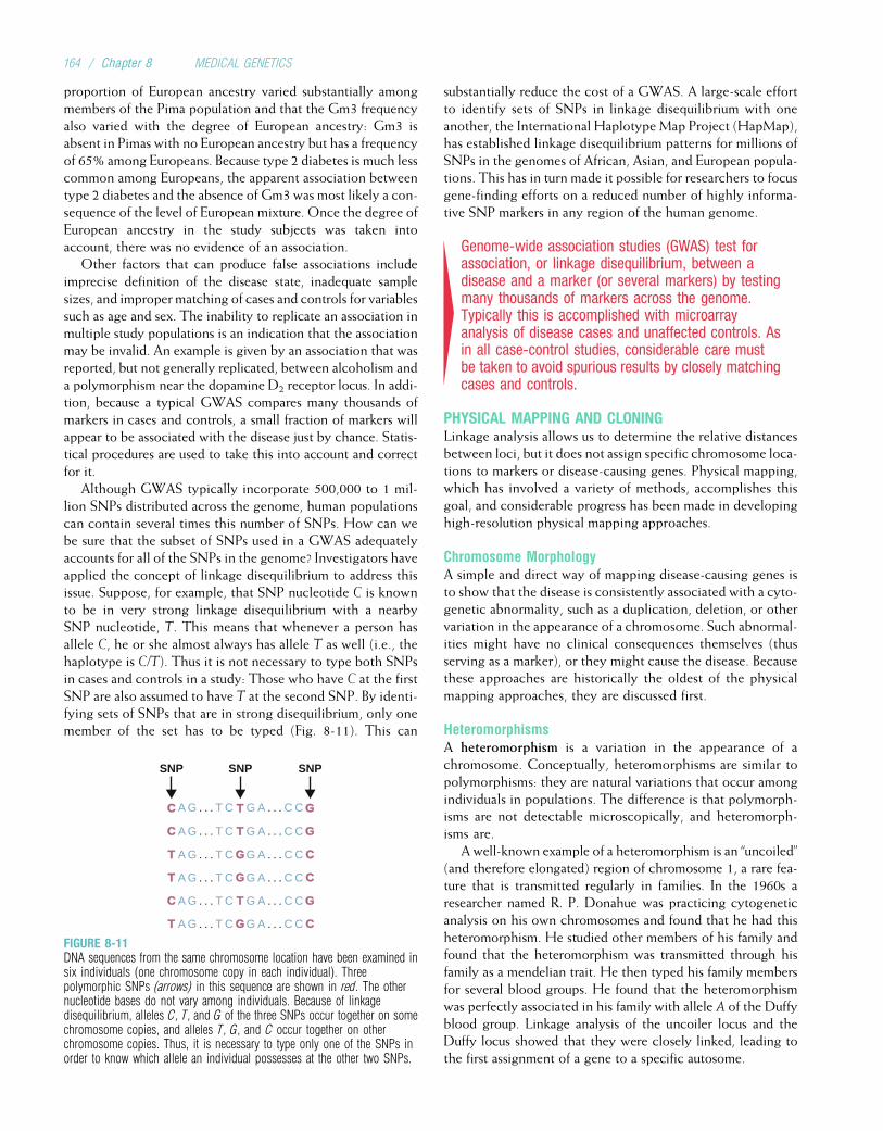
proportion of European ancestry varied substantially amongmembers of the Pima population and that the Gm3 frequencyalso varied with the degree of European ancestry: Gm3 isabsent in Pimas with no European ancestry but has a frequencyof 65% among Europeans. Because type 2 diabetes is much lesscommon among Europeans, the apparent association betweentype 2 diabetes and the absence of Gm3 was most likely a con-sequence of the level of European mixture. Once the degree ofEuropean ancestry in the study subjects was taken intoaccount, there was no evidence of an association.
Other factors that can produce false associations includeimprecise definition of the disease state, inadequate samplesizes, and improper matching of cases and controls for variablessuch as age and sex. The inability to replicate an association inmultiple study populations is an indication that the associationmay be invalid. An example is given by an association that wasreported, but not generally replicated, between alcoholism anda polymorphism near the dopamine D2 receptor locus. In addi-tion, because a typical GWAS compares many thousands ofmarkers in cases and controls, a small fraction of markers willappear to be associated with the disease just by chance. Statis-tical procedures are used to take this into account and correctfor it.
Although GWAS typically incorporate 500,000 to 1 mil-lion SNPs distributed across the genome, human populationscan contain several times this number of SNPs. How can webe sure that the subset of SNPs used in a GWAS adequatelyaccounts for all of the SNPs in the genome? Investigators haveapplied the concept of linkage disequilibrium to address thisissue. Suppose, for example, that SNP nucleotide C is knownto be in very strong linkage disequilibrium with a nearbySNP nucleotide, T. This means that whenever a person hasallele C, he or she almost always has allele T as well (i.e., thehaplotype is C/T). Thus it is not necessary to type both SNPsin cases and controls in a study: Those who have C at the firstSNP are also assumed to have T at the second SNP. By identi-fying sets of SNPs that are in strong disequilibrium, only onemember of the set has to be typed (Fig. 8-11). This can
substantially reduce the cost of a GWAS. A large-scale effortto identify sets of SNPs in linkage disequilibrium with oneanother, the International HaplotypeMap Project (HapMap),has established linkage disequilibrium patterns for millions ofSNPs in the genomes of African, Asian, and European popula-tions. This has in turn made it possible for researchers to focusgene-finding efforts on a reduced number of highly informa-tive SNP markers in any region of the human genome.
4Genome-wide association studies (GWAS) test forassociation, or linkage disequilibrium, between adisease and a marker (or several markers) by testingmany thousands of markers across the genome.Typically this is accomplished with microarrayanalysis of disease cases and unaffected controls. Asin all case-control studies, considerable care mustbe taken to avoid spurious results by closely matchingcases and controls.
PHYSICAL MAPPING AND CLONINGLinkage analysis allows us to determine the relative distancesbetween loci, but it does not assign specific chromosome loca-tions to markers or disease-causing genes. Physical mapping,which has involved a variety of methods, accomplishes thisgoal, and considerable progress has been made in developinghigh-resolution physical mapping approaches.
Chromosome MorphologyA simple and direct way of mapping disease-causing genes isto show that the disease is consistently associated with a cyto-genetic abnormality, such as a duplication, deletion, or othervariation in the appearance of a chromosome. Such abnormal-ities might have no clinical consequences themselves (thusserving as a marker), or they might cause the disease. Becausethese approaches are historically the oldest of the physicalmapping approaches, they are discussed first.
HeteromorphismsA heteromorphism is a variation in the appearance of achromosome. Conceptually, heteromorphisms are similar topolymorphisms: they are natural variations that occur amongindividuals in populations. The difference is that polymorph-isms are not detectable microscopically, and heteromorph-isms are.
A well-known example of a heteromorphism is an “uncoiled”(and therefore elongated) region of chromosome 1, a rare fea-ture that is transmitted regularly in families. In the 1960s aresearcher named R. P. Donahue was practicing cytogeneticanalysis on his own chromosomes and found that he had thisheteromorphism. He studied other members of his family andfound that the heteromorphism was transmitted through hisfamily as a mendelian trait. He then typed his family membersfor several blood groups. He found that the heteromorphismwas perfectly associated in his family with allele A of the Duffyblood group. Linkage analysis of the uncoiler locus and theDuffy locus showed that they were closely linked, leading tothe first assignment of a gene to a specific autosome.
SNP
A G . . . T C G A . . . C C
SNP SNP
CC T G
A G . . . T C G A . . . C CC T G
A G . . . T C G A . . . C CT G C
A G . . . T C G A . . . C CT G C
A G . . . T C G A . . . C CC T G
A G . . . T C G A . . . C CT G C
FIGURE 8-11DNA sequences from the same chromosome location have been examined insix individuals (one chromosome copy in each individual). Threepolymorphic SNPs (arrows) in this sequence are shown in red. The othernucleotide bases do not vary among individuals. Because of linkagedisequilibrium, alleles C, T, and G of the three SNPs occur together on somechromosome copies, and alleles T, G, and C occur together on otherchromosome copies. Thus, it is necessary to type only one of the SNPs inorder to know which allele an individual possesses at the other two SNPs.
164 / Chapter 8 MEDICAL GENETICS
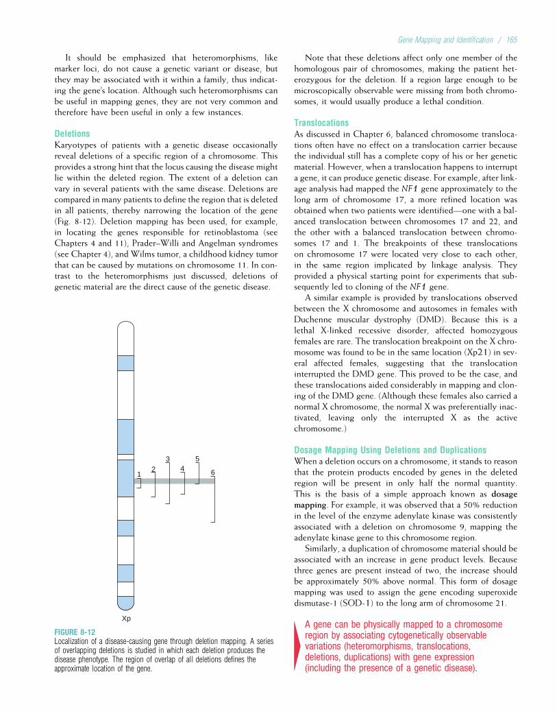
It should be emphasized that heteromorphisms, likemarker loci, do not cause a genetic variant or disease, butthey may be associated with it within a family, thus indicat-ing the gene’s location. Although such heteromorphisms canbe useful in mapping genes, they are not very common andtherefore have been useful in only a few instances.
DeletionsKaryotypes of patients with a genetic disease occasionallyreveal deletions of a specific region of a chromosome. Thisprovides a strong hint that the locus causing the disease mightlie within the deleted region. The extent of a deletion canvary in several patients with the same disease. Deletions arecompared in many patients to define the region that is deletedin all patients, thereby narrowing the location of the gene(Fig. 8-12). Deletion mapping has been used, for example,in locating the genes responsible for retinoblastoma (seeChapters 4 and 11), Prader–Willi and Angelman syndromes(see Chapter 4), and Wilms tumor, a childhood kidney tumorthat can be caused by mutations on chromosome 11. In con-trast to the heteromorphisms just discussed, deletions ofgenetic material are the direct cause of the genetic disease.
Note that these deletions affect only one member of thehomologous pair of chromosomes, making the patient het-erozygous for the deletion. If a region large enough to bemicroscopically observable were missing from both chromo-somes, it would usually produce a lethal condition.
TranslocationsAs discussed in Chapter 6, balanced chromosome transloca-tions often have no effect on a translocation carrier becausethe individual still has a complete copy of his or her geneticmaterial. However, when a translocation happens to interrupta gene, it can produce genetic disease. For example, after link-age analysis had mapped the NF1 gene approximately to thelong arm of chromosome 17, a more refined location wasobtained when two patients were identified—one with a bal-anced translocation between chromosomes 17 and 22, andthe other with a balanced translocation between chromo-somes 17 and 1. The breakpoints of these translocationson chromosome 17 were located very close to each other,in the same region implicated by linkage analysis. Theyprovided a physical starting point for experiments that sub-sequently led to cloning of the NF1 gene.
A similar example is provided by translocations observedbetween the X chromosome and autosomes in females withDuchenne muscular dystrophy (DMD). Because this is alethal X-linked recessive disorder, affected homozygousfemales are rare. The translocation breakpoint on the X chro-mosome was found to be in the same location (Xp21) in sev-eral affected females, suggesting that the translocationinterrupted the DMD gene. This proved to be the case, andthese translocations aided considerably in mapping and clon-ing of the DMD gene. (Although these females also carried anormal X chromosome, the normal X was preferentially inac-tivated, leaving only the interrupted X as the activechromosome.)
Dosage Mapping Using Deletions and DuplicationsWhen a deletion occurs on a chromosome, it stands to reasonthat the protein products encoded by genes in the deletedregion will be present in only half the normal quantity.This is the basis of a simple approach known as dosagemapping. For example, it was observed that a 50% reductionin the level of the enzyme adenylate kinase was consistentlyassociated with a deletion on chromosome 9, mapping theadenylate kinase gene to this chromosome region.
Similarly, a duplication of chromosome material should beassociated with an increase in gene product levels. Becausethree genes are present instead of two, the increase shouldbe approximately 50% above normal. This form of dosagemapping was used to assign the gene encoding superoxidedismutase-1 (SOD-1) to the long arm of chromosome 21.
4A gene can be physically mapped to a chromosomeregion by associating cytogenetically observablevariations (heteromorphisms, translocations,deletions, duplications) with gene expression(including the presence of a genetic disease).
Xp
12
34
5
6
FIGURE 8-12Localization of a disease-causing gene through deletion mapping. A seriesof overlapping deletions is studied in which each deletion produces thedisease phenotype. The region of overlap of all deletions defines theapproximate location of the gene.
Gene Mapping and Identification / 165
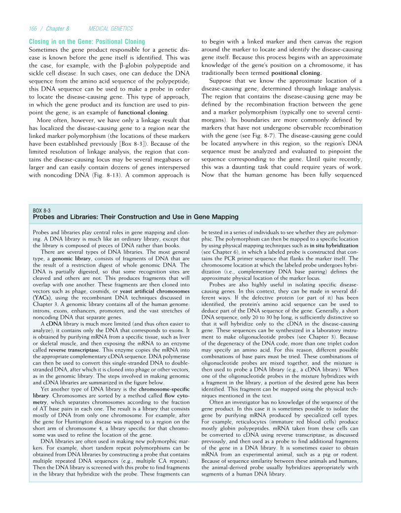
Closing in on the Gene: Positional CloningSometimes the gene product responsible for a genetic dis-ease is known before the gene itself is identified. This wasthe case, for example, with the b-globin polypeptide andsickle cell disease. In such cases, one can deduce the DNAsequence from the amino acid sequence of the polypeptide;this DNA sequence can be used to make a probe in orderto locate the disease-causing gene. This type of approach,in which the gene product and its function are used to pin-point the gene, is an example of functional cloning.
More often, however, we have only a linkage result thathas localized the disease-causing gene to a region near thelinked marker polymorphism (the locations of these markershave been established previously [Box 8-3]). Because of thelimited resolution of linkage analysis, the region that con-tains the disease-causing locus may be several megabases orlarger and can easily contain dozens of genes interspersedwith noncoding DNA (Fig. 8-13). A common approach is
to begin with a linked marker and then canvas the regionaround the marker to locate and identify the disease-causinggene itself. Because this process begins with an approximateknowledge of the gene’s position on a chromosome, it hastraditionally been termed positional cloning.
Suppose that we know the approximate location of adisease-causing gene, determined through linkage analysis.The region that contains the disease-causing gene may bedefined by the recombination fraction between the geneand a marker polymorphism (typically one to several centi-morgans). Its boundaries are more commonly defined bymarkers that have not undergone observable recombinationwith the gene (see Fig. 8-7). The disease-causing gene couldbe located anywhere in this region, so the region’s DNAsequence must be analyzed and evaluated to pinpoint thesequence corresponding to the gene. Until quite recently,this was a daunting task that could require years of work.Now that the human genome has been fully sequenced
BOX 8-3Probes and Libraries: Their Construction and Use in Gene Mapping
Probes and libraries play central roles in gene mapping and clon-ing. A DNA library is much like an ordinary library, except thatthe library is composed of pieces of DNA rather than books.
There are several types of DNA libraries. The most generaltype, a genomic library, consists of fragments of DNA that arethe result of a restriction digest of whole genomic DNA. TheDNA is partially digested, so that some recognition sites arecleaved and others are not. This produces fragments that willoverlap with one another. These fragments are then cloned intovectors such as phage, cosmids, or yeast artificial chromosomes(YACs), using the recombinant DNA techniques discussed inChapter 3. A genomic library contains all of the human genome:introns, exons, enhancers, promoters, and the vast stretches ofnoncoding DNA that separate genes.
A cDNA library is much more limited (and thus often easier toanalyze); it contains only the DNA that corresponds to exons. Itis obtained by purifying mRNA from a specific tissue, such as liveror skeletal muscle, and then exposing the mRNA to an enzymecalled reverse transcriptase. This enzyme copies the mRNA intothe appropriate complementary cDNA sequence. DNA polymerasecan then be used to convert this single-stranded DNA to double-stranded DNA, after which it is cloned into phage or other vectors,as in the genomic library. The steps involved in making genomicand cDNA libraries are summarized in the figure below.
Yet another type of DNA library is the chromosome-specificlibrary. Chromosomes are sorted by a method called flow cyto-metry, which separates chromosomes according to the fractionof AT base pairs in each one. The result is a library that consistsmostly of DNA from only one chromosome. For example, afterthe gene for Huntington disease was mapped to a region on theshort arm of chromosome 4, a library specific for that chromo-some was used to refine the location of the gene.
DNA libraries are often used in making new polymorphic mar-kers. For example, short tandem repeat polymorphisms can beobtained from DNA libraries by constructing a probe that containsmultiple repeated DNA sequences (e.g., multiple CA repeats).Then the DNA library is screened with this probe to find fragmentsin the library that hybridize with the probe. These fragments can
be tested in a series of individuals to see whether they are polymor-phic. The polymorphism can then be mapped to a specific locationby using physical mapping techniques such as in situ hybridization(see Chapter 6), in which a labeled probe is constructed that con-tains the PCR primer sequence that flanks the marker itself. Thechromosome location at which the labeled probe undergoes hybri-dization (i.e., complementary DNA base pairing) defines theapproximate physical location of the marker locus.
Probes are also highly useful in isolating specific disease-causing genes. In this context, they can be made in several dif-ferent ways. If the defective protein (or part of it) has beenidentified, the protein’s amino acid sequence can be used todeduce part of the DNA sequence of the gene. Generally, a shortDNA sequence, only 20 to 30 bp long, is sufficiently distinctive sothat it will hybridize only to the cDNA in the disease-causinggene. These sequences can be synthesized in a laboratory instru-ment to make oligonucleotide probes (see Chapter 3). Becauseof the degeneracy of the DNA code, more than one triplet codonmay specify an amino acid. For this reason, different possiblecombinations of base pairs must be tried. These combinations ofoligonucleotide probes are mixed together, and the mixture isthen used to probe a DNA library (e.g., a cDNA library). Whenone of the oligonucleotide probes in the mixture hybridizes witha fragment in the library, a portion of the desired gene has beenidentified. This fragment can be mapped using the physical tech-niques mentioned in the text.
Often an investigator has no knowledge of the sequence of thegene product. In this case it is sometimes possible to isolate thegene by purifying mRNA produced by specialized cell types.For example, reticulocytes (immature red blood cells) producemostly globin polypeptides. mRNA taken from these cells canbe converted to cDNA using reverse transcriptase, as discussedpreviously, and then used as a probe to find additional fragmentsof the gene in a DNA library. It is sometimes easier to obtainmRNA from an experimental animal, such as a pig or rodent.Because of sequence similarity between these animals and humans,the animal-derived probe usually hybridizes appropriately withsegments of a human DNA library.
166 / Chapter 8 MEDICAL GENETICS
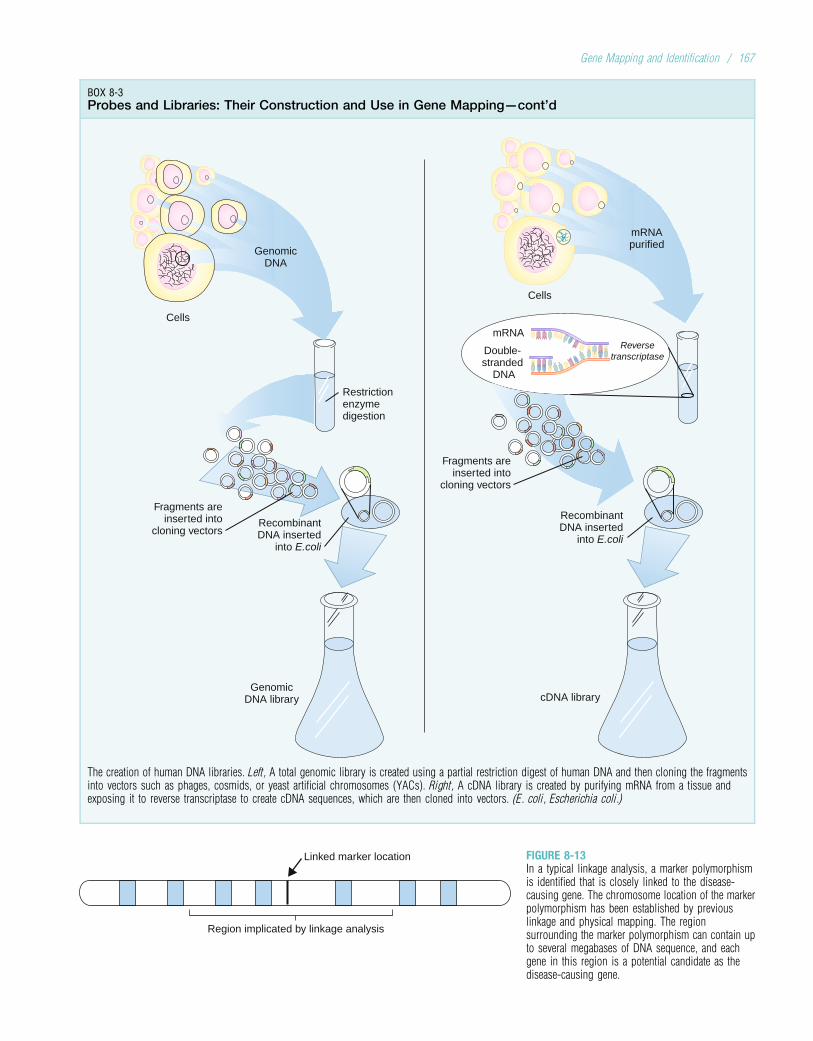
BOX 8-3Probes and Libraries: Their Construction and Use in Gene Mapping—cont’d
Cells
Cells
Restrictionenzymedigestion
Fragments areinserted into
cloning vectors
Fragments areinserted into
cloning vectorsRecombinantDNA inserted
into E.coli
GenomicDNA library
RecombinantDNA inserted
into E.coli
cDNA library
mRNA
Double-stranded
DNA
Reversetranscriptase
mRNApurified
GenomicDNA
The creation of human DNA libraries. Left, A total genomic library is created using a partial restriction digest of human DNA and then cloning the fragmentsinto vectors such as phages, cosmids, or yeast artificial chromosomes (YACs). Right, A cDNA library is created by purifying mRNA from a tissue andexposing it to reverse transcriptase to create cDNA sequences, which are then cloned into vectors. (E. coli, Escherichia coli.)
Region implicated by linkage analysis
Linked marker location FIGURE 8-13In a typical linkage analysis, a marker polymorphismis identified that is closely linked to the disease-causing gene. The chromosome location of the markerpolymorphism has been established by previouslinkage and physical mapping. The regionsurrounding the marker polymorphism can contain upto several megabases of DNA sequence, and eachgene in this region is a potential candidate as thedisease-causing gene.
Gene Mapping and Identification / 167
(see Box 8-2), the process of DNA sequence evaluation canproceed much more rapidly. The finished human genomesequence is now available in computerized databases, soinvestigators often scan a region of interest simply by acces-sing the appropriate DNA sequence on a computer.
As we scan the DNA region containing a disease-causing gene, how do we know when we have actuallyreached the gene? Coding DNA (i.e., DNA that encodesproteins) must be distinguished from noncoding DNA,and the likely function of each gene in the region mustbe determined. Several approaches may be used to accom-plish these tasks.
Functional versus Nonfunctional DNAMost of our DNA sequence has no known function and isunlikely to contribute to disease. Thus, in searching fordisease-causing alterations, we typically wish to focus onDNA that encodes proteins or performs important regulatoryfunctions (i.e., enhancer or promoter sequences). Because oftheir functional significance, coding DNA or regulatoryDNA sequences generally cannot change very much throughthe course of evolution. This means that such DNA sequenceswill be conserved, or similar in base-pair sequence, in manydifferent species. In contrast, nonfunctional DNA sequencesare likely to change rapidly and to differ substantially amongspecies. Published DNA sequences can be compared usingcomputer algorithms (see later) to distinguish functional(conserved) DNA from nonfunctional (nonconserved) DNA.In some cases, a labeled probe containing the human DNAsegment of interest may be constructed and then exposed todenatured DNA from several other species to determinewhether the DNA sequences are similar enough to undergocomplementary base pairing with the probe (this is whimsi-cally termed a “zooblot”). If the human DNA sequence isnonconserved and thus likely nonfunctional, complementarybase pairing between the probe DNA and that of otherspecies is less likely to occur.
As discussed in Chapter 3, most CG dinucleotides aremethylated. However, approximately 60% of human geneshave unmethylated CG dinucleotides (CG islands) in their50 region. (A lack of methylation in the 50 region of the geneprobably makes it more accessible to transcription factorsrequired for active expression.) The identification of a seriesof CG islands has often been used to pinpoint the locationsof coding genes.
4The identification of DNA sequences that are highlyconserved across multiple species, and theidentification of unmethylated CG islands, are bothways to distinguish coding or regulatory (functional)DNA from nonfunctional DNA.
Computer Analysis of DNA SequenceComputer analysis, termed in silico research, has become aneffective and popular approach for identifying genes. Sophis-ticated computer algorithms can test a DNA sequence for
patterns that signal a coding gene (e.g., transcription initia-tion sites, stop codons, intron–exon boundaries). Thisapproach was used, for example, to help identify and charac-terize one of the adult polycystic kidney disease genes(PKD1; see Chapter 4). In addition, these algorithms canoften recognize patterns typical of genes that encode specificclasses of proteins (e.g., transcription factors, transmembraneproteins).
Computerized databases of known DNA sequences alsoplay an important role in gene identification. When studyinga specific region of DNA to find a gene, it is common tosearch for similarity between DNA sequences from theregion and other DNA sequences in the database. Thesequences in the database might derive from genes withknown function or tissue-specific expression patterns. Sup-pose, for example, that we have used linkage analysis toidentify a region containing a gene that causes a develop-mental disorder such as a limb malformation. As we evaluateDNA sequences in the region, we would look for similaritybetween a DNA sequence from this region and a plausiblesequence from the database (e.g., a sequence from a genethat encodes a protein involved in bone development, suchas a fibroblast growth factor). Because genes that encodesimilar protein products usually have similar DNAsequences, a match between the sequence from our regionand a sequence in the database could be a vital clue that thisparticular DNA sequence is actually part of the gene thatcauses the limb malformation.
A computerized database commonly used in thesesearches consists of small DNA sequences known asexpressed sequence tags (ESTs). ESTs are obtained bysequencing several hundred base pairs from both ends ofcDNA clones taken from a cDNA library (see Box 8-3).Because these clones consist of DNA that is complementaryto mRNA, the ESTs represent expressed portions of genes.Often, they are expressed only in certain tissues at certainpoints in time. The investigator can search for sequence sim-ilarity between DNA sequences in a region of interest andESTs known to be located in the same region (and possiblyknown to be expressed in a tissue that would be consistentwith the disease in question). This strategy was used, forexample, to identify one of the genes known to causeAlzheimer disease (see Chapter 12).
The similarity search need not be confined to humangenes. The complete DNA sequences of more than twodozen organisms, including chimpanzee, chicken, mouse,fruit fly, and yeast, are now available in computerized data-bases. Often, sequence similarity is observed with genes ofknown function in other organisms, such as mouse or evenyeast or bacteria. Because genes with important protein pro-ducts tend to be highly conserved throughout evolution, theidentification of a similar gene in another can provide impor-tant information about the gene’s function in the human. Forexample, many of the genes involved in cell cycle regulationare quite similar in yeast and human (e.g., portions of theNF1 gene and the yeast IRA2 gene). Indeed, approximatelyone third of human disease-causing genes identified to date
168 / Chapter 8 MEDICAL GENETICS
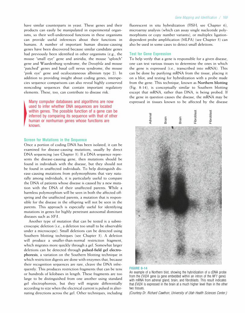
have similar counterparts in yeast. These genes and theirproducts can easily be manipulated in experimental organ-isms, so their well-understood functions in these organismscan provide useful inferences about their functions inhumans. A number of important human disease-causinggenes have been discovered because similar candidate geneshad previously been identified in other organisms (e.g., themouse “small eye” gene and aniridia, the mouse “splotch”gene and Waardenburg syndrome, the Drosophila and mouse“patched” genes and basal cell nevus syndrome, the mouse“pink eye” gene and oculocutaneous albinism type 2). Inaddition to providing insight about coding genes, interspe-cies sequence comparisons can also reveal highly conservednoncoding sequences that contain important regulatoryelements. These, too, can contribute to disease risk.
4Many computer databases and algorithms are nowused to infer whether DNA sequences are locatedwithin genes. The possible function of a gene can beinferred by comparing its sequence with that of otherhuman or nonhuman genes whose functions areknown.
Screen for Mutations in the SequenceOnce a portion of coding DNA has been isolated, it can beexamined for disease-causing mutations, usually by directDNA sequencing (see Chapter 3). If a DNA sequence repre-sents the disease-causing gene, then mutations should befound in individuals with the disease, but they should notbe found in unaffected individuals. To help distinguish dis-ease-causing mutations from polymorphisms that vary natu-rally among individuals, it is particularly useful to comparethe DNA of patients whose disease is caused by a new muta-tion with the DNA of their unaffected parents. While aharmless polymorphism will be seen in both the affected off-spring and the unaffected parents, a mutation that is respon-sible for the disease in the offspring will not be seen in theparents. This approach is especially useful for identifyingmutations in genes for highly penetrant autosomal dominantdiseases such as NF1.
Another type of mutation that can be tested is a submi-croscopic deletion (i.e., a deletion too small to be observableunder a microscope). Small deletions can be detected usingSouthern blotting techniques (see Chapter 3). A deletionwill produce a smaller-than-normal restriction fragment,which migrates more quickly through a gel. Somewhat largerdeletions can be detected through pulsed-field gel electro-phoresis, a variation on the Southern blotting technique inwhich restriction digests are done with enzymes that, becausetheir recognition sequences are rare, cleave the DNA infre-quently. This produces restriction fragments that can be tensor hundreds of kilobases in length. These fragments are toolarge to be distinguished from one another using standardgel electrophoresis, but they will migrate differentiallyaccording to size when the electrical current is pulsed in alter-nating directions across the gel. Other techniques, including
fluorescent in situ hybridization (FISH; see Chapter 6),microarray analysis (which can assay single nucleotide poly-morphisms or copy number variants), or multiplex ligation-dependent probe amplification (MLPA) (see Chapter 3) canalso be used in some cases to detect small deletions.
Test for Gene ExpressionTo help verify that a gene is responsible for a given disease,one can test various tissues to determine the ones in whichthe gene is expressed (i.e., transcribed into mRNA). Thiscan be done by purifying mRNA from the tissue, placing iton a blot, and testing for hybridization with a probe madefrom the gene. This technique, known as Northern blotting(Fig. 8-14), is conceptually similar to Southern blottingexcept that mRNA, rather than DNA, is being probed. Ifthe gene in question causes the disease, the mRNA may beexpressed in tissues known to be affected by the disease
Adr
enal
Bra
in
Bra
in
Fib
robl
ast
Fib
robl
ast
FIGURE 8-14An example of a Northern blot, showing the hybridization of a cDNA probefrom the EVI2A gene (a gene embedded within an intron of the NF1 gene)with mRNA from adrenal gland, brain, and fibroblasts. This result indicatesthat EVI2A is expressed in the brain at a much higher level than in the othertwo tissues.
(Courtesy Dr. Richard Cawthon, University of Utah Health Sciences Center.)
Gene Mapping and Identification / 169
(the same reasoning is applied to the analysis of ESTsobtained from tissue-specific cDNA libraries). For example,one would expect the phenylalanine hydroxylase gene(mutations in which can cause phenylketonuria [PKU]) tobe expressed in the liver, where this enzyme is known tobe synthesized.
Increasingly, microarrays (see Chapter 3) are being usedto test for gene expression. Microarrays containingthousands of oligonucleotide sequences representing genesof interest have been manufactured, and these can beexposed to mRNA from relevant tissues to determine thetissues in which specific genes are expressed.
Another gene expression test involves inserting the nor-mal version of the DNA sequence into a defective cell froman affected person (or animal model), using recombinantDNA techniques. If the normal sequence corrects the defect,then it is very likely to represent the gene of interest. Thisapproach has been used, for example, to show that mutationsin the CFTR gene can cause cystic fibrosis.
4In determining whether a gene contributes to thecause of a disease, a critical test is to screen thegene for mutations that are present in affectedpersons but absent in unaffected controls. Furtherevidence that a gene helps to cause a specificdisease is to show that mRNA corresponding to thatgene is expressed in tissues associated with oraffected by the disease.
Candidate GenesThe gene hunting process can be expedited considerably if acandidate gene is available. As the name implies, this is agene whose known protein product makes it a likely candi-date for the disease in question. For example, the variouscollagen genes were considered to be reasonable candidatesfor Marfan syndrome because collagen is an important
component of connective tissue. However, linkage analysisusing collagen gene markers in Marfan syndrome familiesconsistently yielded negative results. Another candidate geneemerged when the gene that encodes Fibrillin-1 (FBN1) wasidentified on chromosome 15. Fibrillin, as discussed inChapter 4, is also a connective tissue component. Linkageanalysis had localized the Marfan syndrome gene to chromo-some 15, so FBN1 became an even stronger candidate. Anal-ysis of FBN1 mutations showed that they were consistentlyassociated with Marfan syndrome, confirming these muta-tions as a cause of the disease. This combination of linkageanalysis to identify the region containing a gene followedby a search of the region for plausible candidate genes issometimes called the positional candidate approach.
4Candidate genes are those whose characteristics(e.g., protein product) suggest that they may beresponsible for a genetic disease. The analysis ofcandidate genes in a region known to contain thedisease-causing gene is termed the positionalcandidate approach.
Using the techniques described in this chapter, a largenumber of important disease-causing genes have now beenmapped, and many of them have also been cloned. Someexamples are given in Table 8-2.
Although the gene identification examples used in thischapter have focused on mendelian conditions, these sameapproaches are being used to identify genes that contributeto the cause of common, complex diseases such as diabetes,hypertension, and heart disease. Because these diseases areinfluenced by multiple genes (as discussed in Chapter 12),gene identification tends to be especially challenging. Nev-ertheless, the techniques discussed in this chapter are nowbeing applied with considerable success in pinpointinggenes that help to cause the common diseases that affectthe majority of the human population.
TABLE 8-2
Examples of Well-Known Mendelian Disease-Causing Genes that Have Been Mapped and Cloned*
Disease ChromosomeLocation
Gene Product
a1-Antitrypsin deficiency 14q32.1 Serine protease inhibitor
a-Thalassemia 16p13.3 a-Globin component of hemoglobin
b-Thalassemia 11p15.5 b-Globin component of hemoglobin
Achondroplasia 4p16.3 Fibroblast growth factor receptor 3
Albinism, oculocutaneous (type 1) 11q14-q21 Tyrosinase
Albinism, oculocutaneous (type 2) 15q11-q12 Tyrosine transporter
Alzheimer disease* (familial) 14q24.3 Presenilin 1
1q31-q42 Presenilin 2
21q21 b-Amyloid precursor protein
170 / Chapter 8 MEDICAL GENETICS
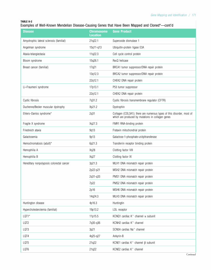
TABLE 8-2
Examples of Well-Known Mendelian Disease-Causing Genes that Have Been Mapped and Cloned*—cont’d
Disease ChromosomeLocation
Gene Product
Amyotrophic lateral sclerosis (familial) 21q22.1 Superoxide dismutase 1
Angelman syndrome 15q11-q13 Ubiquitin-protein ligase E3A
Ataxia-telangiectasia 11q22.3 Cell cycle control protein
Bloom syndrome 15q26.1 RecQ helicase
Breast cancer (familial) 17q21 BRCA1 tumor suppressor/DNA repair protein
13q12.3 BRCA2 tumor suppressor/DNA repair protein
22q12.1 CHEK2 DNA repair protein
Li–Fraumeni syndrome 17p13.1 P53 tumor suppressor
22q12.1 CHEK2 DNA repair protein
Cystic fibrosis 7q31.2 Cystic fibrosis transmembrane regulator (CFTR)
Duchenne/Becker muscular dystrophy Xp21.2 Dystrophin
Ehlers–Danlos syndrome* 2q31 Collagen (COL3A1); there are numerous types of this disorder, most ofwhich are produced by mutations in collagen genes
Fragile X syndrome Xq27.3 FMR1 RNA-binding protein
Friedreich ataxia 9q13 Frataxin mitochondrial protein
Galactosemia 9p13 Galactose-1-phosphate-uridyltransferase
Hemochromatosis (adult)* 6p21.3 Transferrin receptor binding protein
Hemophilia A Xq28 Clotting factor VIII
Hemophilia B Xq27 Clotting factor IX
Hereditary nonpolyposis colorectal cancer 3p21.3 MLH1 DNA mismatch repair protein
2p22-p21 MSH2 DNA mismatch repair protein
2q31-q33 PMS1 DNA mismatch repair protein
7p22 PMS2 DNA mismatch repair protein
2p16 MSH6 DNA mismatch repair protein
14q24.3 MLH3 DNA mismatch repair protein
Huntington disease 4p16.3 Huntingtin
Hypercholesterolemia (familial) 19p13.2 LDL receptor
LQT1* 11p15.5 KCNQ1 cardiac Kþ channel a subunit
LQT2 7q35-q36 KCNH2 cardiac Kþ channel
LQT3 3p21 SCN5A cardiac Naþ channel
LQT4 4q25-q27 Ankyrin-B
LQT5 21q22 KCNE1 cardiac Kþ channel b subunit
LQT6 21q22 KCNE2 cardiac Kþ channel
Continued
Gene Mapping and Identification / 171
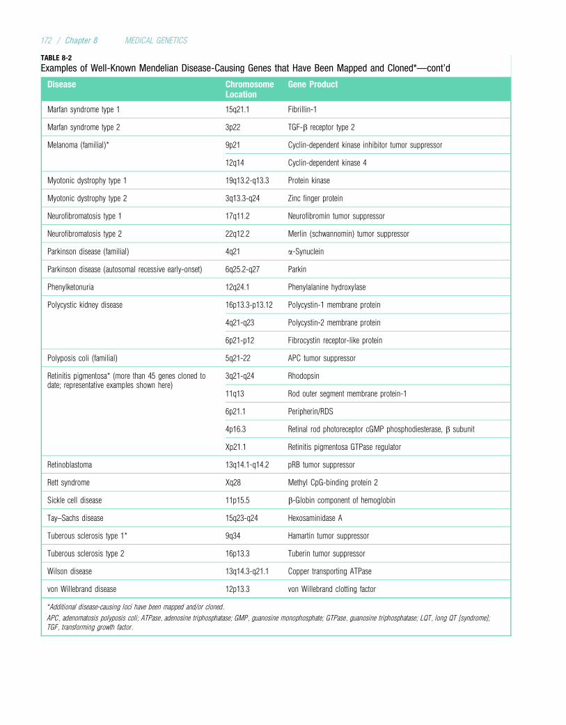
TABLE 8-2
Examples of Well-Known Mendelian Disease-Causing Genes that Have Been Mapped and Cloned*—cont’d
Disease ChromosomeLocation
Gene Product
Marfan syndrome type 1 15q21.1 Fibrillin-1
Marfan syndrome type 2 3p22 TGF-b receptor type 2
Melanoma (familial)* 9p21 Cyclin-dependent kinase inhibitor tumor suppressor
12q14 Cyclin-dependent kinase 4
Myotonic dystrophy type 1 19q13.2-q13.3 Protein kinase
Myotonic dystrophy type 2 3q13.3-q24 Zinc finger protein
Neurofibromatosis type 1 17q11.2 Neurofibromin tumor suppressor
Neurofibromatosis type 2 22q12.2 Merlin (schwannomin) tumor suppressor
Parkinson disease (familial) 4q21 a-Synuclein
Parkinson disease (autosomal recessive early-onset) 6q25.2-q27 Parkin
Phenylketonuria 12q24.1 Phenylalanine hydroxylase
Polycystic kidney disease 16p13.3-p13.12 Polycystin-1 membrane protein
4q21-q23 Polycystin-2 membrane protein
6p21-p12 Fibrocystin receptor-like protein
Polyposis coli (familial) 5q21-22 APC tumor suppressor
Retinitis pigmentosa* (more than 45 genes cloned todate; representative examples shown here)
3q21-q24 Rhodopsin
11q13 Rod outer segment membrane protein-1
6p21.1 Peripherin/RDS
4p16.3 Retinal rod photoreceptor cGMP phosphodiesterase, b subunit
Xp21.1 Retinitis pigmentosa GTPase regulator
Retinoblastoma 13q14.1-q14.2 pRB tumor suppressor
Rett syndrome Xq28 Methyl CpG-binding protein 2
Sickle cell disease 11p15.5 b-Globin component of hemoglobin
Tay–Sachs disease 15q23-q24 Hexosaminidase A
Tuberous sclerosis type 1* 9q34 Hamartin tumor suppressor
Tuberous sclerosis type 2 16p13.3 Tuberin tumor suppressor
Wilson disease 13q14.3-q21.1 Copper transporting ATPase
von Willebrand disease 12p13.3 von Willebrand clotting factor
*Additional disease-causing loci have been mapped and/or cloned.
APC, adenomatosis polyposis coli; ATPase, adenosine triphosphatase; GMP, guanosine monophosphate; GTPase, guanosine triphosphatase; LQT, long QT [syndrome];
TGF, transforming growth factor.
172 / Chapter 8 MEDICAL GENETICS
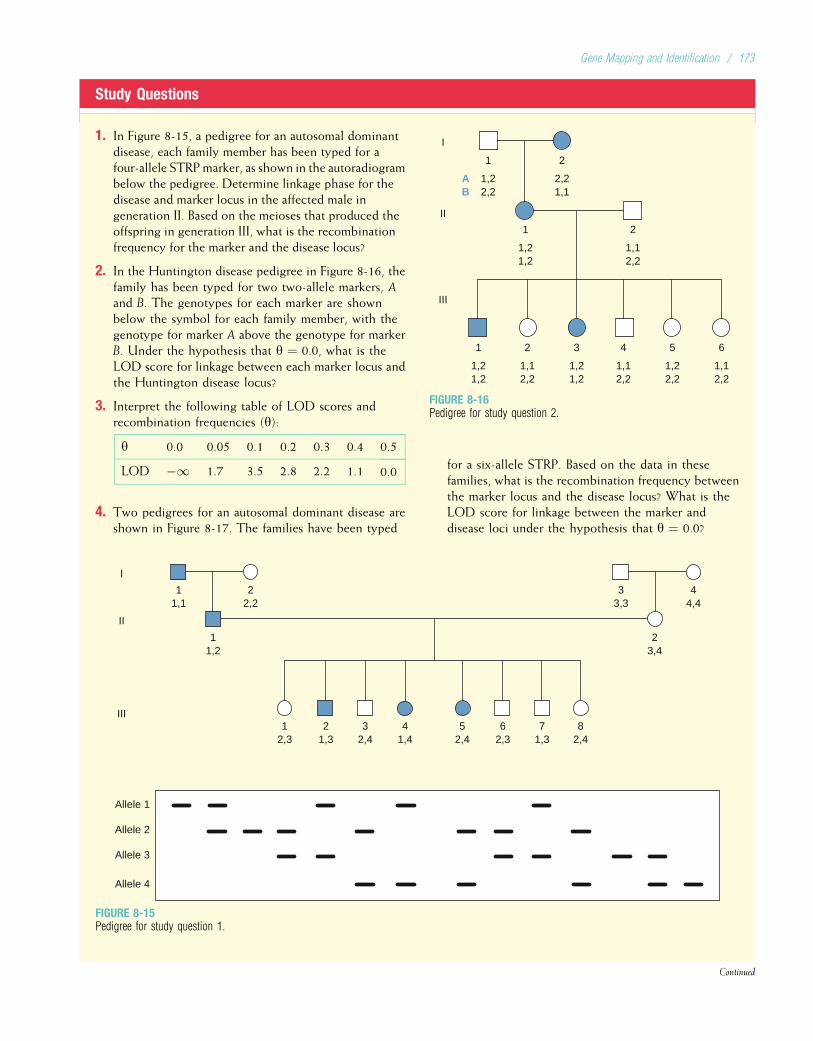
Study Questions
1. In Figure 8-15, a pedigree for an autosomal dominantdisease, each family member has been typed for afour-allele STRPmarker, as shown in the autoradiogrambelow the pedigree. Determine linkage phase for thedisease and marker locus in the affected male ingeneration II. Based on the meioses that produced theoffspring in generation III, what is the recombinationfrequency for the marker and the disease locus?
2. In the Huntington disease pedigree in Figure 8-16, thefamily has been typed for two two-allele markers, Aand B. The genotypes for each marker are shownbelow the symbol for each family member, with thegenotype for marker A above the genotype for markerB. Under the hypothesis that y ¼ 0.0, what is theLOD score for linkage between each marker locus andthe Huntington disease locus?
3. Interpret the following table of LOD scores andrecombination frequencies (y):
y 0.0 0.05 0.1 0.2 0.3 0.4 0.5
LOD �1 1.7 3.5 2.8 2.2 1.1 0.0
4. Two pedigrees for an autosomal dominant disease areshown in Figure 8-17. The families have been typed
1,22,2
I
AB
II
III
1
2,21,1
2
1,21,2
1
1,12,2
2
1,21,2
1
1,21,2
3
1,12,2
2
1,12,2
4
1,12,2
6
1,22,2
5
FIGURE 8-16Pedigree for study question 2.
for a six-allele STRP. Based on the data in thesefamilies, what is the recombination frequency betweenthe marker locus and the disease locus? What is theLOD score for linkage between the marker anddisease loci under the hypothesis that y ¼ 0.0?
22,2
11,1
I
II
III
Allele 1
Allele 2
Allele 3
Allele 4
11,2
44,4
33,3
23,4
12,3
21,3
32,4
41,4
52,4
62,3
71,3
82,4
FIGURE 8-15Pedigree for study question 1.
Continued
Gene Mapping and Identification / 173
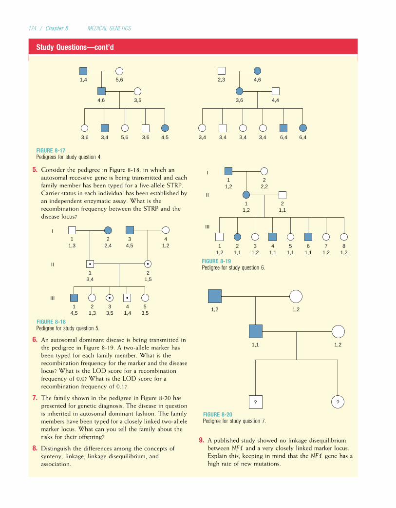
Study Questions—cont’d
5,61,4
3,54,6 3,6 4,4
3,6 3,4 5,6 3,6 4,5 3,4 3,4 3,4 6,4 6,43,4
4,62,3
FIGURE 8-17Pedigrees for study question 4.
5. Consider the pedigree in Figure 8-18, in which anautosomal recessive gene is being transmitted and eachfamily member has been typed for a five-allele STRP.Carrier status in each individual has been established byan independent enzymatic assay. What is therecombination frequency between the STRP and thedisease locus?
11,3
22,4
34,5
41,2
I
II
III
13,4
21,5
33,5
53,5
21,3
14,5
41,4
FIGURE 8-18Pedigree for study question 5.
6. An autosomal dominant disease is being transmitted inthe pedigree in Figure 8-19. A two-allele marker hasbeen typed for each family member. What is therecombination frequency for the marker and the diseaselocus? What is the LOD score for a recombinationfrequency of 0.0? What is the LOD score for arecombination frequency of 0.1?
7. The family shown in the pedigree in Figure 8-20 haspresented for genetic diagnosis. The disease in questionis inherited in autosomal dominant fashion. The familymembers have been typed for a closely linked two-allelemarker locus. What can you tell the family about therisks for their offspring?
8. Distinguish the differences among the concepts ofsynteny, linkage, linkage disequilibrium, andassociation.
11,2
22,2
11,2
21,1
I
II
III
11,2
31,2
21,1
41,1
61,1
51,1
81,2
71,2
FIGURE 8-19Pedigree for study question 6.
1,2 1,2
1,1 1,2
? ?
FIGURE 8-20Pedigree for study question 7.
9. A published study showed no linkage disequilibriumbetween NF1 and a very closely linked marker locus.Explain this, keeping in mind that the NF1 gene has ahigh rate of new mutations.
174 / Chapter 8 MEDICAL GENETICS
Suggested ReadingsAltshuler D, Daly MJ, Lander ES. Genetic mapping in
human disease. Science 2008;322(5903):881–8.Christensen K, Murray JC. What genome-wide association
studies can do for medicine. N Engl J Med 2007;356(11):1094–7.
Collins FS, Morgan M, Patrinos A. The Human GenomeProject: Lessons from large-scale biology. Science2003;300(5617):286–90.
Dawn Teare M, Barrett JH. Genetic linkage studies. Lancet2005;366(9490):1036–44.
Frazer KA, Ballinger DG, Cox DR, et al. A second genera-tion human haplotype map of over 3.1 million SNPs.Nature 2007;449(7164):851–61.
Hartong DT, Berson EL, Dryja TP. Retinitis pigmentosa.Lancet 2006;368(9549):1795–809.
Hirschhorn JN, Daly MJ. Genome-wide association studiesfor common diseases and complex traits. Nat Rev Genet2005;6(2):95–108.
Hopper JL, Bishop DT, Easton DF. Population-based familystudies in genetic epidemiology. Lancet 2005;366(9494):1397–406.
International Human Genome Sequencing Consortium. Fin-ishing the euchromatic sequence of the human genome.Nature 2004;431(7011):931–45.
Kennan A, Aherne A, Humphries P. Light in retinitis pig-mentosa. Trends Genet 2005;21(2):103–10.
Morton NE. LODs past and present. Genetics 1995; 140:7–12.
Ott J. Analysis of Human Genetic Linkage, 3rd ed.Baltimore: Johns Hopkins University Press, 1999.
Palmer LJ, Cardon LR. Shaking the tree: Mapping complexdisease genes with linkage disequilibrium. Lancet 2005;366(9492):1223–34.
Pearson TA, Manolio TA. How to interpret a genome-wideassociation study. JAMA 2008;299(11):1335–44.
Venter JC, Adams MD, Myers EW, et al. The sequence of thehuman genome. Science 2001;291(5507):1304–51.
Internet ResourcesCatalog of Published Genome-Wide Association Studieshttp://www.genome.gov/26525384
Computational Biology at the Oak Ridge NationalLaboratory (contains a number of useful links for theanalysis of DNA sequences and protein structures)http://compbio.ornl.gov/
Ensembl (provides DNA and protein sequences for humansand other organisms, along with descriptive information;includes the BLAST sequence analysis algorithm) http://www.ensembl.org/
Genamics (contains hundreds of links for genetic analysissoftware, including programs for analyzing linkage, link-age disequilibrium, and DNA sequence variation) http://
genamics.com/
GeneCards (database of human genes and their products,with information about the function of each gene product)http://bioinfo.weizmann.ac.il/cards/
International HapMap Project (public database of 1 millionSNPs genotyped in 270 individuals from four humanpopulations) http://www.hapmap.org/
National Center for Biotechnology Information (maps ofchromosomes and disease loci, with links to other usefulgenomics sites such as SNP databases) http://www.ncbi.nlm.nih.gov/genome/guide/human/
UCSC Genome Browser (contains reference sequence forthe genomes of many organisms, along with useful toolsfor sequence analysis) http://genome.ucsc.edu/
Gene Mapping and Identification / 175
Chapter 9
IMMUNOGENETICS
Each day, our bodies are confronted with a formidable seriesof invaders: viruses, bacteria, and many other disease-causingorganisms whose goal is to overcome our natural defenses.These defenses, known collectively as the immune system,consist of a diverse collection of trillions of cells. Theimmune system must be able to cope with a multitude ofinvading microorganisms, and it must be able to distinguish“self” from “nonself” with a high degree of accuracy.
As one might expect, the genetic basis of the immune sys-tem is complex. The study of the genetics of the immunesystem, known as immunogenetics, has benefited enor-mously from new developments in gene mapping and clon-ing. Most of the techniques discussed in earlier chapters(e.g., linkage analysis, positional cloning, DNA sequencing)have been used to study genes responsible for the immuneresponse. Many new genes have been discovered, and theirfunctions and interactions have been studied intensely. Thischapter provides a brief review of basic immunology and dis-cusses the genes underlying the body’s capacity to defendagainst a highly diverse array of pathogens. Aspects ofautoimmune disease are examined, and some of the majorimmunodeficiency disorders are discussed.
THE IMMUNE RESPONSE: BASIC CONCEPTSThe Innate Immune SystemWhen a pathogenic microorganism is encountered, thebody’s first line of defense includes phagocytes (a type ofcell that engulfs and destroys the microorganism) and thecomplement system. The complement proteins can destroymicrobes directly by perforating their cell membranes, andthey can also attract phagocytes and other immune systemagents to microbes by coating the microbial surface (it isbecause of this assisting role that the term complement origi-nated). Natural killer cells, a specific type of lymphocyte,can respond to certain viral infections and some tumor cells.Phagocytes, complement, and natural killer cells are all partof the innate immune system, which is capable of respond-ing very rapidly to pathogens.
The innate immune system is activated by general featuresthat are detected in pathogens but are not found in the host.For example, gram-negative bacteria produce lipopolysac-charides, and gram-positive bacteria produce peptidoglycans.
Some bacteria have a high percentage of unmethylated CGsequences, and some viruses produce double-stranded RNA.These distinctive features of pathogenic organisms can bedetected by receptor molecules located on the surfaces ofinnate immune system cells. An important example is theToll-like family of receptors, named after a cell-surface recep-tor, Toll, that was first described in fruit flies. The genes thatencode the human and fruit-fly versions of Toll-like receptorshave a remarkable similarity to one another, attesting to theirimportance in maintaining an innate immune response in awide variety of organisms. Indeed, all multicellular organismsare thought to possess innate immune systems.
4The innate immune system, which includes somephagocytes, natural killer cells, and the complementsystem, is an early part of the immune response andrecognizes general features of invadingmicroorganisms.
The Adaptive Immune SystemAlthough the innate immune system typically helps to holdan infection in check in its early phases, it is sometimes inca-pable of overcoming the infection. This becomes the task ofa more specialized component of the immune response, theadaptive immune system. As its name suggests, this part ofthe immune system is capable of changing, or adapting tofeatures of the invading microorganism in order to mount amore specific and more effective immune response. Theadaptive immune system is a more recent evolutionary devel-opment than the innate immune system and is found onlyin vertebrates.
Key components of the adaptive immune response(Fig. 9-1) include T lymphocytes (or T cells) and B lympho-cytes (or B cells). These cells develop in the body’s primarylymphoid organs (bone marrow for B cells and the thymusfor T cells). In the thymus, developing T cells are exposedto a wide variety of the body’s peptides. Those that can rec-ognize and tolerate the body’s own peptides are selected,and those that would attack the body’s peptides are elimi-nated. The B and T cells progress to secondary lymphoidtissues, such as the lymph nodes, spleen, and tonsils, wherethey encounter disease-causing microorganisms. Mature B
176
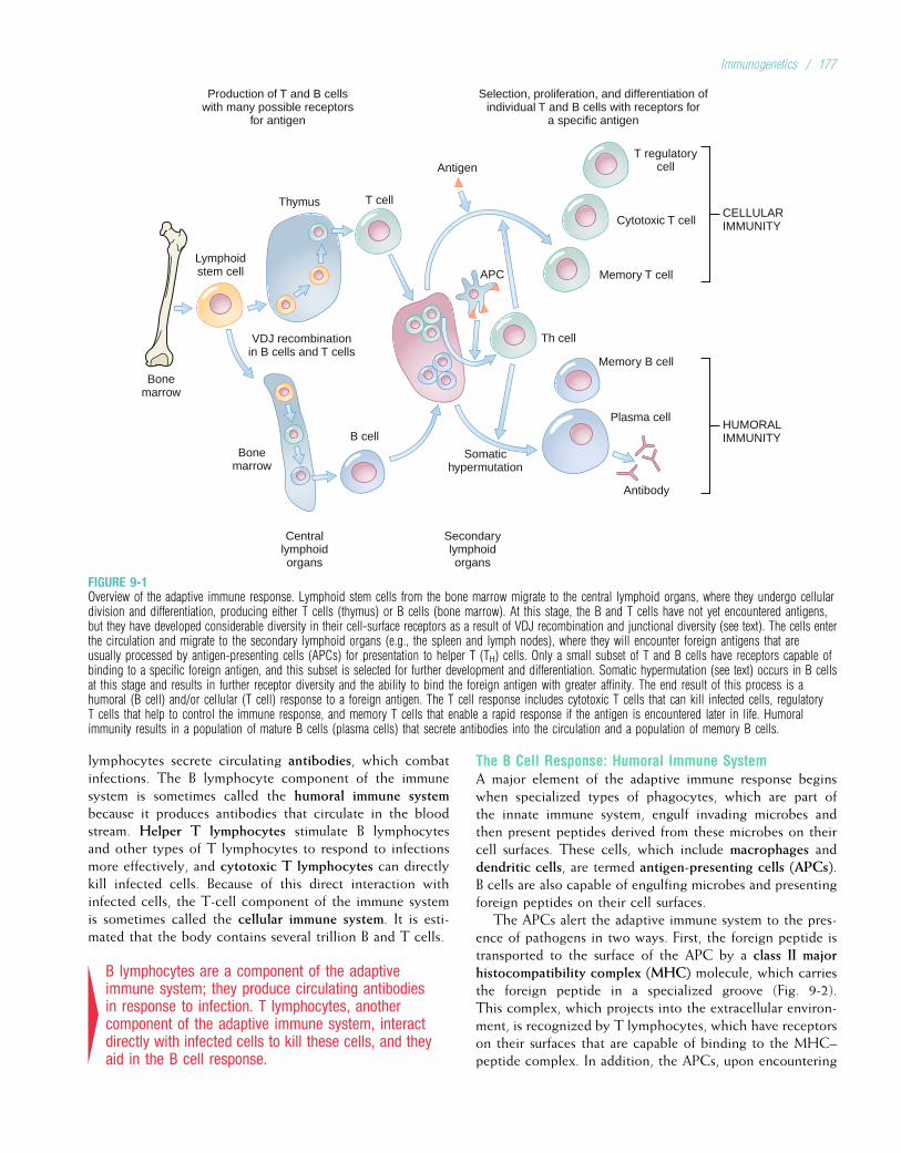
lymphocytes secrete circulating antibodies, which combatinfections. The B lymphocyte component of the immunesystem is sometimes called the humoral immune systembecause it produces antibodies that circulate in the bloodstream. Helper T lymphocytes stimulate B lymphocytesand other types of T lymphocytes to respond to infectionsmore effectively, and cytotoxic T lymphocytes can directlykill infected cells. Because of this direct interaction withinfected cells, the T-cell component of the immune systemis sometimes called the cellular immune system. It is esti-mated that the body contains several trillion B and T cells.
4B lymphocytes are a component of the adaptiveimmune system; they produce circulating antibodiesin response to infection. T lymphocytes, anothercomponent of the adaptive immune system, interactdirectly with infected cells to kill these cells, and theyaid in the B cell response.
The B Cell Response: Humoral Immune SystemA major element of the adaptive immune response beginswhen specialized types of phagocytes, which are part ofthe innate immune system, engulf invading microbes andthen present peptides derived from these microbes on theircell surfaces. These cells, which include macrophages anddendritic cells, are termed antigen-presenting cells (APCs).B cells are also capable of engulfing microbes and presentingforeign peptides on their cell surfaces.
The APCs alert the adaptive immune system to the pres-ence of pathogens in two ways. First, the foreign peptide istransported to the surface of the APC by a class II majorhistocompatibility complex (MHC) molecule, which carriesthe foreign peptide in a specialized groove (Fig. 9-2).This complex, which projects into the extracellular environ-ment, is recognized by T lymphocytes, which have receptorson their surfaces that are capable of binding to the MHC–peptide complex. In addition, the APCs, upon encountering
Bonemarrow
Lymphoidstem cell
Thymus
Production of T and B cellswith many possible receptors
for antigen
Selection, proliferation, and differentiation ofindividual T and B cells with receptors for
a specific antigen
T cell
APC
Bonemarrow
Centrallymphoidorgans
Secondarylymphoidorgans
Somatichypermutation
VDJ recombinationin B cells and T cells
B cell
Antigen
CELLULARIMMUNITY
T regulatorycell
Cytotoxic T cell
Memory T cell
Memory B cell
Plasma cell
Antibody
HUMORALIMMUNITY
Th cell
FIGURE 9-1Overview of the adaptive immune response. Lymphoid stem cells from the bone marrow migrate to the central lymphoid organs, where they undergo cellulardivision and differentiation, producing either T cells (thymus) or B cells (bone marrow). At this stage, the B and T cells have not yet encountered antigens,but they have developed considerable diversity in their cell-surface receptors as a result of VDJ recombination and junctional diversity (see text). The cells enterthe circulation and migrate to the secondary lymphoid organs (e.g., the spleen and lymph nodes), where they will encounter foreign antigens that areusually processed by antigen-presenting cells (APCs) for presentation to helper T (TH) cells. Only a small subset of T and B cells have receptors capable ofbinding to a specific foreign antigen, and this subset is selected for further development and differentiation. Somatic hypermutation (see text) occurs in B cellsat this stage and results in further receptor diversity and the ability to bind the foreign antigen with greater affinity. The end result of this process is ahumoral (B cell) and/or cellular (T cell) response to a foreign antigen. The T cell response includes cytotoxic T cells that can kill infected cells, regulatoryT cells that help to control the immune response, and memory T cells that enable a rapid response if the antigen is encountered later in life. Humoralimmunity results in a population of mature B cells (plasma cells) that secrete antibodies into the circulation and a population of memory B cells.
Immunogenetics / 177
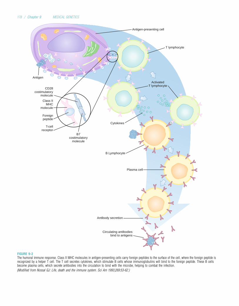
Antigen-presenting cell
Antigen
Class IIMHC
molecule
CD28costimulatory
molecule
B7costimulatory
molecule
Foreignpeptide
Cytokines
Plasma cell
Antibody secretion
Circulating antibodiesbind to antigens
B Lymphocyte
T-cellreceptor
T lymphocyte
ActivatedT lymphocyte
FIGURE 9-2The humoral immune response. Class II MHC molecules in antigen-presenting cells carry foreign peptides to the surface of the cell, where the foreign peptide isrecognized by a helper T cell. The T cell secretes cytokines, which stimulate B cells whose immunoglobulins will bind to the foreign peptide. These B cellsbecome plasma cells, which secrete antibodies into the circulation to bind with the microbe, helping to combat the infection.
(Modified from Nossal GJ: Life, death and the immune system. Sci Am 1993;269:53-62.)
178 / Chapter 9 MEDICAL GENETICS
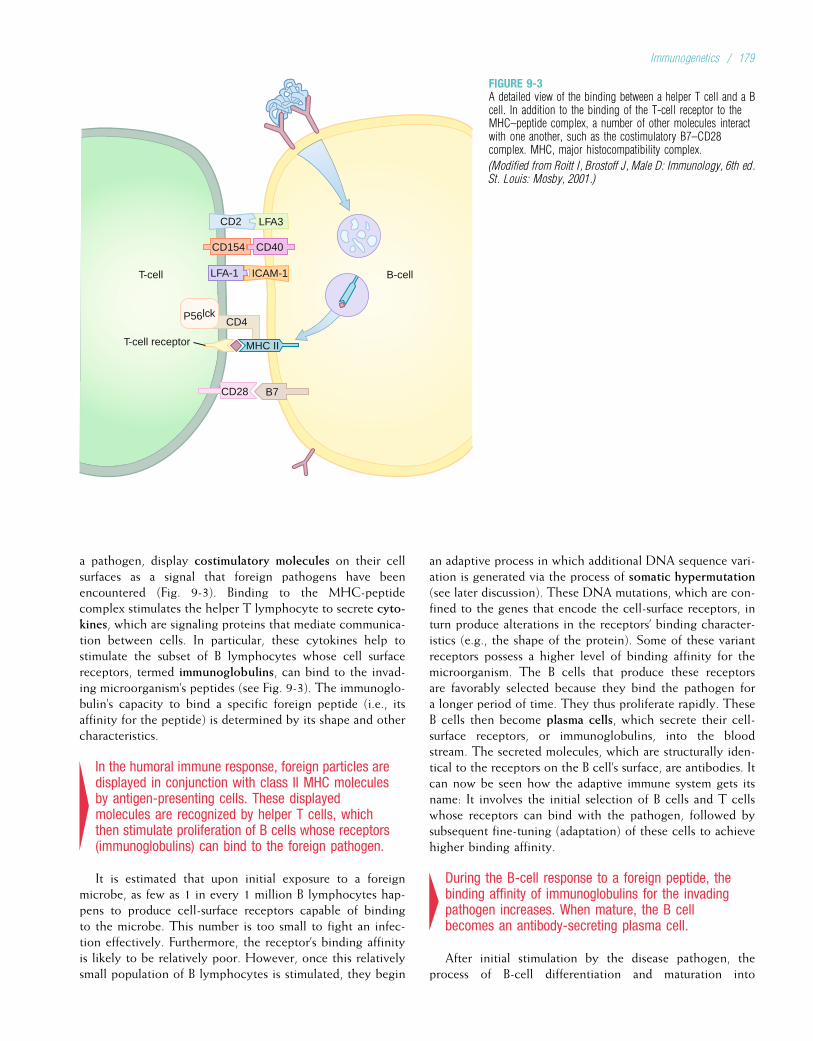
a pathogen, display costimulatory molecules on their cellsurfaces as a signal that foreign pathogens have beenencountered (Fig. 9-3). Binding to the MHC-peptidecomplex stimulates the helper T lymphocyte to secrete cyto-kines, which are signaling proteins that mediate communica-tion between cells. In particular, these cytokines help tostimulate the subset of B lymphocytes whose cell surfacereceptors, termed immunoglobulins, can bind to the invad-ing microorganism’s peptides (see Fig. 9-3). The immunoglo-bulin’s capacity to bind a specific foreign peptide (i.e., itsaffinity for the peptide) is determined by its shape and othercharacteristics.
4In the humoral immune response, foreign particles aredisplayed in conjunction with class II MHC moleculesby antigen-presenting cells. These displayedmolecules are recognized by helper T cells, whichthen stimulate proliferation of B cells whose receptors(immunoglobulins) can bind to the foreign pathogen.
It is estimated that upon initial exposure to a foreignmicrobe, as few as 1 in every 1 million B lymphocytes hap-pens to produce cell-surface receptors capable of bindingto the microbe. This number is too small to fight an infec-tion effectively. Furthermore, the receptor’s binding affinityis likely to be relatively poor. However, once this relativelysmall population of B lymphocytes is stimulated, they begin
an adaptive process in which additional DNA sequence vari-ation is generated via the process of somatic hypermutation(see later discussion). These DNA mutations, which are con-fined to the genes that encode the cell-surface receptors, inturn produce alterations in the receptors’ binding character-istics (e.g., the shape of the protein). Some of these variantreceptors possess a higher level of binding affinity for themicroorganism. The B cells that produce these receptorsare favorably selected because they bind the pathogen fora longer period of time. They thus proliferate rapidly. TheseB cells then become plasma cells, which secrete their cell-surface receptors, or immunoglobulins, into the bloodstream. The secreted molecules, which are structurally iden-tical to the receptors on the B cell’s surface, are antibodies. Itcan now be seen how the adaptive immune system gets itsname: It involves the initial selection of B cells and T cellswhose receptors can bind with the pathogen, followed bysubsequent fine-tuning (adaptation) of these cells to achievehigher binding affinity.
4During the B-cell response to a foreign peptide, thebinding affinity of immunoglobulins for the invadingpathogen increases. When mature, the B cellbecomes an antibody-secreting plasma cell.
After initial stimulation by the disease pathogen, theprocess of B-cell differentiation and maturation into
LFA3CD2
ICAM-1LFA-1
P56lckCD4
MHC II
CD28 B7
T-cell B-cell
CD40CD154
T-cell receptor
FIGURE 9-3A detailed view of the binding between a helper T cell and a Bcell. In addition to the binding of the T-cell receptor to theMHC–peptide complex, a number of other molecules interactwith one another, such as the costimulatory B7–CD28complex. MHC, major histocompatibility complex.
(Modified from Roitt I, Brostoff J, Male D: Immunology, 6th ed.St. Louis: Mosby, 2001.)
Immunogenetics / 179
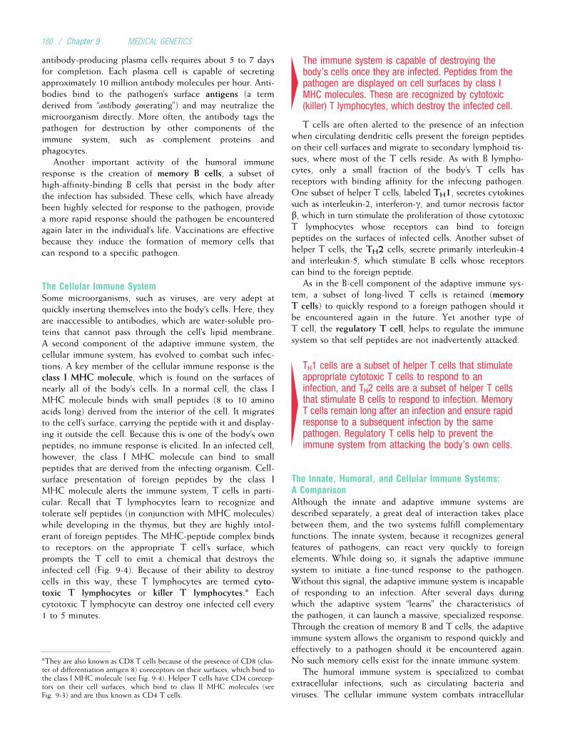
antibody-producing plasma cells requires about 5 to 7 daysfor completion. Each plasma cell is capable of secretingapproximately 10 million antibody molecules per hour. Anti-bodies bind to the pathogen’s surface antigens (a termderived from “antibody generating”) and may neutralize themicroorganism directly. More often, the antibody tags thepathogen for destruction by other components of theimmune system, such as complement proteins andphagocytes.
Another important activity of the humoral immuneresponse is the creation of memory B cells, a subset ofhigh-affinity-binding B cells that persist in the body afterthe infection has subsided. These cells, which have alreadybeen highly selected for response to the pathogen, providea more rapid response should the pathogen be encounteredagain later in the individual’s life. Vaccinations are effectivebecause they induce the formation of memory cells thatcan respond to a specific pathogen.
The Cellular Immune SystemSome microorganisms, such as viruses, are very adept atquickly inserting themselves into the body’s cells. Here, theyare inaccessible to antibodies, which are water-soluble pro-teins that cannot pass through the cell’s lipid membrane.A second component of the adaptive immune system, thecellular immune system, has evolved to combat such infec-tions. A key member of the cellular immune response is theclass I MHC molecule, which is found on the surfaces ofnearly all of the body’s cells. In a normal cell, the class IMHC molecule binds with small peptides (8 to 10 aminoacids long) derived from the interior of the cell. It migratesto the cell’s surface, carrying the peptide with it and display-ing it outside the cell. Because this is one of the body’s ownpeptides, no immune response is elicited. In an infected cell,however, the class I MHC molecule can bind to smallpeptides that are derived from the infecting organism. Cell-surface presentation of foreign peptides by the class IMHC molecule alerts the immune system, T cells in parti-cular. Recall that T lymphocytes learn to recognize andtolerate self peptides (in conjunction with MHC molecules)while developing in the thymus, but they are highly intol-erant of foreign peptides. The MHC-peptide complex bindsto receptors on the appropriate T cell’s surface, whichprompts the T cell to emit a chemical that destroys theinfected cell (Fig. 9-4). Because of their ability to destroycells in this way, these T lymphocytes are termed cyto-toxic T lymphocytes or killer T lymphocytes.* Eachcytotoxic T lymphocyte can destroy one infected cell every1 to 5 minutes.
4The immune system is capable of destroying thebody’s cells once they are infected. Peptides from thepathogen are displayed on cell surfaces by class IMHC molecules. These are recognized by cytotoxic(killer) T lymphocytes, which destroy the infected cell.
T cells are often alerted to the presence of an infectionwhen circulating dendritic cells present the foreign peptideson their cell surfaces and migrate to secondary lymphoid tis-sues, where most of the T cells reside. As with B lympho-cytes, only a small fraction of the body’s T cells hasreceptors with binding affinity for the infecting pathogen.One subset of helper T cells, labeled TH1, secretes cytokinessuch as interleukin-2, interferon-g, and tumor necrosis factorb, which in turn stimulate the proliferation of those cytotoxicT lymphocytes whose receptors can bind to foreignpeptides on the surfaces of infected cells. Another subset ofhelper T cells, the TH2 cells, secrete primarily interleukin-4and interleukin-5, which stimulate B cells whose receptorscan bind to the foreign peptide.
As in the B-cell component of the adaptive immune sys-tem, a subset of long-lived T cells is retained (memoryT cells) to quickly respond to a foreign pathogen should itbe encountered again in the future. Yet another type ofT cell, the regulatory T cell, helps to regulate the immunesystem so that self peptides are not inadvertently attacked.
4TH1 cells are a subset of helper T cells that stimulateappropriate cytotoxic T cells to respond to aninfection, and TH2 cells are a subset of helper T cellsthat stimulate B cells to respond to infection. MemoryT cells remain long after an infection and ensure rapidresponse to a subsequent infection by the samepathogen. Regulatory T cells help to prevent theimmune system from attacking the body’s own cells.
The Innate, Humoral, and Cellular Immune Systems:A ComparisonAlthough the innate and adaptive immune systems aredescribed separately, a great deal of interaction takes placebetween them, and the two systems fulfill complementaryfunctions. The innate system, because it recognizes generalfeatures of pathogens, can react very quickly to foreignelements. While doing so, it signals the adaptive immunesystem to initiate a fine-tuned response to the pathogen.Without this signal, the adaptive immune system is incapableof responding to an infection. After several days duringwhich the adaptive system “learns” the characteristics ofthe pathogen, it can launch a massive, specialized response.Through the creation of memory B and T cells, the adaptiveimmune system allows the organism to respond quickly andeffectively to a pathogen should it be encountered again.No such memory cells exist for the innate immune system.
The humoral immune system is specialized to combatextracellular infections, such as circulating bacteria andviruses. The cellular immune system combats intracellular
*They are also known as CD8 T cells because of the presence of CD8 (clus-ter of differentiation antigen 8) coreceptors on their surfaces, which bind tothe class I MHC molecule (see Fig. 9-4). Helper T cells have CD4 corecep-tors on their cell surfaces, which bind to class II MHC molecules (seeFig. 9-3) and are thus known as CD4 T cells.
180 / Chapter 9 MEDICAL GENETICS
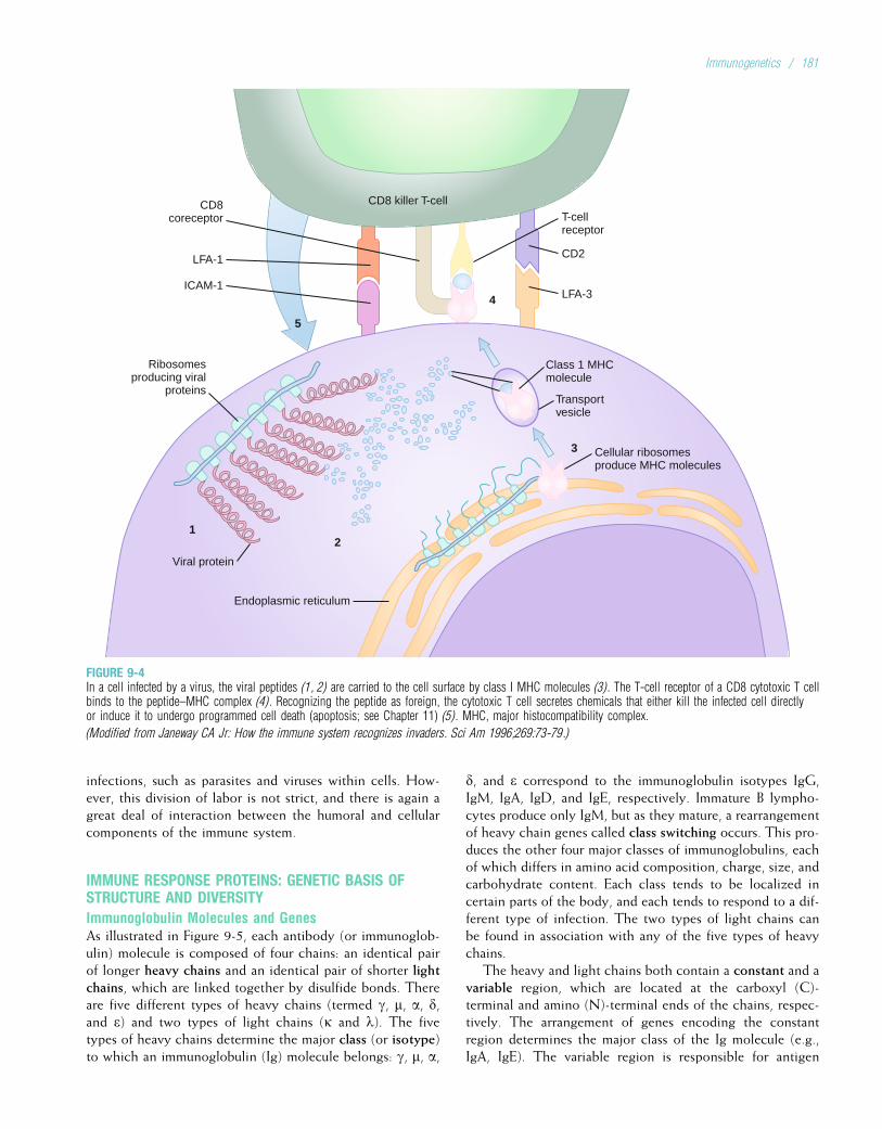
infections, such as parasites and viruses within cells. How-ever, this division of labor is not strict, and there is again agreat deal of interaction between the humoral and cellularcomponents of the immune system.
IMMUNE RESPONSE PROTEINS: GENETIC BASIS OFSTRUCTURE AND DIVERSITYImmunoglobulin Molecules and GenesAs illustrated in Figure 9-5, each antibody (or immunoglob-ulin) molecule is composed of four chains: an identical pairof longer heavy chains and an identical pair of shorter lightchains, which are linked together by disulfide bonds. Thereare five different types of heavy chains (termed g, m, a, d,and e) and two types of light chains (k and l). The fivetypes of heavy chains determine the major class (or isotype)to which an immunoglobulin (Ig) molecule belongs: g, m, a,
d, and e correspond to the immunoglobulin isotypes IgG,IgM, IgA, IgD, and IgE, respectively. Immature B lympho-cytes produce only IgM, but as they mature, a rearrangementof heavy chain genes called class switching occurs. This pro-duces the other four major classes of immunoglobulins, eachof which differs in amino acid composition, charge, size, andcarbohydrate content. Each class tends to be localized incertain parts of the body, and each tends to respond to a dif-ferent type of infection. The two types of light chains canbe found in association with any of the five types of heavychains.
The heavy and light chains both contain a constant and avariable region, which are located at the carboxyl (C)-terminal and amino (N)-terminal ends of the chains, respec-tively. The arrangement of genes encoding the constantregion determines the major class of the Ig molecule (e.g.,IgA, IgE). The variable region is responsible for antigen
CD8coreceptor
5
Class 1 MHCmolecule
Transportvesicle
Ribosomesproducing viral
proteins
1
Viral protein
2
3
4
Cellular ribosomesproduce MHC molecules
Endoplasmic reticulum
CD8 killer T-cellT-cellreceptor
CD2
LFA-3
LFA-1
ICAM-1
FIGURE 9-4In a cell infected by a virus, the viral peptides (1, 2) are carried to the cell surface by class I MHC molecules (3). The T-cell receptor of a CD8 cytotoxic T cellbinds to the peptide–MHC complex (4). Recognizing the peptide as foreign, the cytotoxic T cell secretes chemicals that either kill the infected cell directlyor induce it to undergo programmed cell death (apoptosis; see Chapter 11) (5). MHC, major histocompatibility complex.
(Modified from Janeway CA Jr: How the immune system recognizes invaders. Sci Am 1996;269:73-79.)
Immunogenetics / 181
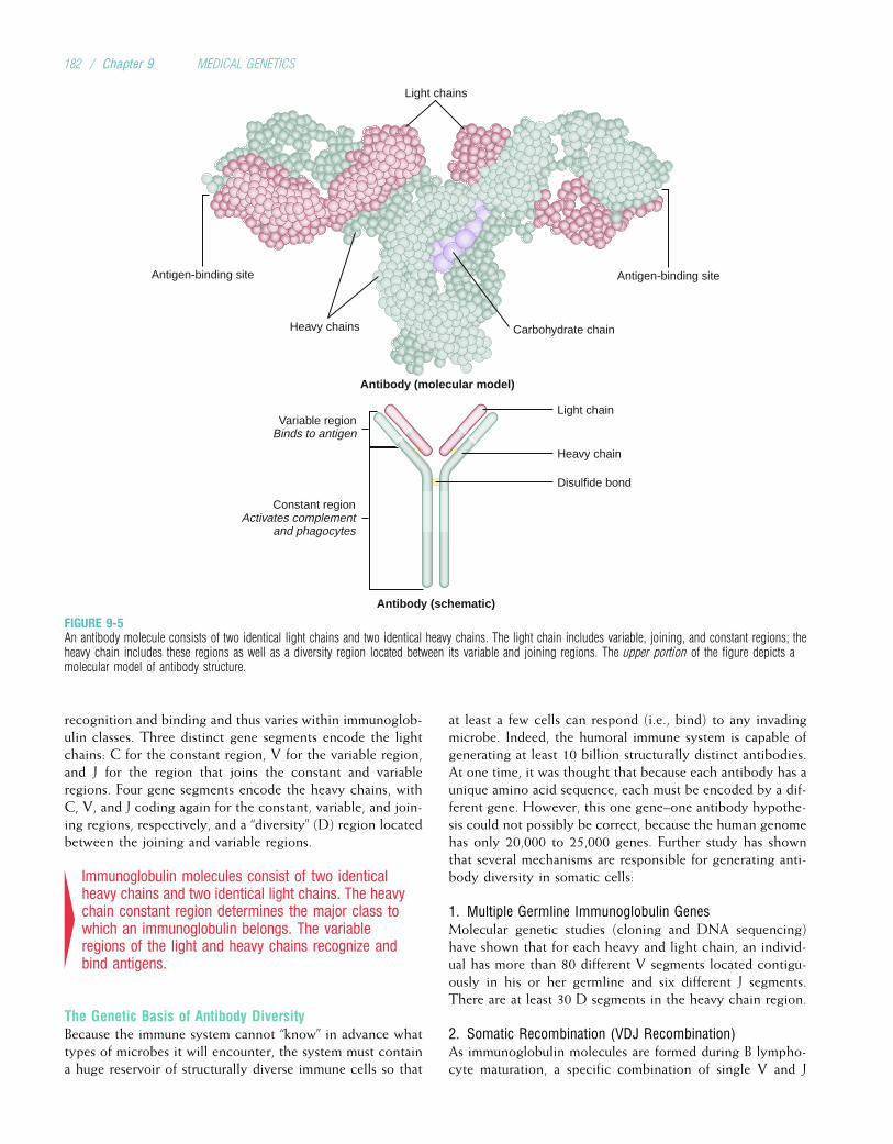
recognition and binding and thus varies within immunoglob-ulin classes. Three distinct gene segments encode the lightchains: C for the constant region, V for the variable region,and J for the region that joins the constant and variableregions. Four gene segments encode the heavy chains, withC, V, and J coding again for the constant, variable, and join-ing regions, respectively, and a “diversity” (D) region locatedbetween the joining and variable regions.
4Immunoglobulin molecules consist of two identicalheavy chains and two identical light chains. The heavychain constant region determines the major class towhich an immunoglobulin belongs. The variableregions of the light and heavy chains recognize andbind antigens.
The Genetic Basis of Antibody DiversityBecause the immune system cannot “know” in advance whattypes of microbes it will encounter, the system must containa huge reservoir of structurally diverse immune cells so that
at least a few cells can respond (i.e., bind) to any invadingmicrobe. Indeed, the humoral immune system is capable ofgenerating at least 10 billion structurally distinct antibodies.At one time, it was thought that because each antibody has aunique amino acid sequence, each must be encoded by a dif-ferent gene. However, this one gene–one antibody hypothe-sis could not possibly be correct, because the human genomehas only 20,000 to 25,000 genes. Further study has shownthat several mechanisms are responsible for generating anti-body diversity in somatic cells:
1. Multiple Germline Immunoglobulin GenesMolecular genetic studies (cloning and DNA sequencing)have shown that for each heavy and light chain, an individ-ual has more than 80 different V segments located contigu-ously in his or her germline and six different J segments.There are at least 30 D segments in the heavy chain region.
2. Somatic Recombination (VDJ Recombination)As immunoglobulin molecules are formed during B lympho-cyte maturation, a specific combination of single V and J
Light chains
Light chain
Antigen-binding siteAntigen-binding site
Carbohydrate chainHeavy chains
Heavy chain
Variable regionBinds to antigen
Constant regionActivates complement
and phagocytes
Disulfide bond
Antibody (molecular model)
Antibody (schematic)
FIGURE 9-5An antibody molecule consists of two identical light chains and two identical heavy chains. The light chain includes variable, joining, and constant regions; theheavy chain includes these regions as well as a diversity region located between its variable and joining regions. The upper portion of the figure depicts amolecular model of antibody structure.
182 / Chapter 9 MEDICAL GENETICS
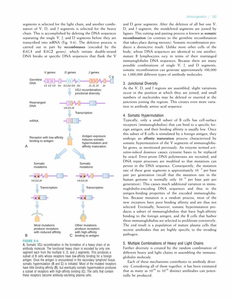
segments is selected for the light chain, and another combi-nation of V, D, and J segments is selected for the heavychain. This is accomplished by deleting the DNA sequencesseparating the single V, J, and D segments before they aretranscribed into mRNA (Fig. 9-6). The deletion process iscarried out in part by recombinases (encoded by theRAG1 and RAG2 genes), which initiate double-strandDNA breaks at specific DNA sequences that flank the V
and D gene segments. After the deletion of all but one V,D, and J segment, the nondeleted segments are joined byligases. This cutting-and-pasting process is known as somaticrecombination (in contrast to the germline recombinationthat takes place during meiosis). Somatic recombination pro-duces a distinctive result: Unlike most other cells of thebody, whose DNA sequences are identical to one another,mature B lymphocytes vary in terms of their rearrangedimmunoglobulin DNA sequences. Because there are manypossible combinations of single V, J, and D segments,somatic recombination can generate approximately 100,000to 1,000,000 different types of antibody molecules.
3. Junctional DiversityAs the V, D, and J regions are assembled, slight variationsoccur in the position at which they are joined, and smallnumbers of nucleotides may be deleted or inserted at thejunctions joining the regions. This creates even more varia-tion in antibody amino acid sequence.
4. Somatic HypermutationTypically, only a small subset of B cells has cell-surfacereceptors (immunoglobulins) that can bind to a specific for-eign antigen, and their binding affinity is usually low. Oncethis subset of B cells is stimulated by a foreign antigen, theyundergo an affinity maturation process characterized bysomatic hypermutation of the V segments of immunoglobu-lin genes, as mentioned previously. An enzyme termed acti-
vation-induced deaminase causes cytosine bases to be replacedby uracil. Error-prone DNA polymerases are recruited, andDNA repair processes are modified so that mutations canpersist in the DNA sequence. Consequently, the mutationrate of these gene segments is approximately 10�3 per basepair per generation (recall that the mutation rate in thehuman genome is normally only 10�9 per base pair pergeneration). This causes much additional variation in immu-noglobulin-encoding DNA sequences and thus in theantigen-binding properties of the encoded immunoglobu-lins. Because mutation is a random process, most of thenew receptors have poor binding affinity and are thus notselected. Eventually, however, somatic hypermutation pro-duces a subset of immunoglobulins that have high-affinitybinding to the foreign antigen, and the B cells that harborthese immunoglobulins are selected to proliferate extensively.The end result is a population of mature plasma cells thatsecrete antibodies that are highly specific to the invadingpathogen.
5. Multiple Combinations of Heavy and Light ChainsFurther diversity is created by the random combination ofdifferent heavy and light chains in assembling the immuno-globulin molecule.
Each of these mechanisms contributes to antibody diver-sity. Considering all of them together, it has been estimatedthat as many as 1010 to 1014 distinct antibodies can poten-tially be produced.
GermlineDNA
Transcription
Transcription
VDJ recombination;junctional diversity
V1 V2
V genes
Somaticmutations
V3 D1 D2
C g
D3
V4 D3 J4
V4 D3 J4
Dn J1 J2 J3 JnVn
RearrangedDNA
mRNA
Receptor with low-affinitybinding to antigen
A
B C
Most mutationsproduce receptorswith reduced affinity
Transcription
Somaticmutations
V4 D3 J4
Other mutationsproduce receptorswith high-affinitybinding to antigen
Antigen exposureinduces somatichypermutation andaffinity maturation
D genes J genes
* * * *
* * * *
FIGURE 9-6A, Somatic VDJ recombination in the formation of a heavy chain of anantibody molecule. The functional heavy chain is encoded by only onesegment each from the multiple V, D, and J segments. This produces asubset of B cells whose receptors have low-affinity binding for a foreignantigen. Once the antigen is encountered in the secondary lymphoid tissue,somatic hypermutation (B and C) is initiated. Most of the mutated receptorshave little binding affinity (B), but eventually somatic hypermutation producesa subset of receptors with high-affinity binding (C). The cells that containthese receptors become antibody-secreting plasma cells.
Immunogenetics / 183
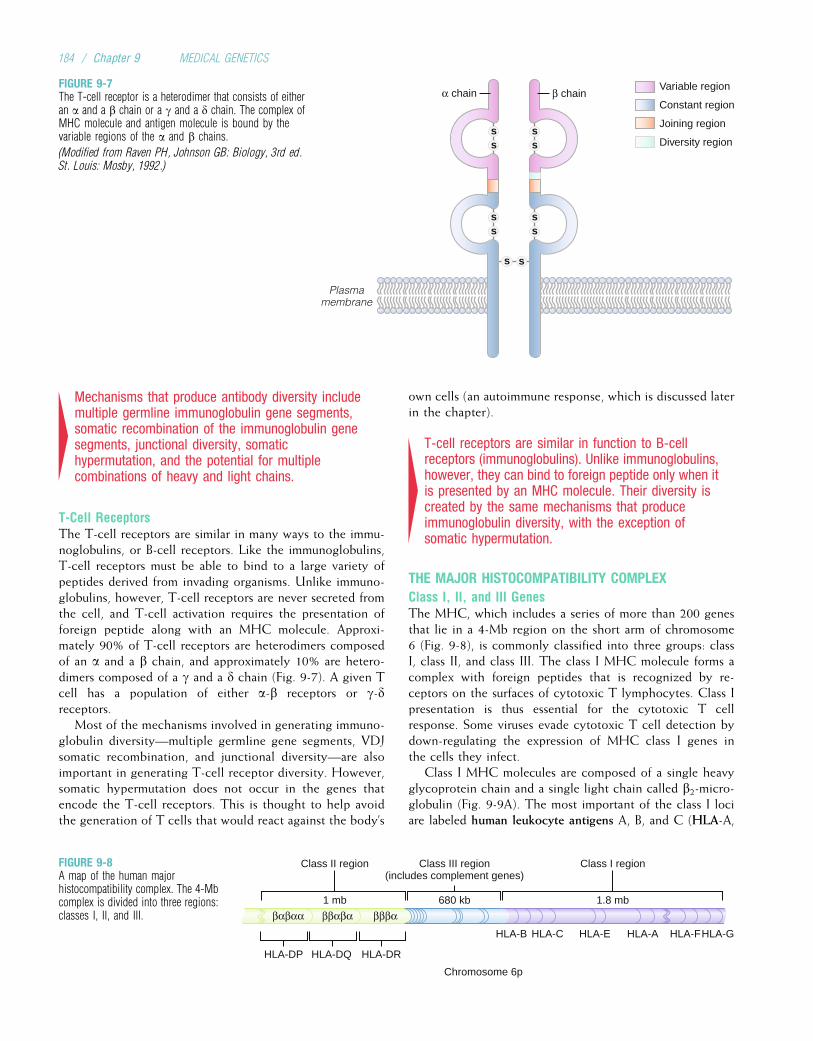
4Mechanisms that produce antibody diversity includemultiple germline immunoglobulin gene segments,somatic recombination of the immunoglobulin genesegments, junctional diversity, somatichypermutation, and the potential for multiplecombinations of heavy and light chains.
T-Cell ReceptorsThe T-cell receptors are similar in many ways to the immu-noglobulins, or B-cell receptors. Like the immunoglobulins,T-cell receptors must be able to bind to a large variety ofpeptides derived from invading organisms. Unlike immuno-globulins, however, T-cell receptors are never secreted fromthe cell, and T-cell activation requires the presentation offoreign peptide along with an MHC molecule. Approxi-mately 90% of T-cell receptors are heterodimers composedof an a and a b chain, and approximately 10% are hetero-dimers composed of a g and a d chain (Fig. 9-7). A given Tcell has a population of either a-b receptors or g-dreceptors.
Most of the mechanisms involved in generating immuno-globulin diversity—multiple germline gene segments, VDJsomatic recombination, and junctional diversity—are alsoimportant in generating T-cell receptor diversity. However,somatic hypermutation does not occur in the genes thatencode the T-cell receptors. This is thought to help avoidthe generation of T cells that would react against the body’s
own cells (an autoimmune response, which is discussed laterin the chapter).
4T-cell receptors are similar in function to B-cellreceptors (immunoglobulins). Unlike immunoglobulins,however, they can bind to foreign peptide only when itis presented by an MHC molecule. Their diversity iscreated by the same mechanisms that produceimmunoglobulin diversity, with the exception ofsomatic hypermutation.
THE MAJOR HISTOCOMPATIBILITY COMPLEXClass I, II, and III GenesThe MHC, which includes a series of more than 200 genesthat lie in a 4-Mb region on the short arm of chromosome6 (Fig. 9-8), is commonly classified into three groups: classI, class II, and class III. The class I MHC molecule forms acomplex with foreign peptides that is recognized by re-ceptors on the surfaces of cytotoxic T lymphocytes. Class Ipresentation is thus essential for the cytotoxic T cellresponse. Some viruses evade cytotoxic T cell detection bydown-regulating the expression of MHC class I genes inthe cells they infect.
Class I MHC molecules are composed of a single heavyglycoprotein chain and a single light chain called b2-micro-globulin (Fig. 9-9A). The most important of the class I lociare labeled human leukocyte antigens A, B, and C (HLA-A,
α chain β chain
Plasmamembrane
ss
ss
ss
s
s s
s
Variable region
Constant region
Joining region
Diversity region
FIGURE 9-7The T-cell receptor is a heterodimer that consists of eitheran a and a b chain or a g and a d chain. The complex ofMHC molecule and antigen molecule is bound by thevariable regions of the a and b chains.
(Modified from Raven PH, Johnson GB: Biology, 3rd ed.St. Louis: Mosby, 1992.)
Class I regionClass III region(includes complement genes)
Class II region
1.8 mb680 kb1 mb
HLA-DP HLA-DQ HLA-DR
HLA-B HLA-C HLA-E HLA-A HLA-FHLA-G
Chromosome 6p
ββαβαβαβαα βββα
FIGURE 9-8A map of the human majorhistocompatibility complex. The 4-Mbcomplex is divided into three regions:classes I, II, and III.
184 / Chapter 9 MEDICAL GENETICS
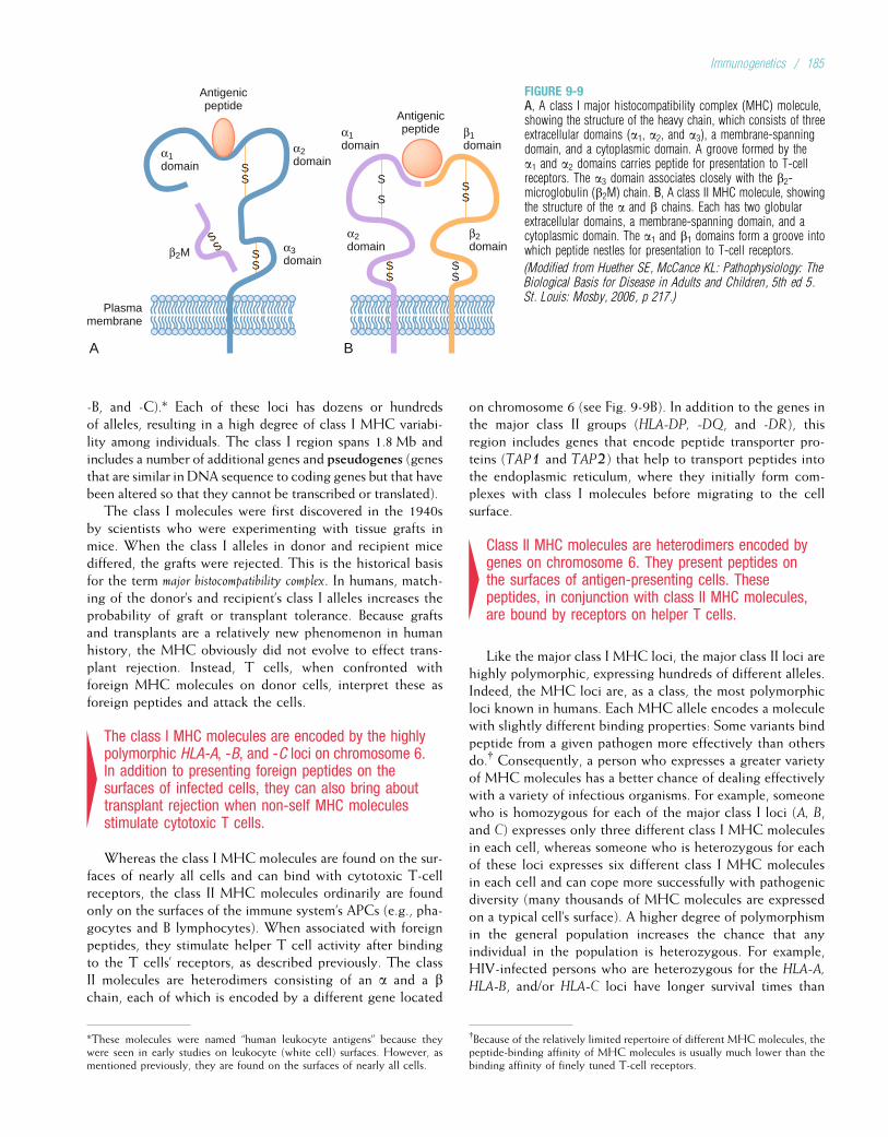
-B, and -C).* Each of these loci has dozens or hundredsof alleles, resulting in a high degree of class I MHC variabi-lity among individuals. The class I region spans 1.8 Mb andincludes a number of additional genes and pseudogenes (genesthat are similar in DNA sequence to coding genes but that havebeen altered so that they cannot be transcribed or translated).
The class I molecules were first discovered in the 1940sby scientists who were experimenting with tissue grafts inmice. When the class I alleles in donor and recipient micediffered, the grafts were rejected. This is the historical basisfor the term major histocompatibility complex. In humans, match-ing of the donor’s and recipient’s class I alleles increases theprobability of graft or transplant tolerance. Because graftsand transplants are a relatively new phenomenon in humanhistory, the MHC obviously did not evolve to effect trans-plant rejection. Instead, T cells, when confronted withforeign MHC molecules on donor cells, interpret these asforeign peptides and attack the cells.
4The class I MHC molecules are encoded by the highlypolymorphic HLA-A, -B, and -C loci on chromosome 6.In addition to presenting foreign peptides on thesurfaces of infected cells, they can also bring abouttransplant rejection when non-self MHC moleculesstimulate cytotoxic T cells.
Whereas the class I MHC molecules are found on the sur-faces of nearly all cells and can bind with cytotoxic T-cellreceptors, the class II MHC molecules ordinarily are foundonly on the surfaces of the immune system’s APCs (e.g., pha-gocytes and B lymphocytes). When associated with foreignpeptides, they stimulate helper T cell activity after bindingto the T cells’ receptors, as described previously. The classII molecules are heterodimers consisting of an a and a bchain, each of which is encoded by a different gene located
on chromosome 6 (see Fig. 9-9B). In addition to the genes inthe major class II groups (HLA-DP, -DQ, and -DR), thisregion includes genes that encode peptide transporter pro-teins (TAP1 and TAP2) that help to transport peptides intothe endoplasmic reticulum, where they initially form com-plexes with class I molecules before migrating to the cellsurface.
4Class II MHC molecules are heterodimers encoded bygenes on chromosome 6. They present peptides onthe surfaces of antigen-presenting cells. Thesepeptides, in conjunction with class II MHC molecules,are bound by receptors on helper T cells.
Like the major class I MHC loci, the major class II loci arehighly polymorphic, expressing hundreds of different alleles.Indeed, the MHC loci are, as a class, the most polymorphicloci known in humans. Each MHC allele encodes a moleculewith slightly different binding properties: Some variants bindpeptide from a given pathogen more effectively than othersdo.{ Consequently, a person who expresses a greater varietyof MHC molecules has a better chance of dealing effectivelywith a variety of infectious organisms. For example, someonewho is homozygous for each of the major class I loci (A, B,and C) expresses only three different class I MHC moleculesin each cell, whereas someone who is heterozygous for eachof these loci expresses six different class I MHC moleculesin each cell and can cope more successfully with pathogenicdiversity (many thousands of MHC molecules are expressedon a typical cell’s surface). A higher degree of polymorphismin the general population increases the chance that anyindividual in the population is heterozygous. For example,HIV-infected persons who are heterozygous for the HLA-A,
HLA-B, and/or HLA-C loci have longer survival times than
B
SS
SS
SS
S
S
Antigenicpeptideα1
domain
α2domain
β1domain
β2domainS
S
SS
SS
Antigenicpeptide
α1domain
β2M
α2domain
α3domain
A
Plasmamembrane
FIGURE 9-9A, A class I major histocompatibility complex (MHC) molecule,showing the structure of the heavy chain, which consists of threeextracellular domains (a1, a2, and a3), a membrane-spanningdomain, and a cytoplasmic domain. A groove formed by thea1 and a2 domains carries peptide for presentation to T-cellreceptors. The a3 domain associates closely with the b2-microglobulin (b2M) chain. B, A class II MHC molecule, showingthe structure of the a and b chains. Each has two globularextracellular domains, a membrane-spanning domain, and acytoplasmic domain. The a1 and b1 domains form a groove intowhich peptide nestles for presentation to T-cell receptors.
(Modified from Huether SE, McCance KL: Pathophysiology: TheBiological Basis for Disease in Adults and Children, 5th ed 5.St. Louis: Mosby, 2006, p 217.)
*These molecules were named “human leukocyte antigens” because theywere seen in early studies on leukocyte (white cell) surfaces. However, asmentioned previously, they are found on the surfaces of nearly all cells.
{Because of the relatively limited repertoire of different MHC molecules, thepeptide-binding affinity of MHC molecules is usually much lower than thebinding affinity of finely tuned T-cell receptors.
Immunogenetics / 185
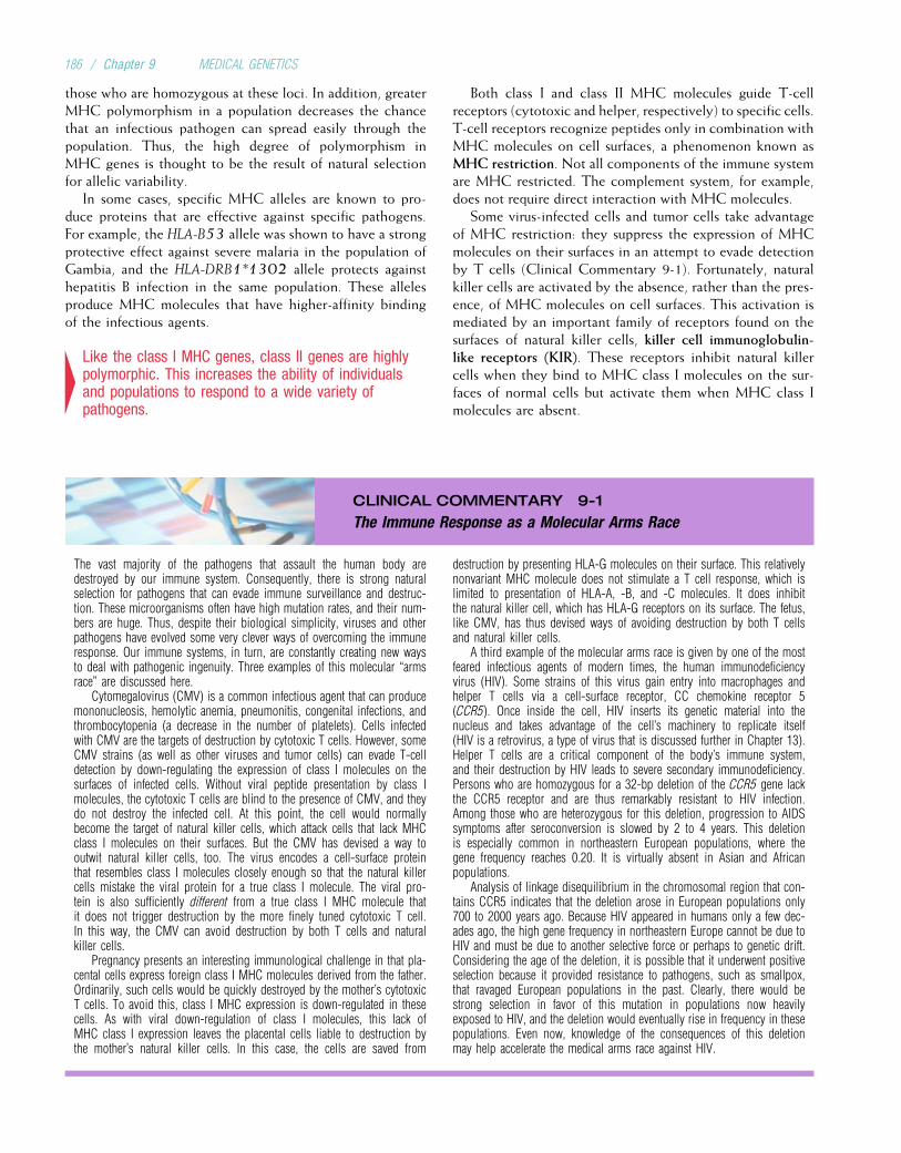
those who are homozygous at these loci. In addition, greaterMHC polymorphism in a population decreases the chancethat an infectious pathogen can spread easily through thepopulation. Thus, the high degree of polymorphism inMHC genes is thought to be the result of natural selectionfor allelic variability.
In some cases, specific MHC alleles are known to pro-duce proteins that are effective against specific pathogens.For example, the HLA-B53 allele was shown to have a strongprotective effect against severe malaria in the population ofGambia, and the HLA-DRB1*1302 allele protects againsthepatitis B infection in the same population. These allelesproduce MHC molecules that have higher-affinity bindingof the infectious agents.
4Like the class I MHC genes, class II genes are highlypolymorphic. This increases the ability of individualsand populations to respond to a wide variety ofpathogens.
Both class I and class II MHC molecules guide T-cellreceptors (cytotoxic and helper, respectively) to specific cells.T-cell receptors recognize peptides only in combination withMHC molecules on cell surfaces, a phenomenon known asMHC restriction. Not all components of the immune systemare MHC restricted. The complement system, for example,does not require direct interaction with MHC molecules.
Some virus-infected cells and tumor cells take advantageof MHC restriction: they suppress the expression of MHCmolecules on their surfaces in an attempt to evade detectionby T cells (Clinical Commentary 9-1). Fortunately, naturalkiller cells are activated by the absence, rather than the pres-ence, of MHC molecules on cell surfaces. This activation ismediated by an important family of receptors found on thesurfaces of natural killer cells, killer cell immunoglobulin-like receptors (KIR). These receptors inhibit natural killercells when they bind to MHC class I molecules on the sur-faces of normal cells but activate them when MHC class Imolecules are absent.
CLINICAL COMMENTARY 9-1
The Immune Response as a Molecular Arms Race
The vast majority of the pathogens that assault the human body aredestroyed by our immune system. Consequently, there is strong naturalselection for pathogens that can evade immune surveillance and destruc-tion. These microorganisms often have high mutation rates, and their num-bers are huge. Thus, despite their biological simplicity, viruses and otherpathogens have evolved some very clever ways of overcoming the immuneresponse. Our immune systems, in turn, are constantly creating new waysto deal with pathogenic ingenuity. Three examples of this molecular “armsrace” are discussed here.
Cytomegalovirus (CMV) is a common infectious agent that can producemononucleosis, hemolytic anemia, pneumonitis, congenital infections, andthrombocytopenia (a decrease in the number of platelets). Cells infectedwith CMV are the targets of destruction by cytotoxic T cells. However, someCMV strains (as well as other viruses and tumor cells) can evade T-celldetection by down-regulating the expression of class I molecules on thesurfaces of infected cells. Without viral peptide presentation by class Imolecules, the cytotoxic T cells are blind to the presence of CMV, and theydo not destroy the infected cell. At this point, the cell would normallybecome the target of natural killer cells, which attack cells that lack MHCclass I molecules on their surfaces. But the CMV has devised a way tooutwit natural killer cells, too. The virus encodes a cell-surface proteinthat resembles class I molecules closely enough so that the natural killercells mistake the viral protein for a true class I molecule. The viral pro-tein is also sufficiently different from a true class I MHC molecule thatit does not trigger destruction by the more finely tuned cytotoxic T cell.In this way, the CMV can avoid destruction by both T cells and naturalkiller cells.
Pregnancy presents an interesting immunological challenge in that pla-cental cells express foreign class I MHC molecules derived from the father.Ordinarily, such cells would be quickly destroyed by the mother’s cytotoxicT cells. To avoid this, class I MHC expression is down-regulated in thesecells. As with viral down-regulation of class I molecules, this lack ofMHC class I expression leaves the placental cells liable to destruction bythe mother’s natural killer cells. In this case, the cells are saved from
destruction by presenting HLA-G molecules on their surface. This relativelynonvariant MHC molecule does not stimulate a T cell response, which islimited to presentation of HLA-A, -B, and -C molecules. It does inhibitthe natural killer cell, which has HLA-G receptors on its surface. The fetus,like CMV, has thus devised ways of avoiding destruction by both T cellsand natural killer cells.
A third example of the molecular arms race is given by one of the mostfeared infectious agents of modern times, the human immunodeficiencyvirus (HIV). Some strains of this virus gain entry into macrophages andhelper T cells via a cell-surface receptor, CC chemokine receptor 5(CCR5). Once inside the cell, HIV inserts its genetic material into thenucleus and takes advantage of the cell’s machinery to replicate itself(HIV is a retrovirus, a type of virus that is discussed further in Chapter 13).Helper T cells are a critical component of the body’s immune system,and their destruction by HIV leads to severe secondary immunodeficiency.Persons who are homozygous for a 32-bp deletion of the CCR5 gene lackthe CCR5 receptor and are thus remarkably resistant to HIV infection.Among those who are heterozygous for this deletion, progression to AIDSsymptoms after seroconversion is slowed by 2 to 4 years. This deletionis especially common in northeastern European populations, where thegene frequency reaches 0.20. It is virtually absent in Asian and Africanpopulations.
Analysis of linkage disequilibrium in the chromosomal region that con-tains CCR5 indicates that the deletion arose in European populations only700 to 2000 years ago. Because HIV appeared in humans only a few dec-ades ago, the high gene frequency in northeastern Europe cannot be due toHIV and must be due to another selective force or perhaps to genetic drift.Considering the age of the deletion, it is possible that it underwent positiveselection because it provided resistance to pathogens, such as smallpox,that ravaged European populations in the past. Clearly, there would bestrong selection in favor of this mutation in populations now heavilyexposed to HIV, and the deletion would eventually rise in frequency in thesepopulations. Even now, knowledge of the consequences of this deletionmay help accelerate the medical arms race against HIV.
186 / Chapter 9 MEDICAL GENETICS
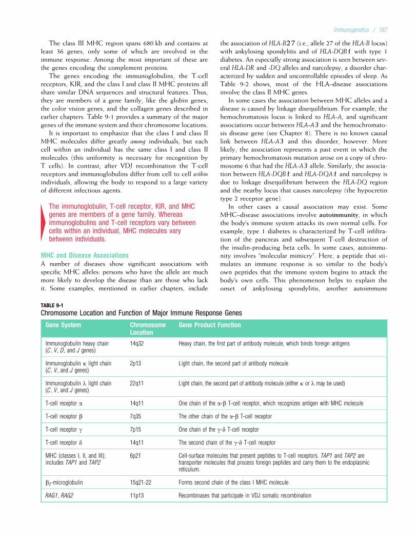
The class III MHC region spans 680 kb and contains atleast 36 genes, only some of which are involved in theimmune response. Among the most important of these arethe genes encoding the complement proteins.
The genes encoding the immunoglobulins, the T-cellreceptors, KIR, and the class I and class II MHC proteins allshare similar DNA sequences and structural features. Thus,they are members of a gene family, like the globin genes,the color vision genes, and the collagen genes described inearlier chapters. Table 9-1 provides a summary of the majorgenes of the immune system and their chromosome locations.
It is important to emphasize that the class I and class IIMHC molecules differ greatly among individuals, but eachcell within an individual has the same class I and class IImolecules (this uniformity is necessary for recognition byT cells). In contrast, after VDJ recombination the T-cellreceptors and immunoglobulins differ from cell to cell withinindividuals, allowing the body to respond to a large varietyof different infectious agents.
4The immunoglobulin, T-cell receptor, KIR, and MHCgenes are members of a gene family. Whereasimmunoglobulins and T-cell receptors vary betweencells within an individual, MHC molecules varybetween individuals.
MHC and Disease AssociationsA number of diseases show significant associations withspecific MHC alleles: persons who have the allele are muchmore likely to develop the disease than are those who lackit. Some examples, mentioned in earlier chapters, include
the association of HLA-B27 (i.e., allele 27 of the HLA-B locus)with ankylosing spondylitis and of HLA-DQB1 with type 1diabetes. An especially strong association is seen between sev-eral HLA-DR and -DQ alleles and narcolepsy, a disorder char-acterized by sudden and uncontrollable episodes of sleep. AsTable 9-2 shows, most of the HLA–disease associationsinvolve the class II MHC genes.
In some cases the association between MHC alleles and adisease is caused by linkage disequilibrium. For example, thehemochromatosis locus is linked to HLA-A, and significantassociations occur between HLA-A3 and the hemochromato-sis disease gene (see Chapter 8). There is no known causallink between HLA-A3 and this disorder, however. Morelikely, the association represents a past event in which theprimary hemochromatosis mutation arose on a copy of chro-mosome 6 that had the HLA-A3 allele. Similarly, the associa-tion between HLA-DQB1 and HLA-DQA1 and narcolepsy isdue to linkage disequilibrium between the HLA-DQ regionand the nearby locus that causes narcolepsy (the hypocretintype 2 receptor gene).
In other cases a causal association may exist. SomeMHC–disease associations involve autoimmunity, in whichthe body’s immune system attacks its own normal cells. Forexample, type 1 diabetes is characterized by T-cell infiltra-tion of the pancreas and subsequent T-cell destruction ofthe insulin-producing beta cells. In some cases, autoimmu-nity involves “molecular mimicry”. Here, a peptide that sti-mulates an immune response is so similar to the body’sown peptides that the immune system begins to attack thebody’s own cells. This phenomenon helps to explain theonset of ankylosing spondylitis, another autoimmune
TABLE 9-1
Chromosome Location and Function of Major Immune Response Genes
Gene System ChromosomeLocation
Gene Product Function
Immunoglobulin heavy chain(C, V, D, and J genes)
14q32 Heavy chain, the first part of antibody molecule, which binds foreign antigens
Immunoglobulin k light chain(C, V, and J genes)
2p13 Light chain, the second part of antibody molecule
Immunoglobulin l light chain(C, V, and J genes)
22q11 Light chain, the second part of antibody molecule (either k or l may be used)
T-cell receptor a 14q11 One chain of the a-b T-cell receptor, which recognizes antigen with MHC molecule
T-cell receptor b 7q35 The other chain of the a-b T-cell receptor
T-cell receptor g 7p15 One chain of the g-d T-cell receptor
T-cell receptor d 14q11 The second chain of the g-d T-cell receptor
MHC (classes I, II, and III);includes TAP1 and TAP2
6p21 Cell-surface molecules that present peptides to T-cell receptors. TAP1 and TAP2 aretransporter molecules that process foreign peptides and carry them to the endoplasmicreticulum.
b2-microglobulin 15q21-22 Forms second chain of the class I MHC molecule
RAG1, RAG2 11p13 Recombinases that participate in VDJ somatic recombination
Immunogenetics / 187
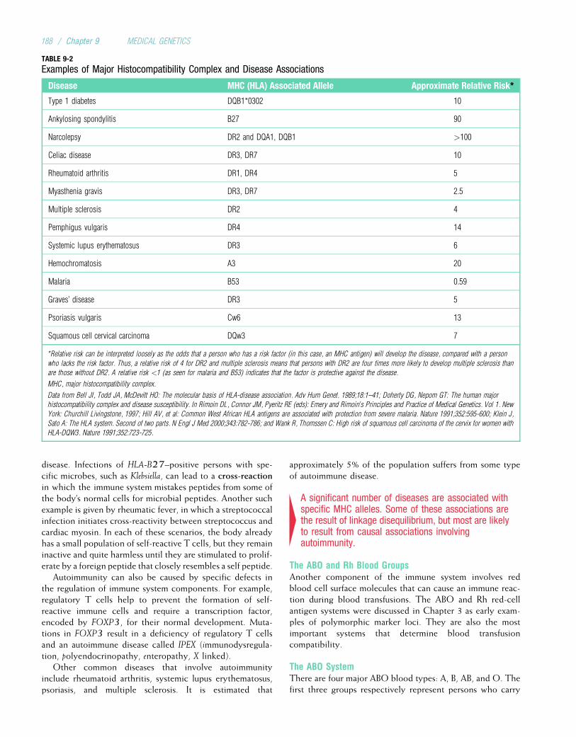
disease. Infections of HLA-B27–positive persons with spe-cific microbes, such as Klebsiella, can lead to a cross-reactionin which the immune system mistakes peptides from some ofthe body’s normal cells for microbial peptides. Another suchexample is given by rheumatic fever, in which a streptococcalinfection initiates cross-reactivity between streptococcus andcardiac myosin. In each of these scenarios, the body alreadyhas a small population of self-reactive T cells, but they remaininactive and quite harmless until they are stimulated to prolif-erate by a foreign peptide that closely resembles a self peptide.
Autoimmunity can also be caused by specific defects inthe regulation of immune system components. For example,regulatory T cells help to prevent the formation of self-reactive immune cells and require a transcription factor,encoded by FOXP3, for their normal development. Muta-tions in FOXP3 result in a deficiency of regulatory T cellsand an autoimmune disease called IPEX (immunodysregula-tion, polyendocrinopathy, enteropathy, X linked).
Other common diseases that involve autoimmunityinclude rheumatoid arthritis, systemic lupus erythematosus,psoriasis, and multiple sclerosis. It is estimated that
approximately 5% of the population suffers from some typeof autoimmune disease.
4A significant number of diseases are associated withspecific MHC alleles. Some of these associations arethe result of linkage disequilibrium, but most are likelyto result from causal associations involvingautoimmunity.
The ABO and Rh Blood GroupsAnother component of the immune system involves redblood cell surface molecules that can cause an immune reac-tion during blood transfusions. The ABO and Rh red-cellantigen systems were discussed in Chapter 3 as early exam-ples of polymorphic marker loci. They are also the mostimportant systems that determine blood transfusioncompatibility.
The ABO SystemThere are four major ABO blood types: A, B, AB, and O. Thefirst three groups respectively represent persons who carry
TABLE 9-2
Examples of Major Histocompatibility Complex and Disease Associations
Disease MHC (HLA) Associated Allele Approximate Relative Risk*
Type 1 diabetes DQB1*0302 10
Ankylosing spondylitis B27 90
Narcolepsy DR2 and DQA1, DQB1 >100
Celiac disease DR3, DR7 10
Rheumatoid arthritis DR1, DR4 5
Myasthenia gravis DR3, DR7 2.5
Multiple sclerosis DR2 4
Pemphigus vulgaris DR4 14
Systemic lupus erythematosus DR3 6
Hemochromatosis A3 20
Malaria B53 0.59
Graves’ disease DR3 5
Psoriasis vulgaris Cw6 13
Squamous cell cervical carcinoma DQw3 7
*Relative risk can be interpreted loosely as the odds that a person who has a risk factor (in this case, an MHC antigen) will develop the disease, compared with a person
who lacks the risk factor. Thus, a relative risk of 4 for DR2 and multiple sclerosis means that persons with DR2 are four times more likely to develop multiple sclerosis than
are those without DR2. A relative risk <1 (as seen for malaria and B53) indicates that the factor is protective against the disease.
MHC, major histocompatibility complex.
Data from Bell JI, Todd JA, McDevitt HO: The molecular basis of HLA-disease association. Adv Hum Genet. 1989;18:1–41; Doherty DG, Nepom GT: The human major
histocompatibility complex and disease susceptibility. In Rimoin DL, Connor JM, Pyeritz RE (eds): Emery and Rimoin’s Principles and Practice of Medical Genetics. Vol 1. New
York: Churchill Livingstone, 1997; Hill AV, et al: Common West African HLA antigens are associated with protection from severe malaria. Nature 1991;352:595-600; Klein J,
Sato A: The HLA system. Second of two parts. N Engl J Med 2000;343:782-786; and Wank R, Thomssen C: High risk of squamous cell carcinoma of the cervix for women with
HLA-DQW3. Nature 1991;352:723-725.
188 / Chapter 9 MEDICAL GENETICS
the A, B, or A and B antigens on their erythrocyte surfaces.Those with type O have neither the A nor the B antigen.Persons who have one of these antigens on their erythrocytesurfaces possess antibodies against all other ABO antigens intheir blood stream. (These antibodies are formed early in lifeas a result of exposure to antigens that are identical to the Aand B antigens but are present in various microorganisms.)Thus, if a type B person received type A or AB blood, hisor her anti-A antibodies would produce a severe and possiblyfatal reaction. Type O persons, who have neither the A northe B antigen and thus both anti-A and anti-B antibodies,would react strongly to blood of the other three types (A,B, and AB). It was once thought that type O persons,because they lack both types of antigens, could be “universaldonors” (anyone could accept their blood). Similarly, typeAB persons were termed “universal recipients” because theylacked both anti-A and anti-B antibodies. However, whenpatients are given transfusions of whole blood containinglarge volumes of serum, the donor’s antibodies can reactagainst the recipient’s erythrocyte antigens. Hence, com-plete ABO matching is nearly always done for bloodtransfusions.
4The ABO locus encodes red cell antigens that cancause a transfusion reaction if donors and recipientsare not properly matched.
The Rh SystemThe Rh blood group is encoded by two tightly linked loci,one of which is labeled D. The other locus produces Rh anti-gens labeled C and E through alternative splicing of themessenger RNA. The D locus is of primary interest becauseit is responsible for Rh maternal–fetal incompatibility andthe resulting disease, hemolytic disease of the newborn(HDN). Persons with the DD or Dd genotype have the Rhantigen on their erythrocytes and are Rh-positive. The reces-sive homozygotes, with genotype dd, are Rh-negative and donot have the Rh antigen. About 85% of North Americans areRh-positive and about 15% are Rh-negative.
Unlike the ABO system, in which antibodies normally areformed in response to antigens presented by other organ-isms, anti-Rh antibody production requires a stimulus bythe human Rh antigen itself. An Rh-negative person doesnot begin to produce anti-Rh antibodies unless he or she isexposed to the Rh antigen, usually through a blood transfu-sion or during pregnancy. Maternal–fetal incompatibilityresults when an Rh-positive man and an Rh-negative womanproduce children. If the man’s genotype is DD, all of theiroffspring will be Rh-positive and will have Rh antigenson their erythrocytes. If the man is a heterozygote, withgenotype Dd, half of their children will be Rh-positive, onaverage.
There are usually no difficulties with the first Rh-incom-patible child, because very few of the fetus’s red blood cellscross the placental barrier during gestation. When the pla-centa detaches at birth, a large number of fetal red bloodcells typically enter the mother’s blood stream. These cells,
carrying the Rh antigens, stimulate production of anti-Rhantibodies by the mother. These antibodies persist in theblood stream for a long time, and if the next offspring isagain Rh-positive, the mother’s anti-Rh antibodies enter thefetus’s bloodstream and destroy its red blood cells. As thisdestruction proceeds, the fetus becomes anemic and beginsto release many erythroblasts (immature nucleated red cells)into its blood stream. This phenomenon is responsible forthe descriptive term erythroblastosis fetalis. The anemia can leadto a spontaneous abortion or stillbirth. Because the maternalantibodies remain in the newborn’s circulatory system,destruction of red cells can continue in the neonate. Thiscauses a buildup of bilirubin and a jaundiced appearanceshortly after birth. Without replacement transfusions, inwhich the child receives Rh-negative red cells, the bilirubinis deposited in the brain, producing cerebral damage andusually death. Infants who do not die can develop mentalretardation, cerebral palsy, and/or high-frequency deafness.
Among North Americans of European descent, approxi-mately 13% of all matings are Rh-incompatible. Fortunately,a simple therapy now exists to avoid Rh sensitization of themother. During and after pregnancy, an Rh-negative motheris given injections of Rh immune globulin, which consists ofanti-Rh antibodies. These antibodies destroy the fetalerythrocytes in the mother’s blood stream before they stim-ulate production of maternal anti-Rh antibodies. Becausethe injected antibodies do not remain in the mother’s bloodstream for long, they do not affect subsequent offspring. Toavoid sensitization, these injections must be administeredwith each pregnancy. The Rh-negative mother must alsobe careful not to receive a transfusion containing Rh-positiveblood, because this would also stimulate production of anti-Rh antibodies.
4Maternal–fetal Rh incompatibility (Rh-negative motherand Rh-positive fetus) can produce hemolytic diseaseof the newborn if the mother’s Rh antibodies attackthe fetus. Administration of Rh immune globulin to themother prevents this reaction.
A rarer form of maternal–fetal incompatibility can resultwhen a mother with type O blood carries a fetus with typeA or B blood. The HDN produced by this combination isusually so mild that it does not require treatment. Interest-ingly, if the mother is also Rh-negative and the child isRh-positive, the ABO incompatibility protects against themore severe Rh incompatibility. This is because any fetalred blood cells entering the mother’s circulatory system arequickly destroyed by her anti-A or anti-B antibodies beforeshe can form anti-Rh antibodies.
IMMUNODEFICIENCY DISEASESImmunodeficiency disease results when one or more compo-nents of the immune system (e.g., T cells, B cells, MHC,complement proteins) are missing or fail to function nor-mally. Primary immunodeficiency diseases are caused by
Immunogenetics / 189
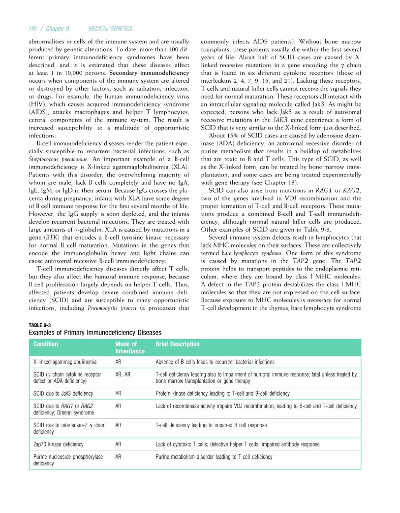
abnormalities in cells of the immune system and are usuallyproduced by genetic alterations. To date, more than 100 dif-ferent primary immunodeficiency syndromes have beendescribed, and it is estimated that these diseases affectat least 1 in 10,000 persons. Secondary immunodeficiencyoccurs when components of the immune system are alteredor destroyed by other factors, such as radiation, infection,or drugs. For example, the human immunodeficiency virus(HIV), which causes acquired immunodeficiency syndrome(AIDS), attacks macrophages and helper T lymphocytes,central components of the immune system. The result isincreased susceptibility to a multitude of opportunisticinfections.
B-cell immunodeficiency diseases render the patient espe-cially susceptible to recurrent bacterial infections, such asStreptococcus pneumoniae. An important example of a B-cellimmunodeficiency is X-linked agammaglobulinemia (XLA).Patients with this disorder, the overwhelming majority ofwhom are male, lack B cells completely and have no IgA,IgE, IgM, or IgD in their serum. Because IgG crosses the pla-centa during pregnancy, infants with XLA have some degreeof B cell immune response for the first several months of life.However, the IgG supply is soon depleted, and the infantsdevelop recurrent bacterial infections. They are treated withlarge amounts of g-globulin. XLA is caused by mutations in agene (BTK) that encodes a B-cell tyrosine kinase necessaryfor normal B cell maturation. Mutations in the genes thatencode the immunoglobulin heavy and light chains cancause autosomal recessive B-cell immunodeficiency.
T-cell immunodeficiency diseases directly affect T cells,but they also affect the humoral immune response, becauseB cell proliferation largely depends on helper T cells. Thus,affected patients develop severe combined immune defi-ciency (SCID) and are susceptible to many opportunisticinfections, including Pneumocystis jiroveci (a protozoan that
commonly infects AIDS patients). Without bone marrowtransplants, these patients usually die within the first severalyears of life. About half of SCID cases are caused by X-linked recessive mutations in a gene encoding the g chainthat is found in six different cytokine receptors (those ofinterleukins 2, 4, 7, 9, 15, and 21). Lacking these receptors,T cells and natural killer cells cannot receive the signals theyneed for normal maturation. These receptors all interact withan intracellular signaling molecule called Jak3. As might beexpected, persons who lack Jak3 as a result of autosomalrecessive mutations in the JAK3 gene experience a form ofSCID that is very similar to the X-linked form just described.
About 15% of SCID cases are caused by adenosine deam-inase (ADA) deficiency, an autosomal recessive disorder ofpurine metabolism that results in a buildup of metabolitesthat are toxic to B and T cells. This type of SCID, as wellas the X-linked form, can be treated by bone marrow trans-plantation, and some cases are being treated experimentallywith gene therapy (see Chapter 13).
SCID can also arise from mutations in RAG1 or RAG2,two of the genes involved in VDJ recombination and theproper formation of T-cell and B-cell receptors. These muta-tions produce a combined B-cell and T-cell immunodefi-ciency, although normal natural killer cells are produced.Other examples of SCID are given in Table 9-3.
Several immune system defects result in lymphocytes thatlack MHC molecules on their surfaces. These are collectivelytermed bare lymphocyte syndrome. One form of this syndromeis caused by mutations in the TAP2 gene. The TAP2protein helps to transport peptides to the endoplasmic reti-culum, where they are bound by class I MHC molecules.A defect in the TAP2 protein destabilizes the class I MHCmolecules so that they are not expressed on the cell surface.Because exposure to MHC molecules is necessary for normalT-cell development in the thymus, bare lymphocyte syndrome
TABLE 9-3
Examples of Primary Immunodeficiency Diseases
Condition Mode ofInheritance
Brief Description
X-linked agammaglobulinemia XR Absence of B cells leads to recurrent bacterial infections
SCID (g chain cytokine receptordefect or ADA deficiency)
XR, AR T-cell deficiency leading also to impairment of humoral immune response; fatal unless treated bybone marrow transplantation or gene therapy
SCID due to Jak3 deficiency AR Protein kinase deficiency leading to T-cell and B-cell deficiency
SCID due to RAG1 or RAG2deficiency; Omenn syndrome
AR Lack of recombinase activity impairs VDJ recombination, leading to B-cell and T-cell deficiency
SCID due to interleukin-7 a chaindeficiency
AR T-cell deficiency leading to impaired B cell response
Zap70 kinase deficiency AR Lack of cytotoxic T cells; defective helper T cells; impaired antibody response
Purine nucleoside phosphorylasedeficiency
AR Purine metabolism disorder leading to T-cell deficiency
190 / Chapter 9 MEDICAL GENETICS
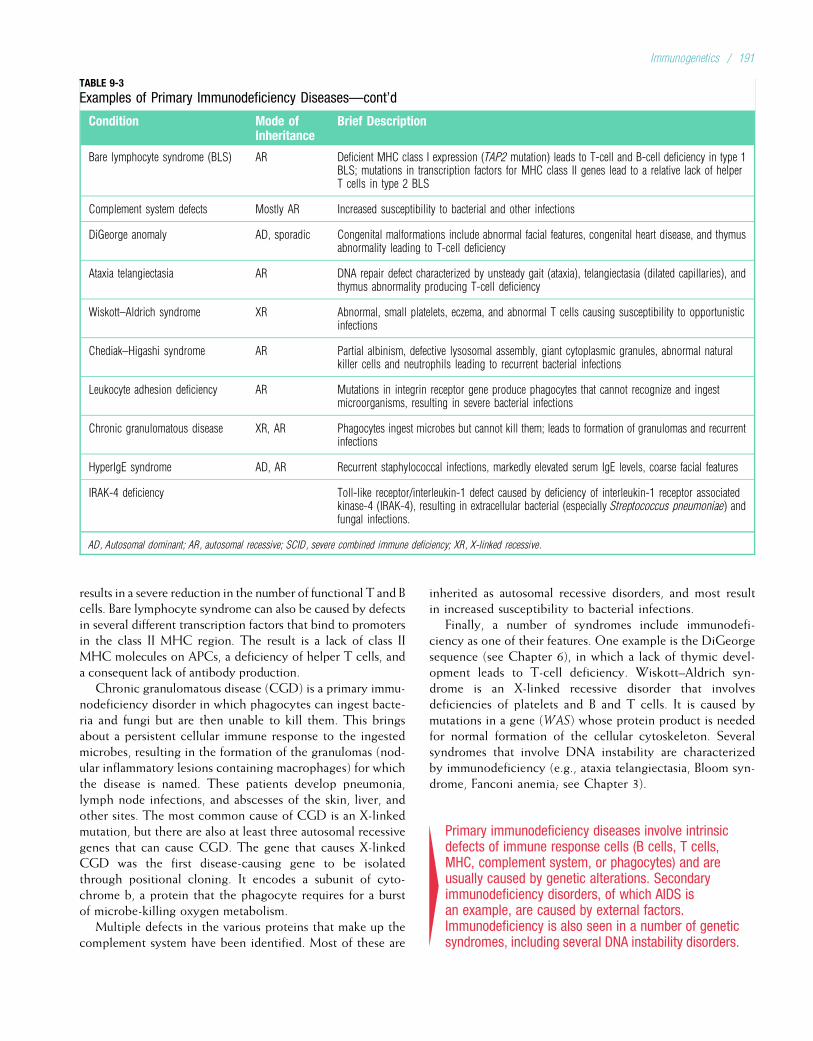
results in a severe reduction in the number of functional T and Bcells. Bare lymphocyte syndrome can also be caused by defectsin several different transcription factors that bind to promotersin the class II MHC region. The result is a lack of class IIMHC molecules on APCs, a deficiency of helper T cells, anda consequent lack of antibody production.
Chronic granulomatous disease (CGD) is a primary immu-nodeficiency disorder in which phagocytes can ingest bacte-ria and fungi but are then unable to kill them. This bringsabout a persistent cellular immune response to the ingestedmicrobes, resulting in the formation of the granulomas (nod-ular inflammatory lesions containing macrophages) for whichthe disease is named. These patients develop pneumonia,lymph node infections, and abscesses of the skin, liver, andother sites. The most common cause of CGD is an X-linkedmutation, but there are also at least three autosomal recessivegenes that can cause CGD. The gene that causes X-linkedCGD was the first disease-causing gene to be isolatedthrough positional cloning. It encodes a subunit of cyto-chrome b, a protein that the phagocyte requires for a burstof microbe-killing oxygen metabolism.
Multiple defects in the various proteins that make up thecomplement system have been identified. Most of these are
inherited as autosomal recessive disorders, and most resultin increased susceptibility to bacterial infections.
Finally, a number of syndromes include immunodefi-ciency as one of their features. One example is the DiGeorgesequence (see Chapter 6), in which a lack of thymic devel-opment leads to T-cell deficiency. Wiskott–Aldrich syn-drome is an X-linked recessive disorder that involvesdeficiencies of platelets and B and T cells. It is caused bymutations in a gene (WAS) whose protein product is neededfor normal formation of the cellular cytoskeleton. Severalsyndromes that involve DNA instability are characterizedby immunodeficiency (e.g., ataxia telangiectasia, Bloom syn-drome, Fanconi anemia; see Chapter 3).
4Primary immunodeficiency diseases involve intrinsicdefects of immune response cells (B cells, T cells,MHC, complement system, or phagocytes) and areusually caused by genetic alterations. Secondaryimmunodeficiency disorders, of which AIDS isan example, are caused by external factors.Immunodeficiency is also seen in a number of geneticsyndromes, including several DNA instability disorders.
TABLE 9-3
Examples of Primary Immunodeficiency Diseases—cont’d
Condition Mode ofInheritance
Brief Description
Bare lymphocyte syndrome (BLS) AR Deficient MHC class I expression (TAP2 mutation) leads to T-cell and B-cell deficiency in type 1BLS; mutations in transcription factors for MHC class II genes lead to a relative lack of helperT cells in type 2 BLS
Complement system defects Mostly AR Increased susceptibility to bacterial and other infections
DiGeorge anomaly AD, sporadic Congenital malformations include abnormal facial features, congenital heart disease, and thymusabnormality leading to T-cell deficiency
Ataxia telangiectasia AR DNA repair defect characterized by unsteady gait (ataxia), telangiectasia (dilated capillaries), andthymus abnormality producing T-cell deficiency
Wiskott–Aldrich syndrome XR Abnormal, small platelets, eczema, and abnormal T cells causing susceptibility to opportunisticinfections
Chediak–Higashi syndrome AR Partial albinism, defective lysosomal assembly, giant cytoplasmic granules, abnormal naturalkiller cells and neutrophils leading to recurrent bacterial infections
Leukocyte adhesion deficiency AR Mutations in integrin receptor gene produce phagocytes that cannot recognize and ingestmicroorganisms, resulting in severe bacterial infections
Chronic granulomatous disease XR, AR Phagocytes ingest microbes but cannot kill them; leads to formation of granulomas and recurrentinfections
HyperIgE syndrome AD, AR Recurrent staphylococcal infections, markedly elevated serum IgE levels, coarse facial features
IRAK-4 deficiency Toll-like receptor/interleukin-1 defect caused by deficiency of interleukin-1 receptor associatedkinase-4 (IRAK-4), resulting in extracellular bacterial (especially Streptococcus pneumoniae) andfungal infections.
AD, Autosomal dominant; AR, autosomal recessive; SCID, severe combined immune deficiency; XR, X-linked recessive.
Immunogenetics / 191
Study Questions
1. Compare the functions of class I and class II MHCmolecules.
2. MHC molecules and immunoglobulins both display agreat deal of diversity but in different ways. How andwhy do these types of diversity differ?
3. In what ways are T-cell receptors andimmunoglobulins similar? In what ways are theydifferent?
4. If there are 80 V segments, 6 J segments, and 30 Dsegments that can encode an immunoglobulin heavychain of one particular class, how many different
immunoglobulins can be formed on the basis ofsomatic recombination alone?
5. When matching donors and recipients for organtransplants, siblings are often desirable donors becausethey are more likely to be HLA-compatible with therecipient. If we assume no crossing over within the HLAloci and assume four distinct HLA haplotypes among theparents, what is the probability that a transplantrecipient will be HLA-identical with a sibling donor?
6. What types of matings will produce Rh maternal–fetalincompatibility?
Suggested ReadingsCharron D. Immunogenetics today: HLA, MHC and much
more. Curr Opin Immunol 2005;17(5): 493–497.Cunningham-Rundles C, Ponda PP. Molecular defects in T-
and B-cell primary immunodeficiency diseases. Nat RevImmunol 2005;5(11):880–892.
Horton R, Wilming L, Rand V, et al. Gene map of theextended human MHC. Nature Rev Genet 2004; 5(12):889–899.
Krogsgaard M, Davis MM. How T cells “see” antigen. NatImmunol 2005;6(3):239–245.
Nossal GJ. The double helix and immunology. Nature2003;421(6921):440–444.
O’Brien SJ, Nelson GW. Human genes that limit AIDS. NatGenet 2004;36(6):565–574.
O’Neill LA. Immunity’s early-warning system. Sci Am2005;292(1):24–31.
Peled JU, Kuang FL, Iglesias-Ussel MD, et al. The biochem-istry of somatic hypermutation. Annu Rev Immunol 2008;26:481–511.
Rioux JD, Abbas AK. Paths to understanding thegenetic basis of autoimmune disease. Nature 2005;435(7042):584–589.
Roitt I, Brostoff J, Male DRoth DA. Immunology. 7th ed.St. Louis: Mosby; 2006.
Schwartz RS. Diversity of the immune repertoire and immuno-regulation. N Engl J Med 2003;348(11):1017–1026.
Trowsdale J. HLA genomics in the third millennium. CurrOpin Immunol 2005;17(5):498–504.
Internet ResourcesImmunogenetics Database http://www.ebi.ac.uk/imgt/
Molecular Immunology Tutorial http://www.mi.interhealth.info/
192 / Chapter 9 MEDICAL GENETICS
Chapter 10
DEVELOPMENTAL GENETICS
Developmental genetics is the study of how the instructionsencoded in genes control and coordinate development of anorganism, beginning with fertilization and ending with death.The field is important to health care providers becausemutationsin genes can perturb developmental processes, resulting inincreased risk for birth defects, mental retardation, and cancer.In the United States, 2% to 3% of all live-born children have amajor birth defect (i.e., one that has a substantial impact onhealth) so about 100,000 children are born each year with a birthdefect. Birth defects are the leading cause of infant* death in theUnited States and are associated with substantial morbidity.
Birth defects can be isolated abnormalities, or they can befeatures of one of several thousand known genetic syndromes.The etiology of most birth defects is unknown; however, a sub-stantial fraction is caused by mutations in genes, either alone orin combination, that control normal development. The charac-terization of genes and developmental processes that coordinateanimal development has revolutionized our understandingof the molecular basis of human birth defects. This chapterprovides a brief review of the genes and proteins that controldevelopment and then discusses some of the cardinal develop-mental processes that, when disturbed, cause birth defects.
DEVELOPMENTBasic ConceptsAnimal development can be defined as the process by which afertilized ovum becomes a mature organism capable of repro-duction. A single fertilized egg divides and grows to form dif-ferent cell types, tissues, and organs, all of which arearranged in a species-specific body plan (i.e., the arrangementand pattern of body parts). Many of the instructions necessaryfor normal development are encoded by an animal’s genes.Because the genes in each cell of an organism are identical, sev-eral questions arise: How do cells with identical genetic consti-tutions form a complex adult organism composed of manydifferent cells and tissues? What controls the fate of each cell,instructing a cell to become, for example, a brain cell or a livercell? How do cells organize into discrete tissues? How is thebody plan of an organism determined? Answering such funda-mental questions has been a major focus of developmental
biology for more than a century. The pace of discovery hasaccelerated dramatically in the past several decades, and thesediscoveries are helping us to understand the causes of humanmalformations and genetic syndromes.
For ethical and technical reasons, it is difficult to study earlydevelopmental events in human embryos. Consequently, avariety of nonhuman model organisms are used to facilitatethe study of development (Table 10-1). This approach is feasi-ble because the major elements (genes and pathways) that con-trol animal development are conserved across a wide range ofspecies and body plans. In addition, many regulatory switchesand signaling pathways are used repeatedly during develop-ment to control various patterning and differentiation events.This underscores the point that the evolution of species pro-ceeds, in part, by continual tinkering with similar developmen-tal programs to effect changes in an organism’s phenotype.
For example, ectopic expression (i.e., expression of thegene product in an abnormal location) of the Drosophila geneeyeless results in the formation of a well-formed but misplacedeye (Fig. 10-1). Mice have a homologous gene, Pax6,{ inwhich mutations can produce abnormally small eyes. Wheninserted ectopically into Drosophila, Pax6 again produces amisplaced fly eye. Mutations in the human homolog,PAX6, cause defects of the eye such as cataracts and aniridia(absence of the iris). PAX6, Pax6, and eyeless are homologousgenes that encode DNA transcription factors (see Chapter 3).Although the ancestors of Drosophila and mouse diverged fromthe lineage leading to humans 500 million and 60 millionyears ago, respectively, the genes and pathways involved indevelopment of the eye have been conserved.
4Approximately 2% to 3% of babies are born with arecognizable birth defect. Many birth defects arecaused by mutations in genes encoding elements inpathways that control development. These pathwaysare highly conserved among animal species. Thus,studies of nonhuman animal models are invaluable forunderstanding human development and the causes ofbirth defects.
*An infant is a person who is younger than 1 year.
{It is conventional to capitalize all letters of the names of human genes butonly the first letter of the names of mouse genes, except for recessive muta-tions, which begin with a lower-case letter.
193
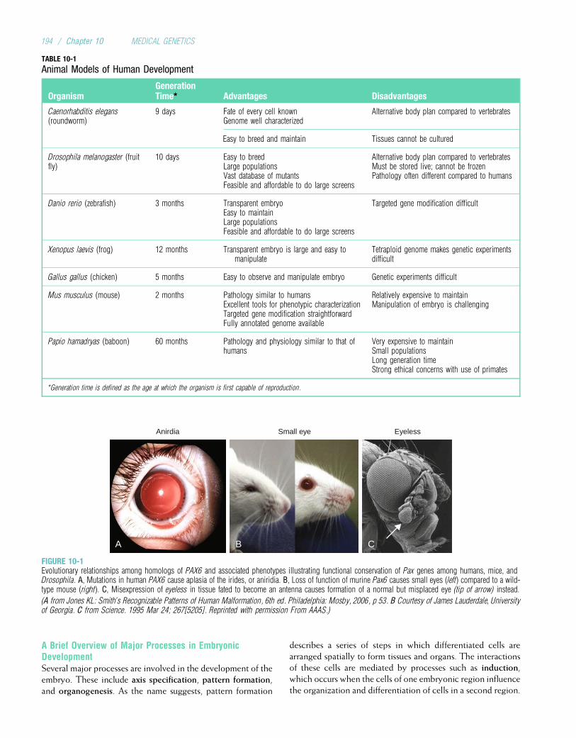
A Brief Overview of Major Processes in EmbryonicDevelopmentSeveral major processes are involved in the development of theembryo. These include axis specification, pattern formation,and organogenesis. As the name suggests, pattern formation
describes a series of steps in which differentiated cells arearranged spatially to form tissues and organs. The interactionsof these cells are mediated by processes such as induction,which occurs when the cells of one embryonic region influencethe organization and differentiation of cells in a second region.
A B C
Anirdia Small eye Eyeless
FIGURE 10-1Evolutionary relationships among homologs of PAX6 and associated phenotypes illustrating functional conservation of Pax genes among humans, mice, andDrosophila. A, Mutations in human PAX6 cause aplasia of the irides, or aniridia. B, Loss of function of murine Pax6 causes small eyes (left) compared to a wild-type mouse (right). C, Misexpression of eyeless in tissue fated to become an antenna causes formation of a normal but misplaced eye (tip of arrow) instead.
(A from Jones KL: Smith’s Recognizable Patterns of Human Malformation, 6th ed. Philadelphia: Mosby, 2006, p 53. B Courtesy of James Lauderdale, Universityof Georgia. C from Science. 1995 Mar 24; 267[5205]. Reprinted with permission From AAAS.)
TABLE 10-1
Animal Models of Human Development
OrganismGenerationTime* Advantages Disadvantages
Caenorhabditis elegans(roundworm)
9 days Fate of every cell knownGenome well characterized
Alternative body plan compared to vertebrates
Easy to breed and maintain Tissues cannot be cultured
Drosophila melanogaster (fruitfly)
10 days Easy to breedLarge populationsVast database of mutantsFeasible and affordable to do large screens
Alternative body plan compared to vertebratesMust be stored live; cannot be frozenPathology often different compared to humans
Danio rerio (zebrafish) 3 months Transparent embryoEasy to maintainLarge populationsFeasible and affordable to do large screens
Targeted gene modification difficult
Xenopus laevis (frog) 12 months Transparent embryo is large and easy tomanipulate
Tetraploid genome makes genetic experimentsdifficult
Gallus gallus (chicken) 5 months Easy to observe and manipulate embryo Genetic experiments difficult
Mus musculus (mouse) 2 months Pathology similar to humansExcellent tools for phenotypic characterizationTargeted gene modification straightforwardFully annotated genome available
Relatively expensive to maintainManipulation of embryo is challenging
Papio hamadryas (baboon) 60 months Pathology and physiology similar to that ofhumans
Very expensive to maintainSmall populationsLong generation timeStrong ethical concerns with use of primates
*Generation time is defined as the age at which the organism is first capable of reproduction.
194 / Chapter 10 MEDICAL GENETICS
Axis specification involves the definition of the major axes ofthe body: ventral/dorsal, anterior/posterior, medial/lateral,and left/right. Specification of polarity (direction) is an impor-tant part of this process. As the axes are specified, the forma-tion of organs and limbs (organogenesis) begins. Each ofthese major processes involves many different proteins thatform structures and provide signals to coordinate the develop-ment of the embryo. The major types of these proteins, and thegenes that encode them, are described next.
4Embryonic development involves the processes ofpattern formation, axis specification, and organogenesis.Each of these processes is controlled by a series ofproteins that provide signals and form structuresnecessary for normal development of the embryo.
GENETIC MEDIATORS OF DEVELOPMENT:THE MOLECULAR TOOLBOXThe genes required for normal development encode manydifferent products, including signaling molecules and theirreceptors, DNA transcription factors, components of theextracellular matrix, enzymes, transport systems, and otherproteins. Each of these genetic mediators is expressed incombinations of spatially and temporally overlappingpatterns that control different developmental processes. Asdetailed in this chapter, mutations in the genes mediatingdevelopment are a common cause of human birth defects.
Paracrine Signaling MoleculesInteractions between neighboring cells are usually mediatedby proteins that can diffuse across small distances to inducea response. These molecules are often called paracrine fac-tors because they are secreted into the space surrounding acell (unlike hormones, which are secreted into the bloodstream). Closely related paracrine factors have been isolatedfrom a variety of organisms, making it clear that homologousmolecules are used throughout the animal kingdom. To date,four major families of paracrine signaling molecules havebeen described: the Fibroblast Growth Factor (FGF) family,the Hedgehog family, the Wingless (Wnt) family, and theTransforming Growth Factor b (TGF-b) family. Each of
these signaling molecules binds to one or more receptors toeffect a response, and mutations in genes encoding thesemolecules may lead to abnormal communication betweencells. Clinical Commentary 10-1 discusses the FGF familyand associated receptors; the other three families aredescribed in this section.
The first member of the Hedgehog family was originallyisolated in a Drosophila mutant that has bristles in an area thatis naked in the normal fly (hence the name “hedgehog”).Vertebrates have several homologs of hedgehog, the mostwidely used of which is termed Sonic hedgehog (Shh). Amongits many roles, Shh participates in axis specification, induc-tion of motor neurons within the neural plate, and patterningof the limbs. The primary receptor of Shh is a transmembraneprotein encoded by a gene called patched. The normal actionof patched is to inhibit the function of another transmembraneprotein called smoothened, encoded by the gene Smo. Bindingof Shh to the patched receptor results in disinhibition ofsmoothened and activation of an intracellular signaling cascadethat targets members of the GLI family of transcriptionfactors (Fig. 10-2).
Mutations in the human homolog, PATCHED (PTC),cause Gorlin syndrome (see Fig. 10-2), a disorder character-ized by rib anomalies, cysts of the jaw, and basal cell carci-nomas (a form of skin cancer). Somatic mutations in PTC
have also been found in sporadic basal cell carcinomas. Thus,germline mutations in PTC alter the regulation of developingcells to cause birth defects, and somatic mutations in PTC
can alter the regulation of terminally differentiated cells tocause cancer.
The Wnt family of genes is named after the Drosophila
gene wingless and one of its vertebrate homologs, integrated.The wingless gene establishes polarity during Drosophila limbformation, and members of the Wnt family play similar rolesin vertebrates. Wnt genes encode secreted glycoproteins thatbind to members of the frizzled and low-density lipoprotein(LDL) receptor–related protein families. In humans, 19 differ-ent Wnt genes have been identified, and they participate in awide variety of developmental processes, including specifica-tion of the dorsal/ventral axis and formation of the brain, mus-cle, gonads, and kidneys. Homozygosity for mutations inWNT3 causes tetra-amelia (absence of all four limbs) in
CLINICAL COMMENTARY 10-1
Disorders of Fibroblast Growth Factor Receptors
Fibroblast growth factor receptors (FGFRs) are highly homologous glyco-proteins, with a common structure that consists of a signal peptide (anamino acid sequence that helps to direct the protein to its proper cellularlocation), three immunoglobulin-like (Ig-like) domains, a membrane-spanning segment, and an intracellular tyrosine kinase domain. FGFRsare receptors for at least 22 fibroblast growth factors (FGFs) that participatein a wide variety of biological processes, including cell migration, growth,and differentiation. With varying affinities, FGFs bind FGFRs, leading tophosphorylation, and hence activation, of the tyrosine kinase domain.
FGFRs are widely expressed in developing bone, and many commonhuman autosomal dominant disorders of generalized bone growth (i.e.,skeletal dysplasias) are caused by mutations in FGFR genes. The mostprevalent of these disorders, affecting more than 250,000 people worldwide,is achondroplasia (ACH), which is characterized by disproportionate shortstature (i.e., the limbs are disproportionately shorter than the trunk) andmacrocephaly (see Chapter 4). Nearly all persons with ACH have a glycine! arginine substitution in the transmembrane domain of FGFR3, resultingin constitutive FGFR3 activation.
Continued
Developmental Genetics / 195
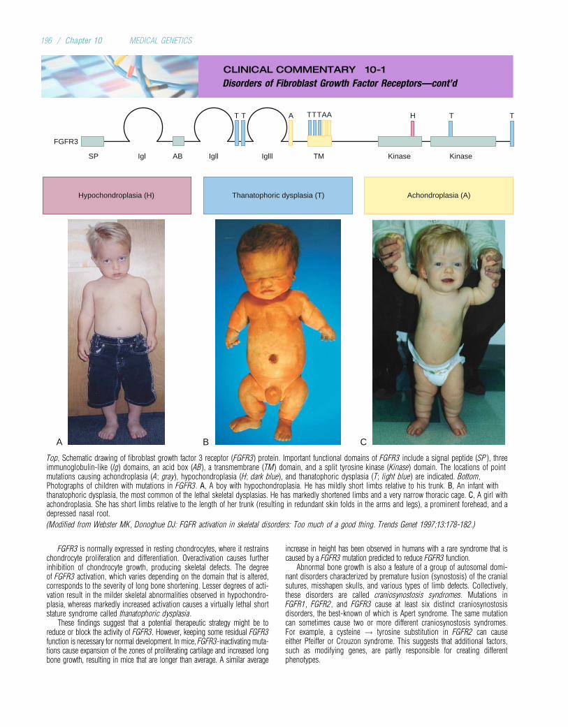
CLINICAL COMMENTARY 10-1
Disorders of Fibroblast Growth Factor Receptors—cont’d
A B C
Achondroplasia (A)Hypochondroplasia (H) Thanatophoric dysplasia (T)
SP Igl AB Igll TMIglll Kinase Kinase
FGFR3
H T TAAATT TTT
Top, Schematic drawing of fibroblast growth factor 3 receptor (FGFR3) protein. Important functional domains of FGFR3 include a signal peptide (SP ), threeimmunoglobulin-like (Ig) domains, an acid box (AB), a transmembrane (TM) domain, and a split tyrosine kinase (Kinase) domain. The locations of pointmutations causing achondroplasia (A; gray), hypochondroplasia (H; dark blue), and thanatophoric dysplasia (T; light blue) are indicated. Bottom,Photographs of children with mutations in FGFR3. A, A boy with hypochondroplasia. He has mildly short limbs relative to his trunk. B, An infant withthanatophoric dysplasia, the most common of the lethal skeletal dysplasias. He has markedly shortened limbs and a very narrow thoracic cage. C, A girl withachondroplasia. She has short limbs relative to the length of her trunk (resulting in redundant skin folds in the arms and legs), a prominent forehead, and adepressed nasal root.
(Modified from Webster MK, Donoghue DJ: FGFR activation in skeletal disorders: Too much of a good thing. Trends Genet 1997;13:178-182.)
FGFR3 is normally expressed in resting chondrocytes, where it restrainschondrocyte proliferation and differentiation. Overactivation causes furtherinhibition of chondrocyte growth, producing skeletal defects. The degreeof FGFR3 activation, which varies depending on the domain that is altered,corresponds to the severity of long bone shortening. Lesser degrees of acti-vation result in the milder skeletal abnormalities observed in hypochondro-plasia, whereas markedly increased activation causes a virtually lethal shortstature syndrome called thanatophoric dysplasia.
These findings suggest that a potential therapeutic strategy might be toreduce or block the activity of FGFR3. However, keeping some residual FGFR3function is necessary for normal development. In mice, FGFR3-inactivating muta-tions cause expansion of the zones of proliferating cartilage and increased longbone growth, resulting in mice that are longer than average. A similar average
increase in height has been observed in humans with a rare syndrome that iscaused by a FGFR3 mutation predicted to reduce FGFR3 function.
Abnormal bone growth is also a feature of a group of autosomal domi-nant disorders characterized by premature fusion (synostosis) of the cranialsutures, misshapen skulls, and various types of limb defects. Collectively,these disorders are called craniosynostosis syndromes. Mutations inFGFR1, FGFR2, and FGFR3 cause at least six distinct craniosynostosisdisorders, the best-known of which is Apert syndrome. The same mutationcan sometimes cause two or more different craniosynostosis syndromes.For example, a cysteine ! tyrosine substitution in FGFR2 can causeeither Pfeiffer or Crouzon syndrome. This suggests that additional factors,such as modifying genes, are partly responsible for creating differentphenotypes.
196 / Chapter 10 MEDICAL GENETICS
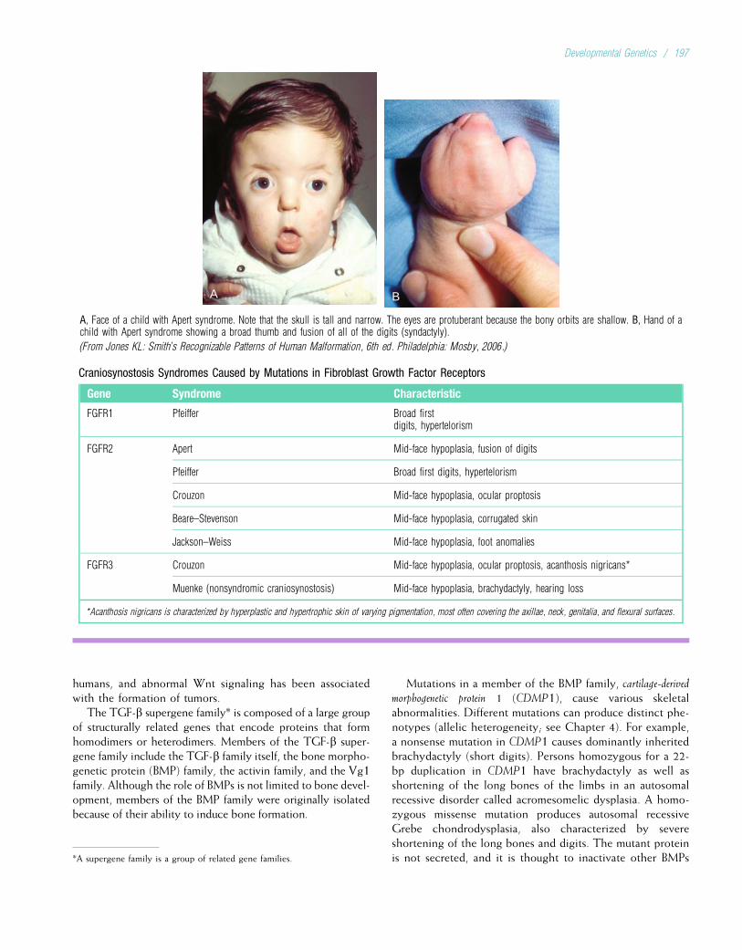
A B
A, Face of a child with Apert syndrome. Note that the skull is tall and narrow. The eyes are protuberant because the bony orbits are shallow. B, Hand of achild with Apert syndrome showing a broad thumb and fusion of all of the digits (syndactyly).
(From Jones KL: Smith’s Recognizable Patterns of Human Malformation, 6th ed. Philadelphia: Mosby, 2006.)
Craniosynostosis Syndromes Caused by Mutations in Fibroblast Growth Factor Receptors
Gene Syndrome Characteristic
FGFR1 Pfeiffer Broad firstdigits, hypertelorism
FGFR2 Apert Mid-face hypoplasia, fusion of digits
Pfeiffer Broad first digits, hypertelorism
Crouzon Mid-face hypoplasia, ocular proptosis
Beare–Stevenson Mid-face hypoplasia, corrugated skin
Jackson–Weiss Mid-face hypoplasia, foot anomalies
FGFR3 Crouzon Mid-face hypoplasia, ocular proptosis, acanthosis nigricans*
Muenke (nonsyndromic craniosynostosis) Mid-face hypoplasia, brachydactyly, hearing loss
*Acanthosis nigricans is characterized by hyperplastic and hypertrophic skin of varying pigmentation, most often covering the axillae, neck, genitalia, and flexural surfaces.
humans, and abnormal Wnt signaling has been associatedwith the formation of tumors.
The TGF-b supergene family* is composed of a large groupof structurally related genes that encode proteins that formhomodimers or heterodimers. Members of the TGF-b super-gene family include the TGF-b family itself, the bone morpho-genetic protein (BMP) family, the activin family, and the Vg1family. Although the role of BMPs is not limited to bone devel-opment, members of the BMP family were originally isolatedbecause of their ability to induce bone formation.
Mutations in a member of the BMP family, cartilage-derivedmorphogenetic protein 1 (CDMP1), cause various skeletalabnormalities. Different mutations can produce distinct phe-notypes (allelic heterogeneity; see Chapter 4). For example,a nonsense mutation in CDMP1 causes dominantly inheritedbrachydactyly (short digits). Persons homozygous for a 22-bp duplication in CDMP1 have brachydactyly as well asshortening of the long bones of the limbs in an autosomalrecessive disorder called acromesomelic dysplasia. A homo-zygous missense mutation produces autosomal recessiveGrebe chondrodysplasia, also characterized by severeshortening of the long bones and digits. The mutant proteinis not secreted, and it is thought to inactivate other BMPs*A supergene family is a group of related gene families.
Developmental Genetics / 197
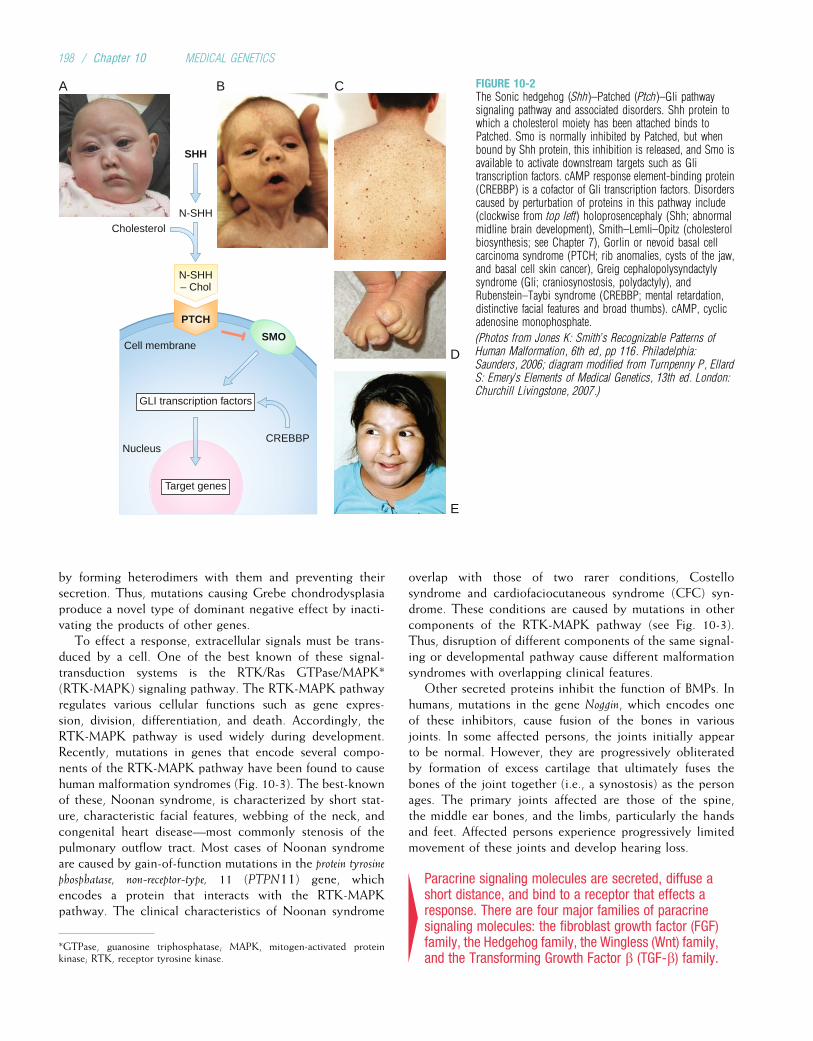
by forming heterodimers with them and preventing theirsecretion. Thus, mutations causing Grebe chondrodysplasiaproduce a novel type of dominant negative effect by inacti-vating the products of other genes.
To effect a response, extracellular signals must be trans-duced by a cell. One of the best known of these signal-transduction systems is the RTK/Ras GTPase/MAPK*(RTK-MAPK) signaling pathway. The RTK-MAPK pathwayregulates various cellular functions such as gene expres-sion, division, differentiation, and death. Accordingly, theRTK-MAPK pathway is used widely during development.Recently, mutations in genes that encode several compo-nents of the RTK-MAPK pathway have been found to causehuman malformation syndromes (Fig. 10-3). The best-knownof these, Noonan syndrome, is characterized by short stat-ure, characteristic facial features, webbing of the neck, andcongenital heart disease—most commonly stenosis of thepulmonary outflow tract. Most cases of Noonan syndromeare caused by gain-of-function mutations in the protein tyrosinephosphatase, non-receptor-type, 11 (PTPN11) gene, whichencodes a protein that interacts with the RTK-MAPKpathway. The clinical characteristics of Noonan syndrome
overlap with those of two rarer conditions, Costellosyndrome and cardiofaciocutaneous syndrome (CFC) syn-drome. These conditions are caused by mutations in othercomponents of the RTK-MAPK pathway (see Fig. 10-3).Thus, disruption of different components of the same signal-ing or developmental pathway cause different malformationsyndromes with overlapping clinical features.
Other secreted proteins inhibit the function of BMPs. Inhumans, mutations in the gene Noggin, which encodes oneof these inhibitors, cause fusion of the bones in variousjoints. In some affected persons, the joints initially appearto be normal. However, they are progressively obliteratedby formation of excess cartilage that ultimately fuses thebones of the joint together (i.e., a synostosis) as the personages. The primary joints affected are those of the spine,the middle ear bones, and the limbs, particularly the handsand feet. Affected persons experience progressively limitedmovement of these joints and develop hearing loss.
4Paracrine signaling molecules are secreted, diffuse ashort distance, and bind to a receptor that effects aresponse. There are four major families of paracrinesignaling molecules: the fibroblast growth factor (FGF)family, the Hedgehog family, the Wingless (Wnt) family,and the Transforming Growth Factor b (TGF-b) family.
Nucleus
Cholesterol
A B C
D
E
Cell membrane
SHH
N-SHH
N-SHH– Chol
PTCH
SMO
CREBBP
GLI transcription factors
Target genes
FIGURE 10-2The Sonic hedgehog (Shh)–Patched (Ptch)–Gli pathwaysignaling pathway and associated disorders. Shh protein towhich a cholesterol moiety has been attached binds toPatched. Smo is normally inhibited by Patched, but whenbound by Shh protein, this inhibition is released, and Smo isavailable to activate downstream targets such as Glitranscription factors. cAMP response element-binding protein(CREBBP) is a cofactor of Gli transcription factors. Disorderscaused by perturbation of proteins in this pathway include(clockwise from top left) holoprosencephaly (Shh; abnormalmidline brain development), Smith–Lemli–Opitz (cholesterolbiosynthesis; see Chapter 7), Gorlin or nevoid basal cellcarcinoma syndrome (PTCH; rib anomalies, cysts of the jaw,and basal cell skin cancer), Greig cephalopolysyndactylysyndrome (Gli; craniosynostosis, polydactyly), andRubenstein–Taybi syndrome (CREBBP; mental retardation,distinctive facial features and broad thumbs). cAMP, cyclicadenosine monophosphate.
(Photos from Jones K: Smith’s Recognizable Patterns ofHuman Malformation, 6th ed, pp 116. Philadelphia:Saunders, 2006; diagram modified from Turnpenny P, EllardS: Emery’s Elements of Medical Genetics, 13th ed. London:Churchill Livingstone, 2007.)
*GTPase, guanosine triphosphatase; MAPK, mitogen-activated proteinkinase; RTK, receptor tyrosine kinase.
198 / Chapter 10 MEDICAL GENETICS
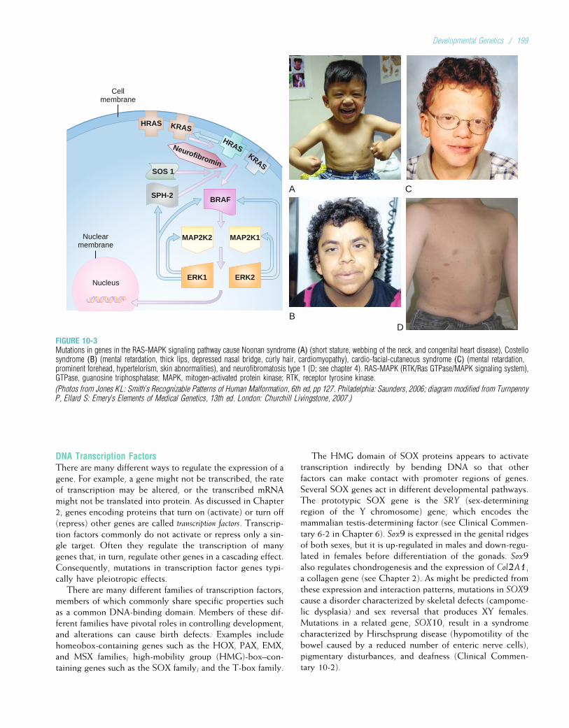
DNA Transcription FactorsThere are many different ways to regulate the expression of agene. For example, a gene might not be transcribed, the rateof transcription may be altered, or the transcribed mRNAmight not be translated into protein. As discussed in Chapter2, genes encoding proteins that turn on (activate) or turn off(repress) other genes are called transcription factors. Transcrip-tion factors commonly do not activate or repress only a sin-gle target. Often they regulate the transcription of manygenes that, in turn, regulate other genes in a cascading effect.Consequently, mutations in transcription factor genes typi-cally have pleiotropic effects.
There are many different families of transcription factors,members of which commonly share specific properties suchas a common DNA-binding domain. Members of these dif-ferent families have pivotal roles in controlling development,and alterations can cause birth defects. Examples includehomeobox-containing genes such as the HOX, PAX, EMX,and MSX families; high-mobility group (HMG)-box–con-taining genes such as the SOX family; and the T-box family.
The HMG domain of SOX proteins appears to activatetranscription indirectly by bending DNA so that otherfactors can make contact with promoter regions of genes.Several SOX genes act in different developmental pathways.The prototypic SOX gene is the SRY (sex-determiningregion of the Y chromosome) gene, which encodes themammalian testis-determining factor (see Clinical Commen-tary 6-2 in Chapter 6). Sox9 is expressed in the genital ridgesof both sexes, but it is up-regulated in males and down-regu-lated in females before differentiation of the gonads. Sox9also regulates chondrogenesis and the expression of Col2A1,a collagen gene (see Chapter 2). As might be predicted fromthese expression and interaction patterns, mutations in SOX9cause a disorder characterized by skeletal defects (campome-lic dysplasia) and sex reversal that produces XY females.Mutations in a related gene, SOX10, result in a syndromecharacterized by Hirschsprung disease (hypomotility of thebowel caused by a reduced number of enteric nerve cells),pigmentary disturbances, and deafness (Clinical Commen-tary 10-2).
Nucleus
A C
BD
Nuclearmembrane
Cellmembrane
MAP2K1MAP2K2
ERK2ERK1
BRAF
SOS 1
SPH-2
HRAS KRAS
HRASKRAS
Neurofibromin
FIGURE 10-3Mutations in genes in the RAS-MAPK signaling pathway cause Noonan syndrome (A) (short stature, webbing of the neck, and congenital heart disease), Costellosyndrome (B) (mental retardation, thick lips, depressed nasal bridge, curly hair, cardiomyopathy), cardio-facial-cutaneous syndrome (C) (mental retardation,prominent forehead, hypertelorism, skin abnormalities), and neurofibromatosis type 1 (D; see chapter 4). RAS-MAPK (RTK/Ras GTPase/MAPK signaling system),GTPase, guanosine triphosphatase; MAPK, mitogen-activated protein kinase; RTK, receptor tyrosine kinase.
(Photos from Jones KL: Smith’s Recognizable Patterns of Human Malformation, 6th ed, pp 127. Philadelphia: Saunders, 2006; diagram modified from TurnpennyP, Ellard S: Emery’s Elements of Medical Genetics, 13th ed. London: Churchill Livingstone, 2007.)
Developmental Genetics / 199
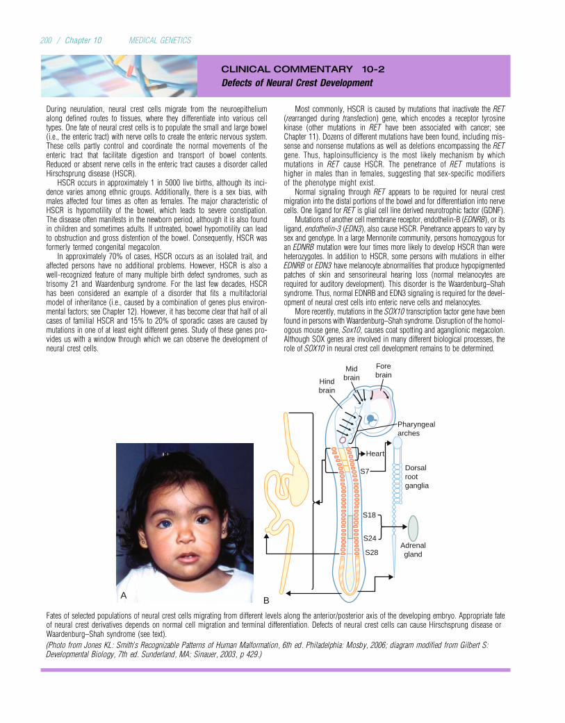
CLINICAL COMMENTARY 10-2
Defects of Neural Crest Development
During neurulation, neural crest cells migrate from the neuroepitheliumalong defined routes to tissues, where they differentiate into various celltypes. One fate of neural crest cells is to populate the small and large bowel(i.e., the enteric tract) with nerve cells to create the enteric nervous system.These cells partly control and coordinate the normal movements of theenteric tract that facilitate digestion and transport of bowel contents.Reduced or absent nerve cells in the enteric tract causes a disorder calledHirschsprung disease (HSCR).
HSCR occurs in approximately 1 in 5000 live births, although its inci-dence varies among ethnic groups. Additionally, there is a sex bias, withmales affected four times as often as females. The major characteristic ofHSCR is hypomotility of the bowel, which leads to severe constipation.The disease often manifests in the newborn period, although it is also foundin children and sometimes adults. If untreated, bowel hypomotility can leadto obstruction and gross distention of the bowel. Consequently, HSCR wasformerly termed congenital megacolon.
In approximately 70% of cases, HSCR occurs as an isolated trait, andaffected persons have no additional problems. However, HSCR is also awell-recognized feature of many multiple birth defect syndromes, such astrisomy 21 and Waardenburg syndrome. For the last few decades, HSCRhas been considered an example of a disorder that fits a multifactorialmodel of inheritance (i.e., caused by a combination of genes plus environ-mental factors; see Chapter 12). However, it has become clear that half of allcases of familial HSCR and 15% to 20% of sporadic cases are caused bymutations in one of at least eight different genes. Study of these genes pro-vides us with a window through which we can observe the development ofneural crest cells.
Most commonly, HSCR is caused by mutations that inactivate the RET(rearranged during transfection) gene, which encodes a receptor tyrosinekinase (other mutations in RET have been associated with cancer; seeChapter 11). Dozens of different mutations have been found, including mis-sense and nonsense mutations as well as deletions encompassing the RETgene. Thus, haploinsufficiency is the most likely mechanism by whichmutations in RET cause HSCR. The penetrance of RET mutations ishigher in males than in females, suggesting that sex-specific modifiersof the phenotype might exist.
Normal signaling through RET appears to be required for neural crestmigration into the distal portions of the bowel and for differentiation into nervecells. One ligand for RET is glial cell line derived neurotrophic factor (GDNF).
Mutations of another cell membrane receptor, endothelin-B (EDNRB), or itsligand, endothelin-3 (EDN3), also cause HSCR. Penetrance appears to vary bysex and genotype. In a large Mennonite community, persons homozygous foran EDNRB mutation were four times more likely to develop HSCR than wereheterozygotes. In addition to HSCR, some persons with mutations in eitherEDNRB or EDN3 have melanocyte abnormalities that produce hypopigmentedpatches of skin and sensorineural hearing loss (normal melanocytes arerequired for auditory development). This disorder is the Waardenburg–Shahsyndrome. Thus, normal EDNRB and EDN3 signaling is required for the devel-opment of neural crest cells into enteric nerve cells and melanocytes.
More recently, mutations in the SOX10 transcription factor gene have beenfound in persons with Waardenburg–Shah syndrome. Disruption of the homol-ogous mouse gene, Sox10, causes coat spotting and aganglionic megacolon.Although SOX genes are involved in many different biological processes, therole of SOX10 in neural crest cell development remains to be determined.
Hindbrain
Midbrain
Forebrain
Pharyngealarches
Heart
Dorsalrootganglia
A B
Adrenalgland
S18
S24
S28
S7
Fates of selected populations of neural crest cells migrating from different levels along the anterior/posterior axis of the developing embryo. Appropriate fateof neural crest derivatives depends on normal cell migration and terminal differentiation. Defects of neural crest cells can cause Hirschsprung disease orWaardenburg–Shah syndrome (see text).
(Photo from Jones KL: Smith’s Recognizable Patterns of Human Malformation, 6th ed. Philadelphia: Mosby, 2006; diagram modified from Gilbert S:Developmental Biology, 7th ed. Sunderland, MA: Sinauer, 2003, p 429.)
200 / Chapter 10 MEDICAL GENETICS
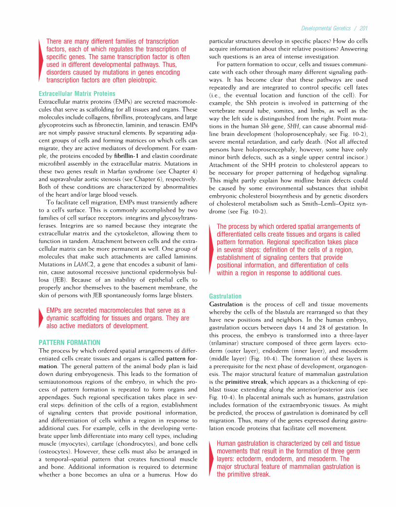
4There are many different families of transcriptionfactors, each of which regulates the transcription ofspecific genes. The same transcription factor is oftenused in different developmental pathways. Thus,disorders caused by mutations in genes encodingtranscription factors are often pleiotropic.
Extracellular Matrix ProteinsExtracellular matrix proteins (EMPs) are secreted macromole-cules that serve as scaffolding for all tissues and organs. Thesemolecules include collagens, fibrillins, proteoglycans, and largeglycoproteins such as fibronectin, laminin, and tenascin. EMPsare not simply passive structural elements. By separating adja-cent groups of cells and forming matrices on which cells canmigrate, they are active mediators of development. For exam-ple, the proteins encoded by fibrillin-1 and elastin coordinatemicrofibril assembly in the extracellular matrix. Mutations inthese two genes result in Marfan syndrome (see Chapter 4)and supravalvular aortic stenosis (see Chapter 6), respectively.Both of these conditions are characterized by abnormalitiesof the heart and/or large blood vessels.
To facilitate cell migration, EMPs must transiently adhereto a cell’s surface. This is commonly accomplished by twofamilies of cell surface receptors: integrins and glycosyltrans-ferases. Integrins are so named because they integrate theextracellular matrix and the cytoskeleton, allowing them tofunction in tandem. Attachment between cells and the extra-cellular matrix can be more permanent as well. One group ofmolecules that make such attachments are called laminins.Mutations in LAMC2, a gene that encodes a subunit of lami-nin, cause autosomal recessive junctional epidermolysis bul-losa (JEB). Because of an inability of epithelial cells toproperly anchor themselves to the basement membrane, theskin of persons with JEB spontaneously forms large blisters.
4EMPs are secreted macromolecules that serve as adynamic scaffolding for tissues and organs. They arealso active mediators of development.
PATTERN FORMATIONThe process by which ordered spatial arrangements of differ-entiated cells create tissues and organs is called pattern for-mation. The general pattern of the animal body plan is laiddown during embryogenesis. This leads to the formation ofsemiautonomous regions of the embryo, in which the pro-cess of pattern formation is repeated to form organs andappendages. Such regional specification takes place in sev-eral steps: definition of the cells of a region, establishmentof signaling centers that provide positional information,and differentiation of cells within a region in response toadditional cues. For example, cells in the developing verte-brate upper limb differentiate into many cell types, includingmuscle (myocytes), cartilage (chondrocytes), and bone cells(osteocytes). However, these cells must also be arranged ina temporal–spatial pattern that creates functional muscleand bone. Additional information is required to determinewhether a bone becomes an ulna or a humerus. How do
particular structures develop in specific places? How do cellsacquire information about their relative positions? Answeringsuch questions is an area of intense investigation.
For pattern formation to occur, cells and tissues communi-cate with each other through many different signaling path-ways. It has become clear that these pathways are usedrepeatedly and are integrated to control specific cell fates(i.e., the eventual location and function of the cell). Forexample, the Shh protein is involved in patterning of thevertebrate neural tube, somites, and limbs, as well as theway the left side is distinguished from the right. Point muta-tions in the human Shh gene, SHH, can cause abnormal mid-line brain development (holoprosencephaly; see Fig. 10-2),severe mental retardation, and early death. (Not all affectedpersons have holoprosencephaly, however; some have onlyminor birth defects, such as a single upper central incisor.)Attachment of the SHH protein to cholesterol appears tobe necessary for proper patterning of hedgehog signaling.This might partly explain how midline brain defects couldbe caused by some environmental substances that inhibitembryonic cholesterol biosynthesis and by genetic disordersof cholesterol metabolism such as Smith–Lemli–Opitz syn-drome (see Fig. 10-2).
4The process by which ordered spatial arrangements ofdifferentiated cells create tissues and organs is calledpattern formation. Regional specification takes placein several steps: definition of the cells of a region,establishment of signaling centers that providepositional information, and differentiation of cellswithin a region in response to additional cues.
GastrulationGastrulation is the process of cell and tissue movementswhereby the cells of the blastula are rearranged so that theyhave new positions and neighbors. In the human embryo,gastrulation occurs between days 14 and 28 of gestation. Inthis process, the embryo is transformed into a three-layer(trilaminar) structure composed of three germ layers: ecto-derm (outer layer), endoderm (inner layer), and mesoderm(middle layer) (Fig. 10-4). The formation of these layers isa prerequisite for the next phase of development, organogen-esis. The major structural feature of mammalian gastrulationis the primitive streak, which appears as a thickening of epi-blast tissue extending along the anterior/posterior axis (seeFig. 10-4). In placental animals such as humans, gastrulationincludes formation of the extraembryonic tissues. As mightbe predicted, the process of gastrulation is dominated by cellmigration. Thus, many of the genes expressed during gastru-lation encode proteins that facilitate cell movement.
4Human gastrulation is characterized by cell and tissuemovements that result in the formation of three germlayers: ectoderm, endoderm, and mesoderm. Themajor structural feature of mammalian gastrulation isthe primitive streak.
Developmental Genetics / 201
Neurulation and EctodermOnce a trilaminar embryo is formed, the dorsal mesodermand the overlying ectoderm interact to form the hollow neu-ral tube. This event, called neurulation, is mediated byinduction. In amphibians, induction of the neural tube andtransformation of the flanking mesoderm to create anembryo with clear anterior/posterior and dorsal/ventral axesis controlled by a group of cells known as the Spemann–Mangold organizer. A number of proteins are expressedalmost exclusively in the organizer. Chordin is a secretedprotein that prevents dorsalized mesoderm from being ven-tralized. Another secreted protein, Noggin, induces neuraltissue from dorsal ectoderm and dorsalizes the mesoderm.Understanding the major functions of the organizer and
the molecules that mediate these functions is an area ofactive research.
Neurulation is a critical event in development because it initi-ates organogenesis and divides the ectoderm into three differentcell populations: the neural tube, which will eventually form thebrain and spinal cord; the epidermis of the skin; and the neuralcrest cells. In humans, neural tube closure begins at five separatesites, which correspond to the locations of common neural tubedefects such as anencephaly (absence of the brain), occipitalencephalocele, and lumbar spina bifida (see Chapter 12). Neuralcrest cells migrate from the neuroepithelium along definedroutes to tissues, where they differentiate into cell types suchas sensory neurons, melanocytes, neurons of the small bowel,and smooth muscle cells (see Clinical Commentary 10-2).
Uterine lining
Epiblast
Epiblast
Hypoblast
Hypoblast
Amniotic cavity
Amniotic cavity
Primitive groove
Primitive groove14–15 days
16 days
Primitivegroove
Primitive streak
EndodermMesoderm
Ectoderm
Yolk sac
Yolk sac
Node
A
B
Endoderm
FIGURE 10-4Human gastrulation. A, Sagittal section through the midline of an embryo embedded in the uterine lining. B, Dorsal surface of an embryo exposed byremoving part of the embryonic mesoderm surrounding the amniotic cavity and yolk sac. Arrows denote ingressing epiblast cells. On days 14 to 15,epiblast cells replace hypoblast cells to form endoderm. A day later, migrating epiblast cells are creating a layer of mesoderm.
202 / Chapter 10 MEDICAL GENETICS
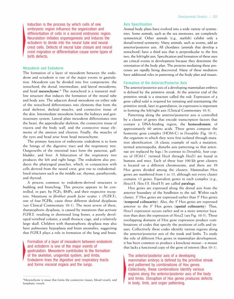
4Induction is the process by which cells of oneembryonic region influence the organization anddifferentiation of cells in a second embryonic region.Neurulation initiates organogenesis and induces theectoderm to divide into the neural tube and neuralcrest cells. Defects of neural tube closure and neuralcrest migration or differentiation cause some types ofbirth defects.
Mesoderm and EndodermThe formation of a layer of mesoderm between the endo-derm and ectoderm is one of the major events in gastrula-tion. Mesoderm can be divided into five components: thenotochord; the dorsal, intermediate, and lateral mesoderms;and head mesenchyme.* The notochord is a transient mid-line structure that induces the formation of the neural tubeand body axis. The adjacent dorsal mesoderm on either sideof the notochord differentiates into elements that form theaxial skeleton, skeletal muscles, and connective tissue ofthe skin. Intermediate mesoderm forms the kidneys and gen-itourinary system. Lateral plate mesoderm differentiates intothe heart, the appendicular skeleton, the connective tissue ofviscera and the body wall, and the connective tissue ele-ments of the amnion and chorion. Finally, the muscles ofthe eyes and head arise from head mesenchyme.
The primary function of embryonic endoderm is to formthe linings of the digestive tract and the respiratory tree.Outgrowths of the intestinal tract form the pancreas, gall-bladder, and liver. A bifurcation of the respiratory treeproduces the left and right lungs. The endoderm also pro-duces the pharyngeal pouches, which, in conjunction withcells derived from the neural crest, give rise to endodermal-lined structures such as the middle ear, thymus, parathyroids,and thyroid.
A process common to endoderm-derived structures isbudding and branching. This process appears to be con-trolled, in part, by FGFs, BMPs, and their respective recep-tors. Mutations in fibroblast growth factor receptor 3 (FGFR3),one of four FGFRs, cause three different skeletal dysplasias(see Clinical Commentary 10-1). The most severe of these,thanatophoric dysplasia, is caused by mutations that activateFGFR3, resulting in shortened long bones, a poorly devel-oped vertebral column, a small thoracic cage, and a relativelylarge skull. Children with thanatophoric dysplasia may alsohave pulmonary hypoplasia and brain anomalies, suggestingthat FGFR3 plays a role in formation of the lung and brain.
4Formation of a layer of mesoderm between endodermand ectoderm is one of the major events ofgastrulation. Mesoderm contributes to the formationof the skeleton, urogenital system, and limbs.Endoderm lines the digestive and respiratory tractsand forms visceral organs and the lungs.
Axis SpecificationAnimal body plans have evolved into a wide variety of symme-tries. Some animals, such as the sea anemones, are completelysymmetrical. Other animals (e.g., starfish) exhibit only adorsal/ventral symmetry. Many animals, such as worms, add ananterior/posterior axis. All chordates (animals that develop anotochord) have a third axis that is perpendicular to the firsttwo, the left/right axis. Specification and formation of these axesare critical events in development because they determine theorientation of the body plan. The proteins mediating these pro-cesses are rapidly being discovered. Many of these mediatorshave additional roles in patterning of the body plan and tissues.
Formation of the Anterior/Posterior AxisThe anterior/posterior axis of a developing mammalian embryois defined by the primitive streak. At the anterior end of theprimitive streak is a structure called the node. Expression of agene called nodal is required for initiating and maintaining theprimitive streak; later in gastrulation, its expression is importantin forming the left/right axis (Clinical Commentary 10-3).
Patterning along the anterior/posterior axis is controlledby a cluster of genes that encode transcription factors thatcontain a DNA-binding region, the homeodomain, ofapproximately 60 amino acids. These genes compose thehomeotic gene complex (HOM-C) in Drosophila (Fig. 10-5),the organism in which they were first isolated through muta-tion identification. (A classic example of such a mutation,termed antennapedia, disturbs axis patterning so that anten-nae are replaced by legs.) In contrast to Drosophila, four cop-ies of HOM-C (termed HoxA through HoxD) are found inhumans and mice. Each of these four 100-kb gene clustersis located on a different chromosome, and there are 39Hox genes divided among the clusters. Mammalian Hoxgenes are numbered from 1 to 13, although not every clustercontains 13 genes. Equivalent genes in each complex (e.g.,Hoxa13, Hoxc13, Hoxd13) are called paralogs.
Hox genes are expressed along the dorsal axis from theanterior boundary of the hindbrain to the tail. Within eachcluster, 30 Hox genes are expressed earlier than 50 Hox genes(temporal colinearity). Also, the 30 Hox genes are expressedanterior to the 50 Hox genes (spatial colinearity). Thus,Hoxa1 expression occurs earlier and in a more anterior loca-tion than does the expression of Hoxa2 (see Fig. 10-5). Theseoverlapping domains of Hox gene expression produce com-binations of codes that specify the positions of cells and tis-sues. Collectively these codes identify various regions alongthe anterior/posterior axis of the trunk and limbs. To studythe role of different Hox genes in mammalian development,it has been common to produce a knockout mouse—a mousethat lacks a functional copy of the gene of interest (Box 10-1).
4The anterior/posterior axis of a developingmammalian embryo is defined by the primitive streakand patterned by combinations of Hox genes.Collectively, these combinations identify variousregions along the anterior/posterior axis of the bodyand limbs. Disruption of Hox genes produces defectsin body, limb, and organ patterning.
*Mesenchyme is tissue that forms the connective tissues, blood vessels, andlymphatic vessels.
Developmental Genetics / 203
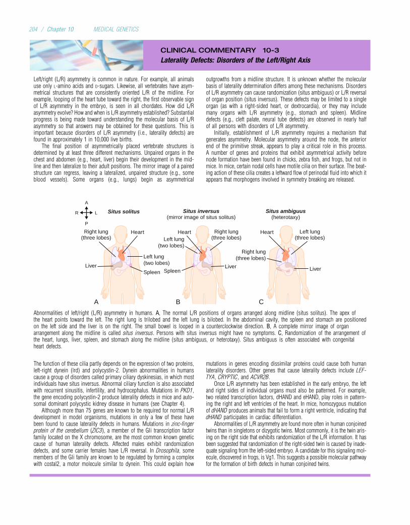
CLINICAL COMMENTARY 10-3
Laterality Defects: Disorders of the Left/Right Axis
Left/right (L/R) asymmetry is common in nature. For example, all animalsuse only L-amino acids and D-sugars. Likewise, all vertebrates have asym-metrical structures that are consistently oriented L/R of the midline. Forexample, looping of the heart tube toward the right, the first observable signof L/R asymmetry in the embryo, is seen in all chordates. How did L/Rasymmetry evolve? How and when is L/R asymmetry established? Substantialprogress is being made toward understanding the molecular basis of L/Rasymmetry so that answers may be obtained for these questions. This isimportant because disorders of L/R asymmetry (i.e., laterality defects) arefound in approximately 1 in 10,000 live births.
The final position of asymmetrically placed vertebrate structures isdetermined by at least three different mechanisms. Unpaired organs in thechest and abdomen (e.g., heart, liver) begin their development in the mid-line and then lateralize to their adult positions. The mirror image of a pairedstructure can regress, leaving a lateralized, unpaired structure (e.g., someblood vessels). Some organs (e.g., lungs) begin as asymmetrical
outgrowths from a midline structure. It is unknown whether the molecularbasis of laterality determination differs among these mechanisms. Disordersof L/R asymmetry can cause randomization (situs ambiguus) or L/R reversalof organ position (situs inversus). These defects may be limited to a singleorgan (as with a right-sided heart, or dextrocardia), or they may includemany organs with L/R asymmetry (e.g., stomach and spleen). Midlinedefects (e.g., cleft palate, neural tube defects) are observed in nearly halfof all persons with disorders of L/R asymmetry.
Initially, establishment of L/R asymmetry requires a mechanism thatgenerates asymmetry. Molecular asymmetry around the node, the anteriorend of the primitive streak, appears to play a critical role in this process.A number of genes and proteins that exhibit asymmetrical activity beforenode formation have been found in chicks, zebra fish, and frogs, but not inmice. In mice, certain nodal cells have motile cilia on their surface. The beat-ing action of these cilia creates a leftward flow of perinodal fluid into which itappears that morphogens involved in symmetry breaking are released.
Left lung(three lobes)
Right lung(three lobes)
Right lung(three lobes)
Right lung(three lobes)
Situs solitus Situs inversus(mirror image of situs solitus)
Situs ambiguus(heterotaxy)
Liver
A B C
Heart Heart Heart
Left lung(two lobes)
Left lung(two lobes)
Spleen Spleen LiverLiver
A
P
R L
Abnormalities of left/right (L/R) asymmetry in humans. A, The normal L/R positions of organs arranged along midline (situs solitus). The apex ofthe heart points toward the left. The right lung is trilobed and the left lung is bilobed. In the abdominal cavity, the spleen and stomach are positionedon the left side and the liver is on the right. The small bowel is looped in a counterclockwise direction. B, A complete mirror image of organarrangement along the midline is called situs inversus. Persons with situs inversus might have no symptoms. C, Randomization of the arrangement ofthe heart, lungs, liver, spleen, and stomach along the midline (situs ambiguus, or heterotaxy). Situs ambiguus is often associated with congenitalheart defects.
The function of these cilia partly depends on the expression of two proteins,left-right dynein (lrd) and polycystin-2. Dynein abnormalities in humanscause a group of disorders called primary ciliary dyskinesias, in which mostindividuals have situs inversus. Abnormal ciliary function is also associatedwith recurrent sinusitis, infertility, and hydrocephalus. Mutations in PKD1,the gene encoding polycystin-2 produce laterality defects in mice and auto-somal dominant polycystic kidney disease in humans (see Chapter 4).
Although more than 75 genes are known to be required for normal L/Rdevelopment in model organisms, mutations in only a few of these havebeen found to cause laterality defects in humans. Mutations in zinc-fingerprotein of the cerebellum (ZIC3), a member of the Gli transcription factorfamily located on the X chromosome, are the most common known geneticcause of human laterality defects. Affected males exhibit randomizationdefects, and some carrier females have L/R reversal. In Drosophila, somemembers of the Gli family are known to be regulated by forming a complexwith costal2, a motor molecule similar to dynein. This could explain how
mutations in genes encoding dissimilar proteins could cause both humanlaterality disorders. Other genes that cause laterality defects include LEF-TYA, CRYPTIC, and ACVR2B.
Once L/R asymmetry has been established in the early embryo, the leftand right sides of individual organs must also be patterned. For example,two related transcription factors, dHAND and eHAND, play roles in pattern-ing the right and left ventricles of the heart. In mice, homozygous mutationof dHAND produces animals that fail to form a right ventricle, indicating thatdHAND participates in cardiac differentiation.
Abnormalities of L/R asymmetry are found more often in human conjoinedtwins than in singletons or dizygotic twins. Most commonly, it is the twin aris-ing on the right side that exhibits randomization of the L/R information. It hasbeen suggested that randomization of the right-sided twin is caused by inade-quate signaling from the left-sided embryo. A candidate for this signaling mol-ecule, discovered in frogs, is Vg1. This suggests a possible molecular pathwayfor the formation of birth defects in human conjoined twins.
204 / Chapter 10 MEDICAL GENETICS
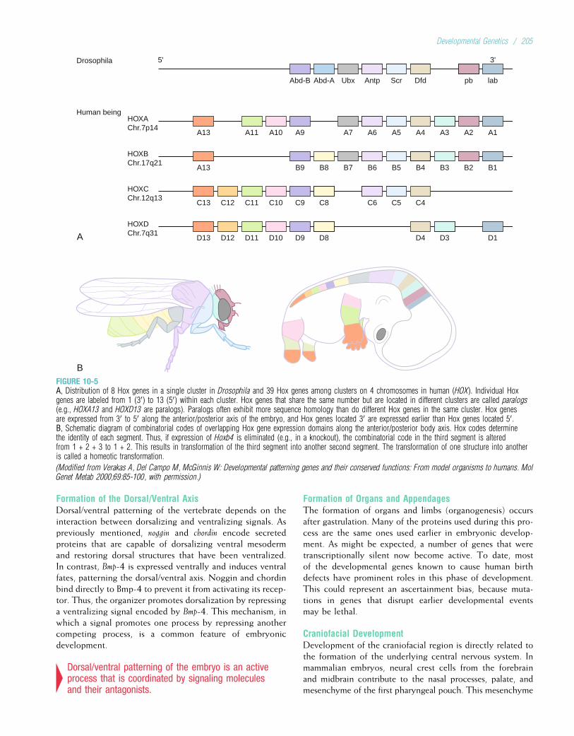
Formation of the Dorsal/Ventral AxisDorsal/ventral patterning of the vertebrate depends on theinteraction between dorsalizing and ventralizing signals. Aspreviously mentioned, noggin and chordin encode secretedproteins that are capable of dorsalizing ventral mesodermand restoring dorsal structures that have been ventralized.In contrast, Bmp-4 is expressed ventrally and induces ventralfates, patterning the dorsal/ventral axis. Noggin and chordinbind directly to Bmp-4 to prevent it from activating its recep-tor. Thus, the organizer promotes dorsalization by repressinga ventralizing signal encoded by Bmp-4. This mechanism, inwhich a signal promotes one process by repressing anothercompeting process, is a common feature of embryonicdevelopment.
4Dorsal/ventral patterning of the embryo is an activeprocess that is coordinated by signaling moleculesand their antagonists.
Formation of Organs and AppendagesThe formation of organs and limbs (organogenesis) occursafter gastrulation. Many of the proteins used during this pro-cess are the same ones used earlier in embryonic develop-ment. As might be expected, a number of genes that weretranscriptionally silent now become active. To date, mostof the developmental genes known to cause human birthdefects have prominent roles in this phase of development.This could represent an ascertainment bias, because muta-tions in genes that disrupt earlier developmental eventsmay be lethal.
Craniofacial DevelopmentDevelopment of the craniofacial region is directly related tothe formation of the underlying central nervous system. Inmammalian embryos, neural crest cells from the forebrainand midbrain contribute to the nasal processes, palate, andmesenchyme of the first pharyngeal pouch. This mesenchyme
A
Drosophila
B
5' 3'
Abd-B Abd-A Ubx Antp Scr Dfd pb lab
A13
HOXAChr.7p14
Human being
A11 A10 A9 A7 A6 A5 A4 A3 A2 A1
A13
HOXBChr.17q21
B9 B8 B7 B6 B5 B4 B3 B2 B1
C13
HOXCChr.12q13
C12 C11 C10 C9 C8 C6 C5 C4
D13
HOXDChr.7q31
D12 D11 D10 D9 D8 D4 D3 D1
FIGURE 10-5A, Distribution of 8 Hox genes in a single cluster in Drosophila and 39 Hox genes among clusters on 4 chromosomes in human (HOX). Individual Hoxgenes are labeled from 1 (30) to 13 (50) within each cluster. Hox genes that share the same number but are located in different clusters are called paralogs(e.g., HOXA13 and HOXD13 are paralogs). Paralogs often exhibit more sequence homology than do different Hox genes in the same cluster. Hox genesare expressed from 30 to 50 along the anterior/posterior axis of the embryo, and Hox genes located 30 are expressed earlier than Hox genes located 50.B, Schematic diagram of combinatorial codes of overlapping Hox gene expression domains along the anterior/posterior body axis. Hox codes determinethe identity of each segment. Thus, if expression of Hoxb4 is eliminated (e.g., in a knockout), the combinatorial code in the third segment is alteredfrom 1 + 2 + 3 to 1 + 2. This results in transformation of the third segment into another second segment. The transformation of one structure into anotheris called a homeotic transformation.
(Modified from Verakas A, Del Campo M, McGinnis W: Developmental patterning genes and their conserved functions: From model organisms to humans. MolGenet Metab 2000;69:85-100, with permission.)
Developmental Genetics / 205
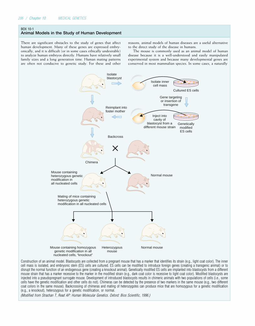
BOX 10-1Animal Models in the Study of Human Development
There are significant obstacles to the study of genes that affecthuman development. Many of these genes are expressed embry-onically, and it is difficult (or in some cases ethically undesirable)to analyze human embryos directly. Humans have relatively smallfamily sizes and a long generation time. Human mating patternsare often not conducive to genetic study. For these and other
reasons, animal models of human diseases are a useful alternativeto the direct study of the disease in humans.
The mouse is commonly used as an animal model of humandisease because it is a well-understood and easily manipulatedexperimental system and because many developmental genes areconserved in most mammalian species. In some cases, a naturally
Chimera
Isolateblastocyst
Reimplant intofoster mother
Mating of mice containingheterozygous geneticmodification in all nucleated cells
Mouse containing homozygousgenetic modification in allnucleated cells, “knockout”
Heterozygousmouse
Normal mouse
Mouse containingheterozygous geneticmodification in all nucleated cells
Normal mouse
Isolate innercell mass
Inject intocavity of
blastocyst from adifferent mouse strain
Cultured ES cells
Gene targetingor insertion of
transgene
GeneticallymodifiedES cells
Backcross
Construction of an animal model. Blastocysts are collected from a pregnant mouse that has a marker that identifies its strain (e.g., light coat color). The innercell mass is isolated, and embryonic stem (ES) cells are cultured. ES cells can be modified to introduce foreign genes (creating a transgenic animal) or todisrupt the normal function of an endogenous gene (creating a knockout animal). Genetically modified ES cells are implanted into blastocysts from a differentmouse strain that has a marker recessive to the marker in the modified strain (e.g., dark coat color is recessive to light coat color). Modified blastocysts areinjected into a pseudopregnant surrogate mouse. Development of introduced blastocysts results in chimeric animals with two populations of cells (i.e., somecells have the genetic modification and other cells do not). Chimeras can be detected by the presence of two markers in the same mouse (e.g., two differentcoat colors in the same mouse). Backcrossing of chimeras and mating of heterozygotes can produce mice that are homozygous for a genetic modification(e.g., a knockout), heterozygous for a genetic modification, or normal.
(Modified from Strachan T, Read AP: Human Molecular Genetics. Oxford: Bios Scientific, 1996.)
206 / Chapter 10 MEDICAL GENETICS
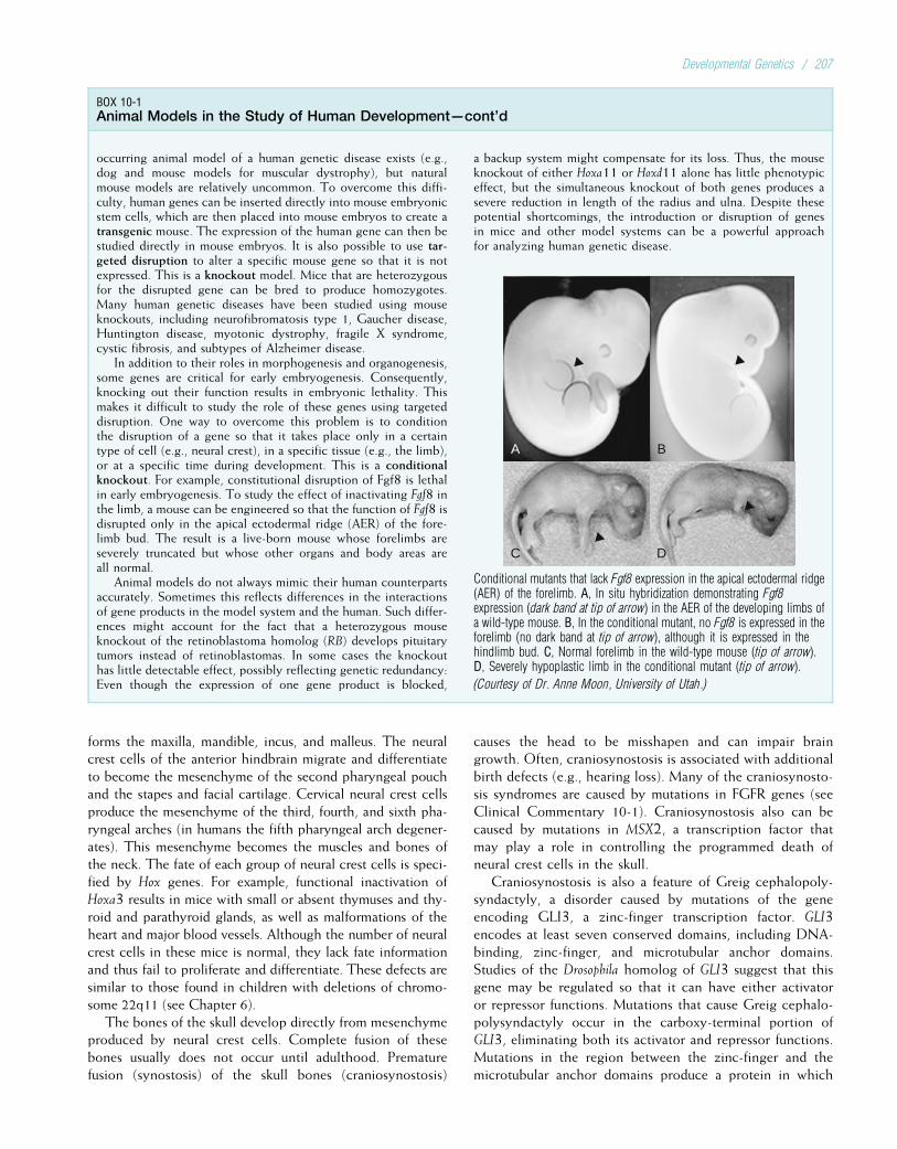
forms the maxilla, mandible, incus, and malleus. The neuralcrest cells of the anterior hindbrain migrate and differentiateto become the mesenchyme of the second pharyngeal pouchand the stapes and facial cartilage. Cervical neural crest cellsproduce the mesenchyme of the third, fourth, and sixth pha-ryngeal arches (in humans the fifth pharyngeal arch degener-ates). This mesenchyme becomes the muscles and bones ofthe neck. The fate of each group of neural crest cells is speci-fied by Hox genes. For example, functional inactivation ofHoxa3 results in mice with small or absent thymuses and thy-roid and parathyroid glands, as well as malformations of theheart and major blood vessels. Although the number of neuralcrest cells in these mice is normal, they lack fate informationand thus fail to proliferate and differentiate. These defects aresimilar to those found in children with deletions of chromo-some 22q11 (see Chapter 6).
The bones of the skull develop directly from mesenchymeproduced by neural crest cells. Complete fusion of thesebones usually does not occur until adulthood. Prematurefusion (synostosis) of the skull bones (craniosynostosis)
causes the head to be misshapen and can impair braingrowth. Often, craniosynostosis is associated with additionalbirth defects (e.g., hearing loss). Many of the craniosynosto-sis syndromes are caused by mutations in FGFR genes (seeClinical Commentary 10-1). Craniosynostosis also can becaused by mutations in MSX2, a transcription factor thatmay play a role in controlling the programmed death ofneural crest cells in the skull.
Craniosynostosis is also a feature of Greig cephalopoly-syndactyly, a disorder caused by mutations of the geneencoding GLI3, a zinc-finger transcription factor. GLI3encodes at least seven conserved domains, including DNA-binding, zinc-finger, and microtubular anchor domains.Studies of the Drosophila homolog of GLI3 suggest that thisgene may be regulated so that it can have either activatoror repressor functions. Mutations that cause Greig cephalo-polysyndactyly occur in the carboxy-terminal portion ofGLI3, eliminating both its activator and repressor functions.Mutations in the region between the zinc-finger and themicrotubular anchor domains produce a protein in which
BOX 10-1Animal Models in the Study of Human Development—cont’d
occurring animal model of a human genetic disease exists (e.g.,dog and mouse models for muscular dystrophy), but naturalmouse models are relatively uncommon. To overcome this diffi-culty, human genes can be inserted directly into mouse embryonicstem cells, which are then placed into mouse embryos to create atransgenic mouse. The expression of the human gene can then bestudied directly in mouse embryos. It is also possible to use tar-geted disruption to alter a specific mouse gene so that it is notexpressed. This is a knockout model. Mice that are heterozygousfor the disrupted gene can be bred to produce homozygotes.Many human genetic diseases have been studied using mouseknockouts, including neurofibromatosis type 1, Gaucher disease,Huntington disease, myotonic dystrophy, fragile X syndrome,cystic fibrosis, and subtypes of Alzheimer disease.
In addition to their roles in morphogenesis and organogenesis,some genes are critical for early embryogenesis. Consequently,knocking out their function results in embryonic lethality. Thismakes it difficult to study the role of these genes using targeteddisruption. One way to overcome this problem is to conditionthe disruption of a gene so that it takes place only in a certaintype of cell (e.g., neural crest), in a specific tissue (e.g., the limb),or at a specific time during development. This is a conditionalknockout. For example, constitutional disruption of Fgf8 is lethalin early embryogenesis. To study the effect of inactivating Fgf8 inthe limb, a mouse can be engineered so that the function of Fgf8 isdisrupted only in the apical ectodermal ridge (AER) of the fore-limb bud. The result is a live-born mouse whose forelimbs areseverely truncated but whose other organs and body areas areall normal.
Animal models do not always mimic their human counterpartsaccurately. Sometimes this reflects differences in the interactionsof gene products in the model system and the human. Such differ-ences might account for the fact that a heterozygous mouseknockout of the retinoblastoma homolog (RB) develops pituitarytumors instead of retinoblastomas. In some cases the knockouthas little detectable effect, possibly reflecting genetic redundancy:Even though the expression of one gene product is blocked,
a backup system might compensate for its loss. Thus, the mouseknockout of either Hoxa11 or Hoxd11 alone has little phenotypiceffect, but the simultaneous knockout of both genes produces asevere reduction in length of the radius and ulna. Despite thesepotential shortcomings, the introduction or disruption of genesin mice and other model systems can be a powerful approachfor analyzing human genetic disease.
A B
C D
Conditional mutants that lack Fgf8 expression in the apical ectodermal ridge(AER) of the forelimb. A, In situ hybridization demonstrating Fgf8expression (dark band at tip of arrow) in the AER of the developing limbs ofa wild-type mouse. B, In the conditional mutant, no Fgf8 is expressed in theforelimb (no dark band at tip of arrow), although it is expressed in thehindlimb bud. C, Normal forelimb in the wild-type mouse (tip of arrow).D, Severely hypoplastic limb in the conditional mutant (tip of arrow).
(Courtesy of Dr. Anne Moon, University of Utah.)
Developmental Genetics / 207
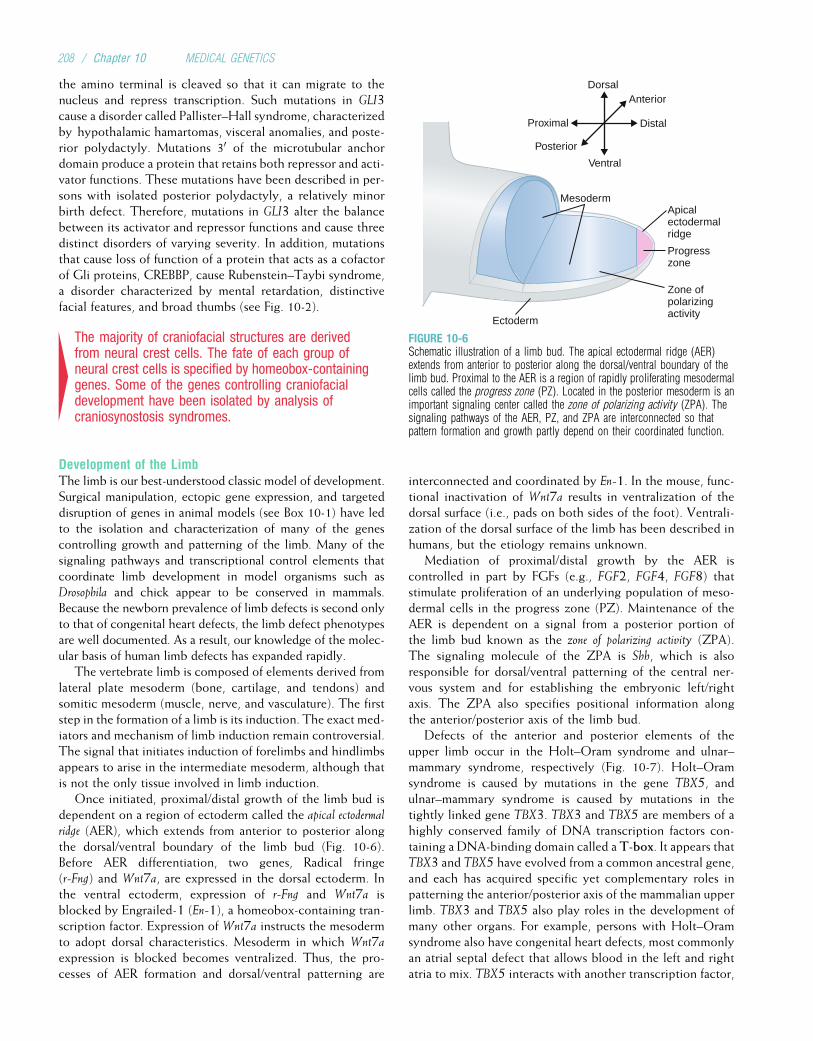
the amino terminal is cleaved so that it can migrate to thenucleus and repress transcription. Such mutations in GLI3cause a disorder called Pallister–Hall syndrome, characterizedby hypothalamic hamartomas, visceral anomalies, and poste-rior polydactyly. Mutations 30 of the microtubular anchordomain produce a protein that retains both repressor and acti-vator functions. These mutations have been described in per-sons with isolated posterior polydactyly, a relatively minorbirth defect. Therefore, mutations in GLI3 alter the balancebetween its activator and repressor functions and cause threedistinct disorders of varying severity. In addition, mutationsthat cause loss of function of a protein that acts as a cofactorof Gli proteins, CREBBP, cause Rubenstein–Taybi syndrome,a disorder characterized by mental retardation, distinctivefacial features, and broad thumbs (see Fig. 10-2).
4The majority of craniofacial structures are derivedfrom neural crest cells. The fate of each group ofneural crest cells is specified by homeobox-containinggenes. Some of the genes controlling craniofacialdevelopment have been isolated by analysis ofcraniosynostosis syndromes.
Development of the LimbThe limb is our best-understood classic model of development.Surgical manipulation, ectopic gene expression, and targeteddisruption of genes in animal models (see Box 10-1) have ledto the isolation and characterization of many of the genescontrolling growth and patterning of the limb. Many of thesignaling pathways and transcriptional control elements thatcoordinate limb development in model organisms such asDrosophila and chick appear to be conserved in mammals.Because the newborn prevalence of limb defects is second onlyto that of congenital heart defects, the limb defect phenotypesare well documented. As a result, our knowledge of the molec-ular basis of human limb defects has expanded rapidly.
The vertebrate limb is composed of elements derived fromlateral plate mesoderm (bone, cartilage, and tendons) andsomitic mesoderm (muscle, nerve, and vasculature). The firststep in the formation of a limb is its induction. The exact med-iators and mechanism of limb induction remain controversial.The signal that initiates induction of forelimbs and hindlimbsappears to arise in the intermediate mesoderm, although thatis not the only tissue involved in limb induction.
Once initiated, proximal/distal growth of the limb bud isdependent on a region of ectoderm called the apical ectodermalridge (AER), which extends from anterior to posterior alongthe dorsal/ventral boundary of the limb bud (Fig. 10-6).Before AER differentiation, two genes, Radical fringe(r-Fng) and Wnt7a, are expressed in the dorsal ectoderm. Inthe ventral ectoderm, expression of r-Fng and Wnt7a isblocked by Engrailed-1 (En-1), a homeobox-containing tran-scription factor. Expression of Wnt7a instructs the mesodermto adopt dorsal characteristics. Mesoderm in which Wnt7aexpression is blocked becomes ventralized. Thus, the pro-cesses of AER formation and dorsal/ventral patterning are
interconnected and coordinated by En-1. In the mouse, func-tional inactivation of Wnt7a results in ventralization of thedorsal surface (i.e., pads on both sides of the foot). Ventrali-zation of the dorsal surface of the limb has been described inhumans, but the etiology remains unknown.
Mediation of proximal/distal growth by the AER iscontrolled in part by FGFs (e.g., FGF2, FGF4, FGF8) thatstimulate proliferation of an underlying population of meso-dermal cells in the progress zone (PZ). Maintenance of theAER is dependent on a signal from a posterior portion ofthe limb bud known as the zone of polarizing activity (ZPA).The signaling molecule of the ZPA is Shh, which is alsoresponsible for dorsal/ventral patterning of the central ner-vous system and for establishing the embryonic left/rightaxis. The ZPA also specifies positional information alongthe anterior/posterior axis of the limb bud.
Defects of the anterior and posterior elements of theupper limb occur in the Holt–Oram syndrome and ulnar–mammary syndrome, respectively (Fig. 10-7). Holt–Oramsyndrome is caused by mutations in the gene TBX5, andulnar–mammary syndrome is caused by mutations in thetightly linked gene TBX3. TBX3 and TBX5 are members of ahighly conserved family of DNA transcription factors con-taining a DNA-binding domain called aT-box. It appears thatTBX3 and TBX5 have evolved from a common ancestral gene,and each has acquired specific yet complementary roles inpatterning the anterior/posterior axis of the mammalian upperlimb. TBX3 and TBX5 also play roles in the development ofmany other organs. For example, persons with Holt–Oramsyndrome also have congenital heart defects, most commonlyan atrial septal defect that allows blood in the left and rightatria to mix. TBX5 interacts with another transcription factor,
Apicalectodermalridge
Progresszone
Ectoderm
Mesoderm
Zone ofpolarizingactivity
DorsalAnterior
Distal
Ventral
Posterior
Proximal
FIGURE 10-6Schematic illustration of a limb bud. The apical ectodermal ridge (AER)extends from anterior to posterior along the dorsal/ventral boundary of thelimb bud. Proximal to the AER is a region of rapidly proliferating mesodermalcells called the progress zone (PZ). Located in the posterior mesoderm is animportant signaling center called the zone of polarizing activity (ZPA). Thesignaling pathways of the AER, PZ, and ZPA are interconnected so thatpattern formation and growth partly depend on their coordinated function.
208 / Chapter 10 MEDICAL GENETICS
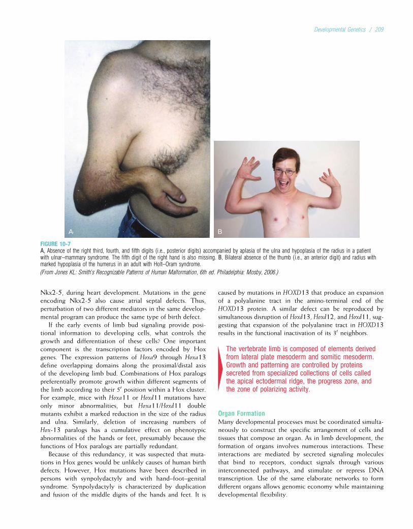
Nkx2-5, during heart development. Mutations in the geneencoding Nkx2-5 also cause atrial septal defects. Thus,perturbation of two different mediators in the same develop-mental program can produce the same type of birth defect.
If the early events of limb bud signaling provide posi-tional information to developing cells, what controls thegrowth and differentiation of these cells? One importantcomponent is the transcription factors encoded by Hoxgenes. The expression patterns of Hoxa9 through Hoxa13define overlapping domains along the proximal/distal axisof the developing limb bud. Combinations of Hox paralogspreferentially promote growth within different segments ofthe limb according to their 50 position within a Hox cluster.For example, mice with Hoxa11 or Hoxd11 mutations haveonly minor abnormalities, but Hoxa11/Hoxd11 doublemutants exhibit a marked reduction in the size of the radiusand ulna. Similarly, deletion of increasing numbers ofHox-13 paralogs has a cumulative effect on phenotypicabnormalities of the hands or feet, presumably because thefunctions of Hox paralogs are partially redundant.
Because of this redundancy, it was suspected that muta-tions in Hox genes would be unlikely causes of human birthdefects. However, Hox mutations have been described inpersons with synpolydactyly and with hand–foot–genitalsyndrome. Synpolydactyly is characterized by duplicationand fusion of the middle digits of the hands and feet. It is
caused by mutations in HOXD13 that produce an expansionof a polyalanine tract in the amino-terminal end of theHOXD13 protein. A similar defect can be reproduced bysimultaneous disruption of Hoxd13, Hoxd12, and Hoxd11, sug-gesting that expansion of the polyalanine tract in HOXD13results in the functional inactivation of its 30 neighbors.
4The vertebrate limb is composed of elements derivedfrom lateral plate mesoderm and somitic mesoderm.Growth and patterning are controlled by proteinssecreted from specialized collections of cells calledthe apical ectodermal ridge, the progress zone, andthe zone of polarizing activity.
Organ FormationMany developmental processes must be coordinated simulta-neously to construct the specific arrangement of cells andtissues that compose an organ. As in limb development, theformation of organs involves numerous interactions. Theseinteractions are mediated by secreted signaling moleculesthat bind to receptors, conduct signals through variousinterconnected pathways, and stimulate or repress DNAtranscription. Use of the same elaborate networks to formdifferent organs allows genomic economy while maintainingdevelopmental flexibility.
A B
FIGURE 10-7A, Absence of the right third, fourth, and fifth digits (i.e., posterior digits) accompanied by aplasia of the ulna and hypoplasia of the radius in a patientwith ulnar–mammary syndrome. The fifth digit of the right hand is also missing. B, Bilateral absence of the thumb (i.e., an anterior digit) and radius withmarked hypoplasia of the humerus in an adult with Holt–Oram syndrome.
(From Jones KL: Smith’s Recognizable Patterns of Human Malformation, 6th ed. Philadelphia: Mosby, 2006.)
Developmental Genetics / 209
Once a specialized cell within an organ is terminally differ-entiated, various proteins turn on its molecular machinery sothat it may perform its intended function. Often, developmentof the organ and function of the differentiated cell are interre-lated. For example, the endocrine pancreas is largely composedof three different cell types: alpha, beta, and gamma. Transcrip-tion of insulin in beta cells is stimulated by the binding of insu-lin promoter factor 1 (IPF1) to the insulin promoter region.Mutations in the gene encoding IPF1 prevent pancreatic devel-opment, indicating that IPF1 is necessary for the maturationand differentiation of pancreatic precursor cells.
Interactions between mesenchymal and epithelial cells areprominent in the development of cutaneous structures (e.g.,hair, sweat glands, breasts), parenchymal organs (e.g., liver,pancreas), lungs, thyroid, kidneys, and teeth. These interac-tions are dynamic in that expression patterns in the epitheliaand mesenchyme change over time and continue to influ-ence one another. For example, during tooth development,the epithelium secretes Bmp-4, which signals the underlyingmesenchyme to express a set of transcription factors, includ-ing Msx1. The mutual exchange of signals between epithe-lium and mesenchyme leads to formation of a dentalpapilla and cusp and finally to terminal differentiation ofmesenchyme into tooth-forming odontoblasts. In humans,mutations in MSX1 disrupt tooth formation and cause lossof the second premolars. Similarly, mutations in the humanhomolog of the mouse gene hairless cause loss of all bodyhair, including that of the scalp, eyebrows, axillae, and pubis.
The integrity of signals exchanged between the epithe-lium and the mesenchyme is dependent on the integrity ofthese tissues. Several proteins produced within the epithe-lium are known to promote the growth and differentiationof the epithelium. One of these proteins is p63, a homologof the prototypic tumor suppressor gene product, p53. Muta-tions that perturb the function of p63 decrease the availabil-ity of epithelial progenitor cells. This results in abnormalitiesof the limbs, skin, teeth, hair, and nails in at least six differ-ent malformation syndromes.
One of the largest organs in the body is the skeleton.Skeletal formation is dependent on bone-forming cells calledosteoblasts. The differentiation of osteoblasts is regulated byan osteoblast-specific transcription factor, Runx2. Targeteddisruption of Runx2 produces mice with a complete lack ofossification of the skeleton. Heterozygous mice have wid-ened cranial sutures, shortened digits, and abnormalities ofthe shoulder girdle. Similar defects are found in persons withcleidocranial dysplasia, which is caused by mutations inRunx2, the human homolog of mouse Runx2.
4Formation of organs involves reciprocal interactionsbetween epithelium and mesenchyme. Thisinteraction is mediated by secreted signalingmolecules that bind to receptors, conduct signalsthrough various interconnected pathways, andstimulate or repress DNA transcription.
Study Questions
1. Explain how nonhuman animal models are useful forstudying human development and birth defects. Giveat least one example.
2. Mutations in fibroblast growth factor receptors(FGFRs) cause at least different craniosynostosissyndromes. Moreover, the same mutation in FGFR2causes Pfeiffer syndrome in some families but Crouzonsyndrome in other families. How can the samemutation produce two distinct disorders?
3. Disorders caused by mutations in genes encodingtranscription factors are often pleiotropic. Explain thisfinding.
4. Define pattern formation, and give an example of abirth defect caused by disruption of this process.
5. If the control of developmental processes is tightlyregulated, how can you explain that mutations in somedevelopmental genes (e.g., Hox genes) produce subtlephenotypes?
6. Loss-of-function mutants in mammals are commonlystudied by creating a knockout mouse model. Explainwhy it is impractical to use some organisms (e.g.,baboons) to generate knockouts. Can you think of away to circumvent some of these obstacles?
7. Give an example of a birth defect that can be causedby a defect in a ligand or its receptor.
8. Explain what may be some of the obstacles to usinggene therapy to treat birth defects.
Suggested ReadingsCapecchi MR. Gene targeting in mice: Functional analysis of
the mammalian genome for the twenty-first century. NatRev Genet 2005;6:507–12.
Chen D, Zhao M, Mundy GR. Bone morphogenetic pro-teins. Growth Factors 2004;22:233–41.
Epstein CP, Erickson RP, Wynshaw-Boris A, (eds): InbornErrors of Development, 2nd ed. New York: Oxford Uni-versity Press, 2008.
Farlie PG, McKeown SJ, Newgreen DF. The neural crest:Basic biology and clinical relationships in the craniofacial
210 / Chapter 10 MEDICAL GENETICS
and enteric nervous systems. Birth Defects Res C EmbryoToday 2004;72:173–89.
Gilbert SF. Developmental Biology, 8th ed. Sunderland,Mass: Sinauer, 2006.
Hoppler S, Kavanagh CL. Wnt signaling: Variety at thecore. J Cell Sci 2007;120:385–93.
Iimura T, Pourquie O. Hox genes in time and space duringvertebrate body formation. Develop Growth Differ2007;49:265–75.
Isphording D, Leylek AM, Yeung J, et al. T-box genes andcongenital heart/limb malformations. Clin Genet 2004;66:253–64.
Itoh N, Ornitz DM. Evolution of the Fgf and Fgfr genefamilies. Trends Genet 2004;20:563–69.
Jorgensen EM, Mango SE. The art and design of geneticscreens: Caenorhabditis elegans. Nat Rev Genet 2002;3:356–69.
Lang D, Powell SK, Plummer RS, et al. PAX genes: Roles indevelopment, pathophysiology, and cancer. BiochemPharm 2007;73:1–14.
Levin M. Left–right asymmetry in embryonic development:A comprehensive review. Mech Dev 2005; 122:3–25.
Lieschke G, Currie PD. Animal models of human disease:Zebra fish swim into view. Nat Rev Genet 2007;8:353–66.
Webster MK, Donoghue DJ. FGFR activation in skeletal dis-orders: Too much of a good thing. Trends Genet1997;13:178–82.
Wilkie AO. Bad bones, absent smell, selfish testes: Thepleiotropic consequences of human FGF receptor muta-tions. Cytokine Growth Factor Rev 2005;16:187–203.
Zhu L, Belmont JW, Ware SM. Genetics of humanheterotaxias. Eur J Hum Genet 2006;14:17–25.
Developmental Genetics / 211
Chapter 11
CANCER GENETICS
Present evidence indicates that approximately one in fourdeaths is now due to cancer and that invasive cancer willbe diagnosed in more than half of the population at somepoint in their lives. Many cancers are increasing in inci-dence, largely as a result of the increased average age ofour population.
As shown in this chapter, the causes of cancer are a mix-ture of environmental and genetic alterations that occur inour tissues. Inherited predisposition plays a role in somefamilies. Dramatic advances in molecular biology and genet-ics have now clarified the basic molecular elements of cancerand provide a schematic outline of the cellular events leadingto cancer. This understanding will be crucially important inthe control of cancer, providing the beginnings of a baseof knowledge that should lead to significantly improvedtherapies and possibly prevention.
“Cancer” is a collection of disorders that share the com-mon feature of uncontrolled cell growth. This leads to a massof cells termed a neoplasm (Greek, “new formation”), ortumor. The formation of tumors is called tumorigenesis.Several key events must occur if cells are to escape the usualconstraints that prevent uncontrolled proliferation. Addi-tional growth signals must be produced and processed, andcells must become resistant to signals that normally inhibitgrowth. Because these abnormal characteristics would typi-cally trigger the process of programmed cell death (apopto-sis), cells must somehow disable this process. The growingcell mass (tumor) requires nourishment, so a new blood sup-ply must be obtained through angiogenesis (the formationof new blood vessels). Additional inhibitory signals mustbe overcome for the tumor to achieve a malignant state, inwhich neoplasms invade nearby tissues and metastasize(spread) to more distant sites in the body. The capacity toinvade and metastasize distinguishes malignant from benignneoplasms.
Tumors are classified according to the tissue type inwhich they arise. Major types of tumors include those of epi-thelial tissue (carcinomas, the most common tumors), con-nective tissue (sarcomas), lymphatic tissue (lymphomas),glial cells of the central nervous system (gliomas), and hema-topoietic organs (leukemias). The cells that compose atumor are usually derived from a single ancestral cell, makingthem a single clone (monoclonal).
Many of the basic biological features of carcinogenesis(cancer development) are now understood. Throughout ourlives, many of our cells continue to grow and differentiate.These cells form, for example, the epithelial layers of ourlungs and colons and the precursor cells of our immune sys-tems. Relatively undifferentiated stem cells produce largenumbers of progeny cells to repopulate and renew our worndefensive layers. Through integration of informationprovided by a complex array of biochemical signals, thenew cells eventually stop dividing and terminally differenti-ate into a cell type appropriate for their role in the body(Fig. 11-1). Alternatively, if the cell is abnormal or damaged,it may undergo apoptosis.
Occasionally one of these cells fails to differentiate andbegins to divide without restraint. The descendants of suchcells can become the founders of neoplasms, capable of fur-ther transformation into invasive, metastatic cancer. Wewish to understand in detail what has gone wrong in thesecells, to detect them early, and ultimately to intervene intheir development so as to eliminate them.
4Cells in the body are programmed to develop, grow,differentiate, and die in response to a complex systemof biochemical signals. Cancer results from theemergence of a clone of cells freed of thesedevelopmental programming constraints and capableof inappropriate proliferation.
CAUSES OF CANCERGenetic ConsiderationsGenetic alterations of cell regulatory systems are the primarybasis of carcinogenesis. We can create cancer in animal mod-els by damaging specific genes. In cell culture systems, wecan reverse a cancer phenotype by introducing normal cop-ies of the damaged genes into the cell. Most of the geneticevents that cause cancer occur in somatic cells. The fre-quency of these events can be altered by exposure to muta-gens, thus establishing a link to environmental carcinogens(cancer-causing agents). However, these genetic events arenot transmitted to future generations because they occur insomatic, rather than germline, cells. Even though they aregenetic events, they are not inherited.
212
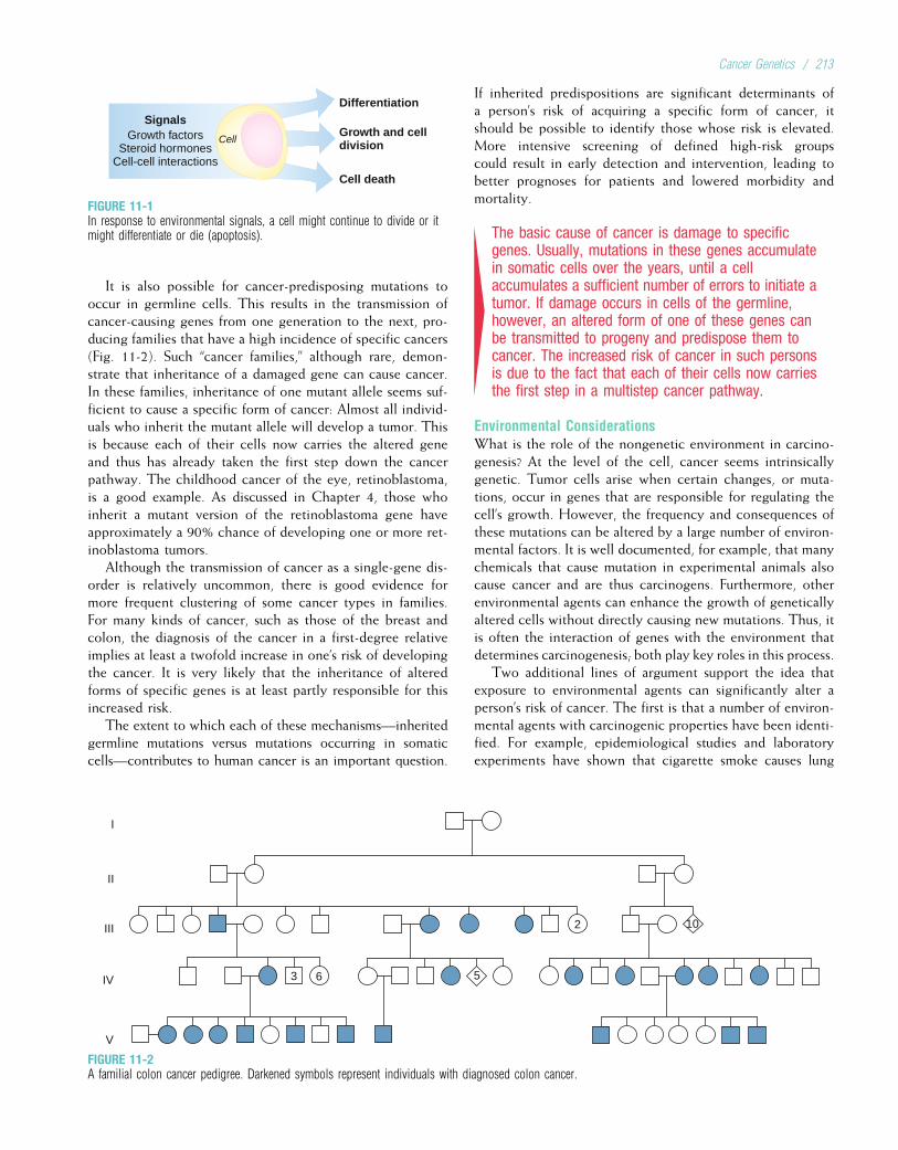
It is also possible for cancer-predisposing mutations tooccur in germline cells. This results in the transmission ofcancer-causing genes from one generation to the next, pro-ducing families that have a high incidence of specific cancers(Fig. 11-2). Such “cancer families,” although rare, demon-strate that inheritance of a damaged gene can cause cancer.In these families, inheritance of one mutant allele seems suf-ficient to cause a specific form of cancer: Almost all individ-uals who inherit the mutant allele will develop a tumor. Thisis because each of their cells now carries the altered geneand thus has already taken the first step down the cancerpathway. The childhood cancer of the eye, retinoblastoma,is a good example. As discussed in Chapter 4, those whoinherit a mutant version of the retinoblastoma gene haveapproximately a 90% chance of developing one or more ret-inoblastoma tumors.
Although the transmission of cancer as a single-gene dis-order is relatively uncommon, there is good evidence formore frequent clustering of some cancer types in families.For many kinds of cancer, such as those of the breast andcolon, the diagnosis of the cancer in a first-degree relativeimplies at least a twofold increase in one’s risk of developingthe cancer. It is very likely that the inheritance of alteredforms of specific genes is at least partly responsible for thisincreased risk.
The extent to which each of these mechanisms—inheritedgermline mutations versus mutations occurring in somaticcells—contributes to human cancer is an important question.
If inherited predispositions are significant determinants ofa person’s risk of acquiring a specific form of cancer, itshould be possible to identify those whose risk is elevated.More intensive screening of defined high-risk groupscould result in early detection and intervention, leading tobetter prognoses for patients and lowered morbidity andmortality.
4The basic cause of cancer is damage to specificgenes. Usually, mutations in these genes accumulatein somatic cells over the years, until a cellaccumulates a sufficient number of errors to initiate atumor. If damage occurs in cells of the germline,however, an altered form of one of these genes canbe transmitted to progeny and predispose them tocancer. The increased risk of cancer in such personsis due to the fact that each of their cells now carriesthe first step in a multistep cancer pathway.
Environmental ConsiderationsWhat is the role of the nongenetic environment in carcino-genesis? At the level of the cell, cancer seems intrinsicallygenetic. Tumor cells arise when certain changes, or muta-tions, occur in genes that are responsible for regulating thecell’s growth. However, the frequency and consequences ofthese mutations can be altered by a large number of environ-mental factors. It is well documented, for example, that manychemicals that cause mutation in experimental animals alsocause cancer and are thus carcinogens. Furthermore, otherenvironmental agents can enhance the growth of geneticallyaltered cells without directly causing new mutations. Thus, itis often the interaction of genes with the environment thatdetermines carcinogenesis; both play key roles in this process.
Two additional lines of argument support the idea thatexposure to environmental agents can significantly alter aperson’s risk of cancer. The first is that a number of environ-mental agents with carcinogenic properties have been identi-fied. For example, epidemiological studies and laboratoryexperiments have shown that cigarette smoke causes lung
Differentiation
Growth and celldivision
Cell death
CellGrowth factorsSteroid hormones
Cell-cell interactions
Signals
FIGURE 11-1In response to environmental signals, a cell might continue to divide or itmight differentiate or die (apoptosis).
3
I
II
III
IV
V
6
2 10
5
FIGURE 11-2A familial colon cancer pedigree. Darkened symbols represent individuals with diagnosed colon cancer.
Cancer Genetics / 213
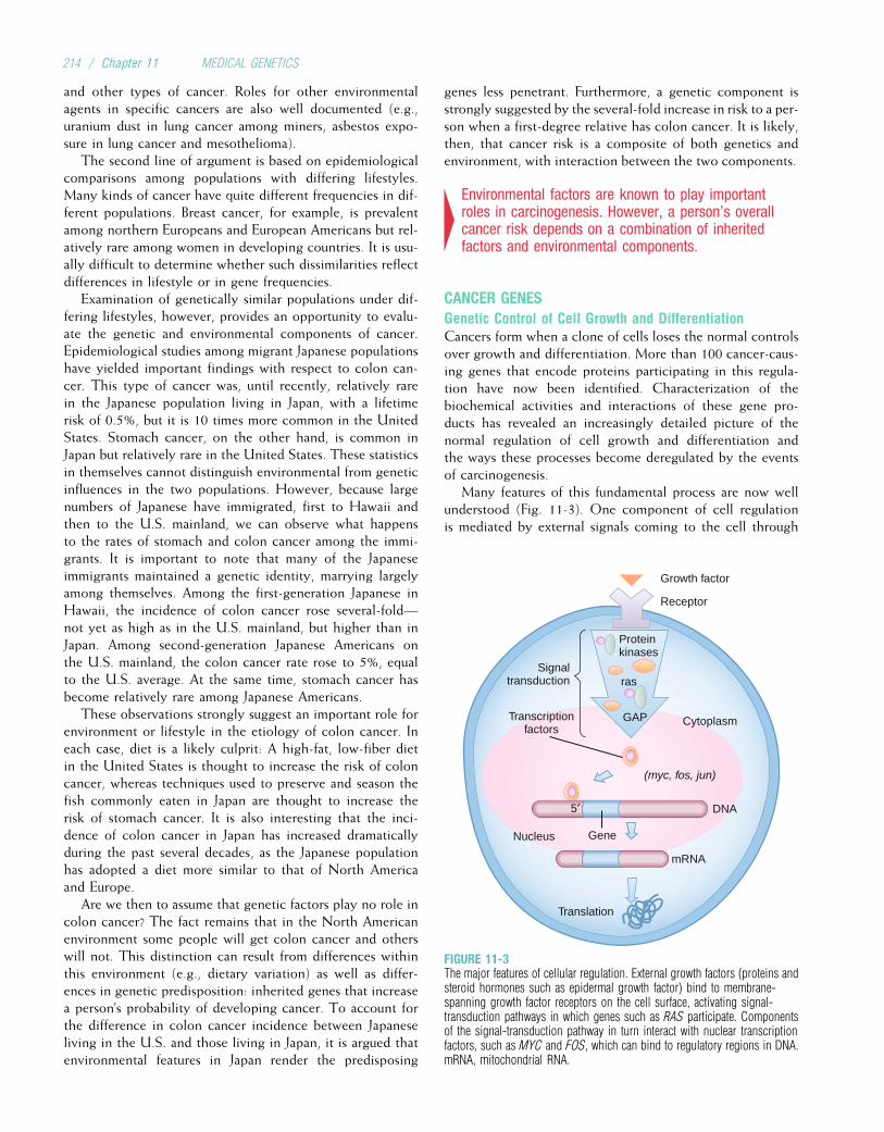
and other types of cancer. Roles for other environmentalagents in specific cancers are also well documented (e.g.,uranium dust in lung cancer among miners, asbestos expo-sure in lung cancer and mesothelioma).
The second line of argument is based on epidemiologicalcomparisons among populations with differing lifestyles.Many kinds of cancer have quite different frequencies in dif-ferent populations. Breast cancer, for example, is prevalentamong northern Europeans and European Americans but rel-atively rare among women in developing countries. It is usu-ally difficult to determine whether such dissimilarities reflectdifferences in lifestyle or in gene frequencies.
Examination of genetically similar populations under dif-fering lifestyles, however, provides an opportunity to evalu-ate the genetic and environmental components of cancer.Epidemiological studies among migrant Japanese populationshave yielded important findings with respect to colon can-cer. This type of cancer was, until recently, relatively rarein the Japanese population living in Japan, with a lifetimerisk of 0.5%, but it is 10 times more common in the UnitedStates. Stomach cancer, on the other hand, is common inJapan but relatively rare in the United States. These statisticsin themselves cannot distinguish environmental from geneticinfluences in the two populations. However, because largenumbers of Japanese have immigrated, first to Hawaii andthen to the U.S. mainland, we can observe what happensto the rates of stomach and colon cancer among the immi-grants. It is important to note that many of the Japaneseimmigrants maintained a genetic identity, marrying largelyamong themselves. Among the first-generation Japanese inHawaii, the incidence of colon cancer rose several-fold—not yet as high as in the U.S. mainland, but higher than inJapan. Among second-generation Japanese Americans onthe U.S. mainland, the colon cancer rate rose to 5%, equalto the U.S. average. At the same time, stomach cancer hasbecome relatively rare among Japanese Americans.
These observations strongly suggest an important role forenvironment or lifestyle in the etiology of colon cancer. Ineach case, diet is a likely culprit: A high-fat, low-fiber dietin the United States is thought to increase the risk of coloncancer, whereas techniques used to preserve and season thefish commonly eaten in Japan are thought to increase therisk of stomach cancer. It is also interesting that the inci-dence of colon cancer in Japan has increased dramaticallyduring the past several decades, as the Japanese populationhas adopted a diet more similar to that of North Americaand Europe.
Are we then to assume that genetic factors play no role incolon cancer? The fact remains that in the North Americanenvironment some people will get colon cancer and otherswill not. This distinction can result from differences withinthis environment (e.g., dietary variation) as well as differ-ences in genetic predisposition: inherited genes that increasea person’s probability of developing cancer. To account forthe difference in colon cancer incidence between Japaneseliving in the U.S. and those living in Japan, it is argued thatenvironmental features in Japan render the predisposing
genes less penetrant. Furthermore, a genetic component isstrongly suggested by the several-fold increase in risk to a per-son when a first-degree relative has colon cancer. It is likely,then, that cancer risk is a composite of both genetics andenvironment, with interaction between the two components.
4Environmental factors are known to play importantroles in carcinogenesis. However, a person’s overallcancer risk depends on a combination of inheritedfactors and environmental components.
CANCER GENESGenetic Control of Cell Growth and DifferentiationCancers form when a clone of cells loses the normal controlsover growth and differentiation. More than 100 cancer-caus-ing genes that encode proteins participating in this regula-tion have now been identified. Characterization of thebiochemical activities and interactions of these gene pro-ducts has revealed an increasingly detailed picture of thenormal regulation of cell growth and differentiation andthe ways these processes become deregulated by the eventsof carcinogenesis.
Many features of this fundamental process are now wellunderstood (Fig. 11-3). One component of cell regulationis mediated by external signals coming to the cell through
Growth factor
Receptor
Proteinkinases
CytoplasmGAP
rasSignal
transduction
Transcriptionfactors
(myc, fos, jun)
DNA
mRNA
Nucleus Gene
5′
Translation
FIGURE 11-3The major features of cellular regulation. External growth factors (proteins andsteroid hormones such as epidermal growth factor) bind to membrane-spanning growth factor receptors on the cell surface, activating signal-transduction pathways in which genes such as RAS participate. Componentsof the signal-transduction pathway in turn interact with nuclear transcriptionfactors, such as MYC and FOS, which can bind to regulatory regions in DNA.mRNA, mitochondrial RNA.
214 / Chapter 11 MEDICAL GENETICS
polypeptide growth factors (e.g., platelet-derived growthfactor, epidermal growth factor, steroid hormones) producedin other cells. Each growth factor interacts with specificgrowth factor receptors located on the cell surface. Bindingof a growth factor activates the receptor, triggering mole-cules that send messages to the cell’s nucleus in the processof signal transduction. These signal transduction moleculesinclude protein kinases, such as src tyrosine kinase, mito-gen-activated protein kinase (MAPK), and jun kinase (JunK),which can alter the activity of target proteins by taggingthem at a specific site with a phosphate molecule (phosphor-ylation). The ultimate stage of the signal transduction path-way is regulation of DNA transcription in the nucleus.Components of the signal transduction cascade interact withnuclear transcription factors that regulate the activity of spe-cific genes whose protein products influence cellular growthand proliferation. Genes that encode these transcriptionfactors include MYC, FOS, and JUN.
After several rounds of cell division, cells normallyreceive signals that tell them to stop growing and to differ-entiate into specialized cells. The signals may come frompolypeptides, from steroid hormones, from direct contactwith adjacent cells, or from internal programs that definethe number of cell divisions that are allowed. The signalsare transduced to the nucleus of the recipient cell. Here, byaltering the transcription patterns of genes that govern thesteps of the cell cycle, they repress genes that promote celldivision and induce genes that inhibit entry into the cell divi-sion cycle.
4The regulation of cell growth is accomplished bysubstances that include: (1) growth factors thattransmit signals from one cell to another; (2) specificreceptors for the growth factors; (3) signaltransduction molecules that activate a cascade ofphosphorylating reactions within the cell; and(4) nuclear transcription factors. The cell integratesand interprets the host of signals it receives from itsenvironment. Decisions to grow and divide, or to stopgrowing and differentiate, result from processing ofthese signals.
A cancer cell can emerge from within a population ofgrowing cells through the accumulation of mutations inthese genes. Although such mutations occur only rarely,these cells can fail to respond to differentiation signalsand continue to divide instead of undergoing their normaldifferentiation program. Furthermore, cancers seem usuallyto result from a progressive series of events that incremen-tally increase the extent of deregulation within a cell line-age. Eventually, a cell emerges whose descendants multiplywithout appropriate restraints. Further changes give these cellsthe capacity to invade adjacent tissues and form metastases.Each of these changes involves mutations, and the require-ment for more than one mutation has been characterizedas themulti-hit concept of carcinogenesis. An example of this
concept is given by colorectal cancer, in which several geneticevents are required to complete the progression from abenign growth to a malignant neoplasm (see later discussion).
4Mutations can occur in any of the steps involved inregulation of cell growth and differentiation.Accumulation of such mutations within a cell lineagecan result in a progressive deregulation of growth,eventually producing a tumor cell.
The Inherited Cancer Gene versus the SomaticallyAltered GeneAlthough “cancer families” have long been recognized, it wasnot until the early 1970s that we began to understand therelationship between the inherited genetic aberrations andthe carcinogenic events that occur in somatic tissue. In1971, A. G. Knudson’s analysis of retinoblastoma, a diseasealready mentioned as a model of inherited cancer, led himto a hypothesis that opened a new window into the mecha-nism of carcinogenesis. In the inherited form of retinoblas-toma (see Chapter 4), an affected individual usually hasan affected parent, and there is a 50% chance of genetictransmission to each of the offspring. In the sporadic (nonin-herited) form, neither parent is affected, nor is there addi-tional risk to other progeny. A key feature distinguishingthe two forms is that inherited retinoblastoma is usuallybilateral (affecting both eyes), whereas sporadic retinoblas-toma usually involves only a single tumor and thereforeaffects only one eye (unilateral).
Knudson reasoned that at least two mutations may berequired to create a retinoblastoma. One of the mutationswould alter the retinoblastoma gene; if this happened inthe germline, it would be present in all cells of a child whoreceived the mutant allele. The second mutation would bean additional, unspecified genetic event occurring in analready-altered cell. The hypothesis of a second event wasrequired to explain why only a tiny fraction of theretinoblasts of a person who has inherited a mutant reti-noblastoma gene actually give rise to tumors. Knudson’shypothesis is known as the two-hit model of carcinogenesis.
Familial retinoblastoma would thus be caused by theinheritance of one of the genetic “hits” as a constitutionalmutation (i.e., a mutation present in all cells of the body).Persons who inherited one hit would require only one addi-tional mutational event in a single retinoblast for that cell toseed a tumor clone. In sporadic cases, on the other hand,both mutations would have to occur somatically in thedeveloping fetus (Fig. 11-4). This is a highly improbablecombination of rare events, even considering the several mil-lion cells of the target tissue. The child who developed a ret-inoblastoma by this two-hit somatic route would be unlikelyto develop more than one tumor. The child inheriting amutant retinoblastoma gene, however, would need only asingle, additional genetic hit in a retinoblast for a tumorclone to develop. Knudson argued that such an event was
Cancer Genetics / 215
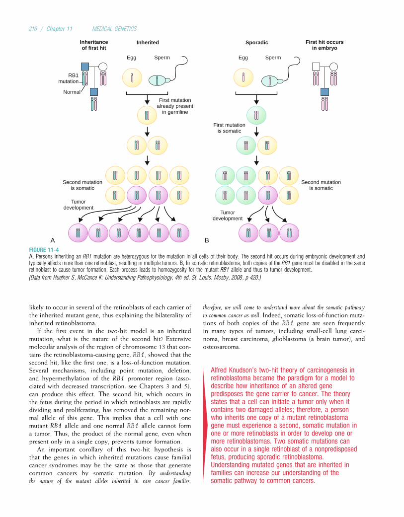
likely to occur in several of the retinoblasts of each carrier ofthe inherited mutant gene, thus explaining the bilaterality ofinherited retinoblastoma.
If the first event in the two-hit model is an inheritedmutation, what is the nature of the second hit? Extensivemolecular analysis of the region of chromosome 13 that con-tains the retinoblastoma-causing gene, RB1, showed that thesecond hit, like the first one, is a loss-of-function mutation.Several mechanisms, including point mutation, deletion,and hypermethylation of the RB1 promoter region (asso-ciated with decreased transcription; see Chapters 3 and 5),can produce this effect. The second hit, which occurs inthe fetus during the period in which retinoblasts are rapidlydividing and proliferating, has removed the remaining nor-mal allele of this gene. This implies that a cell with onemutant RB1 allele and one normal RB1 allele cannot forma tumor. Thus, the product of the normal gene, even whenpresent only in a single copy, prevents tumor formation.
An important corollary of this two-hit hypothesis isthat the genes in which inherited mutations cause familialcancer syndromes may be the same as those that generatecommon cancers by somatic mutation. By understanding
the nature of the mutant alleles inherited in rare cancer families,
therefore, we will come to understand more about the somatic pathway
to common cancer as well. Indeed, somatic loss-of-function muta-tions of both copies of the RB1 gene are seen frequentlyin many types of tumors, including small-cell lung carci-noma, breast carcinoma, glioblastoma (a brain tumor), andosteosarcoma.
4Alfred Knudson’s two-hit theory of carcinogenesis inretinoblastoma became the paradigm for a model todescribe how inheritance of an altered genepredisposes the gene carrier to cancer. The theorystates that a cell can initiate a tumor only when itcontains two damaged alleles; therefore, a personwho inherits one copy of a mutant retinoblastomagene must experience a second, somatic mutation inone or more retinoblasts in order to develop one ormore retinoblastomas. Two somatic mutations canalso occur in a single retinoblast of a nonpredisposedfetus, producing sporadic retinoblastoma.Understanding mutated genes that are inherited infamilies can increase our understanding of thesomatic pathway to common cancers.
InheritedInheritanceof first hit
Egg
RB1mutation
Normal
First mutationalready present
in germline
Second mutationis somatic
A B
Sperm
Sporadic
Egg
First mutationis somatic
Second mutationis somatic
Tumordevelopment
Sperm
Tumordevelopment
First hit occursin embryo
FIGURE 11-4A, Persons inheriting an RB1 mutation are heterozygous for the mutation in all cells of their body. The second hit occurs during embryonic development andtypically affects more than one retinoblast, resulting in multiple tumors. B, In somatic retinoblastoma, both copies of the RB1 gene must be disabled in the sameretinoblast to cause tumor formation. Each process leads to homozygosity for the mutant RB1 allele and thus to tumor development.
(Data from Huether S, McCance K: Understanding Pathophysiology, 4th ed. St. Louis: Mosby, 2008, p 420.)
216 / Chapter 11 MEDICAL GENETICS
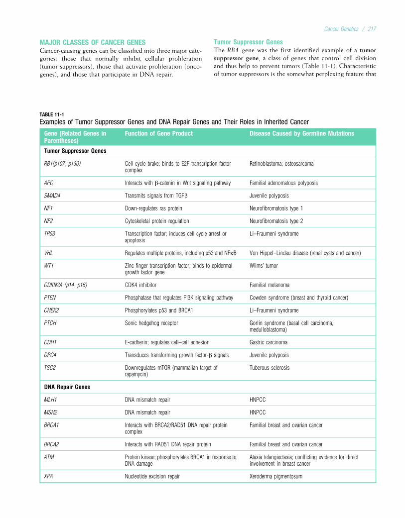
MAJOR CLASSES OF CANCER GENESCancer-causing genes can be classified into three major cate-gories: those that normally inhibit cellular proliferation(tumor suppressors), those that activate proliferation (onco-genes), and those that participate in DNA repair.
Tumor Suppressor GenesThe RB1 gene was the first identified example of a tumorsuppressor gene, a class of genes that control cell divisionand thus help to prevent tumors (Table 11-1). Characteristicof tumor suppressors is the somewhat perplexing feature that
TABLE 11-1
Examples of Tumor Suppressor Genes and DNA Repair Genes and Their Roles in Inherited Cancer
Gene (Related Genes inParentheses)
Function of Gene Product Disease Caused by Germline Mutations
Tumor Suppressor Genes
RB1(p107, p130) Cell cycle brake; binds to E2F transcription factorcomplex
Retinoblastoma; osteosarcoma
APC Interacts with b-catenin in Wnt signaling pathway Familial adenomatous polyposis
SMAD4 Transmits signals from TGFb Juvenile polyposis
NF1 Down-regulates ras protein Neurofibromatosis type 1
NF2 Cytoskeletal protein regulation Neurofibromatosis type 2
TP53 Transcription factor; induces cell cycle arrest orapoptosis
Li–Fraumeni syndrome
VHL Regulates multiple proteins, including p53 and NFkB Von Hippel–Lindau disease (renal cysts and cancer)
WT1 Zinc finger transcription factor; binds to epidermalgrowth factor gene
Wilms’ tumor
CDKN2A (p14, p16) CDK4 inhibitor Familial melanoma
PTEN Phosphatase that regulates PI3K signaling pathway Cowden syndrome (breast and thyroid cancer)
CHEK2 Phosphorylates p53 and BRCA1 Li–Fraumeni syndrome
PTCH Sonic hedgehog receptor Gorlin syndrome (basal cell carcinoma,medulloblastoma)
CDH1 E-cadherin; regulates cell–cell adhesion Gastric carcinoma
DPC4 Transduces transforming growth factor-b signals Juvenile polyposis
TSC2 Downregulates mTOR (mammalian target ofrapamycin)
Tuberous sclerosis
DNA Repair Genes
MLH1 DNA mismatch repair HNPCC
MSH2 DNA mismatch repair HNPCC
BRCA1 Interacts with BRCA2/RAD51 DNA repair proteincomplex
Familial breast and ovarian cancer
BRCA2 Interacts with RAD51 DNA repair protein Familial breast and ovarian cancer
ATM Protein kinase; phosphorylates BRCA1 in response toDNA damage
Ataxia telangiectasia; conflicting evidence for directinvolvement in breast cancer
XPA Nucleotide excision repair Xeroderma pigmentosum
Cancer Genetics / 217
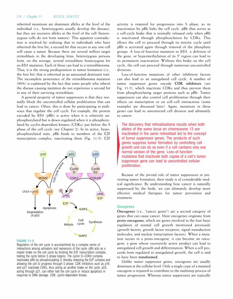
inherited mutations are dominant alleles at the level of theindividual (i.e., heterozygotes usually develop the disease),but they are recessive alleles at the level of the cell (hetero-zygous cells do not form tumors). This apparent contradic-tion is resolved by realizing that in individuals who haveinherited the first hit, a second hit that occurs in any one cellwill cause a tumor. Because there are several million targetretinoblasts in the developing fetus, heterozygous personsform, on the average, several retinoblasts homozygous foran RB1 mutation. Each of these can lead to a retinoblastoma.Thus, it is the strong predisposition to tumor formation (i.e.,the first hit) that is inherited as an autosomal dominant trait.The incomplete penetrance of the retinoblastoma mutation(90%) is explained by the fact that some people who inheritthe disease-causing mutation do not experience a second hitin any of their surviving retinoblasts.
A general property of tumor suppressors is that they nor-mally block the uncontrolled cellular proliferation that canlead to cancer. Often, this is done by participating in path-ways that regulate the cell cycle. For example, the proteinencoded by RB1 (pRb) is active when it is relatively un-phosphorylated but is down-regulated when it is phosphory-lated by cyclin-dependent kinases (CDKs) just before the Sphase of the cell cycle (see Chapter 2). In its active, hypo-phosphorylated state, pRb binds to members of the E2Ftranscription complex, inactivating them (Fig. 11-5). E2F
activity is required for progression into S phase, so itsinactivation by pRb halts the cell cycle. pRb thus serves asa cell-cycle brake that is normally released only when pRbis inactivated through phosphorylation by CDKs. Thisallows the cell to proceed through its mitotic cycle untilpRb is activated again through removal of the phosphategroups. A loss-of-function mutation in RB1, a deletion ofthe gene, or hypermethylation of its 50 region can lead toits permanent inactivation. Without this brake on the cellcycle, the cell can proceed through numerous uncontrolleddivisions.
Loss-of-function mutations of other inhibitory factorscan also lead to an unregulated cell cycle. A number oftumor suppressor genes encode CDK inhibitors (seeFig. 11-5), which inactivate CDKs and thus prevent themfrom phosphorylating target proteins such as pRb. Tumorsuppressors can also control cell proliferation through theireffects on transcription or on cell–cell interactions (someexamples are discussed later). Again, mutations in thesegenes can lead to unrestricted cell division and ultimatelyto cancer.
4The discovery that retinoblastoma results when bothalleles of the same locus on chromosome 13 areinactivated in the same retinoblast led to the conceptof tumor suppressor genes. The products of suchgenes suppress tumor formation by controlling cellgrowth and can do so even if a cell contains only onenormal version of the gene. Loss-of-functionmutations that inactivate both copies of a cell’s tumorsuppressor gene can lead to uncontrolled cellularproliferation.
Because of the pivotal role of tumor suppressors in pre-venting tumor formation, their study is of considerable med-ical significance. By understanding how cancer is naturallysuppressed by the body, we can ultimately develop moreeffective medical therapies for tumor prevention andtreatment.
OncogenesOncogenes (i.e., “cancer genes”) are a second category ofgenes that can cause cancer. Most oncogenes originate fromproto-oncogenes, which are genes involved in the four basicregulators of normal cell growth mentioned previously(growth factors, growth factor receptors, signal transductionmolecules, and nuclear transcription factors). When a muta-tion occurs in a proto-oncogene, it can become an onco-gene, a gene whose excessively active product can lead tounregulated cell growth and differentiation. When a cell pro-ceeds from regulated to unregulated growth, the cell is saidto have been transformed.
Unlike tumor suppressor genes, oncogenes are usuallydominant at the cellular level: Only a single copy of a mutatedoncogene is required to contribute to the multistep process oftumor progression. Whereas tumor suppressors are typically
p14
p53
Degradationof p53
Apoptosis
UUU p53 P P
p21
pRb
GAP1
GAP2
MitosisCellcycle Synthesis
E2F
pRbE2F
p16
CyclinE
CDK2MDM2 CDK4/6
CyclinD1
Cel
l cyc
le a
rrest
FIGURE 11-5Regulation of the cell cycle is accomplished by a complex series ofinteractions among activators and repressors of the cycle. pRb acts as amaster brake on the cell cycle by binding the E2F transcription complex,halting the cycle before S phase begins. The cyclin D–CDK4 complexinactivates pRb by phosphorylating it, thereby releasing the E2F complex andallowing the cell to progress through S phase. CDK inhibitors such as p16and p21 inactivate CDKs, thus acting as another brake on the cycle. p53,acting through p21, can either halt the cell cycle or induce apoptosis inresponse to DNA damage. CDK, cyclin-dependent kinase.
218 / Chapter 11 MEDICAL GENETICS
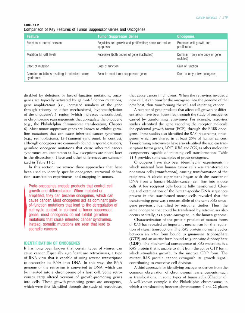
disabled by deletions or loss-of-function mutations, onco-genes are typically activated by gain-of-function mutations,gene amplification (i.e., increased numbers of the genethrough trisomy or other mechanisms), hypomethylationof the oncogene’s 50 region (which increases transcription),or chromosome rearrangements that upregulate the oncogene(e.g., the Philadelphia chromosome translocation, Chapter6). Most tumor suppressor genes are known to exhibit germ-line mutations that can cause inherited cancer syndromes(e.g., retinoblastoma, Li–Fraumeni syndrome). In contrast,although oncogenes are commonly found in sporadic tumors,germline oncogene mutations that cause inherited cancersyndromes are uncommon (a few exceptions are noted laterin the discussion). These and other differences are summar-ized in Table 11-2.
In this section, we review three approaches that havebeen used to identify specific oncogenes: retroviral defini-tion, transfection experiments, and mapping in tumors.
4Proto-oncogenes encode products that control cellgrowth and differentiation. When mutated oramplified, they can become oncogenes, which cancause cancer. Most oncogenes act as dominant gain-of-function mutations that lead to the deregulation ofcell cycle control. In contrast to tumor suppressorgenes, most oncogenes do not exhibit germlinemutations that cause inherited cancer syndromes.Instead, somatic mutations are seen that lead tosporadic cancers.
IDENTIFICATION OF ONCOGENESIt has long been known that certain types of viruses cancause cancer. Especially significant are retroviruses, a typeof RNA virus that is capable of using reverse transcriptaseto transcribe its RNA into DNA. In this way, the RNAgenome of the retrovirus is converted to DNA, which canbe inserted into a chromosome of a host cell. Some retro-viruses carry altered versions of growth-promoting genesinto cells. These growth-promoting genes are oncogenes,which were first identified through the study of retroviruses
that cause cancer in chickens. When the retrovirus invades anew cell, it can transfer the oncogene into the genome of thenew host, thus transforming the cell and initiating cancer.
A number of gene products that affect cell growth or differ-entiation have been identified through the study of oncogenescarried by transforming retroviruses. For example, retrovirusstudies identified the gene encoding the receptor moleculefor epidermal growth factor (EGF), through the ERBB onco-gene. These studies also identified the RAS (rat sarcoma) onco-genes, which are altered in at least 25% of human cancers.Transforming retroviruses have also identified the nuclear tran-scription factor genes,MYC, JUN, and FOS, as other molecularcomponents capable of initiating cell transformation. Table11-3 provides some examples of proto-oncogenes.
Oncogenes have also been identified in experiments inwhich material from human tumor cells was transferred tonontumor cells (transfection), causing transformation of therecipients. A classic experiment began with the transfer ofDNA from a human bladder-cancer cell line into mousecells. A few recipient cells became fully transformed. Clon-ing and examination of the human-specific DNA sequencespresent in the transformed mouse cells revealed that thetransforming gene was a mutant allele of the same RAS onco-gene previously identified by retroviral studies. Thus, thesame oncogene that could be transferred by retroviruses alsooccurs naturally, as a proto-oncogene, in the human genome.
Characterization of the protein product of mutant formsof RAS has revealed an important mechanism for the regula-tion of signal transduction. The RAS protein normally cyclesbetween an active form bound to guanosine triphosphate(GTP) and an inactive form bound to guanosine diphosphate(GDP). The biochemical consequence of RAS mutations is aRAS protein that is unable to shift from the active GTP form,which stimulates growth, to the inactive GDP form. Themutant RAS protein cannot extinguish its growth signal,contributing to excessive cell division.
A third approach for identifying oncogenes derives from thecommon observation of chromosomal rearrangements, suchas translocations, in some types of tumor cells (Chapter 6).A well-known example is the Philadelphia chromosome, inwhich a translocation between chromosomes 9 and 22 places
TABLE 11-2
Comparison of Key Features of Tumor Suppressor Genes and Oncogenes
Feature Tumor Suppressor Genes Oncogenes
Function of normal version Regulates cell growth and proliferation; some can induceapoptosis
Promotes cell growth andproliferation
Mutation (at cell level) Recessive (both copies of gene inactivated) Dominant (only one copy of genemutated)
Effect of mutation Loss of function Gain of function
Germline mutations resulting in inherited cancersyndromes
Seen in most tumor suppressor genes Seen in only a few oncogenes
Cancer Genetics / 219
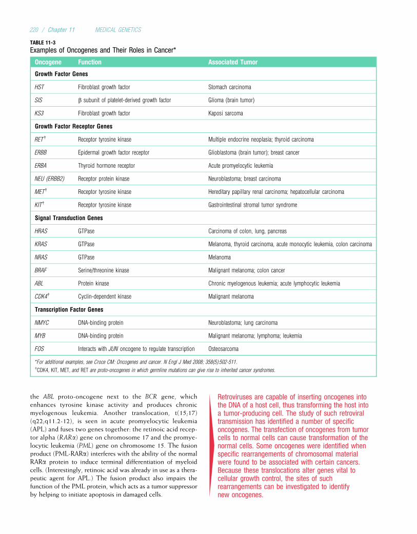
the ABL proto-oncogene next to the BCR gene, whichenhances tyrosine kinase activity and produces chronicmyelogenous leukemia. Another translocation, t(15;17)(q22;q11.2-12), is seen in acute promyelocytic leukemia(APL) and fuses two genes together: the retinoic acid recep-tor alpha (RARa) gene on chromosome 17 and the promye-locytic leukemia (PML) gene on chromosome 15. The fusionproduct (PML-RARa) interferes with the ability of the normalRARa protein to induce terminal differentiation of myeloidcells. (Interestingly, retinoic acid was already in use as a thera-peutic agent for APL.) The fusion product also impairs thefunction of the PML protein, which acts as a tumor suppressorby helping to initiate apoptosis in damaged cells. 4
Retroviruses are capable of inserting oncogenes intothe DNA of a host cell, thus transforming the host intoa tumor-producing cell. The study of such retroviraltransmission has identified a number of specificoncogenes. The transfection of oncogenes from tumorcells to normal cells can cause transformation of thenormal cells. Some oncogenes were identified whenspecific rearrangements of chromosomal materialwere found to be associated with certain cancers.Because these translocations alter genes vital tocellular growth control, the sites of suchrearrangements can be investigated to identifynew oncogenes.
TABLE 11-3
Examples of Oncogenes and Their Roles in Cancer*
Oncogene Function Associated Tumor
Growth Factor Genes
HST Fibroblast growth factor Stomach carcinoma
SIS b subunit of platelet-derived growth factor Glioma (brain tumor)
KS3 Fibroblast growth factor Kaposi sarcoma
Growth Factor Receptor Genes
RET{ Receptor tyrosine kinase Multiple endocrine neoplasia; thyroid carcinoma
ERBB Epidermal growth factor receptor Glioblastoma (brain tumor); breast cancer
ERBA Thyroid hormone receptor Acute promyelocytic leukemia
NEU (ERBB2) Receptor protein kinase Neuroblastoma; breast carcinoma
MET{ Receptor tyrosine kinase Hereditary papillary renal carcinoma; hepatocellular carcinoma
KIT{ Receptor tyrosine kinase Gastrointestinal stromal tumor syndrome
Signal Transduction Genes
HRAS GTPase Carcinoma of colon, lung, pancreas
KRAS GTPase Melanoma, thyroid carcinoma, acute monocytic leukemia, colon carcinoma
NRAS GTPase Melanoma
BRAF Serine/threonine kinase Malignant melanoma; colon cancer
ABL Protein kinase Chronic myelogenous leukemia; acute lymphocytic leukemia
CDK4{ Cyclin-dependent kinase Malignant melanoma
Transcription Factor Genes
NMYC DNA-binding protein Neuroblastoma; lung carcinoma
MYB DNA-binding protein Malignant melanoma; lymphoma; leukemia
FOS Interacts with JUN oncogene to regulate transcription Osteosarcoma
*For additional examples, see Croce CM: Oncogenes and cancer. N Engl J Med 2008; 358(5):502-511.{CDK4, KIT, MET, and RET are proto-oncogenes in which germline mutations can give rise to inherited cancer syndromes.
220 / Chapter 11 MEDICAL GENETICS
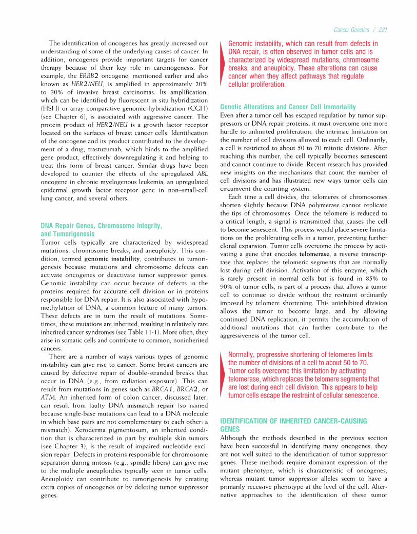
The identification of oncogenes has greatly increased ourunderstanding of some of the underlying causes of cancer. Inaddition, oncogenes provide important targets for cancertherapy because of their key role in carcinogenesis. Forexample, the ERBB2 oncogene, mentioned earlier and alsoknown as HER2/NEU, is amplified in approximately 20%to 30% of invasive breast carcinomas. Its amplification,which can be identified by fluorescent in situ hybridization(FISH) or array comparative genomic hybridization (CGH)(see Chapter 6), is associated with aggressive cancer. Theprotein product of HER2/NEU is a growth factor receptorlocated on the surfaces of breast cancer cells. Identificationof the oncogene and its product contributed to the develop-ment of a drug, trastuzumab, which binds to the amplifiedgene product, effectively downregulating it and helping totreat this form of breast cancer. Similar drugs have beendeveloped to counter the effects of the upregulated ABL
oncogene in chronic myelogenous leukemia, an upregulatedepidermal growth factor receptor gene in non–small-celllung cancer, and several others.
DNA Repair Genes, Chromosome Integrity,and TumorigenesisTumor cells typically are characterized by widespreadmutations, chromosome breaks, and aneuploidy. This con-dition, termed genomic instability, contributes to tumori-genesis because mutations and chromosome defects canactivate oncogenes or deactivate tumor suppressor genes.Genomic instability can occur because of defects in theproteins required for accurate cell division or in proteinsresponsible for DNA repair. It is also associated with hypo-methylation of DNA, a common feature of many tumors.These defects are in turn the result of mutations. Some-times, these mutations are inherited, resulting in relatively rareinherited cancer syndromes (see Table 11-1). More often, theyarise in somatic cells and contribute to common, noninheritedcancers.
There are a number of ways various types of genomicinstability can give rise to cancer. Some breast cancers arecaused by defective repair of double-stranded breaks thatoccur in DNA (e.g., from radiation exposure). This canresult from mutations in genes such as BRCA1, BRCA2, orATM. An inherited form of colon cancer, discussed later,can result from faulty DNA mismatch repair (so namedbecause single-base mutations can lead to a DNA moleculein which base pairs are not complementary to each other: amismatch). Xeroderma pigmentosum, an inherited condi-tion that is characterized in part by multiple skin tumors(see Chapter 3), is the result of impaired nucleotide exci-sion repair. Defects in proteins responsible for chromosomeseparation during mitosis (e.g., spindle fibers) can give riseto the multiple aneuploidies typically seen in tumor cells.Aneuploidy can contribute to tumorigenesis by creatingextra copies of oncogenes or by deleting tumor suppressorgenes.
4Genomic instability, which can result from defects inDNA repair, is often observed in tumor cells and ischaracterized by widespread mutations, chromosomebreaks, and aneuploidy. These alterations can causecancer when they affect pathways that regulatecellular proliferation.
Genetic Alterations and Cancer Cell ImmortalityEven after a tumor cell has escaped regulation by tumor sup-pressors or DNA repair proteins, it must overcome one morehurdle to unlimited proliferation: the intrinsic limitation onthe number of cell divisions allowed to each cell. Ordinarily,a cell is restricted to about 50 to 70 mitotic divisions. Afterreaching this number, the cell typically becomes senescentand cannot continue to divide. Recent research has providednew insights on the mechanisms that count the number ofcell divisions and has illustrated new ways tumor cells cancircumvent the counting system.
Each time a cell divides, the telomeres of chromosomesshorten slightly because DNA polymerase cannot replicatethe tips of chromosomes. Once the telomere is reduced toa critical length, a signal is transmitted that causes the cellto become senescent. This process would place severe limita-tions on the proliferating cells in a tumor, preventing furtherclonal expansion. Tumor cells overcome the process by acti-vating a gene that encodes telomerase, a reverse transcrip-tase that replaces the telomeric segments that are normallylost during cell division. Activation of this enzyme, whichis rarely present in normal cells but is found in 85% to90% of tumor cells, is part of a process that allows a tumorcell to continue to divide without the restraint ordinarilyimposed by telomere shortening. This uninhibited divisionallows the tumor to become large, and, by allowingcontinued DNA replication, it permits the accumulation ofadditional mutations that can further contribute to theaggressiveness of the tumor cell.
4Normally, progressive shortening of telomeres limitsthe number of divisions of a cell to about 50 to 70.Tumor cells overcome this limitation by activatingtelomerase, which replaces the telomere segments thatare lost during each cell division. This appears to helptumor cells escape the restraint of cellular senescence.
IDENTIFICATION OF INHERITED CANCER-CAUSINGGENESAlthough the methods described in the previous sectionhave been successful in identifying many oncogenes, theyare not well suited to the identification of tumor suppressorgenes. These methods require dominant expression of themutant phenotype, which is characteristic of oncogenes,whereas mutant tumor suppressor alleles seem to have aprimarily recessive phenotype at the level of the cell. Alter-native approaches to the identification of these tumor
Cancer Genetics / 221
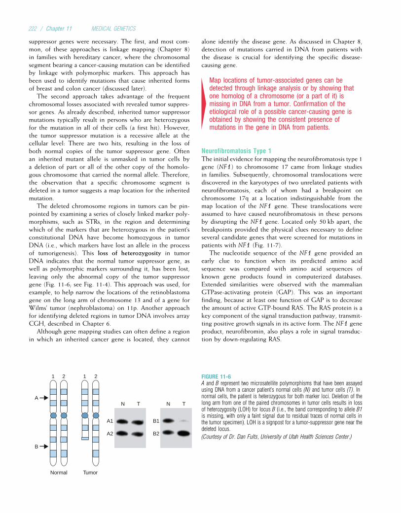
suppressor genes were necessary. The first, and most com-mon, of these approaches is linkage mapping (Chapter 8)in families with hereditary cancer, where the chromosomalsegment bearing a cancer-causing mutation can be identifiedby linkage with polymorphic markers. This approach hasbeen used to identify mutations that cause inherited formsof breast and colon cancer (discussed later).
The second approach takes advantage of the frequentchromosomal losses associated with revealed tumor suppres-sor genes. As already described, inherited tumor suppressormutations typically result in persons who are heterozygousfor the mutation in all of their cells (a first hit). However,the tumor suppressor mutation is a recessive allele at thecellular level: There are two hits, resulting in the loss ofboth normal copies of the tumor suppressor gene. Oftenan inherited mutant allele is unmasked in tumor cells bya deletion of part or all of the other copy of the homolo-gous chromosome that carried the normal allele. Therefore,the observation that a specific chromosome segment isdeleted in a tumor suggests a map location for the inheritedmutation.
The deleted chromosome regions in tumors can be pin-pointed by examining a series of closely linked marker poly-morphisms, such as STRs, in the region and determiningwhich of the markers that are heterozygous in the patient’sconstitutional DNA have become homozygous in tumorDNA (i.e., which markers have lost an allele in the processof tumorigenesis). This loss of heterozygosity in tumorDNA indicates that the normal tumor suppressor gene, aswell as polymorphic markers surrounding it, has been lost,leaving only the abnormal copy of the tumor suppressorgene (Fig. 11-6; see Fig. 11-4). This approach was used, forexample, to help narrow the locations of the retinoblastomagene on the long arm of chromosome 13 and of a gene forWilms’ tumor (nephroblastoma) on 11p. Another approachfor identifying deleted regions in tumor DNA involves arrayCGH, described in Chapter 6.
Although gene mapping studies can often define a regionin which an inherited cancer gene is located, they cannot
alone identify the disease gene. As discussed in Chapter 8,detection of mutations carried in DNA from patients withthe disease is crucial for identifying the specific disease-causing gene.
4Map locations of tumor-associated genes can bedetected through linkage analysis or by showing thatone homolog of a chromosome (or a part of it) ismissing in DNA from a tumor. Confirmation of theetiological role of a possible cancer-causing gene isobtained by showing the consistent presence ofmutations in the gene in DNA from patients.
Neurofibromatosis Type 1The initial evidence for mapping the neurofibromatosis type 1gene (NF1) to chromosome 17 came from linkage studiesin families. Subsequently, chromosomal translocations werediscovered in the karyotypes of two unrelated patients withneurofibromatosis, each of whom had a breakpoint onchromosome 17q at a location indistinguishable from themap location of the NF1 gene. These translocations wereassumed to have caused neurofibromatosis in these personsby disrupting the NF1 gene. Located only 50 kb apart, thebreakpoints provided the physical clues necessary to defineseveral candidate genes that were screened for mutations inpatients with NF1 (Fig. 11-7).
The nucleotide sequence of the NF1 gene provided anearly clue to function when its predicted amino acidsequence was compared with amino acid sequences ofknown gene products found in computerized databases.Extended similarities were observed with the mammalianGTPase-activating protein (GAP). This was an importantfinding, because at least one function of GAP is to decreasethe amount of active GTP-bound RAS. The RAS protein is akey component of the signal transduction pathway, transmit-ting positive growth signals in its active form. The NF1 geneproduct, neurofibromin, also plays a role in signal transduc-tion by down-regulating RAS.
A
B
Normal Tumor
N T
A1
A2 B2
B1
N T
1 2 1 2 FIGURE 11-6A and B represent two microsatellite polymorphisms that have been assayedusing DNA from a cancer patient’s normal cells (N) and tumor cells (T). Innormal cells, the patient is heterozygous for both marker loci. Deletion of thelong arm from one of the paired chromosomes in tumor cells results in lossof heterozygosity (LOH) for locus B (i.e., the band corresponding to allele B1is missing, with only a faint signal due to residual traces of normal cells inthe tumor specimen). LOH is a signpost for a tumor-suppressor gene near thedeleted locus.
(Courtesy of Dr. Dan Fults, University of Utah Health Sciences Center.)
222 / Chapter 11 MEDICAL GENETICS
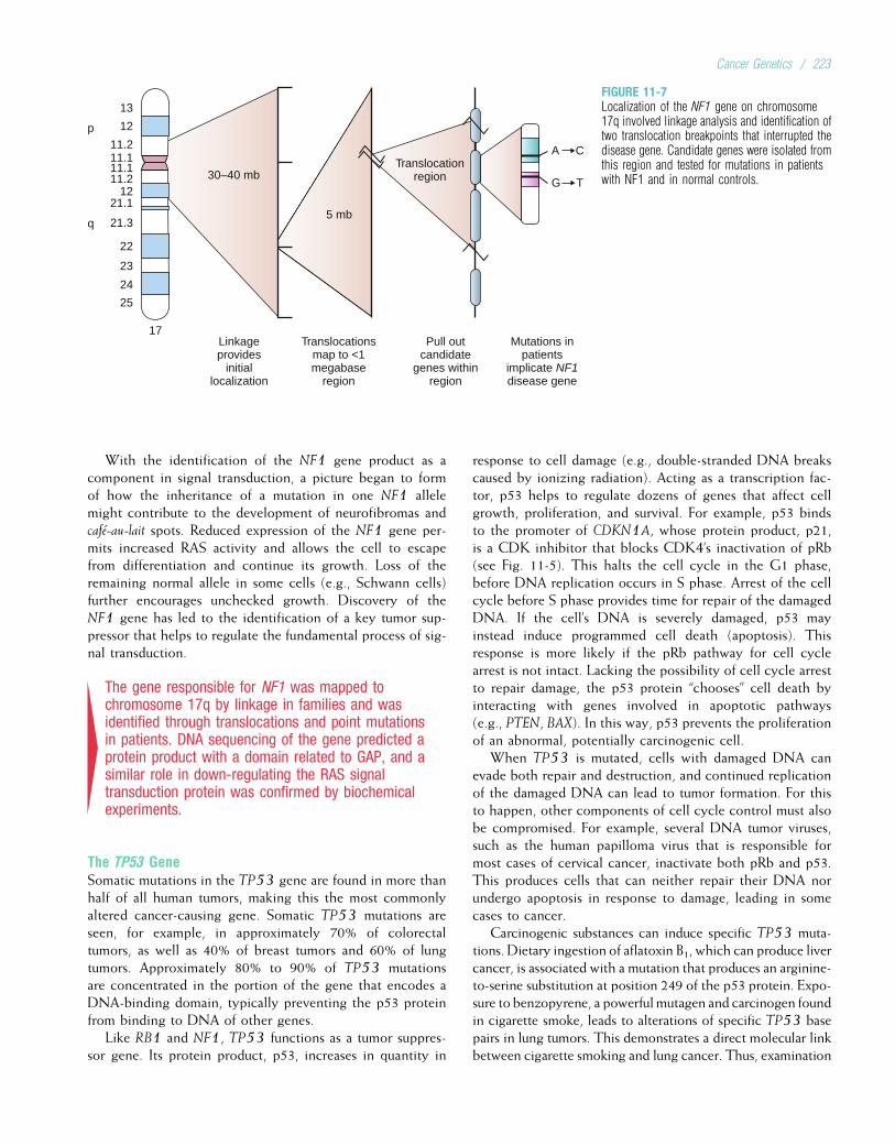
With the identification of the NF1 gene product as acomponent in signal transduction, a picture began to formof how the inheritance of a mutation in one NF1 allelemight contribute to the development of neurofibromas andcafe-au-lait spots. Reduced expression of the NF1 gene per-mits increased RAS activity and allows the cell to escapefrom differentiation and continue its growth. Loss of theremaining normal allele in some cells (e.g., Schwann cells)further encourages unchecked growth. Discovery of theNF1 gene has led to the identification of a key tumor sup-pressor that helps to regulate the fundamental process of sig-nal transduction.
4The gene responsible for NF1 was mapped tochromosome 17q by linkage in families and wasidentified through translocations and point mutationsin patients. DNA sequencing of the gene predicted aprotein product with a domain related to GAP, and asimilar role in down-regulating the RAS signaltransduction protein was confirmed by biochemicalexperiments.
The TP53 GeneSomatic mutations in the TP53 gene are found in more thanhalf of all human tumors, making this the most commonlyaltered cancer-causing gene. Somatic TP53 mutations areseen, for example, in approximately 70% of colorectaltumors, as well as 40% of breast tumors and 60% of lungtumors. Approximately 80% to 90% of TP53 mutationsare concentrated in the portion of the gene that encodes aDNA-binding domain, typically preventing the p53 proteinfrom binding to DNA of other genes.
Like RB1 and NF1, TP53 functions as a tumor suppres-sor gene. Its protein product, p53, increases in quantity in
response to cell damage (e.g., double-stranded DNA breakscaused by ionizing radiation). Acting as a transcription fac-tor, p53 helps to regulate dozens of genes that affect cellgrowth, proliferation, and survival. For example, p53 bindsto the promoter of CDKN1A, whose protein product, p21,is a CDK inhibitor that blocks CDK4’s inactivation of pRb(see Fig. 11-5). This halts the cell cycle in the G1 phase,before DNA replication occurs in S phase. Arrest of the cellcycle before S phase provides time for repair of the damagedDNA. If the cell’s DNA is severely damaged, p53 mayinstead induce programmed cell death (apoptosis). Thisresponse is more likely if the pRb pathway for cell cyclearrest is not intact. Lacking the possibility of cell cycle arrestto repair damage, the p53 protein “chooses” cell death byinteracting with genes involved in apoptotic pathways(e.g., PTEN, BAX). In this way, p53 prevents the proliferationof an abnormal, potentially carcinogenic cell.
When TP53 is mutated, cells with damaged DNA canevade both repair and destruction, and continued replicationof the damaged DNA can lead to tumor formation. For thisto happen, other components of cell cycle control must alsobe compromised. For example, several DNA tumor viruses,such as the human papilloma virus that is responsible formost cases of cervical cancer, inactivate both pRb and p53.This produces cells that can neither repair their DNA norundergo apoptosis in response to damage, leading in somecases to cancer.
Carcinogenic substances can induce specific TP53 muta-tions. Dietary ingestion of aflatoxin B1, which can produce livercancer, is associated with a mutation that produces an arginine-to-serine substitution at position 249 of the p53 protein. Expo-sure to benzopyrene, a powerful mutagen and carcinogen foundin cigarette smoke, leads to alterations of specific TP53 basepairs in lung tumors. This demonstrates a direct molecular linkbetween cigarette smoking and lung cancer. Thus, examination
13
12p
q
11.211.111.111.2
1221.1
21.3
22
23
24
25
A C
G T
Translocationregion
5 mb
30–40 mb
Mutations inpatients
implicate NF1disease gene
Pull outcandidate
genes withinregion
Translocationsmap to <1megabase
region
Linkageprovides
initiallocalization
17
FIGURE 11-7Localization of the NF1 gene on chromosome17q involved linkage analysis and identification oftwo translocation breakpoints that interrupted thedisease gene. Candidate genes were isolated fromthis region and tested for mutations in patientswith NF1 and in normal controls.
Cancer Genetics / 223
of the type of TP53mutation seen in a tumor can provide cluesto the identity of the causative carcinogenic agent.
Although tumor-causing TP53 mutations have beenobserved mostly in somatic cells, germline mutations ofTP53 are responsible for an inherited cancer conditionknown as the Li–Fraumeni syndrome (LFS). This rare syn-drome is transmitted in autosomal dominant fashion andinvolves breast and colon carcinomas, soft-tissue sarcomas,osteosarcomas, brain tumors, leukemia, and adrenocorticalcarcinomas. These tumors usually develop at early ages inmembers of LFS families, and multiple primary tumors arecommonly seen in an affected person. The demonstration ofconsistent TP53 mutations in the constitutional DNA ofpatients with LFS confirmed the causative role of this gene.As in retinoblastoma, the inheritance of a mutated TP53 genegreatly increases the person’s susceptibility to subsequent celltransformation and tumor development when a cell loses theother, normal copy of TP53 (two-hit model). Among LFSfamily members who inherit an abnormal TP53 gene, approx-imately 50% develop invasive cancer by 30 years of age, andmore than 90% develop invasive cancer by age 70 years.
TP53 mutations account for only about 75% of LFS cases.Some of the remaining cases are the result of mutations inanother tumor suppressor gene, CHEK2. This gene encodesa kinase that normally phosphorylates p53 in response to ion-izing radiation, resulting in the accumulation and activationof p53. Loss-of-function mutations in CHEK2 result in a lackof p53 activation, causing LFS via the p53 pathway.
TP53 is medically important in at least two ways. First,the presence of TP53 mutations in tumors, particularly thoseof the breast and colon, often signals a more-aggressive can-cer with relatively poor survival prospects. It is thus a usefulprognostic indicator. Second, TP53 might ultimately proveimportant in tumor prevention. Laboratory experimentsshow that the insertion of a normal TP53 gene into tumorcells can induce tumor regression by inducing abnormal can-cer cells to undergo apoptosis. This has led to gene therapyprotocols (see Chapter 13) in which normal TP53 copiesare inserted into tumors in an effort to eliminate cancerouscells.
4Somatic mutations of the TP53 gene are found in themajority of tumors. This gene encodes a protein thatcan induce either cell cycle arrest or apoptosis inresponse to damaged DNA. Inherited TP53 mutationscan cause Li–Fraumeni syndrome.
The Familial Adenomatous Polyposis Gene, APCColon cancer affects approximately 1 in 20 Americans,and, like most common cancers, it is more likely to occurin persons with a positive family history. One’s risk of devel-oping colon cancer approximately doubles if a first-degreerelative is affected. In addition, a small fraction of coloncancer cases are inherited as autosomal dominant syn-dromes. The two most important of these syndromes arediscussed next.
Familial adenomatous polyposis (FAP), also called adeno-matous polyposis coli (APC), is characterized by the appear-ance of large numbers adenomas, or polyps, of the colon inthe second or third decade of life. Colonic adenomas arenow understood to be the immediate precursors to colorectalcancer. The multiple adenomas of the patient with FAP there-fore present a grave risk of early malignancy. Because earlydetection and removal of adenomatous polyps can signifi-cantly reduce the occurrence of cancer, it is important tounderstand the causative gene and its role in the developmentof polyps (Clinical Commentary 11-1).
The gene responsible for FAP was localized to the longarm of chromosome 5 by linkage analysis in families. Discov-ery of small, overlapping deletions in two unrelated patientsprovided the key to isolation of the disease-causing gene,termed APC. Among the genes that lay within the 100-kbregion that was deleted in both patients, one showed appar-ent mutations in other patients. This mutation was seen inone patient but not in his unaffected parents, confirmingthe identification of the APC gene.
Like RB1 and TP53, APC is a tumor suppressor gene, andboth copies of APC must be inactivated in a cell for tumorprogression to begin. Persons who inherit an APC mutationtypically experience somatic loss-of-function mutations inhundreds of their colonic epithelial cells, giving rise to mul-tiple adenomas. In some cases, loss of function of APC occursbecause of hypermethylation of APC’s promoter region,which results in reduced transcription (see Chapter 5).(Hypermethylation has been observed in the inactivationof a number of tumor suppressor and DNA repair genes,including those associated with retinoblastoma [RB1], breastcancer [BRCA1], hereditary nonpolyposis colorectal cancer[MLH1], malignant melanoma [CDKN2A], and von Hip-pel–Lindau disease [VHL]).
Identification of the APC gene has been important indiagnosing and managing colon cancer in families withFAP (see Clinical Commentary 11-1). In addition, and per-haps even more importantly, APC mutations are found in85% of all sporadic, noninherited cases of colon cancer.Somatic APC mutations (i.e., those that disable both copiesof the gene in a colonic cell) are among the earliest altera-tions that give rise to colon cancer. But APC mutations arenot sufficient by themselves to complete the progression tometastatic disease. As shown in Figure 11-8, other genesmay also be altered. For example, gain-of-function mutationsare seen in the KRAS gene in approximately 50% of colontumors. This gene encodes a signal transduction molecule,and a gain-of-function mutation increases signaling and thuscellular proliferation. Loss-of-function mutations in theTP53 gene are also seen in more than 50% of colon tumorsand usually occur relatively late in the pathway to cancer.Ordinarily, p53 would be activated in response to mutationslike those of APC and KRAS, leading to DNA repair or apo-ptosis. Cells that lack p53 activity are free to continue alongthe path to malignancy in spite of their damaged DNA. Stillanother tumor suppressor gene, SMAD4, appears also to be
224 / Chapter 11 MEDICAL GENETICS
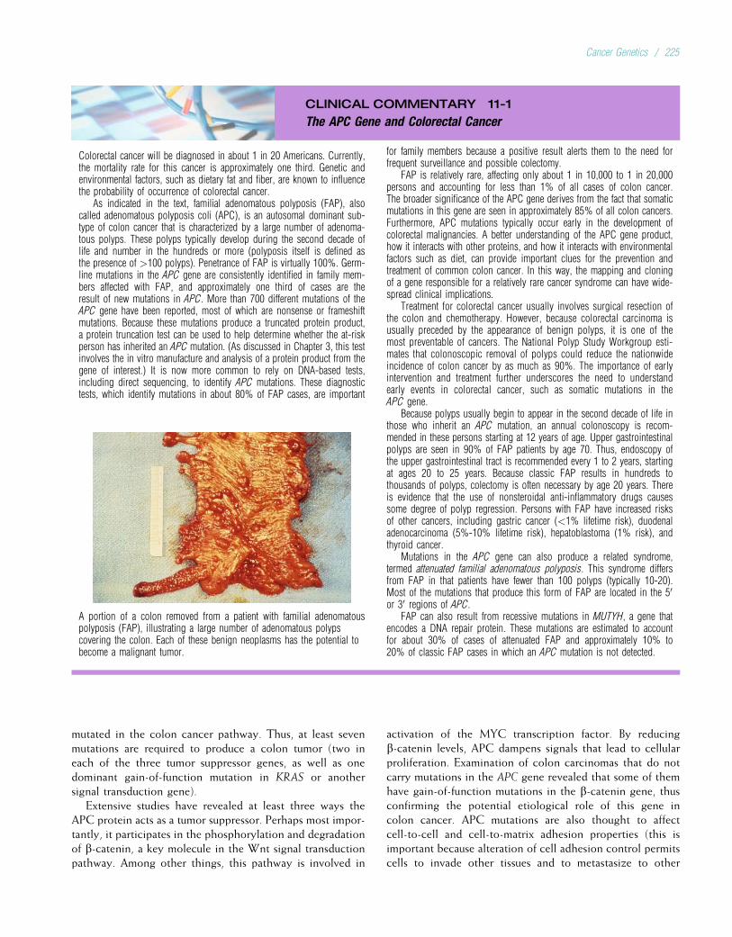
CLINICAL COMMENTARY 11-1
The APC Gene and Colorectal Cancer
Colorectal cancer will be diagnosed in about 1 in 20 Americans. Currently,the mortality rate for this cancer is approximately one third. Genetic andenvironmental factors, such as dietary fat and fiber, are known to influencethe probability of occurrence of colorectal cancer.
As indicated in the text, familial adenomatous polyposis (FAP), alsocalled adenomatous polyposis coli (APC), is an autosomal dominant sub-type of colon cancer that is characterized by a large number of adenoma-tous polyps. These polyps typically develop during the second decade oflife and number in the hundreds or more (polyposis itself is defined asthe presence of >100 polyps). Penetrance of FAP is virtually 100%. Germ-line mutations in the APC gene are consistently identified in family mem-bers affected with FAP, and approximately one third of cases are theresult of new mutations in APC. More than 700 different mutations of theAPC gene have been reported, most of which are nonsense or frameshiftmutations. Because these mutations produce a truncated protein product,a protein truncation test can be used to help determine whether the at-riskperson has inherited an APC mutation. (As discussed in Chapter 3, this testinvolves the in vitro manufacture and analysis of a protein product from thegene of interest.) It is now more common to rely on DNA-based tests,including direct sequencing, to identify APC mutations. These diagnostictests, which identify mutations in about 80% of FAP cases, are important
A portion of a colon removed from a patient with familial adenomatouspolyposis (FAP), illustrating a large number of adenomatous polypscovering the colon. Each of these benign neoplasms has the potential tobecome a malignant tumor.
for family members because a positive result alerts them to the need forfrequent surveillance and possible colectomy.
FAP is relatively rare, affecting only about 1 in 10,000 to 1 in 20,000persons and accounting for less than 1% of all cases of colon cancer.The broader significance of the APC gene derives from the fact that somaticmutations in this gene are seen in approximately 85% of all colon cancers.Furthermore, APC mutations typically occur early in the development ofcolorectal malignancies. A better understanding of the APC gene product,how it interacts with other proteins, and how it interacts with environmentalfactors such as diet, can provide important clues for the prevention andtreatment of common colon cancer. In this way, the mapping and cloningof a gene responsible for a relatively rare cancer syndrome can have wide-spread clinical implications.
Treatment for colorectal cancer usually involves surgical resection ofthe colon and chemotherapy. However, because colorectal carcinoma isusually preceded by the appearance of benign polyps, it is one of themost preventable of cancers. The National Polyp Study Workgroup esti-mates that colonoscopic removal of polyps could reduce the nationwideincidence of colon cancer by as much as 90%. The importance of earlyintervention and treatment further underscores the need to understandearly events in colorectal cancer, such as somatic mutations in theAPC gene.
Because polyps usually begin to appear in the second decade of life inthose who inherit an APC mutation, an annual colonoscopy is recom-mended in these persons starting at 12 years of age. Upper gastrointestinalpolyps are seen in 90% of FAP patients by age 70. Thus, endoscopy ofthe upper gastrointestinal tract is recommended every 1 to 2 years, startingat ages 20 to 25 years. Because classic FAP results in hundreds tothousands of polyps, colectomy is often necessary by age 20 years. Thereis evidence that the use of nonsteroidal anti-inflammatory drugs causessome degree of polyp regression. Persons with FAP have increased risksof other cancers, including gastric cancer (<1% lifetime risk), duodenaladenocarcinoma (5%-10% lifetime risk), hepatoblastoma (1% risk), andthyroid cancer.
Mutations in the APC gene can also produce a related syndrome,termed attenuated familial adenomatous polyposis. This syndrome differsfrom FAP in that patients have fewer than 100 polyps (typically 10-20).Most of the mutations that produce this form of FAP are located in the 50or 30 regions of APC.
FAP can also result from recessive mutations in MUTYH, a gene thatencodes a DNA repair protein. These mutations are estimated to accountfor about 30% of cases of attenuated FAP and approximately 10% to20% of classic FAP cases in which an APC mutation is not detected.
mutated in the colon cancer pathway. Thus, at least sevenmutations are required to produce a colon tumor (two ineach of the three tumor suppressor genes, as well as onedominant gain-of-function mutation in KRAS or anothersignal transduction gene).
Extensive studies have revealed at least three ways theAPC protein acts as a tumor suppressor. Perhaps most impor-tantly, it participates in the phosphorylation and degradationof b-catenin, a key molecule in the Wnt signal transductionpathway. Among other things, this pathway is involved in
activation of the MYC transcription factor. By reducingb-catenin levels, APC dampens signals that lead to cellularproliferation. Examination of colon carcinomas that do notcarry mutations in the APC gene revealed that some of themhave gain-of-function mutations in the b-catenin gene, thusconfirming the potential etiological role of this gene incolon cancer. APC mutations are also thought to affectcell-to-cell and cell-to-matrix adhesion properties (this isimportant because alteration of cell adhesion control permitscells to invade other tissues and to metastasize to other
Cancer Genetics / 225
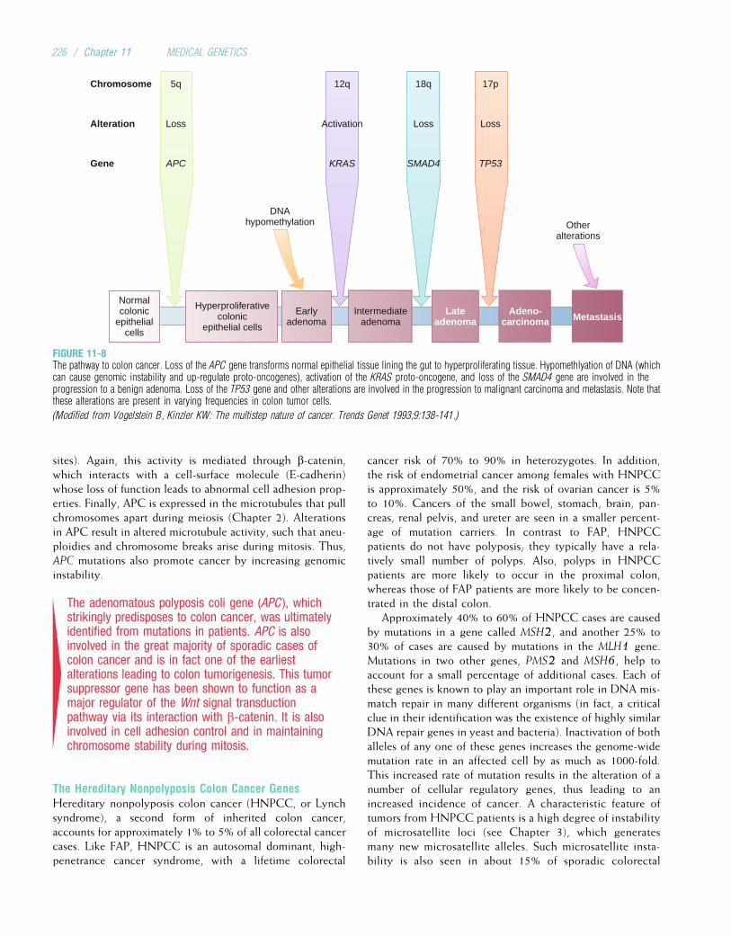
sites). Again, this activity is mediated through b-catenin,which interacts with a cell-surface molecule (E-cadherin)whose loss of function leads to abnormal cell adhesion prop-erties. Finally, APC is expressed in the microtubules that pullchromosomes apart during meiosis (Chapter 2). Alterationsin APC result in altered microtubule activity, such that aneu-ploidies and chromosome breaks arise during mitosis. Thus,APC mutations also promote cancer by increasing genomicinstability.
4The adenomatous polyposis coli gene (APC ), whichstrikingly predisposes to colon cancer, was ultimatelyidentified from mutations in patients. APC is alsoinvolved in the great majority of sporadic cases ofcolon cancer and is in fact one of the earliestalterations leading to colon tumorigenesis. This tumorsuppressor gene has been shown to function as amajor regulator of the Wnt signal transductionpathway via its interaction with b-catenin. It is alsoinvolved in cell adhesion control and in maintainingchromosome stability during mitosis.
The Hereditary Nonpolyposis Colon Cancer GenesHereditary nonpolyposis colon cancer (HNPCC, or Lynchsyndrome), a second form of inherited colon cancer,accounts for approximately 1% to 5% of all colorectal cancercases. Like FAP, HNPCC is an autosomal dominant, high-penetrance cancer syndrome, with a lifetime colorectal
cancer risk of 70% to 90% in heterozygotes. In addition,the risk of endometrial cancer among females with HNPCCis approximately 50%, and the risk of ovarian cancer is 5%to 10%. Cancers of the small bowel, stomach, brain, pan-creas, renal pelvis, and ureter are seen in a smaller percent-age of mutation carriers. In contrast to FAP, HNPCCpatients do not have polyposis; they typically have a rela-tively small number of polyps. Also, polyps in HNPCCpatients are more likely to occur in the proximal colon,whereas those of FAP patients are more likely to be concen-trated in the distal colon.
Approximately 40% to 60% of HNPCC cases are causedby mutations in a gene called MSH2, and another 25% to30% of cases are caused by mutations in the MLH1 gene.Mutations in two other genes, PMS2 and MSH6, help toaccount for a small percentage of additional cases. Each ofthese genes is known to play an important role in DNA mis-match repair in many different organisms (in fact, a criticalclue in their identification was the existence of highly similarDNA repair genes in yeast and bacteria). Inactivation of bothalleles of any one of these genes increases the genome-widemutation rate in an affected cell by as much as 1000-fold.This increased rate of mutation results in the alteration of anumber of cellular regulatory genes, thus leading to anincreased incidence of cancer. A characteristic feature oftumors from HNPCC patients is a high degree of instabilityof microsatellite loci (see Chapter 3), which generatesmany new microsatellite alleles. Such microsatellite insta-bility is also seen in about 15% of sporadic colorectal
DNAhypomethylation Other
alterations
17p18q12q
LossLossActivation
TP53SMAD4KRAS
5q
Loss
APC
Normalcolonic
epithelialcells
Hyperproliferativecolonic
epithelial cells
Earlyadenoma
Intermediateadenoma
Lateadenoma
Adeno-carcinoma Metastasis
Chromosome
Alteration
Gene
FIGURE 11-8The pathway to colon cancer. Loss of the APC gene transforms normal epithelial tissue lining the gut to hyperproliferating tissue. Hypomethlyation of DNA (whichcan cause genomic instability and up-regulate proto-oncogenes), activation of the KRAS proto-oncogene, and loss of the SMAD4 gene are involved in theprogression to a benign adenoma. Loss of the TP53 gene and other alterations are involved in the progression to malignant carcinoma and metastasis. Note thatthese alterations are present in varying frequencies in colon tumor cells.
(Modified from Vogelstein B, Kinzler KW: The multistep nature of cancer. Trends Genet 1993;9:138-141.)
226 / Chapter 11 MEDICAL GENETICS
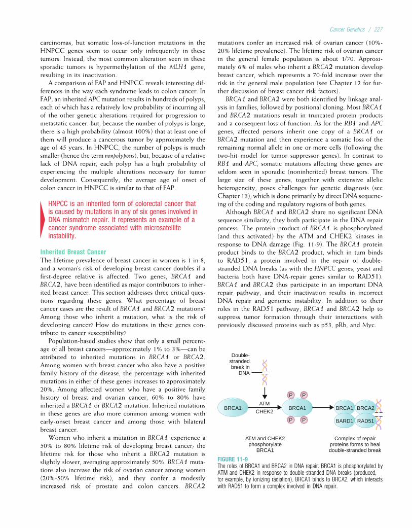
carcinomas, but somatic loss-of-function mutations in theHNPCC genes seem to occur only infrequently in thesetumors. Instead, the most common alteration seen in thesesporadic tumors is hypermethylation of the MLH1 gene,resulting in its inactivation.
A comparison of FAP and HNPCC reveals interesting dif-ferences in the way each syndrome leads to colon cancer. InFAP, an inherited APCmutation results in hundreds of polyps,each of which has a relatively low probability of incurring allof the other genetic alterations required for progression tometastatic cancer. But, because the number of polyps is large,there is a high probability (almost 100%) that at least one ofthem will produce a cancerous tumor by approximately theage of 45 years. In HNPCC, the number of polyps is muchsmaller (hence the term nonpolyposis), but, because of a relativelack of DNA repair, each polyp has a high probability ofexperiencing the multiple alterations necessary for tumordevelopment. Consequently, the average age of onset ofcolon cancer in HNPCC is similar to that of FAP.
4HNPCC is an inherited form of colorectal cancer thatis caused by mutations in any of six genes involved inDNA mismatch repair. It represents an example of acancer syndrome associated with microsatelliteinstability.
Inherited Breast CancerThe lifetime prevalence of breast cancer in women is 1 in 8,and a woman’s risk of developing breast cancer doubles if afirst-degree relative is affected. Two genes, BRCA1 andBRCA2, have been identified as major contributors to inher-ited breast cancer. This section addresses three critical ques-tions regarding these genes: What percentage of breastcancer cases are the result of BRCA1 and BRCA2 mutations?Among those who inherit a mutation, what is the risk ofdeveloping cancer? How do mutations in these genes con-tribute to cancer susceptibility?
Population-based studies show that only a small percent-age of all breast cancers—approximately 1% to 3%—can beattributed to inherited mutations in BRCA1 or BRCA2.Among women with breast cancer who also have a positivefamily history of the disease, the percentage with inheritedmutations in either of these genes increases to approximately20%. Among affected women who have a positive familyhistory of breast and ovarian cancer, 60% to 80% haveinherited a BRCA1 or BRCA2 mutation. Inherited mutationsin these genes are also more common among women withearly-onset breast cancer and among those with bilateralbreast cancer.
Women who inherit a mutation in BRCA1 experience a50% to 80% lifetime risk of developing breast cancer; thelifetime risk for those who inherit a BRCA2 mutation isslightly slower, averaging approximately 50%. BRCA1 muta-tions also increase the risk of ovarian cancer among women(20%-50% lifetime risk), and they confer a modestlyincreased risk of prostate and colon cancers. BRCA2
mutations confer an increased risk of ovarian cancer (10%-20% lifetime prevalence). The lifetime risk of ovarian cancerin the general female population is about 1/70. Approxi-mately 6% of males who inherit a BRCA2 mutation developbreast cancer, which represents a 70-fold increase over therisk in the general male population (see Chapter 12 for fur-ther discussion of breast cancer risk factors).
BRCA1 and BRCA2 were both identified by linkage anal-ysis in families, followed by positional cloning. Most BRCA1and BRCA2 mutations result in truncated protein productsand a consequent loss of function. As for the RB1 and APC
genes, affected persons inherit one copy of a BRCA1 orBRCA2 mutation and then experience a somatic loss of theremaining normal allele in one or more cells (following thetwo-hit model for tumor suppressor genes). In contrast toRB1 and APC, somatic mutations affecting these genes areseldom seen in sporadic (noninherited) breast tumors. Thelarge size of these genes, together with extensive allelicheterogeneity, poses challenges for genetic diagnosis (seeChapter 13), which is done primarily by direct DNA sequenc-ing of the coding and regulatory regions of both genes.
Although BRCA1 and BRCA2 share no significant DNAsequence similarity, they both participate in the DNA repairprocess. The protein product of BRCA1 is phosphorylated(and thus activated) by the ATM and CHEK2 kinases inresponse to DNA damage (Fig. 11-9). The BRCA1 proteinproduct binds to the BRCA2 product, which in turn bindsto RAD51, a protein involved in the repair of double-stranded DNA breaks (as with the HNPCC genes, yeast andbacteria both have DNA-repair genes similar to RAD51).BRCA1 and BRCA2 thus participate in an important DNArepair pathway, and their inactivation results in incorrectDNA repair and genomic instability. In addition to theirroles in the RAD51 pathway, BRCA1 and BRCA2 help tosuppress tumor formation through their interactions withpreviously discussed proteins such as p53, pRb, and Myc.
BRCA1 BRCA1 BRCA2
BARD1 RAD51
BRCA1
Complex of repairproteins forms to healdouble-stranded break
ATM and CHEK2phosphorylate
BRCA1
Double-strandedbreak in
DNA
ATM
CHEK2
P P
P P
FIGURE 11-9The roles of BRCA1 and BRCA2 in DNA repair. BRCA1 is phosphorylated byATM and CHEK2 in response to double-stranded DNA breaks (produced,for example, by ionizing radiation). BRCA1 binds to BRCA2, which interactswith RAD51 to form a complex involved in DNA repair.
Cancer Genetics / 227
Because all of the genes illustrated in Figure 11-9 areinvolved in a DNA repair pathway, it might be anticipatedthat mutations in genes other than BRCA1 or BRCA2 couldlead to DNA repair defects and possibly cancer. This isindeed the case. As already discussed, mutations in CHEK2can cause LFS. Mutations in the ATM gene can cause ataxiatelangiectasia (see Chapter 3), an autosomal recessive diseasethat involves extensive genomic instability, cerebellar ataxia,dilated vessels in the eyes and skin (telangiectasia), and can-cers of mainly lymphoid origin. Another autosomal recessivechromosome instability syndrome, Fanconi anemia, can becaused by the inheritance of two copies of a BRCA2mutation.
Although BRCA1 and BRCA2 mutations are the mostcommon known causes of familial breast cancer, this diseasecan also be caused by inherited mutations in several othertumor suppressor genes (e.g., the previously discussed CHEK2and TP53 genes). Germline mutations in a tumor suppressorgene called PTEN are responsible for Cowden disease, whichis characterized by multiple benign tumors and an increasedsusceptibility to breast cancer. The risk of breast canceramong heterozygous carriers of mutations in the ATM geneis approximately double that of the general population.
It is estimated that the major breast cancer genes, such asBRCA1, BRCA2, PTEN, and CHEK2, account for less than25% of the overall inherited predisposition to breast cancer.Other breast cancer–causing genes are likely to exist, buttheir individual effects on cancer risk are thought to be rela-tively small. Large-scale genome-wide association studies(Chapter 8) have now identified multiple additional inher-ited genetic variants that contribute small increases in breastcancer risk. For example, a variant in the gene that encodesfibroblast growth factor receptor 2 (FGFR2) increases breastcancer risk by about 25%.
Most studies indicate that the clinical course of breastcancer among patients with BRCA1 or BRCA2 mutations isnot substantially different from that of other breast cancerpatients. However, the substantial increase in risk of ovariancancer (which has a high mortality rate and is difficult todetect early), has led to the recommendation that womenwho have completed child bearing should undergo a pro-phylactic oophorectomy. This reduces ovarian cancer riskby about 90%, and it reduces breast cancer risk by about50%. Bilateral prophylactic mastectomy, an option chosenby some BRCA1 and BRCA2 carriers, reduces the risk ofbreast cancer by approximately 90%.
4Mutations in BRCA1 and BRCA2 are responsible for asignificant fraction of inherited breast cancer cases,especially those of early onset. These mutationsusually result in a truncated protein product and lossof function. The protein products of these genes playimportant roles in DNA repair.
Familial MelanomaLargely as a result of increased exposure to ultraviolet radiation,the incidence of melanoma has increased about 20-fold in theUnited States during the past 70 years. It is nowone of themost
common cancers, with 54,000 new cases per year. The riskof developing melanoma increases by a factor of 2 when afirst-degree relative is affected. The risk increases further, toapproximately 6.5, when the first-degree relative is affectedbefore 50 years of age. It is estimated that approximately 5%to 10% of melanoma cases occur in inherited, familial forms.
Linkage analysis in families, studies of loss of heterozy-gosity in melanoma tumor cells, and positional cloning ledto the identification of the CDKN2A gene as a cause offamilial melanoma. Mutations in this gene are estimated tobe involved in 10% to 40% of familial melanoma cases.CDKN2A encodes two different proteins, both of whichare important components of the cell cycle. The first pro-tein, p16, is cyclin-dependent kinase inhibitor which inter-acts negatively with a cyclin-dependent kinase (CDK4)that phosphorylates and down-regulates the pRb protein(see Fig. 11-5). Because pRb acts as a brake on the cell cycle,its down-regulation by CDK4 promotes progression throughthe cell cycle and can lead to cell proliferation. By negativelyregulating CDK4, p16 acts as an upstream brake on the cellcycle. When p16 activity is lost through loss-of-functionmutations in CDKN2A, CDK4 activity is increased and cellproliferation can ensue. This can lead to melanomas. The sec-ond protein encoded by CDKN2A, p14, binds to and inhibitsMDM2. The MDM2 protein, as shown in Figure 11-5,binds to and degrades p53. Thus, by inhibiting MDM2, p14increases p53 expression. As discussed earlier, p53 expressionis needed to arrest the cell cycle for DNA repair in response tocell damage, and it can cause damaged cells to undergo apo-ptosis. A loss of p14 activity leads to a loss of p53 activity,and this can also produce melanomas.
Inherited mutations in the gene that encodes CDK4 canalso result in familial melanoma. These gain-of-functionmutations convert the cyclin-dependent kinase from aproto-oncogene to an activated oncogene. The activatedCDK4 down-regulates pRb, resulting again in a lack of cellcycle control and tumor formation. Melanoma provides anexample in which the same tumor type can result from eitherthe activation of a proto-oncogene (CDK4) or the loss of atumor suppressor gene (CDKN2A).
CDKN2A plays a role not only in familial melanoma butalso in most sporadic melanomas, in which somatic loss-of-function mutations of this gene lead to inactivation of thep16 tumor suppressor protein. About 50% of sporadic melano-mas contain somatic deletions of CDKN2A, and loss-of-func-tion point mutations are seen in another 9% of tumors.Hypermethylation of the promoter region, which down-regu-lates the gene, is seen in ¼ to 3/4 of all melanomas. As might beexpected, somatic mutations in other genes are also seen insporadic melanomas. About 9% of these melanomas havesomatic TP53 mutations, and about 6% have somatic RB1mutations (persons who inherit an RB1 mutation also have anincreased risk ofmelanoma). Approximately two thirds of thesetumors contain somatic gain-of-function mutations in BRAF, agene that encodes a kinase involved in the RAS signal-trans-duction pathway. In addition, one of the RAS genes, NRAS,is mutated in 15% to 30% of sporadic melanomas.
228 / Chapter 11 MEDICAL GENETICS
4Familial melanoma can be caused by loss-of-functionmutations in the CDKN2A tumor suppressor gene orby gain-of-function mutations in the CDK4 proto-oncogene. Both mutations result in a loss of cell cyclecontrol via the pRb and p53 pathways. Somaticmutations of CDKN2A and of BRAF are seen in mostsporadic melanomas.
The RET Proto-Oncogene and Multiple EndocrineNeoplasiaThe RET proto-oncogene, initially identified by a transfec-tion assay (rearranged during transfection; see previous dis-cussion), encodes a receptor tyrosine kinase that includes anextracellular receptor domain, a transmembrane domain, andan intracellular tyrosine kinase domain. RET is involved inembryonic neural crest cell migration (see Chapter 10), andit is normally activated by a complex consisting of glial-derived neurotrophic factor (GDNF) and a coreceptor termedGFRa. The RET protein interacts with several signal transduc-tion pathways, including the well-known RAS pathway.
Inherited loss-of-function mutations in RET can produceHirschsprung disease (a lack of enteric nerve cells, resultingin severe, chronic constipation and bowel distention). Gain-of-function mutations in the same gene result in excess tyro-sine kinase activity and increased signal transduction, leadingultimately to cellular proliferation and, depending on thetype and location of the mutation, any of three formsof autosomal dominant multiple endocrine neoplasia type 2(MEN2): (1) MEN2A, which accounts for 80% of MEN2cases, is characterized by medullary thyroid carcinomas(MTC) in nearly 100% of patients, parathyroid hyperplasiain 30% of patients, and pheochromocytoma (an adrenaltumor) in 50% of patients. More than 98% of MEN2A casesare caused by missense mutations that affect cysteine resi-dues in RET’s extracellular domain. (2) MEN2B is similarto MEN2A but lacks parathyroid hyperplasia and includesmultiple mucosal neuromas and a marfanoid appearance.Nearly all MEN2B alterations are missense mutations thataffect RET’s tyrosine kinase domain. MEN2B accounts forabout 5% of MEN2 cases and is the most aggressive formof MEN2. (3) A syndrome consisting only of familial MTCcan be caused by mutations in both the extracellular andtyrosine kinase domains of RET.
RET is one of only a few proto-oncogenes in which muta-tions can cause inherited cancer syndromes (see Table 11-3for other examples).* Identification of the mutations respon-sible for each of these inherited cancer syndromes hasprovided an accurate means of early diagnosis. Patients withapparently sporadic MTC are advised to undergo genetictesting for RET mutations because 1% to 7% of thesepatients have germline RET mutations and thus familial,rather than sporadic, MTC. Prophylactic thyroidectomy
before 6 years of age is recommended for children whoinherit a disease-causing mutation (thyroidectomy beforeage 3 years may be indicated for the more aggressiveMEN2B tumors).
Somatic alterations of RET can produce papillary thyroidcarcinomas, the most common type of thyroid tumor. Theprevalence of this tumor has increased substantially amongpersons who were exposed to radioactive fallout from theChernobyl nuclear reactor accident (see Chapter 3). Sixtypercent of the papillary thyroid carcinomas in these personshave contained somatic RET alterations.
The RET gene provides an example of extraordinary alle-lic heterogeneity. Loss-of-function mutations in this geneproduce defects in the embryonic development of the bowel,and gain-of-function mutations result in increased signaltransduction and various forms of endocrine neoplasia. Thisexample illustrates the critical connection between normaldevelopment and cancer: Both involve a finely tuned geneticregulation of cell growth and differentiation.
4Loss-of-function mutations in the RET proto-oncogenecan produce Hirschsprung disease, a disorder ofembryonic development. Germline gain-of-functionmutations in the same gene can lead to any of threedifferent types of inherited multiple endocrineneoplasia. Somatic alterations in RET can producenoninherited papillary thyroid carcinoma.
Many additional genes responsible for various inheritedcancer syndromes have been identified. These include, forexample, the genes that cause neurofibromatosis type 2, vonHippel–Lindau syndrome, and Beckwith–Wiedemann syn-drome (see Table 11-1). With the current generation ofgenomic resources, including the complete human DNAsequence, it is reasonable to expect the identification of moresuch genes.
IS GENETIC INHERITANCE IMPORTANT IN COMMONCANCERS?The term “common cancers” is often used to designate thosecancers, such as carcinomas of the breast, colon, or prostate,that are not usually part of an inherited cancer syndrome (e.g., Li–Fraumeni syndrome or familial adenomatous polypo-sis). Recall that germline mutations in genes such as BRCA1or APC, although very important in aiding our understandingof the basis of carcinogenesis, are responsible for only asmall proportion of breast or colon cancer cases. Yet thesecancers do cluster in families. Typically, the presence ofone affected first-degree relative increases one’s risk of devel-oping a common cancer by a factor of two or more. It islikely that additional genes, as well as nongenetic factorsthat are shared in families, contribute to this increased famil-ial risk (see Chapter 12 for further discussion of thesegenetic and nongenetic factors in common cancers).
It is likely that many common cancer-predisposing allelesexist, each with a relatively small effect on overall cancer risk(an example was given by an allele of the FGFR2 gene in
*Another inherited form of multiple endocrine neoplasia, MEN1, is charac-terized by tumors of the parathyroids, anterior pituitary, and pancreas.MEN1 is caused by germline mutations in a gene that encodes the menintumor.
Cancer Genetics / 229
breast cancer). How will we identify these relatively minorrisk alleles in the population? One way, perhaps, couldexploit new approaches of gene mapping that identify genesinvolved in cellular pathways to cancer. Each of thesebecomes a candidate gene that might confer a predispositionto cancer. The use of animal models, as well as the increasingapplication of genome-wide association studies, will
continue to uncover new cancer-predisposing alleles. Themost critical test will be to determine whether there arefunctional mutations in a suspected cancer-causing geneamong cancer patients. The identification of such mutations,and characterization of the genes in which they occur, canbe expected to create new diagnostic and therapeutic toolsthat ultimately will reduce the cancer burden.
Study Questions
1. The G6PD locus is located on the X chromosome.Studies of G6PD alleles in tumor cells from womenshow that all tumor cells usually express the samesingle G6PD allele, even though the women areheterozygous at the G6PD locus. What does thisfinding imply about the origin of the tumor cells?
2. If we assume that the somatic mutation rate at the RB1locus is three mutations per million cells and that thereis a population of 2 million retinoblasts per individual,what is the expected frequency of sporadicretinoblastoma in the population? What is the
expected number of tumors per individual amongthose who inherit a mutated copy of the RB1 gene?
3. Compare and contrast oncogenes and tumorsuppressor genes. How have the characteristics ofthese classes of cancer-causing genes affected ourability to detect them?
4. Members of Li–Fraumeni syndrome families nearlyalways develop tumors by 65 to 70 years of age, butmutation carriers do not necessarily develop the sametype of tumor (e.g., one has breast cancer, and anotherhas colon cancer). Explain this.
Suggested ReadingsCampeau PM, Foulkes WD, Tischkowitz MD. Hereditary
breast cancer: New genetic developments, new therapeu-tic avenues. Hum Genet 2008;124(1):31–42.
Croce CM. Oncogenes and cancer. N Engl J Med 2008;358(5):502–11.
de la Chapelle A. Genetic predisposition to colorectal can-cer. Nat Rev Cancer 2004;4(10):769–80.
Damber JE, Aus G. Prostate cancer. Lancet 2008;371(9625):1710–21.
Deng Y, Chan SS, Chang S. Telomere dysfunction andtumour suppression: The senescence connection. Nat RevCancer 2008;8(6):450–8.
Desai TK, Barkel D. Syndromic colon cancer: Lynch syn-drome and familial adenomatous polyposis. GastroenterolClin North Am 2008;37(1):47–72.
Esteller M. Epigenetics in cancer. Engl J Med 2008;358(11):1148–59.
Finkel T, Serrano M, Blasco MA. The common biology ofcancer and ageing. Nature 2007;448(7155):767–74.
Foulkes WD. Inherited susceptibility to common cancers. NEngl J Med 2008;359(20):2143–53.
Galiatsatos P, Foulkes WD. Familial adenomatous polyposis.Am J Gastroenterol 2006;101(2):385–98.
Knudson AG. Two genetic hits (more or less) to cancer. NatRev Cancer 2001;1(2):157–62.
Lakhani VT, You YN,Wells SA. TheMultiple endocrine neo-plasia syndromes. Ann Rev Med 2007; 58(1):253–65.
Leiderman YI, Kiss S, Mukai S. Molecular genetics of RB1—the retinoblastoma gene. Semin Ophthalmol 2007;22(4):247–54.
Lu X. P53: A heavily dictated dictator of life and death.Curr Opin Genet Dev 2005;15(1):27–33.
Robson M, Offit K. Management of an inherited pre-disposition to breast cancer. N Engl J Med 2007;357(2):154–162.
Sekulic A, Haluska P Jr, Miller AJ, et al. Malignant mela-noma in the 21st century: The emerging molecular land-scape. Mayo Clin Proc 2008;83(7):825–46.
Van Dyke T. P53 and tumor suppression. N Engl J Med2007;356(1):79–81.
Vogelstein B, Kinzler KW, editors. The Genetic Basis ofHuman Cancer. 2nd ed. New York: McGraw–Hill; 2002.
Internet ResourcesNational Cancer Institute Information site (generalinformation on cancer and cancer genetics, as well as linksto other useful Web sites) http://cancer.gov/cancerinformation
230 / Chapter 11 MEDICAL GENETICS
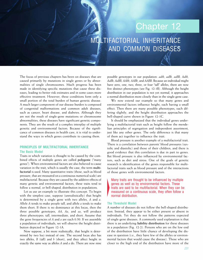
Chapter 12
MULTIFACTORIAL INHERITANCEAND COMMON DISEASES
The focus of previous chapters has been on diseases that arecaused primarily by mutations in single genes or by abnor-malities of single chromosomes. Much progress has beenmade in identifying specific mutations that cause these dis-eases, leading to better risk estimates and in some cases moreeffective treatment. However, these conditions form only asmall portion of the total burden of human genetic disease.A much larger component of our disease burden is composedof congenital malformations and common adult diseases,such as cancer, heart disease, and diabetes. Although theyare not the result of single-gene mutations or chromosomeabnormalities, these diseases have significant genetic compo-nents. They are the result of a complex interplay of multiplegenetic and environmental factors. Because of the signifi-cance of common diseases in health care, it is vital to under-stand the ways in which genes contribute to causing them.
PRINCIPLES OF MULTIFACTORIAL INHERITANCEThe Basic ModelTraits in which variation is thought to be caused by the com-bined effects of multiple genes are called polygenic (“manygenes”). When environmental factors are also believed to causevariation in the trait, which is usually the case, the term multi-factorial is used. Many quantitative traits (those, such as bloodpressure, that are measured on a continuous numerical scale) aremultifactorial. Because they are caused by the additive effects ofmany genetic and environmental factors, these traits tend tofollow a normal, or bell-shaped, distribution in populations.
Let us use an example to illustrate this concept. To beginwith the simplest case, suppose (unrealistically) that heightis determined by a single gene with two alleles, A and a.Allele A tends to make people tall, and allele a tends to makethem short. If there is no dominance at this locus, then thethree possible genotypes, AA, Aa, and aa, will producethree phenotypes: tall, intermediate, and short. Assume thatthe gene frequencies of A and a are each 0.50. If we assemblea population of individuals, we will observe the height distri-bution depicted in Figure 12-1A.
Now suppose, a bit more realistically, that height is deter-mined by two loci instead of one. The second locus also hastwo alleles, B (tall) and b (short), and they affect height inexactly the same way as alleles A and a do. There are now nine
possible genotypes in our population: aabb, aaBb, aaBB, Aabb,AaBb, AaBB, AAbb, AABb, and AABB. Because an individual mighthave zero, one, two, three, or four “tall” alleles, there are nowfive distinct phenotypes (see Fig. 12-1B). Although the heightdistribution in our population is not yet normal, it approachesa normal distribution more closely than in the single-gene case.
We now extend our example so that many genes andenvironmental factors influence height, each having a smalleffect. Then there are many possible phenotypes, each dif-fering slightly, and the height distribution approaches thebell-shaped curve shown in Figure 12-1C.
It should be emphasized that the individual genes under-lying a multifactorial trait such as height follow the mende-lian principles of segregation and independent assortment,just like any other genes. The only difference is that manyof them act together to influence the trait.
Blood pressure is another example of a multifactorial trait.There is a correlation between parents’ blood pressures (sys-tolic and diastolic) and those of their children, and there isgood evidence that this correlation is due in part to genes.But blood pressure is also influenced by environmental fac-tors, such as diet and stress. One of the goals of geneticresearch is identification of the genes responsible for multi-factorial traits such as blood pressure and of the interactionsof those genes with environmental factors.
4Many traits are thought to be influenced by multiplegenes as well as by environmental factors. Thesetraits are said to be multifactorial. When they can bemeasured on a continuous scale, they often follow anormal distribution.
The Threshold ModelA number of diseases do not follow the bell-shaped distribu-tion. Instead, they appear to be either present or absent inindividuals. Yet they do not follow the patterns expectedof single-gene diseases. A commonly used explanation is thatthere is an underlying liability distribution for these diseasesin a population (Fig. 12-2). Persons who are on the low endof the distribution have little chance of developing the dis-ease in question (i.e., they have few of the alleles or environ-mental factors that would cause the disease). Those who arecloser to the high end of the distribution have more of the
231
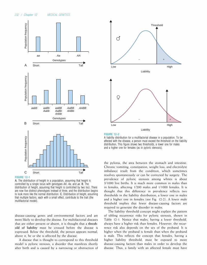
disease-causing genes and environmental factors and aremore likely to develop the disease. For multifactorial diseasesthat are either present or absent, it is thought that a thresh-old of liability must be crossed before the disease isexpressed. Below the threshold, the person appears normal;above it, he or she is affected by the disease.
A disease that is thought to correspond to this thresholdmodel is pyloric stenosis, a disorder that manifests shortlyafter birth and is caused by a narrowing or obstruction of
the pylorus, the area between the stomach and intestine.Chronic vomiting, constipation, weight loss, and electrolyteimbalance result from the condition, which sometimesresolves spontaneously or can be corrected by surgery. Theprevalence of pyloric stenosis among whites is about3/1000 live births. It is much more common in males thanin females, affecting 1/200 males and 1/1000 females. It isthought that this difference in prevalence reflects twothresholds in the liability distribution; a lower one in malesand a higher one in females (see Fig. 12-2). A lower malethreshold implies that fewer disease-causing factors arerequired to generate the disorder in males.
The liability threshold concept might explain the patternof sibling recurrence risks for pyloric stenosis, shown inTable 12-1. Notice that males, having a lower threshold,always have a higher risk than females. However, the recur-rence risk also depends on the sex of the proband. It ishigher when the proband is female than when the probandis male. This reflects the concept that females, having ahigher liability threshold, must be exposed to moredisease-causing factors than males in order to develop thedisease. Thus, a family with an affected female must have
Pop
ulat
ion
freq
uenc
yP
opul
atio
n fr
eque
ncy
Pop
ulat
ion
freq
uenc
y
A
B
C Short Tall
Short Tall
Short Tall
Genotypes
Genotypes
aa Aa AA
aabb aaBbAabb
aaBBAaBbAAbb
AaBBAABb
AABB
FIGURE 12-1A, The distribution of height in a population, assuming that height iscontrolled by a single locus with genotypes AA, Aa, and aa. B, Thedistribution of height, assuming that height is controlled by two loci. Thereare now five distinct phenotypes instead of three, and the distribution beginsto look more like the normal distribution. C, Distribution of height, assumingthat multiple factors, each with a small effect, contribute to the trait (themultifactorial model).
Threshold
+−
+−
High
Liability
Low
High
Liability
Low
FIGURE 12-2A liability distribution for a multifactorial disease in a population. To beaffected with the disease, a person must exceed the threshold on the liabilitydistribution. This figure shows two thresholds, a lower one for malesand a higher one for females (as in pyloric stenosis).
232 / Chapter 12 MEDICAL GENETICS

more genetic and environmental risk factors, producing ahigher recurrence risk for pyloric stenosis in future offspring.In such a situation, we would expect that the highest risk cat-egory would be male relatives of female probands; Table 12-1shows that this is indeed the case.
A number of other congenital malformations are thoughtto correspond to this model. They include isolated* cleft lipand/or cleft palate, neural tube defects (anencephaly andspina bifida), club foot (talipes), and some forms of congeni-tal heart disease.
4The threshold model applies to many multifactorialdiseases. It assumes that there is an underlyingliability distribution in a population and that athreshold on this distribution must be passed before adisease is expressed.
Recurrence Risks and Transmission PatternsWhereas recurrence risks can be given with confidence forsingle-gene diseases (50% for a completely penetrant autoso-mal dominant disease, 25% for autosomal recessive diseases,and so on), risk estimation is more complex for multifactorialdiseases. This is because the number of genes contributing tothe disease is usually not known, the precise allelic constitutionof the parents is not known, and the extent of environmentaleffects can vary substantially. For most multifactorial diseases,empirical risks (i.e., risks based on direct observation of data)have been derived. To estimate empirical risks, a large seriesof families is examined in which one child (the proband) hasdeveloped the disease. The relatives of each proband are sur-veyed in order to calculate the percentage who have also devel-oped the disease. For example, in North America neural tubedefects are seen in about 2% to 3% of the siblings of probandswith this condition (Clinical Commentary 12-1). Thus, therecurrence risk for parents who have had one child with a neu-ral tube defect is 2% to 3%. For conditions that are not lethal orseverely debilitating, such as cleft lip and cleft palate, recur-rence risks can also be estimated for the offspring of affectedparents. Because risk factors vary among diseases, empiricalrecurrence risks are specific for each multifactorial disease.
CLINICAL COMMENTARY 12-1
Neural Tube Defects
Neural tube defects (NTDs) include anencephaly, spina bifida, and ence-phalocele, as well as several other less-common forms. They are one ofthe most important classes of birth defects, with a newborn prevalenceof 1 to 3 per 1000. There is considerable variation in the prevalence ofNTDs among various populations, with an especially high rate amongsome northern Chinese populations (as high as 6 per 1000 births). Forreasons that are not fully known, the prevalence of NTDs has beendecreasing in many parts of the United States and Europe during the pasttwo decades.
Normally the neural tube closes at about the fourth week of gestation. Adefect in closure or a subsequent reopening of the neural tube results in anNTD. Spina bifida is the most commonly observed NTD and consists of aprotrusion of spinal tissue through the vertebral column (the tissue usuallyincludes meninges, spinal cord, and nerve roots). About 75% of spinabifida patients have secondary hydrocephalus, which sometimes in turnproduces mental retardation. Paralysis or muscle weakness, lack of sphinc-ter control, and club feet are often observed. A study conducted in BritishColumbia showed that survival rates for spina bifida patients have improveddramatically over the past several decades. Less than 30% of such childrenborn between 1952 and 1969 survived to 10 years of age, but 65% of thoseborn between 1970 and 1986 survived to this age.
Anencephaly is characterized by partial or complete absence of the cra-nial vault and calvarium and partial or complete absence of the cerebralhemispheres. At least two thirds of anencephalics are stillborn; term deliv-eries do not survive more than a few hours or days. Encephalocele consistsof a protrusion of the brain into an enclosed sac. It is seldom compatiblewith survival.
A
The major neural tube defects (NTDs). A, An infant with an open spina bifida(meningomyelocele).
Continued
TABLE 12-1
Recurrence Risks (%) for Pyloric Stenosis, Subdivided byGender of Affected Probands and Relatives*
Relatives MaleProbands
FemaleProbands
London Belfast London Belfast
Brothers 3.8 9.6 9.2 12.5
Sisters 2.7 3.0 3.8 3.8
*Note that the risks differ somewhat between the two populations.
(Adapted from Carter CO: Genetics of common single malformations. Br Med Bull
1976;32:21-26.)
*In this context, the term “isolated” means that this is the only observed dis-ease feature (i.e., the feature is not part of a larger constellation of findings,as in cleft lip/palate secondary to trisomy 13).
Multifactorial Inheritance and Common Diseases / 233
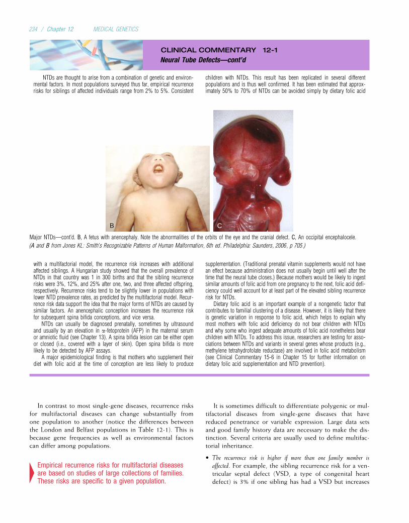
CLINICAL COMMENTARY 12-1
Neural Tube Defects—cont’d
NTDs are thought to arise from a combination of genetic and environ-mental factors. In most populations surveyed thus far, empirical recurrencerisks for siblings of affected individuals range from 2% to 5%. Consistent
with a multifactorial model, the recurrence risk increases with additionalaffected siblings. A Hungarian study showed that the overall prevalence ofNTDs in that country was 1 in 300 births and that the sibling recurrencerisks were 3%, 12%, and 25% after one, two, and three affected offspring,respectively. Recurrence risks tend to be slightly lower in populations withlower NTD prevalence rates, as predicted by the multifactorial model. Recur-rence risk data support the idea that the major forms of NTDs are caused bysimilar factors. An anencephalic conception increases the recurrence riskfor subsequent spina bifida conceptions, and vice versa.
NTDs can usually be diagnosed prenatally, sometimes by ultrasoundand usually by an elevation in a-fetoprotein (AFP) in the maternal serumor amniotic fluid (see Chapter 13). A spina bifida lesion can be either openor closed (i.e., covered with a layer of skin). Open spina bifida is morelikely to be detected by AFP assays.
A major epidemiological finding is that mothers who supplement theirdiet with folic acid at the time of conception are less likely to produce
children with NTDs. This result has been replicated in several differentpopulations and is thus well confirmed. It has been estimated that approx-imately 50% to 70% of NTDs can be avoided simply by dietary folic acid
supplementation. (Traditional prenatal vitamin supplements would not havean effect because administration does not usually begin until well after thetime that the neural tube closes.) Because mothers would be likely to ingestsimilar amounts of folic acid from one pregnancy to the next, folic acid defi-ciency could well account for at least part of the elevated sibling recurrencerisk for NTDs.
Dietary folic acid is an important example of a nongenetic factor thatcontributes to familial clustering of a disease. However, it is likely that thereis genetic variation in response to folic acid, which helps to explain whymost mothers with folic acid deficiency do not bear children with NTDsand why some who ingest adequate amounts of folic acid nonetheless bearchildren with NTDs. To address this issue, researchers are testing for asso-ciations between NTDs and variants in several genes whose products (e.g.,methylene tetrahydrofolate reductase) are involved in folic acid metabolism(see Clinical Commentary 15-6 in Chapter 15 for further information ondietary folic acid supplementation and NTD prevention).
In contrast to most single-gene diseases, recurrence risksfor multifactorial diseases can change substantially fromone population to another (notice the differences betweenthe London and Belfast populations in Table 12-1). This isbecause gene frequencies as well as environmental factorscan differ among populations.
4Empirical recurrence risks for multifactorial diseasesare based on studies of large collections of families.These risks are specific to a given population.
It is sometimes difficult to differentiate polygenic or mul-tifactorial diseases from single-gene diseases that havereduced penetrance or variable expression. Large data setsand good family history data are necessary to make the dis-tinction. Several criteria are usually used to define multifac-torial inheritance.
• The recurrence risk is higher if more than one family member isaffected. For example, the sibling recurrence risk for a ven-tricular septal defect (VSD, a type of congenital heartdefect) is 3% if one sibling has had a VSD but increases
B C
Major NTDs—cont’d. B, A fetus with anencephaly. Note the abnormalities of the orbits of the eye and the cranial defect. C, An occipital encephalocele.
(A and B from Jones KL: Smith’s Recognizable Patterns of Human Malformation, 6th ed. Philadelphia: Saunders, 2006, p 705.)
234 / Chapter 12 MEDICAL GENETICS
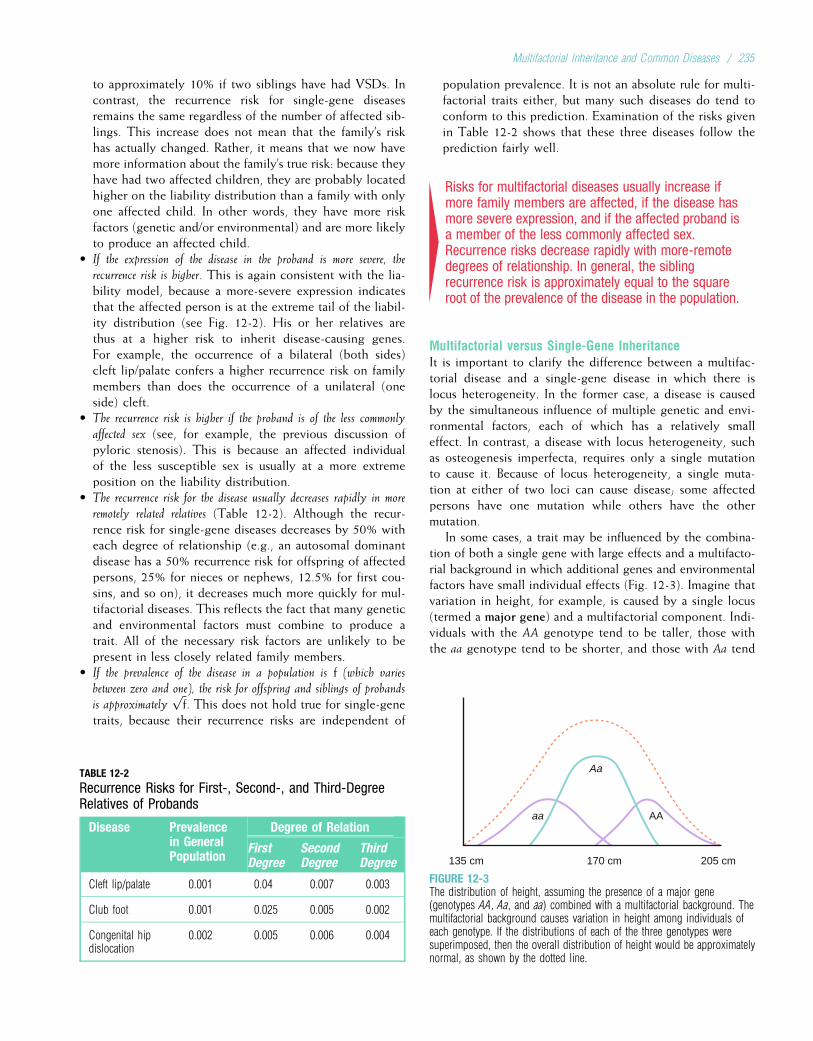
to approximately 10% if two siblings have had VSDs. Incontrast, the recurrence risk for single-gene diseasesremains the same regardless of the number of affected sib-lings. This increase does not mean that the family’s riskhas actually changed. Rather, it means that we now havemore information about the family’s true risk: because theyhave had two affected children, they are probably locatedhigher on the liability distribution than a family with onlyone affected child. In other words, they have more riskfactors (genetic and/or environmental) and are more likelyto produce an affected child.
• If the expression of the disease in the proband is more severe, the
recurrence risk is higher. This is again consistent with the lia-bility model, because a more-severe expression indicatesthat the affected person is at the extreme tail of the liabil-ity distribution (see Fig. 12-2). His or her relatives arethus at a higher risk to inherit disease-causing genes.For example, the occurrence of a bilateral (both sides)cleft lip/palate confers a higher recurrence risk on familymembers than does the occurrence of a unilateral (oneside) cleft.
• The recurrence risk is higher if the proband is of the less commonly
affected sex (see, for example, the previous discussion ofpyloric stenosis). This is because an affected individualof the less susceptible sex is usually at a more extremeposition on the liability distribution.
• The recurrence risk for the disease usually decreases rapidly in more
remotely related relatives (Table 12-2). Although the recur-rence risk for single-gene diseases decreases by 50% witheach degree of relationship (e.g., an autosomal dominantdisease has a 50% recurrence risk for offspring of affectedpersons, 25% for nieces or nephews, 12.5% for first cou-sins, and so on), it decreases much more quickly for mul-tifactorial diseases. This reflects the fact that many geneticand environmental factors must combine to produce atrait. All of the necessary risk factors are unlikely to bepresent in less closely related family members.
• If the prevalence of the disease in a population is f (which varies
between zero and one), the risk for offspring and siblings of probands
is approximatelyffiffif
p. This does not hold true for single-gene
traits, because their recurrence risks are independent of
population prevalence. It is not an absolute rule for multi-factorial traits either, but many such diseases do tend toconform to this prediction. Examination of the risks givenin Table 12-2 shows that these three diseases follow theprediction fairly well.
4Risks for multifactorial diseases usually increase ifmore family members are affected, if the disease hasmore severe expression, and if the affected proband isa member of the less commonly affected sex.Recurrence risks decrease rapidly with more-remotedegrees of relationship. In general, the siblingrecurrence risk is approximately equal to the squareroot of the prevalence of the disease in the population.
Multifactorial versus Single-Gene InheritanceIt is important to clarify the difference between a multifac-torial disease and a single-gene disease in which there islocus heterogeneity. In the former case, a disease is causedby the simultaneous influence of multiple genetic and envi-ronmental factors, each of which has a relatively smalleffect. In contrast, a disease with locus heterogeneity, suchas osteogenesis imperfecta, requires only a single mutationto cause it. Because of locus heterogeneity, a single muta-tion at either of two loci can cause disease; some affectedpersons have one mutation while others have the othermutation.
In some cases, a trait may be influenced by the combina-tion of both a single gene with large effects and a multifacto-rial background in which additional genes and environmentalfactors have small individual effects (Fig. 12-3). Imagine thatvariation in height, for example, is caused by a single locus(termed a major gene) and a multifactorial component. Indi-viduals with the AA genotype tend to be taller, those withthe aa genotype tend to be shorter, and those with Aa tend
TABLE 12-2
Recurrence Risks for First-, Second-, and Third-DegreeRelatives of Probands
Disease Prevalencein GeneralPopulation
Degree of Relation
FirstDegree
SecondDegree
ThirdDegree
Cleft lip/palate 0.001 0.04 0.007 0.003
Club foot 0.001 0.025 0.005 0.002
Congenital hipdislocation
0.002 0.005 0.006 0.004
aa
Aa
AA
135 cm 170 cm 205 cm
FIGURE 12-3The distribution of height, assuming the presence of a major gene(genotypes AA, Aa, and aa) combined with a multifactorial background. Themultifactorial background causes variation in height among individuals ofeach genotype. If the distributions of each of the three genotypes weresuperimposed, then the overall distribution of height would be approximatelynormal, as shown by the dotted line.
Multifactorial Inheritance and Common Diseases / 235
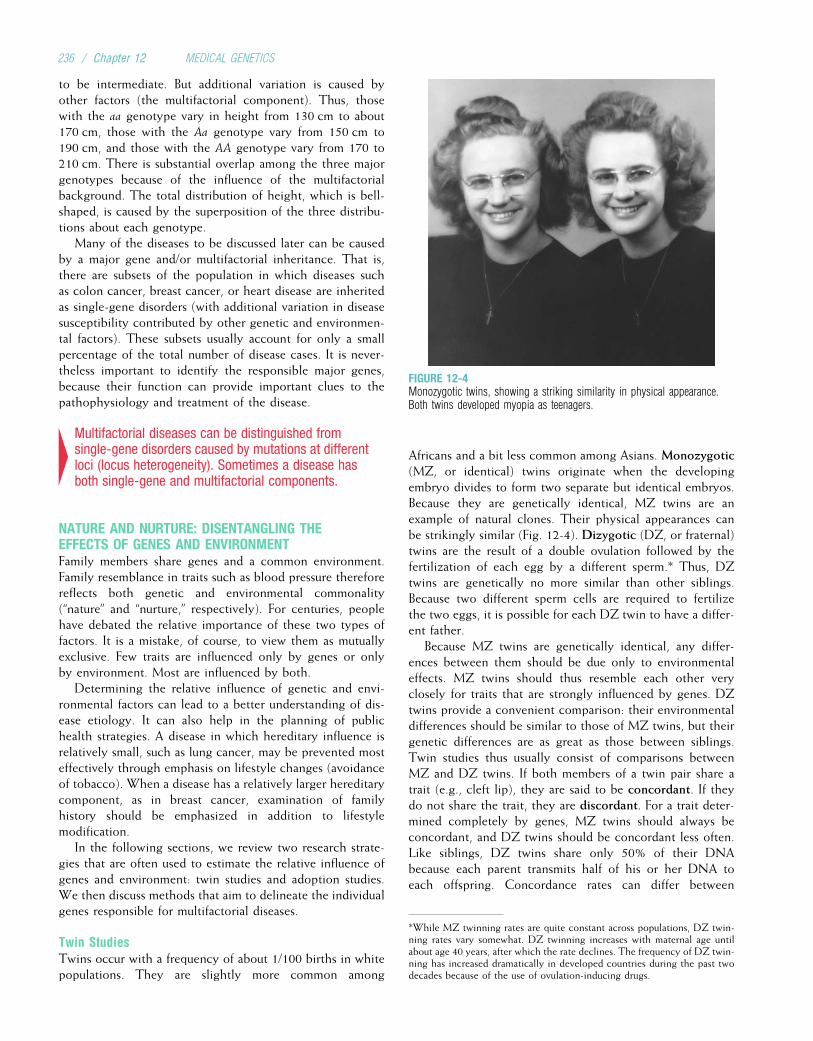
to be intermediate. But additional variation is caused byother factors (the multifactorial component). Thus, thosewith the aa genotype vary in height from 130 cm to about170 cm, those with the Aa genotype vary from 150 cm to190 cm, and those with the AA genotype vary from 170 to210 cm. There is substantial overlap among the three majorgenotypes because of the influence of the multifactorialbackground. The total distribution of height, which is bell-shaped, is caused by the superposition of the three distribu-tions about each genotype.
Many of the diseases to be discussed later can be causedby a major gene and/or multifactorial inheritance. That is,there are subsets of the population in which diseases suchas colon cancer, breast cancer, or heart disease are inheritedas single-gene disorders (with additional variation in diseasesusceptibility contributed by other genetic and environmen-tal factors). These subsets usually account for only a smallpercentage of the total number of disease cases. It is never-theless important to identify the responsible major genes,because their function can provide important clues to thepathophysiology and treatment of the disease.
4Multifactorial diseases can be distinguished fromsingle-gene disorders caused by mutations at differentloci (locus heterogeneity). Sometimes a disease hasboth single-gene and multifactorial components.
NATURE AND NURTURE: DISENTANGLING THEEFFECTS OF GENES AND ENVIRONMENTFamily members share genes and a common environment.Family resemblance in traits such as blood pressure thereforereflects both genetic and environmental commonality(“nature” and “nurture,” respectively). For centuries, peoplehave debated the relative importance of these two types offactors. It is a mistake, of course, to view them as mutuallyexclusive. Few traits are influenced only by genes or onlyby environment. Most are influenced by both.
Determining the relative influence of genetic and envi-ronmental factors can lead to a better understanding of dis-ease etiology. It can also help in the planning of publichealth strategies. A disease in which hereditary influence isrelatively small, such as lung cancer, may be prevented mosteffectively through emphasis on lifestyle changes (avoidanceof tobacco). When a disease has a relatively larger hereditarycomponent, as in breast cancer, examination of familyhistory should be emphasized in addition to lifestylemodification.
In the following sections, we review two research strate-gies that are often used to estimate the relative influence ofgenes and environment: twin studies and adoption studies.We then discuss methods that aim to delineate the individualgenes responsible for multifactorial diseases.
Twin StudiesTwins occur with a frequency of about 1/100 births in whitepopulations. They are slightly more common among
Africans and a bit less common among Asians. Monozygotic(MZ, or identical) twins originate when the developingembryo divides to form two separate but identical embryos.Because they are genetically identical, MZ twins are anexample of natural clones. Their physical appearances canbe strikingly similar (Fig. 12-4). Dizygotic (DZ, or fraternal)twins are the result of a double ovulation followed by thefertilization of each egg by a different sperm.* Thus, DZtwins are genetically no more similar than other siblings.Because two different sperm cells are required to fertilizethe two eggs, it is possible for each DZ twin to have a differ-ent father.
Because MZ twins are genetically identical, any differ-ences between them should be due only to environmentaleffects. MZ twins should thus resemble each other veryclosely for traits that are strongly influenced by genes. DZtwins provide a convenient comparison: their environmentaldifferences should be similar to those of MZ twins, but theirgenetic differences are as great as those between siblings.Twin studies thus usually consist of comparisons betweenMZ and DZ twins. If both members of a twin pair share atrait (e.g., cleft lip), they are said to be concordant. If theydo not share the trait, they are discordant. For a trait deter-mined completely by genes, MZ twins should always beconcordant, and DZ twins should be concordant less often.Like siblings, DZ twins share only 50% of their DNAbecause each parent transmits half of his or her DNA toeach offspring. Concordance rates can differ between
*While MZ twinning rates are quite constant across populations, DZ twin-ning rates vary somewhat. DZ twinning increases with maternal age untilabout age 40 years, after which the rate declines. The frequency of DZ twin-ning has increased dramatically in developed countries during the past twodecades because of the use of ovulation-inducing drugs.
FIGURE 12-4Monozygotic twins, showing a striking similarity in physical appearance.Both twins developed myopia as teenagers.
236 / Chapter 12 MEDICAL GENETICS
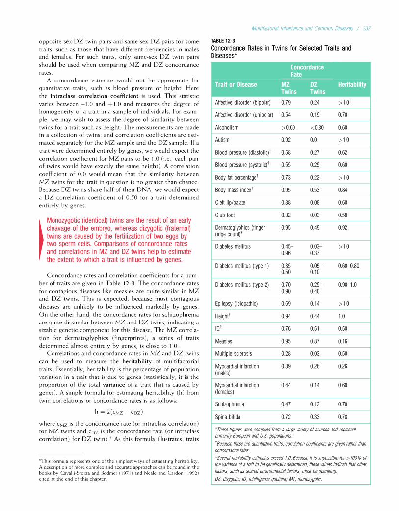
opposite-sex DZ twin pairs and same-sex DZ pairs for sometraits, such as those that have different frequencies in malesand females. For such traits, only same-sex DZ twin pairsshould be used when comparing MZ and DZ concordancerates.
A concordance estimate would not be appropriate forquantitative traits, such as blood pressure or height. Herethe intraclass correlation coefficient is used. This statisticvaries between –1.0 and þ1.0 and measures the degree ofhomogeneity of a trait in a sample of individuals. For exam-ple, we may wish to assess the degree of similarity betweentwins for a trait such as height. The measurements are madein a collection of twins, and correlation coefficients are esti-mated separately for the MZ sample and the DZ sample. If atrait were determined entirely by genes, we would expect thecorrelation coefficient for MZ pairs to be 1.0 (i.e., each pairof twins would have exactly the same height). A correlationcoefficient of 0.0 would mean that the similarity betweenMZ twins for the trait in question is no greater than chance.Because DZ twins share half of their DNA, we would expecta DZ correlation coefficient of 0.50 for a trait determinedentirely by genes.
4Monozygotic (identical) twins are the result of an earlycleavage of the embryo, whereas dizygotic (fraternal)twins are caused by the fertilization of two eggs bytwo sperm cells. Comparisons of concordance ratesand correlations in MZ and DZ twins help to estimatethe extent to which a trait is influenced by genes.
Concordance rates and correlation coefficients for a num-ber of traits are given in Table 12-3. The concordance ratesfor contagious diseases like measles are quite similar in MZand DZ twins. This is expected, because most contagiousdiseases are unlikely to be influenced markedly by genes.On the other hand, the concordance rates for schizophreniaare quite dissimilar between MZ and DZ twins, indicating asizable genetic component for this disease. The MZ correla-tion for dermatoglyphics (fingerprints), a series of traitsdetermined almost entirely by genes, is close to 1.0.
Correlations and concordance rates in MZ and DZ twinscan be used to measure the heritability of multifactorialtraits. Essentially, heritability is the percentage of populationvariation in a trait that is due to genes (statistically, it is theproportion of the total variance of a trait that is caused bygenes). A simple formula for estimating heritability (h) fromtwin correlations or concordance rates is as follows:
h ¼ 2ðcMZ � cDZÞwhere cMZ is the concordance rate (or intraclass correlation)for MZ twins and cDZ is the concordance rate (or intraclasscorrelation) for DZ twins.* As this formula illustrates, traits
*This formula represents one of the simplest ways of estimating heritability.A description of more complex and accurate approaches can be found in thebooks by Cavalli-Sforza and Bodmer (1971) and Neale and Cardon (1992)cited at the end of this chapter.
TABLE 12-3
Concordance Rates in Twins for Selected Traits andDiseases*
ConcordanceRate
Trait or Disease MZTwins
DZTwins
Heritability
Affective disorder (bipolar) 0.79 0.24 >1.0{
Affective disorder (unipolar) 0.54 0.19 0.70
Alcoholism >0.60 <0.30 0.60
Autism 0.92 0.0 >1.0
Blood pressure (diastolic){ 0.58 0.27 0.62
Blood pressure (systolic){ 0.55 0.25 0.60
Body fat percentage{ 0.73 0.22 >1.0
Body mass index{ 0.95 0.53 0.84
Cleft lip/palate 0.38 0.08 0.60
Club foot 0.32 0.03 0.58
Dermatoglyphics (fingerridge count){
0.95 0.49 0.92
Diabetes mellitus 0.45–0.96
0.03–0.37
>1.0
Diabetes mellitus (type 1) 0.35–0.50
0.05–0.10
0.60–0.80
Diabetes mellitus (type 2) 0.70–0.90
0.25–0.40
0.90–1.0
Epilepsy (idiopathic) 0.69 0.14 >1.0
Height{ 0.94 0.44 1.0
IQ{ 0.76 0.51 0.50
Measles 0.95 0.87 0.16
Multiple sclerosis 0.28 0.03 0.50
Myocardial infarction(males)
0.39 0.26 0.26
Myocardial infarction(females)
0.44 0.14 0.60
Schizophrenia 0.47 0.12 0.70
Spina bifida 0.72 0.33 0.78
*These figures were compiled from a large variety of sources and represent
primarily European and U.S. populations.{Because these are quantitative traits, correlation coefficients are given rather than
concordance rates.{Several heritability estimates exceed 1.0. Because it is impossible for >100% of
the variance of a trait to be genetically determined, these values indicate that other
factors, such as shared environmental factors, must be operating.
DZ, dizygotic; IQ, intelligence quotient; MZ, monozygotic.
Multifactorial Inheritance and Common Diseases / 237

that are largely determined by genes result in a heritabilityestimate that approaches 1.0 (i.e., cMZ approaches 1.0, andcDZ approaches 0.5). As the difference between MZ andDZ concordance rates becomes smaller, heritabilityapproaches zero. Correlations and concordance rates inother types of relatives (e.g., between parents and offspring)can also be used to measure heritability.
Like recurrence risks, heritability values are specific forthe population in which they are estimated. However, thereis usually agreement from one population to another regard-ing the general range of heritability estimates of most traits(e.g., the heritability of height is almost always high, andthe heritability of contagious diseases is almost alwayslow). The same is true of empirical recurrence risks.
4Comparisons of correlations and concordance rates inMZ and DZ twins allow the estimation of heritability, ameasure of the percentage of population variation in adisease that can be attributed to genes.
At one time, twins were thought to provide a perfect “nat-ural laboratory” in which to determine the relative influencesof genetics and environment. But several difficulties arise.One of the most important is the assumption that the envi-ronments of MZ and DZ twins are equally similar. MZ twinsare often treated more similarly than DZ twins. The eminentgeneticist L. S. Penrose once joked that, if one were to studythe clothes of twins, it might be concluded that clothes areinherited biologically. A greater similarity in environmentcan make MZ twins more concordant for a trait, inflatingthe apparent influence of genes. In addition, MZ twinsmay be more likely to seek the same type of environment,further reinforcing environmental similarity. On the otherhand, it has been suggested that some MZ twins tend todevelop personality differences in an attempt to assert theirindividuality.
Another difficulty is that the uterine environments of dif-ferent pairs of MZ twins can be more or less similar, depend-ing on whether there are two amnions and two chorions,two amnions and one shared chorion, or one shared amnionand one shared chorion. In addition, somatic mutations canoccur during mitotic divisions of the cells of MZ twinembryos after cleavage occurs. Thus, the MZ twins mightnot be quite “identical,” especially if a mutation occurredearly in the development of one of the twins. Finally, recentstudies indicate that methylation patterns, which can influ-ence the transcription of specific genes, become more dis-similar in MZ twin pairs as they age. This dissimilarity isgreater when the twins adopt markedly different habits andlifestyles (e.g., when one twin smokes cigarettes and theother does not).
Of the various problems with the twin method, thegreater degree of environmental sharing among MZ twinsis perhaps the most serious. One way to circumvent thisproblem, at least in part, is to study MZ twins who wereraised in separate environments. Concordance among these
twin pairs should be caused by genetic, rather than environ-mental, similarities. As one might expect, it is not easy tofind such twin pairs. A major effort to do so has been under-taken by researchers at the University of Minnesota, whosestudies have shown a remarkable congruence among MZtwins reared apart, even for many behavioral traits. However,these studies must be viewed with caution, because the sam-ple sizes are relatively small and because many of the twinpairs had at least some contact with each other before theywere studied.
4Although twin studies provide valuable information,they are also affected by certain biases. The mostserious is greater environmental similarity betweenMZ twins than between DZ twins. Other biasesinclude somatic mutations that might affect only oneMZ twin and differences in the uterine environmentsof twins.
Adoption StudiesStudies of adopted children are also used to estimate thegenetic contribution to a multifactorial trait. Offspring whowere born to parents who have a disease but who wereadopted by parents lacking the disease can be studied to findout whether the offspring develop the disease. In some cases,these adopted persons develop the disease more often thando children in a comparative control population (i.e.,adopted children who were born to parents who do not havethe disease). This provides evidence that genes may beinvolved in causing the disease, because the adopted chil-dren do not share an environment with their affected naturalparents. For example, schizophrenia is seen in 8% to 10% ofadopted children whose natural parent had schizophrenia,whereas it is seen in only 1% of adopted children of unaf-fected parents.
As with twin studies, several precautions must be exer-cised in interpreting the results of adoption studies. First,prenatal environmental influences could have long-lastingeffects on an adopted child. Second, children are sometimesadopted after they are several years old, ensuring that somenongenetic influences have been imparted by the naturalparents. Finally, adoption agencies sometimes try to matchthe adoptive parents with the natural parents in terms ofattributes such as socioeconomic status. All of these factorscould exaggerate the apparent influence of biologicalinheritance.
4Adoption studies provide a second means ofestimating the influence of genes on multifactorialdiseases. They consist of comparing disease ratesamong the adopted offspring of affected parents withthe rates among adopted offspring of unaffectedparents. As with the twin method, several biases caninfluence these studies.
238 / Chapter 12 MEDICAL GENETICS
These reservations, as well as those summarized for twinstudies, underscore the need for caution in basing conclusionson twin and adoption studies. These approaches do not pro-vide definitive measures of the role of genes in multifactorialdisease, nor can they identify specific genes responsible fordisease. Instead, they provide a preliminary indication of theextent to which a multifactorial disease may be influencedby genetic factors. Methods for the direct detection of genesunderlying multifactorial traits are summarized in Box 12-1.
THE GENETICS OF COMMON DISEASESHaving discussed the principles of multifactorial inheritance,we turn next to a discussion of the common multifactorialdisorders themselves. Some of these disorders, the congeni-tal malformations, are by definition present at birth. Others,
including heart disease, cancer, diabetes, and most psychiat-ric disorders, are seen primarily in adolescents and adults.Because of their complexity, unraveling the genetics of thesedisorders is a daunting task. Nonetheless, significant prog-ress is now being made.
Congenital MalformationsApproximately 2% of newborns present with a congenitalmalformation (i.e., one that is present at birth); most of theseconditions are considered to be multifactorial in etiology.Some of the more common congenital malformations arelisted in Table 12-4. In general, sibling recurrence risks formost of these disorders range from 1% to 5%.
Some congenital malformations, such as cleft lip/palateand pyloric stenosis, are relatively easy to repair and thus
BOX 12-1Finding Genes that Contribute to Multifactorial Disease
As mentioned in the text, twin and adoption studies are notdesigned to reveal specific genes that cause multifactorial diseases.The identification of specific causative genes is an important goal,because only then can we begin to understand the underlyingbiology of the disease and undertake to correct the defect. Forcomplex multifactorial traits, this is a formidable task because oflocus heterogeneity, the interactions of multiple genes, decreasedpenetrance, age-dependent onset, and phenocopies (persons whohave a phenotype, such as breast cancer, but who do not carry aknown disease-causing mutation, such as a BRCA1 alteration).Fortunately, recent advances in gene mapping and molecular biol-ogy promise to make this goal more attainable. Here, we discussseveral approaches that are used to identify the genes underlyingmultifactorial traits.
One way to search for these genes is to use conventional link-age analysis, as described in Chapter 8. Disease families are col-lected, a single-gene mode of inheritance is assumed, andlinkage analysis is undertaken with a large series of marker poly-morphisms that span the genome (this is termed a genome scan).If a sufficiently large LOD score (see Chapter 8) is obtained witha polymorphism, it is assumed that the region around this poly-morphism might contain a disease-causing gene. This approachis sometimes successful, especially when there are subsets offamilies in which a single-gene mode of inheritance is seen (e.g.,autosomal dominant, autosomal recessive). This was the case, forexample, with familial breast cancer, where some families pre-sented a clear autosomal dominant mode of inheritance.
With many multifactorial disorders, however, such subsets arenot readily apparent. Because of obstacles such as heterogeneityand phenocopies, traditional linkage analysis may be impractical.One alternative to traditional linkage analysis is the affected sib-pair method. The logic of this approach is simple: if two siblingsare both affected by a genetic disease, we would expect to seeincreased sharing of marker alleles in the genomic region thatcontains a susceptibility gene. To conduct an analysis using thisapproach, we begin by collecting DNA samples from a large num-ber of sib pairs in which both members of the pair are affected bythe disease. Then a genome scan is undertaken, and the propor-tion of affected sib pairs who share the same allele is estimatedfor each polymorphism. Because siblings share half their genes(see Chapter 4), we would expect this proportion to be 50% formarker polymorphisms that are not linked to a disease-susceptibil-ity locus. However, if we find that siblings share the same allele
for a marker polymorphism more than half the time (say, 75%of the time), this would be evidence that the marker is linked toa susceptibility locus. This approach was used, for example, toshow that the genes in the HLA region contribute to susceptibil-ity for type 1 diabetes.
The affected sib-pair method has the advantage that one doesnot have to assume a specific mode of inheritance. In addition, themethod is unaffected by reduced penetrance, because both mem-bers of the sib pair must be affected to be included in the analysis.It is especially useful for disorders with late age of onset (e.g.,prostate cancer), for which it would be difficult to assemble mul-tigenerational families from whom DNA samples could be taken.A weakness of this method is that it tends to require large samplesizes to yield significant results, and it tends to have low resolu-tion (i.e., the genomic region implicated by the analysis tendsto be quite large, often 10 cM or more).
Affected sib-pair analyses are sometimes made more powerfulby selecting subjects with extreme values of a trait (e.g., sib pairswith very high blood pressure) to enrich the sample for geneslikely to contribute to the trait. A variation on this approach isto sample sib pairs that are highly discordant for a trait (e.g.,one with very high blood pressure and one with very low bloodpressure) and then to look for markers in which there is less allelesharing than the expected 50%.
Association tests such as linkage disequilibrium (see Chapter 8)can also be used in the course of a genome scan (these are typi-cally termed genome-wide association studies). These methodsbecame more practical after the Human Genome Project devel-oped dense sets of polymorphic markers (microsatellites, and,more recently, single nucleotide polymorphisms, or SNPs). It isnow common to use microarrays that can assay one million SNPsin a collection of cases and controls. Because the gene frequencydifferences in disease-causing variants can be quite small,thousands of cases and thousands of controls are often tested inthese studies. The likelihood of finding disease-causing genesusing these approaches, as well as sib-pair and traditional linkagemethods, may be enhanced by analyzing isolated populations(e.g., island populations, such as those mentioned in Chapter 3).Because these populations are typically derived from a small num-ber of founders and have experienced little admixture with otherpopulations, it is thought that the number of mutations contribut-ing to a multifactorial disease may be reduced and thus easier topinpoint.
Continued
Multifactorial Inheritance and Common Diseases / 239
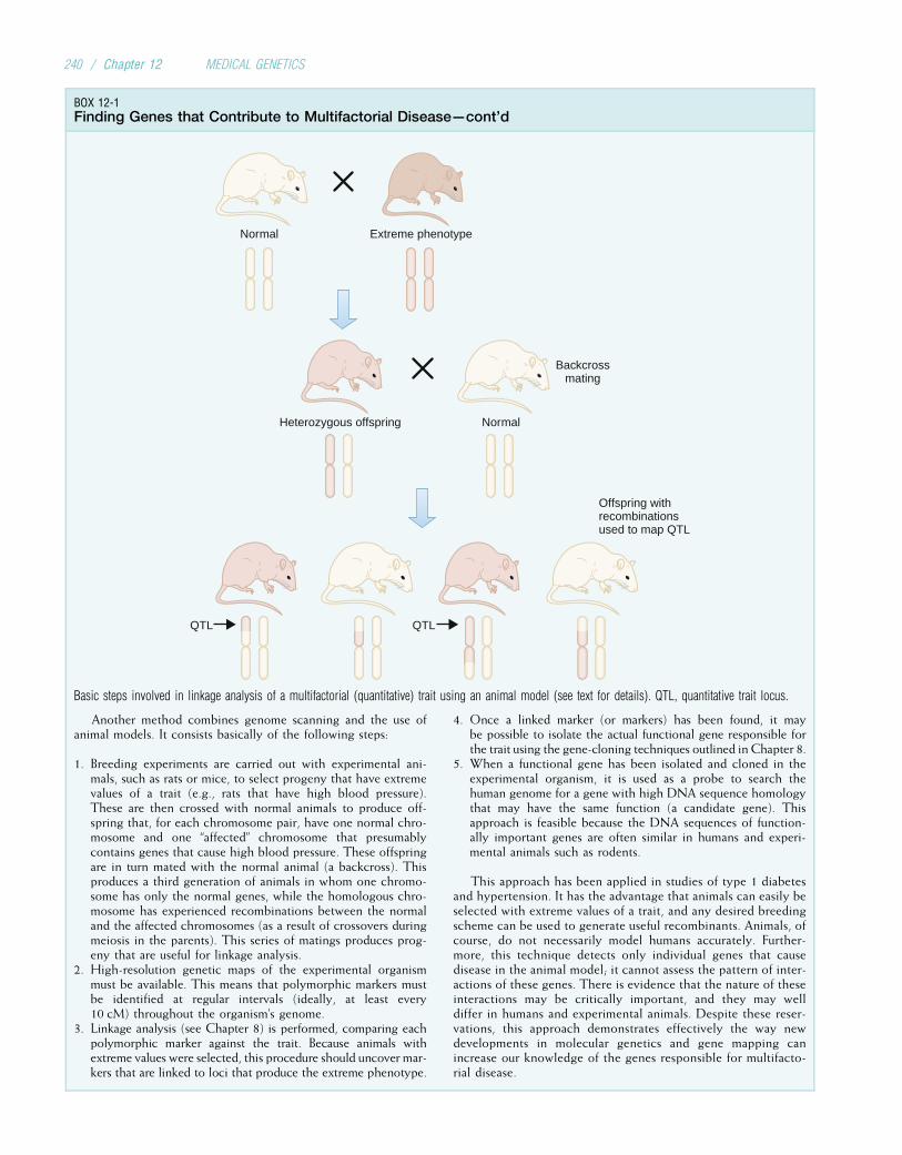
BOX 12-1Finding Genes that Contribute to Multifactorial Disease—cont’d
QTL QTL
Offspring withrecombinationsused to map QTL
Backcrossmating
Normal Extreme phenotype
Heterozygous offspring Normal
Basic steps involved in linkage analysis of a multifactorial (quantitative) trait using an animal model (see text for details). QTL, quantitative trait locus.
Another method combines genome scanning and the use ofanimal models. It consists basically of the following steps:
1. Breeding experiments are carried out with experimental ani-mals, such as rats or mice, to select progeny that have extremevalues of a trait (e.g., rats that have high blood pressure).These are then crossed with normal animals to produce off-spring that, for each chromosome pair, have one normal chro-mosome and one “affected” chromosome that presumablycontains genes that cause high blood pressure. These offspringare in turn mated with the normal animal (a backcross). Thisproduces a third generation of animals in whom one chromo-some has only the normal genes, while the homologous chro-mosome has experienced recombinations between the normaland the affected chromosomes (as a result of crossovers duringmeiosis in the parents). This series of matings produces prog-eny that are useful for linkage analysis.
2. High-resolution genetic maps of the experimental organismmust be available. This means that polymorphic markers mustbe identified at regular intervals (ideally, at least every10 cM) throughout the organism’s genome.
3. Linkage analysis (see Chapter 8) is performed, comparing eachpolymorphic marker against the trait. Because animals withextreme values were selected, this procedure should uncover mar-kers that are linked to loci that produce the extreme phenotype.
4. Once a linked marker (or markers) has been found, it maybe possible to isolate the actual functional gene responsible forthe trait using the gene-cloning techniques outlined in Chapter 8.
5. When a functional gene has been isolated and cloned in theexperimental organism, it is used as a probe to search thehuman genome for a gene with high DNA sequence homologythat may have the same function (a candidate gene). Thisapproach is feasible because the DNA sequences of function-ally important genes are often similar in humans and experi-mental animals such as rodents.
This approach has been applied in studies of type 1 diabetesand hypertension. It has the advantage that animals can easily beselected with extreme values of a trait, and any desired breedingscheme can be used to generate useful recombinants. Animals, ofcourse, do not necessarily model humans accurately. Further-more, this technique detects only individual genes that causedisease in the animal model; it cannot assess the pattern of inter-actions of these genes. There is evidence that the nature of theseinteractions may be critically important, and they may welldiffer in humans and experimental animals. Despite these reser-vations, this approach demonstrates effectively the way newdevelopments in molecular genetics and gene mapping canincrease our knowledge of the genes responsible for multifacto-rial disease.
240 / Chapter 12 MEDICAL GENETICS
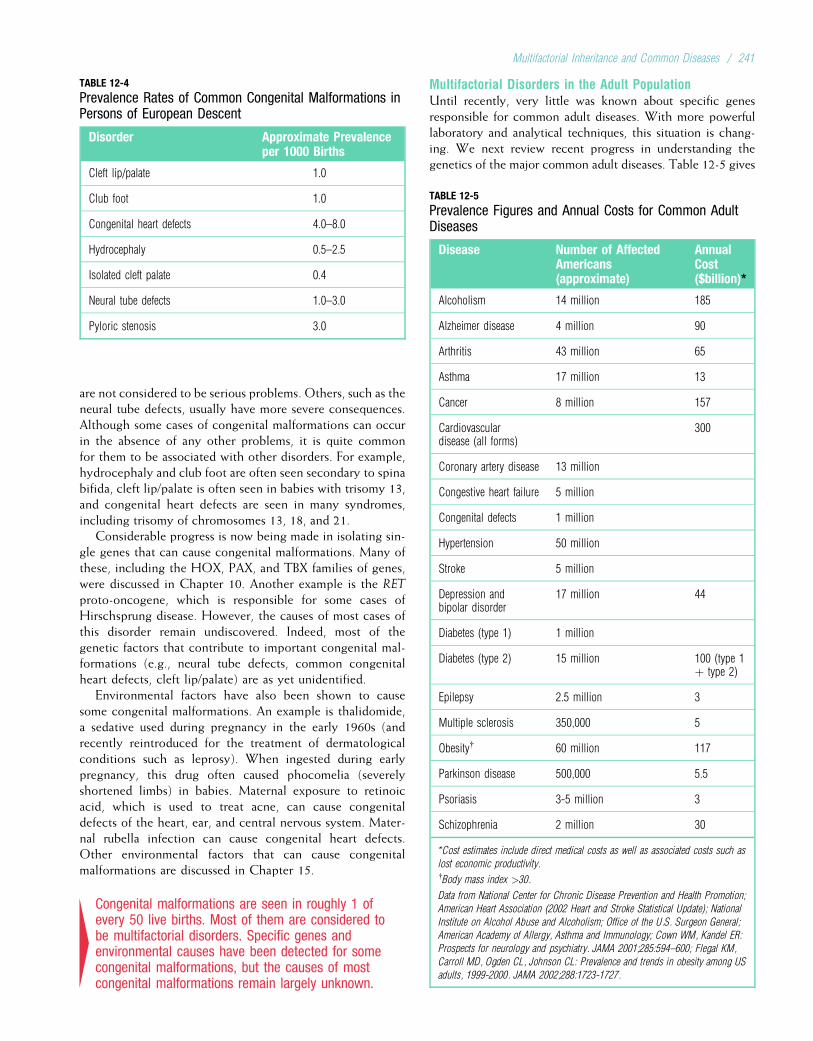
are not considered to be serious problems. Others, such as theneural tube defects, usually have more severe consequences.Although some cases of congenital malformations can occurin the absence of any other problems, it is quite commonfor them to be associated with other disorders. For example,hydrocephaly and club foot are often seen secondary to spinabifida, cleft lip/palate is often seen in babies with trisomy 13,and congenital heart defects are seen in many syndromes,including trisomy of chromosomes 13, 18, and 21.
Considerable progress is now being made in isolating sin-gle genes that can cause congenital malformations. Many ofthese, including the HOX, PAX, and TBX families of genes,were discussed in Chapter 10. Another example is the RET
proto-oncogene, which is responsible for some cases ofHirschsprung disease. However, the causes of most cases ofthis disorder remain undiscovered. Indeed, most of thegenetic factors that contribute to important congenital mal-formations (e.g., neural tube defects, common congenitalheart defects, cleft lip/palate) are as yet unidentified.
Environmental factors have also been shown to causesome congenital malformations. An example is thalidomide,a sedative used during pregnancy in the early 1960s (andrecently reintroduced for the treatment of dermatologicalconditions such as leprosy). When ingested during earlypregnancy, this drug often caused phocomelia (severelyshortened limbs) in babies. Maternal exposure to retinoicacid, which is used to treat acne, can cause congenitaldefects of the heart, ear, and central nervous system. Mater-nal rubella infection can cause congenital heart defects.Other environmental factors that can cause congenitalmalformations are discussed in Chapter 15.
4Congenital malformations are seen in roughly 1 ofevery 50 live births. Most of them are considered tobe multifactorial disorders. Specific genes andenvironmental causes have been detected for somecongenital malformations, but the causes of mostcongenital malformations remain largely unknown.
Multifactorial Disorders in the Adult PopulationUntil recently, very little was known about specific genesresponsible for common adult diseases. With more powerfullaboratory and analytical techniques, this situation is chang-ing. We next review recent progress in understanding thegenetics of the major common adult diseases. Table 12-5 gives
TABLE 12-4
Prevalence Rates of Common Congenital Malformations inPersons of European Descent
Disorder Approximate Prevalenceper 1000 Births
Cleft lip/palate 1.0
Club foot 1.0
Congenital heart defects 4.0–8.0
Hydrocephaly 0.5–2.5
Isolated cleft palate 0.4
Neural tube defects 1.0–3.0
Pyloric stenosis 3.0
TABLE 12-5
Prevalence Figures and Annual Costs for Common AdultDiseases
Disease Number of AffectedAmericans(approximate)
AnnualCost($billion)*
Alcoholism 14 million 185
Alzheimer disease 4 million 90
Arthritis 43 million 65
Asthma 17 million 13
Cancer 8 million 157
Cardiovasculardisease (all forms)
300
Coronary artery disease 13 million
Congestive heart failure 5 million
Congenital defects 1 million
Hypertension 50 million
Stroke 5 million
Depression andbipolar disorder
17 million 44
Diabetes (type 1) 1 million
Diabetes (type 2) 15 million 100 (type 1þ type 2)
Epilepsy 2.5 million 3
Multiple sclerosis 350,000 5
Obesity{ 60 million 117
Parkinson disease 500,000 5.5
Psoriasis 3-5 million 3
Schizophrenia 2 million 30
*Cost estimates include direct medical costs as well as associated costs such as
lost economic productivity.{Body mass index >30.
Data from National Center for Chronic Disease Prevention and Health Promotion;
American Heart Association (2002 Heart and Stroke Statistical Update); National
Institute on Alcohol Abuse and Alcoholism; Office of the U.S. Surgeon General;
American Academy of Allergy, Asthma and Immunology; Cown WM, Kandel ER:
Prospects for neurology and psychiatry. JAMA 2001;285:594–600; Flegal KM,
Carroll MD, Ogden CL, Johnson CL: Prevalence and trends in obesity among US
adults, 1999-2000. JAMA 2002;288:1723-1727.
Multifactorial Inheritance and Common Diseases / 241
approximate prevalence figures for these disorders in theUnited States.
Cardiovascular Disorders
Heart Disease
Heart disease is the leading cause of death worldwide, and itaccounts for approximately 25% of all deaths in the UnitedStates. The most common underlying cause of heart diseaseis coronary artery disease (CAD), which is caused by athero-sclerosis (a narrowing of the coronary arteries resulting fromthe formation of lipid-laden lesions). This narrowingimpedes blood flow to the heart and can eventually resultin a myocardial infarction (death of heart tissue caused byan inadequate supply of oxygen). When atherosclerosisoccurs in arteries that supply blood to the brain, a strokecan result. A number of risk factors for CAD have been iden-tified, including obesity, cigarette smoking, hypertension,elevated cholesterol level, and positive family history (usu-ally defined as having one or more affected first-degreerelatives). Many studies have examined the role of familyhistory in CAD, and they show that a person with a positivefamily history is at least twice as likely to suffer from CADthan is a person with no family history. Generally, thesestudies also show that the risk is higher if there are moreaffected relatives, if the affected relative is female (the lesscommonly affected sex) rather than male, and if the age ofonset in the affected relative is early (before 55 years ofage). For example, one study showed that men between theages of 20 and 39 years had a three-fold increase in CADrisk if they had one affected first-degree relative. This riskincreased to 13-fold if there were two first-degree relativesaffected with CAD before 55 years of age.
What part do genes play in the familial clustering ofCAD? Because of the key role of lipids in atherosclerosis,many studies have focused on the genetic determination ofvariation in circulating lipoprotein levels. An importantadvance was the isolation and cloning of the gene thatencodes the low-density lipoprotein (LDL) receptor. Het-erozygosity for a mutation in this gene roughly doublesLDL cholesterol levels and is seen in approximately 1 in500 persons. (This condition, known as familial hypercholes-terolemia, is described further in Clinical Commentary 12-2.)Mutations in the gene encoding apolipoprotein B, which areseen in about 1 in 1000 persons, are another common geneticcause of elevated LDL cholesterol. These mutations occurin the portion of the gene that is responsible for binding ofapolipoprotein B to the LDL receptor, and they increase cir-culating LDL cholesterol levels by 50% to 100%. More thana dozen other genes involved in lipid metabolism and trans-port have been identified, including those genes that encodevarious apolipoproteins (these are the protein componentsof lipoproteins) (Table 12-6). In addition, several geneswhose protein products contribute to inflammation have beenassociated with CAD, reflecting the critical role of inflamma-tion in generating atherosclerotic plaques. Functional analysisof these genes is leading to increased understanding and moreeffective treatment of CAD.
Environmental factors, many of which are easily modi-fied, are also important causes of CAD. There is abundantepidemiological evidence that cigarette smoking and obesityincrease the risk of CAD, whereas exercise and a diet low insaturated fats decrease the risk. Indeed, the approximate 60%reduction in age-adjusted mortality due to CAD and strokein the United States since 1950 is usually attributed to adecrease in the percentage of adults who smoke cigarettes,decreased consumption of saturated fats, improved medicalcare, and increased emphasis on healthy lifestyle factors suchas exercise.
Another form of heart disease is cardiomyopathy, anabnormality of the heart muscle that leads to inadequate car-diac function. Cardiomyopathy is a common cause of heartfailure, resulting in approximately 10,000 deaths annuallyin the United States. Hypertrophic cardiomyopathy, onemajor form of the disease, is characterized by thickening(hypertrophy) of portions of the left ventricle and is seenin as many as 1 in 500 adults. About half of hypertrophiccardiomyopathy cases are familial and are caused by autoso-mal dominant mutations in any of the multiple genes thatencode various components of the cardiac sarcomere. Themost commonly mutated genes are those that encode theb-myosin heavy chain (35% of familial cases), myosin-binding protein C (20% of cases), and troponin T (15% ofcases).
In contrast to the hypertrophic form of cardiomyopathy,dilated cardiomyopathy, which is seen in about 1 in 2500persons, consists of increased size and impaired contractionof the ventricles. The end result is impaired pumping of theheart. This disease is familial in about one third of affectedpersons; although autosomal dominant mutations are mostcommon, mutations can also be X-linked or mitochondrial.The genes affected by these mutations encode various cyto-skeletal proteins, including actin, cardiac troponin T, desmin,and components of the dystroglycan–sarcoglycan complex.(Recall from Chapter 5 that abnormalities of the latterproteins can also cause muscular dystrophies.)
Mutations have also been identified in several genes thatcause the long QT (LQT) syndrome. LQT describes the char-acteristically elongated QT interval in the electrocardiogramof affected individuals, indicative of delayed cardiac repolari-zation. This disorder, which can be caused either by inheritedmutations or by exposure to drugs that block potassium chan-nels, predisposes affected person to potentially fatal cardiacarrhythmia. An autosomal dominant form, known asRomano–Ward syndrome, can be caused by loss-of-functionmutations in genes that encode potassium channels (such asKCNQ1, KCNH2, KCNE1, KCNE2, or KCNJ2). These muta-tions delay cardiac repolarization. Gain-of-function mutationsin several of these same genes have been shown to produce ashortened QT interval, as might be expected. Romano–Wardsyndrome can also be caused by gain-of-function mutationsin sodium or calcium channel genes (SCN5A and CACNA1C,respectively), which result in a prolonged depolarizing current.(Other examples of mutations that can cause LQT are given inTable 12-7.)
242 / Chapter 12 MEDICAL GENETICS
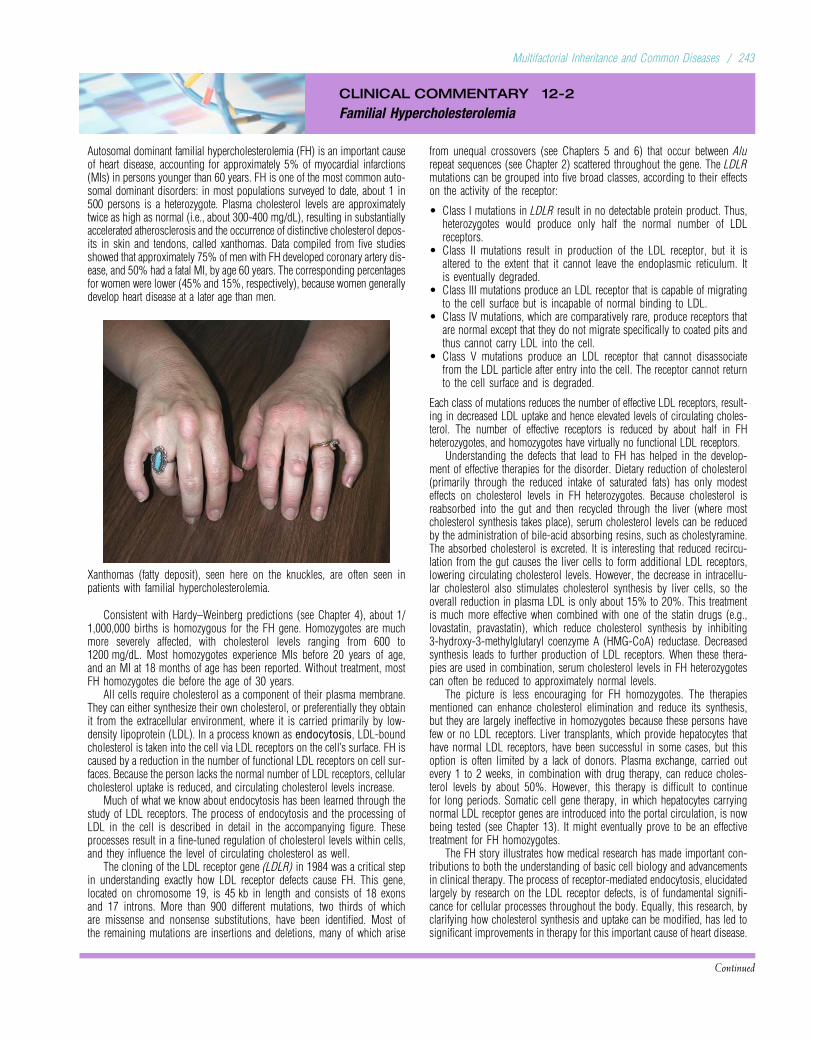
CLINICAL COMMENTARY 12-2
Familial Hypercholesterolemia
Autosomal dominant familial hypercholesterolemia (FH) is an important causeof heart disease, accounting for approximately 5% of myocardial infarctions(MIs) in persons younger than 60 years. FH is one of the most common auto-somal dominant disorders: in most populations surveyed to date, about 1 in500 persons is a heterozygote. Plasma cholesterol levels are approximatelytwice as high as normal (i.e., about 300-400 mg/dL), resulting in substantiallyaccelerated atherosclerosis and the occurrence of distinctive cholesterol depos-its in skin and tendons, called xanthomas. Data compiled from five studiesshowed that approximately 75% of men with FH developed coronary artery dis-ease, and 50% had a fatal MI, by age 60 years. The corresponding percentagesfor women were lower (45% and 15%, respectively), because women generallydevelop heart disease at a later age than men.
Xanthomas (fatty deposit), seen here on the knuckles, are often seen inpatients with familial hypercholesterolemia.
Consistent with Hardy–Weinberg predictions (see Chapter 4), about 1/1,000,000 births is homozygous for the FH gene. Homozygotes are muchmore severely affected, with cholesterol levels ranging from 600 to1200 mg/dL. Most homozygotes experience MIs before 20 years of age,and an MI at 18 months of age has been reported. Without treatment, mostFH homozygotes die before the age of 30 years.
All cells require cholesterol as a component of their plasma membrane.They can either synthesize their own cholesterol, or preferentially they obtainit from the extracellular environment, where it is carried primarily by low-density lipoprotein (LDL). In a process known as endocytosis, LDL-boundcholesterol is taken into the cell via LDL receptors on the cell’s surface. FH iscaused by a reduction in the number of functional LDL receptors on cell sur-faces. Because the person lacks the normal number of LDL receptors, cellularcholesterol uptake is reduced, and circulating cholesterol levels increase.
Much of what we know about endocytosis has been learned through thestudy of LDL receptors. The process of endocytosis and the processing ofLDL in the cell is described in detail in the accompanying figure. Theseprocesses result in a fine-tuned regulation of cholesterol levels within cells,and they influence the level of circulating cholesterol as well.
The cloning of the LDL receptor gene (LDLR) in 1984 was a critical stepin understanding exactly how LDL receptor defects cause FH. This gene,located on chromosome 19, is 45 kb in length and consists of 18 exonsand 17 introns. More than 900 different mutations, two thirds of whichare missense and nonsense substitutions, have been identified. Most ofthe remaining mutations are insertions and deletions, many of which arise
from unequal crossovers (see Chapters 5 and 6) that occur between Alurepeat sequences (see Chapter 2) scattered throughout the gene. The LDLRmutations can be grouped into five broad classes, according to their effectson the activity of the receptor:
• Class I mutations in LDLR result in no detectable protein product. Thus,heterozygotes would produce only half the normal number of LDLreceptors.
• Class II mutations result in production of the LDL receptor, but it isaltered to the extent that it cannot leave the endoplasmic reticulum. Itis eventually degraded.
• Class III mutations produce an LDL receptor that is capable of migratingto the cell surface but is incapable of normal binding to LDL.
• Class IV mutations, which are comparatively rare, produce receptors thatare normal except that they do not migrate specifically to coated pits andthus cannot carry LDL into the cell.
• Class V mutations produce an LDL receptor that cannot disassociatefrom the LDL particle after entry into the cell. The receptor cannot returnto the cell surface and is degraded.
Each class of mutations reduces the number of effective LDL receptors, result-ing in decreased LDL uptake and hence elevated levels of circulating choles-terol. The number of effective receptors is reduced by about half in FHheterozygotes, and homozygotes have virtually no functional LDL receptors.
Understanding the defects that lead to FH has helped in the develop-ment of effective therapies for the disorder. Dietary reduction of cholesterol(primarily through the reduced intake of saturated fats) has only modesteffects on cholesterol levels in FH heterozygotes. Because cholesterol isreabsorbed into the gut and then recycled through the liver (where mostcholesterol synthesis takes place), serum cholesterol levels can be reducedby the administration of bile-acid absorbing resins, such as cholestyramine.The absorbed cholesterol is excreted. It is interesting that reduced recircu-lation from the gut causes the liver cells to form additional LDL receptors,lowering circulating cholesterol levels. However, the decrease in intracellu-lar cholesterol also stimulates cholesterol synthesis by liver cells, so theoverall reduction in plasma LDL is only about 15% to 20%. This treatmentis much more effective when combined with one of the statin drugs (e.g.,lovastatin, pravastatin), which reduce cholesterol synthesis by inhibiting3-hydroxy-3-methylglutaryl coenzyme A (HMG-CoA) reductase. Decreasedsynthesis leads to further production of LDL receptors. When these thera-pies are used in combination, serum cholesterol levels in FH heterozygotescan often be reduced to approximately normal levels.
The picture is less encouraging for FH homozygotes. The therapiesmentioned can enhance cholesterol elimination and reduce its synthesis,but they are largely ineffective in homozygotes because these persons havefew or no LDL receptors. Liver transplants, which provide hepatocytes thathave normal LDL receptors, have been successful in some cases, but thisoption is often limited by a lack of donors. Plasma exchange, carried outevery 1 to 2 weeks, in combination with drug therapy, can reduce choles-terol levels by about 50%. However, this therapy is difficult to continuefor long periods. Somatic cell gene therapy, in which hepatocytes carryingnormal LDL receptor genes are introduced into the portal circulation, is nowbeing tested (see Chapter 13). It might eventually prove to be an effectivetreatment for FH homozygotes.
The FH story illustrates how medical research has made important con-tributions to both the understanding of basic cell biology and advancementsin clinical therapy. The process of receptor-mediated endocytosis, elucidatedlargely by research on the LDL receptor defects, is of fundamental signifi-cance for cellular processes throughout the body. Equally, this research, byclarifying how cholesterol synthesis and uptake can be modified, has led tosignificant improvements in therapy for this important cause of heart disease.
Continued
Multifactorial Inheritance and Common Diseases / 243
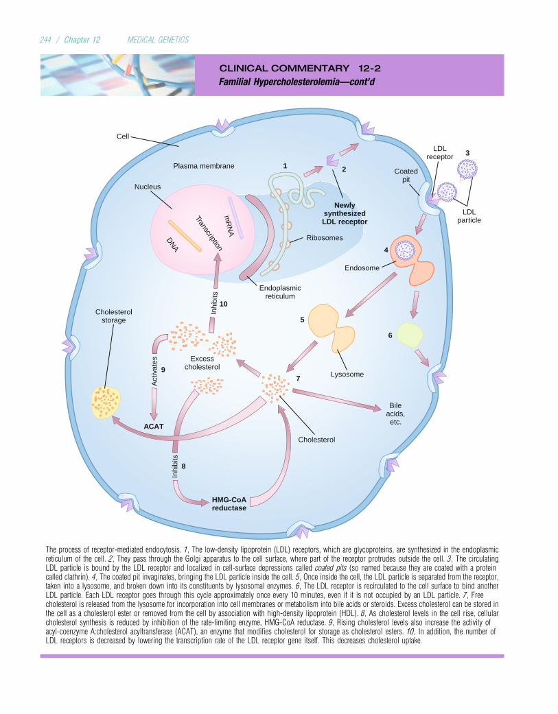
CLINICAL COMMENTARY 12-2
Familial Hypercholesterolemia—cont’d
Lysosome
Cholesterol
Cholesterolstorage
Bile acids, etc.
3
5
ACAT
HMG-CoA reductase
Excess cholesterol
Inhi
bits
Plasma membrane
Cell
9
6
1 2
Transcription
DN
A
mR
NA
Newly synthesized LDL receptor
Ribosomes
Endoplasmic reticulum
Nucleus
LDL particle
LDL receptor
Act
ivat
es
Inhi
bits
Endosome
4
Coated pit
7
10
8
The process of receptor-mediated endocytosis. 1, The low-density lipoprotein (LDL) receptors, which are glycoproteins, are synthesized in the endoplasmicreticulum of the cell. 2, They pass through the Golgi apparatus to the cell surface, where part of the receptor protrudes outside the cell. 3, The circulatingLDL particle is bound by the LDL receptor and localized in cell-surface depressions called coated pits (so named because they are coated with a proteincalled clathrin). 4, The coated pit invaginates, bringing the LDL particle inside the cell. 5, Once inside the cell, the LDL particle is separated from the receptor,taken into a lysosome, and broken down into its constituents by lysosomal enzymes. 6, The LDL receptor is recirculated to the cell surface to bind anotherLDL particle. Each LDL receptor goes through this cycle approximately once every 10 minutes, even if it is not occupied by an LDL particle. 7, Freecholesterol is released from the lysosome for incorporation into cell membranes or metabolism into bile acids or steroids. Excess cholesterol can be stored inthe cell as a cholesterol ester or removed from the cell by association with high-density lipoprotein (HDL). 8, As cholesterol levels in the cell rise, cellularcholesterol synthesis is reduced by inhibition of the rate-limiting enzyme, HMG-CoA reductase. 9, Rising cholesterol levels also increase the activity ofacyl-coenzyme A:cholesterol acyltransferase (ACAT), an enzyme that modifies cholesterol for storage as cholesterol esters. 10, In addition, the number ofLDL receptors is decreased by lowering the transcription rate of the LDL receptor gene itself. This decreases cholesterol uptake.
244 / Chapter 12 MEDICAL GENETICS
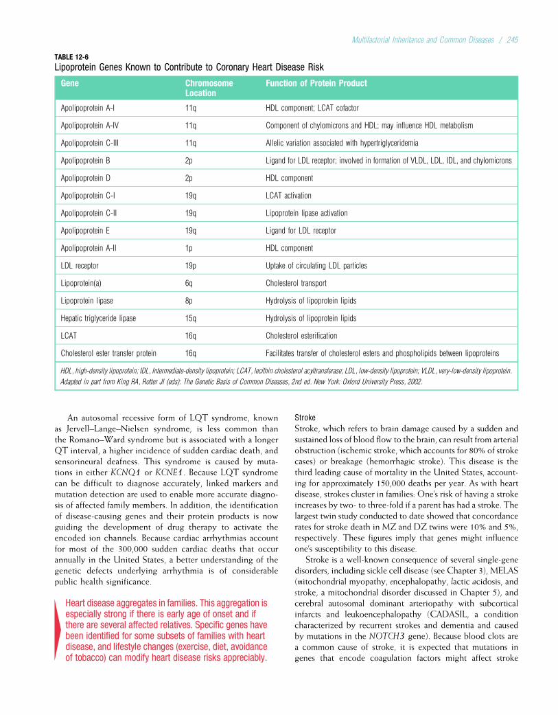
An autosomal recessive form of LQT syndrome, knownas Jervell–Lange–Nielsen syndrome, is less common thanthe Romano–Ward syndrome but is associated with a longerQT interval, a higher incidence of sudden cardiac death, andsensorineural deafness. This syndrome is caused by muta-tions in either KCNQ1 or KCNE1. Because LQT syndromecan be difficult to diagnose accurately, linked markers andmutation detection are used to enable more accurate diagno-sis of affected family members. In addition, the identificationof disease-causing genes and their protein products is nowguiding the development of drug therapy to activate theencoded ion channels. Because cardiac arrhythmias accountfor most of the 300,000 sudden cardiac deaths that occurannually in the United States, a better understanding of thegenetic defects underlying arrhythmia is of considerablepublic health significance.
4Heart disease aggregates in families. This aggregation isespecially strong if there is early age of onset and ifthere are several affected relatives. Specific genes havebeen identified for some subsets of families with heartdisease, and lifestyle changes (exercise, diet, avoidanceof tobacco) can modify heart disease risks appreciably.
Stroke
Stroke, which refers to brain damage caused by a sudden andsustained loss of blood flow to the brain, can result from arterialobstruction (ischemic stroke, which accounts for 80% of strokecases) or breakage (hemorrhagic stroke). This disease is thethird leading cause of mortality in the United States, account-ing for approximately 150,000 deaths per year. As with heartdisease, strokes cluster in families: One’s risk of having a strokeincreases by two- to three-fold if a parent has had a stroke. Thelargest twin study conducted to date showed that concordancerates for stroke death in MZ and DZ twins were 10% and 5%,respectively. These figures imply that genes might influenceone’s susceptibility to this disease.
Stroke is a well-known consequence of several single-genedisorders, including sickle cell disease (see Chapter 3), MELAS(mitochondrial myopathy, encephalopathy, lactic acidosis, andstroke, a mitochondrial disorder discussed in Chapter 5), andcerebral autosomal dominant arteriopathy with subcorticalinfarcts and leukoencephalopathy (CADASIL, a conditioncharacterized by recurrent strokes and dementia and causedby mutations in the NOTCH3 gene). Because blood clots area common cause of stroke, it is expected that mutations ingenes that encode coagulation factors might affect stroke
TABLE 12-6
Lipoprotein Genes Known to Contribute to Coronary Heart Disease Risk
Gene ChromosomeLocation
Function of Protein Product
Apolipoprotein A-I 11q HDL component; LCAT cofactor
Apolipoprotein A-IV 11q Component of chylomicrons and HDL; may influence HDL metabolism
Apolipoprotein C-III 11q Allelic variation associated with hypertriglyceridemia
Apolipoprotein B 2p Ligand for LDL receptor; involved in formation of VLDL, LDL, IDL, and chylomicrons
Apolipoprotein D 2p HDL component
Apolipoprotein C-I 19q LCAT activation
Apolipoprotein C-II 19q Lipoprotein lipase activation
Apolipoprotein E 19q Ligand for LDL receptor
Apolipoprotein A-II 1p HDL component
LDL receptor 19p Uptake of circulating LDL particles
Lipoprotein(a) 6q Cholesterol transport
Lipoprotein lipase 8p Hydrolysis of lipoprotein lipids
Hepatic triglyceride lipase 15q Hydrolysis of lipoprotein lipids
LCAT 16q Cholesterol esterification
Cholesterol ester transfer protein 16q Facilitates transfer of cholesterol esters and phospholipids between lipoproteins
HDL, high-density lipoprotein; IDL, Intermediate-density lipoprotein; LCAT, lecithin cholesterol acyltransferase; LDL, low-density lipoprotein; VLDL, very-low-density lipoprotein.
Adapted in part from King RA, Rotter JI (eds): The Genetic Basis of Common Diseases, 2nd ed. New York: Oxford University Press, 2002.
Multifactorial Inheritance and Common Diseases / 245
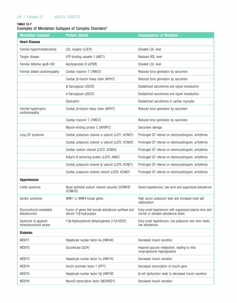
TABLE 12-7
Examples of Mendelian Subtypes of Complex Disorders*
Mendelian Subtype Protein (Gene) Consequence of Mutation
Heart Disease
Familial hypercholesterolemia LDL receptor (LDLR) Elevated LDL level
Tangier disease ATP-binding cassette 1 (ABC1) Reduced HDL level
Familial defective apoB-100 Apolipoprotein B (APOB) Elevated LDL level
Familial dilated cardiomyopathy Cardiac troponin T (TNNT2) Reduced force generation by sarcomere
Cardiac b-myosin heavy chain (MYH7) Reduced force generation by sarcomere
b-Sarcoglycan (SGCB) Destabilized sarcolemma and signal transduction
d-Sarcoglycan (SGCD) Destabilized sarcolemma and signal transduction
Dystrophin Destabilized sarcolemma in cardiac myocytes
Familial hypertrophiccardiomyopathy
Cardiac b-myosin heavy chain (MYH7) Reduced force generation by sarcomere
Cardiac troponin T (TNNT2) Reduced force generation by sarcomere
Myosin-binding protein C (MYBPC) Sarcomere damage
Long QT syndrome Cardiac potassium channel a subunit (LQT1, KCNQ1) Prolonged QT interval on electrocardiogram, arrhythmia
Cardiac potassium channel a subunit (LQT2, KCNH2) Prolonged QT interval on electrocardiogram, arrhythmia
Cardiac sodium channel (LQT3, SCN5A) Prolonged QT interval on electrocardiogram, arrhythmia
Ankyrin B anchoring protein (LQT4, ANK2) Prolonged QT interval on electrocardiogram, arrhythmia
Cardiac potassium channel b subunit (LQT5, KCNE1) Prolonged QT interval on electrocardiogram, arrhythmia
Cardiac potassium channel subunit (LQT6, KCNE2) Prolonged QT interval on electrocardiogram, arrhythmia
Hypertension
Liddle syndrome Renal epithelial sodium channel subunits (SCNN1B,SCNN1G)
Severe hypertension, low renin and suppressed aldosterone
Gordon syndrome WNK1 or WNK4 kinase genes High serum potassium level and increased renal saltreabsorption
Glucocorticoid-remediablealdosteronism
Fusion of genes that encode aldosterone synthase andsteroid 11b-hydroxylase
Early-onset hypertension with suppressed plasma renin andnormal or elevated aldosterone levels
Syndrome of apparentmineralocorticoid excess
11b-Hydroxysteroid dehydrogenase (11b-HSD2) Early-onset hypertension, low potassium and renin levels,low aldosterone
Diabetes
MODY1 Hepatocyte nuclear factor-4a (HNF4A) Decreased insulin secretion
MODY2 Glucokinase (GCK) Impaired glucose metabolism, leading to mildnonprogressive hyperglycemia
MODY3 Hepatocyte nuclear factor-1a (HNF1A) Decreased insulin secretion
MODY4 Insulin promoter factor-1 (IPF1) Decreased transcription of insulin gene
MODY5 Hepatocyte nuclear factor-1b (HNF1B) b-cell dysfunction leads to decreased insulin secretion
MODY6 NeuroD transcription factor (NEUROD1) Decreased insulin secretion
246 / Chapter 12 MEDICAL GENETICS
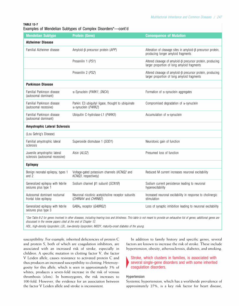
susceptibility. For example, inherited deficiencies of protein Cand protein S, both of which are coagulation inhibitors, areassociated with an increased risk of stroke, especially inchildren. A specific mutation in clotting factor V, the factorV Leiden allele, causes resistance to activated protein C andthus produces an increased susceptibility to clotting. Heterozy-gosity for this allele, which is seen in approximately 5% ofwhites, produces a seven-fold increase in the risk of venousthrombosis (clots). In homozygotes, the risk increases to100-fold. However, the evidence for an association betweenthe factor V Leiden allele and stroke is inconsistent.
In addition to family history and specific genes, severalfactors are known to increase the risk of stroke. These includehypertension, obesity, atherosclerosis, diabetes, and smoking.
4Stroke, which clusters in families, is associated withseveral single-gene disorders and with some inheritedcoagulation disorders.
Hypertension
Systemic hypertension, which has a worldwide prevalence ofapproximately 27%, is a key risk factor for heart disease,
TABLE 12-7
Examples of Mendelian Subtypes of Complex Disorders*—cont’d
Mendelian Subtype Protein (Gene) Consequence of Mutation
Alzheimer Disease
Familial Alzheimer disease Amyloid-b precursor protein (APP) Alteration of cleavage sites in amyloid-b precursor protein,producing longer amyloid fragments
Presenilin 1 (PS1) Altered cleavage of amyloid-b precursor protein, producinglarger proportion of long amyloid fragments
Presenilin 2 (PS2) Altered cleavage of amyloid-b precursor protein, producinglarger proportion of long amyloid fragments
Parkinson Disease
Familial Parkinson disease(autosomal dominant)
a-Synuclein (PARK1, SNCA) Formation of a-synuclein aggregates
Familial Parkinson disease(autosomal recessive)
Parkin: E3 ubiquityl ligase, thought to ubiquinatea-synuclein (PARK2)
Compromised degradation of a-synuclein
Familial Parkinson disease(autosomal dominant)
Ubiquitin C-hydrolase-L1 (PARK5) Accumulation of a-synuclein
Amyotrophic Lateral Sclerosis
(Lou Gehrig’s Disease)
Familial amyotrophic lateralsclerosis
Superoxide dismutase 1 (SOD1) Neurotoxic gain of function
Juvenile amyotrophic lateralsclerosis (autosomal recessive)
Alsin (ALS2) Presumed loss of function
Epilepsy
Benign neonatal epilepsy, types 1and 2
Voltage-gated potassium channels (KCNQ2 andKCNQ3, respectively)
Reduced M current increases neuronal excitability
Generalized epilepsy with febrileseizures plus type 1
Sodium channel b1 subunit (SCN1B) Sodium current persistence leading to neuronalhyperexcitability
Autosomal dominant nocturnalfrontal lobe epilepsy
Neuronal nicotinic acetylcholine receptor subunits(CHRNA4 and CHRNB2)
Increased neuronal excitability in response to cholinergicstimulation
Generalized epilepsy with febrileseizures plus type 3
GABAA receptor (GABRG2) Loss of synaptic inhibition leading to neuronal excitability
*See Table 8-2 for genes involved in other diseases, including hearing loss and blindness. This table is not meant to provide an exhaustive list of genes; additional genes are
discussed in the review papers cited at the end of Chapter 12.
HDL, high-density lipoprotein; LDL, low-density lipoprotein; MODY, maturity-onset diabetes of the young.
Multifactorial Inheritance and Common Diseases / 247
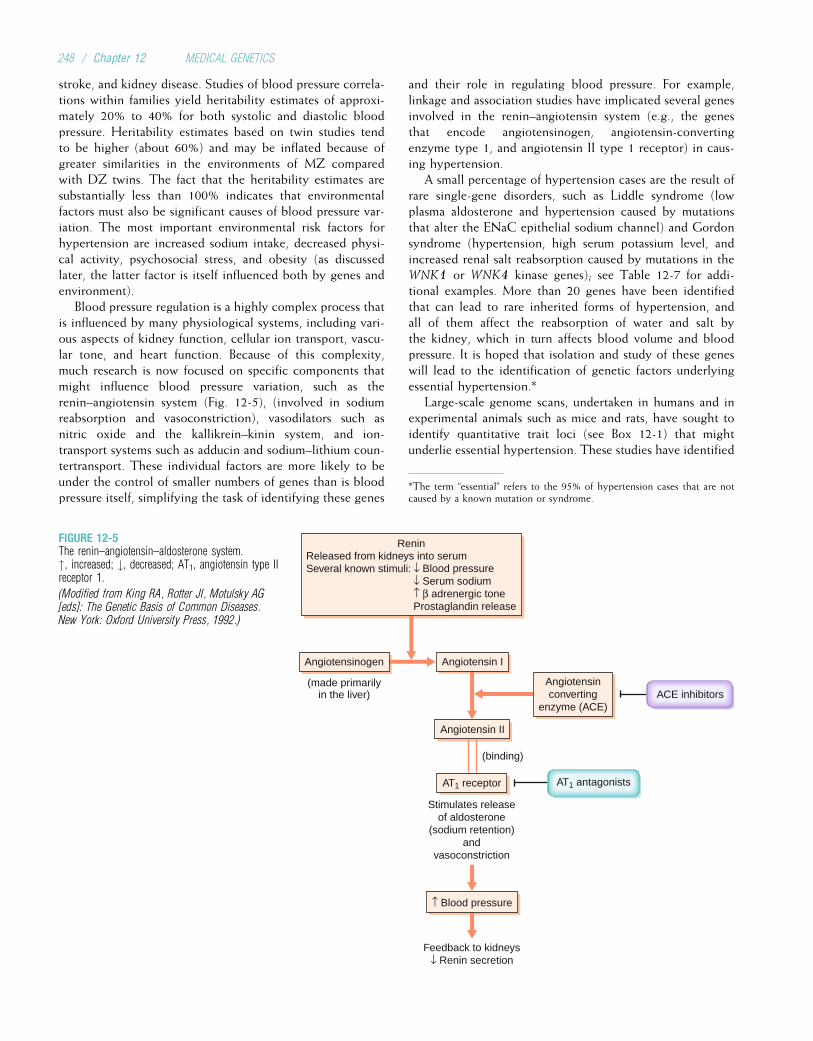
stroke, and kidney disease. Studies of blood pressure correla-tions within families yield heritability estimates of approxi-mately 20% to 40% for both systolic and diastolic bloodpressure. Heritability estimates based on twin studies tendto be higher (about 60%) and may be inflated because ofgreater similarities in the environments of MZ comparedwith DZ twins. The fact that the heritability estimates aresubstantially less than 100% indicates that environmentalfactors must also be significant causes of blood pressure var-iation. The most important environmental risk factors forhypertension are increased sodium intake, decreased physi-cal activity, psychosocial stress, and obesity (as discussedlater, the latter factor is itself influenced both by genes andenvironment).
Blood pressure regulation is a highly complex process thatis influenced by many physiological systems, including vari-ous aspects of kidney function, cellular ion transport, vascu-lar tone, and heart function. Because of this complexity,much research is now focused on specific components thatmight influence blood pressure variation, such as therenin–angiotensin system (Fig. 12-5), (involved in sodiumreabsorption and vasoconstriction), vasodilators such asnitric oxide and the kallikrein–kinin system, and ion-transport systems such as adducin and sodium–lithium coun-tertransport. These individual factors are more likely to beunder the control of smaller numbers of genes than is bloodpressure itself, simplifying the task of identifying these genes
and their role in regulating blood pressure. For example,linkage and association studies have implicated several genesinvolved in the renin–angiotensin system (e.g., the genesthat encode angiotensinogen, angiotensin-convertingenzyme type 1, and angiotensin II type 1 receptor) in caus-ing hypertension.
A small percentage of hypertension cases are the result ofrare single-gene disorders, such as Liddle syndrome (lowplasma aldosterone and hypertension caused by mutationsthat alter the ENaC epithelial sodium channel) and Gordonsyndrome (hypertension, high serum potassium level, andincreased renal salt reabsorption caused by mutations in theWNK1 or WNK4 kinase genes); see Table 12-7 for addi-tional examples. More than 20 genes have been identifiedthat can lead to rare inherited forms of hypertension, andall of them affect the reabsorption of water and salt bythe kidney, which in turn affects blood volume and bloodpressure. It is hoped that isolation and study of these geneswill lead to the identification of genetic factors underlyingessential hypertension.*
Large-scale genome scans, undertaken in humans and inexperimental animals such as mice and rats, have sought toidentify quantitative trait loci (see Box 12-1) that mightunderlie essential hypertension. These studies have identified
Angiotensin IAngiotensinogen
Angiotensin II
(binding)
ACE inhibitors
AT1 antagonists
Stimulates releaseof aldosterone
(sodium retention)and
vasoconstriction
↑ Blood pressure
Feedback to kidneys↓ Renin secretion
AT1 receptor
Angiotensinconverting
enzyme (ACE)
ReninReleased from kidneys into serumSeveral known stimuli: ↓ Blood pressure
↓ Serum sodium↑ β adrenergic toneProstaglandin release
(made primarilyin the liver)
FIGURE 12-5The renin–angiotensin–aldosterone system.", increased; #, decreased; AT1, angiotensin type IIreceptor 1.
(Modified from King RA, Rotter JI, Motulsky AG[eds]: The Genetic Basis of Common Diseases.New York: Oxford University Press, 1992.)
*The term “essential” refers to the 95% of hypertension cases that are notcaused by a known mutation or syndrome.
248 / Chapter 12 MEDICAL GENETICS
a number of regions in which LOD scores (see Chapter 8)offer statistical support for the presence of genes that influ-ence susceptibility to hypertension, and in some cases severalstudies have implicated the same genomic region. Such resultsmight help to pinpoint specific genes that underlie suscepti-bility to essential hypertension.
4Heritability estimates for systolic and diastolic bloodpressure range from 20% to 40%. A number of genesresponsible for rare hypertension syndromes have beenidentified, and genome scans have implicated regionsthat might contain genes that underlie susceptibility toessential hypertension. Other risk factors forhypertension include increased sodium intake, lack ofexercise, psychosocial stress, and obesity.
CancerCancer is the second leading cause of death in the UnitedStates, although it is estimated that it might soon surpassheart disease as the leading cause of death. It is well estab-lished that many major types of cancer (e.g., breast, colon,prostate, ovarian) cluster strongly in families. This is dueboth to shared genes and shared environmental factors.Although numerous cancer genes have been isolated, envi-ronmental factors also play an important role in causing can-cer by inducing somatic mutations. In particular, tobacco useis estimated to account for one third of all cancer cases indeveloped countries, making it the most important knowncause of cancer. Diet (i.e., carcinogenic substances and thelack of “anticancer” components such as fiber, fruits, andvegetables) is another leading cause of cancer and may alsoaccount for as much as one third of cancer cases. It is esti-mated that approximately 15% of worldwide cancer casesare caused primarily by infectious agents (e.g., human papil-loma virus for cervical cancer, hepatitis B and C for liver can-cer). Because cancer genetics was the subject of Chapter 11,we confine our attention here to genetic and environmentalfactors that influence susceptibility to some of the mostcommon cancers.
Breast Cancer
Breast cancer is the second most commonly diagnosed can-cer (after skin cancer) among women, affecting approxi-mately 12% of American women who live to age 85 yearsor older. It was diagnosed in approximately 180,000 Ameri-can women in 2008, and about 40,000 women die from thisdisease each year. Breast cancer was formerly the leadingcause of cancer death among women, but it has been sur-passed by lung cancer. Breast cancer can also occur in men,with a lifetime prevalence that is roughly 100 times lowerthan that of women. The familial aggregation of breast can-cer has been recognized for centuries, having been describedby physicians in ancient Rome. If a woman has one affectedfirst-degree relative, her risk of developing breast cancerdoubles. The risk increases further with additional affected
relatives, and it increases if those relatives developed cancerat a relatively early age (before 45 years of age).
Several genes are now known to predispose women todeveloping hereditary breast cancer. Most important amongthese are BRCA1 and BRCA2, two genes involved in DNArepair (see Chapter 11). Germline mutations in the TP53and CHK2 genes can cause Li–Fraumeni syndrome, whichalso predisposes to breast cancer. Cowden disease, a rareautosomal dominant condition that includes multiple hamar-tomas and breast cancer, is caused by mutations in the PTENtumor suppressor gene (see Chapter 11). Ataxia-telangiectasia,an autosomal recessive disorder caused by defective DNArepair, includes breast cancer in its presentation. Mutationsin the MSH2 and MLH1 DNA repair genes, which lead tohereditary nonpolyposis colorectal cancer (HNPCC), alsoconfer an increased risk of breast cancer. Despite the signifi-cance of these genes, it should be emphasized that more than90% of breast cancer cases are not inherited as mendeliandiseases.
A number of environmental factors are known to increasethe risk of developing breast cancer. These include nullipar-ity (never bearing children), bearing the first child after 30years of age, a high-fat diet, alcohol use, and estrogenreplacement therapy.
Colorectal Cancer
It is estimated that 1 in 20 Americans will develop colorectalcancer, and roughly one third of those with this cancer willdie from it. With approximately 150,000 new cases and50,000 deaths in the United States in 2008, colorectal canceris second only to lung cancer in the total number of annualcancer deaths. Like breast cancer, it clusters in families;familial clustering of this form of cancer was reported inthe medical literature as early as 1881. The risk of colorectalcancer in people with one affected first-degree relative is twoto three times higher than that of the general population.
As discussed in Chapter 11, familial colon cancer can be theresult of mutations in the APC tumor suppressor gene or in oneof several DNA mismatch-repair genes (HNPCC). Another,less common, inherited cause of colon cancer is the autosomaldominant Peutz–Jeghers syndrome. About half of Peutz–Jeghers cases are caused by mutations in the STK11 tumorsuppressor gene, which encodes a protein kinase. Juvenileintestinal polyposis, an autosomal dominant disease definedby the presence of 10 or more polyps before adulthood, canbe caused by mutations in SMAD4 (see Chapter 11), inBMPRA1 (a receptor serine–threonine kinase gene), or, in rarecases, in PTEN. PTEN mutations can also cause Cowdendisease, which, in addition to breast tumors, often includespolyps in the intestinal tract.
As with breast cancer, most colon cancer cases (>90%)are not inherited as mendelian conditions and are likely tobe caused by a complex interaction of inherited and somaticgenetic alterations and environmental factors. The latter riskfactors include a lack of physical activity and a high-fat,low-fiber diet.
Multifactorial Inheritance and Common Diseases / 249
Prostate Cancer
Prostate cancer is the second most commonly diagnosedcancer in men (after skin cancer), with approximately185,000 new cases annually in the United States. Prostatecancer is second only to lung cancer as a cause of cancerdeath in men, causing more than 29,000 deaths in 2008.Having an affected first-degree relative increases the risk ofdeveloping prostate cancer by a factor of two to three. It isestimated that about 5% to 10% of prostate cancer casesare the result of inherited mutations.
The relatively late age of onset of most prostate cancer cases(median age, 72 years) makes genetic analysis especially diffi-cult. However, loss of heterozygosity (see Chapter 11) hasbeen observed in a number of genomic regions in prostatetumor cells, possibly indicating the presence of genetic altera-tions in these regions. In addition, genome scans have indi-cated that several chromosome regions might containprostate cancer susceptibility genes. One of these regions,8q24, has been associated with a significantly increased riskof prostate cancer in several populations. The RNASEL genehas also been associated with prostate cancer risk in severalstudies. The product of this gene, ribonuclease L, regulates cellproliferation and apoptosis. Mutations in RNASEL account for asmall percentage of familial prostate cancer cases.
Nongenetic risk factors for prostate cancer may include ahigh-fat diet. Because prostate cancer usually progressesslowly and because it can be detected by digital examinationand by the prostate-specific antigen (PSA) test, fatal metastasiscan usually be prevented.
4Most common cancers have genetic components.Recurrence risks tend to be higher if there are severalaffected relatives and if those relatives developedcancer at an early age. Specific genes have beendiscovered that cause inherited colon, breast, andprostate cancer in some families.
Diabetes MellitusLike the other disorders discussed in this chapter, the etiologyof diabetes mellitus is complex and not fully understood. Nev-ertheless, progress is being made in understanding the geneticbasis of this disorder, which is the leading cause of adultblindness, kidney failure, and lower-limb amputation and amajor cause of heart disease and stroke. An important advancehas been the recognition that diabetes mellitus is actually aheterogeneous group of disorders, all characterized by ele-vated blood sugar. We focus here on the three major typesof diabetes, type 1 (formerly termed insulin-dependent diabetesmellitus, or IDDM), type 2 (formerly termed non–insulin-dependent diabetes mellitus, or NIDDM), and maturity-onsetdiabetes of the young (MODY).
Type 1 Diabetes
Type 1 diabetes, which is characterized by T-cell infiltrationof the pancreas and destruction of the insulin-producing betacells, usually (though not always) manifests before 40 years
of age. Patients with type 1 diabetes must receive exogenousinsulin to survive. In addition to T-cell infiltration of thepancreas, autoantibodies are formed against pancreatic cells,insulin, and enzymes such as glutamic acid decarboxylase;these autoantibodies can be observed long before clinicalsymptoms occur. These findings, along with a strong associ-ation between type 1 diabetes and the presence of severalhuman leukocyte antigen (HLA) class II alleles, indicate thatthis is an autoimmune disease.
Siblings of persons with type 1 diabetes face a substantialelevation in risk: approximately 6%, as opposed to a risk ofabout 0.3% to 0.5% in the general population. The recur-rence risk is also elevated when there is a diabetic parent,although this risk varies with the sex of the affected parent.The risk for offspring of diabetic mothers is only 1% to3%, but it is 4% to 6% for the offspring of diabetic fathers.(Because type 1 diabetes affects males and females in roughlyequal proportions in the general population, this risk differ-ence is inconsistent with the sex-specific threshold modelfor multifactorial traits.) Twin studies show that the empiri-cal risk for MZ twins of type 1 diabetes patients ranges from30% to 50%. In contrast, the concordance rate for DZ twinsis 5% to 10%. The fact that type 1 diabetes is not 100% con-cordant among identical twins indicates that genetic factorsare not solely responsible for the disorder. There is evidencethat specific viral infections contribute to the cause of type 1diabetes in at least some persons, possibly by activating anautoimmune response.
The association of specific HLA class II alleles and type 1diabetes has been studied extensively, and it is estimated thatthe HLA loci account for about 40% to 50% of the geneticsusceptibility to type 1 diabetes. Approximately 95% ofwhites with type 1 diabetes have the HLA DR3 and/orDR4 alleles, whereas only about 50% of the general whitepopulation has either of these alleles. If an affected probandand a sibling are both heterozygous for the DR3 and DR4alleles, the sibling’s risk of developing type 1 diabetes is nearly20% (i.e., about 40 times higher than the risk in the generalpopulation). This association may in part reflect linkage dis-equilibrium between alleles of the DR locus and those of theHLA-DQ locus. The absence of aspartic acid at position 57of the DQ polypeptide is strongly associated with susceptibil-ity to type 1 diabetes; in fact, those who do not have thisamino acid at position 57 (and instead are homozygous fora different amino acid) are 100 times more likely to developtype 1 diabetes. The aspartic acid substitution alters the shapeof the HLA class II molecule and thus its ability to bind andpresent peptides to T cells (see Chapter 9). Altered T-cell rec-ognition might help to protect persons with the aspartic acidsubstitution from an autoimmune episode.
The insulin gene, which is located on the short arm ofchromosome 11, is another logical candidate for type 1 dia-betes susceptibility. Polymorphisms within and near thisgene have been tested for association with type 1 diabetes.Intriguingly, a strong risk association is seen with allelic var-iation in a VNTR polymorphism (see Chapter 3) located just50 of the insulin gene. Differences in the number of VNTR
250 / Chapter 12 MEDICAL GENETICS
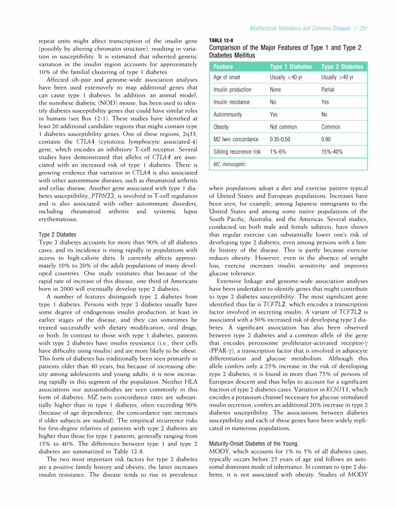
repeat units might affect transcription of the insulin gene(possibly by altering chromatin structure), resulting in varia-tion in susceptibility. It is estimated that inherited geneticvariation in the insulin region accounts for approximately10% of the familial clustering of type 1 diabetes.
Affected sib-pair and genome-wide association analyseshave been used extensively to map additional genes thatcan cause type 1 diabetes. In addition, an animal model,the nonobese diabetic (NOD) mouse, has been used to iden-tify diabetes susceptibility genes that could have similar rolesin humans (see Box 12-1). These studies have identified atleast 20 additional candidate regions that might contain type1 diabetes susceptibility genes. One of these regions, 2q33,contains the CTLA4 (cytotoxic lymphocyte associated-4)gene, which encodes an inhibitory T-cell receptor. Severalstudies have demonstrated that alleles of CTLA4 are asso-ciated with an increased risk of type 1 diabetes. There isgrowing evidence that variation in CTLA4 is also associatedwith other autoimmune diseases, such as rheumatoid arthritisand celiac disease. Another gene associated with type 1 dia-betes susceptibility, PTPN22, is involved in T-cell regulationand is also associated with other autoimmune disorders,including rheumatoid arthritis and systemic lupuserythematosus.
Type 2 Diabetes
Type 2 diabetes accounts for more than 90% of all diabetescases, and its incidence is rising rapidly in populations withaccess to high-calorie diets. It currently affects approxi-mately 10% to 20% of the adult populations of many devel-oped countries. One study estimates that because of therapid rate of increase of this disease, one third of Americansborn in 2000 will eventually develop type 2 diabetes.
A number of features distinguish type 2 diabetes fromtype 1 diabetes. Persons with type 2 diabetes usually havesome degree of endogenous insulin production, at least inearlier stages of the disease, and they can sometimes betreated successfully with dietary modification, oral drugs,or both. In contrast to those with type 1 diabetes, patientswith type 2 diabetes have insulin resistance (i.e., their cellshave difficulty using insulin) and are more likely to be obese.This form of diabetes has traditionally been seen primarily inpatients older than 40 years, but because of increasing obe-sity among adolescents and young adults, it is now increas-ing rapidly in this segment of the population. Neither HLAassociations nor autoantibodies are seen commonly in thisform of diabetes. MZ twin concordance rates are substan-tially higher than in type 1 diabetes, often exceeding 90%(because of age dependence, the concordance rate increasesif older subjects are studied). The empirical recurrence risksfor first-degree relatives of patients with type 2 diabetes arehigher than those for type 1 patients, generally ranging from15% to 40%. The differences between type 1 and type 2diabetes are summarized in Table 12-8.
The two most important risk factors for type 2 diabetesare a positive family history and obesity; the latter increasesinsulin resistance. The disease tends to rise in prevalence
when populations adopt a diet and exercise pattern typicalof United States and European populations. Increases havebeen seen, for example, among Japanese immigrants to theUnited States and among some native populations of theSouth Pacific, Australia, and the Americas. Several studies,conducted on both male and female subjects, have shownthat regular exercise can substantially lower one’s risk ofdeveloping type 2 diabetes, even among persons with a fam-ily history of the disease. This is partly because exercisereduces obesity. However, even in the absence of weightloss, exercise increases insulin sensitivity and improvesglucose tolerance.
Extensive linkage and genome-wide association analyseshave been undertaken to identify genes that might contributeto type 2 diabetes susceptibility. The most significant geneidentified thus far is TCF7L2, which encodes a transcriptionfactor involved in secreting insulin. A variant of TCF7L2 isassociated with a 50% increased risk of developing type 2 dia-betes. A significant association has also been observedbetween type 2 diabetes and a common allele of the genethat encodes peroxisome proliferator-activated receptor-g(PPAR-g), a transcription factor that is involved in adipocytedifferentiation and glucose metabolism. Although thisallele confers only a 25% increase in the risk of developingtype 2 diabetes, it is found in more than 75% of persons ofEuropean descent and thus helps to account for a significantfraction of type 2 diabetes cases. Variation in KCNJ11, whichencodes a potassium channel necessary for glucose-stimulatedinsulin secretion, confers an additional 20% increase in type 2diabetes susceptibility. The associations between diabetessusceptibility and each of these genes have been widely repli-cated in numerous populations.
Maturity-Onset Diabetes of the Young
MODY, which accounts for 1% to 5% of all diabetes cases,typically occurs before 25 years of age and follows an auto-somal dominant mode of inheritance. In contrast to type 2 dia-betes, it is not associated with obesity. Studies of MODY
TABLE 12-8
Comparison of the Major Features of Type 1 and Type 2Diabetes Mellitus
Feature Type 1 Diabetes Type 2 Diabetes
Age of onset Usually <40 yr Usually >40 yr
Insulin production None Partial
Insulin resistance No Yes
Autoimmunity Yes No
Obesity Not common Common
MZ twin concordance 0.35-0.50 0.90
Sibling recurrence risk 1%-6% 15%-40%
MZ, monozygotic.
Multifactorial Inheritance and Common Diseases / 251
pedigrees have shown that about 50% of cases are caused bymutations in the gene that encodes glucokinase, a rate-limitingenzyme in the conversion of glucose to glucose-6-phosphate inthe pancreas. Another 40% of MODY cases are caused bymutations in any of five genes that encode transcription factorsinvolved in pancreatic development or insulin regulation: hepa-tocyte nuclear factor 1-a (HNF1a), hepatocyte nuclear factor1-b (HNF1b), hepatocyte nuclear factor 4-a (HNF4a), insulinpromoter factor 1 (IPF1), and neurogenic differentiation 1(NEUROD1). Mutations in these genes, all of which areexpressed in pancreatic beta cells, lead to beta cell abnormal-ities and thus to diabetes.
4Type 1 (insulin-dependent) and type 2 (non–insulin-dependent) diabetes both cluster in families,with stronger familial clustering observed for type 2diabetes. Type 1 has an earlier average age of onset,is HLA-associated, and is an autoimmune disease.Type 2 is not an autoimmune disorder and is morelikely to be seen in obese persons. Several geneshave been identified that increase susceptibility totype 1 or type 2 diabetes. Most cases of autosomaldominant MODY are caused by mutations in any of sixspecific genes.
ObesityThe worldwide prevalence of obesity is increasing rapidlyamong adults and children. Approximately 70% of Americanadults and 60% of British adults are overweight (body massindex [BMI] >25),* and about half of these overweight per-sons are obese (BMI >30). Although obesity itself is not adisease, it is an important risk factor for several common dis-eases, including heart disease, stroke, hypertension, and type2 diabetes.
As one might expect, there is a strong correlationbetween obesity in parents and obesity in their children.This could easily be ascribed to common environmentaleffects: parents and children usually share similar diet andexercise habits. However, there is good evidence for geneticcomponents as well. Four adoption studies each showed thatthe body weights of adopted persons correlated significantlywith their natural parents’ body weights but not with thoseof their adoptive parents. Twin studies also provide evidencefor a genetic effect on body weight, with most studies yield-ing heritability estimates between 0.60 and 0.80. The herita-bility of “fatness” (measured, for example, by skinfoldthickness) is approximately 0.40 to 0.50.
Research, aided substantially by mouse models, hasshown that several genes each play a role in human obesity.Important among these are the genes that encode leptin(Greek, “thin”) and its receptor. The leptin hormone issecreted by adipocytes (fat storage cells) and binds to recep-
tors in the hypothalamus, the site of the body’s appetite con-trol center. Increased fat stores lead to an elevated leptinlevel, which produces satiety and a loss of appetite. Lowerleptin levels lead to increased appetite. Mice with loss-of-function mutations in the leptin gene have uncontrolledappetites and become obese. When injected with leptin,these mice lose weight. Mice with mutations in the leptinreceptor gene cannot respond to increased leptin levels andalso develop obesity.
Identification of the leptin gene and its receptor in miceled to their identification in humans, which in turn promptedoptimistic predictions that leptin could be a key to weightloss in humans (without the perceived unpleasantness ofdieting and exercise). However, most obese humans havehigh levels of leptin, indicating that the leptin gene is func-tioning normally. Leptin receptor defects were then sus-pected, but these are also uncommon in humans. Althoughmutations in the human leptin gene and its receptor havenow been identified in a few humans with severe obesity(BMI >40), they both appear to be extremely rare. Unfortu-nately, these genes will not solve the problem of human obe-sity. However, clinical trials using recombinant leptin havedemonstrated moderate weight loss in a subset of obeseindividuals.
In addition, leptin participates in important interactionswith other components of appetite control, such as neuro-peptide Y, as well as a-melanocyte-stimulating hormone andits receptor, the melanocortin-4 receptor (MC4R). Mutationsin the gene that encodes MC4R have been found in 3% to5% of severely obese individuals. Several genome-wide associ-ation studies have demonstrated an association between avariant in the brain-expressed FTO gene and obesity inwhites. Homozygosity for this variant, which is seen inabout 16% of whites, confers increased risks of overweightand obesity of 40% and 70%, respectively. Identification ofthese and other obesity-presdisposing genes is leading to abetter understanding of appetite control in humans and couldeventually lead to effective treatments for some cases ofobesity.
4Adoption and twin studies indicate that at least half ofthe population variation in obesity may be causedby genes. Specific genes and gene products involvedin appetite control and susceptibility to obesity,including leptin and its receptor, MC4R, and FTO,are now being studied.
Alzheimer DiseaseAlzheimer disease (AD), which is responsible for 60% to70% of cases of progressive cognitive impairment amongthe elderly, affects approximately 10% of the populationolder than 65 years and 40% of the population older than85 years. Because of the aging of the population, the numberof Americans with AD, which was about 5 million in 2007,continues to increase. Alzheimer disease is characterized by
*BMI is defined as W/H2, where W is weight in kilograms and H is height inmeters.
252 / Chapter 12 MEDICAL GENETICS

progressive dementia and memory loss and by the formationof amyloid plaques and neurofibrillary tangles in the brain,particularly in the cerebral cortex and hippocampus. Theplaques and tangles lead to progressive neuronal loss, anddeath usually occurs within 7 to 10 years after the firstappearance of symptoms.
The risk of developing AD doubles in persons who havean affected first-degree relative. Although most cases donot appear to be caused by single genes, approximately10% follow an autosomal dominant mode of transmission.About 3% to 5% of AD cases occur before age 65 yearsand are considered early onset; these are much more likelyto be inherited in autosomal dominant fashion.
Alzheimer disease is a genetically heterogeneous disorder.Approximately half of early-onset cases can be attributed tomutations in any of three genes, all of which affect amy-loid-b deposition. Two of the genes, presenilin 1 (PS1) andpresenilin 2 (PS2), are very similar to each other, and theirprotein products are involved in cleavage of the amyloid-bprecursor protein (APP) by g-secretase (posttranslationalmodification; see Chapter 2). Gain-of-function mutations inPS1 or PS2 affect the cleavage of APP such that amyloid-pro-ducing forms of it accumulate excessively and are deposited inthe brain (Fig. 12-6). This is thought to be a primary cause ofAD. Mutations in PS1 typically result in especially early onsetof AD, with the first occurrence of symptoms in the fifthdecade of life.
A small number of cases of early-onset AD are caused bymutations of the gene (APP) that encodes APP itself, whichis located on chromosome 21. These mutations disrupt nor-mal secretase cleavage sites in APP (see Fig. 12-6), againleading to the accumulation of the longer protein product.It is interesting that this gene is present in three copies inpersons with trisomy 21, where the extra gene copy leadsto amyloid deposition and the occurrence of AD in Downsyndrome patients (see Chapter 6).
An important risk factor for the more common late-onsetform of AD is allelic variation in the apolipoprotein E(APOE) locus, which has three major alleles: e2, e3, and e4.Studies conducted in diverse populations have shown that
persons who have one copy of the e4 allele are 2 to 5 timesmore likely to develop AD, and those with two copies of thisallele are 5 to 10 times more likely to develop AD. The riskvaries somewhat by population, with higher e4-associatedrisks in whites and Japanese and relatively lower risks inLatin Americans and African Americans. Despite the strongassociation between e4 and AD, approximately half of per-sons who develop late-onset AD do not have a copy of thee4 allele, and many who are homozygous for e4 remain freeof AD even at advanced age. The apolipoprotein E proteinproduct is not involved in cleavage of APP but insteadappears to be associated with clearance of amyloid fromthe brain.
Genome scans indicate that there are additional ADgenes, with especially strong evidence for susceptibility lociin regions of chromosomes 10 and 12. A gene located withinthe chromosome 12 region encodes a2-macroglobulin, aprotease inhibitor that interacts with apolipoprotein E.Another gene in this region encodes the low-density lipo-protein receptor–related protein (LRP), which also interactswith apolipoprotein E. Some studies support an associationbetween alleles of these genes and late-onset AD, and othersfail to replicate the association. It remains to be seenwhether these genes play significant roles in causing AD.
AD has several features that have made it refractory togenetic analysis. Its genetic heterogeneity has already beendescribed. In addition, because a definitive diagnosis canbe obtained only by a brain autopsy, it is often difficult todiagnose AD in living family members (although clinicalfeatures and brain imaging studies can provide strong evi-dence that a person is affected with AD). Finally, becauseonset of the disease can occur very late in life, persons car-rying an AD-predisposing mutation could die from anothercause before developing the disease. They would then bemisidentified as noncarriers. These types of difficulties arisenot only with AD but with many other common adult dis-eases as well. Despite these obstacles, several AD genes havenow been identified, leading to a better understanding ofthe disorder and to the possibility of more effective ADtreatment.
Normal APP cleavage
Aβ
α-secretase
APP cleavage in Alzheimer disease
Aβ
β-Secretase γ-Secretase
β-amyloid plaques40 42
FIGURE 12-6A, Cleavage of the amyloid-b precursor protein (APP) bya-secretase disrupts the amyloid-b protein and prevents theformation of amyloid plaques. B, An alternate cleavage pathwayinvolves cleavage of APP by b-secretase at the N terminus andg-secretase at the C terminus, producing a protein product of 40to 42 amino acids. Gain-of-function mutations in the presenilingenes result in increased cleavage activity via this pathway. Thisyields an excessive amount of the 42–amino acid form of APP,which leads to formation of amyloid plaques. Mutations in theAPP gene can also alter the a-secretase cleavage sites such thatexcess amounts of the longer form of APP are produced.
Multifactorial Inheritance and Common Diseases / 253
4Approximately 10% of AD cases are caused byautosomal dominant genes. Early-onset cases clustermore strongly in families and are more likely to follow anautosomal dominant inheritance pattern. This disease isgenetically heterogeneous: At least four ADsusceptibility genes have been identified. Three of thegenes (encoding presenilin 1, presenilin 2, and amyloid-bprecursor protein) cause early-onset AD and affect thecleavage and processing of the amyloid precursorprotein. A fourth encodes the apolipoprotein E proteinand is strongly associated with late-age onset of AD.
AlcoholismAt some point in their lives, alcoholism is diagnosed inapproximately 10% of men and 3% to 5% of women in theUnited States (see Table 12-7). More than 100 studies haveshown that this disease clusters in families: the risk of devel-oping alcoholism among persons with one affected parent isthree to five times higher than for those with unaffected par-ents. Most twin studies have yielded concordance rates forDZ twins of less than 30% and for MZ twins in excess of60%. Adoption studies have shown that the offspring of analcoholic parent, even when raised by nonalcoholic parents,have a four-fold increased risk of developing the disorder.To control for possible prenatal effects from an alcoholicmother, some studies have included the offspring of alco-holic fathers only. The results have remained the same.These data argue that there may be genes that predisposesome people to alcoholism.
Some researchers distinguish two major subtypes of alco-holism. Type I is characterized by a later age of onset (after25 years of age), occurrence in both males and females, andgreater psychological dependency on alcohol. Type I alco-holics are more likely to be introverted, solitary drinkers.This form of alcoholism is less likely to cluster in families(one study yielded a heritability estimate of 0.21), has aless-severe course, and is more easily treated. Type II alco-holism is seen predominantly in males, typically occursbefore 25 years of age, and tends to involve persons whoare extroverted and thrill-seeking. This form is more difficultto treat successfully and tends to cluster more strongly infamilies, with heritability estimates ranging from 0.55 tomore than 0.80.
It has long been known that an individual’s physiologicalresponse to alcohol can be influenced by variation in the keyenzymes responsible for alcohol metabolism: alcohol dehy-drogenase (ADH), which converts ethanol to acetaldehyde,and aldehyde dehydrogenase (ALDH), which converts acet-aldehyde to acetate. In particular, an allele of the ALDH2gene (ALDH2*2) results in excessive accumulation of acetal-dehyde and thus in facial flushing, nausea, palpitations, andlightheadedness. Because of these unpleasant effects, personswho have the ALDH2*2 allele are much less likely to becomealcoholics. This protective allele is common in some Asianpopulations but is rare in other populations.
A number of genome scans have been undertaken in largecohorts of alcoholics and controls. One of the most consis-tent findings is that variants in genes that encode compo-nents of gamma-aminobutyric acid (GABA) receptors areassociated with addiction to alcohol. This finding is biologi-cally plausible, because the GABA neurotransmitter systeminhibits excitatory signals in neurons, exerting a calmingeffect. Alcohol has been shown to increase GABA release,and allelic variation in GABA receptor genes might modulatethis effect.
It should be underscored that we refer to genes that mightincrease one’s susceptibility to alcoholism. This is obviously adisease that requires an environmental component, regard-less of one’s genetic constitution.
4Twin and adoption studies show that alcoholismclusters quite strongly in families, reflecting a possiblegenetic contribution to this disease. Familialclustering is particularly strong for type II alcoholism(early-onset form primarily affecting males).
Psychiatric DisordersTwo of the major psychiatric diseases, schizophrenia andbipolar disorder, have been the subjects of numerous geneticstudies. Twin, adoption, and family studies have shown thatboth disorders aggregate in families.
Schizophrenia
Schizophrenia is a severe emotional disorder characterizedby delusions, hallucinations, retreat from reality, and bizarre,withdrawn, or inappropriate behavior (contrary to popularbelief, schizophrenia is not a “split personality” disorder).The lifetime recurrence risk for schizophrenia among theoffspring of one affected parent is approximately 8% to10%, which is about 10 times higher than the risk in thegeneral population. The empirical risks increase when morerelatives are affected. For example, a person with an affectedsibling and an affected parent has a risk of about 17%, and aperson with two affected parents has a risk of 40% to 50%.The risks decrease when the affected family member is asecond- or third-degree relative. Details are given in Table12-9. On inspection of this table, it may seem puzzling thatthe proportion of schizophrenic probands who have aschizophrenic parent is only about 5%, which is substan-tially lower than the risk for other first-degree relatives(e.g., siblings, affected parents, and their offspring). Thiscan be explained by the fact that schizophrenics are lesslikely to marry and produce children than are other persons.There is thus substantial selection against schizophrenia inthe population.
Twin and adoption studies indicate that genetic factorsare likely to be involved in schizophrenia. Data pooled fromfive different twin studies show a 47% concordance rate forMZ twins, compared with only 12% for DZ twins. The con-cordance rate for MZ twins reared apart, 46%, is about thesame as the rate for MZ twins reared together. The risk of
254 / Chapter 12 MEDICAL GENETICS
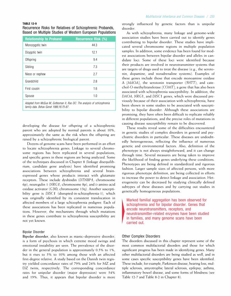
developing the disease for offspring of a schizophrenicparent who are adopted by normal parents is about 10%,approximately the same as the risk when the offspring areraised by a schizophrenic biological parent.
Dozens of genome scans have been performed in an effortto locate schizophrenia genes. Linkage to several chromo-some regions has been replicated in several populations,and specific genes in these regions are being analyzed. Someof the techniques discussed in Chapter 8 (linkage disequilib-rium, candidate gene analysis) have identified promisingassociations between schizophrenia and several brain-expressed genes whose products interact with glutamatereceptors. These include dysbindin (DTNBP1; chromosome6p), neuregulin 1 (NRG1; chromosome 8p), and D-amino-acidoxidase activator (G30; chromosome 13q). Another suscepti-bility gene is DISC1 (disrupted-in-schizophrenia-1), whichwas originally identified by its consistent translocation inaffected members of a large schizophrenia pedigree. Each ofthese associations has been replicated in numerous popula-tions. However, the mechanisms through which mutationsin these genes contribute to schizophrenia susceptibility arenot yet known.
Bipolar Disorder
Bipolar disorder, also known as manic–depressive disorder,is a form of psychosis in which extreme mood swings andemotional instability are seen. The prevalence of the disor-der in the general population is approximately 0.5% to 1%,but it rises to 5% to 10% among those with an affectedfirst-degree relative. A study based on the Danish twin regis-try yielded concordance rates of 79% and 24% for MZ andDZ twins, respectively. The corresponding concordancerates for unipolar disorder (major depression) were 54%and 19%. Thus, it appears that bipolar disorder is more
strongly influenced by genetic factors than is unipolardisorder.
As with schizophrenia, many linkage and genome-wideassociation studies have been carried out to identify genescontributing to bipolar disorder. These studies have impli-cated several chromosome regions in multiple populationsamples. In addition, some evidence has been found for mod-est associations between bipolar disorder and alleles in can-didate loci. Some of these loci were identified becausetheir products are involved in neurotransmitter systems thatare targets of drugs used to treat the disease (e.g., the seroto-nin, dopamine, and noradrenaline systems). Examples ofthese genes include those that encode monoamine oxidaseA (MAOA), the serotonin transporter (5HTT), and cate-chol-O-methyltransferase (COMT), a gene that has also beenassociated with schizophrenia susceptibility. In addition, theDAOA, NRG1, and DISC1 genes, which were discussed pre-viously because of their association with schizophrenia, havebeen shown in some studies to be associated with suscepti-bility to bipolar disorder. Although these associations arepromising, they have often been difficult to replicate reliablyin different populations, and the precise roles of mutations incausing disease susceptibility remain to be discovered.
These results reveal some of the difficulties encounteredin genetic studies of complex disorders in general and psy-chiatric disorders in particular. These diseases are undoubt-edly heterogeneous, reflecting the influence of numerousgenetic and environmental factors. Also, definition of thephenotype is not always straightforward, and it can changethrough time. Several measures are being taken to improvethe likelihood of finding genes underlying these conditions.Phenotypes are being defined in standardized and rigorousfashion. Larger sample sizes of affected persons, with morerigorous phenotype definition, are being collected in effortsto increase the power to detect linkage and association. Het-erogeneity can be decreased by studying clinically definedsubtypes of these diseases and by carrying out studies ingenetically homogeneous populations.
4Marked familial aggregation has been observed forschizophrenia and for bipolar disorder. Genes thatencode neurotransmitters, receptors, andneurotransmitter-related enzymes have been studiedin families, and many genome scans have beencarried out.
Other Complex DisordersThe disorders discussed in this chapter represent some of themost common multifactorial disorders and those for whichsignificant progress has been made in identifying genes. Manyother multifactorial disorders are being studied as well, and insome cases specific susceptibility genes have been identified.These include, for example, Parkinson disease, hearing loss, mul-tiple sclerosis, amyotrophic lateral sclerosis, epilepsy, asthma,inflammatory bowel disease, and some forms of blindness (seeTable 12-7 and Table 8-2 in Chapter 8).
TABLE 12-9
Recurrence Risks for Relatives of Schizophrenic Probands,Based on Multiple Studies of Western European Populations
Relationship to Proband Recurrence Risk (%)
Monozygotic twin 44.3
Dizygotic twin 12.1
Offspring 9.4
Sibling 7.3
Niece or nephew 2.7
Grandchild 2.8
First cousin 1.6
Spouse 1.0
Adapted from McGue M, Gottesman II, Rao DC: The analysis of schizophrenia
family data. Behav Genet 1986;16:75-87.
Multifactorial Inheritance and Common Diseases / 255
SOME GENERAL PRINCIPLES AND CONCLUSIONSSome general principles can be deduced from the resultsobtained thus far regarding the genetics of complex disor-ders. First, the more strongly inherited forms of complexdisorders generally have an earlier age of onset (examplesinclude breast cancer, Alzheimer disease, and heart disease).Often, these represent subsets of cases in which there issingle-gene inheritance. Second, when there is laterality,bilateral forms sometimes cluster more strongly in families(e.g., cleft lip/palate). Third, while the sex-specific thresholdmodel fits some of the complex disorders (e.g., pyloric ste-nosis, cleft lip/palate, autism, heart disease), it fails to fitothers (e.g., type 1 diabetes).
There is a tendency, particularly among the lay public, toassume that the presence of a genetic component means thatthe course of a disease cannot be altered (“If it’s genetic, youcan’t change it”). This is incorrect. Most of the diseases dis-cussed in this chapter have both genetic and environmentalcomponents. Thus, environmental modification (e.g., diet,exercise, stress reduction) can often reduce risk significantly.Such modifications may be especially important for personswho have a family history of a disease, because they are likelyto develop the disease earlier in life. Those with a family
history of heart disease, for example, can often add manyyears of productive living with relatively minor lifestylealterations. By targeting those who can benefit most fromintervention, genetics helps to serve the goal of preventivemedicine.
In addition, it should be stressed that the identification ofa specific genetic alteration can lead to more effective pre-vention and treatment of the disease. Identification of muta-tions causing familial colon cancer can allow early screeningand prevention of metastasis. Pinpointing a gene responsiblefor a neurotransmitter defect in a psychiatric disorder suchas schizophrenia could lead to the development of moreeffective drug treatments. In some cases, such as familialhypercholesterolemia, gene therapy may be useful. It isimportant for health care practitioners to make their patientsaware of these facts.
Although the genetics of common disorders is complexand often confusing, the public health impact of these dis-eases and the evidence for hereditary factors in their etiol-ogy demand that genetic studies be pursued. Substantialprogress is already being made. The next decade willundoubtedly witness many advancements in our understand-ing and treatment of these disorders.
Study Questions
1. Consider a multifactorial trait that is twice as commonin females as in males. Indicate which type of matingis at higher risk for producing affected children(affected father and normal mother versus normalfather and affected mother). Is the recurrence riskhigher for their sons or their daughters?
2. Consider a disease that is known to have a 5% siblingrecurrence risk. This recurrence risk could be theresult of either multifactorial inheritance or a singleautosomal dominant gene with 10% penetrance. Howwould you test which of these possibilities is correct?
3. One member of a pair of monozygotic twins isaffected by an autosomal dominant disease, andthe other is not. List two different ways this couldhappen.
4. Suppose that the heritability of body fat percentageis 0.80 when correlations between siblings are studiedbut only 0.50 when correlations between parents andoffspring are studied. Suppose also that a significantpositive correlation is observed in the body fatpercentages of spouses. How would you interpretthese results?
Suggested ReadingsAbrahams BS, Geschwind DH. Advances in autism genetics:
On the threshold of a new neurobiology. Nat Rev Genet2008;9(5):341–55.
Bell CG, Walley AJ, Froguel P. The genetics of human obe-sity. Nat Rev Genet 2005;6:221–34.
Bird TD. Genetic aspects of Alzheimer disease. Genet Med2008;10(4):231–39.
Blennow K, de Leon MJ, Zetterberg H. Alzheimer’s disease.Lancet 2006;368(9533):387–403.
Boomsma D, Busjahn A, Peltonen L. Classical twin studies andbeyond. Nat Rev Genet 2002;3(11):872–82.
Burmeister M, McInnis MG, Zollner S. Psychiatric genetics:Progress amid controversy. Nat Rev Genet 2008;9(7):527–40.
Cowley AW Jr. The genetic dissection of essential hyperten-sion. Nat Rev Genet 2006;7(11):829–40.
Edenberg HJ, Foroud T. The genetics of alcoholism: Identi-fying specific genes through family studies. Addict Biol2006;11(3–4):386–96.
Hirschhorn JN, Daly MJ. Genome-wide association studiesfor common diseases and complex traits. Nat Rev Genet2005;6:95–108.
Kibar Z, Capra V, Gros P. Toward understanding thegenetic basis of neural tube defects. Clin Genet 2007;71(4):295–310.
King RA, Rotter JI, Motulsky AG (eds). The Genetic Basis ofCommon Diseases, 2nd ed. New York: Oxford University,2002.
256 / Chapter 12 MEDICAL GENETICS
MacGregor AJ, Snieder H, Schork NJ, Spector TD. Twins:Novel uses to study complex traits and genetic diseases.Trends Genet 2000;16:131–4.
Manolio TA, Brooks LD, Collins FS. A hapmap harvest ofinsights into the genetics of common disease. J Clin Invest2008;118(5):1590–605.
McCarthy MI, Abecasis GR, Cardon LR, et al. Genome-wideassociation studies for complex traits: Consensus, uncer-tainty and challenges. Nat Rev Genet 2008;9(5):356–69.
Morita H, Seidman J, Seidman CE. Genetic causes of humanheart failure. J Clin Invest 2005;115:518–26.
Neale MC, Cardon LR. Methodology for Genetic Studies ofTwins and Families. Dordrecht, Netherlands: Kluwer,1992.
Newton-Cheh C, Shah R. Genetic determinants of QTinterval variation and sudden cardiac death. Curr OpinGenet Dev 2007;17(3):213–21.
Owen KR, McCarthy MI. Genetics of type 2 diabetes. CurrOpin Genet Dev 2007;17(3):239–44.
Roden DM. Long-QT syndrome. N Engl J Med 2008;358(2):169–76.
Shih PA, O’Connor DT. Hereditary determinants of humanhypertension: Strategies in the setting of genetic complex-ity. Hypertension 2008;51(6):1456–64.
Stumvoll M, Goldstein BJ, van Haeften TW. Type 2 diabe-tes: Principles of pathogenesis and therapy. Lancet2005;365:1333–1346.
Visscher PM, Hill WG, Wray NR. Heritability in the geno-mics era—concepts and misconceptions. Nat Rev Genet2008;9(4):255–66.
Watkins H, Farrall M. Genetic susceptibility to coronaryartery disease: From promise to progress. Nat Rev Genet2006;7(3):163–73.
Internet ResourcesHuman Genome Epidemiology Network Reviews (containslinks to review articles on genetics of mendelian andcommon diseases) http://www.cdc.gov/genomics/hugenet/reviews.
htm
International Clearinghouse for Birth Defects Web Guidehttp://www.icbdsr.org/page.asp?n¼WebGuide
Multifactorial Inheritance and Common Diseases / 257

Chapter 13
GENETIC TESTING AND GENE THERAPY
As we have seen in previous chapters, significant advanceshave occurred in many areas of medical genetics, includingDNA technology, gene mapping and cloning, and cytoge-netics. These developments have paved the way for moreaccurate and efficient testing of genetic disorders. Genetictesting can be defined as the analysis of chromosomes,DNA, RNA, proteins, or other analytes* to detect abnormal-ities that can cause a genetic disease. Examples of genetictesting include prenatal diagnosis, heterozygote carrierdetection, and presymptomatic diagnosis of genetic disease.The principles and applications of genetic testing in thesecontexts are one focus of this chapter.
The other focus is the treatment of genetic disease. Manyaspects of disease management involve areas of medicine,such as surgery and drug treatment, that are beyond thescope of this book. However, gene therapy, in whichpatients’ cells are genetically altered to combat specific dis-eases, is discussed here in some detail.
POPULATION SCREENING FOR GENETIC DISEASEScreening tests represent an important component of routinehealth care. These tests are usually designed to detect treat-able human diseases in their presymptomatic stage. Papani-colaou (Pap) tests for detecting cervical dysplasia andpopulation screening for hypercholesterolemia are well-known examples of this public health strategy. Populationscreening is large-scale testing of populations for disease inan effort to identify persons who probably have the diseaseand those who probably do not. Screening tests are notintended to provide definitive diagnoses; rather, they areaimed at identifying a subset of the population on whom fur-ther, more exact, diagnostic tests should be carried out. Thisimportant distinction is commonly misunderstood by the laypublic and seldom clarified by the popular media.
Genetic screening is population screening for a gene thatcan cause the disease in the person carrying the gene or inthe descendents of the carrier. Newborn screening for inher-ited metabolic diseases (see Chapter 7) is a good example ofthe first type of genetic screening, and heterozygote detec-tion for Tay–Sachs disease (discussed later) exemplifies the
second. These two examples involve screening of popula-tions, but genetic screening can also be applied to membersof families with a positive history of a genetic condition. Anexample is testing for a balanced reciprocal translocation infamilies in which one or more members have had a chromo-some disorder (see Chapter 6). Box 13-1 lists the varioustypes of genetic screening, including several forms of prena-tal diagnosis, that are discussed in this chapter.
4The goal of screening is early recognition of a disorderso that intervention will prevent or reverse the diseaseprocess (as in newborn screening for inborn errors ofmetabolism) or so that informed reproductive decisionscan be made (as in screening for heterozygouscarriers of an autosomal recessive mutation). A positiveresult from a genetic screening test is typicallyfollowed by a more precise diagnostic test.
Principles of ScreeningThe basic principles of screening were developed in the 1960sand are still widely recognized. Characteristics of the disease,the test, and the health care system should be consideredwhen deciding whether population screening is appropriate.
First, the condition should be serious and relatively com-mon. This ensures that the benefits to be derived from thescreening program will justify its costs. The natural historyof the disease should be clearly understood. There shouldbe an acceptable and effective treatment, or, in the case ofsome genetic conditions, prenatal diagnosis should be avail-able. As for the screening test itself, it should be acceptableto the population, easy to perform, and relatively inexpen-sive. The screening test should be valid and reliable. Finally,the resources for diagnosis and treatment of the disordermust be accessible. A strategy for communicating results effi-ciently and effectively must be in place.
Screening programs typically use tests that are widelyapplicable and inexpensive to identify an at-risk population(e.g., the phenylketonuria (PKU) screening program, dis-cussed in Clinical Commentary 13-1). Members of this pop-ulation are then targeted for subsequent tests that are moreaccurate but also more expensive and time consuming. Inthis context, the screening test should be able to effectively*An analyte is any substance that is subject to analysis.
258
CLINICAL COMMENTARY 13-1
Neonatal Screening for Phenylketonuria
DISEASE CHARACTERISTICSPopulation screening of newborns for PKU represents an excellent exampleof the application of the screening model to genetic disease. As discussedin Chapter 4, the prevalence of this autosomal recessive disorder of phenyl-alanine metabolism is about 1 in 10,000 to 15,000 white births. The naturalhistory of PKU is well understood. More than 95% of untreated PKUpatients become moderately to severely mentally retarded. The conditionis not identified clinically in the first year of life, because the physical signsare subtle and PKU usually manifests only as developmental delay. Dietaryrestriction of phenylalanine, when begun before 4 weeks of age, is highlyeffective in altering the course of the disease. A low-phenylalanine diet,although not particularly palatable, largely eliminates the IQ loss that wouldotherwise occur (an important exception is those who have a defect in biop-terin metabolism, in whom a different therapy is used).
TEST CHARACTERISTICSPKU is typically detected by the measurement of blood phenylalanine usinga bacterial inhibition assay, the Guthrie test. Blood is collected in the new-born period, usually by heel stick, and placed on filter paper. The driedblood is placed on an agar plate and incubated with a strain of bacteria(Bacillus subtilis) that requires phenylalanine for growth. Measurement ofbacterial growth permits quantification of the amount of phenylalanine in
the blood sample. Increasingly, tandem mass spectrometry is used toscreen for PKU. Positive test results are usually repeated and followed bya quantitative assay of plasma phenylalanine and tyrosine.
If the test is performed after 2 days of age and after regular feeding on aprotein diet, the detection rate (sensitivity) is about 98%. If it is performedat less than 24 hours of age, the sensitivity is about 84% and a repeat testis recommended a few weeks after birth. Specificity is close to 100%.
SYSTEM CHARACTERISTICSBecause of the requirement of normal protein in the diet, many statesrequest rescreening at 2 to 4 weeks of age. At that point, sensitivityapproaches 100%. A high sensitivity level is important because of thesevere impact of a misdiagnosis.
Phenylalanine levels in children with classic PKU typically exceed 20mg/dL. For every 20 positive PKU screening results, only one infant hasclassic PKU. The others represent either false-positive findings (usuallydue to a transient reversible tyrosinemia) or infants with a form of hyper-phenylalaninemia (elevated phenylalanine) not caused by classic PKU.
The cost of a Guthrie test is typically less than a few dollars. Severalstudies have shown that the cost of nationwide PKU screening is signifi-cantly less than the savings it achieves by avoiding institutionalization costsand lost productivity.
separate persons who have the disease from those who donot. This attribute, which defines the test’s validity, involvestwo components: sensitivity and specificity. Sensitivityreflects the ability of the test to correctly identify those with
the disease. It is measured as the fraction of affected personsin whom the test is positive (i.e., true positives). Specificity
is the ability of the test to correctly identify those without
the disease. It is measured as the fraction of unaffected per-sons in whom the test is negative (i.e., true negatives). Sen-sitivity and specificity are determined by comparing thescreening results with those of a definitive diagnostic test(Table 13-1).
BOX 13-1 Types of Genetic Screening and Prenatal Diagnosis
POPULATION SCREENING FOR GENETIC DISORDERS
Newborn ScreeningBlood• Phenylketonuria, all 50 states in the United States• Galactosemia, all 50 states in the United States• Hypothyroidism, all 50 states in the United States• Hemoglobinopathies, nearly all states• Other: maple syrup urine disease, homocystinuria, tyrosinemia,and several other diseases are screened in many states
Universal newborn hearing screening (>60% of congenitalhearing loss is due to genetic factors)
Heterozygote ScreeningTay–Sachs disease, Ashkenazi Jewish populationSickle cell disease, African-American populationThalassemias in at-risk ethnic groupsCystic fibrosis in some populations (persons of European descent,
Ashkenazi Jews)
PRENATAL DIAGNOSIS OF GENETIC DISORDERS
Diagnostic Testing (Invasive Prenatal Diagnosis)AmniocentesisChorionic villus sampling
Percutaneous umbilical blood sampling (PUBS)Preimplantation genetic diagnosis
Fetal Visualization TechniquesUltrasonographyRadiographyMagnetic resonance imaging
Population ScreeningMaternal age >35 yearsFamily history of condition diagnosable by prenatal techniquesQuadruple screen: maternal serum a-fetoprotein, estriol, human
chorionic gonadotropin, inhibin-AFirst trimester screening: ultrasonography, PAPP-A, and free b
subunit of human chorionic gonadotropin
FAMILY SCREENING FOR GENETIC DISORDERSFamily history of chromosomal rearrangement (e.g., translocation)Screening female relatives in an X-linked pedigree (e.g., Du-
chenne muscular dystrophy, fragile X syndrome)Heterozygote screening within at-risk families (e.g., cystic fibrosis)Presymptomatic screening (e.g., Huntington disease, breast
cancer, colon cancer)
Genetic Testing and Gene Therapy / 259
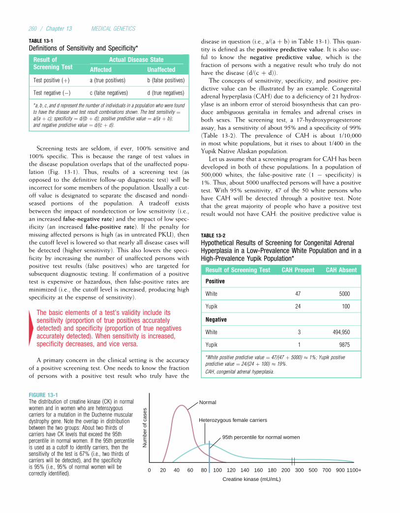
Screening tests are seldom, if ever, 100% sensitive and100% specific. This is because the range of test values inthe disease population overlaps that of the unaffected popu-lation (Fig. 13-1). Thus, results of a screening test (asopposed to the definitive follow-up diagnostic test) will beincorrect for some members of the population. Usually a cut-off value is designated to separate the diseased and nondi-seased portions of the population. A tradeoff existsbetween the impact of nondetection or low sensitivity (i.e.,an increased false-negative rate) and the impact of low spec-ificity (an increased false-positive rate). If the penalty formissing affected persons is high (as in untreated PKU), thenthe cutoff level is lowered so that nearly all disease cases willbe detected (higher sensitivity). This also lowers the speci-ficity by increasing the number of unaffected persons withpositive test results (false positives) who are targeted forsubsequent diagnostic testing. If confirmation of a positivetest is expensive or hazardous, then false-positive rates areminimized (i.e., the cutoff level is increased, producing highspecificity at the expense of sensitivity).
4The basic elements of a test’s validity include itssensitivity (proportion of true positives accuratelydetected) and specificity (proportion of true negativesaccurately detected). When sensitivity is increased,specificity decreases, and vice versa.
A primary concern in the clinical setting is the accuracyof a positive screening test. One needs to know the fractionof persons with a positive test result who truly have the
disease in question (i.e., a/(a þ b) in Table 13-1). This quan-tity is defined as the positive predictive value. It is also use-ful to know the negative predictive value, which is thefraction of persons with a negative result who truly do nothave the disease (d/(c þ d)).
The concepts of sensitivity, specificity, and positive pre-dictive value can be illustrated by an example. Congenitaladrenal hyperplasia (CAH) due to a deficiency of 21 hydrox-ylase is an inborn error of steroid biosynthesis that can pro-duce ambiguous genitalia in females and adrenal crises inboth sexes. The screening test, a 17-hydroxyprogesteroneassay, has a sensitivity of about 95% and a specificity of 99%(Table 13-2). The prevalence of CAH is about 1/10,000in most white populations, but it rises to about 1/400 in theYupik Native Alaskan population.
Let us assume that a screening program for CAH has beendeveloped in both of these populations. In a population of500,000 whites, the false-positive rate (1 � specificity) is1%. Thus, about 5000 unaffected persons will have a positivetest. With 95% sensitivity, 47 of the 50 white persons whohave CAH will be detected through a positive test. Notethat the great majority of people who have a positive testresult would not have CAH: the positive predictive value is
TABLE 13-1
Definitions of Sensitivity and Specificity*
Result of Actual Disease StateScreening Test Affected Unaffected
Test positive (þ) a (true positives) b (false positives)
Test negative (�) c (false negatives) d (true negatives)
*a, b, c, and d represent the number of individuals in a population who were found
to have the disease and test result combinations shown. The test sensitivity ¼a/(a þ c); specificity ¼ d/(b þ d); positive predictive value ¼ a/(a þ b);
and negative predictive value ¼ d/(c þ d).
Creatine kinase (mU/mL)
0 20 40 60 80 100 120
Num
ber
of c
ases
140 160 180 200 300 500 700 900 1100+
Normal
Heterozygous female carriers
95th percentile for normal women
FIGURE 13-1The distribution of creatine kinase (CK) in normalwomen and in women who are heterozygouscarriers for a mutation in the Duchenne musculardystrophy gene. Note the overlap in distributionbetween the two groups: About two thirds ofcarriers have CK levels that exceed the 95thpercentile in normal women. If the 95th percentileis used as a cutoff to identify carriers, then thesensitivity of the test is 67% (i.e., two thirds ofcarriers will be detected), and the specificityis 95% (i.e., 95% of normal women will becorrectly identified).
TABLE 13-2
Hypothetical Results of Screening for Congenital AdrenalHyperplasia in a Low-Prevalence White Population and in aHigh-Prevalence Yupik Population*
Result of Screening Test CAH Present CAH Absent
Positive
White 47 5000
Yupik 24 100
Negative
White 3 494,950
Yupik 1 9875
*White positive predictive value ¼ 47/(47 þ 5000) � 1%; Yupik positive
predictive value ¼ 24/(24 þ 100) � 19%.
CAH, congenital adrenal hyperplasia.
260 / Chapter 13 MEDICAL GENETICS
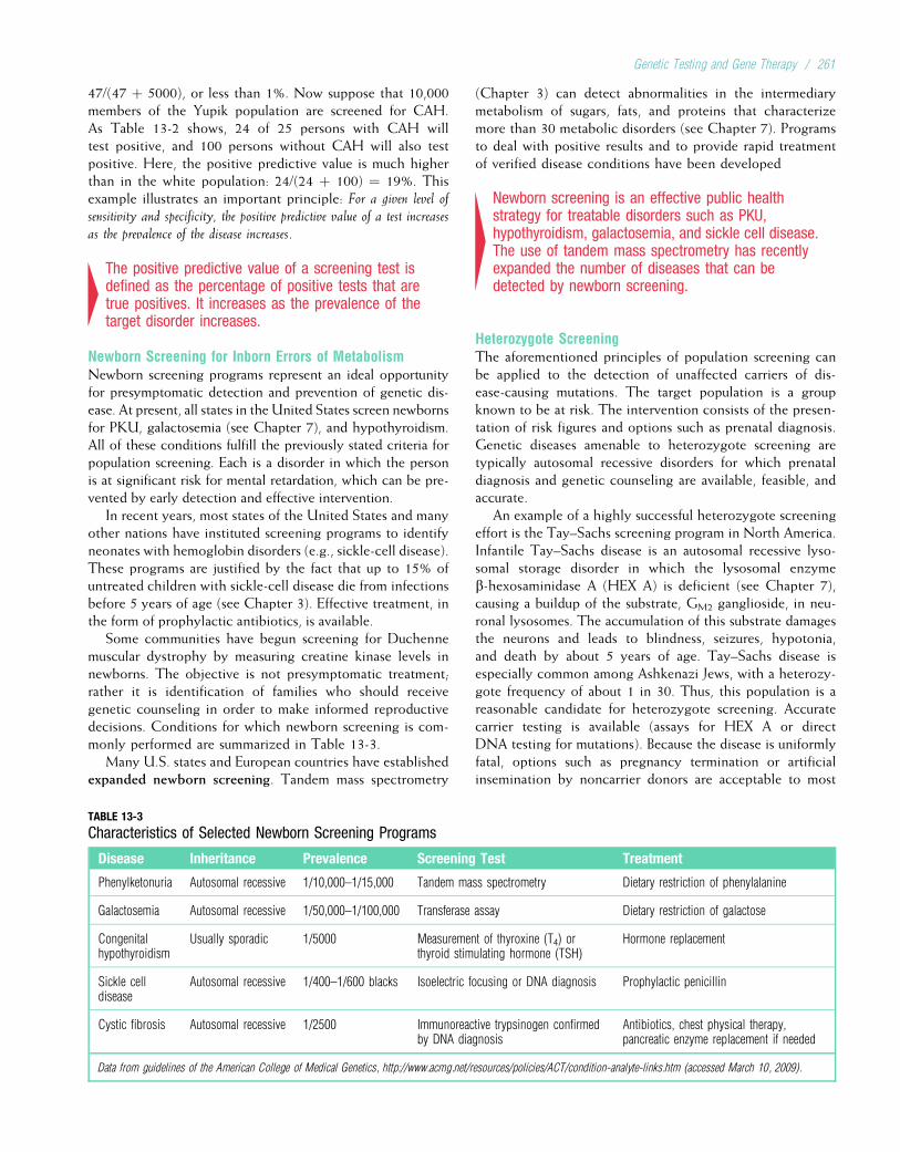
47/(47 þ 5000), or less than 1%. Now suppose that 10,000members of the Yupik population are screened for CAH.As Table 13-2 shows, 24 of 25 persons with CAH willtest positive, and 100 persons without CAH will also testpositive. Here, the positive predictive value is much higherthan in the white population: 24/(24 þ 100) ¼ 19%. Thisexample illustrates an important principle: For a given level of
sensitivity and specificity, the positive predictive value of a test increases
as the prevalence of the disease increases.
4The positive predictive value of a screening test isdefined as the percentage of positive tests that aretrue positives. It increases as the prevalence of thetarget disorder increases.
Newborn Screening for Inborn Errors of MetabolismNewborn screening programs represent an ideal opportunityfor presymptomatic detection and prevention of genetic dis-ease. At present, all states in the United States screen newbornsfor PKU, galactosemia (see Chapter 7), and hypothyroidism.All of these conditions fulfill the previously stated criteria forpopulation screening. Each is a disorder in which the personis at significant risk for mental retardation, which can be pre-vented by early detection and effective intervention.
In recent years, most states of the United States and manyother nations have instituted screening programs to identifyneonates with hemoglobin disorders (e.g., sickle-cell disease).These programs are justified by the fact that up to 15% ofuntreated children with sickle-cell disease die from infectionsbefore 5 years of age (see Chapter 3). Effective treatment, inthe form of prophylactic antibiotics, is available.
Some communities have begun screening for Duchennemuscular dystrophy by measuring creatine kinase levels innewborns. The objective is not presymptomatic treatment;rather it is identification of families who should receivegenetic counseling in order to make informed reproductivedecisions. Conditions for which newborn screening is com-monly performed are summarized in Table 13-3.
Many U.S. states and European countries have establishedexpanded newborn screening. Tandem mass spectrometry
(Chapter 3) can detect abnormalities in the intermediarymetabolism of sugars, fats, and proteins that characterizemore than 30 metabolic disorders (see Chapter 7). Programsto deal with positive results and to provide rapid treatmentof verified disease conditions have been developed
4Newborn screening is an effective public healthstrategy for treatable disorders such as PKU,hypothyroidism, galactosemia, and sickle cell disease.The use of tandem mass spectrometry has recentlyexpanded the number of diseases that can bedetected by newborn screening.
Heterozygote ScreeningThe aforementioned principles of population screening canbe applied to the detection of unaffected carriers of dis-ease-causing mutations. The target population is a groupknown to be at risk. The intervention consists of the presen-tation of risk figures and options such as prenatal diagnosis.Genetic diseases amenable to heterozygote screening aretypically autosomal recessive disorders for which prenataldiagnosis and genetic counseling are available, feasible, andaccurate.
An example of a highly successful heterozygote screeningeffort is the Tay–Sachs screening program in North America.Infantile Tay–Sachs disease is an autosomal recessive lyso-somal storage disorder in which the lysosomal enzymeb-hexosaminidase A (HEX A) is deficient (see Chapter 7),causing a buildup of the substrate, GM2 ganglioside, in neu-ronal lysosomes. The accumulation of this substrate damagesthe neurons and leads to blindness, seizures, hypotonia,and death by about 5 years of age. Tay–Sachs disease isespecially common among Ashkenazi Jews, with a heterozy-gote frequency of about 1 in 30. Thus, this population is areasonable candidate for heterozygote screening. Accuratecarrier testing is available (assays for HEX A or directDNA testing for mutations). Because the disease is uniformlyfatal, options such as pregnancy termination or artificialinsemination by noncarrier donors are acceptable to most
TABLE 13-3
Characteristics of Selected Newborn Screening Programs
Disease Inheritance Prevalence Screening Test Treatment
Phenylketonuria Autosomal recessive 1/10,000–1/15,000 Tandem mass spectrometry Dietary restriction of phenylalanine
Galactosemia Autosomal recessive 1/50,000–1/100,000 Transferase assay Dietary restriction of galactose
Congenitalhypothyroidism
Usually sporadic 1/5000 Measurement of thyroxine (T4) orthyroid stimulating hormone (TSH)
Hormone replacement
Sickle celldisease
Autosomal recessive 1/400–1/600 blacks Isoelectric focusing or DNA diagnosis Prophylactic penicillin
Cystic fibrosis Autosomal recessive 1/2500 Immunoreactive trypsinogen confirmedby DNA diagnosis
Antibiotics, chest physical therapy,pancreatic enzyme replacement if needed
Data from guidelines of the American College of Medical Genetics, http://www.acmg.net/resources/policies/ACT/condition-analyte-links.htm (accessed March 10, 2009).
Genetic Testing and Gene Therapy / 261
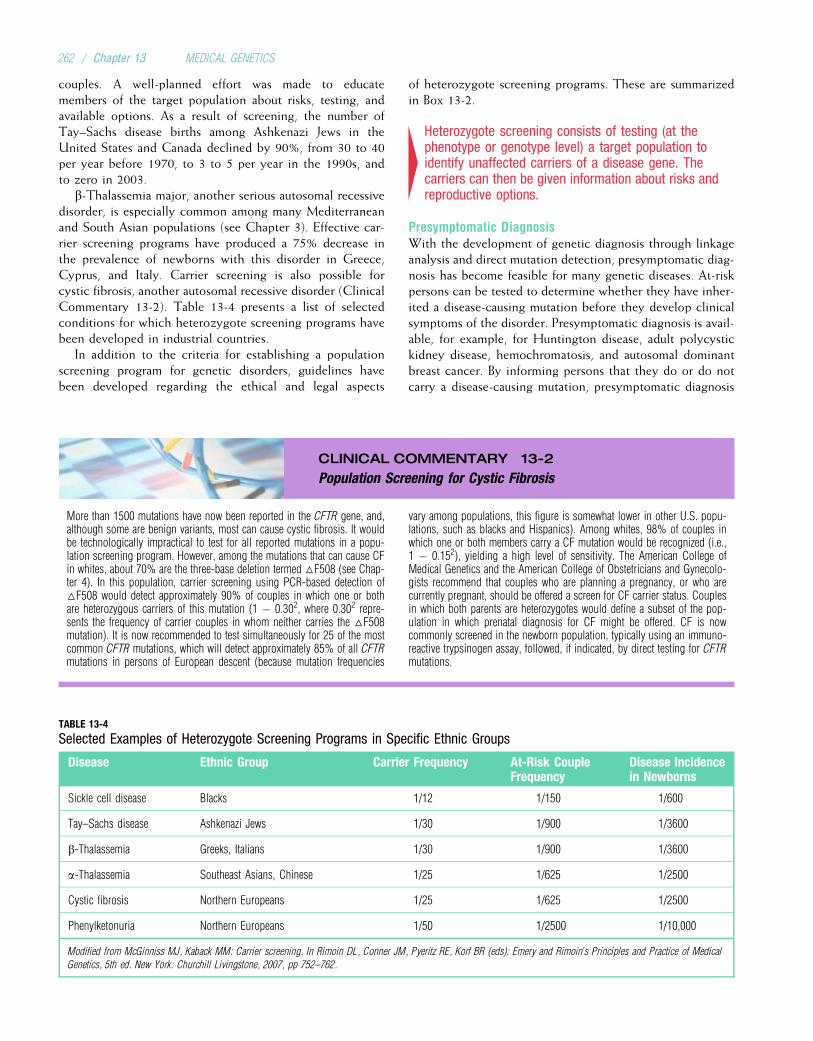
couples. A well-planned effort was made to educatemembers of the target population about risks, testing, andavailable options. As a result of screening, the number ofTay–Sachs disease births among Ashkenazi Jews in theUnited States and Canada declined by 90%, from 30 to 40per year before 1970, to 3 to 5 per year in the 1990s, andto zero in 2003.
b-Thalassemia major, another serious autosomal recessivedisorder, is especially common among many Mediterraneanand South Asian populations (see Chapter 3). Effective car-rier screening programs have produced a 75% decrease inthe prevalence of newborns with this disorder in Greece,Cyprus, and Italy. Carrier screening is also possible forcystic fibrosis, another autosomal recessive disorder (ClinicalCommentary 13-2). Table 13-4 presents a list of selectedconditions for which heterozygote screening programs havebeen developed in industrial countries.
In addition to the criteria for establishing a populationscreening program for genetic disorders, guidelines havebeen developed regarding the ethical and legal aspects
of heterozygote screening programs. These are summarizedin Box 13-2.
4Heterozygote screening consists of testing (at thephenotype or genotype level) a target population toidentify unaffected carriers of a disease gene. Thecarriers can then be given information about risks andreproductive options.
Presymptomatic DiagnosisWith the development of genetic diagnosis through linkageanalysis and direct mutation detection, presymptomatic diag-nosis has become feasible for many genetic diseases. At-riskpersons can be tested to determine whether they have inher-ited a disease-causing mutation before they develop clinicalsymptoms of the disorder. Presymptomatic diagnosis is avail-able, for example, for Huntington disease, adult polycystickidney disease, hemochromatosis, and autosomal dominantbreast cancer. By informing persons that they do or do notcarry a disease-causing mutation, presymptomatic diagnosis
CLINICAL COMMENTARY 13-2
Population Screening for Cystic Fibrosis
More than 1500 mutations have now been reported in the CFTR gene, and,although some are benign variants, most can cause cystic fibrosis. It wouldbe technologically impractical to test for all reported mutations in a popu-lation screening program. However, among the mutations that can cause CFin whites, about 70% are the three-base deletion termed △F508 (see Chap-ter 4). In this population, carrier screening using PCR-based detection of△F508 would detect approximately 90% of couples in which one or bothare heterozygous carriers of this mutation (1 � 0.302, where 0.302 repre-sents the frequency of carrier couples in whom neither carries the △F508mutation). It is now recommended to test simultaneously for 25 of the mostcommon CFTR mutations, which will detect approximately 85% of all CFTRmutations in persons of European descent (because mutation frequencies
vary among populations, this figure is somewhat lower in other U.S. popu-lations, such as blacks and Hispanics). Among whites, 98% of couples inwhich one or both members carry a CF mutation would be recognized (i.e.,1 � 0.152), yielding a high level of sensitivity. The American College ofMedical Genetics and the American College of Obstetricians and Gynecolo-gists recommend that couples who are planning a pregnancy, or who arecurrently pregnant, should be offered a screen for CF carrier status. Couplesin which both parents are heterozygotes would define a subset of the pop-ulation in which prenatal diagnosis for CF might be offered. CF is nowcommonly screened in the newborn population, typically using an immuno-reactive trypsinogen assay, followed, if indicated, by direct testing for CFTRmutations.
TABLE 13-4
Selected Examples of Heterozygote Screening Programs in Specific Ethnic Groups
Disease Ethnic Group Carrier Frequency At-Risk CoupleFrequency
Disease Incidencein Newborns
Sickle cell disease Blacks 1/12 1/150 1/600
Tay–Sachs disease Ashkenazi Jews 1/30 1/900 1/3600
b-Thalassemia Greeks, Italians 1/30 1/900 1/3600
a-Thalassemia Southeast Asians, Chinese 1/25 1/625 1/2500
Cystic fibrosis Northern Europeans 1/25 1/625 1/2500
Phenylketonuria Northern Europeans 1/50 1/2500 1/10,000
Modified from McGinniss MJ, Kaback MM: Carrier screening. In Rimoin DL, Conner JM, Pyeritz RE, Korf BR (eds): Emery and Rimoin’s Principles and Practice of Medical
Genetics, 5th ed. New York: Churchill Livingstone, 2007, pp 752–762.
262 / Chapter 13 MEDICAL GENETICS
can aid in making reproductive decisions. It can providereassurance to those who learn that they do not carry a dis-ease-causing mutation. In some cases, early diagnosis canimprove health supervision. For example, persons whoinherit an autosomal dominant breast cancer mutation canundergo mammography at an earlier age to increase thechances of early tumor detection. Persons at risk for the inher-itance of RET mutations (see Chapter 11), who are highlylikely to develop multiple endocrine neoplasia type2 (MEN2), can undergo a prophylactic thyroidectomy toreduce their chance of developing a malignancy. Those whoinherit mutations that cause some forms of familial coloncancer (adenomatous polyposis coli [APC] and hereditarynonpolyposis colorectal cancer [HNPCC]; see Chapter 11)can also benefit from early diagnosis and treatment.
Because most genetic diseases are relatively uncommon inthe general population, universal presymptomatic screeningis currently impractical. It is usually recommended only forpersons who are known to be at risk for the disease, gener-ally because of a positive family history.
4Genetic testing can sometimes be performed toidentify persons who have inherited a disease-causinggene before they develop symptoms. This is termedpresymptomatic diagnosis.
Psychosocial Implications of Genetic Screeningand DiagnosisScreening for genetic diseases has many social and psycho-logical implications. The burden of anxiety, cost, and poten-tial stigmatization surrounding a positive test result must beweighed against the need for detection. Often, screeningtests are misperceived as diagnostically definitive. The con-cept that a positive screening test does not necessarily indi-cate disease presence must be emphasized to those whoundergo screening (see Box 13-2).
The initial screening programs for sickle-cell carrier statusin the 1970s were plagued by misunderstandings about theimplications of carrier status. Occasionally, carrier detectionled to cancellation of health insurance or employer discrimi-nation. Such experiences underscore the need for effectivegenetic counseling and public education. Other issuesinclude the right to choose not to be tested and the potentialfor invasion of privacy.
The social, psychological, and ethical aspects of geneticscreening will become more complicated as newer tech-niques of DNA diagnosis become more accessible. For exam-ple, even though presymptomatic diagnosis of Huntingtondisease is available, several studies have shown that fewerthan 20% of at-risk persons elected this option. Largely, thisreflects the fact that no effective treatment is currently avail-able for this disorder. Presymptomatic diagnosis of BRCA1and BRCA2 carrier status in breast-and-ovarian cancerfamilies has also met with mixed responses. In part, this isa reaction to the cost of the test: because of the large numberof different mutations in BRCA1 or BRCA2 that can causebreast cancer, testing typically consists of sequencing allexons and promoters of both genes, as well as some intronicnucleotides near each exon. This is an expensive procedure.Preventive measures such as prophylactic mastectomy andoophorectomy (removal of the breasts and ovaries, respec-tively) are known to reduce cancer risk substantially, butthey do not completely eliminate it.
For some genetic diseases, such as autosomal dominantcolon cancer syndromes, early diagnosis can lead to improvedsurvival because effective preventive treatments are readilyavailable (colectomy or polypectomy for precancerous colonpolyps). In addition, many at-risk persons will find that theydo not carry the disease-causing gene, allowing them to avoidunpleasant (and possibly hazardous) diagnostic proceduressuch as colonoscopy or mammography. However, as screen-ing for such diseases becomes more common, the issues ofprivacy and confidentiality and the need for accurate commu-nication of risk information must also be addressed.
MOLECULAR TOOLS FOR SCREENING AND DIAGNOSISUntil recently, genetic screening usually relied on assays ofthe disease phenotype, such as a b-hexosaminidase assay forTay–Sachs disease or a creatine kinase assay for Duchennemuscular dystrophy. Advances in DNA technology have ledto diagnosis at the level of the genotype. In some cases, link-age analysis is used to determine whether a person has inher-ited a disease-causing gene, but in most cases direct assays ofdisease-causing mutations have been developed. Geneticdiagnosis at the DNA level is now supplementing, and inmany cases supplanting, tests based on phenotypic assays.
Linkage analysis and direct mutation diagnosis have beenused for diagnostic testing within families, for prenatal diag-nosis of genetic disorders, and, more recently, in popula-tion screening. Improved technology and an increaseddemand for testing have led to the establishment of clinicalmolecular laboratories in many medical centers throughoutthe world.
BOX 13-2 Public Policy Guidelines for HeterozygoteScreening
Recommended guidelines:
Screening should be voluntary, and confidentiality must beensured.
Screening requires informed consent.Providers of screening services have an obligation to ensure that
adequate education and counseling are included in theprogram.
Quality control of all aspects of the laboratory testing, includingsystematic proficiency testing, is required and should beimplemented as soon as possible.
There should be equal access to testing.
From Elias S, Annas GJ, Simpson JL: Carrier screening for cystic fibrosis:
implications for obstetric and gynecologic practice. Am J Obstet Gynecol
1991;164:1077–1083.
Genetic Testing and Gene Therapy / 263
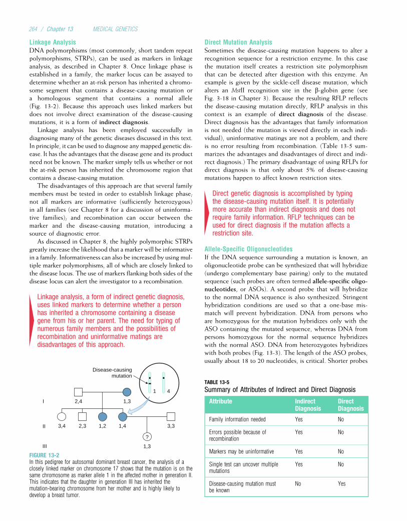
Linkage AnalysisDNA polymorphisms (most commonly, short tandem repeatpolymorphisms, STRPs), can be used as markers in linkageanalysis, as described in Chapter 8. Once linkage phase isestablished in a family, the marker locus can be assayed todetermine whether an at-risk person has inherited a chromo-some segment that contains a disease-causing mutation ora homologous segment that contains a normal allele(Fig. 13-2). Because this approach uses linked markers butdoes not involve direct examination of the disease-causingmutations, it is a form of indirect diagnosis.
Linkage analysis has been employed successfully indiagnosing many of the genetic diseases discussed in this text.In principle, it can be used to diagnose anymapped genetic dis-ease. It has the advantages that the disease gene and its productneed not be known. The marker simply tells us whether or notthe at-risk person has inherited the chromosome region thatcontains a disease-causing mutation.
The disadvantages of this approach are that several familymembers must be tested in order to establish linkage phase;not all markers are informative (sufficiently heterozygous)in all families (see Chapter 8 for a discussion of uninforma-tive families); and recombination can occur between themarker and the disease-causing mutation, introducing asource of diagnostic error.
As discussed in Chapter 8, the highly polymorphic STRPsgreatly increase the likelihood that a marker will be informativein a family. Informativeness can also be increased by using mul-tiple marker polymorphisms, all of which are closely linked tothe disease locus. The use of markers flanking both sides of thedisease locus can alert the investigator to a recombination.
4Linkage analysis, a form of indirect genetic diagnosis,uses linked markers to determine whether a personhas inherited a chromosome containing a diseasegene from his or her parent. The need for typing ofnumerous family members and the possibilities ofrecombination and uninformative matings aredisadvantages of this approach.
Direct Mutation AnalysisSometimes the disease-causing mutation happens to alter arecognition sequence for a restriction enzyme. In this casethe mutation itself creates a restriction site polymorphismthat can be detected after digestion with this enzyme. Anexample is given by the sickle-cell disease mutation, whichalters an MstII recognition site in the b-globin gene (seeFig. 3-18 in Chapter 3). Because the resulting RFLP reflectsthe disease-causing mutation directly, RFLP analysis in thiscontext is an example of direct diagnosis of the disease.Direct diagnosis has the advantages that family informationis not needed (the mutation is viewed directly in each indi-vidual), uninformative matings are not a problem, and thereis no error resulting from recombination. (Table 13-5 sum-marizes the advantages and disadvantages of direct and indi-rect diagnosis.) The primary disadvantage of using RFLPs fordirect diagnosis is that only about 5% of disease-causingmutations happen to affect known restriction sites.
4Direct genetic diagnosis is accomplished by typingthe disease-causing mutation itself. It is potentiallymore accurate than indirect diagnosis and does notrequire family information. RFLP techniques can beused for direct diagnosis if the mutation affects arestriction site.
Allele-Specific OligonucleotidesIf the DNA sequence surrounding a mutation is known, anoligonucleotide probe can be synthesized that will hybridize(undergo complementary base pairing) only to the mutatedsequence (such probes are often termed allele-specific oligo-nucleotides, or ASOs). A second probe that will hybridizeto the normal DNA sequence is also synthesized. Stringenthybridization conditions are used so that a one-base mis-match will prevent hybridization. DNA from persons whoare homozygous for the mutation hybridizes only with theASO containing the mutated sequence, whereas DNA frompersons homozygous for the normal sequence hybridizeswith the normal ASO. DNA from heterozygotes hybridizeswith both probes (Fig. 13-3). The length of the ASO probes,usually about 18 to 20 nucleotides, is critical. Shorter probes
2,4 1,3
3,31,41,22,33,4
1,3
I
II
III
1 4
Disease-causingmutation
?
FIGURE 13-2In this pedigree for autosomal dominant breast cancer, the analysis of aclosely linked marker on chromosome 17 shows that the mutation is on thesame chromosome as marker allele 1 in the affected mother in generation II.This indicates that the daughter in generation III has inherited themutation-bearing chromosome from her mother and is highly likely todevelop a breast tumor.
TABLE 13-5
Summary of Attributes of Indirect and Direct Diagnosis
Attribute IndirectDiagnosis
DirectDiagnosis
Family information needed Yes No
Errors possible because ofrecombination
Yes No
Markers may be uninformative Yes No
Single test can uncover multiplemutations
Yes No
Disease-causing mutation mustbe known
No Yes
264 / Chapter 13 MEDICAL GENETICS
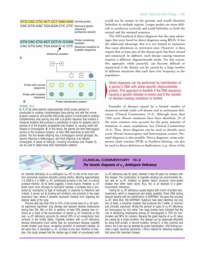
would not be unique in the genome and would thereforehybridize to multiple regions. Longer probes are more diffi-cult to synthesize correctly and could hybridize to both thenormal and the mutated sequence.
The ASO method of direct diagnosis has the same advan-tages that were listed for direct diagnosis using RFLPs. It hasthe additional advantage that it is not limited to mutationsthat cause alterations in restriction sites. However, it doesrequire that at least part of the disease gene has been clonedand sequenced. In addition, each disease-causing mutationrequires a different oligonucleotide probe. For this reason,this approach, while powerful, can become difficult orimpractical if the disease can be caused by a large numberof different mutations that each have low frequency in thepopulation.
4Direct diagnosis can be performed by hybridization ofa person’s DNA with allele-specific oligonucleotideprobes. This approach is feasible if the DNA sequencecausing a genetic disease is known and if the numberof disease-causing mutations is limited.
Examples of diseases caused by a limited number ofmutations include sickle cell disease and a1-antitrypsin defi-ciency (Clinical Commentary 13-3). Although more than1500 cystic fibrosis mutations have been identified, 25 ofthe most common ones account for the great majority ofmutations in many populations (see Clinical Commentary13-2). Thus, direct diagnosis can be used to identify mostcystic fibrosis homozygotes and heterozygous carriers. Pre-natal diagnosis is also possible. Direct diagnosis, using poly-merase chain reaction (PCR) or Southern blotting, can alsobe used to detect deletions or duplications (e.g., those of the
CLINICAL COMMENTARY 13-3
The Genetic Diagnosis of a1-Antitrypsin Deficiency
An inherited deficiency of a1-antitrypsin (a1-AT) is one of the most com-mon autosomal recessive disorders among whites, affecting approximately1 in 2000 to 1 in 5000. a1-AT, synthesized primarily in the liver, is a serineprotease inhibitor. As its name suggests, it binds trypsin. However, a1-ATbinds much more strongly to neutrophil elastase, a protease that is pro-duced by neutrophils (a type of leukocyte) in response to infections andirritants. It carries out its binding and inhibitory role primarily in the lowerrespiratory tract, where it prevents neutrophil elastase from digesting thealveolar septa of the lung.
Persons with less than 10% to 15% of the normal level of a1-AT activ-ity experience significant lung damage and typically develop emphysemaduring their 30s, 40s, or 50s. In addition, at least 10% develop liver cir-rhosis as a result of the accumulation of variant a1-AT molecules in theliver. a1-AT deficiency accounts for almost 20% of all nonalcoholic livercirrhosis in the United States. Cigarette smokers with a1-AT deficiencydevelop emphysema much earlier than do nonsmokers because cigarettesmoke irritates lung tissue, increasing secretion of neutrophil elastase. Atthe same time, it inactivates a1-AT, so there is also less inhibition of elas-tase. One study showed that the median age of death of nonsmokers with
a1-AT deficiency was 62 years, whereas it was 40 years for smokers withthis disease. The combination of cigarette smoking (an environmental fac-tor) and an a1-AT mutation (a genetic factor) produces more-severedisease than either factor alone; thus, this is an example of a gene–environment interaction.
Testing for a1-AT deficiency usually begins with a form of protein elec-trophoresis, which is inexpensive and widely available. Direct DNA testingbecame feasible with the identification of SERPINA1, the gene that encodesa1-AT. More than 100 SERPINA1 mutations have been identified, but onlyone of these, a missense mutation that produces the Z allele, is commonand clinically significant. Ninety-five percent of cases of a1-AT deficiencyare homozygous for this allele. Two large studies have indicated that therisk of developing emphysema among ZZ homozygotes is 70% for non-smokers and 90% for smokers. Because the great majority of a1-AT casesare caused by a single mutation, this disease can be diagnosed efficientlyby using ASO probes. A second mutation, termed S, is less common andsevere, but it can also be detected by probe hybridization. ASO testing pro-vides a rapid, sensitive (sensitivity >95%) method for detecting mutationsthat cause this important disease.
Normal probe
Normal β-globinsequence(antisense strand)
Probe containingmutation
Missense mutation inβ-globin sequence
Missense mutation
A
GTG CAC CTG ACT CCT
GTG CAC CTG ACT CCT
GAG GAG
GTG GAG
TGACAC GTG GAC GGA CTC CTC
TGACAC GTG GAC GGA CAC CTC
B Probe hybridization pattern
Probe with normalsequence
Probe with mutatedsequence
FIGURE 13-3A, A 21-bp allele-specific oligonucleotide (ASO) probe (yellow) isconstructed to undergo complementary base pairing only with the normalb-globin sequence, and another ASO probe (green) is constructed to undergocomplementary base pairing only with a b-globin sequence that contains amissense mutation that produces a substitution of valine for glutamic acid atposition 6 of the b-globin polypeptide (see Chapter 3), causing sickle celldisease in homozygotes. B, In this family, the parents are both heterozygouscarriers of the missense mutation, so their DNA hybridizes to both ASOprobes. The first female offspring has a homozygous normal genotype, thesecond offspring is heterozygous, and third offspring is an affectedhomozygote. A variety of methods, including microarrays (see Chapter 3),can be used to detect these ASO hybridization patterns.
Genetic Testing and Gene Therapy / 265
dystrophin gene that cause most cases of Duchenne musculardystrophy). Currently, clinical genetic testing is available formore than almost 1500 genetic diseases, including almost allof the single-gene conditions discussed in this textbook.Despite this wide availability, it should be kept in mind thatgenetic testing, like all testing procedures, has a number oflimitations (Box 13-3).
Other Methods of Direct DiagnosisThe ASO method (see Fig. 13-3) is commonly used in detect-ing direct mutations. Many other techniques can also be usedto detect mutations, including several discussed in Chapter 3(e.g., direct sequencing, DNA mismatch cleavage, andMLPA). Microarrays (DNA chips, also discussed in Chapter3) are now widely used to detect large-scale mutations.Microarrays have many convenient properties, including min-iaturization and automated computerized processing. Theycan be designed to assay large numbers of sequence variants(including disease-causing mutations) in a single rapid analy-sis (Box 13-4). For example, one microarray containsthousands of oligonucleotide probes that hybridize to largenumbers of possible sequence variants in the CYP2D6 andCYP2C19 genes. The products of these genes influence therate of metabolism of about 25% of all prescription drugs,and analysis of their variation might help to predict how indi-vidual patients will respond to these drugs.
Mass spectrometry, a method commonly used in chemis-try, is also being explored as a rapid means of detectingmutations. This technique detects minute differences in themass of PCR-amplified DNA molecules, which representvariations in DNA sequence. Mass spectrometry, whichoffers the advantages of high speed and great accuracy, hasbeen used, for example, to detect mutations in the CFTR
and APOE genes.
Another form of mass spectrometry, tandem mass spec-trometry, is being used increasingly to screen newborns forprotein variants that signal amino acid disorders (e.g.,PKU, tyrosinemia, homocystinuria), organic acid disorders,and fatty acid oxidation disorders (e.g., MCAD and LCHADdeficiencies; see Chapter 7 and see earlier). This methodbegins with a sample of material from a dried blood spot,which is subjected to analysis by two mass spectrometers.The first spectrometer separates ionized molecules accordingto their mass, and the molecules are fragmented. The secondspectrometer assesses the mass and charge of these frag-ments, allowing a computer to generate a molecular profileof the sample. Tandem mass spectrometry is highly accurateand very rapid: more than two dozen disorders can bescreened in approximately 2 minutes.
4New methods of direct mutation detection, includingthe use of microarrays and mass spectrometry, havegreatly increased the speed and accuracy of geneticdiagnosis. Tandem mass spectrometry can be used totest for protein variants that are characteristic of anumber of newborn disorders and is thus a usefulscreening tool.
PRENATAL DIAGNOSIS OF GENETIC DISORDERSAND CONGENITAL DEFECTSPrenatal diagnosis is a major focus of genetic testing, andseveral important areas of technology have evolved to pro-vide this service. The principal aim of prenatal diagnosis isto supply at-risk families with information so that they canmake informed choices during pregnancy. The potentialbenefits of prenatal testing include providing reassurance toat-risk families when the result is normal; providing risk
BOX 13-3 Limitations of Genetic Testing
Although genetic testing offers many advantages, its limitationsmust also be borne in mind. These limitations can be summarizedas follows:
• No genetic test is 100% accurate. Although most genetic testsdo achieve a high level of accuracy, factors such as mosaicismcan complicate cytogenetic diagnosis, and genotyping errorscan occur in the diagnosis of single-gene disorders.
• Genetic tests reveal mutations, not the presence of disease,because many disease-causing mutations have incomplete pene-trance. For example, approximately 50% to 80% of womenwith BRCA1 or BRCA2 mutations develop breast cancer, and70% to 90% of persons with mutations in one of the HNPCCgenes develop colorectal cancer. Even when penetranceapproaches 100% (as in neurofibromatosis type 1 or Hunting-ton disease), detection of the mutation often reveals little aboutthe severity or age of onset of the disease.
• Genetic testing might not detect all of the mutations that cancause a disease. Even in the absence of genotyping or sequenc-ing errors, many genetic tests lack sensitivity. For example, thepanels commonly used to test for cystic fibrosis mutations have
typically less than 90% sensitivity to detect homozygotes (seeClinical Commentary 13.2). When a large number of differentmutations can produce a genetic disease (e.g., neurofibromato-sis, autosomal dominant breast cancer, Marfan syndrome), itmight not be practical to test for all possible mutations. In thiscase, the analysis of linked markers can provide additional diag-nostic accuracy if multiple family members are affected. Otherfactors that can reduce accuracy include locus heterogeneityand the presence of phenocopies.
• Genetic testing can lead to complex ethical and social consid-erations. The results of a genetic test might lead to stigmatiza-tion or to discrimination by employers or insurance companies.Effective treatment is not available for some genetic diseases(e.g., Huntington disease, familial Alzheimer disease), decreas-ing the value of early diagnosis through genetic testing. Becausegenes are shared in families, the results of a genetic test mightaffect not only the tested person but also other members ofthe family (who might not wish to know about their risk for agenetic disease). These and other ethical and social issues arediscussed further in Chapter 15.
266 / Chapter 13 MEDICAL GENETICS
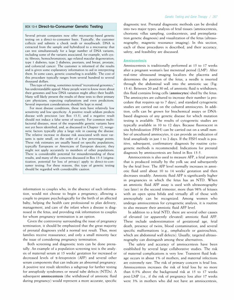
information to couples who, in the absence of such informa-tion, would not choose to begin a pregnancy; allowing acouple to prepare psychologically for the birth of an affectedbaby; helping the health care professional to plan delivery,management, and care of the infant when a disease is diag-nosed in the fetus; and providing risk information to couplesfor whom pregnancy termination is an option.
Given the controversy surrounding the issue of pregnancytermination, it should be emphasized that the great majorityof prenatal diagnoses yield a normal test result. Thus, mostfamilies receive reassurance, and only a small minority facethe issue of considering pregnancy termination.
Both screening and diagnostic tests can be done prena-tally. An example of a population screening test is the analy-sis of maternal serum at 15 weeks’ gestation for increased ordecreased levels of a-fetoprotein (AFP) and several otherserum components that can indicate an abnormal pregnancy.A positive test result identifies a subgroup for further testingfor aneuploidy syndromes or neural tube defects (NTDs). Asubsequent amniocentesis (the withdrawal of amniotic fluidduring pregnancy) would represent a more accurate, specific
diagnostic test. Prenatal diagnostic methods can be dividedinto two major types: analysis of fetal tissues (amniocentesis,chorionic villus sampling, cordocentesis, and preimplanta-tion genetic diagnosis) and visualization of the fetus (ultraso-nography, magnetic resonance imaging). In this section,each of these procedures is described, and their accuracy,safety, and feasibility are discussed.
AmniocentesisAmniocentesis is traditionally performed at 15 to 17 weeksafter a pregnant woman’s last menstrual period (LMP). Afterreal-time ultrasound imaging localizes the placenta anddetermines the position of the fetus, a needle is insertedthrough the abdominal wall into the amniotic sac (Fig.13-4). Between 20 and 30 mL of amniotic fluid is withdrawn;this fluid contains living cells (amniocytes) shed by the fetus.The amniocytes are cultured to increase their number (a pro-cedure that requires up to 7 days), and standard cytogeneticstudies are carried out on the cultured amniocytes. In addi-tion, cells can be grown for biochemical assays or DNA-based diagnosis of any genetic disease for which mutationtesting is available. The results of cytogenetic studies aretypically available in 10 to 12 days. Because fluorescent insitu hybridization (FISH) can be carried out on a small num-ber of uncultured amniocytes, it can provide an indication offetal aneuploidy in just 1 to 2 days. If the FISH result is pos-itive, subsequent, confirmatory diagnosis by routine cyto-genetic methods is recommended. Indications for prenataldiagnosis by amniocentesis are listed in Box 13-5.
Amniocentesis is also used to measure AFP, a fetal proteinthat is produced initially by the yolk sac and subsequentlyby the fetal liver. The AFP level normally increases in amni-otic fluid until about 10 to 14 weeks’ gestation and thendecreases steadily. Amniotic fluid AFP is significantly higherin pregnancies in which the fetus has an NTD. Whenan amniotic fluid AFP assay is used with ultrasonography(see later) in the second trimester, more than 98% of fetuseswith an open spina bifida and virtually all of those withanencephaly can be recognized. Among women whoundergo amniocentesis for cytogenetic analysis, it is routineto also measure their amniotic fluid AFP level.
In addition to a fetal NTD, there are several other causesof elevated (or apparently elevated) amniotic fluid AFP.These include underestimation of gestational age, fetaldeath, presence of twins, blood contamination, and severalspecific malformations (e.g., omphalocele or gastroschisis,which are abdominal wall defects). Usually, targeted ultraso-nography can distinguish among these alternatives.
The safety and accuracy of amniocentesis have beenestablished by several large collaborative studies. The riskof maternal complications is very low. Transient fluid leak-age occurs in about 1% of mothers, and maternal infectionsare extremely rare. The risk of primary concern is fetal loss.Amniocentesis increases the risk of fetal loss by no morethan 0.5% above the background risk at 15 to 17 weekspost-LMP (i.e., if the risk of pregnancy loss after 17 weekswere 3% in mothers who did not have an amniocentesis,
BOX 13-4 Direct-to-Consumer Genetic Testing
Several private companies now offer microarray-based genetictesting on a direct-to-consumer basis. Typically, the customercollects and submits a cheek swab or mouthwash. DNA isextracted from the sample and hybridized to a microarray thatcan test simultaneously for a large number of DNA variants,including some of the variants associated, for example, with cys-tic fibrosis; hemochromatosis; age-related macular degeneration;type 1 diabetes; type 2 diabetes; psoriasis; and breast, prostate,and colorectal cancer. The customer is informed of the resultsand is given some explanatory information to aid in interpretingthem. In some cases, genetic counseling is available. The cost ofthis procedure typically ranges from several hundred to severalthousand dollars.
This type of testing, sometimes termed “recreational genomics,”has understandable appeal. Many people want to know more abouttheir genomes and how DNA variation might affect their health.Many will likely present the results of these tests to their primarycare physicians, expecting explanations and even predictions.Several important considerations should be kept in mind.
For most disease conditions, these tests have relatively lowsensitivity and low specificity. A positive result seldom predictsdisease with precision (see Box 13.3), and a negative resultshould not induce a false sense of security. For common multi-factorial diseases, most of the responsible genetic variants havenot yet been identified, and, as discussed in Chapter 12, nonge-netic factors typically play a large role in causing the disease.The relative increase in disease risk associated with most var-iants is quite small, on the order of a few percentage points.These risk estimates are usually based on specific populations,typically Europeans or Americans of European descent; theymight not apply accurately to members of other populations.There is considerable potential for misunderstanding of theseresults, and many of the concerns discussed in Box 13-3 (stigma-tization, potential for loss of privacy) apply to direct-to-con-sumer testing. For these reasons, this type of genetic testingshould be regarded with considerable caution.
Genetic Testing and Gene Therapy / 267
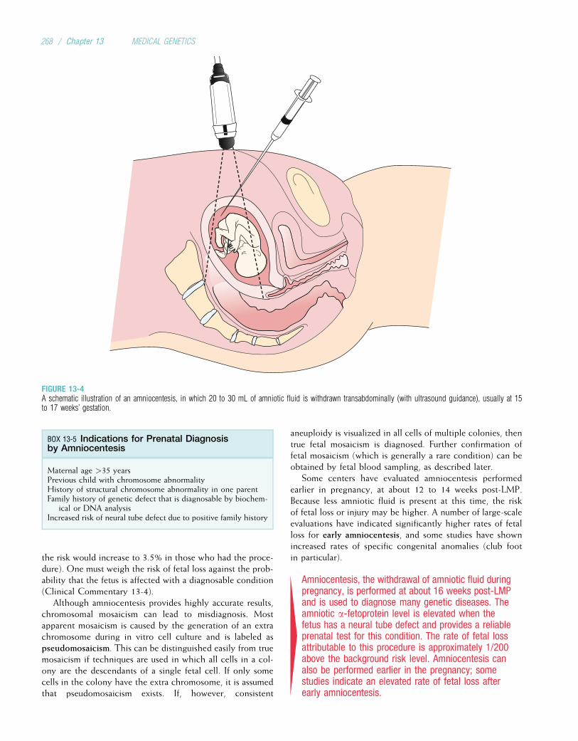
the risk would increase to 3.5% in those who had the proce-dure). One must weigh the risk of fetal loss against the prob-ability that the fetus is affected with a diagnosable condition(Clinical Commentary 13-4).
Although amniocentesis provides highly accurate results,chromosomal mosaicism can lead to misdiagnosis. Mostapparent mosaicism is caused by the generation of an extrachromosome during in vitro cell culture and is labeled aspseudomosaicism. This can be distinguished easily from truemosaicism if techniques are used in which all cells in a col-ony are the descendants of a single fetal cell. If only somecells in the colony have the extra chromosome, it is assumedthat pseudomosaicism exists. If, however, consistent
aneuploidy is visualized in all cells of multiple colonies, thentrue fetal mosaicism is diagnosed. Further confirmation offetal mosaicism (which is generally a rare condition) can beobtained by fetal blood sampling, as described later.
Some centers have evaluated amniocentesis performedearlier in pregnancy, at about 12 to 14 weeks post-LMP.Because less amniotic fluid is present at this time, the riskof fetal loss or injury may be higher. A number of large-scaleevaluations have indicated significantly higher rates of fetalloss for early amniocentesis, and some studies have shownincreased rates of specific congenital anomalies (club footin particular).
4Amniocentesis, the withdrawal of amniotic fluid duringpregnancy, is performed at about 16 weeks post-LMPand is used to diagnose many genetic diseases. Theamniotic a-fetoprotein level is elevated when thefetus has a neural tube defect and provides a reliableprenatal test for this condition. The rate of fetal lossattributable to this procedure is approximately 1/200above the background risk level. Amniocentesis canalso be performed earlier in the pregnancy; somestudies indicate an elevated rate of fetal loss afterearly amniocentesis.
FIGURE 13-4A schematic illustration of an amniocentesis, in which 20 to 30 mL of amniotic fluid is withdrawn transabdominally (with ultrasound guidance), usually at 15to 17 weeks’ gestation.
BOX 13-5 Indications for Prenatal Diagnosisby Amniocentesis
Maternal age >35 yearsPrevious child with chromosome abnormalityHistory of structural chromosome abnormality in one parentFamily history of genetic defect that is diagnosable by biochem-
ical or DNA analysisIncreased risk of neural tube defect due to positive family history
268 / Chapter 13 MEDICAL GENETICS
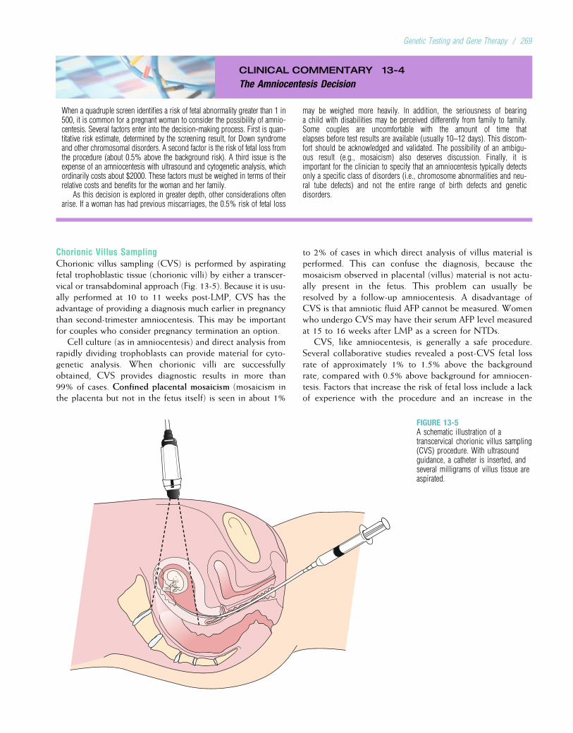
CLINICAL COMMENTARY 13-4
The Amniocentesis Decision
When a quadruple screen identifies a risk of fetal abnormality greater than 1 in500, it is common for a pregnant woman to consider the possibility of amnio-centesis. Several factors enter into the decision-making process. First is quan-titative risk estimate, determined by the screening result, for Down syndromeand other chromosomal disorders. A second factor is the risk of fetal loss fromthe procedure (about 0.5% above the background risk). A third issue is theexpense of an amniocentesis with ultrasound and cytogenetic analysis, whichordinarily costs about $2000. These factors must be weighed in terms of theirrelative costs and benefits for the woman and her family.
As this decision is explored in greater depth, other considerations oftenarise. If a woman has had previous miscarriages, the 0.5% risk of fetal loss
may be weighed more heavily. In addition, the seriousness of bearinga child with disabilities may be perceived differently from family to family.Some couples are uncomfortable with the amount of time thatelapses before test results are available (usually 10–12 days). This discom-fort should be acknowledged and validated. The possibility of an ambigu-ous result (e.g., mosaicism) also deserves discussion. Finally, it isimportant for the clinician to specify that an amniocentesis typically detectsonly a specific class of disorders (i.e., chromosome abnormalities and neu-ral tube defects) and not the entire range of birth defects and geneticdisorders.
Chorionic Villus SamplingChorionic villus sampling (CVS) is performed by aspiratingfetal trophoblastic tissue (chorionic villi) by either a transcer-vical or transabdominal approach (Fig. 13-5). Because it is usu-ally performed at 10 to 11 weeks post-LMP, CVS has theadvantage of providing a diagnosis much earlier in pregnancythan second-trimester amniocentesis. This may be importantfor couples who consider pregnancy termination an option.
Cell culture (as in amniocentesis) and direct analysis fromrapidly dividing trophoblasts can provide material for cyto-genetic analysis. When chorionic villi are successfullyobtained, CVS provides diagnostic results in more than99% of cases. Confined placental mosaicism (mosaicism inthe placenta but not in the fetus itself) is seen in about 1%
to 2% of cases in which direct analysis of villus material isperformed. This can confuse the diagnosis, because themosaicism observed in placental (villus) material is not actu-ally present in the fetus. This problem can usually beresolved by a follow-up amniocentesis. A disadvantage ofCVS is that amniotic fluid AFP cannot be measured. Womenwho undergo CVS may have their serum AFP level measuredat 15 to 16 weeks after LMP as a screen for NTDs.
CVS, like amniocentesis, is generally a safe procedure.Several collaborative studies revealed a post-CVS fetal lossrate of approximately 1% to 1.5% above the backgroundrate, compared with 0.5% above background for amniocen-tesis. Factors that increase the risk of fetal loss include a lackof experience with the procedure and an increase in the
FIGURE 13-5A schematic illustration of atranscervical chorionic villus sampling(CVS) procedure. With ultrasoundguidance, a catheter is inserted, andseveral milligrams of villus tissue areaspirated.
Genetic Testing and Gene Therapy / 269
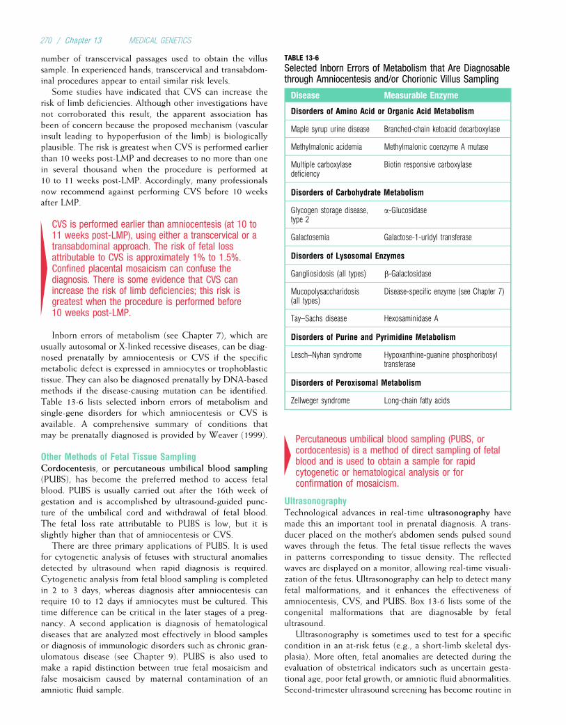
number of transcervical passages used to obtain the villussample. In experienced hands, transcervical and transabdom-inal procedures appear to entail similar risk levels.
Some studies have indicated that CVS can increase therisk of limb deficiencies. Although other investigations havenot corroborated this result, the apparent association hasbeen of concern because the proposed mechanism (vascularinsult leading to hypoperfusion of the limb) is biologicallyplausible. The risk is greatest when CVS is performed earlierthan 10 weeks post-LMP and decreases to no more than onein several thousand when the procedure is performed at10 to 11 weeks post-LMP. Accordingly, many professionalsnow recommend against performing CVS before 10 weeksafter LMP.
4CVS is performed earlier than amniocentesis (at 10 to11 weeks post-LMP), using either a transcervical or atransabdominal approach. The risk of fetal lossattributable to CVS is approximately 1% to 1.5%.Confined placental mosaicism can confuse thediagnosis. There is some evidence that CVS canincrease the risk of limb deficiencies; this risk isgreatest when the procedure is performed before10 weeks post-LMP.
Inborn errors of metabolism (see Chapter 7), which areusually autosomal or X-linked recessive diseases, can be diag-nosed prenatally by amniocentesis or CVS if the specificmetabolic defect is expressed in amniocytes or trophoblastictissue. They can also be diagnosed prenatally by DNA-basedmethods if the disease-causing mutation can be identified.Table 13-6 lists selected inborn errors of metabolism andsingle-gene disorders for which amniocentesis or CVS isavailable. A comprehensive summary of conditions thatmay be prenatally diagnosed is provided by Weaver (1999).
Other Methods of Fetal Tissue SamplingCordocentesis, or percutaneous umbilical blood sampling(PUBS), has become the preferred method to access fetalblood. PUBS is usually carried out after the 16th week ofgestation and is accomplished by ultrasound-guided punc-ture of the umbilical cord and withdrawal of fetal blood.The fetal loss rate attributable to PUBS is low, but it isslightly higher than that of amniocentesis or CVS.
There are three primary applications of PUBS. It is usedfor cytogenetic analysis of fetuses with structural anomaliesdetected by ultrasound when rapid diagnosis is required.Cytogenetic analysis from fetal blood sampling is completedin 2 to 3 days, whereas diagnosis after amniocentesis canrequire 10 to 12 days if amniocytes must be cultured. Thistime difference can be critical in the later stages of a preg-nancy. A second application is diagnosis of hematologicaldiseases that are analyzed most effectively in blood samplesor diagnosis of immunologic disorders such as chronic gran-ulomatous disease (see Chapter 9). PUBS is also used tomake a rapid distinction between true fetal mosaicism andfalse mosaicism caused by maternal contamination of anamniotic fluid sample.
4Percutaneous umbilical blood sampling (PUBS, orcordocentesis) is a method of direct sampling of fetalblood and is used to obtain a sample for rapidcytogenetic or hematological analysis or forconfirmation of mosaicism.
UltrasonographyTechnological advances in real-time ultrasonography havemade this an important tool in prenatal diagnosis. A trans-ducer placed on the mother’s abdomen sends pulsed soundwaves through the fetus. The fetal tissue reflects the wavesin patterns corresponding to tissue density. The reflectedwaves are displayed on a monitor, allowing real-time visuali-zation of the fetus. Ultrasonography can help to detect manyfetal malformations, and it enhances the effectiveness ofamniocentesis, CVS, and PUBS. Box 13-6 lists some of thecongenital malformations that are diagnosable by fetalultrasound.
Ultrasonography is sometimes used to test for a specificcondition in an at-risk fetus (e.g., a short-limb skeletal dys-plasia). More often, fetal anomalies are detected during theevaluation of obstetrical indicators such as uncertain gesta-tional age, poor fetal growth, or amniotic fluid abnormalities.Second-trimester ultrasound screening has become routine in
TABLE 13-6
Selected Inborn Errors of Metabolism that Are Diagnosablethrough Amniocentesis and/or Chorionic Villus Sampling
Disease Measurable Enzyme
Disorders of Amino Acid or Organic Acid Metabolism
Maple syrup urine disease Branched-chain ketoacid decarboxylase
Methylmalonic acidemia Methylmalonic coenzyme A mutase
Multiple carboxylasedeficiency
Biotin responsive carboxylase
Disorders of Carbohydrate Metabolism
Glycogen storage disease,type 2
a-Glucosidase
Galactosemia Galactose-1-uridyl transferase
Disorders of Lysosomal Enzymes
Gangliosidosis (all types) b-Galactosidase
Mucopolysaccharidosis(all types)
Disease-specific enzyme (see Chapter 7)
Tay–Sachs disease Hexosaminidase A
Disorders of Purine and Pyrimidine Metabolism
Lesch–Nyhan syndrome Hypoxanthine-guanine phosphoribosyltransferase
Disorders of Peroxisomal Metabolism
Zellweger syndrome Long-chain fatty acids
270 / Chapter 13 MEDICAL GENETICS
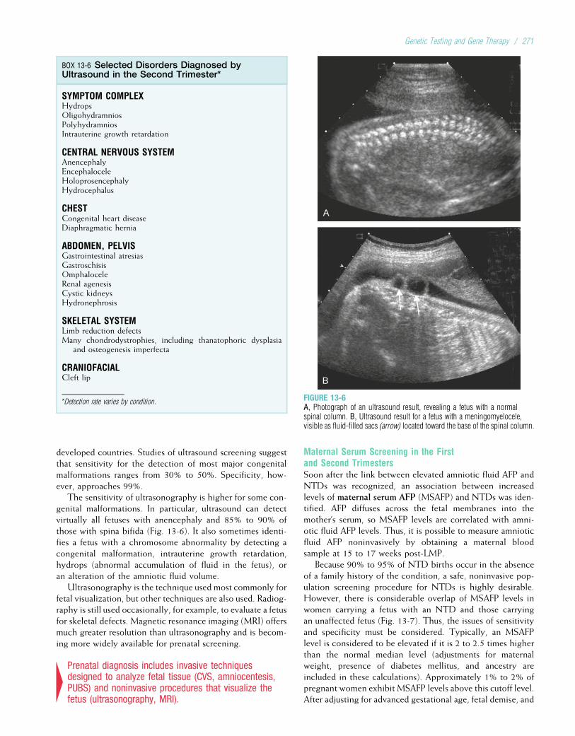
developed countries. Studies of ultrasound screening suggestthat sensitivity for the detection of most major congenitalmalformations ranges from 30% to 50%. Specificity, how-ever, approaches 99%.
The sensitivity of ultrasonography is higher for some con-genital malformations. In particular, ultrasound can detectvirtually all fetuses with anencephaly and 85% to 90% ofthose with spina bifida (Fig. 13-6). It also sometimes identi-fies a fetus with a chromosome abnormality by detecting acongenital malformation, intrauterine growth retardation,hydrops (abnormal accumulation of fluid in the fetus), oran alteration of the amniotic fluid volume.
Ultrasonography is the technique used most commonly forfetal visualization, but other techniques are also used. Radiog-raphy is still used occasionally, for example, to evaluate a fetusfor skeletal defects. Magnetic resonance imaging (MRI) offersmuch greater resolution than ultrasonography and is becom-ing more widely available for prenatal screening.
4Prenatal diagnosis includes invasive techniquesdesigned to analyze fetal tissue (CVS, amniocentesis,PUBS) and noninvasive procedures that visualize thefetus (ultrasonography, MRI).
Maternal Serum Screening in the Firstand Second TrimestersSoon after the link between elevated amniotic fluid AFP andNTDs was recognized, an association between increasedlevels of maternal serum AFP (MSAFP) and NTDs was iden-tified. AFP diffuses across the fetal membranes into themother’s serum, so MSAFP levels are correlated with amni-otic fluid AFP levels. Thus, it is possible to measure amnioticfluid AFP noninvasively by obtaining a maternal bloodsample at 15 to 17 weeks post-LMP.
Because 90% to 95% of NTD births occur in the absenceof a family history of the condition, a safe, noninvasive pop-ulation screening procedure for NTDs is highly desirable.However, there is considerable overlap of MSAFP levels inwomen carrying a fetus with an NTD and those carryingan unaffected fetus (Fig. 13-7). Thus, the issues of sensitivityand specificity must be considered. Typically, an MSAFPlevel is considered to be elevated if it is 2 to 2.5 times higherthan the normal median level (adjustments for maternalweight, presence of diabetes mellitus, and ancestry areincluded in these calculations). Approximately 1% to 2% ofpregnant women exhibit MSAFP levels above this cutoff level.After adjusting for advanced gestational age, fetal demise, and
BOX 13-6 Selected Disorders Diagnosed byUltrasound in the Second Trimester*
SYMPTOM COMPLEXHydropsOligohydramniosPolyhydramniosIntrauterine growth retardation
CENTRAL NERVOUS SYSTEMAnencephalyEncephaloceleHoloprosencephalyHydrocephalus
CHESTCongenital heart diseaseDiaphragmatic hernia
ABDOMEN, PELVISGastrointestinal atresiasGastroschisisOmphaloceleRenal agenesisCystic kidneysHydronephrosis
SKELETAL SYSTEMLimb reduction defectsMany chondrodystrophies, including thanatophoric dysplasia
and osteogenesis imperfecta
CRANIOFACIALCleft lip
*Detection rate varies by condition.
A
B
FIGURE 13-6A, Photograph of an ultrasound result, revealing a fetus with a normalspinal column. B, Ultrasound result for a fetus with a meningomyelocele,visible as fluid-filled sacs (arrow) located toward the base of the spinal column.
Genetic Testing and Gene Therapy / 271
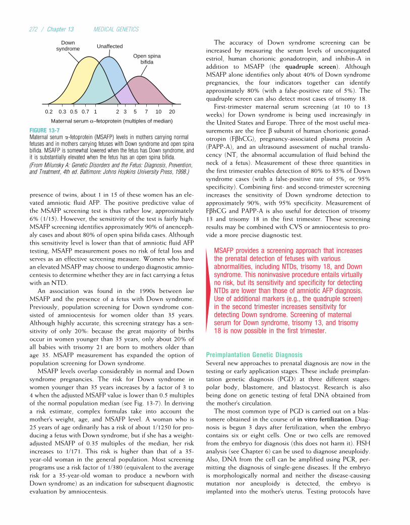
presence of twins, about 1 in 15 of these women has an ele-vated amniotic fluid AFP. The positive predictive value ofthe MSAFP screening test is thus rather low, approximately6% (1/15). However, the sensitivity of the test is fairly high:MSAFP screening identifies approximately 90% of anenceph-aly cases and about 80% of open spina bifida cases. Althoughthis sensitivity level is lower than that of amniotic fluid AFPtesting, MSAFP measurement poses no risk of fetal loss andserves as an effective screening measure. Women who havean elevated MSAFP may choose to undergo diagnostic amnio-centesis to determine whether they are in fact carrying a fetuswith an NTD.
An association was found in the 1990s between low
MSAFP and the presence of a fetus with Down syndrome.Previously, population screening for Down syndrome con-sisted of amniocentesis for women older than 35 years.Although highly accurate, this screening strategy has a sen-sitivity of only 20%: because the great majority of birthsoccur in women younger than 35 years, only about 20% ofall babies with trisomy 21 are born to mothers older thanage 35. MSAFP measurement has expanded the option ofpopulation screening for Down syndrome.
MSAFP levels overlap considerably in normal and Downsyndrome pregnancies. The risk for Down syndrome inwomen younger than 35 years increases by a factor of 3 to4 when the adjusted MSAFP value is lower than 0.5 multiplesof the normal population median (see Fig. 13-7). In derivinga risk estimate, complex formulas take into account themother’s weight, age, and MSAFP level. A woman who is25 years of age ordinarily has a risk of about 1/1250 for pro-ducing a fetus with Down syndrome, but if she has a weight-adjusted MSAFP of 0.35 multiples of the median, her riskincreases to 1/171. This risk is higher than that of a 35-year-old woman in the general population. Most screeningprograms use a risk factor of 1/380 (equivalent to the averagerisk for a 35-year-old woman to produce a newborn withDown syndrome) as an indication for subsequent diagnosticevaluation by amniocentesis.
The accuracy of Down syndrome screening can beincreased by measuring the serum levels of unconjugatedestriol, human chorionic gonadotropin, and inhibin-A inaddition to MSAFP (the quadruple screen). AlthoughMSAFP alone identifies only about 40% of Down syndromepregnancies, the four indicators together can identifyapproximately 80% (with a false-positive rate of 5%). Thequadruple screen can also detect most cases of trisomy 18.
First-trimester maternal serum screening (at 10 to 13weeks) for Down syndrome is being used increasingly inthe United States and Europe. Three of the most useful mea-surements are the free b subunit of human chorionic gonad-otropin (FbhCG), pregnancy-associated plasma protein A(PAPP-A), and an ultrasound assessment of nuchal translu-cency (NT, the abnormal accumulation of fluid behind theneck of a fetus). Measurement of these three quantities inthe first trimester enables detection of 80% to 85% of Downsyndrome cases (with a false-positive rate of 5%, or 95%specificity). Combining first- and second-trimester screeningincreases the sensitivity of Down syndrome detection toapproximately 90%, with 95% specificity. Measurement ofFbhCG and PAPP-A is also useful for detection of trisomy13 and trisomy 18 in the first trimester. These screeningresults may be combined with CVS or amniocentesis to pro-vide a more precise diagnostic test.
4MSAFP provides a screening approach that increasesthe prenatal detection of fetuses with variousabnormalities, including NTDs, trisomy 18, and Downsyndrome. This noninvasive procedure entails virtuallyno risk, but its sensitivity and specificity for detectingNTDs are lower than those of amniotic AFP diagnosis.Use of additional markers (e.g., the quadruple screen)in the second trimester increases sensitivity fordetecting Down syndrome. Screening of maternalserum for Down syndrome, trisomy 13, and trisomy18 is now possible in the first trimester.
Preimplantation Genetic DiagnosisSeveral new approaches to prenatal diagnosis are now in thetesting or early application stages. These include preimplan-tation genetic diagnosis (PGD) at three different stages:polar body, blastomere, and blastocyst. Research is alsobeing done on genetic testing of fetal DNA obtained fromthe mother’s circulation.
The most common type of PGD is carried out on a blas-tomere obtained in the course of in vitro fertilization. Diag-nosis is begun 3 days after fertilization, when the embryocontains six or eight cells. One or two cells are removedfrom the embryo for diagnosis (this does not harm it). FISHanalysis (see Chapter 6) can be used to diagnose aneuploidy.Also, DNA from the cell can be amplified using PCR, per-mitting the diagnosis of single-gene diseases. If the embryois morphologically normal and neither the disease-causingmutation nor aneuploidy is detected, the embryo isimplanted into the mother’s uterus. Testing protocols have
Downsyndrome Unaffected
Open spinabifida
Maternal serum α–fetoprotein (multiples of median)
0.2 0.3 0.5 0.7 1 2 3 5 7 10 20
FIGURE 13-7Maternal serum a-fetoprotein (MSAFP) levels in mothers carrying normalfetuses and in mothers carrying fetuses with Down syndrome and open spinabifida. MSAFP is somewhat lowered when the fetus has Down syndrome, andit is substantially elevated when the fetus has an open spina bifida.
(From Milunsky A: Genetic Disorders and the Fetus: Diagnosis, Prevention,and Treatment, 4th ed. Baltimore: Johns Hopkins University Press, 1998.)
272 / Chapter 13 MEDICAL GENETICS
been developed for dozens of genetic diseases (e.g., cysticfibrosis, Tay–Sachs disease, b-thalassemia, myotonic dystro-phy, Huntington disease, Duchenne muscular dystrophy),and more than 1000 normal babies have been born afterblastomere diagnosis.
An occasional problem with blastomere PGD is that one ofthe two alleles of a locus may be undetectable, which couldcause a heterozygote to appear as a homozygote. This phe-nomenon, termed “allelic dropout,” occurs because of partialPCR amplification failure when using DNA from only a singlecell. This has led to misdiagnosis in a small number of cases,and several methods are used to increase accuracy. For exam-ple, highly heterozygous STRPs closely linked to the disease-causing locus can be tested as part of the PCR analysis. If onlyone of the parents’ STRP alleles can be observed in the blasto-mere’s DNA, it is likely that allelic dropout has also occurredfor the disease-causing locus. The testing of two cells, ratherthan one, helps to avoid allelic dropout.
PGD can also be carried out at the 100-cell blastocyststage, using cells from the trophoectoderm of the blastocyst.This procedure has the advantage that a larger collection ofcells is analyzed, helping to avoid allelic dropout. A disad-vantage is that extraembryonic tissue (the trophoectoderm)is diagnosed, rather than the embryo itself.
Polar body diagnosis involves an examination of the firstor second polar body produced along with the ovum (seeChapter 2). The polar body’s DNA is tested to determinewhether it contains a disease-causing mutation. If it does, itis assumed that the egg does not contain the mutation. Thisegg is then fertilized and implanted using the usual in vitrotechniques. Because only the polar body is examined, pater-nal mutations cannot be evaluated. Polar body diagnosis isthus most useful when only the mother is at risk for transmit-ting a disease-causing mutation or when testing for aneu-ploidy (because most aneuploidies are contributed by themother [see Chapter 6]).
PGD is most commonly used by couples who haveresorted to in vitro fertilization and wish to test for diagnos-able genetic conditions. It can also be useful for coupleswho want prenatal diagnosis but would not consider a preg-nancy termination. PGD, however, is costly and technicallychallenging, and its availability is still limited.
4Preimplantation genetic diagnosis can be carried outon polar bodies, blastomeres, or blastocyst cells, onwhich PCR analysis and/or FISH is performed.Diagnosis of genetic conditions permits implantationof only unaffected embryos and avoids the issue ofpregnancy termination.
Analysis of Fetal DNA in Maternal CirculationDuring pregnancy, a small number of fetal cells cross the pla-cental barrier to enter the mother’s circulation. Some ofthese fetal cells are nucleated red blood cells, which are oth-erwise rare in the adult circulation. These cells can beisolated as early as 6 to 8 weeks post-LMP and can be
identified by cell-sorting techniques. Further specificity forfetal cells can be achieved by testing cells for surface pro-teins specific to the fetus. FISH analysis of these cells hasbeen used to test for fetal conditions such as trisomies 13,18, and 21. PCR has been used to test for a limited numberof single-gene disorders, although this remains a challengebecause of the difficulty of sorting pure populations of fetalcells. The major advantage of this approach is that it requiresonly a blood sample from the mother and thus poses no riskof fetal loss. Its accuracy and feasibility are being evaluated.Cell-free fetal DNA is also present in the mother’s circulationand has been used to identify the sex of the fetus and its Rhblood type (especially important if the mother is Rh-negativeand the fetus may be Rh-positive; see Chapter 9).
4Fetal cells or cell-free DNA that enter the maternalcirculation can be isolated and evaluated formutations using PCR or FISH. This experimentalprocedure entails no risk of fetal loss.
FETAL TREATMENTA potential goal of prenatal diagnosis is treatment of theaffected fetus. Although this is not currently possible formost conditions, some examples can be cited. Many of theseprocedures are experimental.
Two of the best-established forms of in utero interventionare treatment for rare inborn errors of metabolism and treat-ment for hormone deficiencies. An important example of atreatable biochemical disorder is biotin-responsive multiplecarboxylase deficiency, an autosomal recessive condition thatcan be diagnosed by amniocentesis. In one case report, oraladministration of biotin to the mother was initiated at 23weeks of pregnancy and resulted in the birth of a normal baby.
CAH is a second example of a condition for which inutero treatment has been successful after prenatal diagnosis.Because of excessive androgen secretion by the enlargedfetal adrenal glands, female fetuses with CAH become mas-culinized. Administration of dexamethasone to the motherbeginning at 10 weeks post-LMP diminishes or prevents thismasculinization.
Surgical treatment of fetuses, primarily for conditionsinvolving urinary tract obstruction, has met with moderatesuccess. Surgical correction of diaphragmatic hernia at 20weeks’ gestation has also been attempted, but results have beendiscouraging and this approach has been abandoned. Surgicalclosure of myelomeningocele (spina bifida) has been per-formed in more than 200 cases, and there is evidence thatthe procedure helps to restore the normal flow of cerebrospi-nal fluid. Clinical trials are under way to determine the effec-tiveness of this procedure. Some success has been achievedin transplantation of hematopoietic stem cells into fetuses withX-linked severe combined immune deficiency (see Chapter 9).
GENE THERAPYAs we have seen, the identification of disease-causing genesprovides opportunities for improved understanding and
Genetic Testing and Gene Therapy / 273
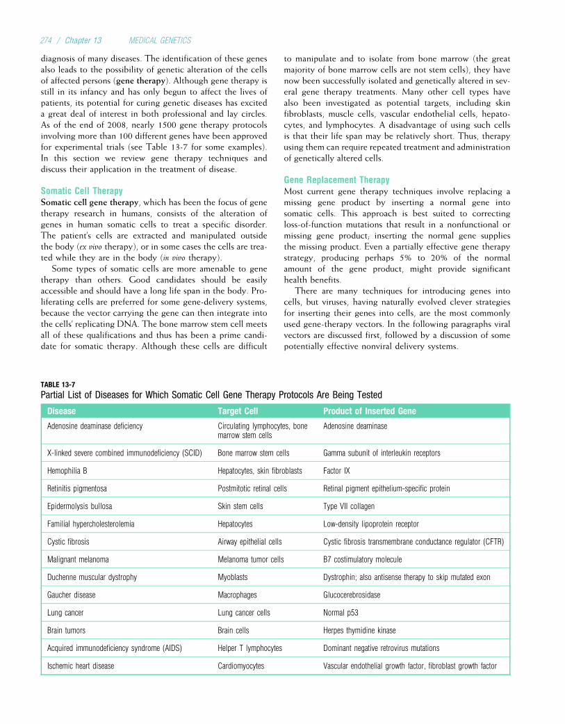
diagnosis of many diseases. The identification of these genesalso leads to the possibility of genetic alteration of the cellsof affected persons (gene therapy). Although gene therapy isstill in its infancy and has only begun to affect the lives ofpatients, its potential for curing genetic diseases has exciteda great deal of interest in both professional and lay circles.As of the end of 2008, nearly 1500 gene therapy protocolsinvolving more than 100 different genes have been approvedfor experimental trials (see Table 13-7 for some examples).In this section we review gene therapy techniques anddiscuss their application in the treatment of disease.
Somatic Cell TherapySomatic cell gene therapy, which has been the focus of genetherapy research in humans, consists of the alteration ofgenes in human somatic cells to treat a specific disorder.The patient’s cells are extracted and manipulated outsidethe body (ex vivo therapy), or in some cases the cells are trea-ted while they are in the body (in vivo therapy).
Some types of somatic cells are more amenable to genetherapy than others. Good candidates should be easilyaccessible and should have a long life span in the body. Pro-liferating cells are preferred for some gene-delivery systems,because the vector carrying the gene can then integrate intothe cells’ replicating DNA. The bone marrow stem cell meetsall of these qualifications and thus has been a prime candi-date for somatic therapy. Although these cells are difficult
to manipulate and to isolate from bone marrow (the greatmajority of bone marrow cells are not stem cells), they havenow been successfully isolated and genetically altered in sev-eral gene therapy treatments. Many other cell types havealso been investigated as potential targets, including skinfibroblasts, muscle cells, vascular endothelial cells, hepato-cytes, and lymphocytes. A disadvantage of using such cellsis that their life span may be relatively short. Thus, therapyusing them can require repeated treatment and administrationof genetically altered cells.
Gene Replacement TherapyMost current gene therapy techniques involve replacing amissing gene product by inserting a normal gene intosomatic cells. This approach is best suited to correctingloss-of-function mutations that result in a nonfunctional ormissing gene product; inserting the normal gene suppliesthe missing product. Even a partially effective gene therapystrategy, producing perhaps 5% to 20% of the normalamount of the gene product, might provide significanthealth benefits.
There are many techniques for introducing genes intocells, but viruses, having naturally evolved clever strategiesfor inserting their genes into cells, are the most commonlyused gene-therapy vectors. In the following paragraphs viralvectors are discussed first, followed by a discussion of somepotentially effective nonviral delivery systems.
TABLE 13-7
Partial List of Diseases for Which Somatic Cell Gene Therapy Protocols Are Being Tested
Disease Target Cell Product of Inserted Gene
Adenosine deaminase deficiency Circulating lymphocytes, bonemarrow stem cells
Adenosine deaminase
X-linked severe combined immunodeficiency (SCID) Bone marrow stem cells Gamma subunit of interleukin receptors
Hemophilia B Hepatocytes, skin fibroblasts Factor IX
Retinitis pigmentosa Postmitotic retinal cells Retinal pigment epithelium-specific protein
Epidermolysis bullosa Skin stem cells Type VII collagen
Familial hypercholesterolemia Hepatocytes Low-density lipoprotein receptor
Cystic fibrosis Airway epithelial cells Cystic fibrosis transmembrane conductance regulator (CFTR)
Malignant melanoma Melanoma tumor cells B7 costimulatory molecule
Duchenne muscular dystrophy Myoblasts Dystrophin; also antisense therapy to skip mutated exon
Gaucher disease Macrophages Glucocerebrosidase
Lung cancer Lung cancer cells Normal p53
Brain tumors Brain cells Herpes thymidine kinase
Acquired immunodeficiency syndrome (AIDS) Helper T lymphocytes Dominant negative retrovirus mutations
Ischemic heart disease Cardiomyocytes Vascular endothelial growth factor, fibroblast growth factor
274 / Chapter 13 MEDICAL GENETICS
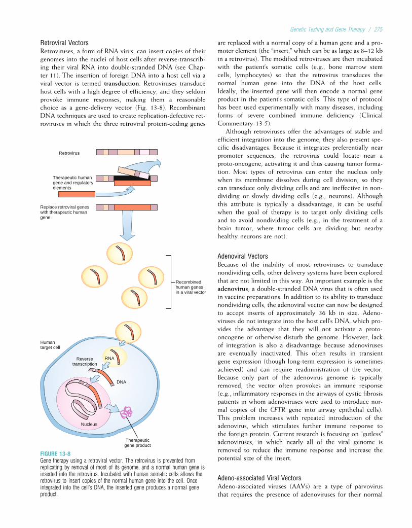
Retroviral VectorsRetroviruses, a form of RNA virus, can insert copies of theirgenomes into the nuclei of host cells after reverse-transcrib-ing their viral RNA into double-stranded DNA (see Chap-ter 11). The insertion of foreign DNA into a host cell via aviral vector is termed transduction. Retroviruses transducehost cells with a high degree of efficiency, and they seldomprovoke immune responses, making them a reasonablechoice as a gene-delivery vector (Fig. 13-8). RecombinantDNA techniques are used to create replication-defective ret-roviruses in which the three retroviral protein-coding genes
are replaced with a normal copy of a human gene and a pro-moter element (the “insert,” which can be as large as 8–12 kbin a retrovirus). The modified retroviruses are then incubatedwith the patient’s somatic cells (e.g., bone marrow stemcells, lymphocytes) so that the retrovirus transduces thenormal human gene into the DNA of the host cells.Ideally, the inserted gene will then encode a normal geneproduct in the patient’s somatic cells. This type of protocolhas been used experimentally with many diseases, includingforms of severe combined immune deficiency (ClinicalCommentary 13-5).
Although retroviruses offer the advantages of stable andefficient integration into the genome, they also present spe-cific disadvantages. Because it integrates preferentially nearpromoter sequences, the retrovirus could locate near aproto-oncogene, activating it and thus causing tumor forma-tion. Most types of retrovirus can enter the nucleus onlywhen its membrane dissolves during cell division, so theycan transduce only dividing cells and are ineffective in non-dividing or slowly dividing cells (e.g., neurons). Althoughthis attribute is typically a disadvantage, it can be usefulwhen the goal of therapy is to target only dividing cellsand to avoid nondividing cells (e.g., in the treatment of abrain tumor, where tumor cells are dividing but nearbyhealthy neurons are not).
Adenoviral VectorsBecause of the inability of most retroviruses to transducenondividing cells, other delivery systems have been exploredthat are not limited in this way. An important example is theadenovirus, a double-stranded DNA virus that is often usedin vaccine preparations. In addition to its ability to transducenondividing cells, the adenoviral vector can now be designedto accept inserts of approximately 36 kb in size. Adeno-viruses do not integrate into the host cell’s DNA, which pro-vides the advantage that they will not activate a proto-oncogene or otherwise disturb the genome. However, lackof integration is also a disadvantage because adenovirusesare eventually inactivated. This often results in transientgene expression (though long-term expression is sometimesachieved) and can require readministration of the vector.Because only part of the adenovirus genome is typicallyremoved, the vector often provokes an immune response(e.g., inflammatory responses in the airways of cystic fibrosispatients in whom adenoviruses were used to introduce nor-mal copies of the CFTR gene into airway epithelial cells).This problem increases with repeated introduction of theadenovirus, which stimulates further immune response tothe foreign protein. Current research is focusing on “gutless”adenoviruses, in which nearly all of the viral genome isremoved to reduce the immune response and increase thepotential size of the insert.
Adeno-associated Viral VectorsAdeno-associated viruses (AAVs) are a type of parvovirusthat requires the presence of adenoviruses for their normal
Therapeuticgene product
Nucleus
Reversetranscription
DNA
RNA
Humantarget cell
Replace retroviral geneswith therapeutic humangene
Therapeutic humangene and regulatoryelements
Retrovirus
Recombinedhuman genesin a viral vector
FIGURE 13-8Gene therapy using a retroviral vector. The retrovirus is prevented fromreplicating by removal of most of its genome, and a normal human gene isinserted into the retrovirus. Incubated with human somatic cells allows theretrovirus to insert copies of the normal human gene into the cell. Onceintegrated into the cell’s DNA, the inserted gene produces a normal geneproduct.
Genetic Testing and Gene Therapy / 275
CLINICAL COMMENTARY 13-5
Gene Therapy and Severe Combined Immunodeficiency
Gene therapy has been attempted for several forms of severe combinedimmunodeficiency (SCID), including adenosine deaminase deficiency SCID(ADA-SCID) and X-linked SCID. ADA, which is produced primarily in lym-phoid tissues, is an important component of the purine salvage pathway.ADA deficiency, an autosomal recessive disorder that accounts for about15% of SCID cases, results in the abnormal accumulation of purine meta-bolites toxic to T lymphocytes. Subsequently, B lymphocytes are alsoreduced in function and number. The resulting SCID is usually fatal bythe age of 2 years if untreated.
The preferred treatment for ADA-SCID is bone marrow transplantation.However, complications of bone marrow transplantation increase patientmorbidity and are sometimes fatal. In addition, major histocompatibilitycomplex (MHC)-compatible sibling donors are available for less than30% of ADA-SCID patients. Patients may be treated with polyethylene gly-col (PEG)-conjugated ADA (administered once or twice weekly by intramus-cular injection), but the response to this treatment is variable, and somepatients develop antibodies against PEG-ADA.
Because it is a systemic disorder caused by an enzyme deficiency, ADA-SCID represents a good candidate for gene replacement therapy. Ideally,proliferating bone marrow stem cells would be modified by retroviral vec-tors containing the normal ADA gene, resulting in a permanent cure forthe disorder. Because of difficulties in dealing with bone marrow stem cells,gene therapy for ADA-SCID was initiated instead in 1990 by retroviral inser-tion of ADA genes into lymphocytes that were extracted from patients. Afterretroviral insertion, the lymphocytes were injected back into the patients’peripheral circulation. This was the first application of gene therapy to aninherited human disorder.
Lymphocyte gene therapy has been applied in more than a dozenpatients with ADA deficiency. In some patients, ADA levels increased, Tlymphocyte counts improved, and the number of infections decreased.Because of the limited life span of T lymphocytes, these patients were rein-jected with modified T cells once every several months; however, the treatedlymphocytes have displayed surprising longevity, surviving in the circula-tion for well over 1 year in some cases. These patients were also typicallytreated with PEG-ADA, so it has been somewhat difficult to establish theefficacy of gene therapy.
More recently, ADA-SCID has been treated by retroviral insertion of theADA gene into bone marrow stem cells, rather than lymphocytes. This treat-
ment has resulted in long-term increases in B cell and T cell counts (up to9 years) and normal immune function in 11 treated patients.
X-linked SCID results from mutations in the gene, SCIDX1, thatencodes subunits of the g chain found in six different cytokine receptors(those of interleukins 2, 4, 7, 9, 15, and 21; see Chapter 9). Lacking thesereceptors, T cells and natural killer cells cannot receive the signals theyneed for normal maturation. The T cell deficiency in turn produces a defi-ciency of normal B cells, resulting in SCID. As with ADA deficiency, thisdisorder can be treated with bone marrow transplantation if an MHC-com-patible donor is available. Without a bone marrow transplant, the disease isfatal early in childhood.
In 1999, retroviral therapy was initiated to introduce SCIDX1 intopatients’ bone marrow stem cells. Less than 1% of bone marrow stem cellswere effectively transduced with the therapeutic gene. However, the trans-duced cells enjoyed a selective growth advantage over other bone marrowstem cells because the inserted gene increased the cytokine signalingneeded for normal cell function. In most treated patients, the number of nat-ural killer cells, T cells, and B cells increased to near-normal levels, withsustained resistance to infections continuing for years after therapy.
These positive outcomes in most ADA and X-linked SCID patients havebeen widely heralded as the first successful uses of somatic cell gene ther-apy in the treatment of an inherited disease. However, five of the X-linkedSCID patients developed leukemia-like disease (clonal T cell proliferation)as a result of random insertion of the retroviral vector in or near LOM2,a proto-oncogene that is activated in about half of all cases of acute lym-phocytic leukemia. This was fatal in one patient, but the others were treatedsuccessfully by chemotherapy and continued to benefit from gene therapy.Although the cause of T cell proliferation in these patients remains some-what unclear, there is evidence that a specific interaction occurs betweenthe inserted g-chain gene and LOM2 to activate the proto-oncogene. Thisinteraction might explain why, among the many different clinical trialsinvolving retroviral transfer of genes to bone marrow stem cells, only thistrial has resulted in cancer.
These examples illustrate some of the promise, as well as some of theperils, of somatic cell gene therapy. Clearly, gene therapy poses hazards thatmust be closely monitored. However, these protocols can lead to effectivetreatment of otherwise lethal diseases, and they provide invaluable informa-tion for the development of gene therapy protocols for other genetic diseases.
replication (hence the term adeno-associated). Like adeno-viruses, AAVs are DNA viruses that can transduce nondivid-ing cells. In addition, they elicit much less immune responsethan do adenoviruses and have little, if any, pathogeniceffect. They are also capable of sustaining protracted thera-peutic expression (months to years). These vectors, however,can accept a DNA insert of only about 4.5 kb. (In somecases, this problem can be circumvented by dividing theinsert into two parts, placing each part in a vector, anddesigning the mRNA products to reassemble.) Because oftheir many useful properties, AAVs have become much morepopular as a gene therapy vector during the past severalyears. They have been tested in clinical trials for the treat-ment of cystic fibrosis, hemophilia B, a1-antitrypsin defi-ciency, Duchenne muscular dystrophy, Parkinson disease,Alzheimer disease, and many other disorders.
Lentiviral VectorsLentiviral viruses are complex RNA retroviruses that, unlikesimple retroviruses, can transduce nondividing cellsthrough pores in the nuclear membrane (human immunode-ficiency virus [HIV] is an example of a lentivirus). Likeother retroviruses, lentiviruses can integrate stably intothe genome, and they can accept reasonably large inserts(8 kb). Because they combine the desirable properties ofstable integration and the ability to transduce nondividingcells, lentiviruses are currently the focus of much researchand development.
Challenges in Viral Gene TherapyAlthough viral gene therapy holds considerable promise,there are several important challenges:
276 / Chapter 13 MEDICAL GENETICS
• Transient and low-level expression. The gene product may beexpressed at subtherapeutic levels, often less than 1% ofthe normal amount. In part, this reflects the fact that onlysome of the target cells successfully incorporate the normalgene. In addition, random insertion of the virus into thehost’s genome can affect gene regulation (e.g., enhancersequences required for normal expression levels are not pres-ent). Cells sometimes respond to foreign inserted DNA bymethylating—and thus inactivating—it. For these reasons,transcription of the gene often ceases after a few weeks ormonths. It should be noted, however, that transient expres-sion is sufficient, and even desirable, for some types of ther-apy, such as provoking an immune response against a tumoror generating new blood vessels (discussed later).
• Difficulties in reaching or specifying target tissue. Although somesystemic disorders are relatively easy to target by modify-ing lymphocytes or bone marrow stem cells, others pre-sent formidable challenges. It may be difficult, forexample, to target affected neurons responsible for centralnervous system disorders. In addition, vectors must bemodified so that they enter only the desired cell type.
• Necessity for precise regulation of gene activity. Accurate regula-tion of gene activity is not a concern for some diseases(e.g., a 50-fold overexpression of adenosine deaminasehas no clinically significant effects). However, it is criticalfor diseases such as thalassemia, in which the number ofa-globin and b-globin chains must be closely balanced(see Chapter 3). It is often difficult to achieve such preci-sion using viral gene therapy.
• Potential for insertional mutagenesis. The unpredictable integrationof a retroviral vector into the host’s DNA can have undesiredconsequences, as discussed earlier. Although insertionalmutagenesis appears to be a rare event, it has occurred inseveral patients (see Clinical Commentary 13-5).
Considerable research is being devoted to overcoming theseand other problems. For example, the levels and permanence ofgene expression are being increased by incorporating strongerpromoter sequences in DNA inserts. Vectors are being modi-fied to reduce immune responses and to increase target-cellspecificity. Methods are being developed for targeted inser-tion of corrected DNA sequences. For example, proteins areengineered to bind to a specific mutated DNA sequence andto induce double-stranded DNA breaks followed by the inser-tion of a normal DNA sequence. With targeted insertion, themutated DNA is corrected in situ, avoiding difficulties withrandom insertion of DNA and taking advantage of the nativepromoter and enhancer sequences in the host’s genome.
4Viral vectors offer highly efficient transfer oftherapeutic genes into somatic cells. However, theyhave several drawbacks, including low or transientexpression of the gene product, limited insert size,generation of immune responses, difficulty in preciseregulation, and, for some vectors, a lack of ability totransduce nondividing cells and the potential foroncogenesis.
Nonviral VectorsAlthough viral vectors provide the advantage of efficientgene transfer into cells, the disadvantages just listed haveprompted researchers to investigate several types of nonviralvectors. One of the most extensively studied is the liposome,a fat body that can accept large DNA inserts. Liposomessometimes fuse with cells, allowing the DNA insert to enterthe cell. Because the liposome has no peptides, it does notelicit an immune response. Its primary disadvantage is thatit lacks the transfer efficiency of viruses: most of the lipo-somes are degraded in the cytoplasm, and most of those thatare not degraded are unable to enter the nucleus.
Surprisingly, it is possible to insert plasmids containinghuman DNA directly into cells without using any delivery vec-tor at all. Although most “naked” DNA is repelled by the cellmembrane, the DNA occasionally enters the cell, escapes deg-radation, and temporarily encodes proteins. Attempts are underway to use naked DNA as a vaccine that encodes a pathogenicprotein against which the body mounts an immune response.
An intriguing development with potential for somatic celltherapy is the synthesis of human artificial chromosomes.Because these synthetically constructed chromosomes con-tain functional centromeres and telomeres, they should beable to integrate and replicate in human cell nuclei. Also,they are capable of accepting inserts as large as the entire2.4 Mb Duchenne muscular dystrophy gene (DMD).
4Gene therapy using nonviral vectors, includingliposomes and naked DNA, offers some advantagesover viral vectors, but they currently lack the transferefficiency of viral vectors.
Gene-Blocking TherapiesGene-replacement techniques are not effective in correctinggain-of-function or dominant negative mutations (e.g.,Huntington disease, Marfan syndrome). To correct theseconditions, the defective gene product must be blocked ordisabled in some way. Although not as well developed asgene-replacement therapy methods, gene-blocking methodsare being developed, and some show promise.
Antisense TherapyThe principle behind antisense therapy is simple: an oligo-nucleotide is engineered whose DNA sequence is comple-mentary to that of the messenger RNA (mRNA) sequenceproduced by a gain-of-function mutation. This antisense oli-gonucleotide binds to the abnormal mRNA, preventing itstranslation into a harmful protein (Fig. 13-9A). Antisenseoligonucleotides can also be engineered to bind to double-stranded DNA containing the disease-causing mutation,creating a triple helix that cannot be transcribed into mRNA.A difficulty with this antisense therapy is that antisenseoligonucleotides are often degraded before they can reachtheir target. Also, because of variation in the shape of thetarget DNA or RNA molecule, the antisense oligonucleotidemight not be able to bind to its complementary sequence.Nevertheless, antisense therapy is being tested in a number
Genetic Testing and Gene Therapy / 277
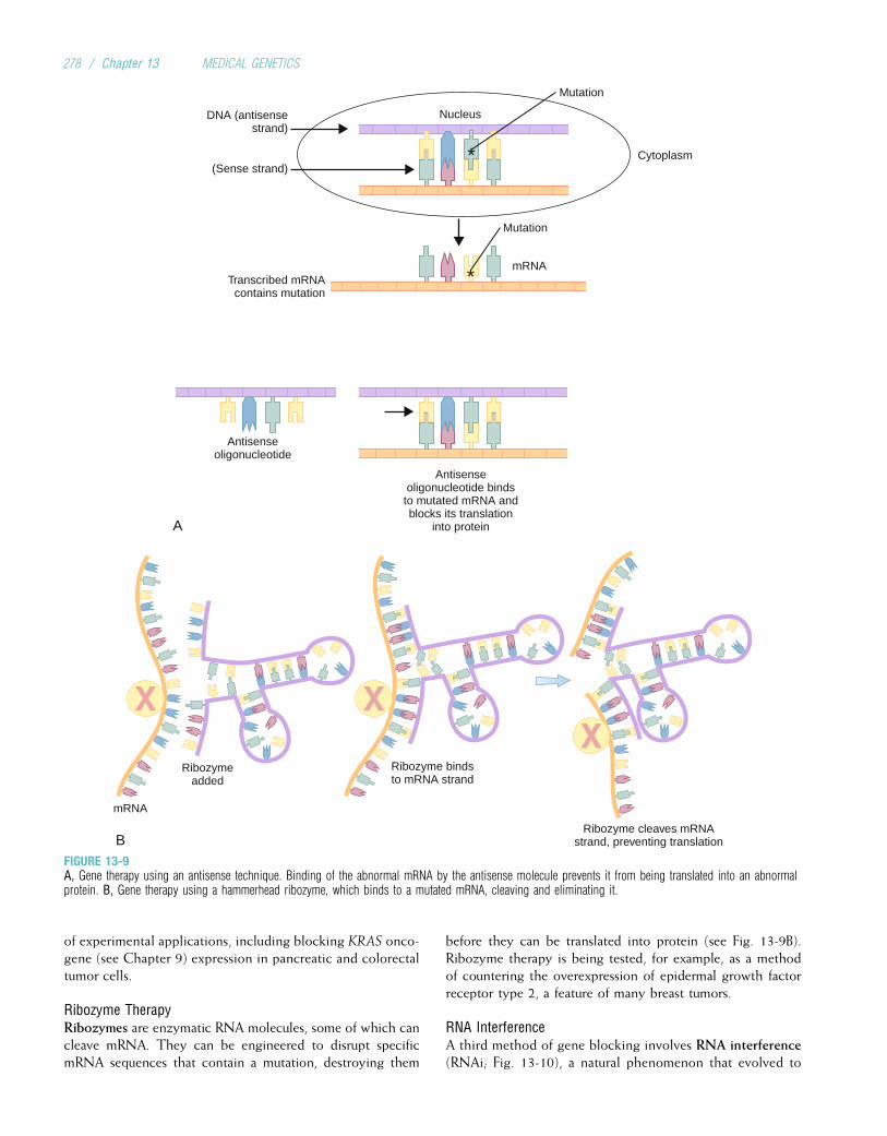
of experimental applications, including blocking KRAS onco-gene (see Chapter 9) expression in pancreatic and colorectaltumor cells.
Ribozyme TherapyRibozymes are enzymatic RNA molecules, some of which cancleave mRNA. They can be engineered to disrupt specificmRNA sequences that contain a mutation, destroying them
before they can be translated into protein (see Fig. 13-9B).Ribozyme therapy is being tested, for example, as a methodof countering the overexpression of epidermal growth factorreceptor type 2, a feature of many breast tumors.
RNA InterferenceA third method of gene blocking involves RNA interference(RNAi; Fig. 13-10), a natural phenomenon that evolved to
Nucleus
Cytoplasm
DNA (antisensestrand)
(Sense strand)
mRNA
Antisenseoligonucleotide
Antisenseoligonucleotide bindsto mutated mRNA andblocks its translation
into proteinA
Mutation
Mutation
Transcribed mRNAcontains mutation
*
*
mRNA
Ribozymeadded
Ribozyme bindsto mRNA strand
Ribozyme cleaves mRNAstrand, preventing translationB
FIGURE 13-9A, Gene therapy using an antisense technique. Binding of the abnormal mRNA by the antisense molecule prevents it from being translated into an abnormalprotein. B, Gene therapy using a hammerhead ribozyme, which binds to a mutated mRNA, cleaving and eliminating it.
278 / Chapter 13 MEDICAL GENETICS
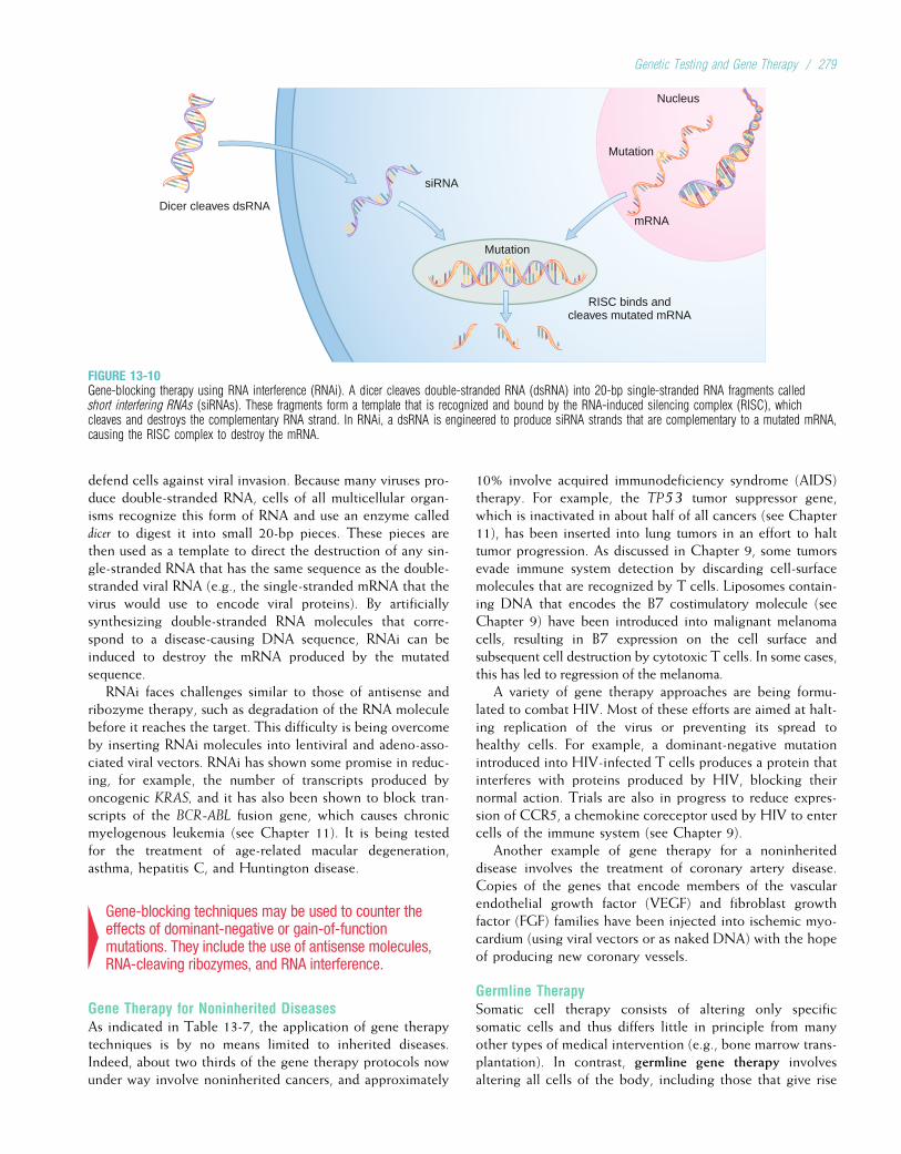
defend cells against viral invasion. Because many viruses pro-duce double-stranded RNA, cells of all multicellular organ-isms recognize this form of RNA and use an enzyme calleddicer to digest it into small 20-bp pieces. These pieces arethen used as a template to direct the destruction of any sin-gle-stranded RNA that has the same sequence as the double-stranded viral RNA (e.g., the single-stranded mRNA that thevirus would use to encode viral proteins). By artificiallysynthesizing double-stranded RNA molecules that corre-spond to a disease-causing DNA sequence, RNAi can beinduced to destroy the mRNA produced by the mutatedsequence.
RNAi faces challenges similar to those of antisense andribozyme therapy, such as degradation of the RNA moleculebefore it reaches the target. This difficulty is being overcomeby inserting RNAi molecules into lentiviral and adeno-asso-ciated viral vectors. RNAi has shown some promise in reduc-ing, for example, the number of transcripts produced byoncogenic KRAS, and it has also been shown to block tran-scripts of the BCR-ABL fusion gene, which causes chronicmyelogenous leukemia (see Chapter 11). It is being testedfor the treatment of age-related macular degeneration,asthma, hepatitis C, and Huntington disease.
4Gene-blocking techniques may be used to counter theeffects of dominant-negative or gain-of-functionmutations. They include the use of antisense molecules,RNA-cleaving ribozymes, and RNA interference.
Gene Therapy for Noninherited DiseasesAs indicated in Table 13-7, the application of gene therapytechniques is by no means limited to inherited diseases.Indeed, about two thirds of the gene therapy protocols nowunder way involve noninherited cancers, and approximately
10% involve acquired immunodeficiency syndrome (AIDS)therapy. For example, the TP53 tumor suppressor gene,which is inactivated in about half of all cancers (see Chapter11), has been inserted into lung tumors in an effort to halttumor progression. As discussed in Chapter 9, some tumorsevade immune system detection by discarding cell-surfacemolecules that are recognized by T cells. Liposomes contain-ing DNA that encodes the B7 costimulatory molecule (seeChapter 9) have been introduced into malignant melanomacells, resulting in B7 expression on the cell surface andsubsequent cell destruction by cytotoxic T cells. In some cases,this has led to regression of the melanoma.
A variety of gene therapy approaches are being formu-lated to combat HIV. Most of these efforts are aimed at halt-ing replication of the virus or preventing its spread tohealthy cells. For example, a dominant-negative mutationintroduced into HIV-infected T cells produces a protein thatinterferes with proteins produced by HIV, blocking theirnormal action. Trials are also in progress to reduce expres-sion of CCR5, a chemokine coreceptor used by HIV to entercells of the immune system (see Chapter 9).
Another example of gene therapy for a noninheriteddisease involves the treatment of coronary artery disease.Copies of the genes that encode members of the vascularendothelial growth factor (VEGF) and fibroblast growthfactor (FGF) families have been injected into ischemic myo-cardium (using viral vectors or as naked DNA) with the hopeof producing new coronary vessels.
Germline TherapySomatic cell therapy consists of altering only specificsomatic cells and thus differs little in principle from manyother types of medical intervention (e.g., bone marrow trans-plantation). In contrast, germline gene therapy involvesaltering all cells of the body, including those that give rise
Dicer cleaves dsRNA
siRNA
RISC binds andcleaves mutated mRNA
Nucleus
Mutation
Mutation
mRNA
FIGURE 13-10Gene-blocking therapy using RNA interference (RNAi). A dicer cleaves double-stranded RNA (dsRNA) into 20-bp single-stranded RNA fragments calledshort interfering RNAs (siRNAs). These fragments form a template that is recognized and bound by the RNA-induced silencing complex (RISC), whichcleaves and destroys the complementary RNA strand. In RNAi, a dsRNA is engineered to produce siRNA strands that are complementary to a mutated mRNA,causing the RISC complex to destroy the mRNA.
Genetic Testing and Gene Therapy / 279
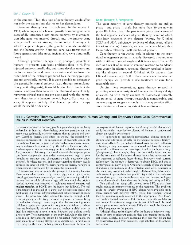
to the gametes. Thus, this type of gene therapy would affectnot only the patient but also his or her descendants.
Germline therapy was first achieved in the mouse in1983, when copies of a human growth hormone gene weresuccessfully introduced into mouse embryos by microinjec-tion (the gene was inserted directly into the embryo usinga very small needle). Among the minority of embryos inwhich the gene integrated, the gametes were also modified,and the human growth hormone gene was transmitted tofuture generations (the mice, incidentally, were abnormallylarge).
Although germline therapy is, in principle, possible inhumans, it presents significant problems (Box 13-7). First,injected embryos usually die, and some develop tumors andmalformations. Second, even in an autosomal dominant dis-order, half of the embryos produced by a heterozygous par-ent are genetically normal. If it were possible to distinguishthe genetically normal embryos (e.g., through preimplanta-tion genetic diagnosis), it would be simpler to implant thenormal embryos than to alter the abnormal ones. Finally,numerous ethical questions are associated with the perma-nent alteration of a human’s genetic legacy. For these rea-sons, it appears unlikely that human germline therapywould be useful or desirable.
Gene Therapy: A PerspectiveThe great majority of gene therapy protocols are still inphase I and phase II trials, but more than 30 are now inphase III clinical trials. The past several years have witnessedthe first arguable successes of gene therapy, some of whichhave been discussed in this chapter (therapy for X-linkedSCID and ADA deficiency; evidence of therapeutic effectsin various cancers). However, success has been achieved thusfar in only a relatively small number of persons.
Gene therapy is not without risk. In addition to the inser-tional mutagenesis potential already discussed, a young manwith ornithine transcarbamylase deficiency (see Chapter 7)died as a result of an adverse immune reaction to an adeno-virus vector. In addition, retroviral therapy resulted in leuke-mia-like disease in several X-linked SCID patients (seeClinical Commentary 13-5). It thus remains unclear whethergene therapy will provide a safe treatment or cure for areasonable cost.
Despite these reservations, gene therapy research isproviding many new insights of fundamental biological sig-nificance. As with many avenues of biomedical research,the potential of gene therapy research is considerable, andcurrent progress suggests strongly that it may provide effica-cious treatment of some important human diseases.
BOX 13-7 Germline Therapy, Genetic Enhancement, Human Cloning, and Embryonic Stem Cells: ControversialIssues in Medical Genetics
For reasons outlined in the text, germline gene therapy is not beingundertaken in humans. Nevertheless, germline gene therapy is inmany ways technically easier to perform than is somatic cell ther-apy. Germline therapy also offers (in theory) the possibility of“genetic enhancement,” the introduction of favorable genes intothe embryo. However, a gene that is favorable in one environmentmay be unfavorable in another (e.g., the sickle-cell mutation, whichis advantageous only for heterozygotes in a malarial environment).And, because of pleiotropy, the introduction of advantageous genescan have completely unintended consequences (e.g., a genethought to enhance one characteristic could negatively affectanother). For these reasons, and because germline therapy usuallydestroys the targeted embryo, neither germline therapy nor geneticenhancement is advocated by the scientific community.
Controversy also surrounds the prospect of cloning humans.Many mammalian species (e.g., sheep, pigs, cattle, goats, mice,cats, dogs) have been successfully cloned by introducing a diploidnucleus from an adult cell into an egg cell from which the originalhaploid nucleus was removed (a technique termed somatic cellnuclear transfer, or SCNT; see the figure that follows). The cellis manipulated so that all of its genes can be expressed (recall thatmost genes in a typical differentiated adult cell are transcriptionallysilent). This procedure, when allowed to proceed through a full-term pregnancy, could likely be used to produce a human being(reproductive cloning). Some argue that human cloning offerschildless couples the opportunity to produce children to whomthey are biologically related or even to replace a child who hasdied. It is important to keep in mind, however, that a clone is onlya genetic copy. The environment of the individual, which also plays alarge role in development, cannot be replicated. Furthermore, thegreat majority of cloning attempts in mammals fail: in most cases,the embryo either dies or has gross malformations. Because the
consequences of human reproductive cloning would almost cer-tainly be similar, reproductive cloning of humans is condemnedalmost universally by scientists.
It is important to distinguish reproductive cloning from thecloning and cultivation of cells for therapeutic purposes. Embry-onic stem cells (ESCs), which are derived from the inner-cell massof blastocyst-stage embryos, can be cloned and have the uniquepotential to differentiate into any type of cell in the human body(pluripotency). For example, they can potentially form neuronsfor the treatment of Parkinson disease or cardiac myocytes forthe treatment of ischemic heart disease. However, with currenttechnology, the embryo is destroyed to obtain ESCs, and this iscontroversial in many circles. Ongoing research efforts are aimedat inducing pluripotency in differentiated adult cells. Research isalso under way to extract usable single cells from 3-day blastomereembryos (as in preimplantation genetic diagnosis) so that embryosare not destroyed. It remains to be seen whether these technologiescan produce cells that have the same flexibility and utility as ESCs.
One difficulty in using cells derived from ESCs is that theymight induce an immune response in the recipient. This problemcould be largely overcome if ESC clones were available frommany persons with different MHC types. The recipient wouldthen be immunologically matched to the appropriate ESC. How-ever, only a limited number of ESC lines are currently available tomost researchers. Another suggestion is that SCNT could be usedwith a patient’s own cells to create ESCs that would be identicalin DNA sequence to the patient.
Although these technologies offer the hope of effective treat-ment for some recalcitrant diseases, they also present thorny eth-ical issues. Clearly, decisions regarding their use must be guidedby constructive input from scientists, legal scholars, philosophers,and others.
280 / Chapter 13 MEDICAL GENETICS
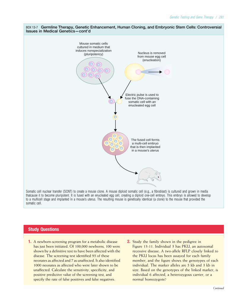
Study Questions
1. A newborn-screening program for a metabolic diseasehas just been initiated. Of 100,000 newborns, 100 wereshown by a definitive test to have been affected with thedisease. The screening test identified 93 of theseneonates as affected and 7 as unaffected. It also identified1000 neonates as affected who were later shown to beunaffected. Calculate the sensitivity, specificity, andpositive predictive value of the screening test, andspecify the rate of false positives and false negatives.
2. Study the family shown in the pedigree inFigure 13-11. Individual 3 has PKU, an autosomalrecessive disease. A two-allele RFLP closely linked tothe PKU locus has been assayed for each familymember, and the figure shows the genotypes of eachindividual. The marker alleles are 5 kb and 3 kb insize. Based on the genotypes of the linked marker, isindividual 6 affected, a heterozygous carrier, or anormal homozygote?
Continued
BOX 13-7 Germline Therapy, Genetic Enhancement, Human Cloning, and Embryonic Stem Cells: ControversialIssues in Medical Genetics—cont’d
Mouse somatic cellscultured in medium that
induces nonspecialization(pluripotency) Nucleus is removed
from mouse egg cell(enucleation)
Electric pulse is used tofuse the DNA-containing
somatic cell with anenucleated egg cell
The fused cell formsa multi-cell embryo
that is then implantedin a mouse’s uterus
Somatic cell nuclear transfer (SCNT) to create a mouse clone. A mouse diploid somatic cell (e.g., a fibroblast) is cultured and grown in mediathatcause it to become pluripotent. It is fused with an enucleated egg cell, creating a diploid one-cell embryo. This embryo is allowed to developto a multicell stage and implanted in a mouse’s uterus. The resulting mouse is genetically identical (a clone) to the mouse that provided thesomatic cell.
Genetic Testing and Gene Therapy / 281
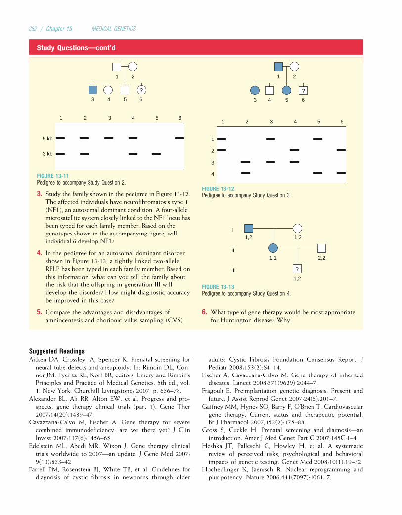
Study Questions—cont’d
1 2
3
5 kb
3 kb
4 5 6
3 41 2 5 6
?
FIGURE 13-11Pedigree to accompany Study Question 2.
3. Study the family shown in the pedigree in Figure 13-12.The affected individuals have neurofibromatosis type 1(NF1), an autosomal dominant condition. A four-allelemicrosatellite system closely linked to the NF1 locus hasbeen typed for each family member. Based on thegenotypes shown in the accompanying figure, willindividual 6 develop NF1?
4. In the pedigree for an autosomal dominant disordershown in Figure 13-13, a tightly linked two-alleleRFLP has been typed in each family member. Based onthis information, what can you tell the family aboutthe risk that the offspring in generation III willdevelop the disorder? How might diagnostic accuracybe improved in this case?
5. Compare the advantages and disadvantages ofamniocentesis and chorionic villus sampling (CVS).
1 2
3
1
2
3
4
4 5 6
3 41 2 5 6
?
FIGURE 13-12Pedigree to accompany Study Question 3.
1,2 1,2
1,1 2,2
1,2
I
II
III ?
FIGURE 13-13Pedigree to accompany Study Question 4.
6. What type of gene therapy would be most appropriatefor Huntington disease? Why?
Suggested ReadingsAitken DA, Crossley JA, Spencer K. Prenatal screening for
neural tube defects and aneuploidy. In: Rimoin DL, Con-nor JM, Pyeritz RE, Korf BR, editors. Emery and Rimoin’sPrinciples and Practice of Medical Genetics. 5th ed., vol.1. New York: Churchill Livingstone; 2007. p. 636–78.
Alexander BL, Ali RR, Alton EW, et al. Progress and pro-spects: gene therapy clinical trials (part 1). Gene Ther2007;14(20):1439–47.
Cavazzana-Calvo M, Fischer A. Gene therapy for severecombined immunodeficiency: are we there yet? J ClinInvest 2007;117(6):1456–65.
Edelstein ML, Abedi MR, Wixon J. Gene therapy clinicaltrials worldwide to 2007—an update. J Gene Med 2007;9(10):833–42.
Farrell PM, Rosenstein BJ, White TB, et al. Guidelines fordiagnosis of cystic fibrosis in newborns through older
adults: Cystic Fibrosis Foundation Consensus Report. JPediatr 2008;153(2):S4–14.
Fischer A, Cavazzana-Calvo M. Gene therapy of inheriteddiseases. Lancet 2008;371(9629):2044–7.
Fragouli E. Preimplantation genetic diagnosis: Present andfuture. J Assist Reprod Genet 2007;24(6):201–7.
Gaffney MM, Hynes SO, Barry F, O’Brien T. Cardiovasculargene therapy: Current status and therapeutic potential.Br J Pharmacol 2007;152(2):175–88.
Gross S, Cuckle H. Prenatal screening and diagnosis—anintroduction. Amer J Med Genet Part C 2007;145C:1–4.
Heshka JT, Palleschi C, Howley H, et al. A systematicreview of perceived risks, psychological and behavioralimpacts of genetic testing. Genet Med 2008;10(1):19–32.
Hochedlinger K, Jaenisch R. Nuclear reprogramming andpluripotency. Nature 2006;441(7097):1061–7.
282 / Chapter 13 MEDICAL GENETICS
Hunter DJ, Khoury MJ, Drazen JM. Letting the genome outof the bottle–will we get our wish? N Engl J Med2008;358(2):105–7.
Jaenisch R. Human cloning—the science and ethics of nucleartransplantation. N Engl J Med 2004;351: 2878–91.
Kim DH, Rossi JJ. Strategies for silencing human diseaseusing RNA interference. Nat Rev Genet 2007; 8(3):173–84.
Lau TK, Leung TN. Genetic screening and diagnosis. CurrOpin Obstet Gynecol 2005;17(2):163–9.
Malone FD, Canick JA, Ball RH, et al. First-trimester orsecond-trimester screening, or both, for Down syndrome.N Engl J Med 2005;353(19):2001–11.
McCabe LL, McCabe ER. Expanded newborn screening:implications for genomic medicine. Annu Rev Med 2008;59:163–75.
O’Connor TP, Crystal RG. Genetic medicines: treatmentstrategies for hereditary disorders. Nat Rev Genet 2006;7(4):261–76.
Pagon RA, Tarczy-Hornoch P, Baskin PK, et al. Genetests—gene clinics: genetic testing information for agrowing audience. Hum Mutat 2002;19:501–9.
Riordan JR. CFTR function and prospects for therapy. AnnuRev Biochem 2008;77:701–26.
Scheuner MT, Sieverding P, Shekelle PG. Delivery of geno-mic medicine for common chronic adult diseases: a sys-tematic review. JAMA 2008;299(11):1320–34.
Sekizawa A, Purwosunu Y, Matsuoka R, et al. Recentadvances in non-invasive prenatal DNA diagnosis throughanalysis of maternal blood. J Obstet Gynaecol Res 2007;33(6):747–64.
Sermon K, Van Steirteghem A, Liebaers I. Preimplantationgenetic diagnosis. Lancet 2004;363:1633–41.
Shulman LP, Simpson JL. Techniques for prenatal diagnosis.In: Rimoin DL, Connor JM, Pyeritz RE, Korf BR, editors.Emery and Rimoin’s Principles and Practice of MedicalGenetics. 5th ed., vol. 1. New York: Churchill Living-stone; 2007. p. 679–702.
South ST, Chen Z, Brothman AR. Genomic medicine in pre-natal diagnosis. Clin Obstet Gynecol 2008; 51(1):62–73.
Spencer K. Aneuploidy screening in the first trimester. AmerJ Med Genet Part C 2007;145C:18–32.
Stoller JK. Aboussouan LS: a1-Antitrypsin deficiency. Lan-cet 2005;365(9478):2225–36.
Van Voorhis BJ. Clinical practice. In vitro fertilization.N Engl J Med 2007;356(4):379–86.
Verma IM, Weitzman MD. Gene therapy: twenty-first century medicine. Annu Rev Biochem 2005; 74:711–38.
Waisbren SE. Expanded newborn screening: information andresources for the family physician. Am Fam Physician2008;77:987–94.
Warrington Jr KH, Herzog RW. Treatment of human dis-ease by adeno-associated viral gene transfer. Hum Genet2006;119(6):571–603.
Weaver D. Catalog of Prenatally Diagnosed Conditions. 3rded. Baltimore: Johns Hopkins University Press; 1999.
Wilcken B. Recent advances in newborn screening. J InheritMetab Dis 2007;30(2):129–33.
Wolfberg AJ. Genes on the web—direct-to-consumer mar-keting. N Engl J Med 2006;355:543–5.
Internet ResourcesGene Therapy Clinical Trials Worldwide (updated list ofall gene therapy protocols) http://www.wiley.co.uk/genetherapy/clinical/
Gene Clinics/Gene Tests (reviews of genetic diseases andlists of laboratories that perform diagnostic tests) http://www.geneclinics.org/
Human Genome Project Information (includes informa-tion on genetic testing and gene therapy, with relevantlinks)http://www.ornl.gov/sci/techresources/Human_Genome/medi-cine/genetest.shtml
http://www.ornl.gov/sci/techresources/Human_Genome/medicine/gen-
etherapy.shtml
National Newborn Screening and Genetics ResourceCenter http://genes-r-us.uthscsa.edu/
National Organization for Rare Diseases (database ofrare disorders that includes brief reviews and informationon diagnostic tests and treatment for families andprofessionals) http://www.rarediseases.org
Genetic Testing and Gene Therapy / 283
Chapter 14
GENETICS AND PERSONALIZED MEDICINE
Scientific, technological, and medical advances have made itpossible to detect, diagnose, and treat most common dis-eases (e.g., asthma, diabetes, hypertension) early in theircourse and more effectively than ever before. Such advancesare, however, highly dependent on the skills and knowledgeof clinicians, access to health care services, and the availabil-ity and affordability of diagnostic technologies. Most healthcare providers follow a conventional model in which apatient presents with a set of symptoms and signs, whichthe provider uses to make a “most likely” diagnosis. The pro-vider then prescribes a treatment that he or she thinks willbe most effective. If this treatment fails, the process isrepeated until a correct diagnosis or more effective treatmentis found. In such a model, preventive health maintenance isencouraged. Compliance, however, is challenging becauseinformation about risk factors, as well as the patient’s percep-tion of risk, is at best approximate.
Personalized medicine is a model of practice in whicheach patient’s personal risk for common diseases and theeffectiveness of various treatments are estimated directlyfrom the patient’s unique combination of genetic and envi-ronmental risk factors. Accordingly, a health care providercan predict a person’s risk for common diseases, select diag-nostic tests to confirm the presence of disease, and prescribethe best therapeutic regimen to treat it. Ideally, knowledgeof disease risk promotes interventions (e.g., modification ofdiet, choice of drug therapy) that not only can treat diseaseearly in its course but also can delay its onset or prevent italtogether.
The effectiveness of personalized medicine depends on anumber of factors. These include identifying genetic andenvironmental risk factors (and their interactions) thatenable accurate prediction of clinically significant risk;demonstrating that individual risk assessment improvesdiagnostic accuracy and treatment outcome; developingtechnologies for cost-efficient assessment of a person’sgenome; building an infrastructure for clinicians to accessrisk data, interpret risk information, and explain risk esti-mates to patients; and developing guidelines and policiesfor how risk assessment information should be used in clin-ical and research applications. Not all of these aims will beachieved for every common disease. Indeed, for many com-plex diseases, it is likely that there will be no alternative to
the conventional model of practice in the near futurebecause so little is known about their etiology and patho-physiology. Nevertheless, for some common diseases anddrug responses, genetic testing and, in several cases perso-nalized medicine, are already being adapted to the clinicalsetting.
In this chapter, we discuss how new technologies aremaking the assessment of individual human genomes widelyaccessible, how such genome-wide information is being usedcurrently to make personal decisions about health, and theimplications of personalized care.
4Personalized medicine is the use of each person’sunique combination of genetic and environmental riskfactors to make predictions about the individualperson’s disease risk and response to varioustreatments.
A TECHNOLOGY-DRIVEN TRANSFORMATIONTraditionally, the search for genetic variants that influencecommon complex diseases has been a daunting task and hasbeen one of the major obstacles to developing personalizedmedicine. The most common approach to finding such var-iants involved testing whether polymorphisms in candidategenes were associated with disease risk in a small group ofunrelated patients with the same phenotype (e.g., diabetes,obesity). This was problematic, in part because choosingthe most appropriate candidate genes was difficult, smallcohorts provided limited statistical power, and the processof genotyping or sequencing was labor intensive andexpensive. This situation has changed dramatically overthe past decade with the development of technologies tointerrogate millions of polymorphisms per person cheaplyand efficiently (Box 14-1). These technologies, coupledwith advances in statistics and computing, enabled theapplication of new approaches such as genome-wide associ-ation studies (see Chapter 8), as well as the study of muchlarger cohorts of thousands or tens of thousands of persons.In addition, these new technologies for genotyping andDNA sequencing make it possible to develop cost-effectiveclinical tests that take advantage of newly discovered riskvariants.
284
THE IMPACT OF GENOMICSPharmacogeneticsMany of the drinks and foods that we ingest each day (e.g.,coffee, tea) contain thousands of complex compounds thateach of us must process. Some of these compounds neverleave the gastrointestinal tract, but most are absorbed,distributed, metabolized, and eliminated (i.e., biotrans-formed) to a variety of products that are used immediately,stored, or excreted. Exogenously synthesized compoundsthat are administered to achieve a specific effect on thehuman body (e.g., pharmaceuticals) also undergo biotrans-formation, and humans vary in the efficiency and speed withwhich they do this. Moreover, the response of a drug’s target(e.g., enzymes, receptors) can also vary among individuals.The study of the individual genetic variants that modifyhuman responses to pharmacological agents is called pharma-cogenetics; the assessment of the action of many genes simul-taneously is called pharmacogenomics.
Genetic Prediction of Serious Adverse Drug ResponsesOver the last decade, ambitious efforts have been undertakento advance the knowledge of pharmacogenetics. This hasbeen driven, in part, by the expectation that through the
use of pharmacogenetics, we will be able to profile DNA dif-ferences among individuals and thereby predict responses todifferent medicines. For example, a genetic profile (i.e., asummary of a person’s risk alleles) might predict who is moreor less likely to respond to a drug or to suffer a seriousadverse drug reaction (SADR).
Many drugs have a response rate between 25% and 75%.For example, ACE inhibitors and beta blockers have beenfound to be ineffective or only partially effective in up to70% of hypertensive patients. The use of such drugs in per-sons who are unlikely to respond increases the incidence ofSADRs and adds to the burden of health care costs. Yet,for most drugs, no tests are available to determine who willor will not respond, so these drugs are administered largelyon a trial-and-error basis.
Many drugs have adverse effects that are of clinicalimportance, and of the approximately 1200 drugs approvedfor use in the United States, about 15% are associated witha significant incidence of SADRs. A widely cited analysisconducted in the mid-1990s suggested that nearly 2 millionpeople are hospitalized each year as a result of adverse drugeffects, and approximately 100,000 people die from them,even when the drugs are appropriately prescribed and adm-inistered. Studies in Europe and Australia have yielded similarresults. Thus, identification of genetic profiles that predict aperson’s response to drugs is likely to increase the overallefficacy and safety of pharmaceuticals.
Testing is currently available for a handful of alleles thatpredict SADRs. For example, thiopurine methyl transferase(TPMT) is an enzyme that inactivates thiopurine drugs(e.g., 6-mercaptopurine, azathioprine), which are frequentlyused to treat acute lymphatic leukemia and to prevent rejec-tion of organ transplants. A mutation of the TPMT genereduces enzyme activity. About one in 300 persons of Euro-pean ancestry is homozygous for this mutation, and thesepatients can experience life-threatening bone marrow sup-pression upon exposure to thiopurine drugs. The presenceof such variants can be assessed by genotyping or by enzymeassays, which are now commonly done before administeringthiopurines.
4Each person’s response to natural and syntheticchemicals is determined in part by polymorphismsin genes that control pathways of biotransformationand the chemical’s target.
Personalized Drug TherapyOne of the major challenges of pharmacogenetics is theselection of appropriate targets (e.g., a specific enzyme,cytokine, or cell-surface receptor) that might be amenableto manipulation by a drug. The results of genetic studiesare used to identify polymorphisms associated with varyingsusceptibility to disease (i.e., a potential target for a drug)or polymorphisms that modify the human response to adrug. For example, long QT syndrome (LQT syndrome;see Chapter 12) can be caused by mutations in one of at least
BOX 14-1Assessing Your Genome
Knowledge of a person’s genetic makeup will clearly be animportant tool for making better decisions about health, medicalcare, and perhaps lifestyle as well. Until recently, assessing thegenome as a whole was fairly expensive and done only inresearch laboratories. However, new technologies have dramati-cally lowered the cost of whole-genome analysis and havespurred the development of consumer services that offerwhole-genome studies directly to the public (see Chapter 13).These services have rapidly made headlines, as much for theirnovelty as for their potential to inform persons about theirgenetic composition.
Most whole-genome consumer services offer to genotypehundreds of thousands to millions of common single nucleotidepolymorphisms (SNPs). The SNPs typed for consumers are thesame ones commonly being used by researchers to identify dis-ease–gene associations for common multifactorial disorders suchas hypertension, diabetes, and obesity. As gene–disease associa-tions are reported, consumers who have access to their geneticinformation can evaluate their risk for genetic diseases. Also,because each person’s genetic data are permanent, evaluationof risk can be reassessed with each new discovery. However,many of the SNP–disease associations reported to consumersare relatively weak and may be misunderstood or misinterpretedby the lay consumer (see Chapter 13).
More recently, whole-genome sequencing has been madeavailable to the public. This service is still expensive and there-fore very limited in application. Also, it is debatable whetherone’s understanding of health-related risks will be increased byknowing about the 99% of the genome that does not encodeproteins. An alternative strategy is to sequence only the pro-tein-coding exons. In any case, the same caveats raised in theprevious paragraph for whole-genome SNP typing apply equallyto whole-genome sequencing.
Genetics and Personalized Medicine / 285
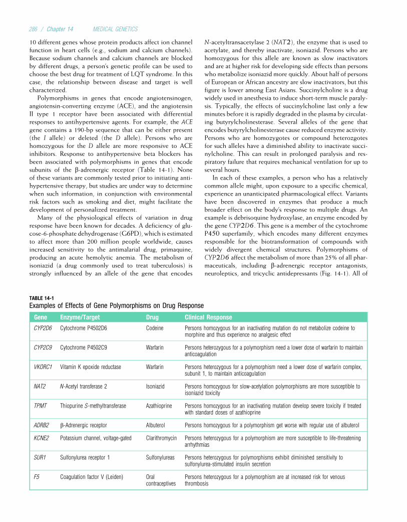
10 different genes whose protein products affect ion channelfunction in heart cells (e.g., sodium and calcium channels).Because sodium channels and calcium channels are blockedby different drugs, a person’s genetic profile can be used tochoose the best drug for treatment of LQT syndrome. In thiscase, the relationship between disease and target is wellcharacterized.
Polymorphisms in genes that encode angiotensinogen,angiotensin-converting enzyme (ACE), and the angiotensinII type 1 receptor have been associated with differentialresponses to antihypertensive agents. For example, the ACE
gene contains a 190-bp sequence that can be either present(the I allele) or deleted (the D allele). Persons who arehomozygous for the D allele are more responsive to ACEinhibitors. Response to antihypertensive beta blockers hasbeen associated with polymorphisms in genes that encodesubunits of the b-adrenergic receptor (Table 14-1). Noneof these variants are commonly tested prior to initiating anti-hypertensive therapy, but studies are under way to determinewhen such information, in conjunction with environmentalrisk factors such as smoking and diet, might facilitate thedevelopment of personalized treatment.
Many of the physiological effects of variation in drugresponse have been known for decades. A deficiency of glu-cose-6-phosphate dehydrogenase (G6PD), which is estimatedto affect more than 200 million people worldwide, causesincreased sensitivity to the antimalarial drug, primaquine,producing an acute hemolytic anemia. The metabolism ofisoniazid (a drug commonly used to treat tuberculosis) isstrongly influenced by an allele of the gene that encodes
N-acetyltransacetylase 2 (NAT2), the enzyme that is used toacetylate, and thereby inactivate, isoniazid. Persons who arehomozygous for this allele are known as slow inactivatorsand are at higher risk for developing side effects than personswho metabolize isoniazid more quickly. About half of personsof European or African ancestry are slow inactivators, but thisfigure is lower among East Asians. Succinylcholine is a drugwidely used in anesthesia to induce short-term muscle paraly-sis. Typically, the effects of succinylcholine last only a fewminutes before it is rapidly degraded in the plasma by circulat-ing butyrylcholinesterase. Several alleles of the gene thatencodes butyrylcholinesterase cause reduced enzyme activity.Persons who are homozygotes or compound heterozgotesfor such alleles have a diminished ability to inactivate succi-nylcholine. This can result in prolonged paralysis and res-piratory failure that requires mechanical ventilation for up toseveral hours.
In each of these examples, a person who has a relativelycommon allele might, upon exposure to a specific chemical,experience an unanticipated pharmacological effect. Variantshave been discovered in enzymes that produce a muchbroader effect on the body’s response to multiple drugs. Anexample is debrisoquine hydroxylase, an enzyme encoded bythe gene CYP2D6. This gene is a member of the cytochromeP450 superfamily, which encodes many different enzymesresponsible for the biotransformation of compounds withwidely divergent chemical structures. Polymorphisms ofCYP2D6 affect the metabolism of more than 25% of all phar-maceuticals, including b-adrenergic receptor antagonists,neuroleptics, and tricyclic antidepressants (Fig. 14-1). All of
TABLE 14-1
Examples of Effects of Gene Polymorphisms on Drug Response
Gene Enzyme/Target Drug Clinical Response
CYP2D6 Cytochrome P4502D6 Codeine Persons homozygous for an inactivating mutation do not metabolize codeine tomorphine and thus experience no analgesic effect
CYP2C9 Cytochrome P4502C9 Warfarin Persons heterozygous for a polymorphism need a lower dose of warfarin to maintainanticoagulation
VKORC1 Vitamin K epoxide reductase Warfarin Persons heterozygous for a polymorphism need a lower dose of warfarin complex,subunit 1, to maintain anticoagulation
NAT2 N-Acetyl transferase 2 Isoniazid Persons homozygous for slow-acetylation polymorphisms are more susceptible toisoniazid toxicity
TPMT Thiopurine S-methyltransferase Azathioprine Persons homozygous for an inactivating mutation develop severe toxicity if treatedwith standard doses of azathioprine
ADRB2 b-Adrenergic receptor Albuterol Persons homozygous for a polymorphism get worse with regular use of albuterol
KCNE2 Potassium channel, voltage-gated Clarithromycin Persons heterozygous for a polymorphism are more susceptible to life-threateningarrhythmias
SUR1 Sulfonylurea receptor 1 Sulfonylureas Persons heterozygous for polymorphisms exhibit diminished sensitivity tosulfonylurea-stimulated insulin secretion
F5 Coagulation factor V (Leiden) Oralcontraceptives
Persons heterozygous for a polymorphism are at increased risk for venousthrombosis
286 / Chapter 14 MEDICAL GENETICS
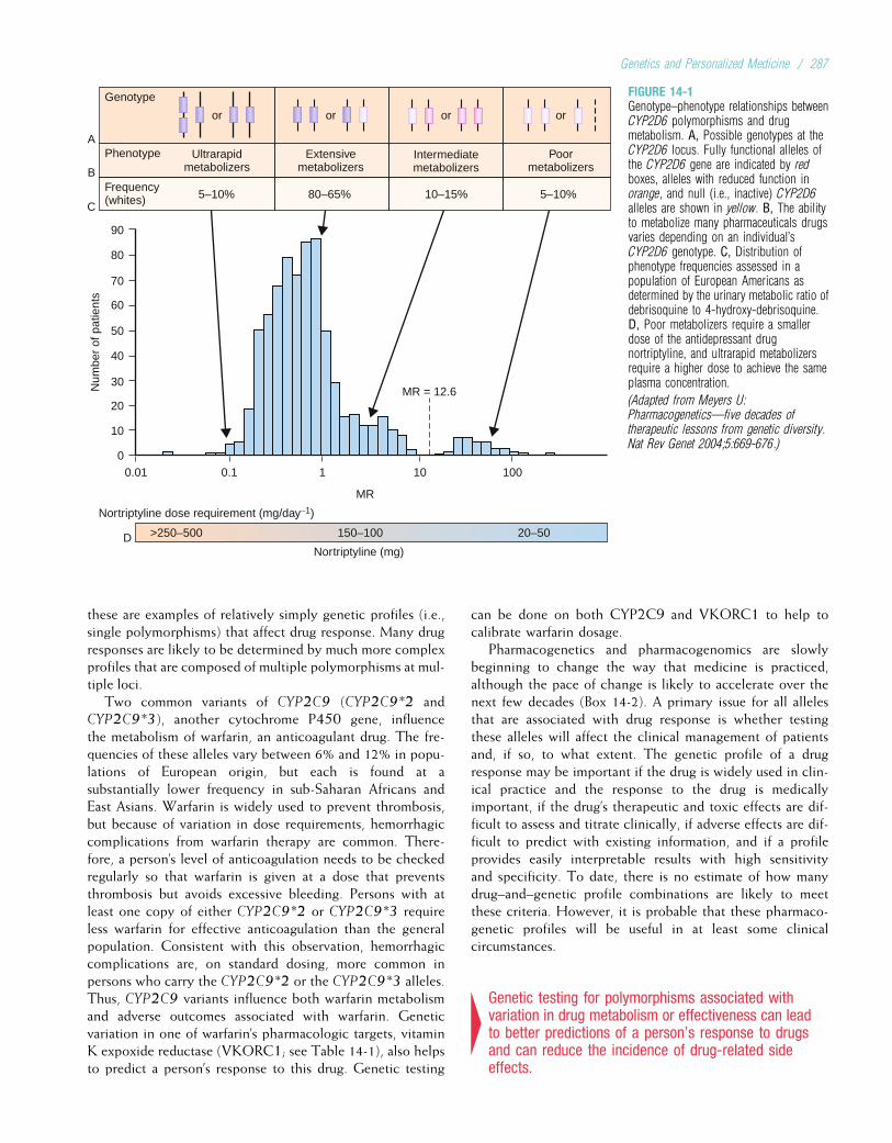
these are examples of relatively simply genetic profiles (i.e.,single polymorphisms) that affect drug response. Many drugresponses are likely to be determined by much more complexprofiles that are composed of multiple polymorphisms at mul-tiple loci.
Two common variants of CYP2C9 (CYP2C9*2 andCYP2C9*3), another cytochrome P450 gene, influencethe metabolism of warfarin, an anticoagulant drug. The fre-quencies of these alleles vary between 6% and 12% in popu-lations of European origin, but each is found at asubstantially lower frequency in sub-Saharan Africans andEast Asians. Warfarin is widely used to prevent thrombosis,but because of variation in dose requirements, hemorrhagiccomplications from warfarin therapy are common. There-fore, a person’s level of anticoagulation needs to be checkedregularly so that warfarin is given at a dose that preventsthrombosis but avoids excessive bleeding. Persons with atleast one copy of either CYP2C9*2 or CYP2C9*3 requireless warfarin for effective anticoagulation than the generalpopulation. Consistent with this observation, hemorrhagiccomplications are, on standard dosing, more common inpersons who carry the CYP2C9*2 or the CYP2C9*3 alleles.Thus, CYP2C9 variants influence both warfarin metabolismand adverse outcomes associated with warfarin. Geneticvariation in one of warfarin’s pharmacologic targets, vitaminK expoxide reductase (VKORC1; see Table 14-1), also helpsto predict a person’s response to this drug. Genetic testing
can be done on both CYP2C9 and VKORC1 to help tocalibrate warfarin dosage.
Pharmacogenetics and pharmacogenomics are slowlybeginning to change the way that medicine is practiced,although the pace of change is likely to accelerate over thenext few decades (Box 14-2). A primary issue for all allelesthat are associated with drug response is whether testingthese alleles will affect the clinical management of patientsand, if so, to what extent. The genetic profile of a drugresponse may be important if the drug is widely used in clin-ical practice and the response to the drug is medicallyimportant, if the drug’s therapeutic and toxic effects are dif-ficult to assess and titrate clinically, if adverse effects are dif-ficult to predict with existing information, and if a profileprovides easily interpretable results with high sensitivityand specificity. To date, there is no estimate of how manydrug–and–genetic profile combinations are likely to meetthese criteria. However, it is probable that these pharmaco-genetic profiles will be useful in at least some clinicalcircumstances.
4Genetic testing for polymorphisms associated withvariation in drug metabolism or effectiveness can leadto better predictions of a person’s response to drugsand can reduce the incidence of drug-related sideeffects.
Num
ber
of p
atie
nts
MR
Nortriptyline dose requirement (mg/day−1)
Frequency(whites)
Phenotype
Genotype
A
B
C
D
or
Ultrarapidmetabolizers
or
Extensivemetabolizers
or
Poormetabolizers
or
Intermediatemetabolizers
0.010
10
20
30
40
50
60
70
80
90
0.1 1 10
MR = 12.6
5–10% 80–65% 10–15% 5–10%
100
>250–500 150–100
Nortriptyline (mg)
20–50
FIGURE 14-1Genotype–phenotype relationships betweenCYP2D6 polymorphisms and drugmetabolism. A, Possible genotypes at theCYP2D6 locus. Fully functional alleles ofthe CYP2D6 gene are indicated by redboxes, alleles with reduced function inorange, and null (i.e., inactive) CYP2D6alleles are shown in yellow. B, The abilityto metabolize many pharmaceuticals drugsvaries depending on an individual’sCYP2D6 genotype. C, Distribution ofphenotype frequencies assessed in apopulation of European Americans asdetermined by the urinary metabolic ratio ofdebrisoquine to 4-hydroxy-debrisoquine.D, Poor metabolizers require a smallerdose of the antidepressant drugnortriptyline, and ultrarapid metabolizersrequire a higher dose to achieve the sameplasma concentration.
(Adapted from Meyers U:Pharmacogenetics—five decades oftherapeutic lessons from genetic diversity.Nat Rev Genet 2004;5:669-676.)
Genetics and Personalized Medicine / 287
Diagnosing and Monitoring Common DiseaseIn the previous sections, we have explained how genomicinformation can be used to personalize risk assessments forcommon diseases and drug responses. Genomic informationcan also be used to facilitate disease diagnosis and to moni-tor therapeutic responses. For example, a microarray (Chap-ter 3) can be used to estimate the expression level of eachgene (i.e., the amount of mRNA that is transcribed) in a spe-cific tissue. These gene-expression profiles can be used toidentify patterns of gene expression that are associated withspecific diseases (e.g., increased transcription of an oncogeneor reduced transcription of a tumor suppressor gene in tumortissue). Such information can help to distinguish differenttypes of cancers, different types of infections, or otherphenotypes associated with disease.
Cancer GenomicsEvery cancer cell harbors numerous alterations in DNAsequence and copy number that affect genes or regulatorysequences, often accompanied by reversible, epigenetic,modifications. These changes perturb the expression and/orfunction of hundreds to thousands of genes. Collectively,these changes result in the activation or inhibition of variouscellular pathways that control the characteristics of cancerssuch as growth or metastasis, and they determine, in part,prognosis and response to treatment. Cancer genomics isthe study of the DNA-associated changes that accompanycancer with the overall goal of better preventing, detecting,diagnosing, and treating common cancers.
A particularly powerful application of genomics to cancerhas been the use of genome-wide gene expression analyses
to provide a snapshot of gene activity within a tumor at agiven point in time. This has facilitated the development ofclassification schemes based on expression profiles for manytypes of cancer, including leukemia, lymphoma, and cancersof the breast, lung, colon, and brain. This information can beused, for example, in refining prognosis, directing the appli-cation of conventional and targeted biological therapies, andidentifying targets for new drug development (Fig. 14-2).
Currently, it is often difficult to predict the prognosis ofcancer patients based on traditional phenotypic informationsuch as the type of tumor (T), whether the cancer is foundin nearby lymph nodes (N), and evidence of metastasis(M). Staging using this TMN system is currently the stan-dard for most solid tumors, yet these stages are often notpredictive of prognosis or treatment response. Gene-expres-sion profiling can help to distinguish between cancers thatare easily confused (e.g., Burkitt lymphoma vs. diffuse largeB-cell lymphoma). It can also facilitate the identification ofsubsets of tumors of the same TMN stage that might havequite different outcomes. Several gene-expression profilesare currently available for assessing breast cancer prognosis,and gene-expression profiles that predict recurrence of sev-eral other types of cancer have been established. Prospectivetrials will determine the extent to which the use of expres-sion profiling is of clinical benefit, but it is anticipated thatits use will lead to a substantial improvement in cancermanagement.
The conventional approach to cancer therapy has been toprovide treatment based on the tissue or organ in which thecancer originated. However, persons with the same type ofcancer often have different genetic abnormalities in their
BOX 14-2Personal Genomics
It is 2025. Jonathan is a 1 hour-old baby sleeping comfortably inhis mother’s arms in the room in which he was just born. A nurseenters and swabs the inside of Jonathan’s mouth with a brush tocollect buccal epithelial cells. DNA is extracted from these cells,and a week later an electronic summary of Jonathan’s completegenome sequence is deposited in a national health informationdatabase (NHID). A subset of genotypes that represent aunique genetic profile are put into a national forensics database.Mutation data for conditions covered in the newborn screeningprogram, including PKU, galactosemia, cystic fibrosis, andsickle-cell disease, are forwarded to the state health department.Jonathan’s parents are notified that he is a carrier for sickle-celldisease.
Jonathan’s parents control access to genetic risk data bankedin the NHID for disorders that commonly manifest inchildhood, and they elect to provide Jonathan’s pediatric careproviders with these data. On Jonathan’s well-child visit at1 month of age, a genetic counselor explains that Jonathan has ahigher-than-average genetic risk for autism, peanut allergy,chronic otitis media, and adverse responses to penicillin. It isrecommended to his parents that he avoid both penicillin andproducts containing peanuts until he can undergo direct testing.Jonathan is also found to have a lower-than-average genetic riskfor asthma.
At 1 year of age, it is apparent that Jonathan’s speech and lan-guage development is delayed. His genetic profile confirms thathe has no risk variants known to be associated with hearing loss,suggesting that his delay might be an early indication of autism.An optimal therapy for autism is selected based on his geneticprofile. Jonathan responds well to this intervention and, in con-junction with speech training, his development by the age of5 years is appropriate.
Jonathan remains healthy throughout childhood, and, whenhe turns 18 years old, control over his banked genetic risk datais switched from his parents to him. At the same time, Jonathan’smedical care is transferred to a family physician. At his firstappointment, Jonathan’s physician explains his risk for heartdisease, hypertension, obesity, type 2 diabetes, and colon cancer.Jonathan is alerted to his high risk of developing both diabetesand obesity, and a program of exercise and diet that has beenshown to delay disease onset is recommended to him.
Ten years later, Jonathan informs his physician that he and hiswife are planning to start a family. His wife is also a carrier forsickle-cell disease and has several risk variants for asthma, so theyare referred for further counseling about prenatal genetic testingoptions.When Jonathan is 45, he develops hypertension, and, basedonhis profile of drug response variants, therapy is initiatedwith a spe-cific antihypertensive agent to which he is likely to respond.
288 / Chapter 14 MEDICAL GENETICS
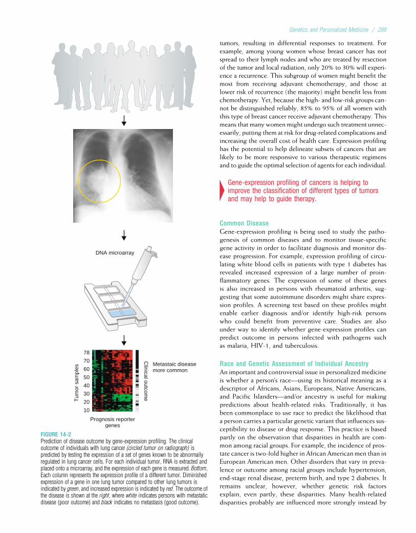
tumors, resulting in differential responses to treatment. Forexample, among young women whose breast cancer has notspread to their lymph nodes and who are treated by resectionof the tumor and local radiation, only 20% to 30% will experi-ence a recurrence. This subgroup of women might benefit themost from receiving adjuvant chemotherapy, and those atlower risk of recurrence (the majority) might benefit less fromchemotherapy. Yet, because the high- and low-risk groups can-not be distinguished reliably, 85% to 95% of all women withthis type of breast cancer receive adjuvant chemotherapy. Thismeans that many women might undergo such treatment unnec-essarily, putting them at risk for drug-related complications andincreasing the overall cost of health care. Expression profilinghas the potential to help delineate subsets of cancers that arelikely to be more responsive to various therapeutic regimensand to guide the optimal selection of agents for each individual.
4Gene-expression profiling of cancers is helping toimprove the classification of different types of tumorsand may help to guide therapy.
Common DiseaseGene-expression profiling is being used to study the patho-genesis of common diseases and to monitor tissue-specificgene activity in order to facilitate diagnosis and monitor dis-ease progression. For example, expression profiling of circu-lating white blood cells in patients with type 1 diabetes hasrevealed increased expression of a large number of proin-flammatory genes. The expression of some of these genesis also increased in persons with rheumatoid arthritis, sug-gesting that some autoimmune disorders might share expres-sion profiles. A screening test based on these profiles mightenable earlier diagnosis and/or identify high-risk personswho could benefit from preventive care. Studies are alsounder way to identify whether gene-expression profiles canpredict outcome in persons infected with pathogens suchas malaria, HIV-1, and tuberculosis.
Race and Genetic Assessment of Individual AncestryAn important and controversial issue in personalized medicineis whether a person’s race—using its historical meaning as adescriptor of Africans, Asians, Europeans, Native Americans,and Pacific Islanders—and/or ancestry is useful for makingpredictions about health-related risks. Traditionally, it hasbeen commonplace to use race to predict the likelihood thata person carries a particular genetic variant that influences sus-ceptibility to disease or drug response. This practice is basedpartly on the observation that disparities in health are com-mon among racial groups. For example, the incidence of pros-tate cancer is two-fold higher in African American men than inEuropean American men. Other disorders that vary in preva-lence or outcome among racial groups include hypertension,end-stage renal disease, preterm birth, and type 2 diabetes. Itremains unclear, however, whether genetic risk factorsexplain, even partly, these disparities. Many health-relateddisparities probably are influenced more strongly instead by
Metastaic diseasemore common
Prognosis reportergenes
Tum
or s
ampl
es
78
70
60
50
40
30
20
10
Clinical outcom
e
DNA microarray
FIGURE 14-2Prediction of disease outcome by gene-expression profiling. The clinicaloutcome of individuals with lung cancer (circled tumor on radiograph) ispredicted by testing the expression of a set of genes known to be abnormallyregulated in lung cancer cells. For each individual tumor, RNA is extracted andplaced onto a microarray, and the expression of each gene is measured. Bottom,Each column represents the expression profile of a different tumor. Diminishedexpression of a gene in one lung tumor compared to other lung tumors isindicated by green, and increased expression is indicated by red. The outcome ofthe disease is shown at the right, where white indicates persons with metastaticdisease (poor outcome) and black indicates no metastasis (good outcome).
Genetics and Personalized Medicine / 289
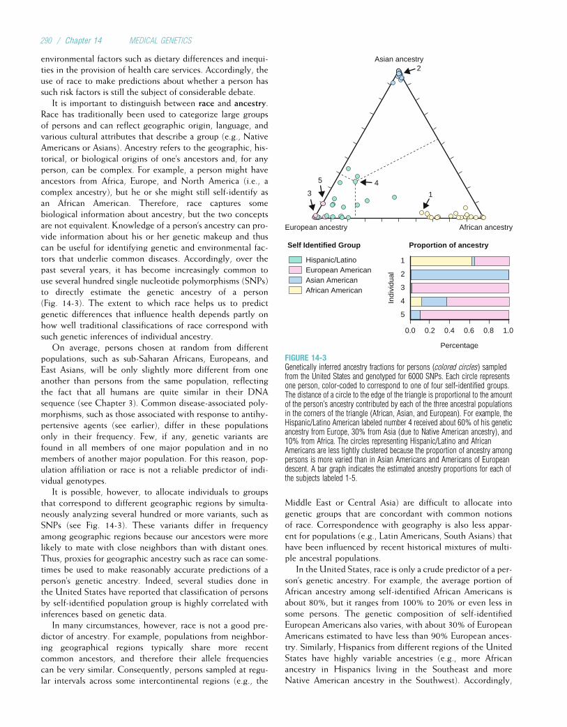
environmental factors such as dietary differences and inequi-ties in the provision of health care services. Accordingly, theuse of race to make predictions about whether a person hassuch risk factors is still the subject of considerable debate.
It is important to distinguish between race and ancestry.Race has traditionally been used to categorize large groupsof persons and can reflect geographic origin, language, andvarious cultural attributes that describe a group (e.g., NativeAmericans or Asians). Ancestry refers to the geographic, his-torical, or biological origins of one’s ancestors and, for anyperson, can be complex. For example, a person might haveancestors from Africa, Europe, and North America (i.e., acomplex ancestry), but he or she might still self-identify asan African American. Therefore, race captures somebiological information about ancestry, but the two conceptsare not equivalent. Knowledge of a person’s ancestry can pro-vide information about his or her genetic makeup and thuscan be useful for identifying genetic and environmental fac-tors that underlie common diseases. Accordingly, over thepast several years, it has become increasingly common touse several hundred single nucleotide polymorphisms (SNPs)to directly estimate the genetic ancestry of a person(Fig. 14-3). The extent to which race helps us to predictgenetic differences that influence health depends partly onhow well traditional classifications of race correspond withsuch genetic inferences of individual ancestry.
On average, persons chosen at random from differentpopulations, such as sub-Saharan Africans, Europeans, andEast Asians, will be only slightly more different from oneanother than persons from the same population, reflectingthe fact that all humans are quite similar in their DNAsequence (see Chapter 3). Common disease-associated poly-morphisms, such as those associated with response to antihy-pertensive agents (see earlier), differ in these populationsonly in their frequency. Few, if any, genetic variants arefound in all members of one major population and in nomembers of another major population. For this reason, pop-ulation affiliation or race is not a reliable predictor of indi-vidual genotypes.
It is possible, however, to allocate individuals to groupsthat correspond to different geographic regions by simulta-neously analyzing several hundred or more variants, such asSNPs (see Fig. 14-3). These variants differ in frequencyamong geographic regions because our ancestors were morelikely to mate with close neighbors than with distant ones.Thus, proxies for geographic ancestry such as race can some-times be used to make reasonably accurate predictions of aperson’s genetic ancestry. Indeed, several studies done inthe United States have reported that classification of personsby self-identified population group is highly correlated withinferences based on genetic data.
In many circumstances, however, race is not a good pre-dictor of ancestry. For example, populations from neighbor-ing geographical regions typically share more recentcommon ancestors, and therefore their allele frequenciescan be very similar. Consequently, persons sampled at regu-lar intervals across some intercontinental regions (e.g., the
Middle East or Central Asia) are difficult to allocate intogenetic groups that are concordant with common notionsof race. Correspondence with geography is also less appar-ent for populations (e.g., Latin Americans, South Asians) thathave been influenced by recent historical mixtures of multi-ple ancestral populations.
In the United States, race is only a crude predictor of a per-son’s genetic ancestry. For example, the average portion ofAfrican ancestry among self-identified African Americans isabout 80%, but it ranges from 100% to 20% or even less insome persons. The genetic composition of self-identifiedEuropean Americans also varies, with about 30% of EuropeanAmericans estimated to have less than 90% European ances-try. Similarly, Hispanics from different regions of the UnitedStates have highly variable ancestries (e.g., more Africanancestry in Hispanics living in the Southeast and moreNative American ancestry in the Southwest). Accordingly,
2
43
Asian ancestry
European ancestry African ancestry
5
0.0 0.2 0.4 0.6 0.8 1.0
Hispanic/LatinoEuropean AmericanAsian AmericanAfrican American
5
4
3
2
1
Self Identified Group Proportion of ancestry
Percentage
Indi
vidu
al
1
FIGURE 14-3Genetically inferred ancestry fractions for persons (colored circles) sampledfrom the United States and genotyped for 6000 SNPs. Each circle representsone person, color-coded to correspond to one of four self-identified groups.The distance of a circle to the edge of the triangle is proportional to the amountof the person’s ancestry contributed by each of the three ancestral populationsin the corners of the triangle (African, Asian, and European). For example, theHispanic/Latino American labeled number 4 received about 60% of his geneticancestry from Europe, 30% from Asia (due to Native American ancestry), and10% from Africa. The circles representing Hispanic/Latino and AfricanAmericans are less tightly clustered because the proportion of ancestry amongpersons is more varied than in Asian Americans and Americans of Europeandescent. A bar graph indicates the estimated ancestry proportions for each ofthe subjects labeled 1-5.
290 / Chapter 14 MEDICAL GENETICS
membership in a group does not mean that all members of thegroup necessarily have similar genetic ancestries.
Although it is clear that explicit genetic information,rather than race, can be used to make more accurate infer-ences of ancestry, it is not yet known whether personalancestry information can make useful predictions about one’srisk of common disease. The consequences of using detailedancestry information in a clinical setting are also largelyunknown. It is possible that personal ancestry informationcould have adverse effects on a person’s perception of riskand cultural identity. Similarly, such information could rein-force unfair stereotypes about specific populations. Furtherresearch is needed to examine the potential benefits and risksof using ancestry information in clinical practice.
4The relationship between ancestry and traditionalconcepts of race is complex. Genetic information,rather than race, is a better predictor of ancestry.
THE FUTURE OF PERSONALIZED MEDICINEGenetic variants that increase the risk of common disease arenow being found with increasing speed and efficiency (seeChapter 12), but only a small fraction of the genetic basis ofdisease risk has thus far been defined. In addition, the inter-actions of multiple disease-predisposing gene products, andtheir interactions with nongenetic factors, remain almostcompletely unknown. Thus, the promise of personalized med-icine, in which a detailed genetic profile can provide clinicallyuseful risk information for common diseases such as diabetes,cancer, or heart disease, remains largely unfulfilled. It is hopedthat with increasing knowledge of alleles that predispose per-sons to disease, genetic testing will begin to contribute moresubstantially to the diagnosis and treatment of common dis-ease. It must also be borne in mind that nongenetic factors,such as diet and exercise, are also part of each person’s risk pro-file. These factors can and should be assessed and modified tomaximize each person’s potential for a healthy life.
Study Questions
1. Explain how genetic information can be used toenhance the practice of preventive medicine comparedto a conventional model of medical service. Give atleast one example.
2. Different individuals with the same type of canceroften respond differently to therapy. Provide at leasttwo possible explanations of this observation.
3. Define race and ancestry; explain the differencesbetween them.
4. Consider how explicit genetic information about yourancestry might change your perception of yourbiological and cultural identities.
5. Give an example of a polymorphism that affects drugmetabolism and/or the response to a drug.
6. Explain some of the possible obstacles to the use ofgenetic information in practicing personalizedmedicine.
7. Distinguish between genetic medicine and genomicmedicine.
8. Give examples of how the availability of whole-genome data from individuals might change the waysmedicine is currently practiced.
Suggested ReadingsBamshad MJ. Genetic influences on health: Does race mat-
ter? JAMA 2006;294:937–46.Belle DJ, Singh H. Genetic factors in drug metabolism. Am
Fam Physician 2008;77:1553–60.Chin L, Gray JW. Translating insights from the
cancer genome to clinical practice. Nature 2008;452:553–63.
Feero GW, Guttmacher AE, Collins FS. The genome getspersonal—almost. JAMA 2008;299:1351–2.
Hunter DJ, Khoury MJ, Drazen JM. Letting the genome outof the bottle—will we get our wish? N Engl J Med2008;358:105–7.
Khoury MJ, Gwinn M, Yoon PW, et al. The continuum oftranslation research in genomic medicine: how can weaccelerate the appropriate integration of human genomediscoveries into health care and disease prevention? GenetMed 2007;9:665–74.
Olson MV. Dr. Watson’s base pairs. Nature 2008; 452:819–20.
Rothstein MA. Keeping your genes private. Sci Amer2008;299(3):64–9.
Swen JJ, Huizinga TW, Gelderblom H, et al. Translatingpharmacogenomics: challenges on the road to the clinic.PLOSMedicine 2008;4:1317–24.
Wheeler DA, Srinivasan M, Egholm M, et al. The completegenome of an individual by massively parallel DNAsequencing. Nature 2008;452:872–6.
Wilke RA, Lin DW, Roden DM, et al. Identifying genetic riskfactors for serious adverse drug reactions: current progressand challenges. Nat Rev Drug Discovery 2007;6:904–16.
Internet ResourcesNational Institutes of Health–sponsoredpharmacogeneticsresearch network http://www.nigms.nih.gov/Initiatives/PGRN
National Cancer Institute–sponsored tutorial on cancer genomicshttp://www.cancer.gov/cancertopics/understandingcancer/cancergenomics
Genetics and Personalized Medicine / 291
Chapter 15
CLINICAL GENETICS AND GENETIC COUNSELING
Medical genetics has recently emerged as a true specialty inmainstream medicine. In the 1960s, the fields of biochemicalgenetics, clinical cytogenetics, and dysmorphology (thestudy of abnormal physical development) developed. The1970s witnessed the establishment of the techniques neces-sary for prenatal diagnosis of genetic disorders. By the endof the 1970s, discussions about forming the American Boardof Medical Genetics had occurred, and in 1981 the firstcertification examination was administered. The AmericanBoard of Genetic Counseling was established in the early1990s, and now various types of geneticists, includinggenetic counselors, medical geneticists, and basic humangeneticists, can be certified. In 1991 the American Board ofMedical Specialties recognized this new field, and medicalgenetics has now become an integral part of medicine.
Whereas medical genetics involves the study of thegenetics of human disease, clinical genetics deals withthe direct clinical care of persons with genetic diseases. Thediagnostic, counseling, and management issues surroundinggenetic disease are the principal foci of clinical genetics.
In this chapter, we summarize the principles of clinicalgenetics and the process of genetic counseling. In addition,we provide an overview of the field of dysmorphology,because the growth of this area has influenced and paralleledthe emergence of clinical genetics.
THE PRINCIPLES AND PRACTICE OF CLINICALGENETICSAs mentioned in Chapter 1, genetic conditions as a groupare common and are a significant cause of human mortalityand morbidity. Typically, genetic disorders are complex,multiorgan, systemic conditions, and the care of personswith these disorders can also involve multiple medical spe-cialties. Thus, genetic disorders are among the differentialdiagnosis of most symptoms and clinical presentations. Forexample, when evaluating an infant with a blistering skin dis-ease, the ability to distinguish between one of the manyforms of epidermolysis bullosa (an inherited disorder of ker-atinocytes in which skin blisters develop after mild trauma)and staphylococcal skin disease must be part of the clini-cian’s repertoire.
Because of the complexity and number of human geneticdiseases, their clinical diagnosis and treatment can seem
overwhelming. To help manage this information, we providean overview of the most important concepts, including theimportance of accurate diagnosis, the application of thetenets of medical genetics to medical practice, and the roleof genetic counseling in the care of persons with geneticdisease.
Accurate DiagnosisThe significance of the basic medical principle of accuratediagnosis cannot be overemphasized. The process of geneticcounseling, one of the principal services of medical genetics,begins with correct diagnosis. All discussions of natural his-tory, prognosis, management, risk determination, optionsfor prenatal diagnosis, and referral to genetic advocacygroups (also termed genetic support groups) depend on an accu-rate diagnosis of the patient’s condition. For example,genetic counseling for a family who has a son with mentalretardation usually involves questions of risk for this condi-tion in future offspring. An accurate answer requires the cli-nician to identify a condition of known etiology. If a specificdiagnosis (e.g., fragile X syndrome) is made, then the rest ofthe genetic counseling process starts: Current informationcan be shared and management can be initiated (ClinicalCommentary 15-1).
4In clinical genetics, as in all of medicine, accuratediagnosis is the most important first step in patientcare.
The process of diagnosing a genetic disorder is a complexsequence of events. It depends upon diagnostic decisionmaking, recognition of important phenotypic signs, applica-tion of principles of dysmorphology and medical genetics,and laboratory diagnosis. For diseases in which the diagnos-tic criteria are well established, the practitioner has guide-lines for making a diagnosis. An example of such criteria isthe criteria recommended by the National Institutes ofHealth Consensus Development Conference for the diagno-sis of neurofibromatosis type 1 (NF1; see Chapter 4). Forconditions that are defined by a specific laboratory marker,such as an abnormal karyotype or biochemical assay, thediagnostic procedure is generally straightforward. For manygenetic diseases, however, there are no well-established
292
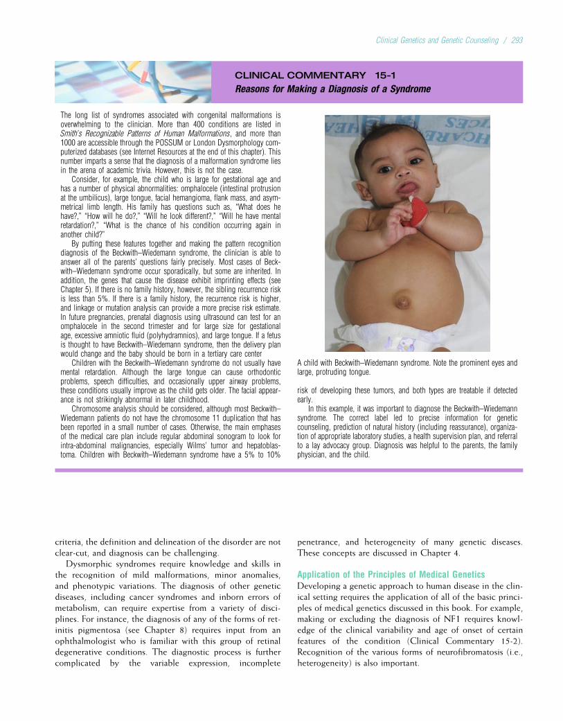
CLINICAL COMMENTARY 15-1
Reasons for Making a Diagnosis of a Syndrome
The long list of syndromes associated with congenital malformations isoverwhelming to the clinician. More than 400 conditions are listed inSmith’s Recognizable Patterns of Human Malformations, and more than1000 are accessible through the POSSUM or London Dysmorphology com-puterized databases (see Internet Resources at the end of this chapter). Thisnumber imparts a sense that the diagnosis of a malformation syndrome liesin the arena of academic trivia. However, this is not the case.
Consider, for example, the child who is large for gestational age andhas a number of physical abnormalities: omphalocele (intestinal protrusionat the umbilicus), large tongue, facial hemangioma, flank mass, and asym-metrical limb length. His family has questions such as, “What does hehave?,” “How will he do?,” “Will he look different?,” “Will he have mentalretardation?,” “What is the chance of his condition occurring again inanother child?”
By putting these features together and making the pattern recognitiondiagnosis of the Beckwith–Wiedemann syndrome, the clinician is able toanswer all of the parents’ questions fairly precisely. Most cases of Beck-with–Wiedemann syndrome occur sporadically, but some are inherited. Inaddition, the genes that cause the disease exhibit imprinting effects (seeChapter 5). If there is no family history, however, the sibling recurrence riskis less than 5%. If there is a family history, the recurrence risk is higher,and linkage or mutation analysis can provide a more precise risk estimate.In future pregnancies, prenatal diagnosis using ultrasound can test for anomphalocele in the second trimester and for large size for gestationalage, excessive amniotic fluid (polyhydramnios), and large tongue. If a fetusis thought to have Beckwith–Wiedemann syndrome, then the delivery planwould change and the baby should be born in a tertiary care center
Children with the Beckwith–Wiedemann syndrome do not usually havemental retardation. Although the large tongue can cause orthodonticproblems, speech difficulties, and occasionally upper airway problems,these conditions usually improve as the child gets older. The facial appear-ance is not strikingly abnormal in later childhood.
Chromosome analysis should be considered, although most Beckwith–Wiedemann patients do not have the chromosome 11 duplication that hasbeen reported in a small number of cases. Otherwise, the main emphasesof the medical care plan include regular abdominal sonogram to look forintra-abdominal malignancies, especially Wilms’ tumor and hepatoblas-toma. Children with Beckwith–Wiedemann syndrome have a 5% to 10%
A child with Beckwith–Wiedemann syndrome. Note the prominent eyes andlarge, protruding tongue.
risk of developing these tumors, and both types are treatable if detectedearly.
In this example, it was important to diagnose the Beckwith–Wiedemannsyndrome. The correct label led to precise information for geneticcounseling, prediction of natural history (including reassurance), organiza-tion of appropriate laboratory studies, a health supervision plan, and referralto a lay advocacy group. Diagnosis was helpful to the parents, the familyphysician, and the child.
criteria, the definition and delineation of the disorder are notclear-cut, and diagnosis can be challenging.
Dysmorphic syndromes require knowledge and skills inthe recognition of mild malformations, minor anomalies,and phenotypic variations. The diagnosis of other geneticdiseases, including cancer syndromes and inborn errors ofmetabolism, can require expertise from a variety of disci-plines. For instance, the diagnosis of any of the forms of ret-initis pigmentosa (see Chapter 8) requires input from anophthalmologist who is familiar with this group of retinaldegenerative conditions. The diagnostic process is furthercomplicated by the variable expression, incomplete
penetrance, and heterogeneity of many genetic diseases.These concepts are discussed in Chapter 4.
Application of the Principles of Medical GeneticsDeveloping a genetic approach to human disease in the clin-ical setting requires the application of all of the basic princi-ples of medical genetics discussed in this book. For example,making or excluding the diagnosis of NF1 requires knowl-edge of the clinical variability and age of onset of certainfeatures of the condition (Clinical Commentary 15-2).Recognition of the various forms of neurofibromatosis (i.e.,heterogeneity) is also important.
Clinical Genetics and Genetic Counseling / 293
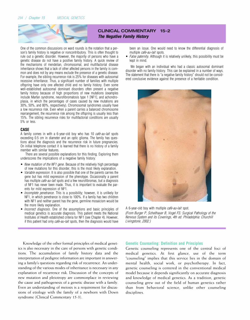
CLINICAL COMMENTARY 15-2
The Negative Family History
One of the common discussions on ward rounds is the notation that a per-son’s family history is negative or noncontributory. This is often thought torule out a genetic disorder. However, the majority of persons who have agenetic disease do not have a positive family history. A quick review ofthe mechanisms of mendelian, chromosomal, and multifactorial diseaseinheritance shows that a lack of other affected persons in the family is com-mon and does not by any means exclude the presence of a genetic disease.For example, the sibling recurrence risk is 25% for diseases with autosomalrecessive inheritance. Thus, a significant number of families with multipleoffspring have only one affected child and no family history. Even somewell-established autosomal dominant disorders often present a negativefamily history because of high proportions of new mutations (examplesinclude Marfan syndrome, neurofibromatosis type 1 [NF1], and achondro-plasia, in which the percentages of cases caused by new mutations are30%, 50%, and 80%, respectively). Chromosomal syndromes usually havea low recurrence risk. Even when a parent carries a balanced chromosomerearrangement, the recurrence risk among the offspring is usually less than15%. The sibling recurrence risks for multifactorial conditions are usually5% or less.
CASEA family comes in with a 6-year-old boy who has 10 cafe-au-lait spotsexceeding 0.5 cm in diameter and an optic glioma. The family has ques-tions about the diagnosis and the recurrence risk in future pregnancies.On initial telephone contact it is learned that there is no history of a familymember with similar features.
There are several possible explanations for this finding. Exploring themunderscores the implications of a negative family history:
• New mutation of the NF1 gene. Because of the relatively high percentageof new mutations for this disorder, this is the most likely explanation.
• Variable expression. It is also possible that one of the parents carries thegene but has mild expression of the phenotype. Occasionally a parenthas multiple cafe-au-lait spots and a few neurofibromas, but a diagnosisof NF1 has never been made. Thus, it is important to evaluate the par-ents for mild expression of NF1.
• Incomplete penetrance. This is a possibility; however, it is unlikely forNF1, in which penetrance is close to 100%. If a family has two childrenwith NF1 and neither parent has the gene, germline mosaicism would bethe more likely explanation.
• Incorrect diagnosis. One of the assumptions and basic principles ofmedical genetics is accurate diagnosis. This patient meets the NationalInstitutes of Health established criteria for NF1 (see Chapter 4). However,if this patient had only cafe-au-lait spots, then the diagnosis would have
been an issue. One would need to know the differential diagnosis ofmultiple cafe-au-lait spots.
• False paternity. Although it is relatively unlikely, this possibility must bekept in mind.
We began with an individual who had a classic autosomal dominantdisorder with no family history. This can be explained in a number of ways.The statement that there is “a negative family history” should not be consid-ered conclusive evidence against the presence of a heritable condition.
A 6-year-old boy with multiple cafe-au-lait spot.
(From Burger P, Scheithauer B, Vogel FS: Surgical Pathology of theNervous System and its Coverings, 4th ed. Philadelphia: ChurchillLivingstone, 2002.)
Knowledge of the other formal principles of medical genet-ics is also necessary in the care of persons with genetic condi-tions. The accumulation of family history data and theinterpretation of pedigree information are important in answer-ing a family’s questions regarding risk of recurrence. An under-standing of the various modes of inheritance is necessary in anyexplanation of recurrence risk. Discussion of the concepts ofnew mutation and pleiotropy are commonplace in reviewingthe cause and pathogenesis of a genetic disease with a family.Even an understanding of meiosis is a requirement for discus-sions of etiology with the family of a newborn with Downsyndrome (Clinical Commentary 15-3).
Genetic Counseling: Definition and PrinciplesGenetic counseling represents one of the central foci ofmedical genetics. At first glance, use of the term“counseling” implies that this service lies in the domain ofmental health, social work, or psychotherapy. In fact,genetic counseling is centered in the conventional medicalmodel because it depends significantly on accurate diagnosisand knowledge of medical genetics. As a tradition, geneticcounseling grew out of the field of human genetics ratherthan from behavioral science, unlike other counselingdisciplines.
294 / Chapter 15 MEDICAL GENETICS
CLINICAL COMMENTARY 15-3
Talking to the Parents of a Newborn with Down Syndrome
The birth of a newborn with Down syndrome presents many challenges.Typically, the infant is not acutely ill and the parents are not aware of thediagnosis before the birth. Thus, the practitioner must approach the parents,often strangers, with unexpected and sometimes disappointing news. Thefamily can experience a series of emotions that are somewhat similar tothe reactions after a loss: anger, denial, sadness, and then usually reorga-nization and adaptation. Families face these situations with markedly differ-ent backgrounds: varying attitudes toward crisis, varied demographic andsocioeconomic circumstances, and even a wide range of differences inthe cultural meaning of a disability or defect. All of these variables, plusthe fact that physicians are often not trained in being the bearers of difficultnews, can make this a challenging situation. Parents remember in detail theway the news is presented. The practitioner has both the opportunity andthe challenge to help the family through these events.
A number of practical suggestions have come from studies investigatingthe recommendations of parents who have experienced this event:
• Prepare yourself. Set up the interview scenario, and think about how youwill begin the discussion.
• Talk to both parents together whenever possible. This is sometimes notpractical, but when it can be accomplished, it is critical.
• Communicate the diagnosis as soon as possible. All studies of parentalinterviews show that they prefer early communication of the diagnosis.
• Choose a place that is private and quiet where both the parents and theprofessionals can sit down. Avoid standing up with the parents seated.Always be sure to introduce yourself. Structure the interview from thebeginning.
• Humanize the situation as much as possible. Learn the baby’s first nameif it has been decided on, and always know the baby’s gender. Refer to
the infant by name or as a son or daughter, and be aware of the use ofall language. Phrases such as “mental retardation” have great impact.Terms such as “mongolism” are not appropriate because they are stig-matizing, pejorative, and incorrect.
• Develop a sense of realistic positivism. It is important to discuss thedevelopmental limitations in a patient with Down syndrome, but it is alsoimportant to have an optimistic and positive attitude. This suggestioncomes from the advocacy and parents’ organizations that have developedin the last three decades.
• Answer the parents’ questions, but avoid technical overload. It is impor-tant to be accurate and current on the biological and medical aspects ofthe condition under discussion. When an answer is not known, mentionthat the question can be reviewed or referred to a consultant.
• Listen actively. Assume that almost all feelings are natural and thatparents will be wrestling with their own guilt and shame. Validate allfeelings that arise. Most parents can meet this challenge quite effectivelyand do not require psychiatric consultation.
• Refer the family to the appropriate resources early. This would includeparents’ advocacy groups or even individual parents who have a childwith Down syndrome. Share available written material or web pages,but make certain that it is accurate and current.
Above all, be aware of the unique plight of families in such a situation, andmake an effort to spend time with them. Although it is difficult to present inwritten form how one can develop attributes such as kindness and empathy,it is important for physicians in training to learn from their mentors and usetheir own individual communication style as a strength. Clearly, the recommen-dations provided here apply not only to genetic counseling but also to anysituation in which difficult information is presented to patients or families.
In 1975 the American Society of Human Geneticsadopted a definition of genetic counseling. Newer languagehas been proposed recently to modernize and simplify thisdefinition, but the original language stands the test of time:
“Genetic counseling is a communication process that deals with
the human problems associated with the occurrence or risk of
occurrence of a genetic disorder in a family. This process involves
an attempt by one or more appropriately trained persons to help
the individual or family to (1) comprehend the medical facts,
including the diagnosis, probable course of the disorder, and
the available management; (2) appreciate the way heredity con-
tributes to the disorder and the risk of recurrence in specified rela-
tives; (3) understand the alternatives for dealing with the risk of
recurrence; (4) choose a course of action that seems to them
appropriate in their view of their risk, their family goals, and their
ethical and religious standards, and act in accordance with that
decision; and (5) make the best possible adjustment to the disor-
der in an affected family member and/or to the risk of recurrence
of that disorder.”
This definition illustrates the complex tasks that face thepractitioner. The first task involves establishing the diagnosisand discussing the natural history and management of thecondition. In this regard, the medical care of a patient witha genetic disease does not differ from that of a patient withany other type of disease.
The second task requires an understanding of the basictenets of medical genetics, especially the principles ofhuman genetics and risk determination. For chromosomaland multifactorial disorders, empirical figures are used toestimate recurrence risk. Inheritance patterns are used to pre-dict the recurrence risk of mendelian disorders. However,the clinical issues are often complicated by incomplete pen-etrance, variable expression, delayed age of onset, and allelicand locus heterogeneity. In some cases, incorporation ofadditional information using the Bayesian probabilityapproach can significantly alter estimates (Box 15-1).
The third and fourth objectives of the genetic counselingprocess underlie the primary differences between the geneticmodel and the traditional biomedical approach. These tasksinvolve discussing reproductive options and facilitating deci-sion making. Implicit in the fourth part of the definition isthe notion of respect for the family’s autonomy and theirperceptions of risk and of the disorder itself. This approachhas been called nondirectiveness: the counselor leaves alldecisions about future reproduction up to the family. Thisdiffers somewhat from the more traditional medicalapproach, in which recommendations for treatment or interven-tion are often made in a more directed fashion. This is animportant issue, because nondirectiveness sometimes con-flicts with the broader view of preventive medicine, which
Clinical Genetics and Genetic Counseling / 295
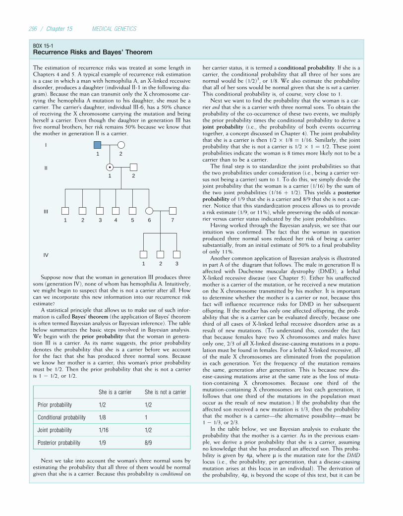
BOX 15-1Recurrence Risks and Bayes’ Theorem
The estimation of recurrence risks was treated at some length inChapters 4 and 5. A typical example of recurrence risk estimationis a case in which a man with hemophilia A, an X-linked recessivedisorder, produces a daughter (individual II-1 in the following dia-gram). Because the man can transmit only the X chromosome car-rying the hemophilia A mutation to his daughter, she must be acarrier. The carrier’s daughter, individual III-6, has a 50% chanceof receiving the X chromosome carrying the mutation and beingherself a carrier. Even though the daughter in generation III hasfive normal brothers, her risk remains 50% because we know thatthe mother in generation II is a carrier.
1
I
II
III
IV
2
1 2 3
1 2 3
4 75 6
1 2
Suppose now that the woman in generation III produces threesons (generation IV), none of whom has hemophilia A. Intuitively,we might begin to suspect that she is not a carrier after all. Howcan we incorporate this new information into our recurrence riskestimate?
A statistical principle that allows us to make use of such infor-mation is called Bayes’ theorem (the application of Bayes’ theoremis often termed Bayesian analysis or Bayesian inference). The tablebelow summarizes the basic steps involved in Bayesian analysis.We begin with the prior probability that the woman in genera-tion III is a carrier. As its name suggests, the prior probabilitydenotes the probability that she is a carrier before we accountfor the fact that she has produced three normal sons. Becausewe know her mother is a carrier, this woman’s prior probabilitymust be 1/2. Then the prior probability that she is not a carrieris 1 � 1/2, or 1/2.
She is a carrier She is not a carrier
Prior probability 1/2 1/2
Conditional probability 1/8 1
Joint probability 1/16 1/2
Posterior probability 1/9 8/9
Next we take into account the woman’s three normal sons byestimating the probability that all three of them would be normalgiven that she is a carrier. Because this probability is conditioned on
her carrier status, it is termed a conditional probability. If she is acarrier, the conditional probability that all three of her sons arenormal would be (1/2)3, or 1/8. We also estimate the probabilitythat all of her sons would be normal given that she is not a carrier.This conditional probability is, of course, very close to 1.
Next we want to find the probability that the woman is a car-rier and that she is a carrier with three normal sons. To obtain theprobability of the co-occurrence of these two events, we multiplythe prior probability times the conditional probability to derive ajoint probability (i.e., the probability of both events occurringtogether, a concept discussed in Chapter 4). The joint probabilitythat she is a carrier is then 1/2 � 1/8 ¼ 1/16. Similarly, the jointprobability that she is not a carrier is 1/2 � 1 ¼ 1/2. These jointprobabilities indicate the woman is 8 times more likely not to be acarrier than to be a carrier.
The final step is to standardize the joint probabilities so thatthe two probabilities under consideration (i.e., being a carrier ver-sus not being a carrier) sum to 1. To do this, we simply divide thejoint probability that the woman is a carrier (1/16) by the sum ofthe two joint probabilities (1/16 þ 1/2). This yields a posteriorprobability of 1/9 that she is a carrier and 8/9 that she is not a car-rier. Notice that this standardization process allows us to providea risk estimate (1/9, or 11%), while preserving the odds of noncar-rier versus carrier status indicated by the joint probabilities.
Having worked through the Bayesian analysis, we see that ourintuition was confirmed: The fact that the woman in questionproduced three normal sons reduced her risk of being a carriersubstantially, from an initial estimate of 50% to a final probabilityof only 11%.
Another common application of Bayesian analysis is illustratedin part A of the diagram that follows. The male in generation II isaffected with Duchenne muscular dystrophy (DMD), a lethalX-linked recessive disease (see Chapter 5). Either his unaffectedmother is a carrier of the mutation, or he received a new mutationon the X chromosome transmitted by his mother. It is importantto determine whether the mother is a carrier or not, because thisfact will influence recurrence risks for DMD in her subsequentoffspring. If the mother has only one affected offspring, the prob-ability that she is a carrier can be evaluated directly, because onethird of all cases of X-linked lethal recessive disorders arise as aresult of new mutations. (To understand this, consider the factthat because females have two X chromosomes and males haveonly one, 2/3 of all X-linked disease-causing mutations in a popu-lation must be found in females. For a lethal X-linked recessive, allof the male X chromosomes are eliminated from the populationin each generation. Yet the frequency of the mutation remainsthe same, generation after generation. This is because new dis-ease-causing mutations arise at the same rate as the loss of muta-tion-containing X chromosomes. Because one third of themutation-containing X chromosomes are lost each generation, itfollows that one third of the mutations in the population mustoccur as the result of new mutation.) If the probability that theaffected son received a new mutation is 1/3, then the probabilitythat the mother is a carrier—the alternative possibility—must be1 � 1/3, or 2/3.
In the table below, we use Bayesian analysis to evaluate theprobability that the mother is a carrier. As in the previous exam-ple, we derive a prior probability that she is a carrier, assumingno knowledge that she has produced an affected son. This proba-bility is given by 4m, where m is the mutation rate for the DMDlocus (i.e., the probability, per generation, that a disease-causingmutation arises at this locus in an individual). The derivation ofthe probability, 4m, is beyond the scope of this text, but it can be
296 / Chapter 15 MEDICAL GENETICS
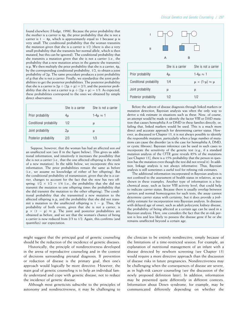
might suggest that the principal goal of genetic counselingshould be the reduction of the incidence of genetic diseases.
Historically, the principle of nondirectiveness developedin the arena of reproductive counseling and in the contextof decisions surrounding prenatal diagnosis. If preventionor reduction of disease is the primary goal, then one’sapproach would logically be more directive. However, themain goal of genetic counseling is to help an individual fam-ily understand and cope with genetic disease, not to reducethe incidence of genetic disease.
Although most geneticists subscribe to the principles ofautonomy and nondirectiveness, it may be challenging to
the clinician to be entirely nondirective, simply because ofthe limitations of a time-restricted session. For example, anexplanation of nutritional management of an infant with adisease detected by newborn screening (see Chapter 13)would require a more directive approach than the discussionof disease risks in future pregnancies. Nondirectiveness maybe challenging when the consequences of disease are severe,as in high-risk cancer counseling (see the discussion of thenewly proposed definition later). In addition, informationmay be presented quite differently in different contexts.Information about Down syndrome, for example, may becommunicated differently depending on whether the
found elsewhere (Hodge, 1998). Because the prior probability thatthe mother is a carrier is 4m, the prior probability that she is not acarrier is 1 � 4m, which is approximately equal to 1 because m isvery small. The conditional probability that the woman transmitsthe mutation given that she is a carrier is 1/2 (there is also a verysmall probability that she transmits her normal allele, which is thenmutated, but this can be ignored). The conditional probability thatshe transmits a mutation given that she is not a carrier (i.e., theprobability that a new mutation arises in the gamete she transmits)is m. We then multiply the prior probability that she is a carrier, 4m,by the corresponding conditional probability, 1/2, to obtain a jointprobability of 2m. The same procedure produces a joint probabilityof m that she is not a carrier. Finally, we standardize the joint prob-abilities to get the posterior probabilities. The posterior probabilitythat she is a carrier is 2m � (2m þ m) ¼ 2/3, and the posterior prob-ability that she is not a carrier is m � (2m þ m) ¼ 1/3. As expected,these probabilities correspond to the ones we obtained by simpledirect observation.
She is a carrier She is not a carrier
Prior probability 4m 1-4m � 1
Conditional probability 1/2 m
Joint probability 2m m
Posterior probability 2/3 1/3
Suppose, however, that the woman has had an affected son andan unaffected son (see B in the figure below). This gives us addi-tional information, and intuitively it increases the possibility thatshe is not a carrier (i.e., that the one affected offspring is the resultof a new mutation). In the table below, we incorporate this newinformation. The prior probabilities remain the same as before(i.e., we assume no knowledge of either of her offspring). Butthe conditional probability of transmission, given that she is a car-rier, changes to account for the fact that she now has two off-spring: 1/2 � 1/2 ¼ 1/4 (i.e., the probability that she did nottransmit the mutation to one offspring times the probability thatshe did transmit the mutation to the other offspring). The condi-tional probability that she transmitted a new mutation to theaffected offspring is m, and the probability that she did not trans-mit a mutation to the unaffected offspring is 1 � m. Thus, theprobability of both events, given that she is not a carrier, ism � (1 � m) � m. The joint and posterior probabilities areobtained as before, and we see that the woman’s chance of beinga carrier is now reduced from 2/3 to 1/2. Again, this confirms (andquantifies) our expectation.
A
I
II
B
She is a carrier She is not a carrier
Prior probability 4m 1-4m � 1
Conditional probability 1/4 m � (1-m) � m
Joint probability m m
Posterior probability 1/2 1/2
Before the advent of disease diagnosis through linked markers ormutation detection, Bayesian analysis was often the only way toderive a risk estimate in situations such as these. Now, of course,an attempt would be made to identify the factor VIII or DMD muta-tion that causes hemophilia A or DMD in these families directly, or,failing that, linked markers would be used. This is a much moredirect and accurate approach for determining carrier status. How-ever, as discussed in Chapter 13, it is not always possible to identifythe responsible mutation, particularly when a large number of muta-tions can cause the disorder (as is the case for hemophilia A, DMD,or cystic fibrosis). Bayesian inference can be used in such cases toincorporate the sensitivity of the genetic test (e.g., if a standardmutation analysis of the CFTR gene reveals 85% of the mutations[see Chapter 13], there is a 15% probability that the person in ques-tion has the mutation even though the test did not reveal it). In addi-tion, linkage analysis is not always informative. Thus, Bayesiananalysis is still sometimes a useful tool for refining risk estimates.
The additional information incorporated in Bayesian analysis isnot confined to the assessment of health status in relatives, as wasshown in these examples. Another type of information is a bio-chemical assay, such as factor VIII activity level, that could helpto indicate carrier status. Because there is usually overlap betweencarriers and normal homozygotes for such tests, the assay cannotdetermine carrier status with certainty, but it does provide a prob-ability estimate for incorporation into Bayesian analysis. In diseaseswith delayed age of onset, such as adult polycystic kidney disease,the probability of being affected at a certain age can be used in aBayesian analysis. Here, one considers the fact that the at-risk per-son is less and less likely to possess the disease gene if he or sheremains unaffected beyond a certain age.
Clinical Genetics and Genetic Counseling / 297
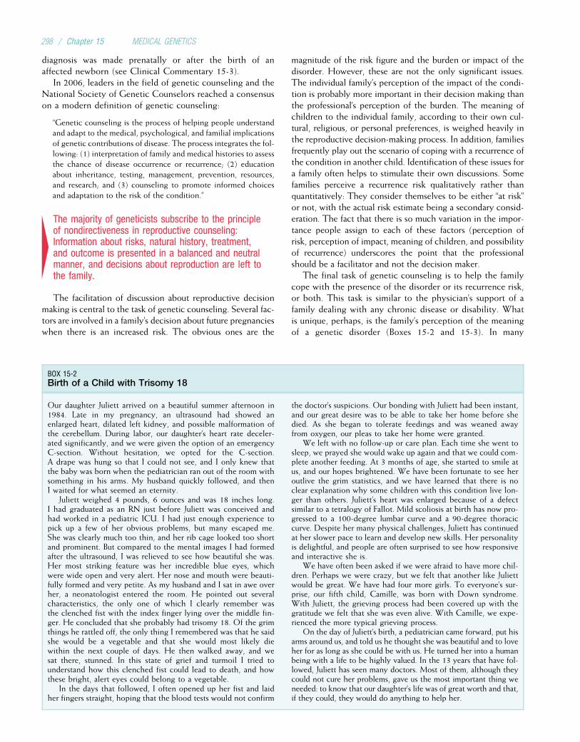
diagnosis was made prenatally or after the birth of anaffected newborn (see Clinical Commentary 15-3).
In 2006, leaders in the field of genetic counseling and theNational Society of Genetic Counselors reached a consensuson a modern definition of genetic counseling:
“Genetic counseling is the process of helping people understand
and adapt to the medical, psychological, and familial implications
of genetic contributions of disease. The process integrates the fol-
lowing: (1) interpretation of family and medical histories to assess
the chance of disease occurrence or recurrence; (2) education
about inheritance, testing, management, prevention, resources,
and research; and (3) counseling to promote informed choices
and adaptation to the risk of the condition.”
4The majority of geneticists subscribe to the principleof nondirectiveness in reproductive counseling:Information about risks, natural history, treatment,and outcome is presented in a balanced and neutralmanner, and decisions about reproduction are left tothe family.
The facilitation of discussion about reproductive decisionmaking is central to the task of genetic counseling. Several fac-tors are involved in a family’s decision about future pregnancieswhen there is an increased risk. The obvious ones are the
magnitude of the risk figure and the burden or impact of thedisorder. However, these are not the only significant issues.The individual family’s perception of the impact of the condi-tion is probably more important in their decision making thanthe professional’s perception of the burden. The meaning ofchildren to the individual family, according to their own cul-tural, religious, or personal preferences, is weighed heavily inthe reproductive decision-making process. In addition, familiesfrequently play out the scenario of coping with a recurrence ofthe condition in another child. Identification of these issues fora family often helps to stimulate their own discussions. Somefamilies perceive a recurrence risk qualitatively rather thanquantitatively: They consider themselves to be either “at risk”or not, with the actual risk estimate being a secondary consid-eration. The fact that there is so much variation in the impor-tance people assign to each of these factors (perception ofrisk, perception of impact, meaning of children, and possibilityof recurrence) underscores the point that the professionalshould be a facilitator and not the decision maker.
The final task of genetic counseling is to help the familycope with the presence of the disorder or its recurrence risk,or both. This task is similar to the physician’s support of afamily dealing with any chronic disease or disability. Whatis unique, perhaps, is the family’s perception of the meaningof a genetic disorder (Boxes 15-2 and 15-3). In many
BOX 15-2Birth of a Child with Trisomy 18
Our daughter Juliett arrived on a beautiful summer afternoon in1984. Late in my pregnancy, an ultrasound had showed anenlarged heart, dilated left kidney, and possible malformation ofthe cerebellum. During labor, our daughter’s heart rate deceler-ated significantly, and we were given the option of an emergencyC-section. Without hesitation, we opted for the C-section.A drape was hung so that I could not see, and I only knew thatthe baby was born when the pediatrician ran out of the room withsomething in his arms. My husband quickly followed, and thenI waited for what seemed an eternity.
Juliett weighed 4 pounds, 6 ounces and was 18 inches long.I had graduated as an RN just before Juliett was conceived andhad worked in a pediatric ICU. I had just enough experience topick up a few of her obvious problems, but many escaped me.She was clearly much too thin, and her rib cage looked too shortand prominent. But compared to the mental images I had formedafter the ultrasound, I was relieved to see how beautiful she was.Her most striking feature was her incredible blue eyes, whichwere wide open and very alert. Her nose and mouth were beauti-fully formed and very petite. As my husband and I sat in awe overher, a neonatologist entered the room. He pointed out severalcharacteristics, the only one of which I clearly remember wasthe clenched fist with the index finger lying over the middle fin-ger. He concluded that she probably had trisomy 18. Of the grimthings he rattled off, the only thing I remembered was that he saidshe would be a vegetable and that she would most likely diewithin the next couple of days. He then walked away, and wesat there, stunned. In this state of grief and turmoil I tried tounderstand how this clenched fist could lead to death, and howthese bright, alert eyes could belong to a vegetable.
In the days that followed, I often opened up her fist and laidher fingers straight, hoping that the blood tests would not confirm
the doctor’s suspicions. Our bonding with Juliett had been instant,and our great desire was to be able to take her home before shedied. As she began to tolerate feedings and was weaned awayfrom oxygen, our pleas to take her home were granted.
We left with no follow-up or care plan. Each time she went tosleep, we prayed she would wake up again and that we could com-plete another feeding. At 3 months of age, she started to smile atus, and our hopes brightened. We have been fortunate to see heroutlive the grim statistics, and we have learned that there is noclear explanation why some children with this condition live lon-ger than others. Juliett’s heart was enlarged because of a defectsimilar to a tetralogy of Fallot. Mild scoliosis at birth has now pro-gressed to a 100-degree lumbar curve and a 90-degree thoraciccurve. Despite her many physical challenges, Juliett has continuedat her slower pace to learn and develop new skills. Her personalityis delightful, and people are often surprised to see how responsiveand interactive she is.
We have often been asked if we were afraid to have more chil-dren. Perhaps we were crazy, but we felt that another like Juliettwould be great. We have had four more girls. To everyone’s sur-prise, our fifth child, Camille, was born with Down syndrome.With Juliett, the grieving process had been covered up with thegratitude we felt that she was even alive. With Camille, we expe-rienced the more typical grieving process.
On the day of Juliett’s birth, a pediatrician came forward, put hisarms around us, and told us he thought she was beautiful and to loveher for as long as she could be with us. He turned her into a humanbeing with a life to be highly valued. In the 13 years that have fol-lowed, Juliett has seen many doctors. Most of them, although theycould not cure her problems, gave us the most important thing weneeded: to know that our daughter’s life was of great worth and that,if they could, they would do anything to help her.
298 / Chapter 15 MEDICAL GENETICS
acquired conditions, such as infections or accidents, the ulti-mate meaning of the condition is externalized. In geneticdisorders, the condition is more intrinsic to the individualand the family; it thus often presents a complex personaldilemma. Validation of the plight of families is vital and isprobably more effective than simplistic attempts to wipeaway guilt. Feelings of guilt and shame are natural to thesituation and also need acknowledgment.
The primary care practitioner plays a vital role in theongoing support of families in which a member has a geneticdisease. Additional support strategies include referral of thefamily to a genetic advocacy group, distribution of currentprinted and Internet information on the disorder, referral tomental health professionals for ongoing counseling, and fre-quent follow-up visits that include time for discussions offeelings and thoughts.
4Genetic counseling includes many themes: medicaldiagnosis and management, determining risk ofrecurrence, options for addressing the risk,reproductive decision making, and support services.
Numerous studies in the past 3 decades have attempted toevaluate the effectiveness of genetic counseling. The meth-odology of these studies is complicated, and the evaluationof the results depends on one’s interpretation of the goal ofgenetic counseling. A few general points, however, can bemade. Families tend to recall recurrence risks relatively well.A letter sent to them after the visit improves this recall.
Families who perceive their offspring’s condition as beingserious and one of “burden” recall risk figures better. Moststudies suggest that genetic counseling is relatively effectivein providing information about the medical aspects andgenetic risks of the condition. Issues surrounding decisionmaking and psychosocial support require additionalinvestigation.
Genetic Counselors and the Delivery of GeneticCounselingAs the discipline of medical genetics (including geneticcounseling) evolved in the 1970s, it became clear that thedelivery of this service is complex and time-consuming.Not only did the geneticist need to have skills in most spe-cialties of medicine, but facilitation of decision making andprovision of psychological support were also necessary. Asa need for genetics professionals other than physiciansbecame apparent, a number of genetic counseling trainingprograms emerged in the United States and Canada. Cur-rently, more than 30 accredited programs in North Americaprovide master’s level training in genetic counseling. Geneticcounselors have become integral partners with physiciansand other professionals in the delivery of medical geneticservices. From this growth evolved a professional society,The National Society of Genetic Counselors, and, the certi-fying and accrediting body, the American Board of GeneticCounseling. Although the range of skills is wide and jobdescriptions vary in different medical centers, genetic coun-selors have established themselves as experts in the
BOX 15-3Raising a Child with Bloom Syndrome
Tommy was born via an emergency cesarean section, because 1week before his delivery date his fetal movements markedlydecreased. At birth, he weighed only 4 pounds, and the first timethat I saw him, he was in an incubator connected to all sorts oftubes. He spent his first month of life in the neonatal intensivecare unit so that his weight gain could be closely monitored.Because he was so small, he was fed through a feeding tube formany months, and as a consequence, he refused to drink from abottle. Eventually, he overcame his aversion to using his mouthto eat but only after substantial training. Nevertheless, despiteour care, Tommy remained small for his age.
The following summer, Tommy developed dark red marks onhis cheeks and under his eyes. Our pediatrician referred us to a der-matologist, who suspected that the marks on Tommy’s face wererelated to his growth failure. We were very surprised. How couldthese two findings be related? That is when we were told thatTommy might have a genetic disorder called Bloom syndrome.We hoped that the doctor was wrong, but soon thereafter Tommyhad a genetic test that measured the number of sister chromatidexchanges per cell (see Chapter 2). This test confirmed thatTommy had Bloom syndrome. Although I insisted that it was afalse-positive result, I learned to accept that our son had a very rarecancer syndrome.
We were barraged by questions from family, friends, and doc-tors. As a result, we became very protective of our son and hisprivacy. Nevertheless, there was only so much we could do to
protect him because he is such a social little boy, loving to playwith family and friends. This also made choosing an appropriateelementary school a very difficult decision for us. We expectedthat children would pick on him because of his small size. How-ever, to our surprise, he easily developed friendships and adjustedwell to his classmates. In fact, the problems that he did developwere largely because of his misbehavior. Thus, we struggled tofind a balance between protecting Tommy while not permittinghim special privileges because of his small stature.
In our home, we try to treat Tommy like any of our other chil-dren. One challenge for us is that because of Tommy’s small size,people wrongly perceive that he is much younger than his chrono-logical age. This is very frustrating for Tommy, yet we occasionallyreinforce this image because of our concern for his safety. Forexample, although Tommy is 6 years old, he weighs only 21pounds. Thus, he must sit in an infant seat when he travels in acar and we explain to Tommy’s friends that it helps him seethrough the windows. Another safety problem is that many ofthe sensors for the automatic doors at supermarkets cannot detecthis presence and easily slam into him.
Overall, Tommy has adapted well. He climbs or jumps in orderto reach things. To keep up with his peers, he often runs, hops, orjumps instead of walking. We constantly worry about his safety,but we cannot control all that happens to him. To date, he has beenhealthy, and although it seems like we have been riding an emo-tional roller-coaster, we wouldn’t trade our experiences for anything.
Clinical Genetics and Genetic Counseling / 299
determination of recurrence chances, reproductive decisionmaking, and psychosocial support (Box 15-4). In the prenataland cancer genetics settings, genetic counselors function rel-atively independently as practitioners. More recently,genetic counselors have become important professionals onresearch teams and in genetics laboratory services.
Genetic Advocacy GroupsGenetic advocacy groups can provide critical support inassisting families who have a member with a genetic disorder(see, for example, Genetic Alliance under Internet Resourcesat the end of this chapter). These support organizationsprovide the family with the sense of a fellow traveler in away that the professional is not able to do. The sense of iso-lation that often accompanies genetic disorders (and rareconditions in general) is often alleviated by meeting some-one else in the same situation. Immediate bonds are formed
that often assist in the coping process. In the last few dec-ades, a clear partnership of professionals and persons withgenetic disorders and disabilities has developed. Not onlyhave these groups provided a needed service, but they alsohave promoted the establishment of databases and researchstudies. Referral to a genetic support group and distributionof their written information are now a routine part of thecare and management of all genetic disorders.
4The delivery of genetic services including geneticcounseling involves a partnership of physicians,genetic counselors, and genetic advocacy groups.
Clinical Genetics Evaluation and ServicesWith the development of medical genetics as a medical spe-cialty, clinical genetics services have become part of the
BOX 15-4An Insider’s View of Genetic Counseling
What Is a Genetic Counselor?In its most general usage, the term genetic counselor refers to anymedical professional who is professionally qualified to providegenetic counseling. Typically, a genetic counselor is a geneticsprofessional with a master’s degree or Ph.D. in genetic counseling.Degree programs in genetic counseling provide education andclinical training in medical genetics and counseling. A certifiedgenetic counselor has also passed a certification examinationadministered by the American Board of Genetic Counseling orthe American Board of Medical Genetics.
What Do Genetic Counselors Do?Some of the primary responsibilities of a genetic counselor are tointerview individuals and families with genetic disorders and toanswer questions about the possibilities of a genetic disorder. Agenetic counselor often works as part of a team that may includemedical geneticists, other physicians (e.g., obstetricians, oncologists,neurologists), social workers, psychologists, nutritionists, or nurses.Genetic counselors help to collect and assess medical informationleading to a diagnosis, provide patient education, provide psychoso-cial support and counseling, provide genetic counseling and riskassessment for genetic testing, and help physicians with the manage-ment of genetic conditions. They often triage inquiries and referralsto the genetics service in which they practice. They might manageor coordinate clinics and personnel. They are active in genetic edu-cation programs for medical professionals and the lay public.
In What Settings Do Genetic Counselors Work?Genetic counselors often work in general genetics settings in pedi-atrics and adult medicine. They also work in obstetrics settings,providing counseling for prenatal diagnosis and screening, genetictesting for couples with multiple pregnancy loss, diagnosis andmanagement of pregnancies affected with abnormalities detectedby radiological imaging, and, most recently, alternative reproduc-tive technologies. They work in multidisciplinary specialty clinicsfor groups of diseases (e.g., metabolic, craniofacial, bone dysplasia,neurogenetic) or for single diseases (e.g., Down syndrome, neuro-fibromatosis, hemophilia). More recently, genetic counselors arebecoming increasingly integrated into cancer genetics clinics.
Many counselors participate in research related to clinicalgenetics and genetic counseling. For example, predictive testing
for disorders such as Huntington disease and hereditary cancershas occurred mainly in research studies aimed at assessing themedical, ethical, legal, and social consequences. Within thisresearch setting, genetic counselors provide counseling and helpto design, implement, and evaluate research protocols.
Some genetic counselors work in laboratories to provide aninterface between the laboratory and its clients and to helpdevelop laboratory protocols. A small percentage of genetic coun-selors are in private practice, and some work in administrativepositions for the state or federal government. Many genetic coun-selors are active at regional and national levels in professionalorganizations, and some counselors help to start, maintain, oradvise lay advocacy groups for genetic disorders.
What Skills and Personal Qualities Make a GoodGenetic Counselor?A good genetic counselor needs both a strong background in thebiological sciences and genetics and training in the theory and prac-tice of psychosocial techniques (e.g., family systems, crisiscounseling, interviewing skills). Because most genetic counselorsprovide direct patient service, it is essential to work well with people.Genetic counselors must work well both independently and on ateam. There is a high level of responsibility involved in patient careaspects, and counselors must successfully learn to handle the stress ofthe difficult situations of the families with whom they work.
What Is the Future of Genetic Counseling?It is difficult to predict the extent to which medical genetics willcontinue to move into mainstream medicine. Will geneticistsand genetic counselors increase in number, or will genetics profes-sionals remain small in number and limit their role to advisinggeneralists and seeing only the most complicated cases? In eithercase, there is clearly a need for an increase in the genetics educa-tion of medical professionals and the public. Many observersthink that medical genetics and genetic counseling have a highpotential for expansion. What is undisputed is the striking emer-gence of medical genetics from an obscure medical subspecialtyto an area of knowledge that is fast becoming integrated intoevery field of medicine.
(Courtesy of Bonnie J. Baty, M.S.)
300 / Chapter 15 MEDICAL GENETICS
health care delivery system. Most university medical centersin North America include a genetics clinic whose majorobjective is to provide genetic diagnosis, management, andcounseling services.
As in all medical visits, evaluation of a person or family fora potential genetic condition requires a thorough history andphysical examination. The history includes information aboutthe family’s concerns, the prenatal period, labor, delivery, anddocumentation of family relationships (the pedigree). Thephysical examination should focus on the physical variationsor minor anomalies that provide clues to a diagnosis. Addi-tional family members might need evaluation for the presenceor absence of a genetic disorder. Photographs and recordingof certain physical measurements are a standard componentof the genetic evaluation. Ancillary tests may be required todocument specific physical features (e.g., an echocardiogramor MRI for aortic dilatation in Marfan syndrome or radio-graphs to diagnose achondroplasia).
An important type of clinical data gathered in this processis the family history (Box 15-5). The data obtained in a fam-ily history are often useful in obtaining an accurate diagnosisof a condition. For example, a strong family history of early-onset coronary disease might indicate the presence of a low-density lipoprotein–receptor defect causing familial hyper-cholesterolemia. A family history of early-onset colon cancercould indicate that a gene for familial adenomatous polypo-sis or hereditary nonpolyposis colorectal cancer is present inthe family. Family history information can also guide theestimation of recurrence risks by helping to determinewhether a genetic disease has been transmitted by one’s par-ents or has occurred as a new mutation (this is especiallyimportant for diseases with reduced penetrance). The knowl-edge and skills required to take an accurate and thoroughfamily history are important for all clinicians, not only clini-cal geneticists.
Routinely, the clinician sends the family a letter summar-izing the diagnosis, natural history, and risk informationregarding the condition. This letter is a valuable resourcefor the family, because it helps to document the risk
information for later review. Information regarding lay advo-cacy groups, including pamphlets, booklets, and brochures,is often provided. Follow-up visits are recommendeddepending on the individual situation. Box 15-6 provides alist of clinical genetics services.
4Clinical genetic evaluations include physicalexamination, detailed family history, ancillary tests asneeded, and communication of information to thefamily through letters and the distribution of publishedliterature.
In recent years, the care of persons with genetic diseasehas included the development of guidelines for follow-upand routine care. Knowledge of the natural history of a con-dition, coupled with a critical review of screening tests andinterventions, can provide a framework for health supervi-sion and anticipatory guidance. The management plan cansubsequently be used by the primary care provider. It is pri-marily for this purpose that many of the specialized clinics,such as those for NF1 or hemophilia, have been established.An example of this approach is the management checklist forthe health maintenance of infants and children with Downsyndrome (see Chapter 6).
As treatment options for mendelian disorders becomemore numerous (e.g., the treatment of aortic dilatation inMarfan syndrome, see Chapter 4), the role of clinical geneti-cists will likely change. Since the turn of the 21st century,geneticists have become increasingly involved in the designand implementation of clinical trials, and this trend will cer-tainly continue and change the nature of the practice.
Traditionally, genetic counseling involves the family whocomes in with questions about the diagnosis, management,and recurrence risk of the condition in question. Thus, inthe majority of situations, genetic counseling is carried outretrospectively. With the increased availability of prenatal,carrier, and presymptomatic testing, prospective geneticcounseling will become more common. Box 15-7 lists com-mon reasons for referral for genetic evaluation.
BOX 15-5The Family History
A thorough, accurate family history is an indispensable part of amedical evaluation, and a pedigree should be part of the patient’schart. At a minimum, the following items should be included:
• The sex of each individual and his or her relationship to otherfamily members. This information should be indicated usingstandard pedigree symbols (see Chapter 4).
• A three-generation family history should be obtained. Forexample, male relatives on the mother’s side of the family willbe especially important when considering an X-linked recessivedisorder.
• The age of each individual. A record must be kept of whethereach individual is affected with the disease in question, andinquiries should be made about diseases that may be related
to the disease in question (e.g., ovarian cancer in a family beingseen for familial breast cancer).
• All known miscarriages and stillbirths.• The ethnic origin of the family. This is important because manydiseases vary considerably in prevalence among different ethnicgroups.
• Information about consanguinity. Although it is relatively rare inmost Western populations, consanguinity is common in many ofthe world’s populations, and immigrant populations often main-tain relatively high rates of consanguinity (see Chapter 4).
• Changes in family histories. Family members develop newlydiagnosed diseases, and additional children are born. Thesechanges can affect diagnosis and risk estimation, so the familyhistory and pedigree should be updated periodically.
Clinical Genetics and Genetic Counseling / 301
DYSMORPHOLOGY AND CLINICAL TERATOLOGYDysmorphology was defined at the beginning of this chapter asthe study of abnormal physical development (morphogenesis).Congenital defects are caused by altered morphogenesis.Although the term dysmorphology may seem synonymouswith teratology, the latter term usually implies the study ofthe environmental causes of congenital anomalies, even thoughits literal meaning does not refer to etiology. (The term
teratology is derived from teras, the Greek word for “monster.”The term dysmorphology was proposed by Dr. David Smithas a reaction to the pejorative connotation of teratology.)
Congenital defects represent an important cause of infantmortality and morbidity. Current studies indicate that thefrequency of medically significant malformations diagnosedin the newborn period is 2% to 3%. Investigations that haveobserved children for a longer period demonstrate that thisfrequency increases to 3% to 4% by the age of 1 year. Inthe United States, congenital malformations represent themost common cause of mortality during the first year of life.Table 15-1 lists some of the most common and importantmalformation syndromes.
BOX 15-6Types of Clinical Genetic Services and Programs
Center-based genetics clinicsOutreach clinicsInpatient consultationsSpecialty clinics• Metabolic clinics• Cancer genetics clinics• Spina bifida clinics• Hemophilia clinics• Craniofacial clinics• Other single-disorder clinics (e.g., NF1 clinics)Prenatal diagnosis programs: Perinatal and reproductive genetics
clinics• Amniocentesis and chorionic villus sampling clinics• Ultrasound programs• Maternal serum triple screen programs• Preimplantation diagnosis programs• Presymptomatic diagnosis in families (e.g., familial breast can-cer diagnosis)
Genetic screening• Newborn screening program/follow-up clinics• Other population screening programs (e.g., Tay–Sachs disease)Education and training• Health care professionals, including clinical geneticists andgenetic counselors
• General public• School system• Teratology information services
BOX 15-7Common Indications for Genetics Referral
Evaluation of a person with mental retardation or developmentaldelay
Evaluation of a person with single or multiple malformations;question of a dysmorphic syndrome
Evaluation of a person with a possible inherited metabolic diseasePresence of a possible single-gene disorderPresence of a chromosomal disorder, including balanced
rearrangementsPerson at risk for a genetic condition, including questions of
presymptomatic diagnosis or cancer riskPerson or family with questions about the genetic aspects of any
medical conditionCouples with a history of recurrent miscarriagesConsanguinity in a couple, usually first cousin or closer relationshipTeratogen counselingPreconceptional counseling and risk-factor counseling, including
advanced maternal age and other potential indications forprenatal diagnosis
TABLE 15-1
Examples of Common Multiple Congenital Anomaly andDysplasia Syndromes Referred to a Medical Genetics Clinic
Syndromes Etiology
Down syndrome Chromosomal
Neurofibromatosis type 1 Single gene (AD)
Angelman syndrome Microdeletion of chromosome 15q;uniparental disomy
Amnion disruptionsequence
Unknown
Osteogenesis imperfecta Single gene; heterogeneous, AD, AR;type I collagen, related genes
Trisomy 18 Chromosomal
VATER association Unknown
Marfan syndrome Single gene (AD)
Prader–Willi syndrome Microdeletion of chromosome 15q;uniparental disomy
Noonan syndrome Single gene (AD)
Williams syndrome Microdeletion of chromosome 7
Achondroplasia Single gene (AD)
Trisomy 13 Chromosomal
Turner syndrome Chromosomal (45,X)
Rett syndrome X-linked gene
Rubinstein–Taybi syndrome Single gene (AD)
Klippel–Trenaunay Heterogeneous; one susceptibilitysyndrome gene identified
Fetal alcohol syndrome Excessive alcohol
Cornelia de Langesyndrome
Single gene (AD)
AD, Autosomal dominant, AR autosomal recessive; VATER, vertebral anomalies,
anal atresia, tracheoesophageal fistula, esophageal atresia, renal anomalies.
302 / Chapter 15 MEDICAL GENETICS
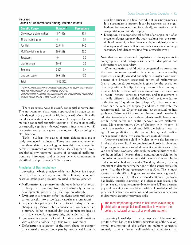
There are several ways to classify congenital abnormalities.The most common classification approach is by organ systemor body region (e.g., craniofacial, limb, heart). More clinicallyuseful classification schemes include (1) single defect versusmultiple congenital anomaly syndrome, (2) major (medicallyor surgically significant defects) versus minor anomalies, (3)categorization by pathogenic process, and (4) an etiologicalclassification.
Table 15-2 lists the causes of main defects in a majorstudy conducted in Boston. Three key messages emergedfrom these data: the etiology of two thirds of congenitaldefects is unknown or multifactorial (see Chapter 12), well-established environmental causes of congenital malforma-tions are infrequent, and a known genetic component isidentified in approximately 30% of cases.
Principles of DysmorphologyIn discussing the basic principles of dysmorphology, it is impor-tant to define certain key terms. The following definitions,based on pathogenic processes, are used in clinical practice:
• Malformation is a primary morphologic defect of an organor body part resulting from an intrinsically abnormaldevelopmental process (e.g., cleft lip, polydactyly).
• Dysplasia is a primary defect involving abnormal organi-zation of cells into tissue (e.g., vascular malformation).
• Sequence is a primary defect with its secondary structuralchanges (e.g., Pierre Robin sequence, a disorder in whicha primary defect in mandibular development produces asmall jaw, secondary glossoptosis, and a cleft palate)
• Syndrome is a pattern of multiple primary malformationswith a single etiology (e.g., trisomy 13 syndrome).
• Deformation is alteration of the form, shape, or positionof a normally formed body part by mechanical forces. It
usually occurs in the fetal period, not in embryogenesis.It is a secondary alteration. It can be extrinsic, as in oligo-hydramnios (reduced amniotic fluid), or intrinsic, as incongenital myotonic dystrophy.
• Disruption is a morphological defect of an organ, part of anorgan, or a larger region of the body resulting from the extrin-sic breakdown of, or interference with, an originally normaldevelopmental process. It is a secondary malformation (e.g.,secondary limb defect resulting from a vascular event).
Note that malformations and dysplasias are primary events inembryogenesis and histogenesis, whereas disruptions anddeformations are secondary.
When evaluating a child with a congenital malformation,the most important question is whether the abnormalityrepresents a single, isolated anomaly or is instead one com-ponent of a broader, organized pattern of malformation(i.e., a syndrome). An example is given by the evaluationof a baby with a cleft lip. If a baby has an isolated, nonsyn-dromic cleft lip with no other malformations, the discussionof natural history, genetics, prognosis, and management ismarkedly different than if the baby’s cleft lip is one featureof the trisomy 13 syndrome (see Chapter 6). The former con-dition can be repaired surgically and has a relatively lowrecurrence risk (see Chapter 12) and few associated medicalproblems. Trisomy 13 is a serious chromosomal disorder. Inaddition to oral–facial clefts, these infants usually have a con-genital heart defect and central nervous system malforma-tions. More importantly, 50% of children with trisomy 13die in the newborn period, and 90% die before 1 year ofage. Thus, prediction of the natural history and medicalmanagement in these two examples are quite different.
Another example is a child with cleft lip who also has pits orfistulas of the lower lip. The combination of orofacial clefts andlip pits signifies an autosomal dominant condition called thevan der Woude syndrome. Although the natural history of thiscondition differs little from that of nonsyndromic cleft lip, thediscussion of genetic recurrence risks is much different. In theevaluation of a child with van der Woude syndrome, it is veryimportant to determine whether one of the parents carries thegene. If so, the sibling recurrence risk is 50%. This is muchgreater than the 4% sibling recurrence risk usually given fornonsyndromic cleft lip. Because van der Woude syndromehas highly variable expression and often is manifested onlyby lip fistulas, it is quite commonly overlooked. Thus, a carefulphysical examination, combined with a knowledge of thegenetics of isolated malformations and syndromes, is necessaryto determine accurate recurrence risks.
4The most important question to ask when evaluating achild with a congenital malformation is whether thedefect is isolated or part of a syndrome pattern.
Increasing knowledge of the pathogenesis of human con-genital defects has led to a better understanding of the develop-mental relationship of the defects in multiple congenitalanomaly patterns. Some well-established conditions that
TABLE 15-2
Causes of Malformations among Affected Infants
Genetic Cause Number* Percentage
Chromosome abnormalities 157 (45) 10.1
Single mutant genes 48 3.1
Familial 225 (3) 14.5
Multifactorial inheritance 356 (23) 23.0
Teratogens 49 3.2
Uterine factors 39 (5) 2.5
Twinning 6 (2) 0.4
Unknown cause 669 (24) 43.2
Total 1549 (102)
*Values in parentheses denote therapeutic abortions; of the 69,277 infants studied,
1549 had malformations, for an incidence of 2.24%.
Data from Nelson K, Holmes LB: Malformations due to spontaneous mutations in
newborn infants. N Engl J Med 1989;320:19-23.
Clinical Genetics and Genetic Counseling / 303
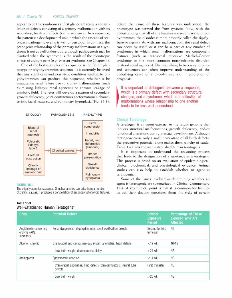
appear to be true syndromes at first glance are really a constel-lation of defects consisting of a primary malformation with itssecondary, localized effects (i.e., a sequence). In a sequence,the pattern is a developmental unit in which the cascade of sec-ondary pathogenic events is well understood. In contrast, thepathogenic relationship of the primary malformations in a syn-drome is not as well understood, although pathogenesis may beclarified when the syndrome is the result of the pleiotropiceffects of a single gene (e.g., Marfan syndrome; see Chapter 4).
One of the best examples of a sequence is the Potter phe-notype or oligohydramnios sequence. It is currently believedthat any significant and persistent condition leading to oli-gohydramnios can produce this sequence, whether it beintrauterine renal failure due to kidney malformations (suchas missing kidneys, renal agenesis) or chronic leakage ofamniotic fluid. The fetus will develop a pattern of secondarygrowth deficiency, joint contractures (deformations), charac-teristic facial features, and pulmonary hypoplasia (Fig. 15-1).
Before the cause of these features was understood, thephenotype was termed the Potter syndrome. Now, with theunderstanding that all of the features are secondary to oligo-hydramnios, the disorder is more properly called the oligohy-dramnios sequence. As with any malformation, the renal defectcan occur by itself, or it can be a part of any number ofsyndromes in which renal malformations are componentfeatures (such as autosomal recessive Meckel–Grubersyndrome or the more common nonsyndromic disorder,bilateral renal agenesis). Distinguishing between syndromesand sequences can often improve understanding of theunderlying cause of a disorder and aid in prediction ofprognosis.
4It is important to distinguish between a sequence,which is a primary defect with secondary structuralchanges, and a syndrome, which is a collection ofmalformations whose relationship to one anothertends to be less well understood.
Clinical TeratologyA teratogen is an agent external to the fetus’s genome thatinduces structural malformations, growth deficiency, and/orfunctional alterations during prenatal development. Althoughteratogens cause only a small percentage of all birth defects,the preventive potential alone makes them worthy of study.Table 15-3 lists the well-established human teratogens.
It is important to understand the reasoning processthat leads to the designation of a substance as a teratogen.This process is based on an evaluation of epidemiological,clinical, biochemical, and physiological evidence. Animalstudies can also help to establish whether an agent isteratogenic.
Some of the issues involved in determining whether anagent is teratogenic are summarized in Clinical Commentary15-4. A key clinical point is that it is common for familiesto ask their doctors questions about the risks of certain
Oligohydramnios
ETIOLOGY PATHOGENESIS PHENOTYPE
Bilateralrenal
agenesis
Polycystickidneys,type 1
Urethralobstruction
Chronicleakage of
amniotic fluid
Fetalcompression
Immobility
Facial, limbdeformities(club foot)
Breechpresentation
Growthdeficiency
Pulmonaryhypoplasia
FIGURE 15-1The oligohydramnios sequence. Oligohydramnios can arise from a numberof distinct causes. It produces a constellation of secondary phenotypic features.
TABLE 15-3
Well-Established Human Teratogens*
Drug Potential Defect CriticalExposurePeriod
Percentage of ThoseExposed Who AreAffected
Angiotensin-convertingenzyme (ACE)inhibitors
Renal dysgenesis; oligohydramnios; skull ossification defects Second to thirdtrimester
NE
Alcohol, chronic Craniofacial and central nervous system anomalies; heart defects <12 wk 10-15
Low birth weight; developmental delay >24 wk NE
Aminopterin Spontaneous abortion <14 wk NE
Craniofacial anomalies; limb defects; craniosynostosis; neural tubedefects
First trimester NE
Low birth weight >20 wk NE
304 / Chapter 15 MEDICAL GENETICS
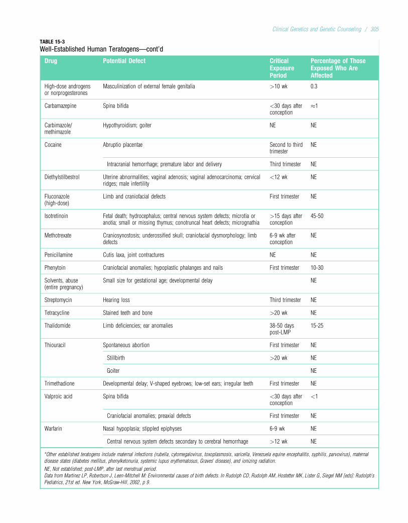
TABLE 15-3
Well-Established Human Teratogens—cont’d
Drug Potential Defect CriticalExposurePeriod
Percentage of ThoseExposed Who AreAffected
High-dose androgensor norprogesterones
Masculinization of external female genitalia >10 wk 0.3
Carbamazepine Spina bifida <30 days afterconception
�1
Carbimazole/methimazole
Hypothyroidism; goiter NE NE
Cocaine Abruptio placentae Second to thirdtrimester
NE
Intracranial hemorrhage; premature labor and delivery Third trimester NE
Diethylstilbestrol Uterine abnormalities; vaginal adenosis; vaginal adenocarcinoma; cervicalridges; male infertility
<12 wk NE
Fluconazole(high-dose)
Limb and craniofacial defects First trimester NE
Isotretinoin Fetal death; hydrocephalus; central nervous system defects; microtia oranotia; small or missing thymus; conotruncal heart defects; micrognathia
>15 days afterconception
45-50
Methotrexate Craniosynostosis; underossified skull; craniofacial dysmorphology; limbdefects
6-9 wk afterconception
NE
Penicillamine Cutis laxa, joint contractures NE NE
Phenytoin Craniofacial anomalies; hypoplastic phalanges and nails First trimester 10-30
Solvents, abuse(entire pregnancy)
Small size for gestational age; developmental delay NE
Streptomycin Hearing loss Third trimester NE
Tetracycline Stained teeth and bone >20 wk NE
Thalidomide Limb deficiencies; ear anomalies 38-50 dayspost-LMP
15-25
Thiouracil Spontaneous abortion First trimester NE
Stillbirth >20 wk NE
Goiter NE
Trimethadione Developmental delay; V-shaped eyebrows; low-set ears; irregular teeth First trimester NE
Valproic acid Spina bifida <30 days afterconception
<1
Craniofacial anomalies; preaxial defects First trimester NE
Warfarin Nasal hypoplasia; stippled epiphyses 6-9 wk NE
Central nervous system defects secondary to cerebral hemorrhage >12 wk NE
*Other established teratogens include maternal infections (rubella, cytomegalovirus, toxoplasmosis, varicella, Venezuela equine encephalitis, syphilis, parvovirus), maternal
disease states (diabetes mellitus, phenylketonuria, systemic lupus erythematosus, Graves’ disease), and ionizing radiation.
NE, Not established; post-LMP, after last menstrual period.
Data from Martinez LP, Robertson J, Leen-Mitchell M: Environmental causes of birth defects. In Rudolph CD, Rudolph AM, Hostetter MK, Lister G, Siegel NM [eds]: Rudolph’s
Pediatrics, 21st ed. New York, McGraw-Hill, 2002, p 9.
Clinical Genetics and Genetic Counseling / 305
CLINICAL COMMENTARY 15-4
The Bendectin Saga
Bendectin, or doxyclamine, was an agent introduced in the 1960s for “morn-ing sickness” (nausea and vomiting during pregnancy). The agent was par-ticularly efficacious, and during the 1970s about one third of Americanwomen took Bendectin at some time during their first trimester of pregnancy.
Bendectin has probably been studied more than any other individualpregnancy medication. Several epidemiological studies have found no con-clusive evidence of an increased risk of congenital malformations with useof Bendectin during pregnancy. The few studies that have demonstrated weakassociations between Bendectin and birth defects did not reveal any consis-tent patterns. Animal studies also indicate no association. Despite these data,a number of lawsuits were filed in the 1980s against the company that mar-keted Bendectin. As a result, the company removed the drug from the market.
The reasoning process used to determine causation in such cases iscomplex. Before judgments about etiology are made, a critical review ofthe literature is required. The available epidemiological studies must beevaluated in terms of their methodology, design, and biases. Up-to-dateknowledge of the etiology and pathogenesis of congenital malformationsmust be included in the design of the study. The basic principles of teratol-ogy must be applied. This includes an evaluation of the critical period (i.e.,did exposure occur during the period of pregnancy in which the malformedfetal structures were developing?). Clinical evidence includes the search fora pattern of defects (i.e., a specific syndrome), because all well-establishedteratogens produce consistent patterns (see Table 15-3). Animal modelsnever prove causation in humans but can provide supportive evidenceand information about pathogenesis. In addition, the proposed effect ofthe agent should be biologically plausible.
A review of the evidence regarding Bendectin shows that it meets noneof these criteria. Indeed, because of its extensive testing, Bendectin satisfiesstandard criteria for safety as well as any known medication. What, then,could have produced this litigation? An important part of the answer to thisquestion involves the coincidental occurrence of congenital malformationsand Bendectin exposure. Considering that a major congenital malformationwill be diagnosed in 3% of infants by 1 year of age and that about one thirdof women were taking Bendectin in the first trimester of pregnancy, thenabout 1% (1/3 � 3%) of all pregnancies in the 1970s would have experi-enced the co-occurrence of these two events by chance alone. Becauseabout two thirds of congenital malformations have no known cause, it isnot surprising that many families of children with these disorders (and theirlawyers) would attribute them to Bendectin use.
Bendectin was removed from the market in 1983. Since then, the per-centage of babies born with congenital malformations has remained thesame, and the number of women admitted to hospitals for complaints ofmorning sickness has doubled.
It is very difficult to prove epidemiologically that any exposure is“safe.” The statistical power of such studies does not permit an absolutestatement that there is no effect. All that can be demonstrated is that thereis no definitive evidence that a particular agent (in this case, Bendectin)causes an adverse outcome. Blanket reassurance or absolute statementsof complete safety are not appropriate. On the other hand, when the evi-dence is relatively conclusive, as in the case of Bendectin, it is clinicallyappropriate to have a reassuring tone in discussing exposure in the preg-nancy setting.
exposures during pregnancy. When faced with such a ques-tion, the physician has a number of options. One is to reviewthe literature on particular exposures in humans, make ajudgment regarding the degree of risk, and then providecounseling. Various online and printed sources available tothe clinician are listed at the end of the chapter. An alterna-tive is to refer the patient to a teratology information serviceor to a clinical genetics unit. Because of the complexity ofthese issues, teratology information services have sprung upthroughout the United States, Canada, and Europe. TheOrganization of Teratology Information Services providesup-to-date and comprehensive information on exposures inpregnancy and lists the available teratology information ser-vices in North America.
4Teratogens are external agents that cause a small butimportant percentage of congenital malformations. Itis often difficult to prove conclusively that a substanceis teratogenic.
Prevention of Congenital MalformationsBecause the majority of structural defects have no obviouscause, their prevention presents a challenge. An exampleis fetal alcohol syndrome (FAS), one of the more common
preventable causes of human malformation (Clinical Com-mentary 15-5). The institution of rubella immunizationprograms and the preconceptional administration of folicacid exemplify successful prevention (Clinical Commentary15-6).
Preconceptional counseling is a model for primary pre-vention. Women who have diabetes mellitus, phenylketon-uria, or systemic lupus erythematosus (an autoimmunedisorder involving production of autoantibodies that affectmultiple organs) can decrease their risk of producing aninfant with a structural defect through appropriate pre-conceptional management, a primary prevention strategy.Examples of secondary and tertiary levels of prevention,respectively, include newborn screening for hearing lossand the provision of high-quality medical care for infantsand children with congenital malformations. Institutingappropriate guidelines for health supervision and anticipa-tory guidance can decrease some of the complications ofthese disorders. Also important is public education regardingthe limitations of scientific knowledge and the emotionaldifficulties of families in which a child has a congenitaldefect. Such information has the potential to diminish anxi-ety, improve the family’s coping process, and reduce thestigma that surrounds congenital malformations and geneticdisorders.
306 / Chapter 15 MEDICAL GENETICS
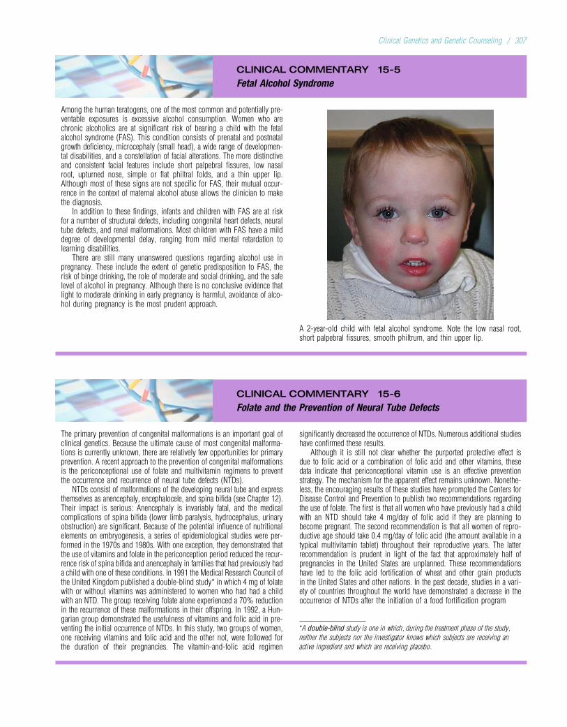
CLINICAL COMMENTARY 15-5
Fetal Alcohol Syndrome
Among the human teratogens, one of the most common and potentially pre-ventable exposures is excessive alcohol consumption. Women who arechronic alcoholics are at significant risk of bearing a child with the fetalalcohol syndrome (FAS). This condition consists of prenatal and postnatalgrowth deficiency, microcephaly (small head), a wide range of developmen-tal disabilities, and a constellation of facial alterations. The more distinctiveand consistent facial features include short palpebral fissures, low nasalroot, upturned nose, simple or flat philtral folds, and a thin upper lip.Although most of these signs are not specific for FAS, their mutual occur-rence in the context of maternal alcohol abuse allows the clinician to makethe diagnosis.
In addition to these findings, infants and children with FAS are at riskfor a number of structural defects, including congenital heart defects, neuraltube defects, and renal malformations. Most children with FAS have a milddegree of developmental delay, ranging from mild mental retardation tolearning disabilities.
There are still many unanswered questions regarding alcohol use inpregnancy. These include the extent of genetic predisposition to FAS, therisk of binge drinking, the role of moderate and social drinking, and the safelevel of alcohol in pregnancy. Although there is no conclusive evidence thatlight to moderate drinking in early pregnancy is harmful, avoidance of alco-hol during pregnancy is the most prudent approach.
A 2-year-old child with fetal alcohol syndrome. Note the low nasal root,short palpebral fissures, smooth philtrum, and thin upper lip.
CLINICAL COMMENTARY 15-6
Folate and the Prevention of Neural Tube Defects
The primary prevention of congenital malformations is an important goal ofclinical genetics. Because the ultimate cause of most congenital malforma-tions is currently unknown, there are relatively few opportunities for primaryprevention. A recent approach to the prevention of congenital malformationsis the periconceptional use of folate and multivitamin regimens to preventthe occurrence and recurrence of neural tube defects (NTDs).
NTDs consist of malformations of the developing neural tube and expressthemselves as anencephaly, encephalocele, and spina bifida (see Chapter 12).Their impact is serious: Anencephaly is invariably fatal, and the medicalcomplications of spina bifida (lower limb paralysis, hydrocephalus, urinaryobstruction) are significant. Because of the potential influence of nutritionalelements on embryogenesis, a series of epidemiological studies were per-formed in the 1970s and 1980s. With one exception, they demonstrated thatthe use of vitamins and folate in the periconception period reduced the recur-rence risk of spina bifida and anencephaly in families that had previously hada child with one of these conditions. In 1991 the Medical Research Council ofthe United Kingdom published a double-blind study* in which 4 mg of folatewith or without vitamins was administered to women who had had a childwith an NTD. The group receiving folate alone experienced a 70% reductionin the recurrence of these malformations in their offspring. In 1992, a Hun-garian group demonstrated the usefulness of vitamins and folic acid in pre-venting the initial occurrence of NTDs. In this study, two groups of women,one receiving vitamins and folic acid and the other not, were followed forthe duration of their pregnancies. The vitamin-and-folic acid regimen
significantly decreased the occurrence of NTDs. Numerous additional studieshave confirmed these results.
Although it is still not clear whether the purported protective effect isdue to folic acid or a combination of folic acid and other vitamins, thesedata indicate that periconceptional vitamin use is an effective preventionstrategy. The mechanism for the apparent effect remains unknown. Nonethe-less, the encouraging results of these studies have prompted the Centers forDisease Control and Prevention to publish two recommendations regardingthe use of folate. The first is that all women who have previously had a childwith an NTD should take 4 mg/day of folic acid if they are planning tobecome pregnant. The second recommendation is that all women of repro-ductive age should take 0.4 mg/day of folic acid (the amount available in atypical multivitamin tablet) throughout their reproductive years. The latterrecommendation is prudent in light of the fact that approximately half ofpregnancies in the United States are unplanned. These recommendationshave led to the folic acid fortification of wheat and other grain productsin the United States and other nations. In the past decade, studies in a vari-ety of countries throughout the world have demonstrated a decrease in theoccurrence of NTDs after the initiation of a food fortification program
*A double-blind study is one in which, during the treatment phase of the study,
neither the subjects nor the investigator knows which subjects are receiving an
active ingredient and which are receiving placebo.
Clinical Genetics and Genetic Counseling / 307
BIOETHICS AND MEDICAL GENETICSWith new discoveries and advances in medical technologycome new choices for patients, families, and society. As med-ical genetics came to be defined as a medical specialty duringthe past several decades, a number of new issues in bioethicsalso emerged. Because of the significance and complexity ofthese issues, a significant portion of the budget of the HumanGenome Project has been devoted to the ethical, legal, andsocial implications of human genetics. Some of these implica-tions have been touched upon previously in this textbook(e.g., genetic testing, gene therapy, embryonic stem cellresearch). Our goal here is to provide a sampling of the majorethical questions currently confronting the medical andgenetics communities.
The combination of advances in prenatal diagnosis (e.g.,ultrasound, amniocentesis), the ability to determine thehuman karyotype, and the option of pregnancy terminationset the stage for the rise of prenatal diagnosis as a clinicalservice in the 1970s. By that time, most tertiary medical cen-ters in developed nations offered amniocentesis for variousindications, most commonly advanced maternal age (seeChapter 13). In the early discussions of the ethics of prenataldiagnosis, the central controversy involved a woman’s (orcouple’s) right to terminate a pregnancy. In the 1990s, theissue took on a new dimension with the concern that personswith disabilities may be devalued by society when prenataldiagnosis can lead to the selective termination of fetuseswith a disability. Similar concerns revolve around the issueof withdrawal of support for newborns with severe birthdefects (e.g., trisomy 13, some neural tube defects). The mainprinciples guiding decisions about these issues are to considerthe interests of the child and to provide genetic counseling sothat the parents can make an informed decision.
Ethical concerns have arisen about other types of genetictesting, including carrier testing and presymptomatic testing(see Chapter 13). Genetic tests differ from other types ofmedical testing because genes (including mutations that pre-dispose to disease) can be shared in families. Thus, a genetictest performed in one person might reveal risk informationabout a relative who might not wish to know about his orher risk (e.g., testing of a young adult for an autosomal dom-inant disease might indicate that one of the parents musthave transmitted the disease-causing mutation). In addition,many people perceive that their genetic inheritance is verymuch an intrinsic part of themselves (and their families).Genetic risk is often misperceived as “unchangeable.” All ofthese factors can lead to unfair stigmatization of individuals,families, and even whole populations. To help counter this,care providers must be sensitive to the needs and concernsof individuals and their families. They should avoid makingvalue judgments that could lead to or reinforce stigmatiza-tion. They should dispel notions of genetic determinism,making it clear to families that genes are but one part ofthe cause of a disease. Nongenetic factors, which oftencan be altered, can also play an important role. As with allmedical information, privacy and confidentiality must berespected.
Genetic testing also raises the specter of discrimination byinsurance companies or employers. Insurance companies havelong collected information about family history as a means ofassessing risk. In some cases (e.g., an individual at risk for inher-iting a BRCA1 mutation), a genetic test can provide a muchmore accurate measure of disease risk. At-risk persons areunderstandably concerned about the possibility of losing theirinsurance benefits or their employment because of the outcomeof a genetic test. A paradoxical result is that some choose not tobe tested, even when it could lead to a potentially life-savingintervention. Insurance providers and employers reason thatdenying coverage (or increasing premiums) to at-risk indivi-duals serves the general interest by minimizing costs. Othersrespond that, unlike lifestyle choices such as cigarette smokingor exercise, one does not choose one’s genes, so it is unfair todiscriminate on the basis of genetic tests.
Because of concerns in the United States regarding dis-crimination in employment and health insurance, the genet-ics community and supportive legislators worked togetherduring the early 2000s to enact legislation to ensure the con-fidentiality of genetic test results. The Genetic InformationNondiscrimination Act (GINA) was formulated to preventthe discriminatory use of genetic test results by employersor health insurers. GINA became a federal law in 2008 andtook effect in 2009.
Preimplantation genetic testing (see Chapter 13) has alsobeen the focus of ethical debates. For example, this form oftesting can be used to determine the sex of an embryo.Indeed, an early application was to avoid implanting maleembryos that had an increased risk of carrying an X-linkedrecessive mutation. Many scientists and ethicists believe thatpreimplantation diagnosis solely for sex selection is inappro-priate, and the practice is currently banned in the UnitedKingdom. Preimplantation diagnosis could also be used toselect preferentially for embryos that do carry disease-causingmutations. For example, an embryo that is homozygous formutations causing autosomal recessive deafness could beselected to match the phenotypes of deaf parents (a casewas reported in which deaf parents deliberately conceiveda deaf child by artificial insemination). Such applicationsmight put the interests of the parents and the interests ofthe child in conflict. Another controversial application ofpreimplantation diagnosis was in the selection of a humanleukocyte antigen (HLA)-matched embryo that could laterprovide bone marrow cells for an older sibling who had Fan-coni anemia. It is argued that such persons could feel thattheir life was devalued because they had been selected partlyon the basis of their suitability as a bone marrow donor.
Questions have arisen about the genetic testing of chil-dren. When such testing can lead to useful diagnostic mea-sures or interventions, it may be warranted. An examplewould be the genetic testing of children at risk for inheritinga mutation in the adenomatous polyposis coli (APC) gene. Asdiscussed in Chapter 11, colonoscopy should begin in genecarriers by age 12 years and is potentially life-saving. In con-trast, childhood diagnosis of Huntington disease currentlyprovides no preventive or therapeutic benefits and increases
308 / Chapter 15 MEDICAL GENETICS
the potential for anxiety and stigmatization. A consensus hasemerged that childhood genetic testing should not be pur-sued unless it provides an avenue to clinically beneficialintervention.
Intense controversy has surrounded the issues of cloningand embryonic stem cell research (see Chapter 13). It isimportant to repeat the distinction between reproductive clon-ing and therapeutic cloning. Because of the high failure rate ofreproductive cloning in other mammals, and because thebenefits of reproductive cloning are unclear, the scientificcommunity is nearly unanimous in its opposition to thecreation of human beings through reproductive cloning.The use of cloning to create embryonic stem cells for ther-apeutic purposes (e.g., pancreatic islet cells for patientswith diabetes or neurons for those with Alzheimer disease)is more controversial and, like pregnancy termination,involves highly charged questions about the definition ofhuman life and the limits of medical intervention. As noted
in Chapter 13, these issues require informed and thoughtfulinput from the scientific community and from patient advo-cacy groups, ethicists, philosophers, legal scholars, theclergy, and others.
The science of genetics is no stranger to controversy andeven to abuse. The eugenics movement (Greek, “goodbirth”), popular in the United States and some Europeancountries in the early part of the 20th century, advocatedboth “positive eugenics” (preferential reproduction of thosedeemed to be genetically more fit) and “negative eugenics”(the prevention of reproduction of those thought to begenetically less fit). Eugenics, in concert with the politicalthinking of the times, led to a series of abuses that culmi-nated in the atrocities of Nazi Germany. These events area sobering reminder of the potential for misuse of geneticinformation. Geneticists must help to ensure that their sci-ence is used for maximum benefit while adhering to thetime-honored maxim primum non nocere (“first, do no harm”).
Study Questions
1. Allen, a 40-year-old man, comes to your officebecause he is concerned about his family history ofheart disease. His father had a fatal myocardialinfarction (MI) at 45 years of age, and his paternalgrandfather had a fatal MI at age 47 years. Allen’sfather had two brothers and two sisters. One of thebrothers had an MI at age 44 years, and one of thesisters had an MI at age 49 years. Allen’s mother had abrother and sister, both of whom are still alive. Allen’smother’s parents both survived into their 80s and diedof “natural causes.” Draw a pedigree summarizing theinformation you have taken about Allen’s family, andmake a recommendation for further study and/ortreatment.
2. Mary’s two brothers and her mother’s brother all hadDuchenne muscular dystrophy (DMD) and are nowdead. Based on only this information, what is theprobability that Mary is a heterozygous carrier for this
disorder? What is the probability that she will produceaffected offspring? Suppose Mary has a serum creatinekinase (CK) test and is told that her level is above the95th percentile for homozygous normal individuals.Approximately two thirds of DMD carriers have CKlevels above the 95th percentile. Given thisinformation, use Bayes’ theorem to calculate theprobability that Mary is a carrier and the probabilitythat she will produce affected offspring.
3. Bob’s father had Huntington disease and is nowdeceased. Bob is now 51 years old and has nosymptoms of Huntington disease. Age-of-onset curvesshow that approximately 85% of individuals with anaffected father show symptoms by this age (thepercentage is slightly lower, about 80%, if the motheris affected). Based on this information, use Bayes’theorem to estimate the probability that Bob inheritedthe Huntington disease mutation from his father.
Suggested ReadingsAase J. Dysmorphologic diagnosis for the pediatric practi-
tioner. Pediatr Clin North Am 1992;39:135–56.Baker DL, Schuette JL, Uhlmann WR. A Guide to Genetic
Counseling. New York: John Wiley; 1998.Baty B Biesecker B. Evidence-based genetic counseling. (Spe-
cial issue on genetic counseling). Am J Med Genet C2007;142C:220.
Biesecker BB. Goals of genetic counseling. Clin Genet2001;60:323–30.
Biesecker BB, Peters KF. Process studies in geneticcounseling: peering into the black box. (Special issue
on genetic counseling). Am J Med Genet 2001; 106:191–8.
Boulet SL, Yang Q, Mai CL, et al. Trends in the postfortifi-cation prevalence of spina bifida and anencephaly in theUnited States. Birth Defects Res A Clin Mol Teratol2008;82:527–32.
Brent RL. Environmental causes of human congenital malfor-mations: the pediatrician’s role in dealing with these com-plex clinical problems caused by a multiplicity ofenvironmental and genetic factors. Pediatrics 2004;113:957–68.
Clinical Genetics and Genetic Counseling / 309
Brent RL. The Bendectin saga: an American tragedy. Teratol-ogy 1983;27:283–6.
Carey JC, Viskochil DH. Status of the human malformationmap: 2007. Am J Med Genet A 2007;143A(24):2868–85.
Cassidy SB, Allanson JE. Management of Genetic Syn-dromes. 3rd ed. Hoboken, NJ: John Wiley; 2009.
Clarke A. Ethical and social issues in clinical genetics. In:Rimoin DL, Connor JM, Pyeritz RE, Korf BR, editors.Emery and Rimoin’s Principles and Practice of MedicalGenetics. 4th ed., vol. 1. Philadelphia: Churchill Living-stone; 2002. p. 897–928.
Clayton EW. Ethical, legal, and social implications of geno-mic medicine. N Engl J Med 2003;349:562–9.
Cohen MM. The Child with Multiple Birth Defects. NewYork: Oxford; 1997.
Dent K, Carey JC. Breaking difficult news in the newbornsetting: Down Syndrome as a paradigm. Am J of MedGenet C 2006;142C:173–9.
Donnai D. Genetic services. Clin Genet 2002;61:1–6.Friedman JM, Polifka JE. Teratogenic Effects of Drugs: A
Resource for Clinicians (TERIS). Baltimore: Johns Hop-kins University Press; 2000.
Hennekam RCM, Allanson J, Krantz I. Gorlin’s Syndromesof the Head and Neck. 5th ed. New York: Oxford Univer-sity Press; 2009.
Harper PS. Practical Genetic Counseling. 5th ed. Oxford:Butterworth Heineman; 1999.
Hodge SE. A simple, unified approach to Bayesian risk calcu-lations. J Genet Couns 1998;7:235–61.
Hunter AG. Medical genetics: 2. The diagnostic approachto the child with dysmorphic signs. CMAJ 2002;167:367–72.
Jones KL. Smith’s Recognizable Patterns of Human Malfor-mation. 6th ed. Philadelphia: WB Saunders; 2006.
Koren G. Medication Safety in Pregnancy and Breast Feed-ing. New York: McGraw-Hill; 2007.
Mahowald MB, Verp MS, Anderson RR. Genetic counseling:clinical and ethical challenges. Annu Rev Genet1998;32:547–59.
Martinez LP, Robertson J, Leen-Mitchell M. Environmentalcauses of birth defects. In:Rudolph CD, Rudolph AM, Hos-tetter MK, Lister G, editors. Rudolph’s Pediatrics. 22nd ed.New York: McGraw-Hill; 2009, p. 774–9.
Nelson K, Holmes LB. Malformations due to presumedspontaneous mutations in newborn infants. N Engl JMed 1989;320:19–23.
Nowlan W. Human genetics: a rational view of insuranceand genetic discrimination. Science 2002; 297:195–6.
Polifka JE, Friedman JM. Medical genetics: 1. Clinical tera-tology in the age of genomics. CMAJ 2002; 167:265–73.
Resta R. Defining and redefining the scope and goals ofgenetic counseling. Am J Med Genet Part C 2007;142C:269–75.
Rothenberg KH, Terry SF. Human genetics: before it’s toolate—addressing fear of genetic information. Science2002;297:196–7.
Schneider KA. Counseling About Cancer: Strategies forGenetic Counselors. 2nd ed. New York: John Wiley;2001.
Walker AP. Genetic counseling. In: Rimoin DL, Connor JM,Pyeritz RE, Korf BR, editors. Emery and Rimoin’s Princi-ples and Practice of Medical Genetics. 4th ed., vol 1.New York: Churchill Livingstone; 2002, p. 842–74.
Weil J. Psychosocial Genetic Counseling. New York:Oxford University Press; 2000.
Weiss JO, Mackta JS. Starting and Sustaining Genetic Sup-port Groups. Baltimore: Johns Hopkins University Press;1996.
Internet ResourcesGene Clinics (reviews of tests for genetic diseases andlaboratories that perform them) http://www.geneclinics.org/
Genetic Alliance (descriptions of genetic conditions,information on health insurance, and links to lay advocacygroups) http://www.geneticalliance.org/
Genetic and Rare Conditions Site (links to lay advocacygroups for a large number of genetic disorders) http://www.kumc.edu/gec/support/
Organization of Teratology Information Specialists (reviewsof various exposures in pregnancy): http://www.otispregnancy.org
Genetic Interest Group (alliance of organizations, with amembership of more than 120 charities that supportchildren, families, and individuals affected by geneticdisorders) http://www.gig.org.uk/
National Institutes of Health Office of Rare Diseases(information on more than 6000 rare diseases)http://rarediseases.info.nih.gov/
POSSUM (computer-aided diagnosis of genetic disordersand other syndromes; paid subscription required) http://www.possum.net.au/
310 / Chapter 15 MEDICAL GENETICS
GLOSSARY
NOTE: When boldface type is used to highlight a word orphrase within a definition, it signifies that the highlightedword or phrase is defined elsewhere in this glossary.
50 cap A chemically modified guanine nucleotide added tothe 50 end of a growing mRNA molecule.
acceptor site AG sequence that defines the splice site atthe 30 end of an intron.
acetylation The addition of an acetyl group to a molecule(as in histone acetylation).
acrocentric A chromosome whose centromere is close tothe end of one arm.
activator A specific transcription factor that binds tocoactivators and to enhancers to help regulate the transcrip-tional activity of certain genes.
adaptive immune system The portion of the immunesystem that is capable of changing its DNA sequence to bindforeign particles more effectively. It includes the humoral andcellular components. Compare with innate immune system.
addition rule A law of probability that states that the prob-ability of one event or another event occurring is derived byadding the probabilities of the two events together, assumingthe events occur independently of one another.
adenine One of the four DNA bases (abbrev.: A).
adeno-associated virus A type of parvovirus sometimesused as a vector for somatic cell gene therapy.
adenovirus A double-stranded RNA virus that is sometimesused in gene therapy.
adjacent segregation A meiotic segregation pattern inwhich the pairing of translocated chromosomes leads tounbalanced gametes. Compare with alternate segregation.
affected sib-pair method A linkage analysis method inwhich sib pairs who are both affected with a disease are assessedfor the extent to which they share alleles at various marker loci.If alleles are shared significantly more often than the expected50%, linkage of the disease to the marker is indicated.
affinity The binding power of an antibody with an antigen(low affinity indicates poor binding; high affinity indicatesprecise binding).
affinity maturation A phase of B cell development inwhich the cell undergoes somatic hypermutation so thatsome cells are formed that can achieve high-affinity bindingto peptides from a pathogen.
allele Conventional abbreviation for “allelomorph.” Refersto the different forms, or DNA sequences, that a gene canhave in a population.
allele-specific oligonucleotide A short DNA sequence,usually 18 to 20 nucleotides, that can hybridize with eitherdisease-causing or normal DNA sequences. Used in directdiagnosis of mutations.
alpha-fetoprotein Albumin-like protein produced by thefetus. The level of alpha-fetoprotein is elevated in pregnan-cies with neural tube defects and may be decreased in preg-nancies with Down syndrome.
alpha-satellite DNA A type of repetitive DNA sequencefound near centromeres.
alternate segregation A meiotic segregation pattern inwhich the pairing of translocated chromosomes leads to bal-anced gametes. Compare with adjacent segregation.
alternative splice site A variation in the location ofintron–exon splice sites in some genes that allow one geneto produce multiple different protein products.
Alu family A major group of dispersed repetitive DNAsequences.
amino acids The major building blocks of polypeptides.Each of the 20 amino acids is encoded by one or more mRNAcodons.
amniocentesis A prenatal diagnostic technique in whicha small amount of amniotic fluid is withdrawn transab-dominally at about 16 weeks after the last menstrualperiod. Fetal cells can then be tested for some geneticdiseases.
amniocentesis, early Amniocentesis carried out at approx-imately 12 to 14 weeks after the last menstrual period.
amniocyte Fetal cell found in the amniotic fluid.
anaphase One of the stages of cell division, in which sisterchromatids separate and move toward opposite sides ofthe cell.
311
ancestry The origins of an individual’s ancestors; mostcommonly used to refer to the geographic origins of one’sancestors.
aneuploid The condition in which the number of chromo-somes is not a multiple of 23, as in trisomy and monosomy.Compare with euploid. (n.: aneuploidy).
antibody Molecule produced by plasma cells; antibodiesbind to invading antigens.
anticipation A feature of pedigrees in which a disease isseen at earlier ages or with increased severity in more recentgenerations.
anticodon A three-nucleotide DNA sequence in a tRNAmolecule that undergoes complementary base pairing withan mRNA codon.
antigen A molecule that provokes antibody formation(from “antibody generator”).
antigen-presenting cell A cell that engulfs foreign bodies,digests them, and then displays the foreign antigens on itscell surface for recognition by T lymphocytes.
antisense strand In a double-stranded DNA molecule, thestrand from which mRNA is transcribed. See sense strand.
antisense therapy A type of somatic cell gene therapy inwhich an oligonucleotide is synthesized that can hybridizewith a mutant mRNA sequence, blocking its translation intoprotein.
apoptosis Programmed cell death.
association The co-occurrence of two traits or eventsmore often than expected by chance.
autoimmunity The condition in which one’s immunesystem attacks one’s own cells.
autoradiogram The image produced by exposing a radio-actively labeled substance, such as a probe, to x-ray film(used, for example, in detecting RFLPs and in performingin situ hybridization).
autosomes The 22 pairs of chromosomes excluding thesex chromosomes (X and Y).
axis specification Definition, during embryonic develop-ment, of the major axes of the embryo: ventral/dorsal andanterior/posterior.
B lymphocyte (also, B cell) A component of the adaptiveimmune system that produces antibodies.
bacterial artificial chromosome (BAC) A recombinantplasmid inserted in bacteria that serves as a cloning vectorcapable of accepting DNA inserts of 50 to 200 kb.
bacteriophage A virus that infects bacteria. In recombinantDNA technology, bacteriophages are used as vectors tocarry inserted DNA sequences.
bacteriophage P1 artificial chromosome (PAC) A cloningvector that consists of bacteriophage P1 and is inserted intoa plasmid; it accepts DNA inserts up to 100 kb.
bands (1) Visibly darkened areas on autoradiograms thatrepresent the location of alleles on a gel. (2) Alternating darkand light areas visible on chromosomes after certain types ofstains are used.
Barr body The inactive X chromosome, visible as a denselystaining chromatin mass in the somatic cells of normalfemales. Also known as sex chromatin.
base One of the four nitrogenous substances (adenine,cytosine, guanine, or thymine) that make up part of theDNA molecule. Combinations of bases specify amino acidsequences.
base analog A substance that can mimic the chemicalbehavior of one of the four DNA bases. Base analogs are atype of mutagen.
base pair (bp) A unit of complementary DNA bases in adouble-stranded DNA molecule (A-T or C-G).
base pair substitution The replacement of one base pairby another. A type of mutation.
Bayes’ theorem A statistical procedure in which prior andconditional probabilities are used to derive an improved esti-mate of probability or risk.
benign Describes a neoplasm (tumor) that does not invadesurrounding tissue or metastasize to other parts of the body.Compare with malignant.
bivalent A pair of intertwined homologous chromosomesseen in prophase I of meiosis. Synonymous with tetrad.
blood group Molecules found on the surfaces of erythro-cytes, some of which (ABO and Rh) determine blood trans-fusion compatibility.
body plan The pattern and arrangement of body segmentsduring embryonic development.
bp Abbreviation for base pair.
breakpoint The location on a chromosome at which atranslocation has occurred.
C-banding A type of chromosome staining that highlights theconstitutive heterochromatin that lies at and near centromeres.
candidate gene A gene that, on the basis of known prop-erties or protein product, is thought to be the gene causinga specific genetic disease.
capillary sequencing A method of DNA sequencing inwhich DNA migrates through a thin capillary tube in orderto separate DNA fragments of different lengths.
carcinogen A substance that can produce cancer (adj.:carcinogenic).
carcinogenesis The process of cancer development.
carrier A person who has a copy of a disease-causing genebut does not express the disease. The term is usually used todenote heterozygotes for a recessive disease gene.
catalyst A substance that increases the rate of a chemicalreaction. Enzymes are an example of a catalyst.
312 / GLOSSARY
cDNA Complementary DNA, formed by reverse transcrip-tion of mRNA purified from a collection of cells. This typeof DNA corresponds only to coding sequence (exons).
cDNA library A collection of segments of complementaryDNA (cDNA) cloned into vehicles such as phages or plas-mids. Compare with genomic library.
cell adhesion molecule Cell-surface molecule that partici-pates in the interaction of T cells and their targets.
cell cycle The alternating sequence of mitosis andinterphase.
cell fate The location and function of cells, programmedduring embryonic development.
cellular immune system The T-cell component of theadaptive immune system.
centimorgan (cM) A unit of measure of the frequency ofrecombination between two loci, also known as a map unit.One cM corresponds to a recombination frequency of 1%.
centriole Structure in cells that helps to pull chromosomesapart during meiosis and mitosis.
centromere The region of a chromosome that separatesthe two arms; centromeres are the sites of attachment ofspindle fibers during cell division.
CG islands Unmethylated CG sequences that are foundnear the 50 ends of many genes.
chiasma (pl. chiasmata) The location of a crossoverbetween two homologous chromosomes during meiosis.
chorionic villus sampling (CVS) A prenatal diagnostictechnique in which a small sample of chorionic villi is aspi-rated. Usually performed at 10 to 12 weeks’ gestation.
chromatin The combination of proteins (e.g., histones)and nucleic acids that makes up chromosomes.
chromatin loop A unit of DNA coiling consisting of agroup of solenoids. Each loop is approximately 100 kb.
chromatin opener Regulatory element that is capable ofdecondensing or “opening” regions of chromatin.
chromosome Threadlike structure (literally “colored body”)consisting of chromatin. Genes are arranged alongchromosomes.
chromosome abnormalities A major group of genetic dis-eases, consisting of microscopically observable alterationsof chromosome number or structure.
chromosome banding The process of applying specificstains to chromosomes in order to produce characteristicpatterns of bands (example: G-banding).
chromosome breakage The fracture of chromosomes;breakage is increased in the presence of clastogens.
chromosome instability syndrome Diseases characterizedby the presence of large numbers of chromosome breaks orexchanges, such as sister chromatid exchange (example:Bloom syndrome).
chromosome-specific library A collection of DNA frag-ments from a single chromosome.
class switching The process in which the heavy chains ofB lymphocytes change from one class, or isotype, to another(e.g., IgM to IgG).
clastogen A substance that can induce chromosomebreakage (example: radiation).
clone (1) A series of identical DNA fragments created byrecombinant DNA techniques. (2) Identical cells that aredescended from a single common ancestor.
cloning, functional A method of isolating genes in whicha gene whose protein product’s function is already knownis evaluated as a candidate gene responsible for a trait ordisease.
cloning, positional The isolation and cloning of a diseasegene after determining its approximate physical location;the gene product is subsequently determined. Formerlytermed “reverse genetics.”
coactivator A type of specific transcription factor thatbinds to activators and to the general transcription factorcomplex to regulate the transcription of specific genes.
codominant Alleles that are both expressed when theyoccur together in the heterozygous state (example: A and B
alleles of the ABO blood group system).
codon A group of three mRNA bases, each of which speci-fies an amino acid when translated.
coefficient of relationship A statistic that measures theproportion of genes shared by two individuals as a result ofdescent from a common ancestor.
cofactors Substances that interact with enzymes to pro-duce chemical reactions, such as various metabolic processes(examples: dietary trace elements, vitamins).
colcemid or colchicine A spindle poison that arrests cells inmetaphase, making them easily discernible microscopically.
comparative genomic hybridization (CGH) Technique inwhich a mixture of DNA from a test source (e.g., a tumor)and a normal control are differentially labeled, mixed, andhybridized with normal metaphase chromosomes or a micro-array (array CGH). Differences in color reveal chromosomelosses or duplications in the test DNA.
complement system A component of the immune system,encoded by genes in the class III MHC region, that candestroy invading organisms. The complement system alsointeracts with other components of the immune system, suchas antibodies and phagocytes.
complementary base pairing A fundamental process inwhich adenosine pairs only with thymine and guanine pairsonly with cytosine. Also sometimes known as Watson–Crickpairing.
compound heterozygote An individual who is heterozy-gous for two different disease-causing mutations at a locus.
Glossary / 313
Compare with homozygote. Compound heterozygotes forrecessive disease mutations are usually affected with thedisorder.
concordant Refers to two individuals who have the sametrait (e.g., monozygotic twins may be concordant for adisease such as diabetes). Compare with discordant.(n.: concordance).
conditional probability The probability that an event willoccur, given that another event has already occurred. Con-ditional probabilities are used, for example, in Bayes’theorem.
confined placental mosaicism A form of mosaicism that isobserved in the placenta but not in the fetus.
congenital Present at birth.
consanguinity The mating of related individuals (adj.:consanguineous).
consensus sequence A sequence that denotes the DNAbases most often seen in a region of interest. The sequencesthat are found near donor and acceptor sites are a type ofconsensus sequence.
conservation The preservation of highly similar DNAsequences among different organisms; conserved sequencesare usually found in functional genes.
conserved See conservation.
constitutional or constitutive Pertaining to DNA in normalcells of the body, usually used in contrast to tumor DNA.
constitutive heterochromatin See heterochromatin, con-stitutive.
contig map A physical map of a chromosome region con-structed by isolating overlapping (contiguous) DNA segments.
contiguous gene syndrome A disease caused by the dele-tion or duplication of multiple consecutive genes. See alsomicrodeletion.
copy number variant (CNV) DNA sequences of 1000 ormore base pairs that are present in variable numbers in differ-ent individuals.
cordocentesis See percutaneous umbilical blood sam-pling (PUBS).
cosmid A phage–plasmid hybrid capable of acceptinglarger DNA inserts (up to 40-50 kb) than either phage orplasmids.
costimulatory molecule Cell-surface molecule that partici-pates in the binding of T-cell receptors to MHC–antigencomplexes.
cross Mating between organisms in genetic studies.
cross-reaction The binding of an antibody to an antigenother than the one that originally stimulated antibody for-mation. Such antigens are usually very similar to the originalantibody-generating antigen.
crossing over or crossover The exchange of genetic mate-rial between homologous chromosomes during meiosis (alsooccurs rarely during mitosis); produces recombination.
cryptic splice site Site at which intron–exon splicing canoccur when the usual splice site is altered.
cyclin-dependent kinases Enzymes that form complexeswith specific cyclins to phosphorylate regulatory proteins(such as pRb) at specific stages of the cell cycle.
cyclin-dependent kinase inhibitors Proteins that inacti-vate cyclin-dependent kinases. Many of these are tumor sup-pressors (examples: p16, p21).
cyclins Proteins that interact with specific cyclin-dependentkinases to regulate the cell cycle at specific stages.
cytogenetics The study of chromosomes and theirabnormalities. Combines cytology, the study of cells, andgenetics.
cytokine A growth factor that causes cells to proliferate(example: interleukins).
cytokinesis Cytoplasmic division that occurs during mito-sis and meiosis.
cytosine One of the four DNA bases (abbrev.: C).
cytotoxic T lymphocyte A type of T lymphocyte thatdestroys a cell when the cell presents a complex of MHCclass I molecule and foreign peptide. Part of the cellularimmune system.
daughter cells Cells that result from the division of a par-ent cell.
deformation Alteration of the form, shape, or position of anormally formed body part by mechanical forces (example:oligohydramnios sequence).
deletion The loss of chromosome material. May be termi-nal or interstitial. Compare with duplication.
deletion, interstitial Deletion that removes part of theinterior of the chromosome.
deletion, terminal Deletion that removes part of a chro-mosome, including one telomere.
denaturing gradient gel electrophoresis (DGGE) See elec-trophoresis, denaturing gradient gel.
deoxyribonucleic acid See DNA.
derivative chromosome A chromosome that has beenaltered as a result of a translocation [example: derivative 9,or der(9)].
dideoxy method A technique for sequencing DNA inwhich dideoxynucleotides, which terminate replication, areincorporated into replicating DNA strands.
diploid Having two copies of each chromosome. In humans,the diploid number is 46. Compare with haploid, polyploid.
direct cDNA selection A method of exon detection inwhich a genomic DNA fragment inserted into a vector suchas a YAC is hybridized with cDNA; hybridization and
314 / GLOSSARY
selection identify the regions of the genomic DNA fragmentthat correspond to cDNA (i.e., exon-containing DNA).
direct diagnosis A form of DNA-based disease diagnosisin which the mutation itself is examined directly. Comparewith indirect diagnosis.
discordant Refers to two individuals who do not share thesame trait. Compare with concordant. (n.: discordance).
dispermy Fertilization of a single ovum by two sperm cells.
dispersed repetitive DNA A class of repeated DNAsequences in which single repeats are scattered throughoutthe genome. Compare with tandem repeat.
disruption Morphological defect that results from thebreakdown of an otherwise normal developmental process(example: limb reduction defect resulting from poorvascularization).
dizygotic A type of twinning in which each twin is pro-duced by the fertilization of a different ovum. Synonymouswith fraternal twin. Compare with monozygotic.
DNA (deoxyribonucleic acid) A double-helix molecule thatconsists of a sugar-phosphate backbone and four nitrogenousbases (A, C, G, and T). DNA bases encode messenger RNA(mRNA), which in turn encodes amino acid sequences.
DNA-binding motifs Portions of transcription factors thatallow them to interact with specific DNA sequences (exam-ples: helix-loop-helix motif, zinc finger motif).
DNA chips See microarrays.
DNA looping The formation of looped structures in DNA;sometimes permits the interaction of various regulatoryelements.
DNA mismatch repair A type of DNA repair in whichnucleotide mismatches (i.e., violations of the G-C, A-T com-plementary base-pairing rule) are corrected by specializedenzymes.
DNA polymerase An enzyme involved in DNA replicationand repair.
DNA profile A series of DNA polymorphisms (usuallyVNTRs or microsatellites) typed in an individual. Becausethese polymorphisms are highly variable, the combined gen-otypes are useful in identifying individuals for forensicpurposes.
DNA repair A process in which mistakes in the DNAsequence are altered to represent the original sequence.
DNA sequence The order of DNA bases along achromosome.
DNA sequencing, automated DNA sequencing techniquesin which automated procedures such as computer-guidedlaser scanning are used to provide more rapid, accurateDNA sequence results.
dominant An allele that is expressed in the same way insingle copy (heterozygotes) as in double copy (homozy-gotes). Compare with recessive.
dominant negative A type of mutation in which the alteredprotein product in a heterozygote forms a complex with thenormal protein product produced by the homologous normalgene, thus disabling it.
donor site The GT sequence that defines the splice site atthe 50 end of an intron.
dosage compensation The situation in which, as a conse-quence of X inactivation, the amount of X chromosome–encoded gene product in females is roughly equal to thatin males.
dosage mapping A technique for mapping genes in whichexcess or deficient gene product is correlated with a chromo-somal duplication or deletion.
dosage sensitivity A condition in which alteration of thelevel of a gene product (e.g., a deletion resulting in 50%expression or a duplication resulting in 150% expression ofthe gene product) causes a substantial alteration of the phe-notype (including disease).
double helix The “twisted-ladder” shape of the double-stranded DNA molecule.
double-stranded DNA breaks A type of DNA breakage inwhich both strands are broken at a specific location.
duplication The presence of an extra copy of chromosomematerial. Compare with deletion.
dysmorphology The study of abnormal physical development.
dysplasia A defect in which cells are abnormally organizedinto tissue (example: bone dysplasia).
ectopic expression Expression of a gene product in anabnormal location or tissue type.
electrophoresis A technique in which charged moleculesare placed in a medium and exposed to an electrical field,causing them to migrate through the medium at differentrates, according to charge, length, or other attributes.
electrophoresis, denaturing gradient gel (DGGE) A methodof mutation detection in which DNA fragments are electro-phoresed through a gel in which there is a changing denatur-ing factor, such as temperature.
embryonic stem cell Cells, found in early embryos, thathave the potential to become any cell type (pluripotency).
empirical risk A risk estimate based on direct observationof data.
endocytosis A process in which molecules are transportedinto the interior of cells.
enhancer A regulatory DNA sequence that interacts withspecific transcription factors to increase the transcriptionof genes. Compare with silencer.
equational division The second major cycle of meiosis:meiosis II. Compare with reduction division.
equatorial plane The middle of the cell’s spindle, alongwhich homologous chromosomes are arranged duringmetaphase.
Glossary / 315
erythroblast A nucleated red blood cell; the precursor ofan erythrocyte.
erythrocyte A red blood cell.
euchromatin Chromatin that is light-staining during inter-phase and tends to be transcriptionally active. Compare withheterochromatin.
eugenics The use of controlled breeding to increase theprevalence of “desirable” genetic traits (positive eugenics)and to decrease the prevalence of “undesirable” traits (nega-tive eugenics).
eukaryotes Organisms whose cells have true nuclei.
euploid Refers to cells whose chromosome number is amultiple of 23 (in humans). (n.: euploidy).
exons Portions of genes that encode amino acids and areretained after the primary mRNA transcript is spliced. Com-pare with intron.
exon trapping A method for isolating exons in a fragmentof genomic DNA by using an in vitro cell system to artifi-cially splice out the introns.
expanded newborn screening See newborn screening.
expanded repeat A type of mutation in which a tandemtrinucleotide repeat increases in number (example: Hunting-ton disease).
expressed sequence tag (EST) Several hundred bp ofknown cDNA sequence, flanked by PCR primers. Becausethey are derived from cDNA libraries, these sequences rep-resent portions of expressed genes.
false negative A test result in which an affected individualis incorrectly identified as being unaffected with the diseasein question. Compare with false positive.
false positive A test result in which an unaffected individ-ual is incorrectly identified as being affected with the diseasein question. Compare with false negative.
fetoscopy A fetal visualization technique in which anendoscope is inserted through the abdominal wall. Some-times used in prenatal diagnosis.
fiber FISH See fluorescence in situ hybridization, fiber.
fibrillin A component of connective tissue; mutations inthe fibrillin gene can cause Marfan syndrome.
flow cytometry A technique whereby chromosomes can beindividually sorted.
fluorescence in situ hybridization (FISH) A molecularcytogenetic technique in which labeled probes are hybri-dized with chromosomes and then visualized under a fluores-cence microscope.
fluorescence in situ hybridization, fiber (fiber FISH) Aversion of the FISH technique in which fluorescentprobes are hybridized to interphase chromosomes thathave been manipulated so that the chromatin fibers arestretched, allowing high-resolution visualization andmapping.
founder effect A large shift in gene frequencies that resultswhen a small “founder” population, which contains limitedgenetic variation, is derived from a larger population.Founder effect can be considered a special case of geneticdrift.
frameshift mutation An alteration of DNA in which aduplication or deletion occurs that is not a multiple of threebase pairs.
fusion gene A gene that results from a combination of twogenes or parts of two genes.
gain of function A class of mutations that results in a pro-tein product that either is increased in quantity or has anovel function. Compare with loss of function.
gamete The haploid germ cell (sperm and ovum).
gametogenesis The process of gamete formation.
gastrulation The embryonic stage in which cells of theblastula are arranged to become a three-layer structure con-sisting of endoderm, mesoderm, and ectoderm.
gene The fundamental unit of heredity.
gene–environment interaction A mutual phenotypic effectof a gene and an environmental factor that is greater thanthe single effect of either factor alone (example: the effectof a1-antitrypsin deficiency and cigarette smoking on pul-monary emphysema).
gene family A group of genes that are similar in DNAsequence and have evolved from a single common ancestralgene; they may or may not be located in the same chromo-some region.
gene flow The exchange of genes between differentpopulations.
gene frequency In a population, the fraction of chromo-somes that contain a specific gene.
gene therapy The insertion or alteration of genes to cor-rect a disease.
gene therapy, germline Gene therapy that alters all cellsof the body, including the germline. Compare with genetherapy, somatic cell.
gene therapy, somatic cell Gene therapy that alterssomatic cells but not cells of the germline. Compare withgene therapy, germline.
genetic code The combinations of mRNA codons thatspecify individual amino acids.
genetic counseling The delivery of information aboutgenetic diseases (risks, natural history, and management) topatients and their families.
genetic drift An evolutionary process in which gene fre-quencies change as a result of random fluctuations in thetransmission of genes from one generation to the next. Driftis greater in smaller populations.
genetic engineering The alteration of genes; it typicallyinvolves recombinant DNA techniques.
316 / GLOSSARY
genetic mapping The ordering of genes on chromosomesaccording to recombination frequency. Compare with phys-ical mapping.
genetic screening The large-scale testing of defined popu-lations to identify those who are at increased risk of having adisease-causing gene.
genetic testing The analysis of DNA, RNA, or proteins totest for the presence of differences that might cause agenetic disease.
genome The totality of an organism’s DNA.
genome scan A gene-mapping approach in which markersfrom the entire human genome are tested for linkage with adisease phenotype.
genome-wide association studies (GWAS) A study designin which allele frequencies at large numbers of loci (typicallysingle nucleotide polymorphisms, SNPs) are compared indisease cases and unaffected controls. SNPs that show largefrequency differences between cases and controls are likelyto be located in or near genes that are responsible for thedisease.
genomic library A collection of DNA fragments from anorganism’s entire genome. It includes cDNA as well as non-coding DNA. Compare with cDNA library.
genomic instability An abnormal condition in which thereis a substantial increase in mutations throughout the genome.It can occur, for example, when a DNA repair system isdisabled.
genotype An individual’s allelic constitution at a locus.
genotype frequency The fraction of individuals in a popu-lation that carry a specific genotype.
genotype–phenotype correlation The relationship betweendifferent possible genotypes (i.e., different alleles) at a locusand the individual’s phenotype. Because of allelic heterogene-ity, different alleles at a locus can produce more or less severeexpression of a disease phenotype (e.g., missense vs. nonsensemutations).
germline Cells responsible for the production of gametes.
Giemsa A type of staining that produces G-bands inchromosomes.
globin A major component of the hemoglobin molecule.Globin is also found in the vertebrate myoglobin molecule.
growth factor A substance capable of stimulating cellproliferation.
growth factor receptor A structure on cell surfaces towhich growth factors can bind.
guanine One of the four DNA bases (abbrev.: G).
guanosine diphosphate (GDP) A partially dephosphory-lated form of guanosine triphosphate.
guanosine triphosphate (GTP) A molecule required for thesynthesis of peptide bonds during translation.
haploid Refers to cells that have one copy of each chromo-some, the typical state for gametes. In humans, the haploidnumber is 23.
haploinsufficiency Describes the situation in which 50%of the normal level of gene expression (i.e., in a heterozy-gote) is not sufficient for normal function.
haplotype The allelic constitution of multiple loci on a sin-gle chromosome. Derived from “haploid genotype.”
Hardy–Weinberg principle Specifies an equilibrium rela-tionship between gene frequencies and genotype frequenciesin populations.
heavy chain A major structural component of an antibodymolecule, having a higher molecular weight than the othermajor component, the light chain. There are five major typesof heavy chains in the human: g, m, a, d, and ε.
helper T lymphocyte A type of T lymphocyte whosereceptors bind to a complex of MHC class II molecule andforeign peptide on the surfaces of antigen-presenting cells.Part of the cellular immune system.
heme The iron-containing component of the hemoglobinmolecule; binds with oxygen.
hemizygous Refers to a gene that is present in only a singlecopy (hemi ¼ “half”). Most commonly refers to genes on thesingle male X chromosome but can refer to other genes inthe haploid state, such as the genes homologous to a deletedregion of a chromosome.
heritability The portion of population variance in a traitthat can be ascribed to genetic factors.
heterochromatin Dark-staining chromatin that is usuallytranscriptionally inactive and consists mostly of repetitiveDNA. Compare with euchromatin.
heterochromatin, constitutive Heterochromatin that con-sists of satellite DNA; located near centromeres and on theshort arms of acrocentric chromosomes.
heterodisomy The presence in a cell of two chromosomesderived from a single parent and none from the other parent(disomy). In heterodisomy, the two chromosomes are thenonidentical homologous chromosomes. Compare withisodisomy.
heterogeneity, allelic Describes conditions in which dif-ferent alleles at a locus can produce variable expression ofa disease. Depending on phenotype definition, allelic het-erogeneity can cause two distinct diseases, as in Duchenneand Becker muscular dystrophy.
heterogeneity, locus Describes diseases in which muta-tions at distinct loci can produce the same disease phenotype(examples: osteogenesis imperfecta; retinitis pigmentosa).
heteromorphism Variation in the microscopic appearanceof a chromosome.
heteroplasmy The existence of differing DNA sequencesat a locus within a single cell. Often seen in mitochondrialgenes.
Glossary / 317
heterotetramer A molecule consisting of four (tetra) subu-nits, at least one of which differs from the others. Comparewith homotetramer.
heterozygote An individual who has two different alleles ata locus. Compare with homozygote.
high-resolution banding Chromosome banding using pro-phase or prometaphase chromosomes, which are moreextended than metaphase chromosomes and thus yield morebands and greater resolution.
histone The protein core around which DNA is wound ina chromosome.
holandric Refers to Y-linked inheritance; a transmissionexclusively from father to son.
homeodomain A DNA-binding portion of transcriptionfactor proteins that are involved in embryonic development.
homologous (1) Refers to DNA or amino acid sequencesthat are highly similar to one another. (2) Describes chromo-somes that pair during meiosis, one derived from the indi-vidual’s father and the other from the mother.
homologs Chromosomes that are homologous.
homotetramer Molecule consisting of four (tetra) identicalsubunits. Compare with heterotetramer.
homozygote An individual in whom the two alleles at alocus are the same. Compare with heterozygote.
housekeeping genes Genes whose protein products arerequired for cellular maintenance or metabolism. Because oftheir central role in the life of the cell, housekeeping genesare transcriptionally active in all cells.
human artificial chromosome A synthetic chromosomeconsisting of an artificial centromere and telomeres and aninsert of human DNA that can be 5 to 10 Mb.
human leukocyte antigen (HLA) Older term for majorhistocompatibility complex (MHC).
humoral immune system The B-cell component of theadaptive immune system, termed humoral because antibodiesare secreted into the circulation.
immunodeficiency disease A class of diseases character-ized by insufficiencies in the immune response (example:severe combined immune deficiency).
immunodeficiency disease, primary Disorder of theimmune system that is caused directly by defects (usuallygenetic) in components or cells of the immune system.
immunodeficiency disease, secondary Disorder of theimmune system that is a consequence of an agent or defectthat originates outside the immune system (e.g., infection,radiation, drugs).
immunogenetics The study of the genetic basis of theimmune system.
immunoglobulin Receptor found on the surfaces of B cells.When secreted into the circulation by B cells that have maturedinto plasma cells, immunoglobulins are known as antibodies.
imprinting, genomic Describes process in which geneticmaterial is expressed differently when inherited from themother than when inherited from the father.
in situ hybridization Molecular gene mapping technique inwhich labeled probes are hybridized to stained metaphasechromosomes and then exposed to x-ray film to reveal theposition of the probe.
in vitro fertilization (IVF) A procedure in which the fertili-zation of an egg cell by a sperm cell is carried out in the lab-oratory. The embryo is then implanted in the mother’suterus.
incest The mating of closely related individuals, usuallydescribing the union of first-degree relatives.
independence A principle, often invoked in statistical anal-ysis, indicating that the occurrence of one event has noeffect on the probability of occurrence of another (adj.:independent).
independent assortment One of Mendel’s fundamentalprinciples; dictates that alleles at different loci are transmit-ted independently of one another.
index case See proband.
indirect diagnosis A form of genetic diagnosis in whichthe disease-causing mutation is not directly observed; usuallyrefers to diagnosis using linked markers. Compare withdirect diagnosis.
induction The influence or determination of developmentof one group of cells by a second group of cells.
innate immune system The portion of the immune systemthat does not change its characteristics to respond to infec-tions. Major part of the initial immune response. Comparewith adaptive immune system.
insert A DNA sequence that is placed into a vector, suchas a plasmid or cosmid, using recombinant DNA techniques.
interphase The portion of the cell cycle that alternateswith meiosis or mitosis (cell division). DNA is replicatedand repaired during this phase.
intraclass correlation coefficient A statistical measurethat varies between �1 and 1 and specifies the degree ofsimilarity of two quantities in a sample or population.
intron DNA sequence found between two exons. It is tran-scribed into primary mRNA but is spliced out in the forma-tion of the mature mRNA transcript.
inversion A structural rearrangement of a chromosome inwhich two breaks occur, followed by reinsertion of the chro-mosome segment, but in reversed order. It may be eitherparacentric or pericentric. See also inversion, paracentricand inversion, pericentric.
inversion, paracentric An inversion that does not includethe centromere.
inversion, pericentric An inversion that includes thecentromere.
318 / GLOSSARY
isochromosome A structural chromosome rearrangementcaused by the division of a chromosome along an axis per-pendicular to the usual axis of division; results in chromo-somes with either two short arms or two long arms.
isodisomy The presence in a cell of two identical chromo-somes derived from one parent and none from the other par-ent. Compare with heterodisomy.
isotype Classes of immunoglobulin molecules (e.g., IgA,IgE, IgG), determined by the type of heavy chain presentin the molecule.
joint probability The probability of two events bothoccurring.
karyotype A display of chromosomes, ordered according tolength.
kilobase (kb) One thousand DNA base pairs.
knockout An animal model in which a specific gene hasbeen disabled.
lentivirus A type of retrovirus that can enter nondividingcells.
light chain A major structural component of the antibodymolecule, consisting of either a k or a l chain. The lightchain has a lower molecular weight than the other majorcomponent, the heavy chain.
likelihood A statistic that measures the probability of anevent or a series of events.
LINEs (long interspersed elements) A class of dispersedrepetitive DNA in which each repeat is relatively long, upto 7 kb. Compare with SINEs.
linkage Describes two loci that are located close enoughon the same chromosome that their recombination fre-quency is less than 50%.
linkage disequilibrium A nonrandom association of allelesat linked loci in populations. Compare with linkageequilibrium.
linkage equilibrium Lack of preferential association ofalleles at linked loci. Compare with linkage disequilibrium.
linkage phase The arrangement of alleles of linked loci onchromosomes.
liposome A fat body sometimes used as a vector forsomatic cell gene therapy.
locus The chromosome location of a specific gene (pl.: loci).
locus control region A DNA sequence in the 50 region ofthe globin gene clusters that is involved in transcriptionalregulation.
LOD score The common logarithm of the ratio of the like-lihood of linkage at a specific recombination fraction to thelikelihood of no linkage.
loss of function A class of mutation in which the alterationresults in a nonfunctional protein product. Compare withgain of function.
loss of heterozygosity Describes a locus or loci at which adeletion or other process has converted the locus from het-erozygosity to homozygosity or hemizygosity.
Lyon hypothesis A proposal (now verified) that one Xchromosome is randomly inactivated in each somatic cellof the normal female embryo (lyonization).
macrophage A type of phagocyte that ingests foreignmicrobes and displays them on its surface for recognitionby T-cell receptors.
major gene A single locus responsible for a trait (some-times contrasted with a polygenic component).
major histocompatibility complex (MHC), class I Amembrane-spanning glycoprotein, found on the surfacesof nearly all cells, that presents antigen for recognition bycytotoxic T lymphocytes. Compare with MHC class II.
major histocompatibility complex (MHC), class II A mem-brane-spanning glycoprotein, found on the surfaces of anti-gen-presenting cells, that presents antigen for recognitionby helper T cells.
malformation A primary morphologic defect resultingfrom an intrinsically abnormal developmental process (exam-ple: polydactyly).
malignant Describes a tumor that is capable of invadingsurrounding tissue and metastasizing to other sites in thebody. Compare with benign.
manifesting heterozygote An individual who is heterozy-gous for a recessive trait but displays the trait. Most oftenused to describe females who are heterozygous for anX-linked trait and display the trait.
markers Polymorphisms, such as RFLPs, VNTRs, microsat-ellite repeats, and blood groups, that are linked to a diseaselocus.
mass spectrometry Analysis of the mass-to-charge ratio ofmolecules; can be used to sequence DNA and detectmutations.
mass spectrometry, tandem A form of mass spectrometryin which two mass spectrometry machines are used, the firstof which separates molecules according to mass, and the sec-ond of which assesses the mass and charge of the moleculesafter they have been fragmented.
maternal serum alpha-fetoprotein (MSAFP) Alpha-feto-protein present in the serum of pregnant women; used inprenatal screening for fetal disorders such as neural tubedefects and Down syndrome.
mature transcript Describes mRNA after the introns havebeen spliced out. Before splicing, the mRNA is referred toas a primary transcript.
maximum likelihood estimate A statistical procedure inwhich the likelihoods of a variety of parameter values are esti-mated and then compared to determine which likelihood isthe largest. Used, for example, in evaluating LOD scores todetermine which recombination frequency is the most likely.
Glossary / 319
megabase (Mb) One million base pairs.
meiosis Cell division process in which haploid gametes areformed from diploid germ cells.
meiotic failure Aberrant meiosis in which a diploid gam-ete is produced rather than the normal haploid gamete.
memory cells A class of high-affinity-binding B cells thatremain in the body after an immune response has ended;they provide a relatively rapid, high-affinity response shouldthe same disease antigen be encountered a second time.
mendelian Referring to Gregor Mendel; describes a traitthat is attributable to a single gene.
mesenchyme Tissue that forms connective tissues and lym-phatic and blood vessels during embryonic development.
messenger RNA (mRNA) An RNA molecule that is formedfrom the transcription of DNA. Before intron splicing,mRNA is termed a primary transcript; after splicing, themature transcript (or mature mRNA) proceeds to the cyto-plasm, where it is translated into an amino acid sequence.
metacentric A chromosome in which the centromere islocated approximately in the middle of the chromosome arm.
metaphase A stage of mitosis and meiosis in which homol-ogous chromosomes are arranged along the equatorial plane,or metaphase plate, of the cell. This is the mitotic stage atwhich chromosomes are maximally condensed and most eas-ily visualized.
metastasis The spread of malignant cells from one site inthe body to another (v.: metastasize).
methylation The attachment of methyl groups; in genetics,refers especially to the addition of methyl groups to cytosinebases, forming 5-methylcytosine. Methylation is correlatedwith reduced transcription of genes.
MHC restriction The limiting of immune response func-tions to MHC-mediated interactions (e.g., the binding ofT-cell receptors is MHC restricted because it requires thepresentation of antigen by MHC class I or MHC class IImolecules).
microarrays Arrangement of large numbers of DNAsequences, such as oligonucleotides consisting of normaland mutated sequences, on glass slides or silicone chips(DNA chips). These oligonucleotides can be hybridized tolabeled DNA from subjects to test for sequence variants, tosequence DNA, or to analyze gene expression patterns.
microdeletion A chromosome deletion that is too small to bevisible under a microscope (examples: DiGeorge syndrome;Prader–Willi syndrome). See also contiguous gene syndrome.
microsatellite A type of satellite DNA that consists ofsmall repeat units (usually 2, 3, 4, or 5 bp) that occur intandem.
microsatellite repeat polymorphism A type of genetic var-iation in populations consisting of differing numbers ofmicrosatellite repeat units at a locus. Also known as shorttandem repeat polymorphisms (STRPs).
minisatellite A type of satellite DNA that consists of tan-dem repeat units that are each about 20 to 70 bp long. Vari-ation in the number of minisatellite repeats is the basis ofVNTR polymorphisms.
mismatch The presence in one chain of double-strandedDNA of a base that is not complementary to the correspondingbase in the other chain. Also known as mispairing.
mismatch repair A DNA repair process in which mis-matched nucleotides are altered so that they becomecomplementary.
missense A type of mutation that results in a single aminoacid change in the translated gene product. Compare withnonsense mutation.
mitochondria Cytoplasmic organelles that are important incellular respiration. The mitochondria have their own uniqueDNA.
mitosis The cell division process in which two identicalprogeny cells are produced from a single parent cell. Com-pare with meiosis.
mobile elements DNA sequences that are capable ofinserting themselves (or copies of themselves) into otherlocations in the genome.
modifier gene A gene that alters the expression of a geneat another locus.
molecular genetics Study of the structure and function ofgenes at the molecular level.
monoclonal Refers to a group of cells that consist of a sin-gle clone (i.e., all cells are derived from the same singleancestral cell).
monogenic Describing a single-gene, or mendelian, trait.
monosomy An aneuploid condition in which a specificchromosome is present in only a single copy, giving the indi-vidual a total of 45 chromosomes.
monozygotic Describes a twin pair in which both membersare derived from a single zygote. Synonymous with identicaltwin. Compare with dizygotic.
morphogenesis The process of development of a cell,organ, or organism.
mosaic The existence of two or more genetically differentcell lines in an individual.
mosaic, germline A type of mosaic in which the germ lineof an individual contains an allele not present in the somaticcells.
multifactorial Describes traits or diseases that are theproduct of the interactions of multiple genetic and environ-mental factors (example: neural tube defects).
multi-hit concept of carcinogenesis The principle thatmost tumors arise from a series of errors, or “hits,” occurringin a cell.
multiplication rule A law of probability that states that theprobability of co-occurrence of two or more independent
320 / GLOSSARY
events can be obtained by multiplying the individual prob-abilities of each event.
multipoint mapping A type of genetic mapping in whichthe recombination frequencies among three or more lociare estimated simultaneously.
mutagen A substance that causes a mutation.
mutation An alteration in DNA sequence.
mutation, induced A mutation that is caused by an exoge-nous factor, such as radiation. Compare with mutation,spontaneous.
mutation, spontaneous A mutation that is not known tobe the result of an exogenous factor. Compare with muta-tion, induced.
mutation–selection balance A state in which the rate ofelimination of an allele from a population (due to naturalselection) is equal to the rate of introduction of the allelein a population (due to mutation). Mutation–selection bal-ance can predict the gene frequency of an allele in apopulation.
natural killer cell A type of lymphocyte that is involved inthe early phase of defense against foreign microbes andtumors and is not MHC restricted.
natural selection An evolutionary process in which indi-viduals with favorable genotypes produce relatively greaternumbers of surviving offspring.
negative predictive value In disease screening, the per-centage of subjects with a negative test result who truly donot have the disease. Compare with positive predictivevalue.
neoplasm or tumor A group of cells characterized byunregulated proliferation (may be benign or malignant).
neurofibromin The protein product of the neurofibromato-sis type 1 gene.
neurulation Formation of the neural tube during embry-onic development.
new mutation An alteration in DNA sequence that appearsfor the first time in a family as the result of a mutation in oneof the parents’ germ cells.
newborn screening Testing the population of newborninfants for a condition, such as PKU, that is detectableshortly after birth. Expanded newborn screening refers tothe use of techniques such as tandem mass spectrometrythat allow a larger number of conditions to be screened inthe newborn population.
nondirectiveness Describes the genetic counselingapproach in which information is provided to a family, whiledecisions about reproduction are left to the family.
nondisjunction Failure of homologous chromosomes (inmitosis or meiosis I) or sister chromatids (in meiosis II) toseparate properly into different progeny cells. Can produceaneuploidy.
nonsense mutation A type of mutation in which an mRNAstop codon is produced, resulting in premature terminationof translation, or removed, resulting in an elongated proteinproduct. Compare with missense mutation.
Northern blotting A gene expression assay in which mRNAon a blot is hybridized with a labeled probe.
nucleosome A structural unit of chromatin in which 140 to150 bp of DNA are wrapped around a core unit of eight his-tone molecules.
nucleotide A basic unit of DNA or RNA, consisting of onedeoxyribose (or ribose in the case of RNA), one phosphategroup, and one nitrogenous base.
nucleotide excision repair A type of DNA repair in whichaltered groups of nucleotides, such as pyrimidine dimers, areremoved and replaced with properly functioning nucleotides.
obligate carrier An individual who is known to possess adisease-causing gene (usually on the basis of pedigree exam-ination) but may or may not be affected with the diseasephenotype.
oligonucleotide DNA sequence consisting of a small num-ber of nucleotide bases.
oncogene A gene that can transform cells into a highlyproliferative state, causing cancer.
oogenesis The process by which ova are produced.
oogonium The diploid germline stem cell from which ovaare ultimately derived.
organogenesis The formation of organs during embryonicdevelopment.
packaging cells or helper cells Cells in which replication-deficient viruses are placed so that the replication machineryof the packaging cell can produce viral copies.
palindrome A DNA sequence whose complementarysequence is the same if read backward (e.g., 50-AATGCG-CATT-30).
panmixia Describes a population in which individuals mateat random with respect to a specific genotype.
paralog Within a species, a member of a set of homolo-gous genes (example: HOXA13 and HOXD13).
partial trisomy A chromosome abnormality in which aportion of a chromosome is present in three copies; maybe produced by reciprocal translocation or unequal cross-over. See also translocation, reciprocal; and crossover.
pattern formation The spatial arrangement of differen-tiated cells to form tissues and organs during embryonicdevelopment.
pedigree A diagram that describes family relationships,sex, disease status, and other attributes.
penetrance The probability of expressing a phenotype,given that an individual has inherited a predisposing geno-type. If this probability is less than 1.0, the disease genotypeis said to have reduced or incomplete penetrance.
Glossary / 321
penetrance, age-dependent Describes disease phenotypesthat have a higher probability of occurrence as the age of theindividual with the at-risk genotype increases (examples:Huntington disease, familial breast cancer).
percutaneous umbilical blood sampling (PUBS) Prenataldiagnostic technique in which fetal blood is obtained bypuncture of the umbilical cord. Also called cordocentesis.
personalized medicine An approach to medical care inwhich treatment is designed specifically for the individualpatient. In genetics, the goal is to incorporate the genetic pro-file of the patient into decisions about diagnosis and treatment.
phagocyte A cell that engulfs foreign particles.
pharmacogenetics Study of genetic variation in drugresponse.
pharmacogenomics Study of genetic variation in drugresponse, using data from many genes assayed across thegenome (compare with pharmacogenetics).
phenocopy A phenotype that resembles the phenotypeproduced by a specific gene but is caused instead by a differ-ent, typically nongenetic, factor.
phenotype The observed characteristics of an individual,produced by the interaction of genes and environment.
Philadelphia chromosome A reciprocal translocationbetween the long arms of chromosomes 9 and 22 in somaticcells; produces chronic myelogenous leukemia.
phosphorylation The addition of a phosphate group to amolecule.
physical mapping The determination of physical distancesbetween genes using cytogenetic and molecular techniques.Compare with genetic mapping, in which recombinationfrequencies are estimated.
plasma cell A mature B lymphocyte capable of secretingantibodies.
plasmid A circular double-stranded DNA molecule foundin bacteria; capable of independent replication. Plasmidsare often used as cloning vectors in recombinant DNAtechniques.
pleiotropy Describes genes that have multiple phenotypiceffects (examples: Marfan syndrome, cystic fibrosis). (adj.:pleiotropic)
pluripotency The ability of a cell to develop into morethan one type of mature, differentiated cell type.
point mutation (1) In molecular genetics, the alteration ofa single nucleotide to a different nucleotide. (2) In classicalgenetics, an alteration of DNA sequence too small to bedetected under a light microscope.
polar body A cell produced during oogenesis that has anucleus but very little cytoplasm.
polar body diagnosis A prenatal diagnostic technique inwhich DNA from a polar body is subjected to PCR amplifi-cation and assayed using molecular methods.
polarity Direction (e.g., definition of anterior versus poste-rior in axis specification).
poly-A tail The addition of several adenine nucleotides tothe 30 end of a primary mRNA transcript.
polygenic Describes a trait caused by the combined addi-tive effects of multiple genes.
polymerase chain reaction (PCR) A technique for amplify-ing a large number of copies of a specific DNA sequenceflanked by two oligonucleotide primers. The DNA is alter-nately heated and cooled in the presence of DNA polymer-ase and free nucleotides so that the specified DNA segmentis denatured, hybridized with primers, and extended byDNA polymerase.
polymorphism A locus in which two or more alleles havegene frequencies greater than 0.01 in a population. Whenthis criterion is not fulfilled, the locus is monomorphic.
polypeptide A series of amino acids linked together bypeptide bonds.
polyploidy A chromosome abnormality in which the num-ber of chromosomes in a cell is a multiple of 23 but is greaterthan the diploid number (examples are triploidy [69 chro-mosomes in the human] and tetraploidy [92 chromosomesin the human]).
population genetics The branch of genetics dealing withgenetic variation and genetic evolution of populations.
population screening The large-scale testing of popula-tions for a disease.
positional candidate A gene-mapping approach in whichlinkage analysis is used to define a gene’s approximate loca-tion. Known candidate genes in the region are then evalu-ated for their possible role in causing the trait or diseasebeing analyzed.
positional cloning See cloning, positional.
positive predictive value Among the individuals identi-fied by a test as having a disease, the percentage thatactually have the disease. Compare with negative predictivevalue.
posterior probability In Bayesian analysis, the final proba-bility of an event after taking into account prior, conditional,and joint probabilities.
posttranslational modification Various types of additionsand alterations of a polypeptide that take place after themature mRNA transcript is translated into a polypeptide(e.g., hydroxylation, glycosylation, cleavage of portions ofthe polypeptide).
preimplantation genetic diagnosis (PGD) A form ofgenetic testing in which one or two cells obtained from anearly embryo (created by IVF) are checked for chromosomeabnormalities (typically by FISH) or single-gene mutations(by PCR amplification of DNA).
prenatal diagnosis The identification of a disease in a fetusor embryo.
322 / GLOSSARY
presymptomatic diagnosis The identification of a diseasebefore the phenotype is clinically observable.
primary oocyte The diploid product of an oogonium. Allprimary oocytes are produced in the female during prenataldevelopment; they undergo meiosis I to produce secondaryoocytes when ovulation begins.
primary spermatocyte The diploid progeny cell of a sper-matogonium, which then undergoes meiosis I to producesecondary spermatocytes.
primary transcript The mRNA molecule directly aftertranscription from DNA. A mature mRNA transcript isformed from the primary transcript when the introns arespliced out.
primer An oligonucleotide sequence that flanks eitherside of the DNA to be amplified by polymerase chainreaction.
primer extension Part of the polymerase chain reactionprocess, in which DNA polymerase extends the DNAsequence beginning at an oligonucleotide primer.
primitive streak A structure formed during mammaliangastrulation, consisting of thickened epiblast tissue alongthe anterior/posterior axis.
prior probability In Bayesian analysis, the probability thatan event will occur, estimated before any additional informa-tion, such as a biochemical carrier test, is incorporated.
probability The proportion of times that a specific eventoccurs in a series of trials.
proband The first person in a pedigree to be identifiedclinically as having the disease in question. Synonymouswith propositus and index case.
probe In molecular genetics, a labeled substance, such as aDNA segment, that is used to identify a gene, mRNA tran-script, or gene product (usually through hybridization ofthe probe with the target).
promoter A DNA sequence located 50 of a gene to whichRNA polymerase binds in order to begin transcription ofthe DNA into mRNA.
proofreading The correction of errors that occur duringreplication, transcription, or translation.
prophase The first stage of mitosis and meiosis.
propositus See proband.
protein electrophoresis A technique in which amino acidvariations are identified on the basis of charge differencesthat cause differential mobility of polypeptides through anelectrically charged medium.
protein kinase An enzyme that phosphorylates serine,threonine, or tyrosine residues in proteins.
protein truncation test A mutation detection test in whichthe encoded protein product is artificially translated toreveal the presence of truncation-causing mutations (e.g.,nonsense or frameshift mutations).
proto-oncogene A gene whose protein product is involvedin the regulation of cell growth. When altered, a proto-oncogene can become a cancer-causing oncogene.
pseudoautosomal region The distal tip of the Y chromo-some short arm, which undergoes crossover with the distaltip of the X chromosome short arm during meiosis in the male.
pseudogene A gene that is highly similar in sequence toanother gene or genes but has been rendered transcription-ally or translationally inactive by mutations.
pseudomosaicism A false indication of fetal mosaicism,caused by an artifact of cell culture.
pulsed-field gel electrophoresis A type of electrophoresissuitable for relatively large DNA fragments; the fragment ismoved through a gel by alternating pulses of electricityacross fields that are 90 degrees in orientation from oneanother.
Punnett square A table specifying the genotypes that canarise from the gametes contributed by a mating pair ofindividuals.
purine The two DNA (also RNA) bases, adenine and gua-nine, that consist of double carbon–nitrogen rings. Comparewith pyrimidine.
pyrimidine The bases (cytosine and thymine in DNA;cytosine and uracil in RNA) that consist of single carbon–nitrogen rings. Compare with purine.
quadruple screen A screening test for fetal Down syn-drome, and several other conditions, that can be carriedout on maternal serum during pregnancy. The quadruplescreen tests maternal serum levels of unconjugated estriol,human chorionic gonadotropin, inhibin-A, and maternalserum alpha-fetoprotein.
quantitative trait A characteristic that can be measured ona continuous scale (e.g., height, weight).
quantitative trait locus methods Methods for findinggenes (quantitative trait loci [QTLs]) that underlie complexmultifactorial traits.
quasidominant A pattern of inheritance that appears to beautosomal dominant but is actually autosomal recessive. Usu-ally the result of a mating between an affected homozygoteand a heterozygote.
quinacrine banding (Q-banding) Chromosome stainingtechnique in which a fluorochrome dye (quinacrine com-pound) is added to chromosomes, which are then viewedunder a fluorescence microscope.
race A grouping of human populations that may be basedon geographic origin, language, or other attributes. Mostcommonly, human races have corresponded roughly to con-tinental origins.
radiation hybrid mapping A technique in which ionizingradiation is used to break human chromosomes into smallpieces that are then hybridized with rodent chromosomes.The physical distance between loci is estimated by assessing
Glossary / 323
how frequently two loci are found together on the samehuman chromosome fragment.
radiation, ionizing A type of energy emission that is capa-ble of removing electrons from atoms, thus causing ions toform (example: x-rays).
radiation, nonionizing A type of energy emission that doesnot remove electrons from atoms but can change their orbits(example: ultraviolet radiation).
random mating See panmixia.
receptor A cell-surface structure that binds to extracellularparticles.
recessive An allele that is phenotypically expressed onlyin the homozygous or hemizygous state. The recessive alleleis masked by a dominant allele when the two occur togetherin a heterozygote. Compare with dominant.
recognition site See restriction site.
recombinant DNA A DNA molecule that consists of com-ponents from more than one parent molecule (e.g., a humanDNA insert placed in a plasmid vector).
recombinase An enzyme that helps to bring about soma-tic recombination (especially important in B and Tlymphocytes).
recombination The occurrence among offspring of newcombinations of alleles, resulting from crossovers that occurduring parental meiosis.
recombination frequency The percentage of meioses inwhich recombinants between two loci are observed. Used toestimate genetic distances between loci. See alsocentimorgan.
recombination hot spot A region of a chromosome inwhich the recombination frequency is elevated.
recurrence risk The probability that another affected off-spring will be produced in families in which one or moreaffected offspring have already been produced.
reduction division The first stage of meiosis (meiosis I), inwhich the chromosome number is reduced from diploid tohaploid.
redundancy, genetic The existence of alternate geneticmechanisms or pathways that can compensate when a mech-anism or pathway is disabled.
repetitive DNA DNA sequences that are found in multiplecopies in the genome. They may be dispersed or repeatedin tandem.
replication The process in which the double-strandedDNA molecule is duplicated.
replication bubble A replication structure that occurs inmultiple locations on a chromosome, allowing replicationto proceed more rapidly.
replication origin The point at which replication beginson a DNA strand; in eukaryotes, each chromosome hasnumerous replication origins.
replicative segregation Refers to changes in the propor-tions of different mitochondrial DNA alleles as the mito-chondria reproduce.
restriction digest Process in which DNA is exposed to arestriction enzyme, causing it to be cleaved into restrictionfragments.
restriction endonuclease A bacterial enzyme that cleavesDNA at a specific DNA sequence (restriction site).
restriction fragment A piece of DNA that has beencleaved by a restriction endonuclease.
restriction fragment length polymorphism (RFLP) Varia-tions in DNA sequence in populations, detected by digestingDNA with a restriction endonuclease, electrophoresing theresulting restriction fragments, transferring the fragments toa solid medium (blot), and hybridizing the DNA on the blotwith a labeled probe.
restriction site A DNA sequence that is cleaved by a spe-cific restriction endonuclease.
restriction site polymorphism (RSP) A variation in DNAsequence that is caused by the presence or absence of arestriction site. This type of polymorphism is the basis formost traditional RFLPs.
retrovirus A type of RNA virus that can reverse-transcribeits RNA into DNA for insertion into the genome of a hostcell; useful as a vector for gene therapy.
reverse banding (R-banding) A chromosome banding tech-nique in which chromosomes are heated in a phosphatebuffer; produces dark and light bands in patterns that arethe reverse of those produced by G-banding.
reverse transcriptase An enzyme that transcribes RNAinto DNA (hence “reverse”).
ribonucleic acid (RNA) A single-stranded molecule thatconsists of a sugar (ribose), a phosphate group, and a seriesof bases (adenine, cytosine, guanine, and uracil). There arethree basic types of RNA: messenger RNA (mRNA), ribo-somal RNA (rRNA), and transfer RNA (tRNA).
ribosomal RNA (rRNA) In conjunction with protein mole-cules, composes the ribosome.
ribosome The site of translation of mature messenger RNAinto amino acid sequences.
ribozyme An mRNA molecule that has catalytic activity.Some ribozymes can be used to cleave mRNA in somatic cellgene therapy.
ring chromosome A structural chromosome that is abnor-mally formed when both ends of a chromosome are lostand the new ends fuse together.
RNA interference (RNAi) A method in which a specificmRNA sequence is recognized and destroyed by a complexof proteins that exist naturally in eukaryotic cells. RNAican be used to block the expression of specific genes or, inthe context of gene therapy, gain-of-function mutations.
324 / GLOSSARY
RNA polymerase An enzyme that binds to a promoter siteand synthesizes messenger RNA from a DNA template.
satellite DNA A portion of the DNA that differs enough inbase composition so that it forms a distinct band on a cesiumchloride gradient centrifugation; usually contains highlyrepetitive DNA sequences.
secondary oocyte A cell containing 23 double-strandedchromosomes, produced from a primary oocyte after meiosisI in the female.
secondary spermatocyte A cell containing 23 double-stranded chromosomes, produced from a primary spermato-cyte after meiosis I in the male.
segregation The distribution of genes from homologouschromosomes to different gametes during meiosis.
selection coefficient A numerical measure of the degreeof natural selection against a specific genotype, typicallymeasured as the number of offspring produced by individualswho have the genotype, relative to other genotypes at thelocus. A coefficient of zero indicates that there is no selec-tion against a genotype, and a coefficient of one indicateslethality of the genotype.
senescent Aged (e.g., an aged, or senescent, cell).
sense strand In a double-stranded DNAmolecule, this is thestrand from which messenger RNA is not transcribed. Becauseof complementary base pairing, the sense strand is identical insequence to the transcribed mRNA (with the exception thatmRNA has uracil instead of thymine). See antisense strand.
sensitivity The percentage of affected individuals who arecorrectly identified by a test (true positives). Compare withspecificity.
sequence (formerly “anomalad”) A primary defect withsecondary structural changes in development (examples:oligohydramnios sequence, Pierre–Robin sequence).
sequence tagged sites (STSs) DNA sequences of severalhundred base pairs that are flanked by PCR primers. Theirchromosome location has been established, making themuseful as indicators of physical positions on the genome.
sex chromatin See Barr body.
sex chromosomes The X and Y chromosomes in humans.Compare with autosomes.
sex-influenced A trait whose expression is modified by thesex of the individual possessing the trait.
sex-limited A trait that is expressed in only one sex.
sex-linked A trait that is caused by genes on the sex chro-mosomes (X or Y).
short tandem repeat polymorphism (STRP) A DNAsequence that contains multiple repeated short sequences,one after another. These sequences are polymorphic becausethe number of repeats varies among individuals.
signal transduction A process in which biochemical mes-sages are transmitted from the cell surface to the nucleus.
silencer A DNA sequence that binds to specific transcrip-tion factors to decrease or repress the activity of certaingenes. Compare with enhancer.
silent substitution A DNA sequence change that does notchange the amino acid sequence because of the degeneracyof the genetic code.
SINEs (short interspersed elements) A class of dispersedrepetitive DNA in which each repeat is relatively short.Compare with LINEs.
single-strand conformation polymorphism (SSCP) A tech-nique for detecting variation in DNA sequence by runningsingle-stranded DNA fragments through a nondenaturinggel; fragments with differing secondary structure (conforma-tion) caused by sequence variation migrate at different rates.
single-copy DNA DNA sequences that occur only once inthe genome. Compare with repetitive DNA.
single-gene disorder or trait A feature or disease that iscaused by a single gene. Compare with polygenic andmultifactorial.
single nucleotide polymorphisms (SNPs) Polymorphismsthat result from variation at a single nucleotide. Comparewith microsatellites and VNTRs.
sister chromatid exchange Crossover between sister chro-matids; can occur either in the sister chromatids of a tetradduring meiosis or between sister chromatids of a duplicatedsomatic chromosome.
sister chromatids The two identical strands of a dupli-cated chromosome, joined by a single centromere.
solenoid A structure of coiled DNA, consisting of approx-imately six nucleosomes.
somatic cell A cell other than those of the gamete-forminggermline. In humans, most somatic cells are diploid.
somatic cell hybridization A physical gene-mapping tech-nique in which somatic cells from two different species arefused and allowed to undergo cell division. Chromosomesfrom one species are selectively lost, resulting in clones withonly one or a few chromosomes from one of the species.
somatic hypermutation An extreme increase in the muta-tion rate of somatic cells; observed in B lymphocytes as theyachieve increased binding affinity for a foreign antigen.
somatic recombination The exchange of genetic materialbetween homologous chromosomes during mitosis insomatic cells; much rarer than meiotic recombination.
Southern transfer (also Southern blot) Laboratory proce-dure in which DNA fragments that have been electrophor-esed through a gel are transferred to a solid membrane, suchas nitrocellulose. The DNA can then be hybridized with alabeled probe and exposed to x-ray film (an autoradiogram).
specificity The percentage of unaffected individuals whoare correctly identified by a test (true negatives). Comparewith sensitivity.
Glossary / 325
spectral karyotype A chromosome display (karyotype) inwhich combinations of fluorescent probes are used with spe-cial cameras and image-processing software so that eachchromosome has a unique color.
spermatid One of the four haploid cells formed from a pri-mary spermatocyte during spermatogenesis. Spermatidsmature into spermatozoa.
spermatogenesis The process of male gamete formation.
spermatogonia Diploid germline stem cells from whichsperm cells (spermatozoa) are ultimately derived.
spindle fiber One of the microtubular threads that formthe spindle in a cell.
splice site mutation DNA sequence alterations in donoror acceptor sites or in the consensus sites near them. Pro-duces altered intron splicing, such that portions of exonsare deleted or portions of introns are included in the maturemRNA transcript.
sporadic Refers to the occurrence of a disease in a familywith no apparent genetic transmission pattern (often theresult of a new mutation).
stop codon mRNA base triplets that specify the point atwhich translation of the mRNA ceases.
structural genes Genes that encode protein products.
submetacentric A chromosome in which the centromere islocated closer to one end of the chromosome arm than theother. Compare with metacentric and acrocentric.
subtelomeric rearrangements Alterations of chromo-somes, principally deletions and duplications, that occurclose to the telomeres and can cause genetic disease.
synapsis The pairing of homologous chromosomes duringprophase I of meiosis.
syndrome A pattern of multiple primary malformations ordefects all resulting from a single underlying cause (exam-ples: Down syndrome, Marfan syndrome).
syntenic Describes two loci located on the same chromo-some; they may or may not be linked.
T lymphocyte or T cell A component of the adaptiveimmune system whose receptors bind to a complex ofMHC molecule and foreign antigen. There are two majorclasses of T lymphocytes, helper T lymphocytes and cyto-toxic T lymphocytes.
tandem repeat DNA sequences that occur in multiple cop-ies located directly next to one another. Compare with dis-persed repetitive DNA.
targeted disruption The disabling of a specific gene sothat it is not expressed.
telomerase A transferase enzyme that replaces the DNAsequences in telomeres during cell division.
telomere The tip of a chromosome.
telophase The final major stage of mitosis and meiosis, inwhich daughter chromosomes are located on opposite edgesof the cell and new nuclear envelopes form.
template A strand of DNA that serves as the model forreplication of a new strand. Also denotes the DNA strandfrom which mRNA is transcribed.
teratogen A substance in the environment that can cause abirth defect.
teratology The study of environmental factors that causebirth defects or congenital malformations.
termination sequence A DNA sequence that signals thecessation of transcription.
tetrad The set of four homologous chromatids (two sisterchromatids from each homologous chromosome) observedduring meiotic prophase I and metaphase I. Synonymouswith bivalent.
tetraploidy A polyploid condition in which the individualhas four copies of each chromosome in each cell, for a totalof 92 chromosomes.
thymine One of the four DNA bases (abbrev.: T).
tissue-specific mosaic A mosaic in whom the mosaicismis confined to only specific tissues of the body.
transcription The process in which an mRNA sequence issynthesized from a DNA template.
transcription factor Protein that binds to DNA to influ-ence and regulate transcription.
transcription factor, general A class of transcription fac-tors that are required for the transcription of all structuralgenes.
transcription factor, specific A class of transcription fac-tors that activate only specific genes at specific points intime.
transduction The transfer of DNA from one cell toanother by a vector such as a plasmid or bacteriophage.
transfection The transfer of a DNA sequence into a cell.
transfer RNA (tRNA) A class of RNA that helps to assemblea polypeptide chain during translation. The anticodon por-tion of the tRNA binds to a complementary mRNA codon,and the 30 end of the tRNA molecule attaches to a specificamino acid.
transformation The oncogenic conversion of a normal cellto a state of unregulated growth.
transgenic Refers to an organism into which a gene hasbeen introduced from an organism of another species(e.g., a transgenic mouse could contain an inserted humangene).
translation The process in which an amino acid sequenceis assembled according to the pattern specified by themature mRNA transcript.
326 / GLOSSARY
translocation The exchange of genetic material betweennonhomologous chromosomes.
translocation, reciprocal A translocation resulting frombreaks on two different chromosomes and a subsequentexchange of material. Carriers of reciprocal translocationsmaintain the normal number of chromosomes and the nor-mal amount of chromosome material.
translocation, Robertsonian A translocation in which thelong arms of two acrocentric chromosomes are fused at thecentromere; the short arms of each chromosome are lost.The translocation carrier has 45 chromosomes instead of46 but is phenotypically normal because the short arms con-tain no essential genetic material.
transposon See mobile element.
triploidy A polyploid condition in which the individual hasthree copies of each chromosome in each cell, for a total of69 chromosomes.
trisomy An aneuploid condition in which the individualhas an extra copy of one chromosome, for a total of 47 chro-mosomes in each cell.
true negative Individual who is correctly identified by atest as not having a disease. See also specificity.
true positive Individual who is correctly identified by atest as having a disease. See also sensitivity.
tumor See neoplasm.
tumor suppressor A gene whose product helps to controlcell growth and proliferation; mutations in tumor suppressorscan lead to cancer (example: retinoblastoma gene, RB1).
two-hit model A model of carcinogenesis in which bothcopies of a gene must be altered before a neoplasm can form.
ultrasonography A technique for fetal visualization inwhich sound waves are transmitted through the fetus andtheir reflection patterns are displayed on a monitor.
unequal crossover Crossing over between improperlyaligned DNA sequences; produces deletions or duplicationsof genetic material.
uninformative mating A mating in which linkage phasecannot be established.
uniparental disomy A condition in which two copies ofone chromosome are derived from a single parent, and nocopies are derived from the other parent. May be either het-erodisomy or isodisomy.
variable expression A trait in which the same genotypecan produce phenotypes of varying severity or expression(example: neurofibromatosis type 1).
variable number of tandem repeats (VNTRs) A type ofpolymorphism created by variations in the number of mini-satellite repeats in a defined region.
variance A statistical measure of variation in a quantity;estimated as the sum of the squared differences from theaverage value.
vector The vehicle used to carry a DNA insert (e.g.,phage, plasmid, cosmid, BAC, or YAC).
X inactivation Process by which genes from one X chro-mosome in each cell of the female embryo are rendered tran-scriptionally inactive.
X inactivation center The location on the X chromosomefrom which the X inactivation signal is transmitted (includesthe XIST gene).
X-linked Refers to genes that are located on the Xchromosome.
yeast artificial chromosome (YAC) A synthesized yeastchromosome capable of carrying a large DNA insert (up to1000 kb).
zygote The diploid fertilized ovum.
Glossary / 327
ANSWERS TO STUDY QUESTIONS
CHAPTER 21. The mRNA sequence is: 50-CAG AAG AAA AUU AAC
AUG UAA-30 (remember that transcription moves alongthe 30–50 DNA strand, allowing the mRNA to besynthesized in the 50 to 30 direction). This mRNAsequence is translated in the 50 to 30 direction to yieldthe following amino acid sequence: Gln-Lys-Lys-Ile-Asn-Met-STOP.
2. The genome is the sum total of our genetic material. It iscomposed of 23 pairs of nuclear chromosomes and themitochondrial chromosome. Each chromosome containsa number of genes, the basic unit of heredity. Genes arecomposed of one or more exons; exons alternate withintrons. Exons encode mRNA codons, which consist ofthree nucleotides each. It is important to remember thatDNA coiling patterns also produce a hierarchy:Chromosomes are composed of 100-kb chromatinloops, which are in turn composed of solenoids.Each solenoid contains approximately six nucleosomes.Each nucleosome contains about 150 DNA base pairsand may or may not include coding material.
3. Approximately 55% of human DNA consists ofrepetitive sequences whose function is largely unknown.Single-copy DNA includes protein-coding genes, butit consists mostly of extragenic sequences and intronsthat do not encode proteins. Because individual cellshave specialized functions, most make only a limitednumber of protein products. Thus only a smallpercentage of the cell’s coding DNA is transcriptionallyactive at any given time. This activation is controlled byelements such as transcription factors, enhancers, andpromoters.
4. Mitosis is the cell division process whereby one diploidcell produces two diploid daughter cells. In meiosis, adiploid cell produces haploid cells (gametes). Meiosisproduces haploid cells because the centromeres are notduplicated in meiosis I and because there is noreplication of DNA in the interphase stage betweenmeiosis I and meiosis II. Another difference betweenmitosis and meiosis is that the homologouschromosomes form pairs and exchange material(crossing over) during meiosis I. Homologs do not pairduring mitosis, and mitotic crossing over is very rare.
5. Each mitotic division doubles the number of cells inthe developing embryo. Thus the embryo proceeds from1 to 2 to 4 to 8 cells, and so on. After n cell divisions,there are 2n cells. For example, after 10 divisions, thereare 210, or 1024, cells. We want a value of n that satisfiesthe simple relationship 2n ¼ 1014. One way to find ouranswer is simply to plug in values of n until we get 1014.A more elegant approach is to take the commonlogarithms of both sides of this equation, yieldingnlog(2) ¼ 14log(10). Because the common logarithmof 10 is 1, we obtain the relationship n ¼ 14/log(2).Thus, n ¼ 46.5. This result, approximately 46 to 47 celldivisions, is only an average value. Some cell lineagesdivide more times than others, and many cells arereplaced as they die.
6. A total of 400 mature sperm and 100 mature egg cellswill be produced. Each primary spermatocyte producesfour mature sperm cells, and each primary oocyteproduces only one mature egg cell (the other productsof meiosis are polar bodies, which degenerate).
CHAPTER 31. Mutation 1 is a nonsense mutation in the fourth codon,
which produces premature termination of translation.Mutation 2 is a frameshift mutation in the third codon,and mutation 3 is a missense mutation in the secondcodon.
2. Transcription mutations generally lower the productionof a gene product, but often they do not eliminate itcompletely. Transcription mutations in the b-globingene usually produce b+-thalassemia, a condition inwhich there is some production of b-globin chains.b+-thalassemia tends to be less severe than b0-thalassemia.Missense mutations alter only a single amino acid in apolypeptide chain, and when they occur in the b-globinchain they can produce b+-thalassemia. (However, keep inmind that sickle cell disease, which is relatively severe, isalso caused by a missense mutation.) In contrast, frameshiftmutations alter many or all codons downstream from thesite of the mutation, so a large number of amino acids maybe changed. Frameshifts can also produce a stop codon.Nonsense mutations produce truncated polypeptides,which are often useless (especially if the nonsensemutation
328
occurs near the 50 end of the gene, eliminating most of thepolypeptide chain). Donor and acceptor mutations candelete whole exons or large portions of them. This deletioncan substantially alter the amino acid composition of thepolypeptide. Nonsense, frameshift, and donor or acceptormutations all tend to produce the more-severeb0-thalassemia, in which no b-globin chains are present.
3. In thalassemia conditions, one of the chains, a- orb-globin, is reduced in quantity. Most of the harmfulconsequences are caused by the relative excess of thechain that is produced in normal quantity. If both chainsare reduced in quantity, there may be a rough balancebetween the two, resulting in less accumulation ofexcess chains.
4. Restriction site polymorphisms (RSPs) reflect thepresence or absence of a restriction site. They thus canhave only two alleles. They are detected with the use ofRFLP technology. VNTRs are also a type of RFLP, buthere the polymorphism is in the number of tandemrepeats that lie between two restriction sites, rather thanin the presence or absence of a restriction site. Becausethe number of tandem repeats can vary considerably,VNTRs can have many different alleles in populations.VNTRs are found in minisatellite regions. STRPs consistof variations in the number of shorter microsatelliterepeats (usually dinucleotides, trinucleotides, andtetranucleotides). They are detected with the use of thepolymerase chain reaction (PCR). RSPs and VNTRs canalso be detected using PCR (instead of Southernblotting), provided that the DNA sequences flankingthe polymorphism are known. The autoradiogramrepresents an STRP. This is indicated by the fact thatthere are multiple alleles (distinguishing it from an RSP)and by the fact that the various alleles differ in size byonly 4 bp (recall from Chapter 2 that the tandemrepeat units in minisatellite regions are generally20 to 70 bp long).
5. Because the disease mutation destroys a recognition site,those who have the disease allele have a longerrestriction fragment. This fragment migrates moreslowly on the gel and is seen higher on theautoradiogram. Individual A has only the longerfragment and thus has two copies of the diseasemutation. This individual has a1-antitrypsin deficiency.Individual B has only the short fragment and isgenetically and physically unaffected. Individual C hasboth fragments and is thus a clinically unaffectedheterozygote.
6. In this sample of 100 individuals, there are 88 � 2 HbAalleles in the HbA homozygotes and 10 HbA alleles inthe heterozygotes. There are thus 186 HbA alleles in thepopulation. The frequency of HbA, p, is 186/200 ¼ 0.93,and the frequency of HbS, q, is 1 – 0.93 ¼ 0.07. Thegenotype frequencies in the population are 88/100 ¼0.88, 10/100 ¼ 0.10, and 2/100 ¼ 0.02 for the HbA/HbA,HbA/HbS, and HbS/HbS genotypes, respectively.
Assuming Hardy–Weinberg proportions, the expectedgenotype frequencies are given by p2, 2pq, and q2,respectively. This yields expected genotype frequenciesof (0.93)2 ¼ 0.865, 2 � 0.93 � 0.07 ¼ 0.130, and(0.07)2 ¼ 0.005, respectively. In this population, theobserved and expected genotype frequencies are fairlysimilar to each other.
7. For an autosomal recessive disease, the prevalence(1/10,000) equals the recessive genotype frequency, q2.Thus the PKU gene frequency, q, is given by √q2 ¼√1/10,000¼ 1/100¼ 0.01. The carrier frequency is givenby 2pq, which is approximately 2q, or 0.02 (i.e., 1/50).
CHAPTER 41. Because this is an autosomal dominant disorder, and
because affected homozygotes die early in life, the manis a heterozygote and has a 50% chance of passing thedisease-causing allele to each of his offspring. Theprobability that all four will be affected is given by theproduct of each probability: (1/2)4 ¼ 1/16. Theprobability that none will be affected (1/16) is obtainedin exactly the same way.
2. The probability that the offspring will inherit theretinoblastoma susceptibility allele is 0.50, becausefamilial retinoblastoma is an autosomal dominantdisease. However, we must also consider the penetranceof the disorder. The probability of both inheriting thedisease-causing allele (0.50) and expressing the diseasephenotype (0.90) is given by multiplying the twoprobabilities together: 0.90 � 0.50 ¼ 0.45.
3. Because the woman’s sister had Tay–Sachs disease, bothparents must be heterozygous carriers. This means that,at birth, one fourth of their offspring will be affected,one half will be carriers, and one fourth will begenetically normal. Note, however, that the woman inquestion is 30 years old. She cannot possibly be anaffected homozygote because affected individuals die byage 6 years. There are thus three equally likelypossibilities: (1) the disease allele was inherited from themother and a normal allele was inherited from thefather; (2) the disease allele was inherited from thefather and a normal allele was inherited from themother; (3) normal alleles were inherited from bothparents. Because two of these three possibilities lead tothe carrier state, the woman’s probability of being aheterozygous carrier is 2/3.
4. Because the mother has neurofibromatosis and can beassumed to be a heterozygote (homozygotes for thiscondition have not been reported), the probability thather daughter (the man’s sister) inherits the disease-causing allele is 1/2. The probability that the sistertransmits the disease-causing allele to her daughter isagain 1/2, so the probability that both events occur is1/2 � 1/2 ¼ 1/4. If we knew that the man’s sister wasaffected, then the probability that his sister’s daughter isaffected is simply 1/2.
Answers To Study Questions / 329
5. The probability that the woman’s disease-causing alleleis transmitted to her offspring is 1/2, and the probabilitythat this offspring in turn transmits the disease-causingallele to his or her offspring (i.e., the grandchild) isagain 1/2. Thus, the probability that one grandchild hasinherited the allele is 1/2 � 1/2, or 1/4. Similarly, theprobability that the other grandchild has inherited theallele is 1/4. The probability that both grandchildrenhave inherited the allele is 1/4 � 1/4 ¼ 1/16. If thegrandmother has PKU, she must be homozygous for thedisease-causing allele. Thus, both of her children mustbe heterozygous carriers (probability ¼ 1). Theprobability that one of these individuals transmits thedisease-causing allele to their offspring is 1/2. Theprobability that they both transmit the disease-causingallele to their offspring (i.e., that both grandchildren areheterozygous carriers) is 1/2 � 1/2 ¼ 1/4.
6. The coefficient of relationship is (1/2)6, or 1/64. Thisgives the probability that the second member of thecouple carries the PKU allele. The probability that twocarriers will produce an affected offspring is 1/4. Theoverall probability that this couple will produce a babyaffected with PKU is given by multiplying theprobability that the mate also carries the allele (1/64) bythe probability that the couple both transmit the alleleto their offspring (1/4): 1/64 � 1/4 ¼ 1/256. Thisdemonstrates that the probability of producing anaffected offspring in this consanguineous mating isactually quite small.
7. The frequency of the heterozygote genotype in thegeneral population is given by 2pq, according to theHardy–Weinberg law. Thus, the frequency of the firstgenotype in the general population is predicted to be2 � 0.05 � 0.10 ¼ 0.01. Similarly, for the secondsystem, the frequency of heterozygotes in the generalpopulation is 2 � 0.07 � 0.02 ¼ 0.0028. Theperpetrator was a homozygote for the third system; thefrequency of the homozygote genotype in the generalpopulation is given by p2, or 0.082 ¼ 0.0064. If we canassume independence of the three STR loci in thegeneral population, we can then multiply thefrequencies of the three genotypes together to get theprobability that a randomly chosen individual in thegeneral population would have the same genotypes asthe perpetrator. We thus multiply 0.01 � 0.0028 �0.0064 to obtain a probability of 0.000000179, or1/5,580,357. This probability can be made smaller bytyping additional loci.
8. As in question 7, we assume independence of the fourSTR loci. In this case, we multiply the four allelefrequencies together to get the probability: 0.05� 0.01�0.01 � 0.02 ¼ 0.0000001 ¼ 1/10,000,000. Notice a keydifference between question 8 and question 7: In thepaternity case, the father has contributed only half ofthe baby’s genotype at each locus, the other half beingcontributed by the mother. Thus, we examine only a
single allele for each locus. In contrast, the rapist hascontributed both alleles of each genotype to theevidentiary sample, so we need to know the frequencyof each genotype in the general population. We use theHardy–Weinberg law to estimate the populationfrequency of each genotype, based on the known allelefrequencies.
CHAPTER 51. There are four Barr bodies, always one less than the
number of X chromosomes.2. This is most likely a result of X inactivation. The
heterozygotes with muscle weakness are the ones withrelatively large proportions of active X chromosomescontaining the mutant allele.
3. The disease frequency in males is q, and in females itis q2. The male-to-female ratio is thus q/q2 or 1/q. Thus,as q decreases, the male-to-female ratio increases.
4. Because the male’s grandfather is affected with thedisorder, his mother must be a carrier. His father isphenotypically normal and therefore does not have thedisease gene. Thus, the male in question has a 50% riskof developing hemophilia A. His sister’s risk of being aheterozygous carrier is also 50%. Her risk of beingaffected with the disorder is close to zero (barring a newmutation on the X chromosome transmitted by herfather).
5. Male-to-male transmission can be observed in autosomaldominant inheritance, but it is not observed in X-linkeddominant inheritance. Thus, males affected withX-linked dominant disorders must always have affectedmothers, unless a new mutation has occurred. Males andfemales are affected in approximately equal proportionsin autosomal dominant inheritance, but there are twiceas many affected females as males in X-linked dominantinheritance (unless the disorder is lethal prenatally inmales, in which case only affected females are seen).In X-linked dominant inheritance, all of the sons of anaffected male are normal, and all of the daughters areaffected. In X-linked dominant diseases, heterozygousfemales tend to be more mildly affected than arehemizygous males. In autosomal dominant inheritance,there is usually no difference in severity of expressionbetween male and female heterozygotes.
6. In mitochondrial inheritance, the disease can beinherited only from an affected mother. In contrast to allother types of inheritance, no descendants of affectedfathers can be affected. Note that males with anX-linked recessive disease who mate with normalfemales cannot transmit the disease to their offspring,but their grandsons can be affected with the disease.
7. Because Becker muscular dystrophy is relatively rare, it isreasonable to assume that the woman is a normalhomozygote. Thus, she can transmit only normalX chromosomes to her offspring. Her male offspring,having received a Y chromosome from the father and a
330 / ANSWERS TO STUDY QUESTIONS
normal X from the mother, will all be unaffected.All female offspring of this mating will receive a mutatedX chromosome from the father and all will beheterozygous carriers of the disorder.
8. If the man is phenotypically normal, his X chromosomecannot carry a Duchenne muscular dystrophy mutation.The female will transmit her mutation-carryingchromosome to half of her offspring, on average.Thus, half of the sons will be affected with thedisorder, and half of the daughters will be heterozygouscarriers.
9. The boys’ mother must be a heterozygous carrier for afactor VIII mutation. Thus, one of the mother’s parentsmust also have carried the mutation. Consequently, theprobability that the mother’s sister is a carrier is 1/2.
10. This is explained by genomic imprinting. For normaldevelopment, differential expression of genes inheritedfrom the father and mother is necessary. If theexpression pattern from only one parent is inherited, theembryo cannot develop normally and dies. Theexperiment works in amphibians because genomicimprinting does not occur in these animals.
CHAPTER 61. Euploid cells have a multiple of 23 chromosomes.
Haploid (n ¼ 23), diploid (n ¼ 46), and polyploid(triploid and tetraploid) cells are all euploid. Aneuploidcells do not have a multiple of 23 chromosomes andinclude trisomies (47 chromosomes in a somatic cell)and monosomies (45 chromosomes in a somatic cell).
2. Conventional FISH analysis involves the hybridizationof a fluorescently labeled probe to denatured metaphasechromosomes and is most useful in detectinganeuploidy, deletions, and chromosome rearrangements.FISH can also be extended to use multiple differentlycolored probes, allowing the detection of severaldifferent aneuploidies simultaneously. Spectralkaryotyping is an extension of FISH in which eachchromosome can be visualized as a different color.This enables accurate and easy characterization ofaneuploidy, loss or duplication of chromosome material,and (especially) chromosome rearrangements such astranslocations. CGH is particularly useful in detectingthe gain or loss of chromosome material, but it cannotdetect balanced rearrangements of chromosomes, suchas reciprocal translocations. This is because, despite therearrangement, the test sample and the reference sampleeach have the same amount of DNA from eachchromosome region.
3. A normal egg can be fertilized by two sperm cells(dispermy, the most common cause of triploidy). An eggand polar body can fuse, creating a diploid egg, which isthen fertilized by a normal sperm cell. Diploid sperm oregg cells can be created by meiotic failure; subsequentunion with a haploid gamete would produce a triploidzygote.
4. The difference in incidence of various chromosomeabnormalities reflects the fact that embryos and fetuseswith chromosome abnormalities are spontaneously lostduring pregnancy. The rate and timing of loss varyamong different types of chromosome abnormalities.
5. A karyotype establishes whether the condition is theresult of a true trisomy or a translocation. If the trisomyis a translocation, the recurrence risk in futurepregnancies is greatly elevated. A karyotype also helpsto establish whether the patient is a mosaic. This canhelp to predict and explain the severity of expression ofthe disorder.
6. The risk, from lowest risk to highest, is: (1) 25-year-oldwoman with one previous Down syndrome child(approximately 1%); (2) 25-year-old male carrier of a21/14 translocation (1%-2%); (3) 45-year-old womanwith no family history (approximately 3%); (4) 25-year-oldfemale carrier of a 21/14 translocation (10%-15%).
7. Nondisjunction of the X chromosome can occur in bothmeiosis I and meiosis II. If these two nondisjunctionsoccur in the same cell, an ovum with fourX chromosomes can be produced. If this is fertilized byan X-bearing sperm cell, the zygote will have the 49,XXXXX karyotype.
8. The meiotic error must have occurred in the father,because his X chromosome carries the gene forhemophilia A. Because the daughter has normal factorVIII activity, she must have inherited her singleX chromosome from her mother.
9. A loss of genetic material usually produces more severeconsequences than does a gain of material, so one wouldexpect the patient with the deletion—46,XY,del(8p)—to be more severely affected than the patient with theduplication.
10. A translocation can place a proto-oncogene near asequence that activates it, producing a cancer-causingoncogene. It can also interrupt a tumor-suppressor gene(see Chapter 11), inactivating it. Because these genesencode tumor-suppressing factors, their inactivation canalso lead to cancer.
CHAPTER 71. In consanguineous matings, a higher fraction of genes in
the offspring are identical by descent. The identicalfraction is measured by the coefficient of relationship.The carrier frequency of autosomal recessive inbornerrors of metabolism such as alkaptonuria decreases asthe prevalence of the disorder diminishes. Thus, thecarrier frequency of very rare disorders is very low.When alkaptonuria is diagnosed in a child, it isreasonable to suspect that parents who might be relatedare more likely to share an individual’s alkaptonuria genethan two individuals chosen at random from thepopulation.
2. Many metabolic reactions can proceed in the completeabsence of an enzyme. For example, a hydroxide ion can
Answers To Study Questions / 331
combine with carbon dioxide to form bicarbonate.Yet this reaction occurs much more efficiently in thepresence of a catalyst, in this case the enzyme carbonicanhydrase. Although many reactions in the human bodywould continue in the absence of an enzymatic catalyst,they would not do so at a rate high enough to supportnormal metabolism and physiology.
3. Although most inborn errors of metabolism are rare, inaggregate they contribute substantially to the morbidityand mortality of children and adults. Additionally,understanding the pathogenetic basis of rare metabolicdisorders has the potential to help physicians andscientists understand similar processes that contribute tocommon diseases. For example, by understanding howmutations in glucokinase cause hyperglycemia, we mightbetter understand the pathogenesis of diabetes mellitus.
4. Metabolic disorders have been classified in manydifferent ways. In Chapter 7, we categorized them bythe types of metabolic processes that are affected. Someexamples include carbohydrate metabolism (e.g.,galactosemia, hereditary fructose intolerance, glycogenstorage disorders); amino acid metabolism (e.g., PKU,MSUD, tyrosinemia); lipid metabolism (e.g., MCAD,LCAD); degradative pathways (e.g., Hurler syndrome,OTC deficiency, Gaucher disease); energy production(e.g., OXPHOS defects); and transport systems (e.g.,cystinosis, cystinuria).
5. Mutations in GAL-1-P uridyl transferase are the mostcommon cause of galactosemia. However, mutations ingenes encoding other enzymes necessary for themetabolism of galactose, such as galactokinase anduridine diphosphate galactose-4-epimerase, can alsoresult in galactosemia. This is an example of geneticheterogeneity. That is, indistinguishable phenotypesmay be produced by mutations in different genes. Otherexamples of metabolic disorders that are geneticallyheterogeneous include hyperphenylalaninemia, MSUD,and cystinuria.
6. The prevalence rates for many inborn errors ofmetabolism vary widely among ethnic groups. In manyinstances this is due to founder effect and genetic drift(see Chapter 3). For example, in Finland more than 30autosomal recessive disorders are found at an unusuallyhigh prevalence compared with closely relatedpopulations. A similar scenario partly explains thefinding of only a single or few disease-causing mutationsin Ashkenazi Jews (lysosomal storage disorders),Mennonites (MSUD), and French Canadians(tyrosinemia type 1). In other cases, it appears that theremay be a selective advantage for carriers of a recessiveallele (remember that heterozygotes for recessivedisorders are unaffected). This might explain the varyingfrequency distribution of LAC*P, which conferspersistent lactase activity and may have beenadvantageous in populations in which dairy productswere a valuable nutritional source.
7. Although the mitochondrial genome is inherited onlyfrom the mother, most of the proteins in themitochondria are encoded by genes in the nucleargenome. Thus, disorders of mitochondrial fatty acidoxidation are inherited in an autosomal recessivepattern. Given the age of this young woman, it isunlikely that she is affected with a mitochondrial fattyacid oxidation disorder, so we can assume that sheeither is a heterozygous carrier or is homozygous for thenormal allele. Consequently, the probability that she is acarrier is 0.67. The probability that her mate is a carrieris given by the carrier rate for MCAD in the generalpopulation: 1 in 70 (� 0.014). In a carrier � carriermating, there is a 25% chance of having an affectedchild (probability of two gametes bearing mutant allelesuniting); then the woman’s risk of having an affectedchild is 0.67 � 0.014 � 0.25 ¼ 0.002 or 1/500. Notethat her risk of being a carrier (0.67/0.014) is 48 timeshigher than in the general population.
8. Five different enzymes control the urea cycle pathway.Deficiencies of four of these enzymes (AS, ASA,arginase, and CPS) are inherited in an autosomalrecessive pattern. OTC deficiency is an X-linkedrecessive condition. Most women who are carriers for anX-linked recessive disorder are not symptomatic.However, there are at least two explanations for asymptomatic female. First, one X chromosome isnormally inactivated (lyonized) at random in eachsomatic cell. Sometimes, inactivation is skewed, withmore normal than abnormal X chromosomes beinginactivated, and the woman becomes symptomatic.Testing for skewed inactivation is not commonlyperformed in diagnostic laboratories. A second possibleexplanation is that only a single X chromosome ispresent in each somatic cell (Turner syndrome; seeChapter 6). Thus, X inactivation does not take placebecause each cell must contain one functional copy ofthe X chromosome. If this X chromosome contains amutated gene (e.g., OTC), a woman will besymptomatic. The simplest test to confirm this diagnosisis a karyotype.
9. A functioning OXPHOS system is necessary tometabolize pyruvate aerobically. Defects of theOXPHOS system cause pyruvate to be metabolizedanaerobically into lactate, increasing the level ofcirculating lactate. Thus, an elevated concentration ofcirculating lactate might signal the presence of adisorder of energy production. Increased levels oflactate are also produced by decreased tissueoxygenation due to decreased circulation (e.g.,strenuous exercise, shock). Often there are otherbiochemical abnormalities that suggest a defect ofOXPHOS; however, the diagnosis can be challenging.
10. Polymorphisms in the genes controlling the metabolismof drugs (e.g., CYP2D6) can affect a patient’stherapeutic response as well as the profile of side effects.
332 / ANSWERS TO STUDY QUESTIONS
Natural foods contain thousands of chemicalcompounds, many of which have pharmacologicalproperties similar to, but less potent than, contemporarydrugs used by health care providers. Thus,polymorphisms in the genes might have enabled somegroups of hunter-gatherers to use resources that othergroups found inedible. Over time this might have beenenough of a selective advantage that the size of one groupgrew more quickly than the size of others.
CHAPTER 81. The affected male in generation II inherited the disease
allele and marker allele 1 from his affected father, andhe inherited a normal allele and marker allele 2 from hismother. Therefore, the disease allele must be on thechromosome that contains marker allele 1 in this male(linkage phase). Because he married a female who isheterozygous for marker alleles 3 and 4, we expect toobserve allele 1 in the affected offspring under thehypothesis of linkage. Individual III-5 has the 2,4marker genotype but is affected, and individual III-7 hasthe 1,3 genotype but is normal. They both representrecombinants. Thus, there are two recombinationsobserved in eight meioses, giving a recombinationfrequency of 2/8, or 25%.
2. For marker A, the affected mother in generation II mustcarry allele 2 on the same chromosome as theHuntington disease allele. Under the hypothesis that y¼ 0.0, all of her children must also inherit allele 2 ifthey are affected. Marker A shows a recombinant withthe disease allele in individual III-5 and thus produces alikelihood of zero for a recombination frequency of 0.0.The LOD score is –1 (the logarithm of 0). For markerB, the disease allele is on the chromosome bearingmarker allele 1 in the affected mother in generation II.All offspring inheriting allele 1 also inherit the diseaseallele, so there are no recombinants. Under thehypothesis that y¼ 0.0, the affected mother can transmitonly two possible haplotypes: the disease allele withmarker allele1 and the normal allele with marker allele2.The probability of each of these events is 1/2. Thus, theprobability of observing six offspring with the markergenotypes shown is (1/2)6 ¼ 1/64. This is the numeratorof the likelihood ratio. Under the hypothesis that y¼ 0.5(no linkage), four possible haplotypes can be transmitted,each with probability 1/4. The probability of observingsix children with these haplotypes under the hypothesisof no linkage is then (1/4)6 ¼ 1/4096. This is thedenominator of the likelihood ratio. The ratio is then(1/64)/(1/4096) ¼ 64. The LOD score is given by thecommon logarithm of 64, 1.8.
3. The table shows a maximum LOD score, 3.5, at arecombination frequency of 10%. Thus, the two loci aremost likely to be linked at a distance of approximately10 cM. The odds in favor of linkage at this q value,versus nonlinkage, are 3162 (or 103.5) to 1.
4. Linkage phase can be established in both families, andno recombinants are observed in either family. Thus, theestimated recombination frequency is 0.0. The LODscore for y ¼ 0.0 in the first family is given bylog10(1/2)
5/(1/4)5 ¼ log10(32) ¼ 1.5. In the secondfamily the LOD score for y ¼ 0.0 is log10(1/2)
6/(1/4)6 ¼log10(64) ¼ 1.8. These two LOD scores can then beadded to obtain an overall LOD score of 3.3.
5. These matings allow us to establish linkage phase inindividuals II-1 and II-2, the parents of the individuals ingeneration III. The disease allele is on the samechromosome as marker 4 in individual II-1 and on thesame chromosome as marker 5 in individual II-2. Underthe hypothesis of linkage, we would predict thatoffspring who inherit markers 4 and 5 will behomozygous for the disease allele and, thus affected,offspring who inherit either 4 or 5 will be heterozygouscarriers, and offspring who inherit neither 4 nor 5 willbe homozygous normal. Note a key difference betweenautosomal recessive and autosomal dominant pedigreesin estimating recombination frequencies: Here, bothparents in generation II contribute informative meiosesbecause both parents carry a disease-causing allele.Thus, we can evaluate all 10 meioses contributing togeneration III for recombination between the diseaseand marker loci. No recombinations are seen in thefirst four offspring; however, individual III-5 ishomozygous normal but has inherited allele 5 fromhis mother. Thus, one recombination in five meioseshas occurred in the mother, and no recombinations infive meioses have occurred in the father. Onerecombination in 10 meioses yields a recombinationfrequency of 1/10 ¼ 10%.
6. These matings allow us to establish linkage phase inindividual II-1. Under the hypothesis of linkage, shecarries the disease-causing allele (labeled D) on the samechromosome as marker allele 1. Her haplotypes are thusD1/d2. Based on her haplotypes and on those of herunaffected mate, we can predict that the offspring whoinherit the 1,1 genotype will be affected, and thosewho inherit the 1,2 genotype will not be affected. Wesee that this is the case for all but one of the offspring(III-5). This means that the likelihood that y ¼ 0.0 iszero, so the LOD score for this recombinationfrequency is –1. To assess the LOD score for y ¼ 0.1,we consider that the probability that the father passeseach recombinant haplotype (D2 or d1) is y/2 ¼ 0.05.The probability that he transmits each nonrecombinanthaplotype (D1 or d2) is (1 – y)/2 ¼ 0.45 (see Box 8-1for the details of this reasoning process). There is onerecombinant and seven nonrecombinants among theoffspring. The probability of observing these eightevents is 0.05 � (0.45)7 ¼ 0.00019. This is thenumerator of the likelihood ratio. For eight offspring,the probability that y ¼ 0.5 is (1/4)8 ¼ 0.000015. This isthe denominator of the ratio. The logarithm of the odds
Answers To Study Questions / 333
ratio is given by log10(12.2) ¼ 1.09. This is the LODscore for y ¼ 0.1.
7. The mating in generation II is uninformative because thefather is a homozygote for the marker allele. It will benecessary to type the family for another closely linkedmarker (preferably one with more alleles) before anyrisk information can be given. At this point, the only riskinformation that can be given is that each child has a 50%risk of inheriting the disease gene from the affected father.
8. Synteny refers to loci that are on the same chromosome.Linkage refers to loci that are less than 50 cM apart ona chromosome; alleles at such loci tend to be transmittedtogether within families. Linked loci are thus syntenic,but syntenic loci are not necessarily linked. Linkagedisequilibrium is the nonrandom association of alleles atlinked loci, observable when chromosome haplotypesare examined in populations. Association indicates thattwo traits are observed together in a population moreoften than expected by chance; the traits might ormight not be genetic. Association thus does notnecessarily have anything to do with linkage, unless weare referring to linkage disequilibrium.
9. Linkage disequilibrium can arise when a single disease-causing mutation first occurs on a chromosome copythat contains specific nearby marker alleles. At first, themutation will be seen only on copies of thechromosome that contain the specific marker alleles. Onthe other hand, if there are multiple disease-causingmutations at a locus like NF1, they are likely to occuron chromosome copies with different marker alleles, andlittle association will be observed between the diseasegenotype (which actually consists of a collection ofdifferent mutations at the disease locus) and a specificmarker allele.
CHAPTER 91. The class I molecules present peptides at the surfaces of
nearly all of the body’s cells. The peptide–class Imolecule complex is recognized by cytotoxic T cells,which kill the cell if the class I MHC molecule presentsforeign peptide. The class II MHC molecules alsopresent peptides at the cell surface, but only in antigen-presenting cells of the immune system (e.g., dendriticcells, macrophages, B cells). If the class II moleculepresents foreign peptides derived from an invadingmicrobe, they are bound by the receptors of helperT-cells; this in turn stimulates appropriate B cells toproliferate and to produce antibodies that will help tokill the microbes.
2. Immunoglobulins differ among B cells withinindividuals, so that a large variety of infections can becombated. MHC molecules are identical on each cellsurface within an individual, but they vary a great dealamong individuals. This interindividual variability mayhave evolved to prevent infectious agents fromspreading easily through a population.
3. T-cell receptors and immunoglobulins are similar in thatthey are both cell-surface receptors that bind to foreignpeptides as part of the immune response. Diversity inboth types of molecules is generated by multiplegermline genes, VDJ recombination, and junctionaldiversity. They differ in that immunoglobulins aresecreted into the circulation (as antibodies) and canbind directly to foreign peptide, whereas T-cellreceptors are not secreted and must “see” foreign peptidesin conjunction with MHC molecules to recognize them.Also, somatic hypermutation generates diversity inimmunoglobulins but not in T-cell receptors.
4. Somatic recombination alone could produce 30 � 6 �80 ¼ 14,400 different heavy chains of this class.
5. The probability that a sibling will be HLA-identical is0.25. The probability that two siblings share one geneor haplotype is 0.50 (the coefficient of relationship forsiblings; see Chapter 4). Then, the probability that theyshare two haplotypes and are HLA-identical is 0.50 �0.50 ¼ 0.25.
6. If a homozygous Rh-positive (DD) man mates withan Rh-negative woman, all of the offspring will beRh-positive heterozygotes (Dd) and incompatible withthe mother. If the man is an Rh-positive heterozygote(Dd), half of the children, on average, will beincompatible Rh-positive heterozygotes. However, ifthe couple is ABO-incompatible, this will largely protectagainst Rh incompatibility.
CHAPTER 101. Animals such as roundworm, fruit fly, frog, zebra fish,
chick, and mouse are commonly used to model humandevelopment. Each of these organisms has a relativelyshort generation time and can be selectively bred, andlarge populations can be maintained in captivity.Furthermore, embryos from each of these animals can bemanipulated either in vitro or in vivo using a variety oftechniques (e.g., surgical ablation or transplantation ofcells or tissues, ectopic expression of native genes ortransgenes, knockouts). Of course, each organism hasadvantages and disadvantages that must be consideredwhen choosing an appropriate model. In some animals,naturally occurring mutant strains have proved to bevaluable models of human genetic disease. For example,mutations in murine Pax6 produce an abnormally smalleye. Mutations in the human homolog, PAX6, causehypoplasia or absence of the iris. Most of what weunderstand about axis determination and patternformation has been learned from studies of nonhumananimal models.
2. Although identical mutations in a developmental genecan produce different birth defects, the defects areusually the same within each family. One possibleexplanation is that other genes may be modifying thephenotype differently in each family. In other words,the same mutation is occurring in different genetic
334 / ANSWERS TO STUDY QUESTIONS
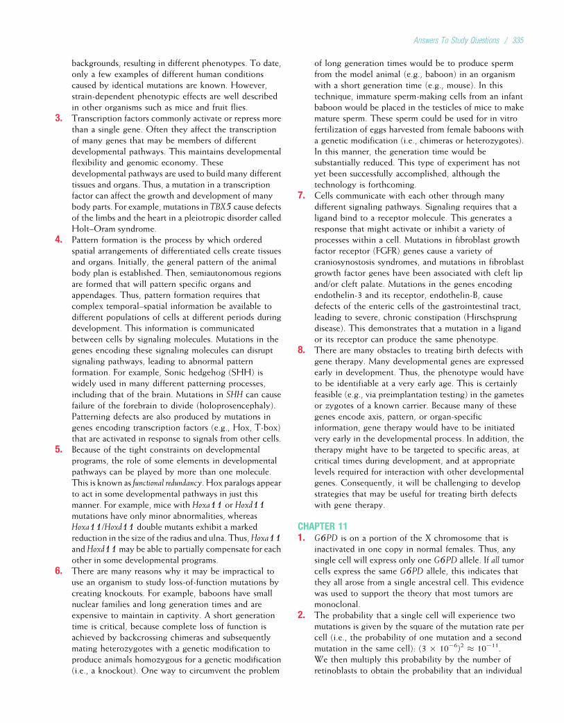
backgrounds, resulting in different phenotypes. To date,only a few examples of different human conditionscaused by identical mutations are known. However,strain-dependent phenotypic effects are well describedin other organisms such as mice and fruit flies.
3. Transcription factors commonly activate or repress morethan a single gene. Often they affect the transcriptionof many genes that may be members of differentdevelopmental pathways. This maintains developmentalflexibility and genomic economy. Thesedevelopmental pathways are used to build many differenttissues and organs. Thus, a mutation in a transcriptionfactor can affect the growth and development of manybody parts. For example, mutations in TBX5 cause defectsof the limbs and the heart in a pleiotropic disorder calledHolt–Oram syndrome.
4. Pattern formation is the process by which orderedspatial arrangements of differentiated cells create tissuesand organs. Initially, the general pattern of the animalbody plan is established. Then, semiautonomous regionsare formed that will pattern specific organs andappendages. Thus, pattern formation requires thatcomplex temporal–spatial information be available todifferent populations of cells at different periods duringdevelopment. This information is communicatedbetween cells by signaling molecules. Mutations in thegenes encoding these signaling molecules can disruptsignaling pathways, leading to abnormal patternformation. For example, Sonic hedgehog (SHH) iswidely used in many different patterning processes,including that of the brain. Mutations in SHH can causefailure of the forebrain to divide (holoprosencephaly).Patterning defects are also produced by mutations ingenes encoding transcription factors (e.g., Hox, T-box)that are activated in response to signals from other cells.
5. Because of the tight constraints on developmentalprograms, the role of some elements in developmentalpathways can be played by more than one molecule.This is known as functional redundancy. Hox paralogs appearto act in some developmental pathways in just thismanner. For example, mice with Hoxa11 or Hoxd11mutations have only minor abnormalities, whereasHoxa11/Hoxd11 double mutants exhibit a markedreduction in the size of the radius and ulna. Thus,Hoxa11and Hoxd11may be able to partially compensate for eachother in some developmental programs.
6. There are many reasons why it may be impractical touse an organism to study loss-of-function mutations bycreating knockouts. For example, baboons have smallnuclear families and long generation times and areexpensive to maintain in captivity. A short generationtime is critical, because complete loss of function isachieved by backcrossing chimeras and subsequentlymating heterozygotes with a genetic modification toproduce animals homozygous for a genetic modification(i.e., a knockout). One way to circumvent the problem
of long generation times would be to produce spermfrom the model animal (e.g., baboon) in an organismwith a short generation time (e.g., mouse). In thistechnique, immature sperm-making cells from an infantbaboon would be placed in the testicles of mice to makemature sperm. These sperm could be used for in vitrofertilization of eggs harvested from female baboons witha genetic modification (i.e., chimeras or heterozygotes).In this manner, the generation time would besubstantially reduced. This type of experiment has notyet been successfully accomplished, although thetechnology is forthcoming.
7. Cells communicate with each other through manydifferent signaling pathways. Signaling requires that aligand bind to a receptor molecule. This generates aresponse that might activate or inhibit a variety ofprocesses within a cell. Mutations in fibroblast growthfactor receptor (FGFR) genes cause a variety ofcraniosynostosis syndromes, and mutations in fibroblastgrowth factor genes have been associated with cleft lipand/or cleft palate. Mutations in the genes encodingendothelin-3 and its receptor, endothelin-B, causedefects of the enteric cells of the gastrointestinal tract,leading to severe, chronic constipation (Hirschsprungdisease). This demonstrates that a mutation in a ligandor its receptor can produce the same phenotype.
8. There are many obstacles to treating birth defects withgene therapy. Many developmental genes are expressedearly in development. Thus, the phenotype would haveto be identifiable at a very early age. This is certainlyfeasible (e.g., via preimplantation testing) in the gametesor zygotes of a known carrier. Because many of thesegenes encode axis, pattern, or organ-specificinformation, gene therapy would have to be initiatedvery early in the developmental process. In addition, thetherapy might have to be targeted to specific areas, atcritical times during development, and at appropriatelevels required for interaction with other developmentalgenes. Consequently, it will be challenging to developstrategies that may be useful for treating birth defectswith gene therapy.
CHAPTER 111. G6PD is on a portion of the X chromosome that is
inactivated in one copy in normal females. Thus, anysingle cell will express only one G6PD allele. If all tumorcells express the same G6PD allele, this indicates thatthey all arose from a single ancestral cell. This evidencewas used to support the theory that most tumors aremonoclonal.
2. The probability that a single cell will experience twomutations is given by the square of the mutation rate percell (i.e., the probability of one mutation and a secondmutation in the same cell): (3 � 10�6)2 � 10�11.We then multiply this probability by the number ofretinoblasts to obtain the probability that an individual
Answers To Study Questions / 335
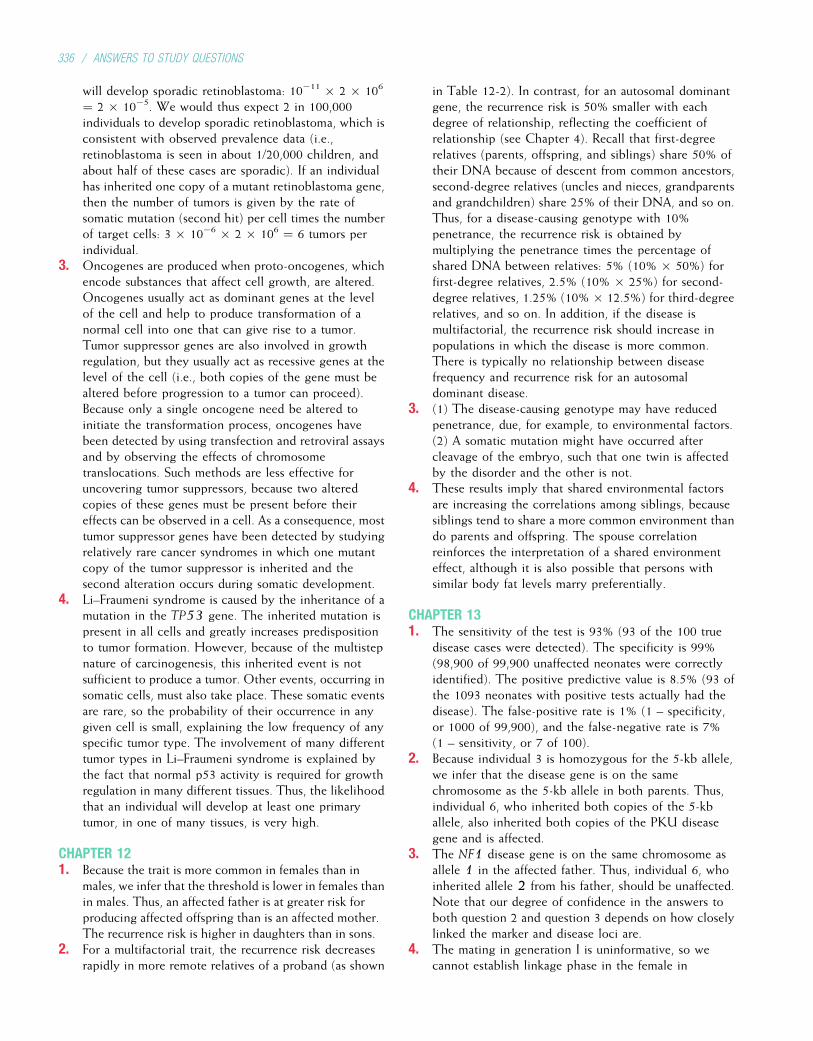
will develop sporadic retinoblastoma: 10�11 � 2 � 106
¼ 2 � 10�5. We would thus expect 2 in 100,000individuals to develop sporadic retinoblastoma, which isconsistent with observed prevalence data (i.e.,retinoblastoma is seen in about 1/20,000 children, andabout half of these cases are sporadic). If an individualhas inherited one copy of a mutant retinoblastoma gene,then the number of tumors is given by the rate ofsomatic mutation (second hit) per cell times the numberof target cells: 3 � 10�6 � 2 � 106 ¼ 6 tumors perindividual.
3. Oncogenes are produced when proto-oncogenes, whichencode substances that affect cell growth, are altered.Oncogenes usually act as dominant genes at the levelof the cell and help to produce transformation of anormal cell into one that can give rise to a tumor.Tumor suppressor genes are also involved in growthregulation, but they usually act as recessive genes at thelevel of the cell (i.e., both copies of the gene must bealtered before progression to a tumor can proceed).Because only a single oncogene need be altered toinitiate the transformation process, oncogenes havebeen detected by using transfection and retroviral assaysand by observing the effects of chromosometranslocations. Such methods are less effective foruncovering tumor suppressors, because two alteredcopies of these genes must be present before theireffects can be observed in a cell. As a consequence, mosttumor suppressor genes have been detected by studyingrelatively rare cancer syndromes in which one mutantcopy of the tumor suppressor is inherited and thesecond alteration occurs during somatic development.
4. Li–Fraumeni syndrome is caused by the inheritance of amutation in the TP53 gene. The inherited mutation ispresent in all cells and greatly increases predispositionto tumor formation. However, because of the multistepnature of carcinogenesis, this inherited event is notsufficient to produce a tumor. Other events, occurring insomatic cells, must also take place. These somatic eventsare rare, so the probability of their occurrence in anygiven cell is small, explaining the low frequency of anyspecific tumor type. The involvement of many differenttumor types in Li–Fraumeni syndrome is explained bythe fact that normal p53 activity is required for growthregulation in many different tissues. Thus, the likelihoodthat an individual will develop at least one primarytumor, in one of many tissues, is very high.
CHAPTER 121. Because the trait is more common in females than in
males, we infer that the threshold is lower in females thanin males. Thus, an affected father is at greater risk forproducing affected offspring than is an affected mother.The recurrence risk is higher in daughters than in sons.
2. For a multifactorial trait, the recurrence risk decreasesrapidly in more remote relatives of a proband (as shown
in Table 12-2). In contrast, for an autosomal dominantgene, the recurrence risk is 50% smaller with eachdegree of relationship, reflecting the coefficient ofrelationship (see Chapter 4). Recall that first-degreerelatives (parents, offspring, and siblings) share 50% oftheir DNA because of descent from common ancestors,second-degree relatives (uncles and nieces, grandparentsand grandchildren) share 25% of their DNA, and so on.Thus, for a disease-causing genotype with 10%penetrance, the recurrence risk is obtained bymultiplying the penetrance times the percentage ofshared DNA between relatives: 5% (10% � 50%) forfirst-degree relatives, 2.5% (10% � 25%) for second-degree relatives, 1.25% (10% � 12.5%) for third-degreerelatives, and so on. In addition, if the disease ismultifactorial, the recurrence risk should increase inpopulations in which the disease is more common.There is typically no relationship between diseasefrequency and recurrence risk for an autosomaldominant disease.
3. (1) The disease-causing genotype may have reducedpenetrance, due, for example, to environmental factors.(2) A somatic mutation might have occurred aftercleavage of the embryo, such that one twin is affectedby the disorder and the other is not.
4. These results imply that shared environmental factorsare increasing the correlations among siblings, becausesiblings tend to share a more common environment thando parents and offspring. The spouse correlationreinforces the interpretation of a shared environmenteffect, although it is also possible that persons withsimilar body fat levels marry preferentially.
CHAPTER 131. The sensitivity of the test is 93% (93 of the 100 true
disease cases were detected). The specificity is 99%(98,900 of 99,900 unaffected neonates were correctlyidentified). The positive predictive value is 8.5% (93 ofthe 1093 neonates with positive tests actually had thedisease). The false-positive rate is 1% (1 – specificity,or 1000 of 99,900), and the false-negative rate is 7%(1 – sensitivity, or 7 of 100).
2. Because individual 3 is homozygous for the 5-kb allele,we infer that the disease gene is on the samechromosome as the 5-kb allele in both parents. Thus,individual 6, who inherited both copies of the 5-kballele, also inherited both copies of the PKU diseasegene and is affected.
3. The NF1 disease gene is on the same chromosome asallele 1 in the affected father. Thus, individual 6, whoinherited allele 2 from his father, should be unaffected.Note that our degree of confidence in the answers toboth question 2 and question 3 depends on how closelylinked the marker and disease loci are.
4. The mating in generation I is uninformative, so wecannot establish linkage phase in the female in
336 / ANSWERS TO STUDY QUESTIONS
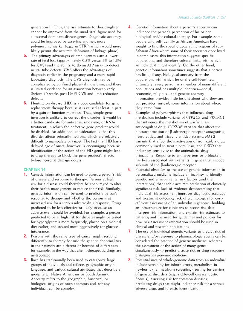
generation II. Thus, the risk estimate for her daughtercannot be improved from the usual 50% figure used forautosomal dominant disease genes. Diagnostic accuracycould be improved by assaying another, morepolymorphic marker (e.g., an STRP, which would morelikely permit the accurate definition of linkage phase).
5. The primary advantages of amniocentesis are a lowerrate of fetal loss (approximately 0.5% versus 1% to 1.3%for CVS) and the ability to do an AFP assay to detectneural tube defects. CVS offers the advantages ofdiagnosis earlier in the pregnancy and a more rapidlaboratory diagnosis. The CVS diagnosis may becomplicated by confined placental mosaicism, and thereis limited evidence for an association between early(before 10 weeks post-LMP) CVS and limb reductiondefects.
6. Huntington disease (HD) is a poor candidate for genereplacement therapy because it is caused at least in partby a gain-of-function mutation. Thus, simple geneinsertion is unlikely to correct the disorder. It would bea better candidate for antisense, ribozyme, or RNAitreatment, in which the defective gene product wouldbe disabled. An additional consideration is that thisdisorder affects primarily neurons, which are relativelydifficult to manipulate or target. The fact that HD has adelayed age of onset, however, is encouraging becauseidentification of the action of the HD gene might leadto drug therapy to block the gene product’s effectsbefore neuronal damage occurs.
CHAPTER 141. Genetic information can be used to assess a person’s risk
of disease and response to therapy. Persons at highrisk for a disease could therefore be encouraged to altertheir health management to reduce their risk. Similarly,genetic information can be used to predict a person’sresponse to therapy and whether the person is atincreased risk for a serious adverse drug response. Drugspredicted to be less effective or likely to cause anadverse event could be avoided. For example, a personpredicted to be at high risk for diabetes might be testedfor hyperglycemia more frequently, placed on a medicaldiet earlier, and treated more aggressively for glucoseintolerance.
2. Persons with the same type of cancer might responddifferently to therapy because the genetic abnormalitiesin their tumors are different or because of differences,for example, in the way that chemotherapeutic drugs aremetabolized.
3. Race has traditionally been used to categorize largegroups of individuals and reflects geographic origin,language, and various cultural attributes that describe agroup (e.g., Native Americans or South Asians).Ancestry refers to the geographic, historical, orbiological origins of one’s ancestors and, for anyindividual, can be complex.
4. Genetic information about a person’s ancestry caninfluence the person’s perception of his or herbiological and/or cultural identity. For example, somepeople who self-identify as African American havesought to find the specific geographic regions of sub-Saharan Africa where some of their ancestors once lived.In some cases, this information suggests specificpopulations, and therefore cultural links, with whichan individual might identify. On the other hand,genetic information sometimes suggests that a personhas little, if any, biological ancestry from thepopulations with which he or she self-identifies.Ultimately, every person is a member of many differentpopulations and has multiple identities—social,economic, religious—and genetic ancestryinformation provides little insight about who they arebut provides, instead, some information about wherethey came from.
5. Examples of polymorphisms that influence drugmetabolism include variants of CYP2C9 and VKORC1that influence the metabolism of warfarin, ananticoagulant drug; CYP2D6 variants that affect thebiotransformation of b-adrenergic receptor antagonists,neuroleptics, and tricyclic antidepressants; NAT2variants that affect the inactivation of isoniazid, a drugcommonly used to treat tuberculosis; and G6PD thatinfluences sensitivity to the antimalarial drug,primaquine. Response to antihypertensive b-blockershas been associated with variants in genes that encodesubunits of the b-adrenergic receptor.
6. Potential obstacles to the use of genetic information inpersonalized medicine include an inability to identifygenetic and environmental risk factors (and theirinteractions) that enable accurate prediction of clinicallysignificant risk; lack of evidence demonstrating thatindividual risk assessment improves diagnostic accuracyand treatment outcome; lack of technologies for cost-efficient assessment of an individual’s genome; buildingan infrastructure for clinicians to access risk data,interpret risk information, and explain risk estimates topatients; and the need for guidelines and policies forhow risk-assessment information should be used inclinical and research applications.
7. The use of individual genetic variants to predict risk ofdisease and/or response to pharmacologic agents can beconsidered the practice of genetic medicine, whereasthe assessment of the action of many genessimultaneously to predict disease risk or drug responsedistinguishes genomic medicine.
8. Potential uses of whole-genome data from an individualinclude screening for inborn errors, metabolism innewborns (i.e., newborn screening), testing for carriersof genetic disorders (e.g., sickle-cell disease, cysticfibrosis), assessing risk for common diseases,predicting drugs that might influence risk for a seriousadverse drug, and forensic identification.
Answers To Study Questions / 337
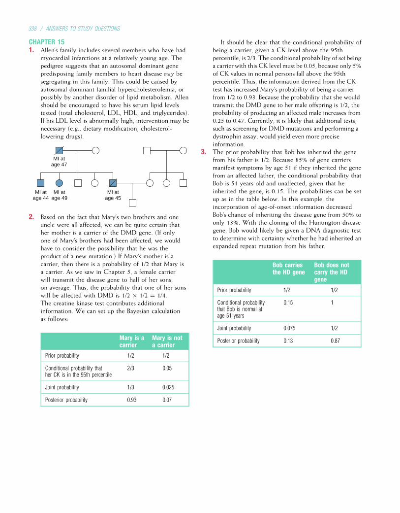
CHAPTER 151. Allen’s family includes several members who have had
myocardial infarctions at a relatively young age. Thepedigree suggests that an autosomal dominant genepredisposing family members to heart disease may besegregating in this family. This could be caused byautosomal dominant familial hypercholesterolemia, orpossibly by another disorder of lipid metabolism. Allenshould be encouraged to have his serum lipid levelstested (total cholesterol, LDL, HDL, and triglycerides).If his LDL level is abnormally high, intervention may benecessary (e.g., dietary modification, cholesterol-lowering drugs).
MI atage 47
MI atage 44
MI atage 49
MI atage 45
2. Based on the fact that Mary’s two brothers and oneuncle were all affected, we can be quite certain thather mother is a carrier of the DMD gene. (If onlyone of Mary’s brothers had been affected, we wouldhave to consider the possibility that he was theproduct of a new mutation.) If Mary’s mother is acarrier, then there is a probability of 1/2 that Mary isa carrier. As we saw in Chapter 5, a female carrierwill transmit the disease gene to half of her sons,on average. Thus, the probability that one of her sonswill be affected with DMD is 1/2 � 1/2 ¼ 1/4.The creatine kinase test contributes additionalinformation. We can set up the Bayesian calculationas follows:
Mary is acarrier
Mary is nota carrier
Prior probability 1/2 1/2
Conditional probability thather CK is in the 95th percentile
2/3 0.05
Joint probability 1/3 0.025
Posterior probability 0.93 0.07
It should be clear that the conditional probability ofbeing a carrier, given a CK level above the 95thpercentile, is 2/3. The conditional probability of not beinga carrier with this CK level must be 0.05, because only 5%of CK values in normal persons fall above the 95thpercentile. Thus, the information derived from the CKtest has increased Mary’s probability of being a carrierfrom 1/2 to 0.93. Because the probability that she wouldtransmit the DMD gene to her male offspring is 1/2, theprobability of producing an affected male increases from0.25 to 0.47. Currently, it is likely that additional tests,such as screening for DMD mutations and performing adystrophin assay, would yield even more preciseinformation.
3. The prior probability that Bob has inherited the genefrom his father is 1/2. Because 85% of gene carriersmanifest symptoms by age 51 if they inherited the genefrom an affected father, the conditional probability thatBob is 51 years old and unaffected, given that heinherited the gene, is 0.15. The probabilities can be setup as in the table below. In this example, theincorporation of age-of-onset information decreasedBob’s chance of inheriting the disease gene from 50% toonly 13%. With the cloning of the Huntington diseasegene, Bob would likely be given a DNA diagnostic testto determine with certainty whether he had inherited anexpanded repeat mutation from his father.
Bob carriesthe HD gene
Bob does notcarry the HDgene
Prior probability 1/2 1/2
Conditional probabilitythat Bob is normal atage 51 years
0.15 1
Joint probability 0.075 1/2
Posterior probability 0.13 0.87
338 / ANSWERS TO STUDY QUESTIONS
INDEX
Note: Page numbers followed by f indicate figures; t indicates tables and b indicates boxes.
AAarskog–Scott syndrome, 85tABL oncogene, 125, 219–220, 220t, 221ABO blood group system, 38–39, 39t, 188–189Abortion
induced. See Pregnancy termination.spontaneous. See Pregnancy loss.
Acceptor site, 28ACE inhibitors. See Angiotensin-converting
enzyme (ACE) inhibitors.Achondroplasia, 63f, 170t, 195–196, 196f
in heterozygote vs. homozygote, 62new mutations for, 63–64
Acidemiaglutaric, type II, 144lactic, 144–145methylmalonic, 129tpropionic, 129t
Acquired immunodeficiency syndrome (AIDS).See also Human immunodeficiency virus (HIV).
CCR5 and, 186gene therapy for, 274t, 279in hemophilia patients, 80
Acrocentric chromosomes, 100, 101–102, 102fRobertsonian translocations of, 115–116, 116f
Acrodermatitis enteropathica, 129t, 148–149, 148fAcromesomelic dysplasia, 197–198Activators, 12, 12fAcute fatty liver of pregnancy, 137Adenine, 5, 6f, 7fAdeno-associated viral vectors, 275–276Adenomatous polyposis coli. See Familial
adenomatous polyposis.Adenosine deaminase deficiency, 190, 274t, 276bAdenoviral vectors, 275Adenylate kinase, 165Adjacent segregation, 115–116, 117fAdoption studies, 238–239Adrenal hyperplasia. See Congenital adrenal
hyperplasia.Adrenoleukodystrophy
childhood cerebral, 140neonatal, 140X-linked, 140
Adrenomyeloneuropathy, 140Advocacy groups, genetic, 300Affected sib-pair method, 239Affinity maturation, 183Aflatoxin B1, 223–224Agammaglobulinemia, X-linked, 190, 190tAge
maternal, trisomies and, 110–111, 110fmitochondrial mutations and, 89
Age (Continued)paternalchromosome abnormalities and, 111, 114single-gene mutations and, 38, 38f
Age-dependent penetrance, 65, 66–67Alagille syndrome, 119tAlbinism
oculocutaneous, 61–62, 61f, 128f, 129t, 170tX-linked ocular, 76–77, 77f
Albuterol, 286tAlcohol, as teratogen, 304t, 306, 307bAlcoholism, 254
pyruvate dehydrogenase complex deficiencyand, 144–145
Aldosterone, 248, 248fAldosteronism, glucocorticoid-remediable, 246tAlkaptonuria, 1, 128, 128f, 129tAlleles, 26Allele-specific oligonucleotide (ASO)
hybridization, 49t, 264–266, 265fAllelic dropout, 273Allelic heterogeneity, 68–69Alpha-fetoprotein
in amniotic fluid, 267in maternal serum, 267, 271–272, 272f
Alternate segregation, 115–116, 117fAlu repeats, 20Alzheimer disease, 252–254
APP gene mutations in, 109, 253, 253fin Down syndrome, 109, 253familial, 70t, 170t, 246tmitochondrial mutations in, 89
Ambiguous genitalia, 138, 139, 140fAmino acid disorders, 128f, 129t, 134–136.
See also Phenylketonuria.Amino acids, 14
branched-chain, 136genetic code for, 14–15, 14ttranslation of mRNA to, 15–18, 16f
Aminopterin, as teratogen, 304tAmish population, genetic drift in, 53Amniocentesis, 267–269, 268f, 269b
Down syndrome and, 272early, 268metabolic disorders and, 270, 270t
Amniocytes, 267Amnion disruption sequence, 302tAmyloid-b, 253, 253fAmyloid-b precursor protein, 246t, 253f, 254
in Down syndrome, 109Amyotrophic lateral sclerosis, 170t, 246tAnalytes, 258Anaphase, 21, 22fAnaphase I, 23, 23f
Anaphase II, 23f, 24Ancestry, 289–291, 290f. See also Ethnic groups,
heterozygote screening in.Anderson disease, 133f, 134tAndrogen insensitivity syndromes, 139, 140Androgen receptor gene, mutations of, 139, 140Androgens, as teratogens, 304tAnemiaFanconi, 35b, 126, 228in hemolytic disease of the newborn, 189in sickle cell disease, 31–32in thalassemia, 32–33
Anencephaly, 233, 234, 234fAneuploidyautosomal, 106–111. See also Trisomy; Trisomy
13; Trisomy 18 (Edwards syndrome);Trisomy 21 (Down syndrome).
prenatal diagnosis ofamniocentesis and, 267a-fetoprotein and, 267
sex chromosome, 111–113tumor cell, 221
Angelman syndrome, 89–91, 90f, 118, 119t,122, 170t
Angiogenesis, 212Angiotensin-converting enzyme (ACE), 248, 248fAngiotensin-converting enzyme (ACE) inhibitorspolymorphisms and response to, 285, 286as teratogens, 304t
AniridiaPAX6 mutations and, 193in WAGR syndrome, 119, 119t
Ankylosing spondylitisautoimmunity in, 187–188clinical features of, 162–163, 163fHLA-B27 and, 162–163, 163t, 187–188, 188t
Anterior/posterior axis, 194–195, 203–204Antibodies, 176–177, 179–180, 182–184.
See also Immunoglobulins.Anticipation, 76, 92–96, 92f, 93fAnticipatory guidance and health
supervision, 108bAnticodon, 15, 15f, 16fAntigen-presenting cells (APCs), 177, 178f, 185Antigens, 179–180Antihypertensive agentsdifferential responses to, 285, 286for Marfan syndrome, 71
Antisense strand, 11, 11fAntisense therapy, 277–278, 278fa1-Antitrypsin deficiency, 170t, 265bAortic stenosis, supravalvular, 118–119, 201APC gene, 217t, 224–226, 225b, 226fAPCs. See Antigen-presenting cells (APCs).
339
Apert syndrome, 196–197, 196fApical ectodermal ridge, 208, 208fApolipoproteins, 242, 245t, 246t
Alzheimer disease and, 253Apoptosis, 212, 213f
TP53 gene and, 223, 224Arachnodactyly, 70, 71, 71fArgininosuccinase, 143, 144fArgininosuccinic acid synthetase, 129t, 143, 144fArray comparative genomic hybridization,
104f, 105Artificial chromosomes
as cloning vectors, 43in Human Genome Project, 156
in comparative genomic hybridization, 105human, 43, 277
Ashkenazi Jewish population, 53, 142, 261–262,262t
ASO (allele-specific oligonucleotide)hybridization, 49t, 264–266, 265f
Association studies, genome-wide, 163–164,164f, 239
Associationsdirect-to-consumer services and, 285bin populations, 162–163, 163t
Ataxia-telangiectasia, 35b, 170t, 217t, 228as chromosome instability syndrome, 126immunodeficiency in, 190t
ATM gene, 217t, 221, 227, 227f, 228ATP7A gene, 147, 148ATP7B gene, 147–148ATRX gene, 85tAutoimmune diseases
gene-expression profiling in, 289type 1 diabetes as, 250, 251
Autoimmunity, 187–188Automated DNA sequencing, 47, 48fAutoradiogram, 40, 41f, 43fAutosomal dominant diseases, 3, 59–61, 60f.
See also Dominant genetic diseases.cautions about category of, 62–63distinguishing characteristics of, 62, 62tprevalence of, 4trecurrence risk for, 60–61severity in homozygotes, 62
Autosomal recessive diseases, 3, 61–62, 61f.See also Recessive genetic diseases.
cautions about category of, 62–63detectability in heterozygotes, 62distinguishing characteristics of, 62, 62tinbreeding and. See Consanguinity.metabolic, 130prevalence of, 4trecurrence risk for, 62
Autosomes, 5Axis specification, 194–195, 203–204, 205Azathioprine, 286t
BB lymphocytes, 176–180, 177f, 178f, 179fBacterial artificial chromosomes, 43, 156
in comparative genomic hybridization, 105Bacteriophage P1 artificial chromosomes, 156Bacteriophages, 43Balanced chromosome rearrangements, 114, 115tBaldness, male-pattern, 88Banding, of chromosomes, 101–102, 102fBands, of autoradiogram, 40, 43fBardet–Biedl syndrome, 160Bare lymphocyte syndrome, 190–191, 190tBarr bodies, 77, 78Basal cell carcinoma, 195, 198fBasal transcription factors, 12, 12fBase analogs, 36–37
Base-pair substitution, 27–28, 27fBases
in DNA, 5, 6f, 7fcomplementary pairing of, 7
in RNA, 10Bayes’ theorem, 296bB-cell immunodeficiency diseases, 190, 190tBeare–Stevenson syndrome, 195bBecker muscular dystrophy, 81, 170tBeckwith–Wiedemann syndrome, 91–92, 92f,
293b, 293fBendectin, 306bBenign neoplasm, 212Beta blockers, 285, 286–287, 286tb sheets, 13tBioethics, 308–309Biotin-responsive multiple carboxylase
deficiency, 273Bipolar disorder, 255Birth defects, 193, 205. See also Chromosome
abnormalities; Congenital malformations;Metabolic disorders.
Bivalent chromosomes, 21, 22Blastocyst
preimplantation genetic diagnosis on, 273stem cells derived from, 280
Blastomerepreimplantation genetic diagnosis on, 272–273stem cells derived from, 280
Blood groups, 38–39, 39t, 188Bloom syndrome, 35b, 126, 170t, 299bBlue cone monochromacy, 83Body plan, 193, 201, 203Bone marrow transplantation
for Gaucher disease, 142for mucopolysaccharidoses, 141for b-thalassemia or sickle cell disease, 33
Bone morphogenetic protein (BMP) family,197–198, 203, 210
Brachydactyly, 197–198Brain tumors, gene therapy for, 274tBranched-chain a-ketoacid dehydrogenase, 136Branched-chain amino acids, 136BRCA1 gene, 217t, 221, 227–228, 227f
hypermethylation of, 224screening for mutations in, 263
BRCA2 gene, 217t, 221, 227–228, 227fscreening for mutations in, 263
Breakpoints, translocation, 125Breast cancer, 249
autosomal dominant, locus heterogeneity in, 70tERBB2 (HER2/neu) oncogene in, 220t, 221familial, 170t, 217t, 227–228frequencies in dissimilar populations, 214gene-expression profiles in, 288–289
Brittle bone disease. See Osteogenesis imperfecta.Burkitt lymphoma, 125, 125t
CCalico cat, 76–77Campomelic dysplasia, 199–201Cancer. See also Cancer genes; Carcinogenesis;
specific cancers.causes of, 212–214chromosome abnormalities in, 125–126, 125f,
125t, 219–220, 221chromosome instability syndromes and, 126comparative genomic hybridization in,
103–105, 104fderegulation of cell growth in, 212, 214, 215developmental genes and, 195environmental factors in, 213–214familial, 213, 213fgenomic instability in, 221, 226
Cancer (Continued)immortality of cells in, 221inherited predispositions to, 213, 229–230as multifactorial disorder, 249–250terminology of, 212
Cancer cytogenetics, 125–126, 125f, 125tCancer genes. See also DNA repair; Oncogenes;
Tumor suppressor genes.cell regulation and, 214–216, 214fclassification of, 217inherited vs. somatically altered, 215–216, 216f
Cancer genomics, 288–289, 289fCandidate gene, 170Capillary sequencing, 47Carbamazepine, as teratogen, 304tCarbamyl phosphate synthetase, 129t, 143, 144fCarbimazole, as teratogen, 304tCarbohydrate disorders, 129t, 131–134, 132f,
133f, 134tCarcinogenesis, 212multi-hit concept of, 215two-hit model of, 215–216, 216f
Carcinogens, 212, 213–214TP53 mutations induced by, 223–224
Carcinomas, 212Cardiofaciocutaneous syndrome, 198, 199fCardiomyopathy, 242Cardiovascular disorders, 242–249, 245t, 246tCarrier. See Heterozygote.b-Catenin, 225–226C-banding, 101–102CCR5 (chemokine receptor 5), 186, 279CD4 T cells, 180CD8 T cells, 180CDK. See Cyclin-dependent kinases (CDKs).CDK4, malignant melanoma and, 220t, 228, 229CDK inhibitors, 217t, 218, 218ffamilial melanoma and, 228p53 protein and, 223
CDKN2A gene, familial melanoma and, 217t,228–229
cDNA library, 166, 167fCeliac disease, 188tCell, anatomy of, 6fCell cycle, 20–25, 20f. See also Meiosis; Mitosis.regulation of, by tumor suppressors, 65, 218,
218f, 223Cell growth and differentiationderegulation of, in cancer, 212, 214, 215in organ formation, 209–210proto-oncogenes and, 218, 219regulation of, 214–215, 214f
Centimorgans (cM), 151, 153Centrioles, 21, 22fCentromeres, 21, 22f, 23, 24, 24fclassification of chromosomes and, 100tandem repeats found near, 20
Cervical cancerhuman papillomavirus and, 223squamous cell, HLA allele and, 188t
CFTR gene, 57b, 57f, 69CG dinucleotides, methylated, 37–38.
See also Methylation.on inactive X chromosome, 78Rett syndrome and, 86
CG islands, 168CGH. See Comparative genomic hybridization
(CGH).Charcot–Marie–Tooth disease, 28, 29–30, 70tChediak–Higashi syndrome, 190tCHEK2 gene, 217t, 224, 227, 227f, 228Chemical mutagens, 36–37Chiasma (chiasmata), 22, 23, 23f, 24fCholesterol. See also Hypercholesterolemia,
familial.pattern formation and, 201
340 / INDEX
Cholesterol (Continued)Smith–Lemli–Opitz syndrome and,
137–138, 201as steroid hormone precursor, 138, 139f
Chordin, 202, 205Chorionic villus sampling, 269–270, 269f
metabolic disorders and, 270, 270tChromatin, 5
transcriptional regulation and, 13Chromatin condensation, imprinting and, 89, 92Chromatin loops, 7, 8fChromosome(s), 5, 6f. See also Artificial
chromosomes; Karyotype.banding of, 101–102, 102fcomparative genomic hybridization with,
103–105, 104ffluorescence in situ hybridization with,
102–103, 103f, 104fgene mapping from morphology of, 164, 165fnomenclature for, 100, 101t, 102fnumber of, in human cell, 2, 5
Chromosome abnormalities, 3in cancer, 125–126, 125f, 125t, 219–220, 221clinical phenotypes and, 123–125gene mapping by observation of, 164, 165fmeiosis and, 24nomenclature for karyotype of, 101tof number, 105–113. See also Aneuploidy;
Polyploidy.pediatric deaths attributable to, 4tprevalence of, 2t, 4t, 100, 115tof structure, 114–123. See also Deletions;
Duplications; Translocations.inversions, 122–123, 123fisochromosomes, 123, 124frings, 122, 122fsubtelomeric rearrangements, 120–121, 121funiparental disomy, 91–92, 92f, 121–122, 121f
Chromosome breakage, 114in chromosome instability syndromes, 126
Chromosome evaluation, clinical indications for,124, 124b
Chromosome instability syndromes, 126Chromosome-specific DNA library, 166Chronic granulomatous disease, 190t, 191Chronic progressive external ophthalmoplegia, 89Ciliary dyskinesias, 204Clarithromycin, 286tClass switching, 181Clastogens, 114Cleft palate, 239–241, 241t
with ankyloglossia, 85tCleidocranial dysplasia, 94t, 210Clinical genetics. See also Genetic counseling.
common indications for referral, 302bdefinition of, 292evaluation and services in, 300–301principles of, 292–301types of clinics and programs, 302b
Clone, definition of, 42Cloning, reproductive, 280, 281f, 309Cloning of DNA sequences, 42b, 42f
functional, 166in Human Genome Project, 156for libraries, 166, 167fpositional, 156, 166–168
Cloning of embryonic stem cells, 280, 309Cloning of genes, 42b, 150cM. See Centimorgans (cM).CNVs (copy number variants), 43–45, 45fCo-activators, 12, 12fCoagulation factors. See also Factor VIII.
stroke and, 245–247Coated pits, 243, 244fCocaine, as teratogen, 304tCockayne syndrome, 35b
Codeine, 286tCodominant alleles, 51Codominant markers, 156–157Codons, 14–15, 14tCoefficient of relationship, 71, 72bCofactors of enzymes, 145–147Collagen
cross-linking of, 147in embryonic development, 199–201type I, 18osteogenesis imperfecta and, 17b, 69, 69fposttranslational modification of, 18, 18f
Color vision, 83bColorectal cancer, 225b, 249. See also APC gene;
Familial adenomatous polyposis; Hereditarynonpolyposis colon cancer.
in migrant Japanese populations, 214screening for syndromes of, 263
Common diseases, 3. See also Multifactorialdisorders.
personalized medicine for, 288, 289Comparative genomic hybridization (CGH),
103–105, 104fComplement system, 176, 179–180, 186, 187
defects of, 190t, 191Complementary base pairing
in DNA replication, 7in mRNA synthesis, 11
Complementary DNA (cDNA) library,166, 167f
Compound heterozygote, 33, 34fConcordance rates, 236–238, 237tConcordant twins, 236–237Conditional knockout, 207, 207fConditional probability, 296, 297Cone cells, 83Congenital adrenal hyperplasia, 138–139, 140f
fetal treatment of, 273screening for, 260–261, 260t
Congenital malformations. See also Chromosomeabnormalities; Dysmorphology.
causes of, 241, 303, 303tclassification of, 303common multiple anomaly syndromes, 302tethical issues about, 308isolated vs. syndromic, 303metabolic disorders and, 137–138with multifactorial inheritance, 239–241, 241tthreshold model of, 233, 256
prevalence of, 2t, 4t, 239, 241t, 302prevention of, 306–307sequence of, 303–304, 304fsingle-gene, 241
Conjoined twins, 204Consanguinity, 71–74
in autosomal recessive disease pedigree,61–62, 61f
definition of, 61–62measurement of, 72bmortality rates associated with, 72–73, 73t
Consensus sequences, 18–19Conserved sequences, 168–169Constant region, of immunoglobulin chain,
181–182, 182fConstitutional mutation, 215–216Constitutive heterochromatin, 101–102Contiguous gene syndromes, 119Cooley’s anemia, 32–33Copper disorders, 147–148Copy number variants (CNVs), 43–45, 45fCordocentesis, 270Cori disease, 133f, 134tCornelia de Lange syndrome, 302tCoronary artery disease, 242–245
gene therapy for, 279Correlation coefficients, 237, 237t
Cortisol deficiency, 138Cosmids, 43Costello syndrome, 198, 199fCostimulatory molecules, 177–179, 179fCounseling. See Genetic counseling;
Preconception counseling.Cousin marriage. See Consanguinity.Cowden disease, 217t, 228Craniofacial development, 205–208Craniosynostosis syndromes, 196–197, 196f,
207–208Creatine kinase, Duchenne muscular dystrophy
and, 80, 260fCri-du-chat syndrome, 116Crossover, 22, 24f. See also Linkage analysis.double, 150–151, 151ffactors affecting rate of, 153recombination in, 150–151, 151f, 152f.
See also Recombination frequencies.unequal, 83, 84, 84f
Cross-reaction, autoimmune, 187–188Crouzon syndrome, 196–197Cryptic splice sites, 28CTLA4 gene, 251Cutis laxa, X-linked, 147Cyclin-dependent kinases (CDKs), 21.
See also CDK inhibitors.oncogenes and, 220t, 228tumor suppressors and, 218, 218f
Cyclins, 21CYP2 genes, 286–287, 286t, 287fCYP21A2 gene, 138, 139Cystic fibrosis, 57b, 57f, 170tcarrier frequency of, 52direct diagnosis of, 265–266gene therapy for, 274theterozygote screening for, 262b, 262tnewborn screening for, 261tuniparental disomy in, 121–122variable expression of, 69
Cystinosis, 129t, 145Cystinuria, 129t, 145Cytochrome c oxidase deficiency, 129tCytochrome P450 superfamily, 286–287, 286tCytogenetics, 2, 100. See also Karyotype.cancer, 125–126, 125f, 125t
Cytokine receptors, severe combined immunedeficiency and, 190
Cytokines, helper T lymphocytes and,177–179, 180
Cytokinesis, 20, 21, 23, 24Cytomegalovirus, immune evasion by, 186Cytosine, 5, 6f, 7f. See also CG dinucleotides,
methylated.methylation of, 37–38, 37f
Cytotoxic T lymphocytes, 176–177, 180, 181f,184, 186
evasion of, by viruses and tumor cells, 186gene therapy and, 279transplant rejection and, 185
DDaughter cells, 21, 22fDeafness, late-onset, mitochondrial mutation
causing, 89Deformation, definition of, 303Degenerate code, 14Dejerine–Sottas syndrome, 28Deletion mapping, 165, 165fDeletionsbase pair, 28chromosomal, 116–118
fluorescence in situ hybridization and,165, 165f
INDEX / 341
Deletions (Continued)mapping with aid of, 165, 165fmicrodeletion syndromes, 118–120, 118f,
119t, 120bsubmicroscopic, detection techniques
for, 169in tumors, 222, 222f22q11 deletion syndrome and, 120, 120f,
205–207Dendritic cells, 177, 180Dentatorubral-pallidoluysian atrophy, 94tDeoxyribonucleic acid. See DNA
(deoxyribonucleic acid).Derivative chromosomes, 114Deuteranomaly, 83, 84, 84fDeuteranopia, 83, 84fDevelopment
animal models of, 193, 194f, 194t, 206b,206f, 207f
basic concepts of, 193–194genetic mediators of, 195–201
DNA transcription factors, 199–201extracellular matrix proteins, 201paracrine signaling molecules, 195–198
major processes in embryo, 194–195pattern formation in, 194–195, 201–210
Developmental delay, 124, 132Developmental genetics, 193Diabetes insipidus, nephrogenic, 85tDiabetes mellitus, 250–252
maturity-onset diabetes of the young, 129t,132–133, 246t, 251–252
type 1, 129t, 132–133, 250–251, 251tanimal models of, 240, 251gene-expression profiling in, 289HLA alleles associated with, 187–188,
188t, 250type 2, 129t, 132–133, 251, 251t
MELAS mutation in, 89in Pima Native Americans, 163–164
Dicer, 278–279, 279fDichromatic vision, 83Dideoxy method, 47, 48fDietary management, of metabolic disorders, 135bDiethylstilbestrol, 304tDifferentially methylated regions (DMRs),
91–92, 92fDifferentiation. See Cell growth and
differentiation.Digenic inheritance, 160DiGeorge sequence, 119t, 120b, 190t, 191Dilated cardiomyopathy, 242, 246tDiploid cells, 5Direct diagnosis
advantages and disadvantages of, 264, 264tmethods of, 264–266, 265f
Disability, ethical issues about, 308Discordant twins, 236–237Diseases. See Genetic diseases.Dispermy, 105Dispersed repetitive DNA, 19, 19f, 20Disruption, definition of, 303Dizygotic twins, 236–238, 237tDMPK gene, 92, 93–94DMRs (differentially methylated regions),
91–92, 92fDNA (deoxyribonucleic acid).
See also Mitochondrial DNA (mtDNA).amplification of, 45–47, 46fcoiling of, 7, 8fconstituents of, 5–6, 6f, 7fearly research on, 1–2genes composed of, 5replication of, 7–10, 9f
cell cycle and, 20, 21
DNA (deoxyribonucleic acid) (Continued)chromosome instability syndromes and, 126
sequencing of, 47, 48ffor detecting mutations, 47–48, 49t
structure of, 2, 5–7, 6f, 7ftypes of sequences in, 19–20, 19f
DNA chips. See Microarrays, DNA.DNA libraries. See Libraries, DNA.DNA mismatch cleavage, 49tDNA polymerase(s), 7
in polymerase chain reaction, 45, 46DNA profiles, forensic, 44b, 44fDNA repair, 37, 217t
BRCA1 and BRCA2 genes and, 217t, 227,227f, 228
diseases caused by defects in, 35b, 35f, 37,126, 217t
genomic instability in tumors and, 221p53 tumor suppressor and, 223
DNA sequencesamplification of, 45, 46cloning of, 42b, 42fcomputer analysis of, 166–169consensus, 18–19conserved, 168–169for mutation detection, 49t
Dominant allele, 26Dominant genetic diseases. See also Autosomal
dominant diseases.gain-of-function mutations in, 29–30mutation–selection balance with, 54
Dominant negative mutations, 30, 30fDonor site, 28, 29fDorsal/ventral axis, 194–195, 205, 208Dosage compensation, 76Dosage mapping, 165Dosage sensitivity, 28Double helix, DNA, 5–6, 7fDouble-stranded DNA breaks, 37, 221, 227Down syndrome. See Trisomy 21 (Down
syndrome).Downstream DNA sequences, 7Drug therapy and genetics, 285
personalized therapy, 285–287, 286t, 287fpredicting serious adverse reactions, 285
Duchenne muscular dystrophy, 80b, 81f, 82fBayesian analysis of, 296creatine kinase and, 80, 260fgene mapping of, 165, 170tgene therapy for, 274tgermline mosaicism in, 64
Duplicationschromosomal, 122dosage mapping and, 165subtelomeric, 120–121
of genes, 28Dynein, 204Dysmorphology. See also Congenital
malformations.definition of, 292, 302isolated vs. syndromic malformations in, 303key terms in, 303sequence concept in, 303–304, 304f
Dysostosis multiplex, 141t, 143tDysplasia, definition of, 303Dystroglycan–sarcoglycan complex, 80–81, 82fDystrophin, 80–81, 82f
EEarly onset, of complex disorders, 256ECORI, 40, 40f, 42Ectoderm, 201, 202–203Ectopic expression, 193
Edwards syndrome. See Trisomy 18 (Edwardssyndrome).
E2F transcription factors, 65Ehlers–Danlos syndrome, 170ttype IX, 147
Elastin, 118–119, 147, 201Electron transfer flavoprotein, 144ElectrophoresisDNA, 40, 40f
for mutation detection, 49tpulsed-field, 169
protein, 39–40, 39fEllis–van Creveld syndrome, 53Embryo. See Development.Embryonic stem cells, 280, 309of mouse, 206, 206f
Employment discrimination, 308Encephalocele, 233, 234fEndocytosis, 243–244, 244fEndoderm, 201, 203Endonucleases. See Restriction enzymes.Endoplasmic reticulum, 6fforeign peptides in, 185posttranslational modifications in, 18
Endothelin mutations, Hirschsprung diseaseand, 200
Energy production defects, 129t, 144–145Enhancers, 12, 12f, 13Environmental factorsin cancer, 213–214congenital malformations and, 241in genetic diseases, 3, 3f
variable expression and, 68–69interventions for modification of, 256multifactorial traits and, 231phenotype and, 56–59relative influence of, 236–239
Enzyme replacementfor Gaucher disease, 142for mucopolysaccharidoses, 141
Epidermolysis bullosa, 274t, 292junctional, 201
Epilepsy, 89, 94t, 246tEpithelium, development of, 210Equational division, 21, 23f, 24Equatorial planein meiosis, 22, 24in mitosis, 21, 22f
ERBB oncogene, 219, 220tERBB2 (HER2/neu) oncogene, 220t, 221Ethics, 308–309Ethnic groups, heterozygote screening in,
261–262, 262t. See also Race and personalizedmedicine.
Euchromatin, 13Eugenics, 309Eukaryotesdefinition of, 13–14intron–exon structure of, 18–19
Euploid cell, 105Exons, 13–14, 14f, 18–19Expanded repeats. See Repeat expansions.Expressed sequence tags (ESTs), 168Expression. See Gene expression; Variable
expression.Extracellular matrixin embryonic development, 201mucopolysaccharidoses and, 140–141
Eye, development of, 193, 194f
FFabry disease, 143tFaciogenital dysplasia, 85t
342 / INDEX
Factor V Leiden allele, 245–247Factor VIII, 79–80, 79b, 86False-negative rate, 259–260False-positive rate, 259–260Familial adenomatous polyposis, 170t, 224–226,
225b, 225f. See also APC gene.Familial hypercholesterolemia, 30, 170t, 242,
243b, 243f, 246t, 274tFamily history, obtaining, 301, 301bFamily screening for genetic disorders, 258, 259Fanconi anemia, 35b, 126, 228Fatty acid metabolism, 137, 137f
defects of, 137Fertilization, 24–25
triploidy caused at time of, 105Fetal alcohol syndrome, 144–145, 306, 307bFetal DNA, in maternal circulation, 273Fetal hemoglobin, 31, 33Fetal treatment, 273a-Fetoprotein
in amniotic fluid, 267in maternal serum, 267, 271–272, 272f
Fibrillin-1, 69–71, 170, 170tin embryonic development, 201
Fibrillin-2, 71Fibroblast growth factor(s)
in gene therapy, 279in limb development, 208oncogenes associated with, 220t
Fibroblast growth factor (FGF) family, 195Fibroblast growth factor receptors
breast cancer risk and, 228disorders of, 195b, 196f, 203, 207
First-degree relatives, 595’ cap, 11Flow cytometry, 166Fluconazole, as teratogen, 304tFluorescence in situ hybridization (FISH),
102–103, 103f, 104fon amniocytes, 267
FMR1 gene, 97, 98FMR2 gene, 98FNB1 gene, 70bFNB2 gene, 71Folate (folic acid), neural tube defects and, 234,
307bFOS gene, 214–215, 214f, 219, 220tFounder effect, 53FOXP3 gene, 188Fragile site (FRAXE), 94t, 98Fragile X syndrome, 94, 94t, 96–98, 96f, 97f, 170tFrameshift mutations, 28–29, 28fFriedreich ataxia, 94t, 170tFructokinase deficiency, 132Fructose, metabolism of, 133fFructose intolerance, hereditary, 129t, 132, 133fFructose-1,6-bisphosphatase deficiency, 132, 133fFructosuria, 129t, 132, 133fFumarylacetoacetate hydrolase deficiency,
128f, 136Functional cloning, 166Fusion genes, 84
GG0 phase, 20–21G1 (gap 1) phase, 20–21, 20fG2 (gap 2) phase, 20, 20fGain-of-function mutations, 29–30, 30fGalactose, metabolism of, 132fGalactosemia, 129t, 131–132, 132f, 170t
newborn screening for, 261tGametes, 5. See also Meiosis.Gametogenesis, 23f, 24–25
Gastric carcinoma. See Stomach carcinoma.Gastrulation, 201, 202f, 203Gaucher disease, 142–143, 143t, 274tGel electrophoresis. See Electrophoresis.Gene(s)
as basic unit of inheritance, 5cloning of, 42b, 150DNA and, 5environmental factors vs., 236–239families of, 20in development, 195, 197–198in immune system, 187, 187tfor opsins, 84
history of genetics and, 1–3, 5number of, in humans, 3, 5structure of, 18–19, 19f
Gene expressioncancer genomics and, 288–289, 289fwith different promoters, 11DNA microarray studies of, 49imprinting and, 92regulation of, 11–13, 12f, 13f, 13ttesting for, 169–170, 169fin common diseases, 288, 289
Gene flow, 53–54Gene frequencies, 51
causes of variation in, 52–55genotype frequencies and, 51–52, 52f
Gene mapping and identification, 150.See also Linkage analysis.
candidate genes in, 170–174chromosome morphology in, 164, 165fcomputer analysis in, 166–169expression testing in, 169–170, 169ffunctional vs. nonfunctional DNA and, 168libraries in, 166bnonhuman genes and, 168–169positional cloning in, 166–168screening for mutations in, 169
Gene splicing, 13–14, 14f, 18–19Gene therapy, 273
germline, 279–280perspective on, 280somatic cell, 274, 274tfor familial hypercholesterolemia, 243gene blocking in, 277–279, 278f, 279fgene replacement in, 274–277for hereditary tyrosinemia type 1, 136for noninherited diseases, 279nonviral vectors for, 277for severe combined immune deficiency,
274t, 276bTP53 gene in, 224, 279viral vectors for, 274, 275f
General transcription factors, 12, 12fGenetic advocacy groups, 300Genetic code, 14–15, 14tGenetic counseling, 294–299. See also Clinical
genetics.accurate diagnosis and, 292, 293b, 295common indications for referral, 302bdefinitions of, 295, 298negative family history and, 293bnondirectiveness in, 295–298recurrence risks and, 295, 296b, 298–299studies on effectiveness of, 299support strategies in, 299tasks of, 295–297, 298–299
Genetic counselors, 299–300, 300bGenetic diseases. See also Chromosome
abnormalities; Metabolic disorders;Multifactorial disorders; Single-gene disorders.
clinical impact of, 3–4environmental influences on, 3, 3f.
See also Environmental factors.
Genetic diseases (Continued)factors affecting expression of, 63guidelines for management of, 108bhistory of research on, 1–3pediatric deaths attributable to, 3, 4tpediatric hospital admissions attributable to, 3prevalence of, 2t, 4ttypes of, 2t, 3
Genetic drift, 53linkage disequilibrium and, 162
Genetic engineering, 42, 42f, 43Genetic enhancement, 280Genetic Information Nondiscrimination Act
(GINA), 308Genetic lethal mutation, 54Genetic mapping, 150. See also Gene mapping and
identification; Linkage analysis.Genetic screening. See also Genetic testing;
Heterozygote screening; Newbornscreening.
definition of, 258molecular tools for, 263–266prenatal, 267, 271–272, 272fas presymptomatic diagnosis, 262–263principles of, 258–261psychosocial implications of, 263types of, 258, 259
Genetic testing, 258–283. See also Geneticscreening; Prenatal diagnosis.
of children, 308–309definition of, 258direct diagnosis in
advantages and disadvantages of, 264, 264tmethods of, 264–266, 265f
direct-to-consumer, 267ethical concerns about, 308–309indirect diagnosis in, 264limitations of, 266number of conditions covered by, 265–266
Genetic variation. See also Mutation(s);Polymorphisms.
causes beyond mutation, 52–55detection and measurement of, 38–49
in blood groups, 38–39, 39tat DNA level, 40–49, 49twith protein electrophoresis, 39–40, 39f
in populations, 49–55Genetics. See also Clinical genetics; Cytogenetics;
Medical genetics.historical background of, 1–3
Genomeconsumer services providing, 285bdefinition of, 2–3number of genes in, 3, 5structure of, 18–20, 19f
Genome scan, 239, 240Genome-wide association studies, 163–164,
164f, 239Genomic imprinting, 76, 89–98, 90f, 92fGenomic instability, 221, 226Genomic library, 166, 167fGenotype, 26phenotype and, 59
Genotype frequency, 51–52, 52fGenotype–phenotype correlations, 68–69Germline mosaicism, 64, 64fGermline mutations, 26radiation-induced, 33–35, 34b
Germline therapy, 279–280Giemsa banding, 101–102, 102fGli transcription factors, 195, 198f, 204, 207–208Gliomas, 212, 220tGlobin chains, 31. See also Hemoglobin disorders.DNA banding patterns of, 43fprotein electrophoresis and, 39–40
INDEX / 343
Glucocerebrosidase deficiency, 142Glucocorticoid-remediable aldosteronism, 246tGlucose, metabolism of, 133fGlucose-6-phosphate dehydrogenase
deficiency of, primaquine sensitivity and, 286X chromosome mosaicism and, 77
b-Glucosidase deficiency, 142, 143tGlucosylceramidase deficiency, 142Glutaric acidemia type II, 144Glycogen storage disorders, 134, 134tGlycosaminoglycans. See Mucopolysaccharidoses.Glycosyltransferases, 201Golgi apparatus, 6fGordon syndrome, 246t, 248Gorlin syndrome, 195, 198f, 217tGranulomatous disease, chronic, 190t, 191Graves disease, 188tGrebe chondrodysplasia, 197–198Greig cephalopolysyndactyly, 198f, 207–208Growth factor genes, 220tGrowth factor receptor genes, 220tGrowth factor receptors, 214–215, 214fGrowth factors, 214–215, 214fGrowth hormone deficiency, isolated familial,
62–63GTPase-activating protein, 222, 223Guanine, 5, 6f, 7fGuanosine diphosphate (GDP), 219Guanosine triphosphate (GTP), 219
HHand–foot–genital syndrome, 209Haploid cells, 5Haploinsufficiency, 30Haplotype, 150–151HapMap (International HaplotypeMap Project), 164Hardy–Weinberg principle, 51–52, 52fHaw River syndrome, 94tHealth supervision and anticipatory guidance, 108bHeart disease, 242–245, 246t
ischemic, gene therapy for, 274t, 279Heavy chains, of immunoglobulins, 181–182,
182f, 183–184genes for, 182–184, 183f
Heavy metal transport defects, 129t, 145–146.See also Hemochromatosis.
Hedgehog family, 195, 198f, 201Helix-loop-helix motif, 13f, 13tHelix-turn-helix motif, 13tHELLP syndrome, 137Helper T lymphocytes, 176–179, 180, 185, 186
HIV infection of, 186Heme, 31Hemizygosity, 76Hemochromatosis, 129t, 146f, 146b, 170t
HLA allele associated with, 146, 162, 187, 188tHemoglobin, genes for, 31, 31fHemoglobin disorders, 30–33, 31t. See also Sickle
cell disease; a-Thalassemia; b-Thalassemia.newborn screening for, 259, 261
Hemoglobin H, 31tHemophilia A, 79b, 79f, 170t
Bayesian analysis of, 296female homozygotes and, 82females affected by, 86germline mosaicism in, 64
Hemophilia B, 80, 170t, 274tHereditary neuropathy with liability to pressure
palsies, 28Hereditary nonpolyposis colon cancer, 35b, 70t,
170t, 217t, 226–227Hereditary persistence of fetal hemoglobin, 31Heritability, of multifactorial traits, 237, 238
Hers disease, 134tHeterochromatin, 13
constitutive, 101–102Heterodisomy, 121–122Heteromorphisms, 164–165Heteroplasmy, 88Heterotaxy, 204fHeterotetramers, 32Heterozygosity, loss of, 222, 222fHeterozygote, 26
compound, 33, 34fmanifesting, 86in metabolic disorders, 130
Heterozygote screening, 259, 261–262, 262tfor cystic fibrosis, 262b, 262tin ethnic groups, 261–262, 262tpublic policy guidelines for, 262, 263
High-resolution banding, 102Hirschsprung disease, 199–201, 200b, 229, 241Histone, 7, 8f
acetylation of, 13deacetylation of, on inactive X chromosome, 78hypoacetylation of, in imprinting, 89
HIV. See Human immunodeficiency virus (HIV).HLA. See Human leukocyte antigen (HLA) loci.HMG (high-mobility group), 199–201Holandric genes, 87Holoprosencephaly, 198f, 201Holt–Oram syndrome, 208–209, 209fHomeobox-containing genes, 199–201, 208Homeodomain, 203Homeotic gene complex, 203, 205fHomocystinuria, 129tHomogentisic acid, 128Homologous chromosomes, 5
meiosis and, 21, 22, 23, 24fHomotetramers, 32Homozygote, 26
compound heterozygote as, 33Housekeeping genes, 11–12, 87Hox genes, 203, 205–207, 205f, 209Human artificial chromosomes, 43, 277Human Genome Project, 2–3, 156b, 239, 308Human immunodeficiency virus (HIV).
See also Acquired immunodeficiencysyndrome (AIDS).
CCR5 and, 186, 279gene therapy aimed at, 279hemophilia patients and, 80
Human leukocyte antigen (HLA) loci, 184,185, 188t
ankylosing spondylitis and, 162–163, 163t,187–188, 188t
diabetes type 1 and, 163hemochromatosis and, 162
Human papillomavirus, cervical cancer and, 223Hunter syndrome, 140–141, 141tHuntington disease, 66b, 66f, 94, 94t, 170t
age of onset, 65–67, 67fHuntington disease–like 2, 94tHurler syndrome, 141, 141t, 142fHurler–Scheie syndrome, 141, 141tHybridization. See Allele-specific oligonucleotide
(ASO) hybridization; Comparative genomichybridization (CGH); Fluorescence in situhybridization (FISH); In situ hybridization.
Hydrops fetalis, 31t, 3221-Hydroxylase deficiency, 138, 139Hyperammonemia, 131, 143Hypercholesterolemia, familial, 30, 170t, 242,
243b, 243f, 246t, 274tHyperglycemia, 132–133, 133fHyperIgE syndrome, 190tHypermethylation, of cancer-related genes, 224,
226–227, 228
Hypermutation, somatic, 179, 183, 184Hyperphenylalaninemias, 128f, 134–136Hypertension, 240, 246t, 247–249Hypertrophic cardiomyopathy, 242, 246tHypochondroplasia, 196, 196fHypohydrotic ectodermal hypoplasia, 85tHypolactasia, 85t, 133Hypomethylation, of cancer-related genes, 221,
226fHypophosphatemic rickets, 86Hypothyroidism, congenital, newborn screening
for, 261t
II-cell disease, 142–143Iduronate sulfatase deficiency, 141, 141tIduronidase deficiency, 141, 141t, 142fIGF2 gene. See Insulin-like growth factor 2.Immune response, 176–181. See also Human
leukocyte antigen (HLA) loci;Immunoglobulins; Major histocompatibilitycomplex (MHC).
adaptive immune system, 176–177, 177f, 180cellular, 176–177, 180–181humoral, 176–181, 178f, 179f
blood groups and, 188innate immune system, 176, 180interactions between components of, 180–181major genes of, summarized, 187, 187tpathogens evasion of, 186–187, 186bplacental evasion of, 186b
Immunodeficiency diseases. See also Humanimmunodeficiency virus (HIV); Severecombined immune deficiency (SCID).
primary, 189–192, 190tsecondary, 189–190
Immunogenetics, 176Immunoglobulins, 177–179, 181–184,
182f, 183fclasses of, 181diversity of, 182–184genes for, 182–184, 183f, 187, 187t
Imprinting, 76, 89–98, 90f, 92fIn situ hybridization, 166. See also Fluorescence
in situ hybridization (FISH).In utero intervention, 273In vitro fertilization, preimplantation genetic
diagnosis and, 272–273Inborn errors of metabolism, 1, 128.
See also Metabolic disorders.Incest, 73Incontinentia pigmenti type 1, 86Independent assortment, 56, 150, 151fIndex case, 59Indirect diagnosis, 264, 264tInduced mutations, 33–35Induction, in embryo, 194–195, 203, 208Informative mating, 157, 158fInsertions, of base pairs, 28, 28fInserts, into plasmids, 42, 42f, 43Insulin, pancreatic development and, 210Insulin gene, 250–251Insulin receptor gene, 133Insulin-like growth factor 2Beckwith-Wiedemann syndrome and,
91–92, 92fRussell–Silver syndrome and, 92, 92f
Insurance providers, discrimination by, 308Integrins, 201International Haplotype Map Project
(HapMap), 164Interphase, 20, 20fInterphase I, 21
344 / INDEX
Interphase II, 24Interstitial deletion, 116Introns, 13–14, 14f, 18–19
possible functions of, 19as single-copy DNA, 19unrelated genes contained in, 19
Inversions, chromosomal, 122–123, 123fIonizing radiation
mutation mechanisms with, 33–35mutation rates due to, 34b
IPEX (immunodysregulation, polyendocrinopathy,enteropathy, X-linked), 188
IRAK-4 deficiency, 190tIron. See Hemochromatosis.Ischemic heart disease, gene therapy for,
274t, 279Isochromosomes, 123, 124fIsodisomy, 121–122Isolated malformations, 303Isoniazid, 286, 286tIsotretinoin, as teratogen, 304tIsotypes, of immunoglobulins, 181
JJackson–Weiss syndrome, 195bJak3, 190Jervell–Lange-Nielsen syndrome, 245Joint probability, 296, 297Joseph disease, external, 94tJUN gene, 214–215, 214f, 219, 220tJunctional diversity, 183, 184Juvenile polyposis, 217tJuvenile retinoschisis, 85t
KKaryogram, 100Karyotype, 100, 101f
banding pattern of, 101–102, 102fclinical indications for, 124, 124bnomenclature for, 101, 101tspectral, 103, 104f
Kearns–Sayre disease, 89Killer cell immunoglobulin-like receptors,
186–187Killer T lymphocytes. See Cytotoxic T
lymphocytes.Kilobases (kb), 7Klinefelter syndrome, 113, 113f
Barr body and, 78Klippel–Trenaunay syndrome, 302tKnockout mice, 206, 206f, 207, 207fKrabbe disease, 143tKRAS gene, in colon cancer, 224–225, 226f
LLactase-phlorizin hydrolase, 133–134Lactic acidemia, 144–145Lactose, 133–134Lactose intolerance, 133Laminins, 201Langer–Giedion syndrome, 119tLaterality, of complex disorders, 256Laterality defects, 204b, 204fLDL (low-density lipoprotein) receptor, 242,
243b, 244fLeber congenital amaurosis, 160Leber hereditary optic neuropathy, 89Left/right asymmetry, 194–195, 204b,
204f, 208
Lentiviral vectors, 276Leptin, 252Leri–Weill dyschondrosis, 85tLethal mutation, genetic, 54Leucine zipper, 13tLeukemias, 212
chromosome alterations in, 125–126,125f, 125t
oncogenes in, 219–220, 220t, 221Leukocyte adhesion deficiency, 190tLeukodystrophy, metachromatic, 143tLiability distribution, 231–233, 232f, 235Libraries, DNA, 166b
cDNA, 166, 167fchromosome-specific, 166genomic, 166, 167f
Liddle syndrome, 246t, 248Li-Fraumeni syndrome, 170t, 217t, 224Light chains, of immunoglobulins, 181–182, 182f,
183–184genes for, 182–183
Likelihood ratio, 153–154Limb development, 208–209, 208f, 209f, 210LINEs (long interspersed elements), 20Linkage, vs. association, 162–163Linkage analysis, 150–153, 151f, 152f, 153f.
See also Markers, polymorphic.candidate genes found by, 166, 167ffor genetic diagnosis, 264, 264fhuman gene map and, 154–158, 157fof inherited cancer-causing genes, 221–222LOD scores in, 153–154, 154bclose to zero, 158
of multifactorial disorders, 239, 240, 240fmultipoint mapping in, 155
Linkage disequilibrium, 158–162, 160f, 161fas form of association, 162, 163in genome-wide association studies, 163, 164,
164f, 239MHC–disease associations and, 187, 188
Linkage equilibrium, 161Linkage phase, 152
polymorphic markers and, 156–157Linked loci, 150, 151fLipid disorders, 129t, 136–138Lipid storage diseases, 142–143, 143tLiposomes, 277, 279Locus (loci), 26
genetic distance between, 151syntenic, 151
Locus control region, 33Locus heterogeneity, 69, 69f, 70t
LOD score of zero and, 158vs. multifactorial inheritance, 235of retinitis pigmentosa, 160b
LOD scores, 153–154, 154bclose to zero, 158
Long interspersed elements (LINEs), 20Long QT syndrome, 170t, 242–245, 246t,
285–286Long-chain L-3-hydroxyacyl-coA dehydrogenase
deficiency, 137Loss of heterozygosity, 222, 222fLoss-of-function mutations, 29–30, 30fLow-copy repeats, 119Low-density lipoprotein (LDL) receptor, 242,
243b, 244fLung cancer, 223–224
gene therapy for, 274t, 279gene-expression profiles in, 288, 289f
Lymphomas, 212Lyon hypothesis, 76–78Lysosomal membrane, cystinosis and, 145Lysosomal storage disorders, 140–143, 141t,
142f, 143t
MMachado atrophy, 94tMacrophages, 177HIV infection of, 186
Magnetic resonance imaging (MRI), prenatal, 271Major gene, 235–236Major histocompatibility complex (MHC),
184–189class I, 180, 181f, 184–185, 184f, 185f, 186–187
immune evasion and, 186class II, 177–179, 178f, 179f, 185–186, 187class III, 187disease associations of, 187–188, 188tsummary of genes for, 184, 184f, 187, 187tT-cell receptors and, 184, 185, 186
MalariaHLA allele associated with, 188tprimaquine for, 286sickle cell heterozygote and, 52–53, 53f
Male-pattern baldness, 88Malformation, definition of, 303Malignant tumor, 212Manifesting heterozygotes, 86Maple syrup urine disease, 129t, 136Mapping. See Gene mapping and identification.Marfan syndrome, 70b, 70f, 71f, 170, 170t, 201Markers, polymorphic, 152–153, 154, 155.
See also Linkage analysis; Polymorphisms.on chromosome 9, 157fdirect observation of families and, 158, 159fDNA libraries in construction of, 166in genome-wide association studies, 163–164,
164fin Human Genome Project, 156locus heterogeneity and, 158positional cloning based on, 166, 167fuseful properties of, 156–157, 158f
Maroteaux–Lamy syndrome, 141, 141tMass spectrometryfor mutation detection, 49t, 266tandem, in newborn screening, 261, 266
Maternal serum screening, 267, 271–272, 272fMature transcript, 13–14, 14fMaturity-onset diabetes of the young, 129t,
132–133, 246t, 251–252Maximum likelihood estimate, 155McArdle disease, 134tMDM2 protein, 218f, 228MECP2 gene, 86Medical genetics. See also Clinical genetics;
Genetic counseling.bioethics and, 308–309historical background of, 1–3importance for practitioners, 1as mainstream specialty, 292scope of, 1
Medium-chain acyl-coA dehydrogenase (MCAD)deficiency, 129t, 131b
Medullary thyroid carcinoma, 229Meiosis, 5, 21–24, 23fgametogenesis and, 23f, 24–25in male, X and Y crossover in, 112, 112fnondisjunction in, 106, 106f
Meiosis I, 21–25, 23fnondisjunction in, 106, 106f
Meiosis II, 23f, 24–25nondisjunction in, 106, 106f
Meiotic failure, 105Melanoma, 170t, 217t, 228–229familial, 70tgene therapy for, 274t, 279
MELAS (mitochondrial encephalomyopathy andstroke-like episodes), 89, 245–247
Memory B cells, 180Memory T cells, 180
INDEX / 345
Mendel, Gregor, 1, 2f, 5, 26, 150key contributions of, 56
Mendelian conditions. See Single-gene disorders.Mendelian Inheritance in Man (McKusick), 3, 56Mendelian traits, 56Menkes disease, 129t, 147, 148MERRF (myoclonic epilepsy with ragged red fiber
syndrome), 89Mesenchyme, 203, 205–207, 210Mesoderm, 201, 203, 208Messenger RNA (mRNA), 10, 10f
DNA libraries and, 166, 167fsplicing of, 13–14, 14f, 18–19synthesis of, 11, 11ftesting for gene expression, 169–170, 169ftranslation of, 15–18, 16f
Metabolic disorders, 129tof amino acid metabolism, 128f, 129t, 134–136.
See also Phenylketonuria.of carbohydrate metabolism, 131–134, 132f,
133f, 134tclassification of, 130–131of degradative pathways, 140, 141t, 143tdiagnosis of, 129–130, 131bdietary management of, 135bof energy production, 129t, 144–145fetal treatment of, 273historical recognition of, 128inheritance of, 130of lipid metabolism, 136–138with peroxisomal defects, 140prenatal diagnosis of, 270, 270tprevalence of, 129–130, 129tof steroid hormone metabolism,
138–139, 140fwith steroid hormone receptor defects,
139–140of transport systems, 129t, 145–148of urea cycle, 143–144
Metabolic pathways, 131Metacentric chromosome, 100, 102fMetachromatic leukodystrophy, 143tMetaphase, 21, 22fMetaphase I, 22, 23fMetaphase II, 23f, 24Metastasis, 212, 215, 225–226Methimazole, as teratogen, 304tMethotrexate, as teratogen, 304tMethylation. See also CG dinucleotides,
methylated.at fragile site FRAXE, 98in fragile X syndrome, 97hypermethylation of cancer-related genes, 224,
226–227, 228hypomethylation of cancer-related genes,
221, 226fof imprinted alleles, 89, 92in monozygotic twins, 238
Methylmalonic acidemia, 129tMHC. See Major histocompatibility complex
(MHC).MHC restriction, 186–187Microarrays, DNA, 48–49, 50f
in comparative genomic hybridization,104f, 105
in gene expression testing, 170in cancer, 289fin common diseases, 288
in genetic diagnosis, 266in genome-wide association studies, 163–164,
164fin mutation detection, 48–49, 49t
Microdeletion syndromes, 118–120, 118f, 119t,120b. See also specific syndromes.
b2-Microglobulin, 184–185, 185f, 187t
MicroRNAs, 13Microsatellite instability, in colorectal carcinomas,
226–227Microsatellites, 20
polymorphisms of, 41, 45fMiller–Dieker syndrome, 119tMineralocorticoid excess, syndrome of apparent, 246tMinisatellites, 20
polymorphisms of, 41, 45fMismatch cleavage, 49tMismatch repair, 37, 221
hereditary nonpolyposis colon cancer and,226–227
Missense mutations, 27–28, 27fMitochondria, oxidative phosphorylation in,
144–145Mitochondrial diseases, 3, 88–89, 89f, 144
prevalence of important examples, 2tstroke associated with, 245–247
Mitochondrial DNA (mtDNA), 88–89, 88fcodons of, 15oxidative phosphorylation and, 144
Mitochondrial encephalomyopathy and stroke-like episodes (MELAS), 89, 245–247
Mitochondrial trifunctional protein, 137Mitosis, 5, 21, 22f
cell cycle and, 20–25, 20fchromosome stability during, 226restriction on number of, 221
MLPA (multiplex ligation-dependent probeamplification), 49t
Mobile elements, 28–29Modes of inheritance, 3Modifier loci, 68–69Molecular genetics, 2–3Monochromatic vision, 83Monoclonal tumors, 212Monogenic traits, 56Monosomy
autosomal, 106, 106fof X chromosome, 111–113, 111f
Monosomy 1p36 syndrome, 119t, 120, 121fMonozygotic twins, 236–238, 236f, 237tMorquio syndrome, 141tMosaicism
chromosome abnormalities with, 108–109confined placental, 269in Turner syndrome, 112
fetal, 268, 270germline, 64, 64frecurrent Down syndrome and, 109
in Klinefelter syndrome, 113NF1 mutation causing, 68tissue-specific, 109in trisomy 21, 108–109in Turner syndrome, 112
mRNA. See Messenger RNA (mRNA).mtDNA. See Mitochondrial DNA (mtDNA).Mucopolysaccharidoses, 140–142, 141t, 142fMuenke syndrome, 195bMultifactorial disorders, 3, 231–257
in adult population, 241–255, 241t. See alsospecific disorders.
alcoholism, 254Alzheimer disease, 252–254, 253fcancer, 249–250cardiovascular diseases, 242–249, 245t, 246tdiabetes mellitus, 250–252, 251tobesity, 252psychiatric disorders, 254–255, 255t
conclusions about, 256congenital malformations as, 239–241, 241tdefining criteria for, 234–235detecting genes for, 239bpediatric deaths attributable to, 4t
Multifactorial disorders (Continued)prevalence of, 2trecurrence risks for, 232–235, 233t, 235t
with neural tube defects, 234vs. single-gene inheritance, 235–236, 235fthreshold model of, 231–233, 232f, 256
Multifactorial traits, 231heritability of, 237, 238normal distribution of, 231, 232f
Multi-hit concept of carcinogenesis, 215Multiple endocrine neoplasia, 220t, 229Multiple epiphyseal dysplasia, 94tMultiple sclerosis, 188, 188tMultiple sulfatase deficiency, 143tMultiplex ligation-dependent probe amplification
(MLPA), 49tMultipoint mapping, 155Muscular dystrophy, 80, 81Becker, 81, 170tDuchenne. See Duchenne muscular dystrophy.myotonic, 92–96, 92f, 93foculopharyngeal, 94t
Mutagens, 33–35carcinogenic, 212
Mutation(s)in cancer, 221causes of, 33–37definition of, 26detection of, at DNA level, 47–49, 49tDNA replication error causing, 7genetic diseases and, 5germline, 26in hemoglobin disorders, 30–33, 31tinduced, 33–35new, 63–64, 293vs. polymorphisms, 26protein product resulting from, 29–30, 30fsex-linked, 76somatic, 26spontaneous, 33–35types of, 27–29
Mutation hot spots, 37–38, 37fMutation rates, 37–38of mitochondrial DNA, 88radiation and, 34b
Mutation–selection balance, 54–55Myasthenia gravis, 188tMYC gene, 214–215, 214f, 219in Burkitt lymphoma, 125
Myelin, 28Myoclonic epilepsy with ragged red fiber
syndrome (MERRF), 89Myotonic dystrophy, 92–96, 92f, 93f, 94t, 170t
NNADH-CoQ reductase deficiency, 129tNarcolepsy, 187, 188tNatural killer cells, 176, 186–187evasion of, 186
Natural selection, 52–53, 54–55linkage disequilibrium and, 162
Nature vs. nurture, 236–239Neanderthals, 47Negative family history, 293bNegative predictive value, 260, 260tNeoplasm, 212Nephrogenic diabetes insipidus, 85tNeural crest cellsin craniofacial development, 205–207developmental defects of, 200b, 200fDiGeorge sequence and, 120neurulation and, 202
Neural tube, 202, 203
346 / INDEX
Neural tube defects, 233b, 234ffetal surgery for, 273a-fetoprotein and
in amniotic fluid, 267in maternal serum, 267, 271–272, 272f
folic acid and, 234, 307bneurulation and, 202, 203ultrasonography of, 271, 271f
Neurofibromatosis type 1, 67b, 68f, 170t, 217t.See also NF1 gene.
clinical management of, 68with negative family history, 293b, 293fRAS-MAPK mutations in, 199fvariable expression of, 67–68, 67b
Neurofibromatosis type 2, 68, 170t, 217tNeurofibromin, 222Neurulation, 202–203New mutation, 63–64, 293Newborn screening, 259, 261, 261t
expanded, 130, 261for phenylketonuria, 259b, 261ttandem mass spectrometry in, 261, 266
NF1 gene, 222–223, 223f.See also Neurofibromatosis type 1.
linkage analysis of, 152, 153flocus and gene product, 170tmutations in, 68translocations in mapping of, 165, 222as tumor suppressor, 217t
NF2 gene, 68as tumor suppressor, 217t
Niemann-Pick disease, 50f, 143tNOD (nonobese diabetic) mouse, 251Node, 202f, 203, 204Noggin gene, 198, 205Noggin protein, 202Nondirectiveness, in genetic counseling, 295–298Nondisjunction, 106, 106f, 114
maternal age and, 110–111Nonionizing radiation, 35–36Nonmendelian inheritance, 76Nonsense mutations, 27–28, 27fNoonan syndrome, 198, 199f, 302tNOR stains, 101–102Norprogesterones, as teratogens, 304tNorthern blotting, 169–170, 169fNotochord, 203NRAS gene, 220t, 228Nucleosomes, 7, 8fNucleotide excision repair, 35b, 37Nucleotides, 6
in DNA replication, 7–10, 9fNucleus of cell, 5, 6f
protein synthesis and, 10, 10f, 14
OObesity, 252Obligate carrier, 64–65Occipital horn syndrome, 147Ocular albinism, X-linked, 76–77, 77fOculocutaneous albinism, 61–62, 61f, 128f,
129t, 170tOculocutaneous tyrosinemia, 136Oculopharyngeal muscular dystrophy, 94tOligohydramnios sequence, 304, 304fOligonucleotide primers, 45Oligonucleotide probe(s). See Probe(s).Oncogenes, 218–219, 220t. See also specific genes.
compared to tumor suppressor genes, 218–219,219t
genomic instability and, 221identification of, 219–221
1p36 deletion syndrome, 119t, 120, 121f
Oocytes, 23f, 24–25chromosome abnormalities in, 114
Oogenesis, 23f, 24–25Oogonia, 21, 24–25Ophthalmoplegia, chronic progressive
external, 89Oral contraceptives, venous thrombosis
secondary to, 286tOrganogenesis, 194–195, 202, 205, 209–210.
See also Limb development.Ornithine transcarbamylase, 144f
deficiency of, 129t, 143Osteogenesis imperfecta, 17b, 17f, 18f
germline mosaicism in, 64locus heterogeneity in, 69, 69f
Otopalatodigital spectrum disorders, 85tOvarian cancer, familial, 217t, 227, 228, 263Ovum, 24–25
X chromosome reactivation in, 77–78Oxidative phosphorylation, 144–145
Pp arm of chromosome, 100p53 protein, 223–224
in colon tumors, 224–225melanomas and, 228
p63 protein, 210Palindromes, restriction enzymes and, 42Pallister–Hall syndrome, 207–208Panmixia, 52Papillary thyroid carcinoma, 229Paracentric inversion, 122Paracrine signaling molecules, 195–198, 195b,
198f, 199fParalogs, 203, 205f, 209Parkinson disease, 246t
autosomal recessive early-onset, 170tfamilial, 170t
Parthenogenesis, 98Partial trisomy, 114–115, 115fPatau syndrome, 110, 110fPATCHED gene, 195, 198fPaternal age effect, 38, 38fPattern formation, 194–195, 201–210PAX6 gene, 193, 194fPCR (polymerase chain reaction), 45–47, 46f
in mutation detection, 49tPearson syndrome, 89Pedigree, 59, 59f
obtaining, 301, 301bPelizaeus–Merzbacher disease, 85tPemphigus vulgaris, 188tPenetrance
age-dependent, 65, 66–67vs. expression, 67reduced, 64–65, 66f
Penicillamine, as teratogen, 304tPercutaneous umbilical blood sampling, 270Pericentric inversion, 122–123, 123fPeroxisomal defects, 140Personalized medicine, 284–291
cancer genomics and, 288–289, 289fcommon diseases and, 288, 289definition of, 284factors for implementation of, 284future of, 291hypothetical life history and, 288bpharmacogenetics in, 285–287, 286t, 287frace and ancestry in, 289–291, 290ftechnologies available for, 284, 285b
Peutz–Jeghers syndrome, 249Pfeiffer syndrome, 196–197Phagocytes, 176, 177, 179–180, 185
Pharmacogenetics and pharmacogenomics, 285personalized drug therapy, 285–287,
286t, 287fpredicting serious drug reactions, 285
Phenocopy, 129–130Phenotype, 56–59Phenylalanine metabolism, 128fPhenylketonuria, 56–59, 128f, 129t, 134–136, 170tdietary management of, 135b, 135f, 135theterozygote screening for, 262tneonatal screening for, 259b, 261t
Phenytoin, as teratogen, 304tPhiladelphia chromosome, 125, 125f, 219–220Physical mapping, 150, 164–174. See also Gene
mapping and identification.Plasma cells, 179–180, 183Plasmids, 42b, 42fPleiotropy, 69–71in myotonic dystrophy, 93–94transcription factors and, 199, 201
PMP22 gene, 28Point mutations, 27–28Polar bodies, 23, 23f, 24–25triploidy associated with, 105
Polar body diagnosis, 273Polarity, in embryo, 194–195Poly-A tail, 11Polycystic kidney disease, 69, 170t, 204Polydactylypostaxial, 60, 60fposterior, 207–208
Polygenic traits, 231Polymerase chain reaction (PCR), 45–47, 46fin mutation detection, 49t
Polymorphisms. See also Markers, polymorphic.definition of, 26detection of, 40–49number in human genome, 40restriction-site, 41single nucleotide. See Single-nucleotide
polymorphisms (SNPs).of tandem repeats, 41, 44f, 45f
Polypeptides, 14ribosomal synthesis of, 15–18, 16f
Polyploidy, 105Polyposis coli. See Familial adenomatous
polyposis.Pompe disease, 134tPopulation associations, 162–163, 163tPopulation genetics, 1–2, 49–55Population screening, 258Positional candidate approach, 170Positional cloning, 166–168Human Genome Project and, 156
Positive predictive value, 260–261, 260tPostaxial polydactyly, 60, 60fPosterior polydactyly, 207–208Posterior probability, 296, 297Posttranslational modification, 15of collagen type I, 18, 18f
Potter phenotype, 304, 304fPrader–Willi syndrome, 89–91, 90f, 91bmicrodeletion in, 118, 119, 119tuniparental disomy in, 121–122
pRb, 65, 218, 223. See also RB1 gene.familial melanoma and, 228
Preconception counseling, 306–308Pregnancyacute fatty liver of, 137immunologic challenge of, 186
Pregnancy loss, with chromosome abnormalities,100, 105, 110, 113, 114
Pregnancy termination, 308Preimplantation genetic diagnosis, 272–273ethical debates on, 308
INDEX / 347
Prenatal diagnosis, 266–273amniocentesis in, 267–269, 268f, 269b
Down syndrome and, 272early, 268metabolic disorders and, 270, 270t
chorionic villus sampling in, 269–270, 269fmetabolic disorders and, 270, 270t
ethical issues in, 308with fetal DNA in maternal circulation, 273fetal treatment and, 273of inborn errors of metabolism, 270, 270tmaternal serum screening in, 267, 271–272, 272fpercutaneous umbilical blood sampling in, 270preimplantation, 272–273types of, 259ultrasonography in, 270–271, 271f, 272
Presenilin 1 and 2, 246t, 253, 254Pressure palsies, hereditary neuropathy with, 28Primaquine, 286Primary care provider, 299, 301Primary transcript, 11
splicing of, 13–14, 14f, 18–19Primer extension, 45, 46, 46fPrimers
in DNA sequencing, 47, 48fin polymerase chain reaction, 45, 46f
Primitive streak, 201, 202f, 203, 204Principle of independent assortment, 56, 150, 151fPrinciple of segregation, 56Prior probability, 296, 297Probability
basic concepts of, 49–51Bayesian analysis and, 296b
Proband, 59Probe(s)
of DNA libraries, 166of DNA microarrays, 48–49, 50fin fluorescence in situ hybridization,
102–103, 103fin gene mapping, 166in Southern blotting, 40, 41f
Progress zone, 208, 208fProgressive myoclonic epilepsy type 1, 94tProkaryotes, 13–14
intron–exon structure and, 18–19Promoter(s), 11
transcriptional regulation and, 12, 12f, 13Promoter mutations, 28Proofreading, in DNA replication, 7Prophase, 21, 22fProphase I, 21, 22, 23fProphase II, 24Propionic acidemia, 129tPropositus (proposita), 59Prostate cancer, 250Protanomaly, 83, 84, 84fProtanopia, 83, 83f, 84fProtein electrophoresis, 39–40, 39fProtein kinases, 214–215, 214fProtein synthesis, 10, 10f. See also Transcription;
Translation.cell cycle and, 20, 21posttranslational modification in, 15
Protein truncation, 49tProto-oncogenes, 218, 219. See also Oncogenes.Pseudoachondroplasia, 94tPseudoautosomal region, 112, 112fPseudogenes, 184Pseudomosaicism, 268Psoriasis vulgaris, 188, 188tPsychiatric disorders, 254–255, 255tPTC gene, 195, 198fPTCH gene, 217tPTEN gene, 217t, 228Pulsed-field gel electrophoresis, 169Punnett square, 26, 26f, 27f
Punnett square (Continued)for autosomal dominant inheritance, 59–60, 60ffor autosomal recessive inheritance, 61, 61f
Purine nucleoside phosphorylase deficiency, 190tPurines, 5, 6fPyloric stenosis, 232–233, 233tPyrimidine dimers, 35–36, 36f
removal of, 35b, 36fPyrimidines, 5, 6fPyruvate carboxylase deficiency, 129tPyruvate dehydrogenase complex deficiency,
129t, 144–145
Qq arm of chromosome, 100Quadruple screen, 272Quasidominant inheritance, 62Quinacrine banding, 101–102
RRace and personalized medicine, 289–291, 290f.
See also Ethnic groups, heterozygotescreening in.
Radiation, mutations caused by, 33–36, 34bthyroid carcinoma and, 229
RAG genes, 182–183, 187tSCID due to mutations in, 190, 190t
Random mating, 52RAS oncogenes, 219, 220t
in sporadic melanomas, 228RAS protein, and NF1 gene, 222, 223RAS-MAPK signaling pathway, 198, 199fRB1 gene, 216, 216f, 217–218, 217t. See also pRb.
hypermethylation of, 224reduced penetrance of, 65
Recessive allele, 26Recessive genetic diseases. See also Autosomal
recessive diseases.carrier frequencies of, 52in compound heterozygotes, 34fgene frequencies of, 54loss-of-function mutations in, 30
Reciprocal translocations, 114–115, 115f, 115tRecombinant DNA, 42, 42fRecombinant 8 syndrome, 123Recombinant gene products, 150Recombinases, 182–183, 187tRecombination, 150–151, 151f, 152f
somatic, 182–183, 183f, 184Recombination frequencies, 151, 152f
factors influencing, 153pedigrees and, 152, 153f
Recombination hot spots, 153Recurrence risk
for autosomal dominant disease, 60–61for autosomal recessive disease, 62definition of, 60in genetic counseling, 295, 296b,
298–299of isolated vs. syndromic malformation, 303for multifactorial disorder, 232–235, 233t, 235tneural tube defects, 234
Red–green color blindness, 83, 83fReduced penetrance, 64–65, 66fReduction division, 21, 23f, 24Refsum disease, infantile, 140Regulatory T cells, 180, 188Renin–angiotensin–aldosterone system, 248, 248fRepeat expansions, 29
at fragile site FRAXE, 98in fragile X syndrome, 97genetic diseases caused by, 94, 94t
Repeat expansions (Continued)in Huntington disease, 66–67in myotonic dystrophy, 92f, 93–94, 93f
Repetitive DNA, 19, 19f, 20Replication bubbles, 8–9, 9fReplication origins, 8–9, 9fRestriction digest, 40Restriction enzymesin recombinant DNA technology, 42, 42fin Southern blotting, 40, 40f
Restriction fragment length polymorphisms(RFLPs), 40–41, 45f
in direct diagnosis, 264, 264tas markers, 155, 156–157PCR in assay of, 47
Restriction fragments, 40, 40fRestriction sites, 40polymorphisms in, 41
RET proto-oncogene, 220t, 229Hirschsprung disease and, 200, 241
Retinitis pigmentosa, 160b, 160f, 170t, 274tRetinoblastoma, 64–65, 65b, 65f, 66f, 170t, 213,
215–216, 216f. See also RB1 gene.Retinoic acid, 241Retinoschisis, juvenile, 85tRetroviral vectors, for gene therapy, 275, 275fRetroviruses, 219Rett syndrome, 86, 170tReverse banding, 101–102Reverse transcriptaseDNA libraries and, 166, 167fof retroviruses, 219telomerase as, 221
Reye syndrome, 129–130, 131, 143RFLPs. See Restriction fragment length
polymorphisms (RFLPs).Rh system, 39, 189Rheumatic fever, 187–188Rheumatoid arthritis, 188, 188t, 289Rhizomelic chondrodysplasia punctata type 1, 140Ribosomal RNA (rRNA), 15Ribosomes, 15Ribozyme therapy, 278, 278fRicketshypophosphatemic, 86vitamin D–resistant, 85t
Ring chromosomes, 122, 122fRisk estimation. See Recurrence risk.RNA (ribonucleic acid), 10. See also Messenger
RNA (mRNA); Ribosomal RNA (rRNA);Transfer RNA (tRNA).
RNA interference, 278–279, 279fRNA polymerase II, 11, 11f, 12, 12fRobertsonian translocations, 115–116, 115t,
116f, 117fRod cells, 83Rod monochromacy, 83Romano–Ward syndrome, 242–245rRNA (ribosomal RNA), 15RTK-MAPK signaling pathway, 198, 199fRubinstein–Taybi syndrome, 119t, 198f, 207–208, 302tRussell–Silver syndrome, 92, 92f
SS (synthesis) phase, 20, 20fSandhoff disease, 143tSanfilippo syndrome, 141tSarcoglycan, 80–81, 82fSarcomas, 212Satellite DNA, 19, 19f, 20Satellites of acrocentric chromosomes,
101–102, 102fScheie syndrome, 141, 141tSchindler disease, 143t
348 / INDEX
Schizophrenia, 254–255, 255tSCID. See Severe combined immune deficiency
(SCID).Second-degree relatives, 59Segregation, principle of, 56Selection coefficient, 54Senescent cell, 221Sense strand, 11, 11fSensitivity of a test, 258–261, 260tSequence, congenital, 303–304, 304fSequence-tagged sites (STSs), 156Serious adverse drug reaction (SADR), 285Severe combined immune deficiency (SCID),
190, 190tgene therapy for, 274t, 276b
Sex chromosome aneuploidy, 111–113Sex chromosomes, 5. See also X chromosome;
Y chromosome.Sex determination, 112bSex selection, 308Sex-influenced traits, 88Sex-limited traits, 88Sex-linked inheritance, 78–87. See also X-linked
dominant inheritance; X-linked recessiveinheritance; Y-linked inheritance.
Sex-linked mutations, 76Sherman paradox, 96–97, 97fSHH gene, 201Short interspersed elements (SINEs), 20Short tandem repeat polymorphisms (STRPs),
41, 45fDNA profiles based on, 44b, 44fas markers, 155, 156–157
SHOX gene, 112–113Shprintzen syndrome, 120Sib-pair method, 239Sickle cell disease, 31–32, 31t, 32f
allele-specific oligonucleotide probes in, 265fheterozygote advantage and, 52–53, 53fheterozygote screening for, 262t, 263locus and gene product in, 170tnewborn screening for, 261tprotein electrophoresis in, 39–40, 39fSouthern blotting in, 40–41, 43ftreatment of, 33
SIDS. See Sudden infant death syndrome (SIDS).Signal transduction, 214–215, 214f
cancer-related genes and, 219, 220tAPC gene, 225–226KRAS gene, 224–225, 226fin melanomas, 228NF1 gene, 222, 223
RTK-MAPK pathway, 198, 199fSilencers, 12, 12fSilent substitutions, 27–28SINEs (short interspersed elements), 20Single-copy DNA, 19, 19fSingle-gene disorders, 3. See also Autosomal
dominant diseases; Autosomal recessivediseases; X-linked diseases.
classification of, 3congenital malformations as, 241vs. multifactorial disorders, 235–236, 235fpaternal age and, 38, 38fpediatric deaths attributable to, 4tprevalence of important examples, 2tvariable expression of, 69
Single-gene mutations, 27–28Single-gene traits, 56Single-nucleotide polymorphisms (SNPs), 41–43, 45f
ancestry and, 290, 290fconsumer services providing, 285bin genome-wide association studies, 163–164, 164fin Human Genome Project, 156in linkage disequilibrium, 163, 164, 164fas useful markers, 156–157
Sister chromatid exchange, 20Sister chromatids, 20, 21, 22f, 24fSitus ambiguus, 204, 204fSitus inversus, 204, 204fSkeletal development, 195b, 210Sly syndrome, 141tSMAD4 gene, 217t, 224–225, 226fSmith–Lemli–Opitz syndrome, 137–138, 138f,
198f, 201Smith–Magenis syndrome, 103, 103f, 119tSmoking, and a1-antitrypsin deficiency, 265SNPs. See Single-nucleotide polymorphisms (SNPs).Solenoids, 7, 8fSolvent abuse, 304tSomatic cell, 5Somatic cell nuclear transfer, 280, 281fSomatic hypermutation, 179, 183, 184Somatic mutations, 26
radiation-induced, 34bSomatic recombination, 182–183, 183f, 184Sonic hedgehog, 195, 198fSouthern blotting, 40–41, 41f
autoradiogram in, 40, 41f, 43fcloning of DNA sequences for, 43for mutation detection, 49tvs. polymerase chain reaction, 47in sickle cell disease, 40–41, 43f
Southern transfer, 40SOX genes, 199–201Spatial colinearity, 203Specific transcription factors, 12Specificity of a test, 258–261, 260tSpectral karyotyping, 103, 104fSpemann–Mangold organizer, 202Sperm cells, 24–25
chromosome abnormalities in, 114Spermatids, 23f, 24Spermatocytes, 23f, 24Spermatogenesis, 23f, 24Spermatogonia, 21, 24Sphingolipidoses, 142–143, 143tSpina bifida, 233, 234, 234f. See also Neural tube
defects.Spinal and bulbar muscular atrophy, 94t, 140Spindle fibers, 21, 22, 22f, 23, 24Spinocerebellar ataxias, 94, 94tSplice-site mutations, 28, 29fSplicing, of messenger RNA, 13–14, 14f, 18–19Spontaneous mutations, 33–35Squamous cell cervical carcinoma, 188tSRY gene, 112b, 112f, 199–201Stalks, of acrocentric chromosomes, 101–102, 102fStem cells
bone marrow, gene therapy using, 274, 276bembryonic, 280, 309of mouse, 206, 206f
Steroid hormone receptors, defects of, 139–140Steroid hormones, synthesis of, 138, 139fStomach carcinoma, 217t, 220t
in migrant Japanese populations, 214Stop codons, 14, 14t, 15
mutations causing, 27–28, 27fStreptomycin, as teratogen, 304tStroke, 245–247STRPs. See Short tandem repeat polymorphisms
(STRPs).STSs (sequence-tagged sites), 156Submetacentric chromosome, 100, 102fSubstitution, of base pairs, 27–28, 27fSubtelomeric rearrangements, 120–121, 121fSuccinylcholine, 286Sudden infant death syndrome (SIDS), metabolic
defect and, 129–130, 131Sulfonylureas, 286tSuperoxide dismutase-1, 165Synapsis, 21
Syndromedefinition of, 303vs. isolated malformation, 303vs. sequence, 303–304
Synpolydactyly, 94t, 209Syntenic loci, 151Systemic lupus erythematosus, 188, 188t
TT lymphocytes, 176–177, 177f, 178f, 179f, 180,
181f. See also Cytotoxic T lymphocytes;Helper T lymphocytes.
Tandem mass spectrometry, in newbornscreening, 261
Tandem repeats, 19, 20. See also Variable numberof tandem repeats (VNTRs).
expanded, 29. See also Repeat expansions.polymorphisms of, 41, 44f, 45f
Tangier disease, 246tTAP genes, 185, 190–191Targeted disruption, 206Tarui disease, 133f, 134tTay–Sachs disease, 143t, 170t, 261–262, 262tT-box, 208–209T-cell immunodeficiency diseases, 190, 190tT-cell receptors, 184, 184f, 185, 186genes for, 184, 187, 187t
Telomerase, 221Telomeres, 100cellular senescence and, 221rearrangements in region of, 120–121recombination frequencies near, 153
Telophase, 21, 22fTelophase I, 23, 23fTelophase II, 24Template, in DNA replication, 7–10Temporal colinearity, 203Teratogens, 304–306, 304tBendectin litigation and, 306b
Teratology, 302, 304–306.See also Dysmorphology.
Terminal deletion, 116Termination sequence, 11Tetracycline, as teratogen, 304tTetrad chromosomes, 21Tetraploidy, 105TGF-b. See Transforming growth factor b (TGF-b).a-Thalassemia, 32–33, 170theterozygote screening for, 262tin X-linked syndrome, 85t
b-Thalassemia, 31t, 32–33, 33f, 170tautosomal dominant, 63heterozygote screening for, 262, 262t
Thalidomide, 241, 304tThanatophoric dysplasia, 196, 196f, 203Thiopurine methyl transferase, 285, 286tThiouracil, as teratogen, 304tThreshold model, of multifactorial disorders,
231–233, 232f, 256Thymine, 5, 6f, 7fThyroid carcinomamedullary, 229papillary, 229
Tissue-specific mosaicism, 109Tooth formation, 210TP53 gene, 217t, 223–224in colon cancer, 224–225, 226f
Trace elements. See Heavy metal transport defects.Transcription, 10, 10f, 11, 11finactivation of, in imprinting, 89of mitochondrial DNA, 88regulation of, 11–13, 12f, 13f, 13t
Transcription factors, 12cell growth and, 214–215, 214f
INDEX / 349
Transcription factors (Continued)diabetes mellitus and, 251–252DNA-bending, 12f, 13, 112, 199–201DNA-binding motifs of, 13, 13f, 13tin embryonic development, 198f, 199–201,
199f, 203, 204, 207, 208–209, 210general, 12, 12fimprinting and, 92oncogenes coding for, 220tspecific, 12
Transduction, 275Transfer RNA (tRNA), 15, 15f, 16f
mitochondrial DNA coding for, 88, 89Transformed cell, 218Transforming growth factor b (TGF-b)
cancer genes and, 217tcystic fibrosis and, 69Marfan syndrome and, 71
Transforming growth factor b (TGF-b) family,195
Transforming growth factor b (TGF-b) supergenefamily, 197–198
Transgenic mouse, 206Translation, 10, 10f, 15–18, 16fTranslocations, 114–116, 115f, 116f, 117f
in cancer, 125–126, 125f, 125t, 219–220mapping with aid of, 165
Transport defects, 129t, 145–148Trichromatic vision, 83Tricyclic antidepressants, 286–287, 287fTrimethadione, as teratogen, 304tTriploidy, 105Trisomy, 106, 106f
maternal age and, 110–111partial, 114–115, 115f
Trisomy 13, 110, 110fprenatal diagnosis of, 272
Trisomy 18 (Edwards syndrome), 109, 109f, 110isochromosome in, 123personal story about, 298bprenatal diagnosis of, 272
Trisomy 21 (Down syndrome), 2, 106–109, 107fanticipatory guidance and health supervision in,
108bprenatal diagnosis of, 272, 272fRobertsonian translocations with phenotype of,
115–116talking to parents about, 295b, 297–298
Trisomy X, 113tRNA. See Transfer RNA (tRNA).True negatives, 258–259, 260tTrue positives, 258–259, 260tTuberous sclerosis, 70t, 170t, 217tTumor suppressor genes, 217–218, 217t, 218f.
See also specific genes.compared to oncogenes, 218–219, 219tgenomic instability and, 221identification of, 221–229, 222f
Tumorigenesis, 212. See also Carcinogenesis.genomic instability and, 221
Tumors, 212. See also Cancer.classification of, 212monoclonal, 212
Turner syndrome, 111–113, 111fwith isochromosome Xq, 123X-linked recessive diseases and, 86
22q11 deletion syndrome, 120, 120f, 205–207Twin studies, 1, 236–238, 236f, 237t
Two-hit model of carcinogenesis, 215–216, 216fTyrosinase-negative albinism, 61–62, 61fTyrosinemias, 128f, 129t, 136
UUlnar–mammary syndrome, 208–209, 209fUltrasonography, prenatal, 270–271, 271f
nuchal translucency in, 272Ultraviolet radiation, DNA damage caused by,
35–36, 36fUmbilical blood sampling, percutaneous, 270Unbalanced chromosome rearrangement, 114Uninformative mating, 157, 158fUniparental disomy, 91–92, 92f, 121–122, 121fUpstream DNA sequences, 7Uracil, 10, 11, 11fUrea cycle, 143, 144fUrea cycle defects, 129t, 143–144Uridine diphosphate (UDP)-galactose-4-
epimerase, 132, 132fUsher syndrome, 160
VValidity of a test, 258–259Valproic acid, as teratogen, 304tVariable expression, 67–69
of chromosome syndromes, 124Variable number of tandem repeats (VNTRs),
41, 45fDNA profiles based on, 44bas markers, 155PCR in assay of, 47
Variable region, of immunoglobulin chain,181–182, 182f
Variation. See Genetic variation.Vascular endothelial growth factor (VEGF),
in gene therapy, 279VATER association, 302tVDJ recombination, 182–183, 183f, 184Vectors
cloning, 43in Human Genome Project, 156
gene therapy, 274, 275f, 277Velocardiofacial syndrome, 119t, 120bVHL gene, 217t, 224Viral gene therapy, 274, 275fVitamin D–resistant rickets, 85tVitamin supplementation, neural tube defects
and, 307bVNTRs. See Variable number of tandem repeats
(VNTRs).von Gierke disease, 133f, 134tvon Hippel–Lindau syndrome, 217tvon Willebrand disease, 80, 170t
WWaardenburg–Shah syndrome, 200WAGR syndrome, 119Warfarin
polymorphisms and response to, 286t, 287as teratogen, 304t
Werner syndrome, 35bWilliams syndrome, 118–119, 118f,
119t, 302tWilms tumor, 217tin WAGR syndrome, 119, 119t
Wilson disease, 129t, 147–148, 170tWingless. See Wnt gene family; Wnt signal
transduction pathway.Wiskott–Aldrich syndrome, 190t, 191Wnt gene family, 195–197Wnt signal transduction pathway,
225–226Wolf–Hirschhorn syndrome, 116–118,
118f, 119
XX chromosome, 5, 76isochromosomes of, 123monosomy of, 111–113, 111f
X inactivation, 76–78, 77fX inactivation center, 77–78Xanthomas, 243, 243fXeroderma pigmentosum, 35b, 35f, 126,
217t, 221XIST gene, 78X-linked adrenoleukodystrophy, 140X-linked agammaglobulinemia, 190, 190tX-linked cutis laxa, 147X-linked diseases, 3, 4t, 76, 78, 85tX-linked dominant inheritance, 86–87, 87fvs. X-linked recessive pattern, 78, 86–87, 87t
X-linked ocular albinism, 76–77, 77fX-linked recessive inheritance, 78–86disorders with, 78, 85tfemales affected by, 84, 86pedigrees for, 82, 85fPunnett squares for, 82–84, 85f, 86fvs. X-linked dominant pattern, 78,
86–87, 87tX-linked severe combined immune deficiency,
274t, 276bXPA gene, 217tXX males, 112b, 112fXY females, 112b, 112f, 199–20147,XYY syndrome, 113
YY chromosome, 5, 76Yeast artificial chromosomes, 43, 156, 166Y-linked inheritance, 3, 87, 87f
ZZap70 kinase deficiency, 190tZellweger syndrome, 140Zinc absorption defect, 148Zinc finger, 13tZinc-finger protein of the cerebellum, 204Zone of polarizing activity, 208, 208fZoo blot, 168Zygote, 20, 24–25
350 / INDEX


![Medical-Genetics-ch5-2009 Medical Students [Compatibility Mode]](https://img.pdfslide.us/doc/110x75/577d35261a28ab3a6b8faacf/medical-genetics-ch5-2009-medical-students-compatibility-mode.jpg)