Embed Size (px)
Citation preview
The Cardiovascular System
• Part 1: The Heart and the Cardiovascular System
• Part 2: The Cardiac Cycle and Cardiac Output • Part 3: The Vasculature and Its Control
The Cardiac Cycle
• the events that must occur in order for the heart to pump blood
• each cycle is completed in 0.86s under resting conditions – assumes resting hr =
70

The Cardiac Cycle• two important concepts:
1. electrical events precede mechanical events • because the
electrical events CAUSE the mechanical events
• for example, depolarization of the atria must come just before contraction of the atria
The Cardiac Cycle2. valves open and
close passively, based on pressure gradients on either side of the valve
• this diagram shows the changes on the left side of the heart – right side is similar,
but at lower pressures
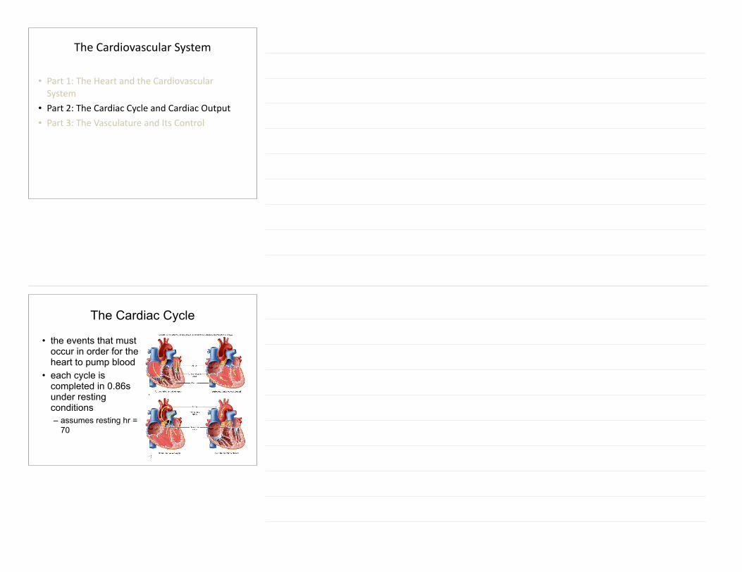
The Cardiac Cycle• valve positions at start
of cycle: – left atrial pressure > left
ventricular pressure • mitral valve is open
– blood can flow from atrium to ventricle (passive filling)
– left ventricular pressure < aortic pressure
• aortic semilunar valve is closed
– blood cannot flow between ventricle and aorta
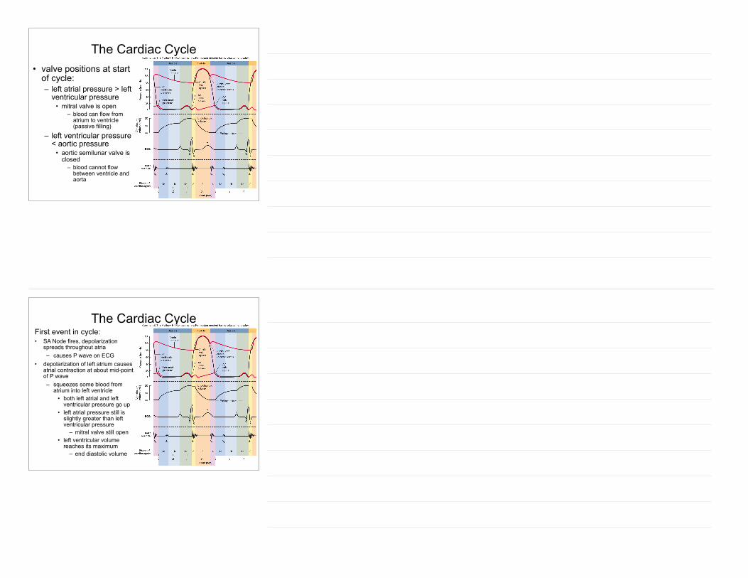
The Cardiac CycleFirst event in cycle: • SA Node fires, depolarization
spreads throughout atria – causes P wave on ECG
• depolarization of left atrium causes atrial contraction at about mid-point of P wave – squeezes some blood from
atrium into left ventricle • both left atrial and left
ventricular pressure go up • left atrial pressure still is
slightly greater than left ventricular pressure
– mitral valve still open • left ventricular volume
reaches its maximum – end diastolic volume

The Cardiac CycleNext event: • AV Node depolarizes, then
Bundle of His, AV bundles, and Purkinje fibers – causes depolarization
of all ventricular muscle fibers
• QRS complex on ECG
• ventricle muscle fibers respond by contracting
– ventricular systole
The Cardiac Cycle• as ventricle contracts, pressure
in the left ventricle goes up – quickly exceeds atrial
pressure • mitral valve closes
– causes first heart sound
– still less than aortic pressure
• aortic semilunar valve still closed
• with both valves closed, blood can neither enter nor leave ventricle – period of isovolumic
contraction • sometimes incorrectly
called isovolumetric contraction
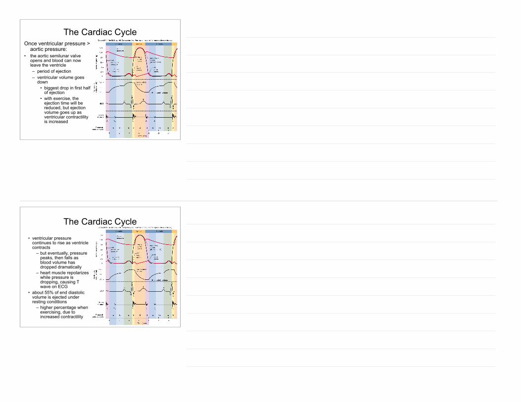
The Cardiac CycleOnce ventricular pressure >
aortic pressure: • the aortic semilunar valve
opens and blood can now leave the ventricle – period of ejection – ventricular volume goes
down • biggest drop in first half
of ejection • with exercise, the
ejection time will be reduced, but ejection volume goes up as ventricular contractility is increased
The Cardiac Cycle
• ventricular pressure continues to rise as ventricle contracts
– but eventually, pressure peaks, then falls as blood volume has dropped dramatically
– heart muscle repolarizes while pressure is dropping, causing T wave on ECG
• about 55% of end diastolic volume is ejected under resting conditions
– higher percentage when exercising, due to increased contractility
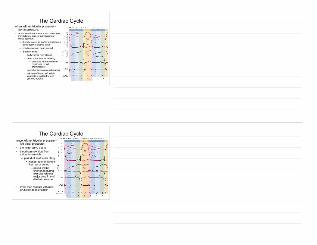
The Cardiac Cyclewhen left ventricular pressure <
aortic pressure: • aortic semilunar valve soon closes (not
immediately due to momentum of blood ejection) – dicrotic notch as aortic blood slams
back against closed valve – creates second heart sound – ejection ends
• both valves now closed • heart muscle now relaxing
– pressure in left ventricle continues to fall dramatically
• period of isovolumic relaxation • volume of blood left in left
ventricle is called the end systolic volume
The Cardiac Cycleonce left ventricular pressure <
left atrial pressure: • the mitral valve opens • blood can now flow from
atrium to ventricle – period of ventricular filling
• highest rate of filling in first half of period
– period will be shortened during exercise without major drop in end diastolic volume
!• cycle then repeats with next
SA Node depolarization
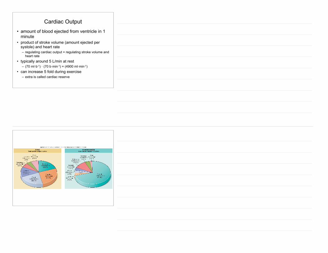
Cardiac Output
• amount of blood ejected from ventricle in 1 minute
• product of stroke volume (amount ejected per systole) and heart rate – regulating cardiac output = regulating stroke volume and
heart rate • typically around 5 L/min at rest
– (70 ml·b-1) · (70 b·min-1) = (4900 ml·min-1) • can increase 5 fold during exercise
– extra is called cardiac reserve
Regulation of Cardiac Output• regulation of stroke volume
– typically, heart ejects 55% of end diastolic volume
• ejection fraction = 0.55 – three factors affect stroke volume:
1.Preload 2.Contractility 3.Afterload
Regulation of Cardiac Output1. preload:
– stretch on heart muscle by end diastolic volume – Frank-Starling Law of the Heart:
• within limits, an increase in preload stretches myocardial fibers, giving better overlap of actin and myosin and hence stronger contraction.
– venous return to heart not only stretches muscle, but it determines the amount of blood that is available for ejection
• muscle pump and thoracic pump increase venous return
Regulation of Cardiac Output
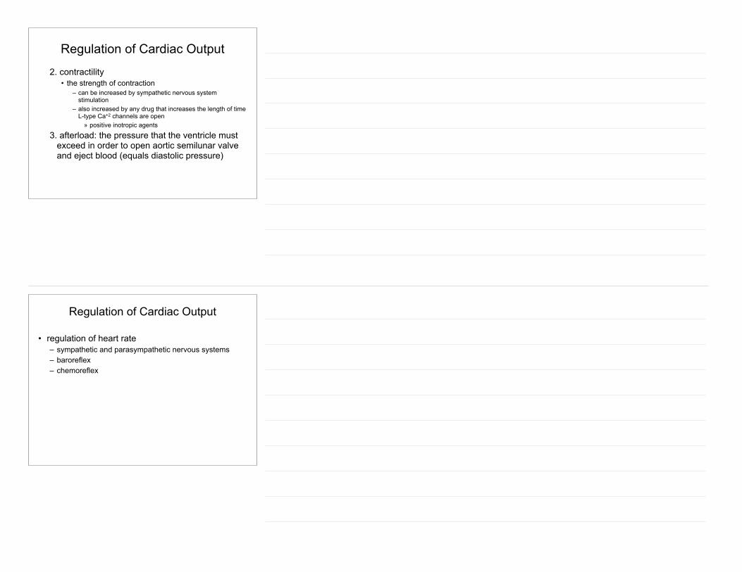
2. contractility • the strength of contraction
– can be increased by sympathetic nervous system stimulation
– also increased by any drug that increases the length of time L-type Ca+2 channels are open
» positive inotropic agents
3. afterload: the pressure that the ventricle must exceed in order to open aortic semilunar valve and eject blood (equals diastolic pressure)
Regulation of Cardiac Output
• regulation of heart rate – sympathetic and parasympathetic nervous systems – baroreflex – chemoreflex


![CYCLE PRESSURE - paintball-team-roterbaron.depaintball-team-roterbaron.de/technik/dye/Dye-DMC-Manual.pdf · CYCLE PRESSURE [75PSI] MAX RATE OF ... TROUBLE SHOOTING GUIDE ... residual](https://img.pdfslide.us/doc/110x75/5b8455c07f8b9a784a8c156c/cycle-pressure-paintball-team-cycle-pressure-75psi-max-rate-of-trouble.jpg)