Embed Size (px)
Citation preview

20/02/2017
1
Cardiovascular health &Health Promotion HH2602
& HH5607
Lecture 2: MicroscopicStructure and Function of
the Heart
2pm 28-02-17 ESGW
Teaching Aims
To introduce you to the micro-structure of heart muscle.
To highlight the link between cardiacstructure and cardiac function
Learning Out-comes:
At the end of the session you will beable to:
Outline the structure of cardiac muscle
Identify the unique functions andproperties of heart cells, and linkstructure to function.

20/02/2017
2
The Myocardium:
Consists of 3 types of excitable cells
1. myocardial cells +++
2. pace-maker cells
3. cells of the intrinsic cardiacconducting system

20/02/2017
3
Figure 18.8 a, b
Cardiac cytoskeleton :
Function of cardiac cytoskeleton:
1. Orientates muscle fibres
2. Acts as a tendon
2. Re-enforces vessel entry / exit points
3. Re-enforces valves
4. Forms a non- excitable zone between atria& ventricles – to safe-guard the independentelectrical and mechanical activity of the 2“hemispheres”
Cardiac cytoskeleton :

20/02/2017
4
The innervation of the heart:
SNS – muscle fibres generally espVentricular fibres, PM cells, cells of theintrinsic conducting system – all via β1
receptors (+ coronary arteries via αreceptors)
Paraympathetic n.s. – predominantlyPM cells, few atrial fibres – virtually NOventricular fibres
Myocardial Cells:
Cardiac muscle is structurally similarto skeletal muscle. However …
there are some important anatomical
and physiological distinctions
that account for…
the different behaviour of cardiac &skeletal muscle

20/02/2017
5
Fibre structure:
Intercalated Discs:
Intercalated discs contain:
Desmosomes
Gap junctions / nexi

20/02/2017
6
Desmosomes:
provide cell to cell cohesion (rivets)
optimise force transmission
Provide attachment site for actin
Gap Junctions: (nexi)Gap junctions form:
Low resistance - high conductancechannels thro’ which ions can flow fromfibre to fibre to …
i.e. AP propagation - in an All or nonemanner.
Mass excitation of a hemisphere masscontraction of each “hemisphere” in turn
Mitochondria: What does the table
suggest?
Cell type % cell volume
Type II Skeletal mm 2%
Type I Skeletal mm 12 – 15%
Cardiac mm 25 – 35%

20/02/2017
7
What is the sarcoplasmic reticulum?
What function does it serve?
How is this function slightly different incardiac muscle?
And what is the significance of thedifference?
SR in skeletal mm
Arrival of an AP in the t-tubule causesthe terminal cisternae of the SR torelease Ca2+ into mm cytoplasm.
This Ca2+ binds to troponin…
This sequence of events also happensin cardiac mm But its also a little bitdifferent… massive functionalsignificance
Cardiac Sarcoplasmic Reticulum:
Arrival of an AP at the sarcolemma
opening of sarcolemmal Ca2+ channels
influx of a small amt Ca2+ from the ECF
causing the SR to release larger amts ofCa2+ - amplifying effect
binding with troponin contraction
Calcuim induced calcium release - CICR

20/02/2017
8
Ca2+ induced Ca2+ release; (CICR)
ECFsarcolemma
Ca++ Ca++Ca++Ca++
Ca++Ca++
Ca++ Ca++
CICR happens when?
All the time / every time an APdepolarises the sarcolemma.
Its just how cardiac muscle works – dayin day out!
BUT CICR can be enhanced to ouradvantage when necessary
A couple of thoughts.
What would happen if the ECF Ca2+influx was increased?
How might the ECF influx beincreased?

20/02/2017
9
CICR is a useful means of increasingthe FORCE of contraction.
When activated sympathetic nerves &sympathetic hormones (catecholamines)
can cause more sarcolemmal Ca2+channels to open than usual.
What is the consequence of sympathetic n.s action?
So how does that work??
Significance of CICR – SNS openingof more SL Ca2+ channels than normal
ECF
Ca++ Ca++
Ca++
Ca++Ca++
Ca++
Ca++Ca++
Ca++
Ca++
Ca++
Ca++
Ca++Ca++
Ca++
Ca++
Ca++
SNS opening more Ca2+ channels in cellmembrane than usual
•Means more Ca2+ enters the cell
•Causes the release of more Ca2+ from SR
• Ca2+ availability to troponin
more XB than usual
more force larger volume ofblood ejected
20/02/2017
10
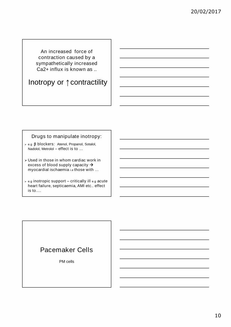
An increased force ofcontraction caused by a
sympathetically increasedCa2+ influx is known as ..
Inotropy or ↑contractility
Drugs to manipulate inotropy:
e.g blockers: Atenol, Propanol, Sotalol,
Nadolol, Metrolol – effect is to …
Used in those in whom cardiac work inexcess of blood supply capacitymyocardial ischaemia i.e those with …
e.g inotropic support – critically ill e.g acuteheart failure, septicaemia, AMI etc.. effectis to….
Pacemaker Cells
PM cells

20/02/2017
11
Myogenicity:
Skeletal mm = neurogenic
Cardiac mm = myogenic
auto-rhythmicity = inherent ability tospontaneously depolarise and createAPs contraction
Figure 18.14
SAN inherent rate of depolarisation
= ~ 100 bpm
What is your resting HR now?
Is it close to 100bpm?
If not – why not?

20/02/2017
12
Myogenic rates of depolarisationare modulated by the ANS
i.e by both the sympathetic & the
parasympathetic n.s.
Sympathetic n.s speeds up HR
Parasympathetic n.s slows down HR
The ANS & HRs: Parasympathetic n.s (vagus nerve) slows
rate of SAN depolarisation HR / HRrest
(negative chronotropy)
Sympathetic n.s. - speeds up SANdepolarisation HR e.g. exercise(positive chronotropy)
Both PNS & SNS nerves must synapsedirectly with the PM cells
Also - SNS hormones esp. epinephrine mustalso make contact with β1 PM cells.
Where does SNS or PNS activationcome from?

20/02/2017
13
Sympathetic n.s also force ofcontraction by increasing CICR
Inotropy or increasedcontractility
To cause inotropy the sympatheticnerves together with epinephrine mustmake direct contact / synapse withthe myocardial muscle cells
Note epinephrine – adrenal medulla
Can cardiac contractions besummated?
Sk mm twitches can be summatedbecause APs are v. short cf to the
twitch. Twitch summation but
Cardiac APs are as long as the twitch
Refractory periods are long
Twitch summation

20/02/2017
14
Summation & tetanisation
At high stimulation frequencies Sk mmcan tetanise force
But the heart can’t.
What would happen to cardiac output ifthe heart were capable of summation /tetanisation?
Conclusion:
Highlighted the main physiologicaldifferences between cardiac muscleand skeletal muscle namely:
Branching structure of cardiac fibres
Presence of intercalated discs, gapjunctions and desmosomes
20/02/2017
15
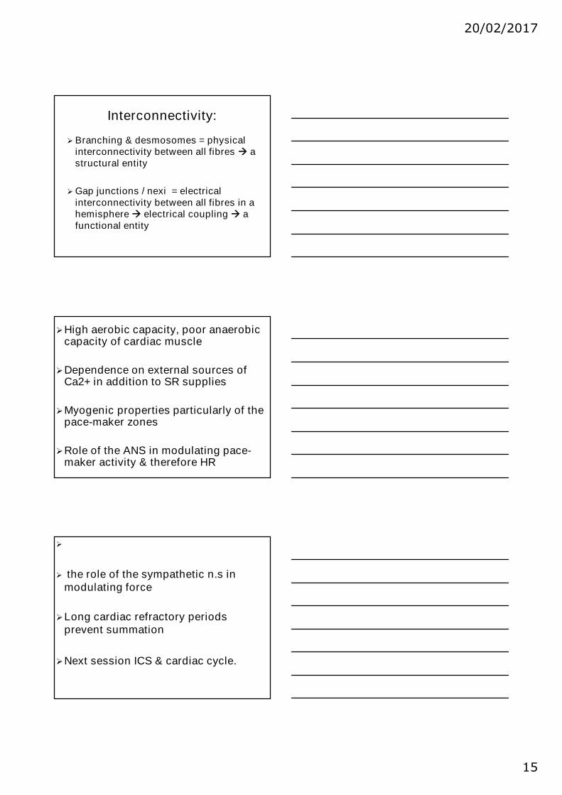
Interconnectivity:
Branching & desmosomes = physicalinterconnectivity between all fibres astructural entity
Gap junctions / nexi = electricalinterconnectivity between all fibres in ahemisphere electrical coupling afunctional entity
High aerobic capacity, poor anaerobiccapacity of cardiac muscle
Dependence on external sources ofCa2+ in addition to SR supplies
Myogenic properties particularly of thepace-maker zones
Role of the ANS in modulating pace-maker activity & therefore HR
the role of the sympathetic n.s inmodulating force
Long cardiac refractory periodsprevent summation
Next session ICS & cardiac cycle.

20/02/2017
16
Knowledge check:
What would happen to your HR rest if theANS supply to your PM cells was cut now?
What would happen to your HR if the SAN –gave up now?
When SNS activity is increased whatchanges HR (& how) ? Or force ofcontraction (& how)?
Possible Viva Qs?
Describe how the Autonomic nervoussystem (ANS) influences
i) pace-maker cell function (5 marks) and
ii) force production ( 5 marks)
Describe how the microscopic structure ofheart muscle facilitates the unique functionof the heart