Embed Size (px)
Citation preview

CARDIOTHORACIC TRANSPLANTATION
A CLINICAL TRIAL COMBINING DONOR BONE MARROW INFUSION AND HEART TRANSPLANTATION: INTERMEDIATE-TERM RESULTS
Si M. Pham, MO"'C Abdul S. Rao, MD. OPhil,,·b.c Adriana Zeevi, PhD",b,e Robert L. Kormos, MD" Kenneth R. McCurry, MD" Brack G. Hattler, MD, PhD" John 1. Fung, MD, PhDa,e Thomas E. Starz!, MD, PhD"·c Bartley P. Griffith, MD"
Background: Donor chimerism (the presence of donor cells of bone matTOW origin) is present for years after transplantation in recipients of solid organs. In lung recipients, chimerism is associated with a lower incidence of chronic rejection. To augment donor chimerism with the aim to enhance graft acceptance and to reduce immunosuppression, we initiated a trial combining infusion of donor bone matTOW with heart transplantation. Reported herein are the intermediate-term results of this ongoing trial.
Methods: Between September 1993 and August 1998, 28 patients received concurrent heart transplantation and infusion of donor bone matTOW at 3.0 x 10⁸ cells/kg (study group). Twenty-four contemporaneous heart recipients who did not receive bone marrow served as controls. All patients received an immunosuppressive regimen consisting of tacrolimus and steroids.
Results: Patient survival was similar between the study and control groups (86% and 87% at 3 years, respectively). However, the proportion of patients free from grade 3A rejection was higher in the study group (64% at 6 months) than in the control group (40%; P = .03). The prevalence of coronary artery disease was similar between the two groups (freedom from disease at 3 years was 78% in study patients and 69% in controls). Similar proportions of study (18%) and control (15%) patients exhibited in vitro evidence of donor-specific hyporesponsiveness.
,Conclusions: The infusion of donor bone marrow reduces the rate of acute rejection in heart recipients. Donor bone matTOW may play an important role in strategies aiming to enhance the graft acceptance. (J Thorac Cardiovasc Surg 2000; 119:673-81 )
We previously reported that donor cells of bone marrow lineages were widely distributed at a low level in
organs of long-surviving recipients of liver l and kidney
transplants.2 Furthermore, this phenomenon of donor microchimerism was present in some recipients who were weaned from immunosuppression prospectively. Subsequently, this phenomenon also occurred in recipients of other organs including hearts3 and lungs,4 and donor microchimerism was found to be associated with a lower incidence of chronic rejection in lung4 and in combined kidney and pancreas5 transplant recipients. To augment donor cell microchimerism with the aim to enhance long-term allograft survival, to attenuate the incidence of acute and chronic rejection, and to reduce the need for maintenance immunosuppression, we initiated a prospective trial combining the infusion of unmodified donor bone matTow with heart transplantation. Reported herein are the intermediate-term results of this trial.
From the Departments of Surgerya and Pathologyh and the Thomas E. Starzl Transplant Institute,e University of Pittsburgh, Pittsburgh, Pa.
Supported in part by the American College of Surgeons Faculty Fellowship, The Thoracic Surgery Research Foundation. American Heart Association grants to Si M. Pham, and National Institutes of Health grant AI40329 to John J. Fung and Abdul S. Rao.
Read at the Seventy-ninth Annual Meeting of The American Association for Thoracic Surgery. New Orleans, La, April 18-21, 1999.
Received for publication May 13, 1999; revisions requested July 7. 1999: revisions received Nov 23, 1999; accepted for publication Nov 24. 1999.
Address for reprints: Si M. Ph am, MD, Jackson Memorial Hospital. Highland Professional Bldg, 1801 NW 9th Ave, 5th Floor, Miami, FL 33136 (E-mail: [email protected]).
Copyright © 2000 by The American Association for Thoracic Surgery.
0022-5223/2000 S 12.00 + 0 12/6/104704
doi: 10.1 067/mtc.2000.1 04704
Patients and methods The Institutional Review Board of the University of
Pittsburgh approved this study on July 14, 1993, and informed
673
674 Pham et al
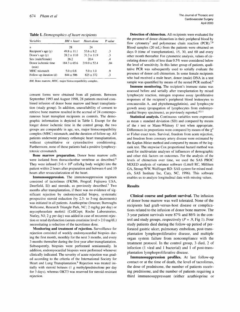
Table I. Demographics of heart recipients
Variables BM + heart Heart alone P value
n 28 24 Recipient's age (y) 49.8 ± 11.1 53.6 ± 8.2 .3 Donor's age (y) 28.2 ± 11.0 31.3 ± 11.9 .3 Sex (male/female) 26:2 20:4 .4 Donor ischemic time 184.3 ± 65.6 219.0 ± 52.4 .04
(min) MHC mismatch 5.0 ± 0.8 5.0± 1.0 .9 Follow-up duratiou (d) 844 ± 506 825 ± 372 .3
BM, Bone marrow; MHC, major histocompatibility complex.
consent forms were obtained from all patients. Between September 1993 and August 1998, 28 patients received combined infusion of donor bone marrow and heart transplantation (study group). In addition, unavailability of consent to retrieve bone marrow resulted in the accrual of 24 contemporaneous heart transplant recipients as controls. The demographic information is depicted in Table I. Except for the longer donor ischemic time in thc control group, the two groups are comparable in age, sex, major histocompatibility complex (MHC) mismatch, and the duration of follow-up. All patients underwent primary orthotopic heart transplantation, without cytoablative or cytoreductive conditioning. Furthermore, none of these patients had a positive lymphocytotoxic crossmatch.
Bone marrow preparation. Donor bone marrow cells were isolated from thoracolumbar vertebrae as described 6
They were infused (3-6 x 10⁸ cells/kg body weight) into the patient within 2 hours after preparation and between 6 and 10 hours after revascularization of the heart.
Immunosuppression. The immunosuppression regimen consisted of tacrolimus (FKS06, Prograf; Fujisawa USA, Deerfield, Ill) and steroids, as previously described.7 Two months after transplantation, if there was no evidence of significant rejection by endomyocardial biopsy, systematic prospective steroid reduction (by 2.S- to S-mg decrements) was initiated in all patients. Azathioprine (Imuran; Burroughs Wellcome, Research Triangle Park, NC; 2 mg/kg per day) or mycophenolate mofetil (CellCept; Roche Laboratories, Nutley, NJ; 2 g per day) was added in case of recurrent rejection or renal dysfunction (serum creatinine level> 2.0 mg/dL) necessitating a reduction of the tacrolimus dose.
Monitoring and treatment of rejection. Surveillance for rejection consisted of weekly endomyocardial biopsies during the first month, monthly for the next 3 months, and every 3 months thereafter during the first year after transplantation. Subsequently. biopsies were performed semiannually. In addition, endomyocardial biopsies were performed whenever clinically indicated. The severity of acute rejection was graded according to the criteria of the International Society for Heart and Lung Transplantation.8 Rejection was treated initially with steroid boluses (1 g methylprednisolone per day for 3 days), whereas OKT3 was reserved for steroid-resistant rejection.
The Journal of Thoracic and
Cardiovascular Surgery
April 2000
Detection of chimerism. All recipients were evaluated for the presence of donor chimerism in their peripheral blood by flow cytometry? and polymerase chain reaction (PCR)7.9 Blood samples (20 mL) from the patients were obtained on days 0 (time of transplantation), IS, 30, and 60 and every other month thereafter. For cytometric analysis, values of circulating donor cells of less than 0.5% were considered below the level of sensitivity. In this latter group of patients, qualitative PCR was subsequently used to serially evaluate the presence of donor cell chimerism. In some female recipients who had received a male heart, donor (male) DNA in a test sample was quantified by means of the nested PCR method.9
Immune monitoring. The recipient's immune status was assessed before and serially after transplantation by mixed lymphocyte reaction, mitogen response assay (proliferative responses of the recipient's peripheral blood leukocytes to concanavalin A, and phytohemagglutinin), and lymphocyte growth assay (propagation of lymphocytes [rom endomyocardial biopsy specimens), as previously reported. 10,1 I
Statistical analysis. Continuous variables were expressed as mean ± standard deviation (SO) and compared by means of the t test or Mann-Whitney U test when appropriate. Differences in proportions were compared by means of the X2 or Fisher exact tests. Survival, freedom from acute rejection, and freedom from coronary artery disease were estimated by the Kaplan-Meier method and compared by means of the logrank test. The stepwise Cox proportional hazard method was used for multivariate analyses of influences of demographic and other risk factors on outcomes. For the analysis of the levels of chimerism over time, we used the SAS PROC MIXED analysis of variance software (Littell RC, Milliken GA, Stroup WW, Wolfinger RD: SAS system for mixed models, SAS Institute Inc, Cary, NC, 1996). This software enables us to analyze longitudinal data with missing values.
Results
Clinical course and patient survival. The infusion of donor bone man-ow was well tolerated. None of the recipients had graft-versus-host disease or complications related to the infusion of donor bone marrow. The 3-year patient survivals were 87% and 86% in the control and study groups, respectively (P = .9, Fig 1). Four study patients died during the follow-up period of perforated gastric ulcer, pulmonary embolism, post-transplantation lymphoproliferative disease, and multiple organ system failure from noncompliance with the treatment protocol. In the control group, 3 died, 2 of infection (l viral and 1 bacterial) and I of post-transplantation lymphoproliferative disease.
Immunosuppression profiles. At last follow-up contact or at the time of death, the level of tacrolimus, the dose of prednisone, the number of patients receiving prednisone, and the number of patients requiring a third immunosuppressant (either azathioprine or

The Journal of Thoracic and
Carqiovascular Surgery
Volume 119, Number 4, Part 1
100
90
Pham et al 675
• • • ()()o
~ ~ .~.~ 1-
80 [J)
p~0.9 c .s;: 70 '1:
:::J en 60 c time (year) 0 t 50 Group 0.5 1.0 2.0 3.0 4.0 8. e 40 BM 93% 86% 86% 86% 86% a. Q) (95% CI) (75·98) (68·95) (68·95) (68·95) (68-95) > 30 ~ NoBM 92% 92% 87% 87% 87% :;
(95%CI) (73-98) (73-98) (ea.95) (Sa.95) (6a.95) E 20 :::J
<.l 10
0 0.5 1.0 1.5 2.0 2.5 3.0 3.5 4.0 4.5
Time after transplantation (year)
Fig 1. Patient survival (Kaplan-Meier estimates) after heart transplantation between bone marrow and control groups. BM, Bone marrow; CI, confidence interval.
100
90
80 (
70 Oil so §
° 50 0>0 o •
40 00' • .. O·
30
20
10
0 0 0.5 1.0 1.5
- Bone Marrow • •• No Bone Marrow
P =0.03
•• • 00 -0 ••• 0
2.0 2.5 3.0 3.5 4.0 4.5
Time after transplantation (year)
Group 0.5 1.0 2.0 3.0 4.0
BM 64% 64% 64% 64% 64% (95% CI) (46-80) (46-80) (46-80) (46-80) (46-80) NoBM 40% 40% 34% 34% 34% (95%CI) (23-60) (23-S0) (18-55) (1a.55) (1a.55)
Fig 2. Freedom from grade 3A rejection (Kaplan-Meier estimates) after heart transplantation between bone marrow and control groups. BM, Bone marrow; CI, confidence interval.
mycophenolate mofetil) were similar between the study and control groups (Table II).
Acute cellular rejection. The freedom from grade 3A rejection at 6 months after transplantation was significantly lower (P = .03) in control patients (40%) than in those who received bone marrow (64%; Fig 2). The infusion of donor bone marrow seems to have its impact early after transplantation. During the first 6 months after transplantation, the average number of episodes of grade 3A rejection per patient was 1.4 ± 0.2 and 0.6 ± 0.2 in the control and study groups, respectively (P =
.02). However, this salutary effect disappears in the subsequent follow-up, with the rates of 3A rejection approaching 0.1 ± 0.1 and 0.2 ± 0.1 in the control and study groups, respectively. Using the stepwise Cox proportional hazard method to assess the int1uence of various factors, which include age (donor and recipient), sex, ischemic time, MHC matching, tacrolimus level, and bone marrow infusion on acute rejection, we found that donor bone marrow infusion is the only independent prognostic factor that reduces acute rejection (P = .03; risk ratio = 2.42; 95% confidence intervals = 1.08-S.4l).

676 Pham et at
100
90 :i< 0
Cl 80 « 0 :; 70 0
- o·fJIt5·1" o. --I! .
---p= 0.6
-
The Journal of Thoracic and
Cardiovascular Surgery
April 2000
.<:
'i 60 o ••• • ·0 • • ·0
c::: 0 50 Time (year) :e 0 a. 40 Group 1.0 2.0 3.0 4.0 e
D- 8M (n=21) 95% 86% 86% 86% CD 30 > (95%CI) (71-99) (59-96) (59-96) (59-96) ~ ""5 20 ••••• No 8M (n=13) 92% 92% 62% 62% E '" (95%CI) (64-99) (64-99) (17-93) (17-93) 0 10
0 0.5 1.0 1.5 2.0 2.5 3.0 3.5 4.0 4.5
Time after transplantation (year)
Fig 3. Freedom from allograft coronary artery disease (Kaplan-Meier estimates) between bone marrow and control groups. BM, Bone marrow; CI, confidence interval; CAD, coronary artery disease.
3.0
2.5
~ 2.0 ·c II>
.S 1.5
..c U
~ 1.0
*
• •
• •• ®
• •• 0
•
• o o
••
• • :eo • y
• · (') • • 0
i 0 Controls i I I
~u Bone marrow I o
• e
• 0
... ~ •.••......•.•• ~ .• .Q. .... ~ ••• e. .... :.g ... ~_.@ ••....••.•••... .8. 0.5 : a:: ::.:t,:-: l .•. ·. ~.... •• A e0 Ie A o @@ -, eo·· •• 00 .(!n~
0.5 - 2 3-5 6-8 9-11 12-14 >14
Time after transplantation (month)
Fig 4. Levels of donor cell chimerism in recipients' peripheral blood leukocytes. Donor cell chimerism was detected in the peripheral blood leukocytes of heart recipients by now cytometry. Each circle represents one patient. Levels below 0.5% were considered not detectable. There was a marginal difference (P = .08) in the overall levels of chimerism between the study and control groups (analysis of variances for repeated measures). However, chimerism persisted much longer in the study group (P = .008). During the first 2 months after transplantation, the bone marrow group had a higher proportion of patients with detectable chimerism. Asterisk (*) indicates P < .05.
Allograft coronary artery disease. Allograft coronary artery disease after transplantation was defined as luminal irregularity and coronary stenosis seen on a coronary angiogram or diffuse arteriopathy at autopsy.
Table II. Immunosuppression profiles of heart recipients with or ,vithout donor bone marrmv infusion
Immunosuppressioll BM + heart Heart alone
FK level (ng/mL) 12.7±4.3 12.8 ± 4.4 Prednisone dose (mg/d) 5.6 ± 5.8 8.3 ± 5.9 Patients on prednisone 17128 (61%) 14/24 (58%)
Patients on a 3rd drug' 20/28 (71%) 14/24 (58%)
Expressed as mean ± -.tandard deviation, when appropriate.
Mann· Whitney U lest.
'Either azathioprine or rnycophenolate mofa!i!.
P value
.9
.07t
.9
.3
In 34 patients in whom allograft coronary arteries could be evaluated, the freedom from coronary artery disease at 3 years after transplantation was 69% (control group, n = 13) and 78% (study group, n = 21) (P = .9) (Fig 3).
Donor cell chimerism. Detection of donor cell chimerism was initially carried out by flow cytometric technique, which can detect chimerism at a level of one donor cell in 200 recipient cells (0.5%) and has been shown to correlate well with the induction and maintenance of tolerance in experimental models. 12 In patients whose levels of donor cells fall below this threshold. molecular detection of donor human leukocyte antigens by peR (which could detect an equivalent of one donor cell in 105 recipient cells) was performed.
Flow cytometric analyses were feasible (when appropriate antibodies to disparate donor HLA antigens were available) in 16 study patients and in 15 controls. Overall, the level of donor chimerism was low «3.0%) (Figs 4 and 5). There was a marginal difference (P =
The Journal of Thoracic and
Cardiovascular Surgery
Volume 119, Number 4, Part 1
A
Pham et al 677
B c
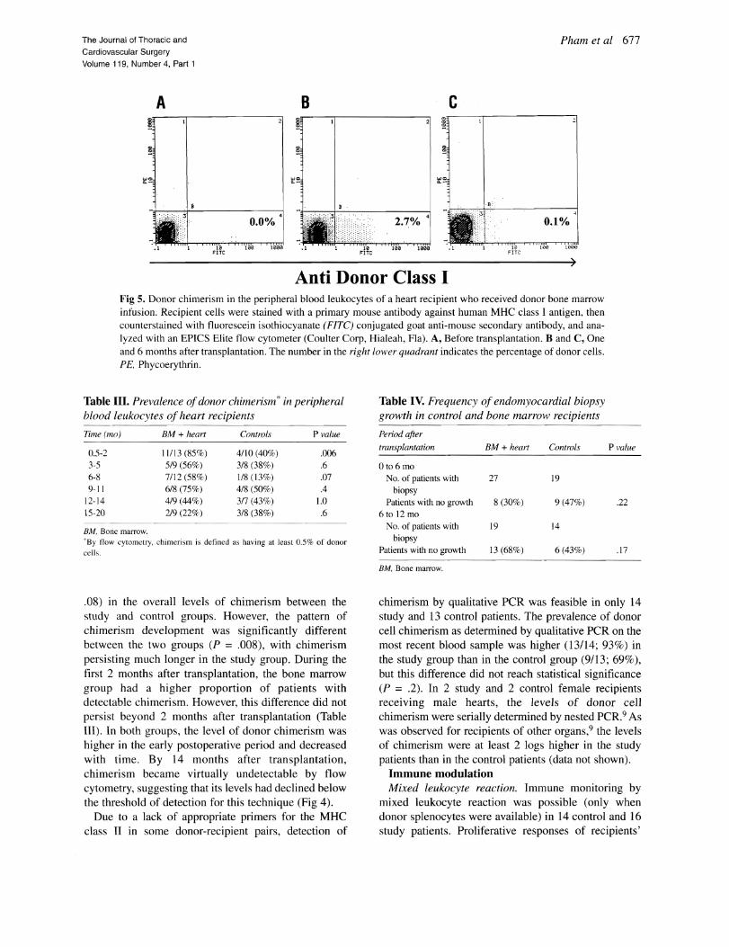
Anti Donor Class I Fig 5. Donor chimerism in the peripheral blood leukocytes of a heart recipient who received donor bone marrow infusion. Recipient cells were stained with a primary mouse antibody against human MHC class I antigen, then counterstained with fluorescein isothiocyanate (FITe) conjugated goat anti-mouse secondary antibody, and analyzed with an EPICS Elite flow cytometer (Coulter Corp, Hialeah, Fla). A, Before transplantation. Band C, One and 6 months after transplantation. The number in the right lo\\'er quadrant indicates the percentage of donor cells. PE, Phycoerythrin.
Table III. Prevalence of donor chimerism * in peripheral blood leukocytes of heart recipients
Time (mo) BM + heart Controls P value
0.5-2 1II13 (85%) 4110 (40%) .006 3-5 5/9 (56%) 3/8 (38%) .6 6-8 7112 (58%) 1/8 (13%) .07 9-11 6/8 (75%) 4/g (50%) .4
12-14 4/9 (44%) 317 (43%) 1.0 15-20 219 (22%) 3/8 (38%) .6
8M, Bone marrow. "'By flow cytomdry, chimerism is defined as having at least 0.5% or donor
cells.
. 08) in the overall levels of chimerism between the study and control groups. However, the pattern of chimerism development was significantly different between the two groups (P = .008), with chimerism persisting much longer in the study group. During the first 2 months after transplantation, the bone marrow group had a higher proportion of patients with detectable chimerism. However, this difference did not persist beyond 2 months after transplantation (Table III). In both groups, the level of donor chimerism was higher in the early postoperative period and decreased with time. By 14 months after transplantation, chimerism became virtually undetectable by flow cytometry, suggesting that its levels had declined below the threshold of detection for this technique (Fig 4).
Due to a lack of appropriate primers for the MHC class Ii in some donor-recipient pairs, detection of
Table IV. Frequency of endomvocardial biopsy growth in control and bone marr01-)) recipients
Period after
transplantation BM + heart Controls
Ot06mo No. of patients with 27 19
biopsy Patients with no growth 8 (30%) 9 (47%)
6to 12mo No. of patients with 19 14
biopsy Patients with no growth 13 (68%) 6 (43'J,,)
8M. Bone marrow .
P value
.22
.17
chimerism by qualitative PCR was feasible in only 14 study and 13 control patients. The prevalence of donor cell chimerism as determined by qualitative PCR on the most recent blood sample was higher (13114; 93%) in the study group than in the control group (9113; 69%), but this difference did not reach statistical significance (P = .2). Tn 2 study and 2 control female recipients receiving male hearts, the levels of donor cell chimerism were serially determined by nested PCR.9 As was observed for recipients of other organs,9 the levels of chimerism were at least 2 logs higher in the study patients than in the control patients (data not shown).
Immune modulation Mixed leukocyte reaction. Immune monitoring by
mixed leukocyte reaction was possible (only when donor splenocytes were available) in 14 control and 16 study patients. Proliferative responses of recipients'

678 Pham et al
peripheral blood leukocytes to donor splenocytes at various times after transplantation were compared with those obtained in pretransplantation samples. No diffcrence in donor-specific immune modulation was discerned between the two groups for up to I year after transplantation. Eighteen percent of patients in the study group and 15% in the control group had donorspccific hyporesponsiveness (data not shown).
Lymphocyte growth assay. We have demonstrated that a positive lymphocyte growth assay in endomyocat'dial specimens that have low-grade rejection (grade < 2) is highly predictive of subsequent high-grade rejection (grade > 2). II Therefore the lymphocyte growth assay may be used as a surrogate marker of the recipient's immune reactivity toward the graft. The absence of lymphocyte growth from the endomyocardial specimens indicates less alloreactivity toward the graft and vice versa. During the first 6 months after transplantation, no difference was noted in the proportion of patients with negative lymphocyte growth assay (non-grower) between the study and control groups (30% vs 47%, respectively; Table IV). In the subsequent 6 months (6-12 months after transplantation), the proportion of non-growers was higher (although not to a statistically significant degree) in the study group than in the control group (68% vs 43%, respectively; Table IV). This observation suggested a trend toward less alloreactivity in recipients who had received concomitant bone marrow infusion.
Discussion
The recognition that bone marrow chimerism is associated with immunologic tolerance dates back more than half a century. Billingham and associates l3
observed that adult freemartin cattle (dizygotic twins that share a common placenta during intrauterine life and possess erythrocyte chimerism) permanently accepted each other's skin grafts. These investigators were able to duplicate this observation in experimental models by inoculating hematopoietic cells obtained from MHC-disparate donors into fetal mice. 14
Subsequently, chimerism with donor-specific tolerance was achieved in adult animals by preconditioning the host with total body irradiation, 12 total lymphoid irradiation,15 and the use of antilymphocyte globulin. 16
Although clinical translation of tolerant models requiring recipient conditioning with total lymphoid radiation 17 and total body radiation (I1dstad ST, personal communication, 2000) have been attempted, it is the Monaco model,16 which used antilymphocyte globulin induction therapy, that has been exploited extensively in clinical transplantation. Monaco and associates l8
The Journal of Thoracic and
Cardiovascular Surgery
April 2000
were the first to apply this strategy to a renal recipient in 1976. The first large-scale clinical trial in which the Monaco model was used was carried out in cadaveric renal recipients by Barber and colleagues. 19 In this study, graft survival was significantly better in the bone marrow group than in the controls (90% vs 71 % at I year). Other clinical evidence of benefits of bone marrow augmentation included a reduced need for immunosuppression and a lower acute rejection rate.
The rationale for the initiation of the current adjuvant bone marrow infusion program was based on the discovery by Starzl and coworkers 1,2 that donor cells of bone marrow lineages persisted at a low level in the peripheral blood, lymphoid organs, and skin of longsurviving kidney (n = 5; lO-29 years) and liver (n = 25; 3-22 years) recipients. 1,2 On the basis of these observations, it was argued that the establishment of microchimerism (the presence of a low level of donor bone marrow-derived cell in solid organ recipients) was perhaps essential for long-term allograft acceptance. It was therefore hypothesized that augmenting this spontaneously occurring event with perioperative donor bone marrow infusion may further enhance the acceptance of the graft, especially of those organs that are not endowed with a large quantity of passenger leukocytes within their interstitium. To test this hypothesis, in December 1992, we6 initiated a trial combining donor bone marrow infusion with solid organ transplantation, without recipient preconditioning.
Following the lead at Pittsburgh, Garcia-Morales and associates20 at the University of Miami have adopted a modified protocol using multiple infusions of donor bone marrow in renal transplant recipients. In this nonrandomized trial, 40 renal transplant recipients who received unmodified donor bone marrow infusion were compared with 100 contemporaneous control subjects. All patients received induction therapy with OKT3 and a maintenance immunosuppression regimen consisting of tacrolimus and steroids; in some patients mycophenolate mofetil was added as a third drug. Cryopreserved bone marrow cells were infused at two intervals: between days 1 and 4 and between days 10 and 14 after transplantation. The bone marrow recipients had higher levels of donor chimerism than control subjects, both in their peripheral blood and in their bone marrow. Notably, the levels of donor chimerism (especially CD3+ and CD34+ cells) were I (}-fold higher in the bone marrow compartment than in the peripheral blood. Patients receiving bone marrow also had more mitigated humoral and cellular immune responses than did the control patients. Using the same induction and maintenance immunosuppression protocol as employed by

The Journal of Thoracic and
Cardiovascular Surgery
Volume 119, Nurnber 4, Part 1
Garcia-Morales and associates,20 Ricordi and coworkers21 have reported that liver allograft survival was significantly higher in patients receiving multiple infusions than in those receiving a single perioperative infusion of donor bone marrow.
The intermediate-term results in heart recipients reported herein confirm our preliminary data7 that the infusion of unmodified donor bone marrow concurrent-1y with heart transplantation is well tolerated and safe. There was no evidence of graft-versus-host disease and no complication that was related specifically to the infusion of donor bone marrow. There was an increase in the level of chimerism in the bone marrow recipients as compared with the controls, a finding reminiscent of that observed in the recipients of other organ allografts. 9 Despite the similarity in the immunosuppression profiles, acute rejection was less prevalent in bone marrow recipients than in the control patients. Furthermore, there was a trend toward less alloreactivity toward the graft, as assessed by the lymphocyte growth assay. in the study group. Although not statistically significant. the freedom from allograft coronary artery disease at 3 years after transplantation was higher in bone marrow recipients (78% vs 69% in the controls). It is conceivable that with a longer duration of follow-up and a larger sample size, this difference may approach statistical significance.
The mechanism that accounts for the lower rejection rate in the bone marrow group remains to be elucidated. However, it is conceivable that donor bone marrow cells may have down-regulated the recipient's immune system by suppressing the development of cytotoxic T cells. In vitro studies by Mathew,22 Rachamin,23 and their associates have demonstrated that human bone marrow cells inhibited both the proliferative and cytotoxic responses in a dose-dependent manner. These in vitro data supporting the existence of a veto mechanism, which was originally described by Miller,24 have been further corroborated by in vivo observations in nonhuman primates25 and in human beings.26
Another possible mechanism by which the donor bone marrow cells could have modulated the recipient's immune system is via clonal exhaustion of donor-reactive cells. It is possible that the infused donor bone marrow cells, which can readily disseminate throughout the recipient's body, expose the recipient's immune cells with an excess of donor antigens, causing clonal activation followed by deletion of the donor-reactive T (and possibly B) cells. This activation-induced tolerance, which occurs exclusively in lymphoid organs or organized lymphoid collections, is analogous to the situation in which all antigen-specific T cells are deleted when the
Pham et al 679
host is exposed to an excess load of that particular antigen, such as a generalized infection with noncytopathic viruses.27•28 This mechanism of tolerance is elegantly described by Zinkemagel27 in the case of lymphocytic choriomeningitis infection in mice. In the realm of transplantation tolerance, the commonality between the tolerance to infectious agents and transplanted allografts has been discussed in detail by Starzl and Zinkemagel.28
Of particular interest is the fact that even though acute rejection is less prevalent in the bone marrow group, there was no in vitro evidence of immune modulation in these patients as assessed by I-way mixed leukocyte reaction. The discrepancy between the in vivo and in vitro responses as observed in this study is not unique. This dichotomy between in vivo hyporesponsiveness (organ acceptance) and in vitro alloreactivity, which has been referred to as "split tolerance,"29.30 further heightens the need for developing more reliable in vitro techniques that would be more predictive of graft tolerance.
In summary, the intermediate-term data from this currently ongoing clinical trial indicate that the infusion of unmodified donor bone marrow concurrently with heart transplantation is safe and is associated with an increased level of donor cell chimerism. Furthermore, the early immunologic events after cardiac transplantation appear to have been altered by the infusion of donor bone marrow. When compared with the control patients, bone marrow recipients had less acute rejection and less alloreactivity (albeit not to a statistically significant degree) by the lymphocyte growth assays. Although the eventual salutary effect of donor-specific bone marrow infusion in heart recipients remains speculative, it is apparent that donor bone marrow infusion is feasible, safe, and associated with less rejection. Inclusion of concomitant infusion of donor bone marrow in future strategies aiming to enhance the acceptance of cardiac allografts must be seriously considered.
We thank Robert Duncan, PhD, Professor of Epidemiology and Public Health, University of Miami, for his assistance with statistical analyses.
REFERENCES l. Starzl TE, Demetris AJ. Trucco M, Ramos H, Zeevi A. Rudert
WA, et al. Systemic chimerism in human female recipients of male livers. Lancet 1992;340:876-7.
2. Starzl TE. Demetris AJ, Trueco M, Zeevi A, Ramos H, Terasaki P, et al. Chimerism and donor-specific nonreactivity 27 to 29 years after kidney allotransplantation. Transplantation 1993;55: 1272-7.
3. Schlitt HJ. Hundrieser J. Hisanaga M, Uthoff K, Karck M, Wahlers T, ct al. Patterns of donor-type microchimerism after heart transplantation. Lancet 1994;343: 1469-71.
~----~-------.----------------
680 Pham et al
4. McSherry C, Hertz MI, Jackson AM, Butters K, Diko M, Matas AJ, et a!. Allogeneic microchimerism and donor antigen-specific hyporeactivity in lung transplant recipients. Clin Transplant 1995:9:442-9.
5. Corry R. Rao AS, Shapiro R. Jordan M, Dvorchik 1, Scantlebury V, et a!. Simultaneous administration of adjuvant donor bone marrow in pancreas transplant recipients. Surgery. In press.
6. Fontes P, Rao AS, Demetris AJ, Zeevi A, Trucco M, Carroll P. et a!. Bone marrow augmentation of donor-cell chimerism in kidney, liver, heart, and pancreas islet transplantation. Lancet 1994; 344:151-5.
7. Pham SM, Keenan RJ, Rao AS. Fontes PA, Kormos RL, AbuElmagd K, et al. Perioperative donor bone marrow infusion augments chimerism in heart and lung transplant recipients. Ann Thorac Surg 1995;60:1015-20.
8. Billingham ME, Cary NR, Hammond ME, Kemnitz J, Marboe C, McCallister HA. et al. A working formulation for the standardization of nomenclature in the diagnosis of heart ami lung rejection: Heart Rejection Study Group. The International Society for Heart Transplantation. J Heart Transplant 1990;9:587-93.
9. Salgar S, Shapiro R, Dodson F, Corry R, McCurry K, Zeevi A, et a!. Infusion of donor leukocytes to induce tolerance in organ allograft recipients. J Leukoc Bioi 1999;66:310-4.
10. Zcevi A. Pavlick M, Lombardozzi S, Banas R, Pappo 0, Rao AS, et al. Immune status of recipients following bone marrow-augmented solid organ transplantation. Transplantation 1995;59: 616-20.
II. Weber T, Zerbe T. Kaufman C, Zcevi A, KornlOS R. Hardesty R. el al. Propagalion of alloreactive lymphocytes from histologically negative endomyocardial biopsies from heart transplant patients: association with subsequent histological evidence of allograft rejection. Transplantation 1989;48:430-5.
12. lIdstad ST, Sachs DH. Reconstitution with syngeneic plus allogeneic or xenogeneic bone marrow leads to specific acceptance of allografts or xenografts. Nature 1984;307: 168-70.
13. Billingham RE, Lamphin HG, Medawar PB, Williams HL. Tolerance of homografts, twin diagnosis, and the freemartin conditions in cattle. Heredity 1952:6:201-21.
14. Billingham RE, Brent L, Medawar PB. Actively acquired tolerance to foreign cells. Nature 1953;172:603-6.
15. Slavin S, Strober S, Fuks Z, Kaplan HS. Induction of specific transplantation tolerance nsing fractionated total lymphoid irradiation in adult mice: Long-term survival of allogeneic bone marrow and skin grafts. J Exp Med 1977: 146:34-48.
16. Monaco AP. Use of antilymphocyte serum in the induction of immunological tolerance to tissue allografts. Fed Proc 1970:29: 153-5.
17. Najarian IS. Ferguson RM, Sutherland DE, Slavin S, Kim T, Kersey J. et a!. Fractionated total lymphoid irradiation as preparative immunosuppression in high risk renal transplantation: clinical and immunological studies. Ann Surg 1982: 196:442-52.
18. Monaco AP, Clark AW, Wood ML, Sahyoun AI, Cod ish SD, Brown RW. Possible active enhancement of a human cadaver renal allograft with antilymphocyte sernm (ALS) and donor bone marrow: case report of an initial attempt. Surgery 1976;79:384-92.
19. Barber WH, Mankin JA, Laskow DA, Deierhoi MH, Julian BA, Curtis JJ, et al. Long-term results of a controlled prospective study with transfusion of donor-specific bone marrow in 57 cadaveric renal allograft recipients. Transplantation 1991;51:70-5.
The Journal of Thoracic and Cardiovascular Surgery
April 2000
20. Garcia-Morales R, Carreno M, Mathew J. Zucker K, Cirocco R, Ciancio G, et al. The effects of chimeric cells following donor bone marrow infusions as detected by PCR-flow assays in kidney transplant recipients [published erratum appears in .I Clin Invest 1997;99:22951. J Clin Invest 1997;99:1118-29.
21. Ricordi C, Karatzas T, Nery J, Webb M. Selvaggi G, Fernandez L, et al. High-dose donor bone marrow infusions to enhance allograft survival: thc effect of timing. Transplantation 19<)7;63:7-11.
22. Mathew JM, Carreno M, Fuller L. Ricordi C, Tzakis A, Esqnenazi Y, ct al. Modulatory effects of human donor bone marrow cells on allogeneic cellular immune responses. Transplantation 1997;63:686-92.
23. Rachamim N, Gan .I. Segall H, Krauthgamer R. Marcus H. Berrebi A, et al. Tolerance induction by "megadose" hematopoietic trarsplants: donor-type human CD34 stem cells induce potent specific reduction of host anti-donor cytotoxic T lymphocyte precursors in mixed lymphocyte culture. Transplantation 1998;65: 1386-93~
24. Miller RG. An immunological suppressor cell inactivating cytotoxic T-lymphocyte precursor cells recognizing it. Nature 1980; 287:544-6.
25. Thomas JM, Carver FM, Cunningham PR, Olson LC, Thomas FT. Kidney allograft tolerance in primates without chronic immunosuppression-the role of veto cells. Transplantation 1991 ;51: 198-207.
26. Burlingham WJ, Grailer AP, Fechner JH Jr, Kusaka S, Trucco M, Kocova M, et al. Microchimerislll linked to cytotoxic T lymphocyte functional unresponsiveness (clonal anergy) in a tolerant renal transplant recipient. Transplantation 1995;59: 1147-55.
27. Zinkernagel RM. Immunology taught by viruses. Science 1996; 271: 173-8.
28. Starzl TE, Zinkcrnagel RM. Antigen localization and migration in immunity and tolerance. N Engl J Med 1998:339: 1905-13.
29. Mayumi H, Himeno K, Tokuda N, Fan JL, Nomoto K. Druginduced tolerance to allografts in mice. X. Augmentation of split tolerance in murine combinations disparate at both H-2 and nonH-2 antigens by the use of spleen cells from donors preimmuni/.ed with recipient antigens. Immllnobiology 1987; 174:274-91.
30. Dahmen U, Qian S, Rao AS, Demetris AJ, Fu F, Sun H, et al. Split tolerance indnced by orthotopic liver transplantation in mice. Transplantation 1994;58: 1-8.
Discussion Dr Verdi J. DiSesa (Chicago. Tlli. Dr Pham and his col
leagues have shown that unmodilied donor bone marrow
administered intravenously shortly after heart transplantation
can reduce the incidence of moderate to severe rejection. This is important since it represents a shift from a strategy of trans
plant immune modulation based on nonspecific pharmacolog
ic immune suppression to one whereby biologic manipulations
are used to induce donor-specific hyporesponsiveness. This is Sir Peter Medawar's actively acquired immune tolerance, and
I congratulate Dr Pham and his coauthors on an elegantly pre
sented report of their attempts to produce it in patients. In heart transplantation, it is not the usual practice to per
form prospective histocompatibility tissue antigen matching,
and the authors have not done so here. However, we and others have shown that fortuitous MHC matching, identified ret-

The Journal of Thoracic and
Cardiovascular Surgery
Volume 119, Number 4, Part 1
rospectively, can produce reductions in rejection similar to those observed in this study.
The authors have reported average MHC mismatching to be the same in both groups, but have they looked at subsets of patients within each group that may have had fortuitous matching? Might this have played a role in the differences in rejection that they saw?
Transient chimerism in their study was also seen in patients treated conventionally without bone marrow infusions. Why did the authors not include a conditioning regimen, such as irradiation, so as to enhance the degree and durability of bone marrow engraftment? What is their explanation for the failure to see a reduced proliferative response in the in vitro immune assays?
Finally, what do the authors think about the importance of the nature of chimerism? Is it low level of all lineages of donor hematopoietic cells that is important, or perhaps the Illere presence of any lineage of circulating donor cells? Does it make any difference?
Dr Pham. Thank you, Dr DiSesa, for your comments. To answer your first question regarding a subset analysis of patients with MHC match, the number of patients in our study is too small to do that. Dr Joshua Miller's group at the University of Miami had identified a subset of renal recipients with DR match who received donor bone marrow infusion. These recipients had better engraftment of the donor cells than those without DR Illatch. Furthermore, rejection was less common in these recipients. Actually, 10 of 10 patients of that group had never had rejection.
The second question is related to the conditioning of the
Pham et al 681
recipient: Why did we not give the recipient radiation or any other cryoablative conditioning? The most robust means to induce tolerance is the mixed chimerislll achieved by radiation that allows the engraftment of the stem cells of the bone marrow. This type of tolerance had been reported initially by Medawar and later by Suzanne Ildstad and David Sachs. However, convincing the institutional review board to allow us to use radiation initially was difficult. Furthermore, the rationale of the current study is different from the classic radiation chimera model. In this study we tried to enhance the naturally occurring microchimerism. We do not know whether donor bone marrow infusion will enhance graft acceptance or not: A great body of experimental data suggests that it would work. In the 1970s, Dr Monaco from Boston showed that this form of treatment induced tolerance in the small animal models. Subsequently, other groups have shown the same results in the larger animal.
The discrepancy between the in vitro immune response versus the in vivo response (ie, graft acceptance) has been well described in animal models. This phenomenon is called split tolerance, where there is acceptance of the organ but no tolerance by in vitro (mixed lymphocyte reaction or cell-mediated Iymphocytotoxity) testing. It reflects the fact that a better or more sensitive technique is needed to detect tolerance.
The stem cell is very important for the induction of tolerance because, if engrafted, it allows the propagation of donor cells and donor antigens that allow clonal deletion of the recipient lymphocytes. However, other mechanisms of tolerance such as clonal anergy and clonal exhaustion may also play an important role in enhancing graft acceptance.


![Kidney Transplantation (Renal Transplantation) Auto Saved]](https://img.pdfslide.us/doc/110x75/577d22b31a28ab4e1e9807d7/kidney-transplantation-renal-transplantation-auto-saved.jpg)