Embed Size (px)
Citation preview
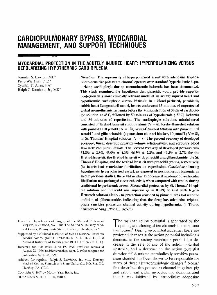
CARDIOPULMONARY BYPASS, MYOCARDIAL MANAGEMENT, AND SUPPORT TECHNIQUES
MYOCARDIAL PROTECTION IN THE ACUTELY INJURED HEART: HYPERPOLARIZING VERSUS DEPOLARIZING HYPOTHERMIC CARDIOPLEGIA
Jennifer S. Lawton, MD a Peng-Wie Hsia, PhD a Cynthia T. Allen, BA d Ralph J. Damiano, Jr., MD a
Objectives: The superiority of hyperpolarized arrest with adenosine triphos- phate-sensitive potassium channel openers over standard hyperkalemic depo- larizing cardioplegia during normothermic ischemia has been documented. This study examined the hypothesis that pinacidil would provide superior protection in a more clinically relevant model of an acutely injured heart and hypothermic cardioplegic arrest. Methods: In a blood-perfused, parabiotic, rabbit heart Langendorff model, hearts underwent 15 minutes of unprotected global normothermic ischemia before the administration of 50 ml of cardiople- gic solution at 4 ° C, followed by 50 minutes of hypothermic (15 ° C) ischemia and 30 minutes of reperfusion. The cardioplegic solutions administered consisted of Krebs-Henseleit solution alone (N = 6), Krebs-Henseleit solution with pinacidil (50 b~mol/L; N = 10), Krebs-Henseleit solution with pinacidil (50 /~mol/L) and glibenclamide (a potassium channel blocker, 10 #mol/L; N = 8), or St. Thomas ' Hospital solution (N = 8). The percent recovery of developed pressure, linear diastolic pressure-volume relationships, and coronary blood flow were compared. Results: The percent recovery of developed pressure was 3 2 . 8 % - 2.8%, 43.0% -+ 4.3%, 46.5% --+ 2.2%, and 49.3% -_. 2.7% for the Krebs-Henseleit, the Krebs-Henseleit with pinacidil and glibenclamide, the St. Thomas' Hospital, and the Krebs-Henseleit with pinacidil groups, respectively. No hearts had ventricular fibrillation on reperfusion. Conclusions: During hypothermic hyperpolarized arrest, as opposed to normothermic ischemia as in our previous studies, there was neither an increased incidence of ventricular fibrillation nor prolonged electrical activity when compared with results during traditional hyperkalemic arrest. Myocardial protection by St. Thomas ' Hospi- tal solution and pinacidil was superior (p = 0.009) to that with Krebs- Henseleit solution alone. The protection provided by pinacidil was lost with the addition of glibenclamide, indicating that the drug has adenosine triphos- phate-sensitive potassium channel activity during hypothermia. (J Thorac Cardiovasc Surg 1997;113:567-75)
From the Departments of Surgery of the Medical College of Virginia, Richmond, Va., a and The Milton S. Hershey Med- ical Center, Pennsylvania State University, Hershey, Pa. b
Supported by a National Institutes of Health National Research Service Award, grant HL09125-02 (J. S. L., R. J. D.) and National Institutes of Health grant RO1 HL51032 (R. J. D.).
Received for publication June 19, 1996; revisions requested August 22, 1996; revisions received Sept. 9, 1996; accepted for publication Sept. 23, 1996.
Address for reprints: Ralph J. Damiano, Jr., MD, Hershey Medical Center, Pennsylvania State University, P.O. Box 850, Hershey, PA 17033.
Copyright © 1997 by Mosby-Year Book, Inc. 0022-5223/97 $5.00 + 0 12/1/78173
T he myocyte act ion potent ia l is genera ted by the opening and closing of ion channels in the p lasma
m e m b r a n e ] Dur ing myocardia l ischemia, there are p ro found changes in the act ion potent ia l including a decrease in the resting m e m b r a n e potential , a de- crease in the rate of rise of the action potent ia l ups t roke, and a decrease in the action potent ia l duration.2, 3 A unique metabol ical ly sensitive potas- s ium channel has been shown to be responsible for many of these electrophysiologic changes. 3 N o m a 4 first descr ibed this po tass ium channel in guinea pig and rabbit ventr icular myocytes and demons t r a t ed that it was inhibited by intracellular adenos ine
5 6 7
5 6 8 Lawton et aL The Journal of Thoracic and
Cardiovascular Surgery
March 1997
triphosphate (ATP) and opened dur ing times of ischemia. The opening of this ATP-sensitive potas- sium (KATP) channel causes an outward potassium current that hyperpolarizes the cell membrane. This results in a significant shortening Of the duration of the plateau phase of the action potential, during which most of the inward calcium transport occurs. This reduction in calcium influx causes a decrease in contractility. Activation of KAT e channels has been shown to underlie the contractile failure seen during periods of ischemia and metabolic inhibition. 5 This decrease in mechanical activity conserves ATP and thus is cardioprotective. 4
Previous Work from our laboratory has demon- strated the benefit of the ATP-sensitive potassium channel openers (PCOs) pinacidil and aprikalim as cardioplegic agents during normothermic ischemia in our blood-perfused, isolated rabbit heart model. During normothermic ischemia, aprikalim was shown to provide superior myocardial protection compared with 20 mmol KC1 in Krebs-Henseleit solution, and pinacidil was shown to provide equiv- alent myocardial protection compared with 20 mmol KC1 in Krebs-Henseleit solution and with St. Thomas ' Hospital solution. 6-8 The optimal dose of pinacidil was determined to be 50 /xmol/L. 7 A shortcoming of these studies was that they examined potassium channel opener cardioplegia only during normothermic global ischemia in normal hearts. However, many cardiac surgeons still use some degree of hypothermia to protect the heart during cardioplegic arrest. Operations also are often com- plicated by acute ongoing ischemia, such as seen after angioplasty failure. In an effort to evaluate pinacidil in a more clinically relevant model, the current study was designed to compare myocardial protection with pinacidil (50/xmol/L), pinacidil with glibenclamide (10 txmol/L; a potassium channel inhibitor), and St. Thomas ' Hospital solution during hypothermic ischemia in an acutely injured heart.
Material and methods
Experimental preparation. Adult New Zealand White rabbits of either sex weighing 3.0 to 5.0 kg were anesthe- tized intramuscularly with acepromazine (1 mg/kg) and xylazine (17.5 mg/kg), followed by ketamine (62.5 mg/kg). Each study involved a support animal and a study animal. The support animal was heparlnized (2500 U) via an ear vein and underwent cannulation of the left carotid artery, the left internal jugular vein, and the left femoral artery. The femoral cannula was connected to a pressure trans- ducer (model P231D, Gould Inc., Cleveland, Ohio) and blood pressure was monitored continuously on a Gould
ES1000 system. The carotid arterial cannula was attached to silicone rubber tubing (internal diameter 0.125 inch, Baxter Scientific Products, McGaw Park, Ill.) and the blood actively pumped (Masterflex model 7013 pump, Cole-Palmer Int., Chicago) to perfuse a modified Langen- dorff apparatus, which has been described previously. 8 The height of the column was 80 cm H20. The venous return from the Langendorff column was returned to the internal jugular vein of the support animal with use of a Travenol pump (model 5Ml155, Travenol Laboratories, Inc., Deerfield, Ill.). The lungs of the support animal were ventilated (model 683, Harvard Apparatus, South Natick, Mass.) via a tracheostomy with 100% 0 2 throughout the experiment.
After support animal preparation was completed, the donor animal was heparinized and the lungs ventilated via a tracheostomy. The animal then underwent rapid ster- notomy and cardiectomy. The aorta was cannulated and coronary perfusion instituted via the Langendorff column. The support animal was given indomethacin (1 mg/kg) intravenously to promote blood pressure stability. 9 The systolic arterial blood pressure of the support animal was maintained higher than 65 mm Hg by transfusion with Plasma-Lyte solution (Baxter Healthcare Corp., Deer- field, Ill.) or blood collected from the donor animal at the time of cardiectomy. Values of arterial blood gases, electrolytes, and the hematocrit of the support animal were monitored at regular intervals and maintained within physiologic limits. The support animal was given supple- mental anesthesia intramuscularly as needed throughout the experiment. Heparin (500 U) was given to the support animal at hourly intervals.
After aortic cannulation, a left atriotomy was done and a vent (polyethylene tubing, internal diameter 0.86 mm, Clay Adams, Parsippany, N.J.) was placed into the left ventricle. A fluid-filled latex balloon was placed into the left ventricle and secured with a purse-string suture in the mitral anulus. The balloon was connected to a pressure transducer (model 42559-01, Abbott Laboratories, North Chicago, Ill.) and to a Gould amplifier (model 134615-50, Gould). The zero pressure reference was set at the level of the aortic valve. Two right atrial electrodes were posi- tioned and connected to a pacemaker (model DTU101, Bloom Associates Ltd., Reading, Pa.). The heart was paced at a constant rate (180 to 240 beats/min) through- out the study. Two left ventricular epicardial bipolar electrodes were positioned and connected to a preampli- fier (model 11-G5407-58, Gould) and to a universal amplifier (model MU13-4615-58, Gould) and filtered be- tween 0.05 and 1000 Hz.
The pressure and electrogram waveforms were dis- played continuously on an oscilloscope (Gould ES1000) and digitized on-line with use of an AT-CODAS system (DATAQ Instruments, Akron, Ohio) at a sampling rate of 1000 Hz.
The heart was enclosed in an air bath surrounded by a glass-jacketed water beaker. Myocardial temperature was monitored throughout the experiment with a temperature probe (model 0112, Shiley Inc., Irvine, Calif.) placed in the right ventricle and was controlled by adjusting the water bath temperature (model 71, Polyscience, Niles, Ill.). Myocardial temperature was maintained at 37°C
The Journal of Thoracic and Cardiovascular Surgery Volume 113, Number 3
Lawton et al. 5 6 9
throughout the experiment except during the 50-minute ischemic period when the heart was maintained between 12.5 ° and 16.6 ° C with use of a second in-line water bath (model 9010, Fisher Scientific, Pittsburgh, Pa.). Coronary flow was monitored with an in-line flow probe (model 2N, Transonic Systems, Ithaca, N.Y.) and an ultrasonic blood flow meter (model 101, Transonic Systems).
Experimental protocol. Hearts were excluded from study if they did not obtain a developed pressure of 80 mm Hg at an end-diastolic pressure of 10 mm Hg or if the baseline developed pressure did not remain stable during the 30 minutes after instrumentation.
After a 30-minute equilibration period, the left ventric- ular pressure-volume relationships were measured. A wide range of volumes was infused into the intraventric- ular latex balloon to generate end-diastolic pressures (EDPs) of 0, 2..5, 5, 10, 15, and 20 mm Hg. After baseline data acquisition, fluid was adjusted in the latex balloon to obtain an EDP of 5 mm Hg before the ischemic period.
All hearts underwent a 15-minute period of unpro- tected, global, normothermic ischemia. This was followed immediately by infusion of cardioplegic solution. Thirty- two hearts were randomly assigned to receive a different cardioplegic solution for myocardial protection during a 50-minute period of global hypothermic ischemia. Hyper- polarizing cardioplegic solution consisted of Krebs- Henseleit solution (in millimolars per liter distilled water: NaCI, 118.5; NaHCO3, 25.0; KC1, 3.2; MgSO4, 1.2; KH2PO4, 1.2; glucose, 5.5; CaC12, 2.5) with pinacidil (50 /xmol/L, N = 8). Depolarizing cardioplegic solution con- sisted of St. Thomas' Hospital solution (N = 8). A control group received Krebs-Henseleit solution alone (N = 6), and a final group received Krebs-Henseleit solution with pinacidil (50 ~Lmol/L) and glibenclamide (a potassium channel blocker, 10 /xmol/L; Sigma Chemical Co., St. Louis, Mo.; N = 8). Heparin (12.5 U/ml) was added to each of the cardioplegic solutions. Pinacidil was provided by Leo Pharmaceuticals, Ballerup, Denmark. St. Thomas' Hospital solution was provided by Abbott Laboratories. Sodium bicarbonate (8.4%) was added to the St. Thomas' Hospital solution before each infusion of cardioplegic solution (0.5 ml/50 ml) to correct the pH to 7.8.
After the 15-minute period of global, normothermic ischemia, 50 ml of hypothermic (4 ° C) cardioplegic solu- tion was infused from a height of 80 cm H20 via a separate column. The cardioplegic solution effluent was collected and discarded. Both the time until cessation of mechanical activity and the time until electrical quies- cence were recorded. Cessation of mechanical activity was defined as the absence of a developed pressure.
After 50 minutes of hypothermic global ischemia, the Langendorff column was unclamped and the heart reper- fused for 30 minutes. If ventricular fibrillation persisted after reperfusion, the heart was defibrillated (model D84, Electrodyne Co., Inc., Westwood, Mass.). Electrolyte, hematocrit, and arterial blood gas measurements of the support animal were repeated to ensure stability. After 30 minutes of reperfusion, data were collected at the identi- cal balloon volumes used during baseline preischemic data acquisition. At the conclusion of the experiment, a sample of the left ventricle was excised, blotted, and weighed to obtain the wet weight. The sample was dried until a
constant dry weight was achieved. Percent tissue water (%TW) was determined by the equation %TW = (wet weight - dry weight)/wet weight.
All animals received humane care in American Associ- ation for Accreditation of Laboratory Animal Care-ac- credited (No. 00036), United States Department of Agri- culture-registered (No. 52-R-007) facilities in compliance with the "Principles of Laboratory Animal Care" formu- lated by the National Society for Medical Research and the "Guide for the Care and Use of Laboratory Animals" prepared by the institute of Laboratory Animal Resources and published by the National Institutes of Health (NIH Publication No. 85-23, revised 1985).
Data analysis. Left ventricular systolic and diastolic pressures were determined from the digitized data files with the use of software developed in our laboratory.
End-systolic pressure. The end-systolic pressure (ESP) of a beat was defined as the maximum point of the digitized pressure waveform. The average ESP was calcu- lated by averaging the ESPs of 10 beats. Average ESP values were obtained for each balloon volume at baseline and postreperfusion. The ESP versus balloon volume (BV) data for baseline and postreperfusion were fitted to the linear end-systolic pressure-volume relationship with the use of a least squares linear regression algorithm:
ESP = Ema x X (BV) + k
where E . . . . is the slope and k is the y-intercept. EDP. The EDP of a beat was defined as the point at
which the increase in slope of the pressure waveform exceeded a threshold of 0.5 mm Hg/msec. This was visually confirmed for each beat. The average EDP was calculated by averaging the EDP values of 10 beats and was obtained for each balloon volume at baseline and postreperfusion. The EDP versus balloon volume (BV) data were fitted to the linear end-diastolic pressure- volume relationship with the use of a least squares linear regression algorithm:
EDP = m(BV - BY0)
where m is the slope and BV o is the balloon volume corresponding to an EDP of zero, or the x-intercept, m' ~ A linear representation of the diastolic pressure-volume relationships was appropriate over the limited range of end-diastolic volumes examined in this model. .2
Developed pressure. The left ventricular developed pres- sure was obtained by subtracting the EDP from the ESP for each data point. An average of 10 data points was used for each balloon volume. The developed pressure (DP) versus balloon volume (BV) data for baseline and pos- treperfusion were fitted to a linear pressure-volume rela- tionship with use of the following linear regression algo- rithm:
DP - ESP EDP = (Ema x X BV + k) - m(BV - BV0)
Percent recovery of developed pressure. The percent re- covery of developed pressure was calculated as the per- centage of the postreperfusion average developed pres- sure to the baseline average developed pressure at the same balloon volume (BV). This was calculated for each of the postreperfusion matched balloon volumes. The
570 Lawton et al. The Journal of Thoracic and
Cardiovascular Surgery March 1997
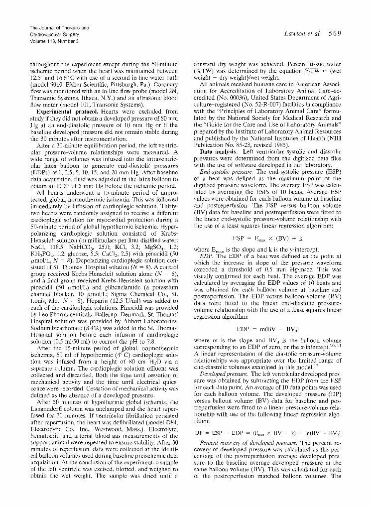
Table I. Mechanical and electrical parameters
Cardioplegic solution Time to infuse
cardioplegic solution (rain)
Time until Time unitt mechanical arrest electrical arrest
(min)* (min)*
Krebs-Henseleit 3£8 +- 2.10 5.58 _+ 0.82 18.58 ± 2.10 St, Thomas' Hospital 2.60 +_ 0.16 7.86 _+ 0,43 15.82 ± 0.13 50 txmol/L pinacidil 1.93 ± 0.39 7.49 +_ 0.62 16.93 ± 0.39 Pinacidil plus glibenclamidc 1,89 + 0.36 6.33 + 0.67 18.53 ± 1.67
Data are represented as mean plus or minus standard error of the mean. *Time from onset of 15-minute normothermic ischemic period.
average percent recovery of developed pressure (%Rec DP) was obtained by use ofthe trapezoidal rule13and the following equation:
Average%RecDP = 100
fBVm~ DP postreperfusion dBV/BVmax - BVmin)
X ) BVmin ~ - - b ~
where B V ~ is the maximum postreperfusion matched balloon volume and BV~,, is the minimum postreperfu- sion matched balloon volume, s
Statistical analysis. Values are represented as the mean plus or minus the standard error of the mean. Analysis of variance was used for comparison of multiple groups. A Student's t test (or Mann-Whitney test when appropriate) was used to compare two means. A paired Student's t test was used to compare means before and after an intervention. Differences were considered statis- tically significant when p < 0.05. Statistical analysis was done with Sigma Stat software (version 1.01, Jandel Corp., San Rafael, Calif.).
Results
Delivery of cardioplegic solution. The mean time to infuse the cardioplegic solution ranged between 1.89 +_ 0.36 and 3.58 -+ 2.10 minutes and was not different among the groups (Table [).
Temporal aspects of the development of electro- mechanical arrest. The mean times for the cessa- tion of electrical and mechanical activity are repre- sented in Table I for each group. Mechanical arrest occurred in all instances during the period of unpro- tected, global, normothermic ischemia and was not different among groups. Electrical quiescence was not achieved until after the administration of cold cardioplegic solution in all groups. Electrical quies- cence was achieved most rapidly in the St. Thomas' Hospital group, but this was not significantly shorter than in the other groups.
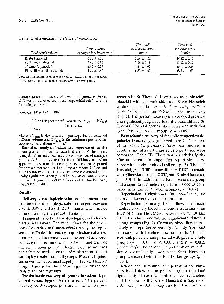
Postisehemic recovery of systolic function: depo- larized versus hyperpolarized arrest. The percent recovery of developed pressure in the hearts pro-
tected with St. Thomas' Hospital solution, pinacidil, pinacidit with glibenclamide, and Krebs-Hense[eit cardioplegic solution was 46.4% _+ 2.2%, 49.3% + 2.4%, 43.0% + 4.3, and 32.8% _+ 2.8%, respectively (Fig. 1). The percent recovery of developed pressure was significantly higher in both the pinacidil and St. Thomas' Hospital groups when compared with that in the Krebs-Henseleit group (p = 0.009).
Postischemic recovery of diastolic properties: de- polarized versus hyperpolarized arrest. The slopes of the diastolic pressure-volume reIationships at baseline and after 30 minutes of reperfusion were compared (Table II). There was a statistically sig- nificant increase in slope after reperfusion com- pared with baseline values in all groups (St. Thomas' Hospital, p < 0.001; pinacidil, p = 0.002; pinacidil with glibenclamide,p = 0.002; and Krebs-Henseleit, p = 0.017). In addition, the Krebs-Henseleit group had a significantly higher reperfusion slope as com- pared with that of all other groups (p = 0.022).
Reperfusion arrhythmias. On reperfflsion, no hearts underwent ventricular fibrillation.
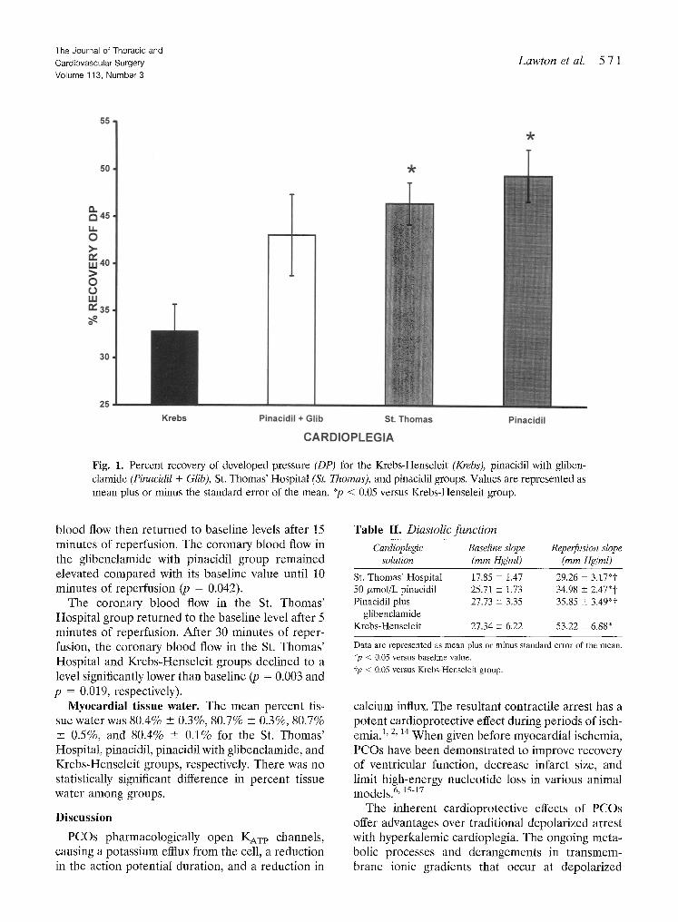
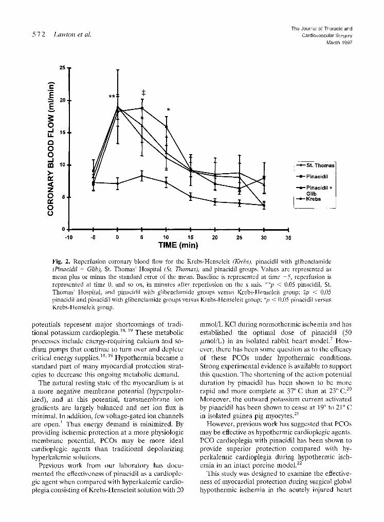
Reperfusion coronary blood flow. The mean baseline coronary blood flow before ischemia at an EDP of 5 mm Hg ranged between 7.0 +- 1.0 and 9.1 -+ 1.7 ml/min and was not significantly different among groups (Fig. 2). Coronary blood flow imme- diately on reperfusion was significantly increased compared with baseline flow in the St. Thomas' Hospital, pinacidil, and pinacidil with glibenclamide groups (p = 0.014, p < 0.001, and p = 0.002, respectively). The coronary blood flow on reperfu- sion was significantly lower in the Krebs-Henseleit group compared with that in all other groups (p = 0.004).
After 5 and 10 minutes of reperfusion, the coro- nary blood flow in the pinacidil group remained significantly higher than both the flow at baseline and the flow in the Krebs-Henseleit group (p < 0.001 and p = 0.021, respectively). The coronary
The Journal of Thoracic and Cardiovascular Surgery Volume 113, Number 3
Lawton et al. 571
55
50
~145 LI. O >-
,,=,40 > 0 rO LLI
35
30
25
l
I I
=k
-k
Krebs Pinacidil + Glib St. Thomas Pinacidil
C A R D I O P L E G I A
Fig. 1. Percent recovery of developed pressure (DP) for the Krebs-Henseleit (Krebs), pinacidil with gliben- clamide (Pinacidil + Glib), St. Thomas' Hospital (St. Thomas), and pinacidil groups. Values are represented as mean plus or minus the standard error of the mean. *p < 0.05 versus Krebs-Henseleit group.
blood flow then returned to baseline levels after 15 minutes of reperfusion. The coronary blood flow in the glibenclamide with pinacidil group remained elevated compared with its baseline value until 10 minutes of reperfusion (p = 0.042).
The coronary blood flow in the St. Thomas' Hospital group returned to the baseline level after 5 minutes of reperfusion. After 30 minutes of reper- fusion, the coronary blood flow in the St. Thomas' Hospital and Krebs-Henseleit groups declined to a level significantly lower than baseline (p = 0.003 and p = 0.019, respectively).
Myocardial tissue water. The mean percent tis- sue water was 80.4% _+ 0.3%, 80.7% + 0.3%, 80.7% _+ 0.5%, and 80.4% _+ 0.1% for the St. Thomas' Hospital, pinacidil, pinacidil with glibenclamide, and Krebs-Henseleit groups, respectively. There was no statistically significant difference in percent tissue water among groups.
Discussion
PCOs pharmacologically open NAT p channels, causing a potassium efflux from the cell, a reduction in the action potential duration, and a reduction in
Table II. Diastolic function
Cardioplegic Baseline slope Reperfusion slope solution (mm Hg/ml) (mm Hg/ml)
St. Thomas' Hospital 17.85 _+ 1.47 29.26 +_ 3.17"t
50/xmol/L pinacidil 25.71 _+ 1.73 34.98 _+ 2.47"t
Pinacidil plus 27.73 _+ 3.35 35.85 _+ 3.49"t
glibenclamide
Krebs-Henseleit 27.34 + 6.22 53.22 + 6.88*
Data are represented as mean plus or minus standard error of the mean.
*p < 0.05 versus baseline value.
?p < 0.05 versus Krebs-Henseleit group.
calcium influx. The resultant contractile arrest has a potent cardioprotective effect during periods of isch- emia.1, 2, 14 When given before myocardial ischemia, PCOs have been demonstrated to improve recovery of ventricular function, decrease infarct size, and limit high-energy nucleotide loss in various animal models,6, 15-17
The inherent cardioprotective effects of PCOs offer advantages over traditional depolarized arrest with hyperkalemic cardioplegia. The ongoing meta- bolic processes and derangements in transmem- brane ionic gradients that occur at depolarized
572 Lawton et al. The Journal of Thoracic and
Cardiovascular Surgery
March 1997
25
20 g
8 o, nn 10
O 5, e~ O U
0
-10
I I I I I I I I I -5 0 5 10 15 20 25 30 35
TIME (min)
St. T h o m a s
- - = - P inac id i l
- = - P inac id i l + Gl ib
- - e - K r e b s
Fig. 2. Reperfusion coronary blood flow for the Krebs-Henseleit (Krebs), pinacidil with glibenclamide (Pinacidil + Glib), St. Thomas' Hospital (St. Thomas), and pinacidil groups. Values are represented as mean plus or minus the standard error of the mean. Baseline is represented at time -5, reperfusion is represented at time 0, and so on, in minutes after reperfusion on the x axis. **p < 0.05 pinacidil, St. Thomas' Hospital, and pinacidil with glibenclamide groups versus Krebs-Henseleit group; Sp < 0.05 pinacidil and pinacidil with glibenclamide groups versus Krebs-Henseleit group; *p < 0.05 pinacidil versus Krebs-Henseleit group.
potentials represent major shortcomings of tradi- tional potassium cardioplegia, is' 19 These metabolic processes include energy-requiring calcium and so- dium pumps that continue to turn over and deplete critical energy supplies. 18' 19 Hypothermia became a standard part of many myocardial protection strat- egies to decrease this ongoing metabolic demand.
The natural resting state of the myocardium is at a more negative membrane potential (hyperpolar- ized), and at this potential, transmembrane ion gradients are largely balanced and net ion flux is minimal. In addition, few voltage-gated ion channels are open. 1 Thus energy demand is minimized. By providing ischemic protection at a more physiologic membrane potential, PCOs may be more ideal cardioplegic agents than traditional depolarizing hyperkalemic solutions.
Previous work from our laboratory has docu- mented the effectiveness of pinacidil as a cardiople- gic agent when compared with hyperkalemic cardio- plegia consisting of Krebs-Henseleit solution with 20
mmol/L KC1 during normothermic ischemia and has established the optimal dose of pinacidil (50 /,mol/L) in an isolated rabbit heart model ] How- ever, there has been some question as to the efficacy of these PCOs under hypothermic conditions. Strong experimental evidence is available to support this question. The shortening of the action potential duration by pinacidil has been shown to be more rapid and more complete at 37°C than at 23 ° C. 2° Moreover, the outward potassium current activated by pinacidil has been shown to cease at 19 ° to 21 ° C in isolated guinea pig myocytes. 21
However, previous work has suggested that PCOs may be effective as hypothermic cardioplegic agents. PCO cardioplegia with pinacidil has been shown to provide superior protection compared with hy- perkalemic cardioplegia during hypothermic isch- emia in an intact porcine model. 22
This study was designed to examine the effective- ness of myocardial protection during surgical global hypothermic ischemia in the acutely injured heart
The Journal of Thoracic and
Cardiovascular Surgery
Volume 113, Number 3
Lawton et aL 573
with pinacidil and to compare this protection with that of the standard hyperkalemic cardioplegia, St. Thomas' Hospital solution. KAT e channel activity was examined under hypothermic conditions with use of the potassium channel inhibitor, gliben- clamide.
Postischemic functional recovery: depolarized versus hyperpolarized arrest. Pinacidil and St. Thomas' Hospital solution provided superior pro- tection during the 50 minutes of hypothermic arrest in the acutely injured heart when compared with Krebs-Henseleit solution alone. However, there was no statistical difference between pinacidil and St. Thomas' Hospital solution. Although this is in con- trast to findings of a previous study, 22 this may be explained by the different experimental models and protocols and species variation. The improved pro- tection with pinacidil was lost with the addition of the potassium channel inhibitor glibenclamide. This indicates that some NAT P channel activity by this drug during hypothermia is responsible for the observed cardioprotection.
The lack of statistical difference in percent recov- ery of developed pressure between the pinacidil and the pinacidil with glibenclamide groups may be related to a suboptimal dose of glibenclamide.
Postischemic recovery of diastolic properties. The end-diastolic pressure-volume relationships were linear over the range of volumes tested in our experiments, as has been described by other inves- tigators.ll, 12 The mean linear regression coefficient for the pressure-volume relationships was 0.99 _+ 0.00, 0.99 _+ 0.00, 0.99 _+ 0.01, and 0.98 _+ 0.01 for the Krebs-Henseleit, Krebs-Henseleit with pinacidil, Krebs-Henseleit with pinacidil and glibenclamide, and St. Thomas' Hospital groups, respectively. All cardioplegia groups showed a significant rise in the slope of the pressure-volume relationship after isch- emia, indicating a decrease in ventrieular compli- ance. The reperfusion slope in the Krebs-Henseleit group was significantly greater than that of any other group, which suggests a more severe diastolic injury in this unprotected group.
The Krebs-Henseleit group had a more severe diastolic injury compared with that in the pinacidil with glibenclamide group. This may be because of the inability of the 10/xmol/L dose of glibenclamide to saturate and block all of the KAT P channels in the presence of 50 ixmol/L pinacidil or because of a non-KaT E channel-mediated mechanism of protec- tion by pinacidil.
The development of electromechanical arrest. Mechanical arrest occurred at approximately the same time in all groups. This occurred during the 15-minute period of unprotected normothermic ischemia and therefore no difference would have been expected among groups. This is indicative of a similar response of the hearts in all groups to unprotected ischemia.
There was no significant difference among groups in the time until electrical arrest. This is probably because hypothermia alone is effective in eliminat- ing electrical activity and this overcame the previ- ously observed inability of pinacidil to achieve rapid electrical asystole.
Reperfusion coronary blood flow. Reperfusion coronary blood flow in the pinacidil group was greater than the coronary blood flow in the Krebs- Henseleit group for the initial 10 minutes of reper- fusion. The coronary blood flow immediately on reperfusion was greater in the St. Thomas' Hospital group compared with that in the Krebs-Henseleit group but this was not different from that seen with pinacidil. These data are in contrast to our previous findings after normothermic ischemia in which the coronary blood flow was significantly higher in the PCO-protected groups than in the St. Thomas' Hospital group. 7 This may be explained by the greater ischemic insult in this protocol (15 minutes of normothermic unprotected plus 50 minutes of hypothermic protected ischemia) compared with that in our previous work under normothermic conditions (30 minutes of protected ischemia). The increased coronary blood flow in this study in the St. Thomas' Hospital group, as opposed to that in the previous study, may be the result of a greater ischemic insult that resulted in reperfusion hyper- emia. This would be consistent with the poorer recovery of developed pressure in this study com- pared with our previous results and the significant diastolic injury seen in each group.
The pinacidil with glibenclamide group exhibited some hyperemia on reperfusion and after 5 minutes of reperfusion, which suggests some KAT e channel activity and resultant vasodilation despite the 10 /xmol/L dose of glibenclamide or a non--KATp chan- nel-mediated endothelial protection in this group. The shortened period of reactive hyperemia in the pinacidil plus glibenclamide group compared with that in the pinaeidil group is consistent with the inhibition of KAT P channel activity by glibenclamide under these hypothermic conditions.
5 7 4 Lawton et al. The Journal of Thoracic and
Cardiovascular Surgery March 1997
The lack of reactive hyperemia in the Krebs- Henseleit group alone is also different from that in our previous work under normothermic conditions. This suggests that the endothelium in the Krebs- Henseleit group was unable to provide the expected hyperemic response because of the magnitude of the injury in this protocol and a no-reflow response.
However, the changes noted in reperfusion coro- nary blood flow in these experiments may be reflec- tive of many factors not limited to the vasodilatory properties of pinacidil and reactive hyperemia. It is likely that the cardioprotective properties of pinaci- dil are independent of the vasodilatory properties, and further research is indicated to elucidate these findings.
Incidence of ventricular fibrillation on reperfu- sion. In contrast to our previous findings with the use of PCOs during normothermic ischemia, no hearts in this study fibrillated on reperfusion. 7 PCOs have been demonstrated to be proarrhythmic by decreasing action potential duration and, as a result, decreasing the refractory period. 16' 23 This finding after hypothermic ischemia may be related to the decreased efficacy of pinacidil at lower temperatures to open KAT P channels and decrease action poten- tial duration. Alternatively, it may represent an electrophysiologically protective effect of hypother- mia itself. Further studies will be needed to clarify this issue.
However, both the absence of reperfusion ventric- ular fibrillation and the lack of prolonged electrical activity by PCO cardioplegia in our study suggest a different electrophysiologic effect of pinacidil on KAT P channels under these hypothermic conditions.
Model shortcomings. The advantages and disad- vantages of the blood-perfused isolated rabbit heart Langendorff model have been previously described. 7' 8 This model provides a closer approximation to the clinical situation than crystalloid-perfused models. There are many advantages of blood over crystalloid perfusion in the investigation of ischemia/reperfusion phenomena. These include a higher oncotic pressure, improved oxygen-carrying capacity, the histidine/imi- dazole buffering capacity, the presence of free radical scavengers, and improved capillary flow distribution. The isolated rabbit model has been shown to be quite stable over 120 minutes of aerobic perfusion as com- pared with that of other animal species. 24 As always, care should be taken when extrapolating these data to the clinical situation.
In addition, the optimal dose of pinacidil (50 /zmol/L) used in these experiments was determined
with use of a dose-response curve generated with normothermic ischemia. 7 This dose may not be the optimal choice under the conditions of hypothermia used in the present experiments.
Summary. Pinacidil (50 /xmol/L) provided com- parable myocardial protection to that of St. Thomas' Hospital solution during hypothermic ischemia in the blood-perfused rabbit heart model. This protec- tion was superior to that of Krebs-Henseleit solu- tion. The superior myocardial protection provided by pinacidil was lost when glibenclamide was added to the cardioplegic solution, indicating KAT P chan- nel activity during hypothermia despite previous results in isolated myocytes. 2°' as
In addition, the previously noted proarrhythmic nature of pinacidil seen in our normothermic experi- ments was absent under the present hypothermic experimental conditions. This observation raises ques- tions as to the nature of the action of pinacidil under hypothermic conditions. This finding is advantageous for the future clinical use of these agents in that reperfusion ventricular fibrillation, previously noted with normothermia, did not occur after hypothermic global ischemia protected with pinacidil.
We gratefully acknowledge the help of Luke Wolfe for statistical analysis and the generous donation of pinacidil by Leo Pharmaceuticals, Ballerup, Denmark.
REFERENCES 1. Katz AM. The cardiac action potential. In: Katz AM, editor.
Physiology of the heart. 2nd ed. New York: Raven Press, 1992:438-9.
2. Damiano RJ. The electrophysiology of ischemia and cardio- plegia: implications for myocardial protection. J Card Surg 1995;10(suppl):445-53.
3. Nichols CG, Lederer WJ. Adenosine triphosphate-sensitive potassium channels in the cardiovascular system. Am J Physiol 1991;261:H1675-86.
4. Noma A. ATP-regulated K + channels in cardiac muscle. Nature 1983;305:147-8.
5. Cohen NM, Lederer WJ, Nichols CG. Activation of ATP- sensitive potassium channels underlies contractile failure in single human cardiac myocytes during complete metabolic blockade. J Cardiovasc Electrophys 1992;3:56-63.
6. Cohen NM, Wise RM, Wechsler AS, Damiano PJ. Elective cardiac arrest with a hyperpolarizing adenosine triphosphate- -sensitive potassium channel opener. J Thorac Cardiovasc Surg 1993;106:317-28.
7. Lawton JS, Harrington GC, Allen CT, Hsia P, Damiano RJ. Myocardial protection with pinacidil cardioplegia in the blood-perfused heart. Ann Thorac Surg 1996;61:1680-8.
8. Maskal SL, Cohen NM, Hsia PW, Wechsler AW, Damiano RJ. Hyperpolarized cardiac arrest with a potassium-channel opener, aprikalim. J Thorac Cardiovasc Surg 1995;110:1083- 95.
9. Goto Y, Slinker BK, LeWinter MM. Decreased contractile
The Journal of Thoracic and Cardiovascular Surgery Volume 113, Number 3
Lawton et al. 5 7 5
efficiency and increased nonmechanical energy cost in hyper- thyroid rabbit heart. Circ Res 1990;66:999-1011.
10. Moon MR, DeAnda A, Daughters GT, Ingels NB, Miller DC. Experimental evaluation of different chordal preserva- tion methods during mitral valve replacement. Ann Thorac Surg 1994;58:931-44.
11. Shintani H, Glantz SA. Effect of disrupting the mitral appa- ratus on left ventricular function in dogs. Circulation 1993; 87:2001-15.
12. Fremes SE, Zhang J, Furukawa RD, Mickle DA, Weisel RD. Adenosine pretreatment for prolonged cardiac storage. J Thorac Cardiovasc Surg 1995;110:293-301.
13. Stark PA. Numerical integration. In: Stark PA, editor. Intro- duction to numerical methods. New York: Macmillan, 1970: 196-8.
14. Sugimoto S, Iwashiro K, Mouti F, Dawodu AA, Schiarit! M, Puddu PE. The risk of myocardial stunning is decreased concentration-dependently by KaT e channel activation with nicorandil before high K + cardioplegia. Int J Cardiol 1995; 48:11-25.
15. Pignat J, Bourgouin J, Dumont L. Cold cardioplegia and the K" channel modulator aprikalim (RP 52891): improved cardioprotection in isolated ischemic rabbit hearts. Can J Physiol Pharmacol 1994;72:126-32.
16. Galinanes M, Shattock MJ, Hearse DJ. Effects of potassium channel modulation during global ischemia in isolated rat heart with and without cardioplegia. Cardiovasc Res 1992;26:1063-8.
17. Grover GJ, Sleph PG, Parham CS. Nicorandil improves
postischemic contractile function independently of direct myocardial effects. J Cardiovasc Res 1990;15:698-705.
18. Kleber AG, Oetliker H. Cellular aspects of early contractile failure in ischemia. In: Fozzard HA, Haber E, Jennings RB, Katz AM, Morgan HE, eds. The heart and cardiovascular system. New York: Raven, 1992:1975-2020.
19. Reimer KA, Jennings RB. Myocardial ischemia, hypoxia and infarction. In: Fozzard HA, Haber E, Jennings RB, Katz AM, Morgan HE, eds. The heart and cardiovascular system. New York: Raven, 1992:1875-973.
20. Martin CL, Chinn K. Pinacidil opens ATP-dependent K + channels in cardiac myocytes in an ATP- and temperature- dependent manner. J Cardiovasc Pharmacol 1990;15:510-4.
21. Escande D, Thuringer D, LeGuern SL, Courteix J, Laville M, Cavero I. Potassium channel openers act through an activa- tion of ATP-sensitive K + channels in guinea-pig cardiac myocytes. Pflugers Arch 1989;414:669-75.
22. Kirvaitis RJ, Krukenkamp IB, Bukhari EA, Gaudette GR, Miyatake T, Levitsky S. Pinacidil-induced hyperpolarized cold blood cardioplegia: a novel myoprotective strategy. Surg Forum 1995;46:224-6.
23. Yao Z, Gross GJ. Effects of the KaTe channel opener bimakalim on coronary blood flow, monophasic action po- tential duration, and infarct size in dogs. Circulation 1994; 89:1769-75.
24. Galinanes M, Hearse DJ. Species differences in susceptibility to ischemic injury and responsiveness to myocardial protec- tion. Cardioscience 1990;2:127-43.


![A Prediction of Blood Flow through a Bypass Graft Using ... · grafts (LIMA, RIMA, SVG) [10], [11], [12]. During myocardial revascularization (CABG on a heart bypass machine), various](https://img.pdfslide.us/doc/110x75/5faefcb978a6db341331311b/a-prediction-of-blood-flow-through-a-bypass-graft-using-grafts-lima-rima.jpg)