Embed Size (px)
DESCRIPTION
cardiology, easy to learning about cardiac disease. complete with electrocardiogram and physical examination. belong to lung, thorak.anamnesis until prognosis and therapy,cardiac attactcardiac failure, chest pain in chief complain.
Citation preview

HANDBOOK OF PATHOLOGY ANDPATHOPHYSIOLOGY OF CARDIOVASCULAR DISEASE
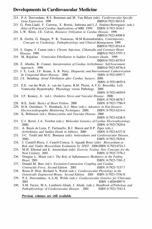
Developments in Cardiovascular Medicine
P. A. Doevendans, R.S. Reneman and M. Van Bilsen (eds): Cardiovascular SpecificGene Expression. 1999 ISBN:0-7923-5633-0
G. Pons-Lladó, F. Carreras, X. Borrás, Subirana and L.J. Jiménez-Borreguero (eds.):Atlas of Practical Cardiac Applications of MRI. 1999 ISBN: 0-7923-5636-5L.W. Klein, J.E. Calvin, Resource Utilization in Cardiac Disease. 1999.
ISBN:0-7923-8509-8R. Gorlin, G. Dangas, P. K. Toutouzas, M.M Konstadoulakis, Contemporary
Concepts in Cardiology, Pathophysiology and Clinical Management. 1999ISBN:0-7923-8514-4
S. Gupta, J. Camm (eds.): Chronic Infection, Chlamydia and Coronary HeartDisease. 1999. ISBN:0-7923-5797-3M. Rajskina: Ventricular Fibrillation in Sudden Coronary Death. 1999.
ISBN:0-7923-8570-5Z. Abedin, R. Conner: Interpretation of Cardiac Arrhythmias: Self AssessmentApproach. 1999. ISBN:0-7923-8576-4J. E. Lock, J.F. Keane, S. B. Perry: Diagnostic and Interventional CatheterizationIn Congenital Heart Disease. 2000. ISBN: 0-7923-8597-7J.S. Steinberg: Atrial Fibrillation after Cardiac Surgery. 2000.
ISBN: 0-7923-8655-8E.E. van der Wall, A. van der Laarse, B.M. Pluim, A.V.G. Bruschke: LeftVentricular Hypertrophy: Physiology versus Pathology. 2000
ISBN: 0-7923-6038-9J.F. Keaney, Jr. (ed.): Oxidative Stress and Vascular Disease. 2000.
ISBN: 0-7923-8678-7B.E. Jaski: Basics of Heart Failure. 2000 ISBN: 0-7923-7786-9H.H. Osterhues, V. Hombach, A.J. Moss (eds.): Advances in Non-InvasiveElectrocardiographic Monitoring Techniques. 2000. ISBN: 0-7923-6214-4K. Robinson (ed.): Homocysteine and Vascular Disease. 2000
ISBN: 0-7923-6248-9C.I. Berul, J.A. Towbin (eds.): Molecular Genetics of Cardiac Electrophysiology.2000. ISBN: 0-7923-7829-6A. Bayés de Luna, F. Furlanello, B.J. Maron and D.P. Zipes (eds.):Arrhythmias and Sudden Death in Athletes. 2000 ISBN: 0-7923-6337-XJ-C. Tardif and M.G. Bourassa (eds): Antioxidants and Cardiovascular Disease.2000. ISBN: 0-7923-7829-6J. Candell-Riera, J. Castell-Conesa, S. Aguadé Bruiz (eds): Myocardium atRisk and Viable Myocardium Evaluation by SPET. 2000.ISBN: 0-7923-6724-3M.H. Ellestad and E. Amsterdam (eds): Exercise Testing: New Concepts for theNew Century. 2001. ISBN: 0-7923-7378-2Douglas L. Mann (ed.): The Role of Inflammatory Mediators in the FailingHeart. 2001 ISBN: 0-7923-7381-2Donald M. Bers (ed.): Excitation-Contraction Coupling and CardiacContractile Force, Second Edition. 2001 ISBN: 0-7923-7157-7Brian D. Hoit, Richard A. Walsh (eds.): Cardiovascular Physiology in theGenetically Engineered Mouse, Second Edition. 2001 ISBN 0-7923-7536-XP.A. Doevendans, A.A.M. Wilde (eds.): Cardiovascular Genetics for Clinicians.2001 ISBN 1-4020-0097-9S.M. Factor, M.A. Lamberti-Abadi, J. Abadi, (eds.): Handbook of Pathology andPathophysiology of Cardiovascular Disease. 2001 ISBN 0-7923-7542-4
Previous volumes are still available
214.
215.
216.
217.
218.
219.
220.
221.
222.
223.
224.
228.229.
230.
231.
232.
233.
234.
235.
236.
237.
238.
239.
240.
HANDBOOK OF PATHOLOGY ANDPATHOPHYSIOLOGY OF CARDIOVASCULAR
DISEASE
by
STEPHEN M. FACTOR, MD, FCAP, ACCProfessor of Pathology and MedicineAlbert Einstein College of Medicine
Bronx, New York
MARIA A. LAMBERTI – ABADI, MDAssistant Professor of Pathology
Albert Einstein College of MedicineBronx, New York
and
JACOBO ABADI, MDAssistant Professor of Pediatrics
Albert Einstein College of MedicineBronx, New York
KLUWER ACADEMIC PUBLISHERS NEW YORK, BOSTON, DORDRECHT, LONDON, MOSCOW

eBook ISBN: 0-306-47575-8Print ISBN: 0-7923-7542-4
©2002 Kluwer Academic PublishersNew York, Boston, Dordrecht, London, Moscow
Print ©2002 Kluwer Academic Publishers
All rights reserved
No part of this eBook may be reproduced or transmitted in any form or by any means, electronic,mechanical, recording, or otherwise, without written consent from the Publisher
Created in the United States of America
Visit Kluwer Online at: http://kluweronline.comand Kluwer's eBookstore at: http://ebooks.kluweronline.com
Dordrecht

To my darling wife, and my lovely children with deepest appreciation forall your help and encouragement along the way. To my parents, formaking me what I am. And, of course, with love to Raja. SMF
With love to my husband Yaco, our children Daniela, Adriana andDavid, my mom Renee, my brother Victor, and especially to my dad JoseA. (Cucho) Lamberti, MD, whose memory is a driving force in my life.MA
To my wife Lala and our kids, with all my HEART. You are the light ofmy life. To my parents, sister, and grandmother Sara, with love. JA

This Page Intentionally Left Blank
TABLE OF CONTENTS
Preface viii
1.2.3.4.
Practical Cardiac AnatomyAtherosclerotic Heart Disease and IschemiaDiabetes and the HeartCardiomyopathies
1196775
HypertrophicDilatedEndocrinopathiesAlcoholic – Nutritional DeficienciesHuman Immunodeficiency Virus Infection
5. ValvulopathiesRheumatic Heart DiseaseMitral Valve Prolapse
6. Congenital Heart DiseaseTetralogy of FallotTurner’s SyndromePatent Ductus Arteriosus
7.8.9.
EndocarditisMyocarditisCardiac Tumors
10.11.12.
The PericardiumAortic AneurysmsCollagen Vascular Diseases and Vasculitis:
SyphilisSclerodermaSystemic Lupus ErythematosusTakayasu’s diseaseKawasaki’s disease
13. Medicine, Law and the Heart
Appendix
Index
119
137
171191203221229241
281
299
305
PREFACE
Autopsy derives from the greek word autopsia, which means act ofseeing with one’s own eyes. It remains the most objective and accuratemethod to understand human.disease. Unfortunately, the volume of autopsiesin teaching hospitals has decreased dramatically over the past years. Thecrucial factors that account for this are the recent progress and development ofnew technologies, especially in diagnostic imaging, immunology, cell biologyand genetics. Additionally, the perpetual fear of legal liability by physiciansaccounts for its further decline. Consequently, physicians and medicalstudents are engaged in fewer autopsies and are not reaping the richeducational rewards that accompany these examinations. The purpose of theautopsy is not only to establish the cause of death, but also to determine thenature and course of the disease process. Our goal with this book is toemphasize the importance of the post-mortem exam and the correlationbetween pathologic material and clinical data by analyzing actual cases withproblem-based methodology. The focus of this handbook is on cardiovasculardisease, and when appropriate, other disease categories are included if theyhave an impact on cardiovascular function.
The approach is more than the usual clinico-pathological correlation.Rather, we attempt to present the material from the perspective of the autopsytable. We use the clinical data as the initial framework and the autopsyfindings to develop a true understanding of the disease and the associatedpathophysiology of the condition. This approach is similar to the “gross”autopsy conferences that have been carried out at our institution for over 30years. The format of the case presentation is logical and uniform withemphasis in teaching current medical concepts. Relevant clinical informationis correlated and evaluated in the context of the macroscopic and microscopicfindings obtained in the post-mortem exam. All cases are followed by acomprehensive discussion and review of the disease and pertinent literature iscited. We use extensive photographic material to illustrate each case andfacilitate learning.
We address the new issue of legal liability by presenting cases thathave medico-legal implications. A thorough discussion on the impact ofsignificant discrepancies between the autopsy findings and clinical diagnosesis also included. This is particularly important in an era that demands qualityassurance and performance improvement from the medical professionals.
Scientific methodology and common sense are still essential despitethe forever growing knowledge of the medical sciences. Hopefully, with thisbook, we will succeed in guiding physicians and medical students in theircontinuous search for answers.
Chapter 1
PRACTICAL CARDIAC ANATOMY:FROM A TO Z
Introduction
It may represent a bias of the authors, but it appears to be a truism tostate that the heart is the most complex anatomical structure in the body. Yes,the brain has an intricate spatial and electrical anatomy with numerous nucleiand interconnections, but it does not approach the complexity of the heartanatomically or physiologically. No organ other than the heart is divided intopumping chambers dependent on blood flow moving between two separatecirculatory circuits, with the timing of blood movement within the chambersoccurring within fractions of a second. This movement must be coordinatedby a neural network (e.g. conduction system), heart valves, vascular supply,connective tissue and muscle cells. The heart is dependent on maintaining itsown nutrition, while at the same time supplying oxygenated blood to theentire body. During the process of pumping, chamber contraction affects thesupply of blood to the tissue performing the pumping, sometimes withadverse consequences. There must be an adequate flow of oxygenated bloodto the heart cells, because they have very high metabolic requirements.Although myocytes are organized as individual cells and do not form asyncytium, they must ‘talk’ to each other electrically and functionally. Thecells must each contract, but this contraction must then be coordinated so thatgroups and layers of cells are shortening sequentially within a very shortperiod of time. Otherwise, this would lead to hypokinesia (decreasedcontraction), dyskinesia (abnormal contraction), or akinesia (absence ofcontraction).
Many books have been written on cardiac anatomy, both descriptiveand illustrated. Detailed descriptions are available of the complex structuraland functional anatomy of the heart; but most of such works do not provideadequate explanations for acquired pathophysiology or contractiledysfunction. The rationale for this book is different: we are attempting tocorrelate actual case material with clinically relevant findings, and to showhow clinical manifestations, pathophysiology and pathology interact toproduce disease. Furthermore, we want to show how there is a certain logicand analytical process that goes into each case, if one understands clinicalfindings and pathology. Our goal in this book is not to re-teach cardiacanatomy. Those who are interested or stimulated to do so can find anynumber of adequate books and atlases on the subject. We want to provide a
brief outline of some basic features of cardiac anatomy, and by doing so,make pathophysiology and disease manifestations meaningful.
The following provides an alphabetical functional anatomicalglossary. It is not proposed as a complete list, but one that illustrates theprinciple that at least some cardiovascular diseases and their complicationsare determined by cardiac anatomy and structure-function relationships.
Annulus
The fibrous ring that supports each of the 4 cardiac valves. Theannulus for the tricuspid, mitral, and aortic valves is virtually a continuousstructure arranged like a pretzel. The pulmonic valve annulus is somewhatseparate from the other 3 (it arises from distinctly independent embryologictissue, e.g. bulbus cordis). The valve base inserts into the annulus with inter-digitating connective tissue fibers. Infection of the valve tissue (endocarditis)can affect these connections thereby causing leaflet or cuspal dehiscence, or itcan lead to an infection of the annulus itself (e.g. valve ring abscess). Theproximity of the annulae of the 3 valves means that infection can spreadreadily from one to the other. The annulus also anchors the base of the heartthrough connective tissue attachment fibers that extend into the muscle of theventricle. Sections of the base of the heart just below the annulus typicallyreveal fibrous tissue; however, this should not be interpreted as pathologicalscar since it represents normal anatomy. Dilatation of the ventricular or atrialchambers can stretch the annulae, most likely through side to side slippage ofthe connective tissue fibers. The dilatation may pull the valve base awayfrom the center channel through which blood flows, and where there isclosure with valve leaflet/cusp coaptation. The result is valvularinsufficiency, as the valve tissue can no longer come together properly toclose the orifice. By providing a surgical shortening of the annulus by placingcrimping sutures, or by sewing in a prosthetic ring (annuloplasty), the annulusmay be repaired with a decrease of insufficiency.
Aortic Valve
The 3 cusped valve separating the outflow tract of the left ventriclefrom the aorta. The cusps attach to the annulus, and insert as thin fibrousbands surfaced by endothelial cells (endocardium) into the aortic wall. Thesefibrous bands, immediately adjacent to each other but separated by less than 1mm, form 3 commissures. Dilatation of the root of the aorta, the areaimmediately superior to the valve, may occur with connective tissue disease(e.g. Marfan’s syndrome), atherosclerosis, or inflammation, and may lead toaortic valvular insufficiency. In particular, with syphilis, Takayasu’s arteritis,
2
or rheumatoid arthritis, the inflammation and damage to these attachmentsleads to commissural separation, thus preventing cusp coaptation. The areabehind the cusps, by the aortic surface, is a pocket known as the sinus ofValsalva (see below).
The sinuses are potential sites of stasis, and with aging, they maydevelop calcification followed by aortic valve degeneration with stenosis.The sinuses also contain the two coronary ostia (see below). The aortic valveis directly continuous with the ventricular surface of the mitral valve. Thus,infection, or degeneration of one valve can spread relatively easy to the other.
Apex, ventricle
A generally disregarded area of the heart, but one that may haveimportant functional significance. It is composed of the most dependentportions of both left and right ventricles, with the myocardial fibers spiralingtowards the tip of the chamber. The muscle and the associated connectivetissue matrix (see below) come together as a button-like structure at this point.In fact, during normal contraction there is dimpling of the apex (dimples inthe face are the result of connective tissue attachments between skeletalmuscle and the dermis). It appears that the apex serves as a fulcrum formuscle shortening; thus, damage to the apex may have significant adverseconsequences for ventricular contractile function, by ‘untethering’ the distalchamber attachments. Since the coronary supply to the apex is dependent onvessels derived from all 3 coronary arteries, obstruction of any one vessel maylead to either generalized infarction or no damage, depending on the collateralinter-connections between the vessels.
Atrio-ventricular node
A modified mass of myocardial cells that serves as the electricalpacemaker of the ventricles. Situated in the lower inter-atrial septum, it liesimmediately superior to the tricuspid valve and its annulus and the mitralvalve and its annulus. Blood supply is provided by a branch of the rightcoronary artery in most individuals, or a branch of the circumflex artery in theminority of cases. Thus disease of either vessel may lead to ischemic damageto the node, with the potential for conduction block. The node sends a bundleof fibers into the ventricles (the bundle of His, see below), by penetrating theannulus of the tricuspid and mitral valves. Thus, partial or complete heartblock may occur with disease processes involving the annulus such as calcificdegeneration of the annulus, infection, or surgical trauma following prostheticvalve replacement or repair of a congenital defect.
3
Bundle of His and conduction bundles
The bundle of His and conduction fibers are composed of modifiedcardiac muscle tissue. The bundle of His crosses through the annulus at thebase of the cardiac septum. Upon reaching the ventricle, it divides into leftand right divisions. The left division skirts around the membranous septum,where it then divides further into an anterior and posterior branch. The rightbundle branch extends over the base of the septum into the right ventricle,where it runs superficially in a muscle known as the moderator bandbetween the ventricular wall and the anterior papillary muscle. All of theconduction bundles are susceptible to damage or disruption, because they runsuperficially just below the endocardium. The bundle of His may beinterrupted if the annulus develops degenerative calcification, or if it isaffected by infection spreading from endocarditis of an adjacent heart valve.Left ventricular septal ischemia or infarction, high in the base around themembranous septum, may lead to left anterior or left posterior hemi-block.More proximal infarction may completely damage the bundle, leading to left,right, or complete heart block. Surgery for congenital heart disease, at oraround the membranous septum may affect the bundle tissue in thesurrounding myocardium. Aortic or mitral valve replacement surgery maylead to damage of conduction tissue if the prosthetic ring sewing suturesextend too deeply into the annulus.
Capillary loops
The myocardial microcirculation is an end-vascular system, whichmeans that as the coronary arteries progressively branch into smallerramifications, they reach a point where they no longer inter-connect. Thislack of collaterals appears to occur when the vessels are approximately 25-50microns in diameter, or at a level when they are pre-capillary arterioles. Atthat point, they give rise to an arcade of capillaries that do not inter-connectwith adjacent arcades derived from other arterioles. The capillaries, usually4-6 in number, provide oxygenated blood to individual myocytes loopingaround them, with the afferent arm draining into venules and then largervenous channels. This pattern of discrete capillary supply has severalconsequences. It provides an explanation for the very sharp histological andvascular border of ischemic myocardium, since there is minimal collateralblood flow at the cellular level. It also means that obstruction of arterioles(e.g. by spasm or microthrombosis), places a small number of myocytes atrisk for necrosis defined by the arcade branching of the arteriole (generallyabout 10-20 myocytes, if examined in 3-dimensions).
4
Chordae tendineae
The fine fibrous bands that extend from the tips of the papillarymuscles to the atrio-ventricular heart valves. The chordae branch at least 3times and become smaller between the papillary muscle tip, known as themyotendon junction, and the valve. In total, there are approximately 120chordae at the valve level. They insert on the undersurface of the valve, at thecommissural junction, and at the free edge. With contraction of the ventricle,pressure within the cavity increases thereby forcing the leaflets of the mitraland tricuspid valves toward the midline. There is concurrent papillary musclecontraction, which pulls the chordae toward the mid portion of the ventricularchamber. This tethers the leaflets of the valves, prevents them fromprolapsing into the atrium, and maintains their coaptation and orifice closure.The chordae from both papillary muscles in the left ventricle insert into bothleaflets of the mitral valve. Therefore, partial disruption of the papillarymuscle or dysfunction due to ischemia, may lead to a less severe degree ofmitral valve insufficiency. Complete rupture of a papillary muscle, whichmay occur with infarction, generally causes massive mitral regurgitation, andusually rapid death due to pulmonary edema. Elongation or rupture ofchordae can cause mitral regurgitation, and may occur as a result ofconnective tissue disease (e.g. Marfan’s syndrome, and possibly the mitralvalve prolapse syndrome). Rupture of one or multiple chordae can developconcomitantly with infection (endocarditis) or post-trauma. Chordae mayalso scar and shorten, or become adherent to adjacent chordae (so-calledfusion) secondary to inflammation (typically seen in rheumatic heart disease).This may lead to valvular insufficiency by drawing the leaflets down into theventricular cavity and preventing their coaptation.
Commissure
The commissures of the aortic valve have been described above; theyare comparable for the pulmonic valve. The commissures of the atrio-ventricular valves are less well defined. The leaflets insert broadly into theannulae along a curved base; at the junction between one leaflet and theadjacent leaflet, they form a commissure. This is a potential site for damage.Inflammation, thrombus, and calcification may scar the commissure andprevent complete opening of the valve. This is typical of rheumatic heartdisease with resulting mitral, or less commonly, tricuspid valve stenosis.Separation of these commissures may occur when the annulus is stretched ordamaged; this results in valvular incompetence.
5
Conduction System
In addition to the atrio-ventricular node with the bundle of His andconduction bundles (see above), and the sino-atrial node (see below), theconduction system includes Purkinje fibers that course in the immediatesubendocardium of the two ventricles. They serve as a fan-like projection ofconductive fibers that rapidly transmit generated electrical impulses from theA-V node to the myocardium. Due to their subendocardial location, they aresusceptible to damage from coronary artery ischemia that initially affects theinner wall of the ventricle. They also can be damaged with trauma (e.g.catheter-induced during invasive cardiology procedures; and open heartsurgery), and endocardial injury secondary to inflammation or thrombosis.The conduction system is affected by external central neural influencethrough a network of nerves and ganglia in the outer atrial wall. Hormonesand sympathetic and para-sympathetic transmitters also may affect it.
Connective Tissue Matrix
The myocardium is completely invested by a fine mesh-like networkof connective tissue fibers that is virtually invisible with standard histologicsections, but can be visualized with special staining techniques or electronmicroscopy. This connective tissue wraps around single myocytes, attachesindividual myocytes laterally, envelops groups of these cells, and maintainsconnections between myocyte layers in the ventricular wall. Although thisskeletal framework is important for maintaining myocyte shape, it is criticalfor normal ventricular function. Damage or lysis of the matrix may result inventricular dysfunction, even in the absence of significant myocyte injury. Ifthe myocytes were not interconnected by matrix, they would be unable togenerate a coordinated contraction to expel blood from the cavity. For theventricular chamber to decrease its dimensions during systole, there must beboth horizontal and vertical shortening. This is accomplished by a spiralmovement of three obliquely oriented layers of myocardium that literally‘squeeze’ the blood out of the chamber. To achieve this goal, there must beinterconnection between the myocytes and the layers. There also must betethering of the ventricle at both ends. In fact, there are parallel coiled fiberslongitudinally oriented that run between myocardial cells that insert both as a‘button’ at the apex (see above), and into the annulae at the base of the heart.Ischemia and inflammation, among other conditions, may lead to activation oflocal and systemic collagenolytic enzymes that may degrade the matrix.Degradation of matrix may cause localized or global remodeling of theventricle with chamber dilatation. Increased synthesis of matrix, whichoccurs with aging and familial hypertrophic cardiomyopathy, among other
6
conditions, decreases ventricular compliance and may lead to diastolicdysfunction.
Coronary Arteries
Muscular blood vessels lined by endothelium with a thin intimacomposed of 1-2 connective tissue cells at birth, and separated from therelatively thick media by an internal elastic lamella. The media usually hastwo obliquely oriented layers of smooth muscle. The outer coat of the arteryis adventitia, or connective tissue that provides both innervation and bloodsupply to the coronary vessels (vasa nervorum and vasa vasorum,respectively). The diameter of the normal epicardial coronary artery, prior toits branching and entering the myocardium (where it is no longer susceptibleto atherosclerosis) is usually 2-3 mm, but it can be variable. With progressivenarrowing of the vessel secondary to atherosclerosis, there may be remodelingof the wall so that the overall diameter of the vessel may be increased, evenwhen the functional lumen is decreased. Failure or inadequate remodelinghas been suggested to be a contributing factor for coronary ischemia. Themain condition that affects coronary arteries leading to disease isatherosclerosis; however, they are susceptible to inflammation (vasculitis),and non-atherosclerotic degeneration (calcification). The location of themajor coronary arteries on the surface of the heart makes them vulnerable toblunt or penetrating chest trauma.
Coronary Ostia
The two openings for the left and right coronary arteries, usuallylocated centrally at the upper portion of the sinus of Valsalva, approximatelyparallel to the free edge of the aortic valve. The location is variable. Highinsertion in the aortic wall has been associated with sudden cardiac death,presumably secondary to transient decrease of coronary flow (with highinsertion, the ostium is affected by systolic pressure that can compress theopening, rather than diastolic pressure with which it is normally perfused).Lower insertion within the sinus of Valsalva may make the ostia moresusceptible to stenosis by conditions that affect the aortic valve (e.g.degenerative calcification). Other acquired disease may lead to ostialstenosis. Atherosclerosis and/or inflammation of the ascending aorta, maycause narrowing of the ostium leading to myocardial ischemia, even in theabsence of significant coronary artery disease. Syphilis is the classic cause ofinflammatory ostial stenosis. Aortic dissection, when it leads to retrograde(e.g. towards the heart) splitting of the aortic wall, can disrupt the ostia.
7
Ductus Arteriosus
It represents one of the physiologic shunts of the fetal circulation (seealso foramen ovale). It is the arterial connection between the left pulmonaryartery and the aorta just distal to the origin of the left subclavian artery. Theductus allows for a bypass of the high-resistance pulmonary circulation, andthe passage of oxygenated blood in the pulmonary artery into the aorta. Itbegins to close shortly after birth, and completes the closure within the firstweeks of life. Persistence of a patent ductus arteriosus may lead to severe,irreversible pulmonary hypertension, if it is not closed.
In the newborn with certain congenital heart defects (e.g. ductaldependent lesions), the closure of the ductus arteriosus generally precipitatescardiovascular collapse. Therefore, it is usually kept open with medicaltherapy (prostaglandin E1), until surgical repair is possible.
Endocardium
The endothelial covering of the entire inner surface of the heart. Thisincludes the cardiac chambers, both surfaces of the valves, and the chordaetendineae. The endocardium has functions similar to endothelium in vessels(the heart is really just a big muscular vessel). It can react to hormones (e.g.prostaglandins), and it has secretory capability with release of endothelin andnitric oxide. It most likely plays an active role in cardiac function, although ithas not been studied extensively. The endocardium is susceptible to damagedue to infection (e.g. endocarditis), thrombosis, and trauma from invasiveprocedures.
Foramen Ovale / Fossa Ovalis
Another one of the normal fetal circulation shunts (see ductusarteriosus). It refers to the opening in the mid-portion of the inter-atrialseptum that allows oxygenated blood returning to the right heart from theplacenta, to cross into the left atrium. The foramen is covered by amembrane, the septum secundum, which grows down on the left atrial sideduring embryogenesis, and overlies a separate membrane. During fetaldevelopment, when pressures are higher in the right atrium, there is flow fromright to left, even when the membrane has completed its development.Following birth, when pressures become higher on the left side, the septumsecundum is forced against the foramen, thereby maintaining it closed.Usually, the membrane fuses to the tissue, and there is an oval depression inthe atrial wall (fossa ovalis). If the fusion is incomplete, one can put a probethrough the foramen between the two membrane layers demonstrating the so-
8
called ‘probe-patent foramen’. This is usually of no clinical significance, andmay only be an incidental finding at autopsy. However, if there is a markedincrease of right-sided pressures as a result of pulmonary hypertension orother causes, the effects of the pressure and the dilatation of the right atrialcavity may lead to a progressive enlargement of the probe-patent foramen .This can then result in a clinically evident right to left shunt, or an acquiredatrial septal defect (ASD).
Growth, Cardiac
The heart grows physiologically proportionally to body mass. Therelationship is maintained from the fetus to the adult, and is equally true forall mammalian species. As a general rule, the heart weight can be estimatedfrom the lean body mass. However, by multiplying the weight expressed inkilograms by 0.50% for a man, and 0.40% for a woman (because woman havea lower lean body mass), an approximation of the normalized heart weight canbe determined. Physiological heart weight (e.g. without associated withfibrosis and myocyte damage) is maximized at 400-450 g for males, and 350-400 g for females, regardless of body size. Beyond these weight limits, thereis virtually always pathology in the heart.
Heart Orientation
Although standard terminology suggests that the heart is oriented in aleft-right direction, this is misleading. The heart is actually positioned so thatthe right ventricle is anterior, and lies just below the sternum. The majorportion of the left ventricle lies posterior and lateral to the right ventricle, withthe apex composed of the left and right ventricles obliquely pointing to theleft. The right atrium is superior and posterior to the right ventricle. The leftatrium is the most superior and posterior structure, lying as it doesimmediately anterior to the esophagus and vertebral column. This providesan explanation for why trans-esophageal echocardiography (TEE) is such asuperior technique for visualizing the mitral valve; the TEE transducerintroduced into the esophagus, is immediately adjacent to the valve. Anotherimportant consequence of the anatomical orientation is the effect of trauma.Blunt trauma to the chest wall (e.g. seat belt or steering wheel injury) maylead to contusion of the anterior surface of the heart below the sternum, or theright ventricle. If the trauma is more lateral and to the left, it may contuse theanterior surface of the left ventricle. Penetrating trauma (e.g. knife or gunshot) often leads to penetration of the anterior right ventricular wall, at aminimum. Marked compressive trauma to the chest wall may lead to rupture
9
of the thin-walled right ventricle, as the competent tricuspid valve allows forexcessive pressure to develop in the ventricular cavity.
Interventricular Septum
The muscular separation between the left and right ventricles. Theseptum has a relatively smooth endocardial surface on the left ventricularoutflow tract, whereas, the right ventricular surface of the septum is diffuselytrabeculated. The septum is functionally considered to be a left ventricularstructure. This is significant since endomyocardial biopsies of the septum areperformed on the right side, for reasons of easy access through the jugularvein as well as increased safety; however, the biopsy findings usually reflectpathology present in the left ventricle (e.g. cardiomyopathy or myocarditis).
Jelly, Cardiac
The relatively acellular layer forming the wall of the embryologiccardiac tube, separating endothelial cells internally from an external layer ofmantle cells. This tube forms the primitive ventricular tube and bulbus cordis.
Koch, Triangle
The triangle of Koch is the area defined inferiorly by the septal leafletof the tricuspid valve inserting into the annulus, posteriorly by the coronarysinus, and superiorly by the ridge-like tendon of Todaro. The triangle issignificant because it contains the atrio-ventricular node. For properexamination of the node, the triangle of Koch is sectioned, along with theannulus of the tricuspid and mitral valves, and the superior interventricularseptum. This block of tissue permits examination of the node, and the bundleof His, as it penetrates the annulus.
Lambl’s Excrescence
A small papillary frond-like vegetation that is found on the closingedge of valve cusps and leaflets. It is typically seen in the central closingedge of the aortic valve cusps. The excrescence resembles a fine thread-likestructure. Microscopically, they have a fibrous core, and a surface ofendothelial cells. They are thought to arise from localized trauma to the valvefrom repetitive closure, with organization of the resulting fibrin and plateletvegetation. They are related to the much less common, and larger, organizedvegetation called papillary fibroelastoma. The latter lesions, for many
10
years, were considered to be benign tumors or hamartomas. Structurally,however, they appear to be due to non-bacterial vegetation that undergoesreplacement of the fibrin and platelets by fibrous and elastic connective tissue.Clinically, they are rarely significant. However, if a larger lesion, particularlyon a stalk, prolapses into a coronary ostium, it may lead to sudden death ormyocardial infarction. In contrast, the Lambl’s excrescence is too small, andis only an incidental finding at autopsy.
Membranous Septum
The round or oval-shaped membranous area immediately below theaortic valve annulus. There are a number of congenital defects of thisstructure, particularly in association with endocardial cushion defects;however, isolated membranous septal defects also occur. Acquired defectsmay develop secondary to infective endocarditis of the aortic valve thatextend to the aortic annulus and then to the septal base. Such defects typicallypresent at or around the tricuspid valve. The membranous septum is theanatomic landmark where the atrio-ventricular conduction tissue progressesthrough the annulus at the base of the septum, and then branches into leftanterior and posterior divisions. Infection of the membranous septum, orsurgical repair of a defect, may lead to interruption of the bundle tissue andthe development of acquired left heart block.
Mitral Valve
The only valve with 2 leaflets. The mitral valve has an unusualconfiguration, with marked asymmetry of the leaflets. The anterior leaflet isa broad, shield-shaped structure that fills approximately two-thirds of thevalve orifice, but only one-third of the annular circumference. It extendsmuch deeper into the ventricular chamber than the posterior leaflet. Thelatter is a shallow, usually scalloped leaflet that has a broad annularattachment, but comprises only one-third of the orifice area. Together, the 2leaflets have approximately equal surface areas. Both are tethered on theirundersurfaces by chordae tendineae and two papillary muscles, whichmaintain them in a flat orientation relative to the annulus during ventricularsystole. When seen from the atrial surface, the 2 leaflets have a curvilinearconvex-concave apposition, with the convex surface from the anterior leaflet;resembling a ‘fish-mouth’. The mitral valve orifice is large, thereby allowinga high volume of blood to be passed into the ventricle at low pressure. Thecommissural attachments of the 2 leaflets are poorly defined areas of junction,but important sites for inflammation (e.g. rheumatic heart disease) andinfection.
11
Nuclei, Myocyte
Myocytic nuclei are relatively large, round to oval-shaped structuresat birth. During embryogenesis, mitotic division of nuclei and cells can beeasily appreciated. Shortly after birth, mitoses become exceedingly rare. Foryears it has been assumed that myocytes are terminally differentiated cells,with no new cell proliferation after birth. Recent evidence suggests thatlimited numbers of cells may proliferate in adulthood around areas of injury(e.g. healing myocardial infarction). It is known that myocyte nuclei maydivide without cytokinesis, thereby giving rise to myocardial cells with 2nuclei. This is common with increasing age, particularly in association withmyocardial hypertrophy. Also with age and hypertrophy, myocyte nuclei mayincrease their chromosome number by meiosis without nuclear division. Thisgives rise to heteroploid nuclei, typically with a doubling of chromosomes.On routine section, stained with hematoxylin, the nuclei have a largerconfiguration with increased blue staining intensity.
Organization, Myocardium
As discussed in connective tissue matrix, the myocardium iscomposed of obliquely oriented layers that ‘wrap’ around the ventricularcavity. This is particular obvious in the left ventricle, with 3 layers; whereas,the thinner, low-pressure right ventricle generally has 2 less well-definedlayers. Since the layers in the left ventricle are oriented obliquely to eachother, sectioning through the ventricular wall reveals groups of myocardialcells parallel to each other, but oriented at approximately 45 degrees toadjacent parallel bundles of cells. Thus, a section across the left ventricle wallhas myocytes oriented longitudinally, obliquely, and in cross-section. Theselayers are attached to each other by connective tissue matrix fibers aspreviously discussed. Contraction of the ventricle takes place in a screw-likemanner, as the obliquely oriented layers shorten in a curvilinear pattern fromapex to base. Upon diastolic relaxation, since the layers and the individualmyocytes are interconnected, and because there are spring-like connectivetissue fibers running along the axis of the ventricle, negative pressuredevelops enhancing diastolic filling by ‘sucking’ blood from the atrialcavities.
Papillary Muscle
There are 2 papillary muscles in the left ventricle, an anterolateral anda posteromedial. They are broad, finger-like projections into the cavity thatenlarge with ventricular hypertrophy, and therefore may contribute to a
12
decreased cavity volume in association with conditions such as hypertension.The papillary muscles also have a terminal coronary circulation with little orno collaterals. Ischemia associated with the left anterior descending orcircumflex coronary arteries makes them particularly susceptible to necrosisor focal scarring. This may lead to papillary muscle dysfunction, and withoutthe tethering effect of the muscle, mitral valve regurgitation. Typically, eachpapillary muscle has multiple smaller heads giving rise to chordae tendineae.The main papillary muscle body is known as the belly. The chordae fromboth papillary muscles fan out to support both leaflets of the mitral valve.The chordae from the 2 right ventricular papillary muscles support theanterior and middle leaflet. The septal leaflet chordae insert directly into theventricular septum without a papillary muscle. In the right ventricle, thepapillary muscles are less prominent, appearing more like hypertrophiedtrabeculae.
Pericardium
A thin fibrous sac that envelops the heart and extends along theascending aorta to the brachiocephalic artery. Thus, rupture of the ascendingaorta (often as a result of aortic dissection) can lead to pericardial tamponade.The pericardium is structurally considered as 2 surfaces: a visceral layer madeup of mesothelial cells overlying the epicardial fibroadipose tissue, and aparietal layer with an inner lining of mesothelial cells, a fibrous body, and anouter layer that blends with the connective tissue of the mediastinum. Themesothelial cells secrete a small volume (usually 10-20 cc) of thin serousfluid that serves to lubricate pericardial and cardiac movement. Thepericardial sac is drained by lymphatic channels. Increased secretion (e.g.associated with inflammation), or reversal of lymphatic flow (e.g. associatedwith elevated pressure in the venous and lymphatic system that occurs inright-sided congestive heart failure) leads to increased fluid volume in thepericardial sac. A slow accumulation of volume allows the fibrouspericardium to accommodate; whereas if it occurs acutely, the volume andpressure can cause tamponade. The fibrous pericardium stretches by aslippage of 2-3 obliquely oriented collagen fiber layers attached by connectivetissue matrix fibers, and also elastic fibers within the tissue. An increase ofpericardial sac volume is possible between 1-2 liters, as long as it occursslowly. This volume includes the actual fluid within the sac, as well as themass of the hypertrophied heart.
13
Pulmonary Valve
Structurally it is similar to the aortic valve, with 3 cusps. The sinusesbehind the cusps are shallower than the sinuses of Valsalva. Compared to theaortic valve, the pulmonary valve is rarely susceptible to degeneration,inflammation, or infection; presumably because it is not chronically damagedby a high pressure system. The valve is the site of congenital anomaliesparticularly that associated with tetralogy of Fallot.
Right Ventricle
As described in the organization of the myocardium, the rightventricle is a thin-walled structure with several poorly defined layers ofmyocardium. The free-wall and septum are heavily trabeculated. The free-wall is often infiltrated by fibroadipose tissue that extends between the musclefibers from the epicardial fat towards the endocardium. This tends to increasewith obesity, and is often associated with diabetes mellitus. There isgenerally no functional consequence of this increase of adipose tissue, but itmay lead to confusion on endomyocardial biopsy interpretation with arelatively rare condition known as right ventricular cardiomyopathy. Thelatter condition, is associated with a marked increase of fat and connectivetissue in the ventricle, and is associated with sudden cardiac death.
The right ventricle undergoes significant remodeling after birth sinceit is relatively hypertrophied compared to the left ventricle, and is usuallyequal in thickness. Within months of delivery it becomes proportionally one-third to one-half the thickness of the left ventricular wall. It can hypertrophy,however, in association with pressure overload usually due to persistent oracquired pulmonary hypertension.
Sinoatrial Node
An elongated, fusiform collection of modified cardiac muscle cellsand connective tissue lying immediately below the epicardium at the junctionof the superior vena cava and the right atrial wall. The node is supplied by abranch of the right coronary artery or a branch of the left circumflex coronaryartery in about equal numbers of people. Degeneration of the node may occurwith ischemia, inflammation, or in association with increased atrial pressure.
14
Tricuspid Valve
Although structurally considered to have 3 leaflets, the tricuspid valvefrequently has a diffusely scalloped appearance with numerous small leaflets.This appearance is enhanced if associated with fibromyxomatous valvedisease (e.g. ‘floppy’ mitral valve, or mitral valve prolapse). In general themost common cause of tricuspid valve regurgitation is secondary to dilatationof the tricuspid annulus in association with right ventricular chamberdilatation and failure. The tricuspid valve commissures are also poorlydefined. Typically there are 3 leaflets: anteriosuperior (or anterior leaflet),posteroinferior (mid or mural leaflet), and septal. As noted previously, theseptal leaflet has chordal attachments that insert directly into the septal wall.The orifice of the tricuspid valve is the largest of the 4 valves, ranging from11.0-13.0 mm, compared to the mitral valve 8.0-10.0 mm, the aortic valve7.0-8.5 mm, and the pulmonic valve 7.5-9.0 mm.
Ultrastructure
The ultrastructural appearance of the myocardium is complex andwould take an entire atlas to describe. Several unique features will beoutlined here. Myocardial cells are generally elongated, with lateral branchesthat attach at each end and at the end of the branches to adjacent cells byelectron-dense intercalated disks. The sarcolemma or myocardial cellmembrane is lined by a basal lamina. Invaginations of the sarcolemma at thelevel of the Z-band extend deeply into the myocyte along with a thinner basallamina. This is called the T-tubule which permits access of electrolytes intothe myocyte where there is interaction at the sarcoplasmic reticulum (aseries of fine tubules that extend along the contractile fibrils) to release oruptake calcium necessary for myocyte contraction and relaxation. Thecontractile unit is known as the sarcomere. It includes myosin, actin, and anumber of associated fibrils. The margins of the sarcomere are the lateral Z-Band into which the thick myosin filaments attach. The central portion of thesarcomere is the A-Band, composed of actin filaments. Although far toocomplex to describe here, the actin and myosin filaments slide over each otherduring each contractile cycle, powered by adenosine triphosphate (ATP). Themitochondria supply the ATP, and lie between the myofibrils (groups ofmyofilaments. Not surprisingly, comparable to the microscopic anatomy ofventricular muscle contraction, the myofilaments and myofibrils areinterconnected through a cytoskeletal framework. These cytoskeletal fibersare attached to the sarcolemma and, through it, to the extracellular space.Other structures of note, that have a potential role in storage diseases affectingheart muscle, are lysosomes. These are typically found adjacent to both ends
15
of the nucleus in the cell center. In adults, the lysosomes usually contain lipiddroplets and cell debris.
Valves
The four valves have a similar microscopic appearance, despite theirdifferent gross anatomy. Each is surfaced entirely by endocardium. Beneaththe endocardial layer, there is a fibrous layer (fibrosa) of variable thickness,which increases with age. The fibrous layer is also thicker in the left-sidedvalves, than those on the right side. The central region of the valve iscomposed of a myxoid layer with scattered spindled and stellate connectivetissue cells, and loose mucopolysaccharide (spongiosa). There is expansionof both the fibrous and the myxoid layer in fibromyxomatous valvedegeneration with a proportionally greater increase in the myxoid zone.Moreover, there are relatively severe changes seen in association withhereditary connective tissue conditions such as Marfan’s syndrome, or Ehlers-Danlos syndrome. In clinically less severe forms of valve degeneration,associated with most cases of floppy mitral valve disease, both the fibrosa andthe spongiosa increase to a variable degree. As a general rule, in associationwith fibromyxomatous valve degeneration, the surface area of the valveleaflets or cusps increases, with the development of tissue redundancy. Thisis not the sole explanation for valvular insufficiency, since the chordaetendineae also increase in length, in the more severe forms. Comparable tothe development of vascular thrombosis, degeneration or injury to theendocardial layer is the initiating event in all forms of valvular vegetation(infective or non-infective). However, some virulent bacterial organisms maydirectly adhere to the valvular endocardium, thereby initiating infectiveendocarditis.
Wall-Thickness
Cardiac organization and structure has been discussed in detail;however, the wall thickness of the left and right ventricles can provide usefulclues to underlying disease, and correlation with clinical symptomatology.The ventricle should be measured from endocardium to inner epicardium,with the measurement not including trabeculae, papillary muscles, orepicardial adipose tissue. The measures are usually carried out in a standardlocation: the outflow tract of both ventricles. However, if there isasymmetrical hypertrophy (e.g. septal hypertrophy associated withhypertrophic cardiomyopathy, or a subaortic hypertrophy associated withhypertensive heart disease), it is worthwhile to measure in multiple sites.Some prefer measuring the mid-portion of the left ventricle, since it can
16
provide useful correlation with echocardiographic measurements. For a‘normal-sized’ heart of 350-400 g in an adult, the left ventricle shouldmeasure between 1.3-1.5 cm in thickness, and the right ventricle 0.3-0.4 cm.Obviously, this is variable, since as discussed previously, heart weight isproportional to body weight, in general. Increases of wall thickness abovethese limits are associated with hypertrophy. Then, it is necessary todetermine the etiology of the hypertrophy, and whether it is diffuse(concentric) or focal (eccentric). Furthermore, the absence of hypertrophy, orsubnormal thickness is also meaningful. A markedly enlarged heart (e.g.increased cardiac mass) with a ‘normal’ wall thickness of 1.5 cm is notnecessarily a positive finding. It generally implies that there has beenventricular remodeling and cavity dilatation, associated with ventriculardysfunction and, possibly, congestive heart failure. Subnormal measurementsalmost always indicate dysfunction or mural damage. Cardiac wall thinningalso may occur with loss of cardiac mass (atrophy associated withmalnutrition or cachexia), but usually the wall maintains proportionality withthe cardiac mass.
Z-Band
This has been discussed previously under ultrastructure. It representsthe borders of the sarcomere, visible under the light microscope as cross-striations. It is composed of a structural protein known asNormally the Z-band is a relatively straight electron dense line that runsperpendicular to the sarcolemma. In myocellular damage, or in hypertrophy,the Z-band may increase in volume, and may take on an irregular streamingappearance, losing its straight borders. The distance between two Z-bandsdefines the sarcomere, and it is a relatively fixed measurement rangingbetween 1.6 and 2.2 microns. This is a physiologically critical distance inwhich there is maximal overlap of the cross-bridges between actin and myosinfilaments. Longer distance virtually always indicates cell damage.Thickening of cross-striations is typically seen in contraction band necrosis,which is an irreversible form of cell necrosis associated with reperfusioninjury. It represents a hypercontraction of the cell secondary to an influx ofcalcium ions across the sarcolemma, leading to a herniation of the actin andmyosin filaments in the central portion of the cell. It does not usually includethe Z-band.
17

This Page Intentionally Left Blank
Chapter 2
ATHEROSCLEROTIC HEART DISEASE ANDISCHEMIA
Case # 1
Clinical Summary
This is the case of an 84 year old man who was admitted withcomplaints of chest pain and shortness of breath for 2 hours. The pain startedat 2:30 AM and he was brought to the hospital at 4:00 AM. His past medicalhistory was remarkable for insulin-dependent diabetes mellitus. There was nohistory of cerebro-vascular accident (CVA), myocardial infarction (MI) orperipheral vascular disease (PVD). He was an alcohol user, but not a smoker.On admission, the vital signs were: BP 140/70 mmHg, T 37°C, P 87, R 32.The physical exam was significant for jugular vein distention and on cardiacauscultation an S4.
Laboratory data and other tests
Laboratory: WBC 11, Hg/Hct 11/34; BUN/Creat 23/1.8,SGOT/SGPT 51/41, LDH 172, CPK 22; PT/PTT 10/17. The ECG showeddegree AV block, and ST elevation in V2-V5.
Hospital course: a diagnosis of anterior myocardial infarction wasmade. The patient was given the thrombolytic agent TPA (tissue plasminogenactivator), aspirin (ASA) and nitroglycerin, with which he had an initialimprovement. No cardiac arrhythmias were recorded. At 7:30 am, hesuddenly went into cardiopulmonary arrest. Resuscitation efforts wereunsuccessful.
Gross Description
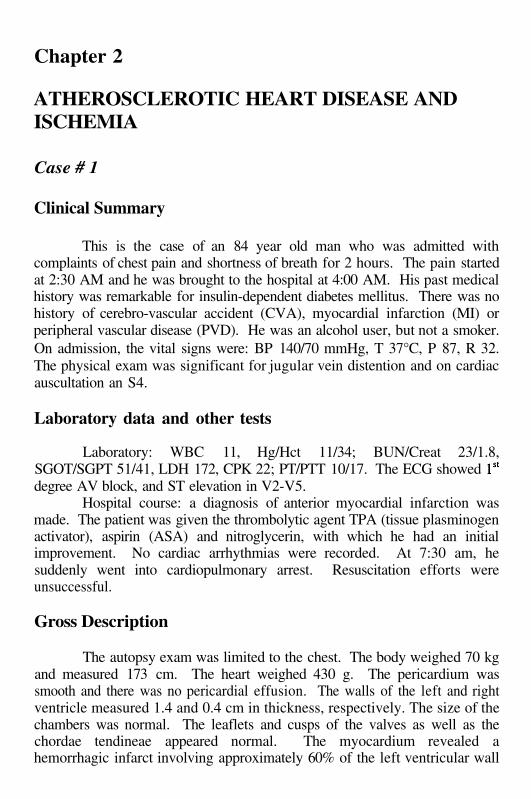
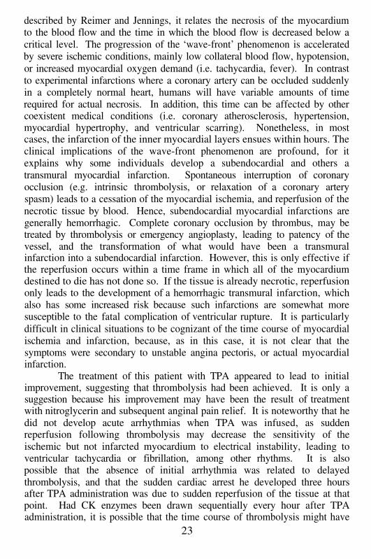
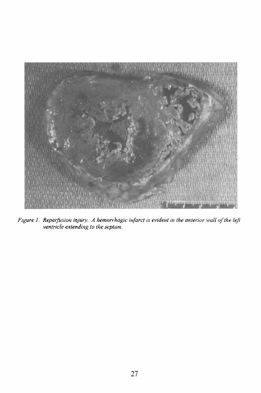
The autopsy exam was limited to the chest. The body weighed 70 kgand measured 173 cm. The heart weighed 430 g. The pericardium wassmooth and there was no pericardial effusion. The walls of the left and rightventricle measured 1.4 and 0.4 cm in thickness, respectively. The size of thechambers was normal. The leaflets and cusps of the valves as well as thechordae tendineae appeared normal. The myocardium revealed ahemorrhagic infarct involving approximately 60% of the left ventricular wall
(Figure 1). The coronary arteries showed severe atherosclerotic changes withup to 95% occlusion of the left main coronary artery, 85% occlusion of theleft anterior descending and circumflex arteries, and 60% occlusion of theright coronary artery. There was no evidence of acute thrombosis of thesevessels. The aorta showed moderate atherosclerosis, but no aneurysms wereidentified. The lungs weighed 1200 g combined and were congested.
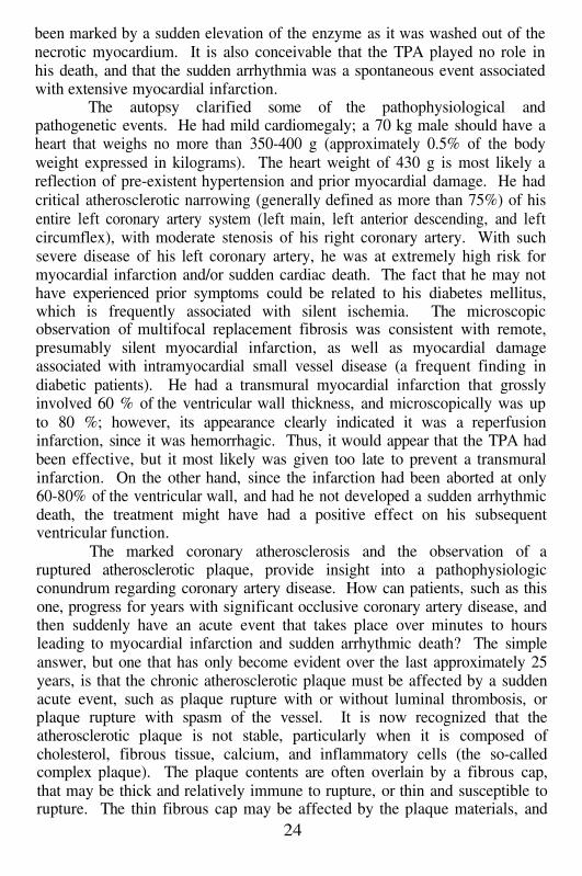
Microscopic Description
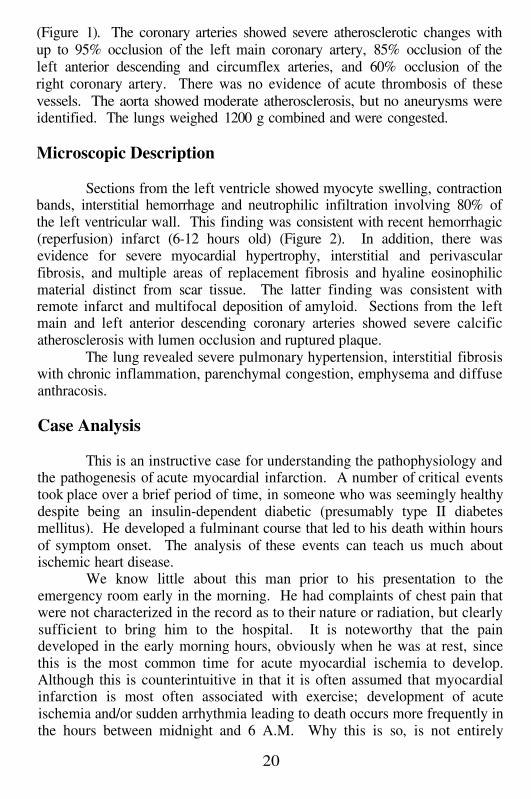
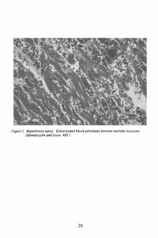
Sections from the left ventricle showed myocyte swelling, contractionbands, interstitial hemorrhage and neutrophilic infiltration involving 80% ofthe left ventricular wall. This finding was consistent with recent hemorrhagic(reperfusion) infarct (6-12 hours old) (Figure 2). In addition, there wasevidence for severe myocardial hypertrophy, interstitial and perivascularfibrosis, and multiple areas of replacement fibrosis and hyaline eosinophilicmaterial distinct from scar tissue. The latter finding was consistent withremote infarct and multifocal deposition of amyloid. Sections from the leftmain and left anterior descending coronary arteries showed severe calcificatherosclerosis with lumen occlusion and ruptured plaque.
The lung revealed severe pulmonary hypertension, interstitial fibrosiswith chronic inflammation, parenchymal congestion, emphysema and diffuseanthracosis.
Case Analysis
This is an instructive case for understanding the pathophysiology andthe pathogenesis of acute myocardial infarction. A number of critical eventstook place over a brief period of time, in someone who was seemingly healthydespite being an insulin-dependent diabetic (presumably type II diabetesmellitus). He developed a fulminant course that led to his death within hoursof symptom onset. The analysis of these events can teach us much aboutischemic heart disease.
We know little about this man prior to his presentation to theemergency room early in the morning. He had complaints of chest pain thatwere not characterized in the record as to their nature or radiation, but clearlysufficient to bring him to the hospital. It is noteworthy that the paindeveloped in the early morning hours, obviously when he was at rest, sincethis is the most common time for acute myocardial ischemia to develop.Although this is counterintuitive in that it is often assumed that myocardialinfarction is most often associated with exercise; development of acuteischemia and/or sudden arrhythmia leading to death occurs more frequently inthe hours between midnight and 6 A.M. Why this is so, is not entirely
20
understood, but some postulate that there may be acute hormonal or neuraleffects on the coronary circulation, possibly related to rapid eye movement(REM) sleep. We do know about his diabetes mellitus, but there is no reportas to whether he was chronically hypertensive. This is an important piece ofinformation lacking in the record, since it may affect the decision to usethrombolytic therapy, which carries somewhat more risk in hypertensivepatients because of the potential for hemorrhagic cerebral infarctions.Regardless, we are told that his blood pressure was normal in the emergencyroom (140/70 mmHg), but this may represent a fall in pressure in someonewith evidence of left ventricular failure.
He did have findings that were strongly suggestive of heart failuredespite maintaining normal blood pressure and pulse rate. His complaints ofchest pain were associated with shortness of breath, consistent withpulmonary congestion secondary to left heart failure. The chest X-raydemonstrated an enlarged heart, which may be secondary to pre-existingcardiomegaly, or to an increase of the cardiac silhouette due to dilatation.However, it also showed increased vascular congestion and interstitial edema,which is one of the earliest signs of elevated left atrial pressure, presumablydue to increased end-diastolic pressure in a failing left ventricle. Moreover,he had an S4 gallop rhythm, which is a pre-systolic extra sound due to atrialcontraction with blood forced into a less compliant left ventricle. Myocardialinfarction decreases the compliance of the left ventricle due to the stiffness ofnon-contracting myocytes, and the presence of interstitial edema. Althoughnot specific for myocardial infarction because other conditions may lead tostiffness of the ventricular wall (e.g. sarcoidosis and scarring), an S3 and/orS4 gallop is a frequent finding in patients with myocardial infarction. Inaddition, the patient presented with jugular venous distention, which is a signof elevated pressure in the right atrium, most likely due to right ventricularfailure.
On clinical grounds, was there sufficient information to support thediagnosis of an acute myocardial infarction? The acute onset of heart failurewith chest pain is presumptive evidence; however, unstable angina pectorismay be associated with ventricular failure due to stunned myocardium (e.g.ischemia-induced contractile dysfunction without myocardial necrosis), andcannot be used as an absolute indicator of infarction. Theelectrocardiographic findings of ST elevation in the anterior leads V2-V5 aresupportive of acute ischemia, but whether the ischemia was transient orassociated with actual tissue damage cannot be determined from thisinformation. The creatine kinase (CK) was within normal limits (22 U/L).Does this mean there was no myocardial necrosis, or was the enzyme drawntoo early in the course of events to be abnormal? Creatine kinase is anintracellular enzyme that is only released into the interstitium from necroticcells. It is then mobilized in the cardiac lymph, where it is eventually drained
21
from cardiac lymphatics into the venous system through the thoracic duct.The measured enzyme activity of CK is thus dependent on the volume ofmyocardium undergoing damage, the time it takes for the enzyme to reach thecirculation, and the velocity of the lymphatic drainage. The latter is at least,partially dependent on blood flow and interstitial pressure in the affectedtissue. This generally correlates with whether there is blood flow (e.g. in aninfarction due to coronary occlusion with profound ischemia, versus atransient occlusion of the coronary artery with subsequent re-perfusion). Inthe situation where profound ischemia develops, the CK may peak at 12-24hours after the onset of infarction. In contrast, with a reperfusion infarction,the CK may be “washed out” of the tissues more rapidly, thereby leading toan earlier peak elevation, which by 12-24 hours post- injury may alreadyreturn to baseline values. The fact that the CK was normal within severalhours after the onset of pain may be due to the early time course of events.Within the last several years, another more sensitive, early marker formyocardial necrosis has been identified: Troponin I enters the serum earlierthan CK, and it persists there longer. The other clinical laboratory finding inthis man that may support a presumptive diagnosis of myocardial infarction isthe elevated white blood count (WBC) of 10,900 cells/nL. Non-specific mildelevations of the WBC are often present in early myocardial infarction butgenerally not with anginal chest pain without myocardial necrosis.
Therefore, based on the initial presentation, it is not absolutely certainthat a completed myocardial infarction had developed by the time the patientreached the emergency room. This may explain why the physicians elected totreat him with TPA, despite the potential risks in an older patient withdiabetes mellitus and possible hypertension. The efficacy of treatment withTPA or streptokinase is directly related to the time in which re-perfusion isachieved, and this is dependent on the pathophysiology of myocardialinfarction. Myocardial infarction is the end result of tissue damage, which isblood flow and time dependent. What this means is that when coronary flowto the tissue is suddenly interrupted, the tissue supplied by that vesselbecomes acutely ischemic. Assuming that the tissue is not protected bysufficient collateral blood flow to prevent ischemic injury (a situation that isnot common in the human heart), or that the myocardium has not been pre-conditioned with previous transient episodes of ischemia in the hours or daysprior to the acute coronary occlusion (this may have the effect of providingpartial myocardium protection, and eventually slow the progression of theinjury), then the myocardium will begin to undergo necrosis.
Myocardial necrosis occurs sequentially from the innersubendocardial layer of the ventricular wall, through the mid andsubepicardial layers of the ventricle until the infarction is transmural (morethan 50% of the ventricular thickness). This process of inner wall to outerwall progression is known as the ‘wave-front’ phenomenon. Originally
22
described by Reimer and Jennings, it relates the necrosis of the myocardiumto the blood flow and the time in which the blood flow is decreased below acritical level. The progression of the ‘wave-front’ phenomenon is acceleratedby severe ischemic conditions, mainly low collateral blood flow, hypotension,or increased myocardial oxygen demand (i.e. tachycardia, fever). In contrastto experimental infarctions where a coronary artery can be occluded suddenlyin a completely normal heart, humans will have variable amounts of timerequired for actual necrosis. In addition, this time can be affected by othercoexistent medical conditions (i.e. coronary atherosclerosis, hypertension,myocardial hypertrophy, and ventricular scarring). Nonetheless, in mostcases, the infarction of the inner myocardial layers ensues within hours. Theclinical implications of the wave-front phenomenon are profound, for itexplains why some individuals develop a subendocardial and others atransmural myocardial infarction. Spontaneous interruption of coronaryocclusion (e.g. intrinsic thrombolysis, or relaxation of a coronary arteryspasm) leads to a cessation of the myocardial ischemia, and reperfusion of thenecrotic tissue by blood. Hence, subendocardial myocardial infarctions aregenerally hemorrhagic. Complete coronary occlusion by thrombus, may betreated by thrombolysis or emergency angioplasty, leading to patency of thevessel, and the transformation of what would have been a transmuralinfarction into a subendocardial infarction. However, this is only effective ifthe reperfusion occurs within a time frame in which all of the myocardiumdestined to die has not done so. If the tissue is already necrotic, reperfusiononly leads to the development of a hemorrhagic transmural infarction, whichalso has some increased risk because such infarctions are somewhat moresusceptible to the fatal complication of ventricular rupture. It is particularlydifficult in clinical situations to be cognizant of the time course of myocardialischemia and infarction, because, as in this case, it is not clear that thesymptoms were secondary to unstable angina pectoris, or actual myocardialinfarction.
The treatment of this patient with TPA appeared to lead to initialimprovement, suggesting that thrombolysis had been achieved. It is only asuggestion because his improvement may have been the result of treatmentwith nitroglycerin and subsequent anginal pain relief. It is noteworthy that hedid not develop acute arrhythmias when TPA was infused, as suddenreperfusion following thrombolysis may decrease the sensitivity of theischemic but not infarcted myocardium to electrical instability, leading toventricular tachycardia or fibrillation, among other rhythms. It is alsopossible that the absence of initial arrhythmia was related to delayedthrombolysis, and that the sudden cardiac arrest he developed three hoursafter TPA administration was due to sudden reperfusion of the tissue at thatpoint. Had CK enzymes been drawn sequentially every hour after TPAadministration, it is possible that the time course of thrombolysis might have
23
been marked by a sudden elevation of the enzyme as it was washed out of thenecrotic myocardium. It is also conceivable that the TPA played no role inhis death, and that the sudden arrhythmia was a spontaneous event associatedwith extensive myocardial infarction.
The autopsy clarified some of the pathophysiological andpathogenetic events. He had mild cardiomegaly; a 70 kg male should have aheart that weighs no more than 350-400 g (approximately 0.5% of the bodyweight expressed in kilograms). The heart weight of 430 g is most likely areflection of pre-existent hypertension and prior myocardial damage. He hadcritical atherosclerotic narrowing (generally defined as more than 75%) of hisentire left coronary artery system (left main, left anterior descending, and leftcircumflex), with moderate stenosis of his right coronary artery. With suchsevere disease of his left coronary artery, he was at extremely high risk formyocardial infarction and/or sudden cardiac death. The fact that he may nothave experienced prior symptoms could be related to his diabetes mellitus,which is frequently associated with silent ischemia. The microscopicobservation of multifocal replacement fibrosis was consistent with remote,presumably silent myocardial infarction, as well as myocardial damageassociated with intramyocardial small vessel disease (a frequent finding indiabetic patients). He had a transmural myocardial infarction that grosslyinvolved 60 % of the ventricular wall thickness, and microscopically was upto 80 %; however, its appearance clearly indicated it was a reperfusioninfarction, since it was hemorrhagic. Thus, it would appear that the TPA hadbeen effective, but it most likely was given too late to prevent a transmuralinfarction. On the other hand, since the infarction had been aborted at only60-80% of the ventricular wall, and had he not developed a sudden arrhythmicdeath, the treatment might have had a positive effect on his subsequentventricular function.
The marked coronary atherosclerosis and the observation of aruptured atherosclerotic plaque, provide insight into a pathophysiologicconundrum regarding coronary artery disease. How can patients, such as thisone, progress for years with significant occlusive coronary artery disease, andthen suddenly have an acute event that takes place over minutes to hoursleading to myocardial infarction and sudden arrhythmic death? The simpleanswer, but one that has only become evident over the last approximately 25years, is that the chronic atherosclerotic plaque must be affected by a suddenacute event, such as plaque rupture with or without luminal thrombosis, orplaque rupture with spasm of the vessel. It is now recognized that theatherosclerotic plaque is not stable, particularly when it is composed ofcholesterol, fibrous tissue, calcium, and inflammatory cells (the so-calledcomplex plaque). The plaque contents are often overlain by a fibrous cap,that may be thick and relatively immune to rupture, or thin and susceptible torupture. The thin fibrous cap may be affected by the plaque materials, and
24
specifically collagenases (tissue metalloproteinases, or TMPs), elaborated byinflammatory cells, and capable of degrading the cap collagen. This may leadto plaque rupture, and the release of thrombosis promoting material into thecoronary lumen. Alternatively, the plaque may be affected by mechanicaldisruption secondary to spasm of intact smooth muscle in the coronary arterywall (particularly if the plaque is eccentrically localized in the vesselcircumference). Thus, stimulation of vessel contraction that may occursecondary to the release of stress related hormones, or secondary to vasogenicsubstances such as nicotine or cocaine, can lead to plaque rupture, also. Theruptured plaque in this case was not associated with luminal thrombosis,which may be explained by the efficacy of the TPA that led to thrombolysis.However, as noted by the presence of a transmural myocardial infarction,there must have been complete left coronary artery occlusion for at least twoto four hours before TPA treatment was instituted. It would appear that thetreatment was appropriate but the extent and severity of the infarction,particularly in a patient with underlying, and probably silent, myocardialdisease, led to his death.
A final comment pertains to the microscopic incidental finding ofmultifocal amyloid deposition in the myocardium. In the absence of systemicamyloidosis associated with a plasma cell dyscrasia, or chronic inflammation,it is most likely that this amyloid represents the so-called senile amyloidosis.This condition, which tends to increase in frequency with age, is of unknownetiology. It is generally due to the abnormal accumulation of transthyreitin,which develops the beta pleated sheet conformation of all of the amyloidproteins, and therefore stains with Congo red dye, producing apple-greenbirefringence when viewed by polarized microscopy. Why this particulartype of amyloid has a predilection for the myocardium is unknown. It usuallyis asymptomatic, but it may occasionally be associated with a restrictivecardiomyopathy characterized by diastolic dysfunction. It is unlikely thatamyloidosis played a direct role in this patient’s course, although it may havemade him more susceptible to develop acute congestive heart failure when hismyocardial infarction developed.
25
Suggested Readings
2.
3.
4.
5.
6.
7.
8.
9.
Jennings RB, Steenbergen C Jr, Reimer KA. Myocardial ischemia andreperfusion. Monogr Pathol. 1995; 37:47-80.Reimer KA, Vander Heide RS, Jennings RB. Ischemic preconditioningslows ischemic metabolism and limits myocardial infarct size. Ann N YAcad Sci. 1994; 723:99-115.Jennings RB, Reimer KA. The cell biology of acute myocardial ischemia.Annu Rev Med. 1991; 42:225-46.Reimer KA, Jennings RB. The "wavefront phenomenon" of myocardialischemic cell death. II. Transmural progression of necrosis within theframework of ischemic bed size (myocardium at risk) and collateral flow.Lab Invest. 1979; 40:633-44.Brockington CD, Lyden PD. Criteria for selection of older patients forthrombolytic therapy. Clin Geriatr Med. 1999; 15:721-39.Hamm CW. New serum markers for acute myocardial infarction. N Engl JMed. 1994; 331:607-8.Straznicky IT, White HD. Thrombolytic therapy for acute myocardialinfarction in the elderly. Coron Artery Dis. 2000; 11:299-304.Zhao M, Zhang H, Robinson TF, Factor SM, Sonneblick H, Eng C.Profound structural alterations of the extracellular collagen matrix in post-ischemic dysfunctional (“stunned”) but viable myocardium. J Am CollCardiol l987; 10:1322-34.Factor SM, Bache R. “ Pathophysiology of Myocardial Ischemia”. InHurt’s The Heart. Ninth Edition. Alexander WR, Schlant RC, and FusterV eds. McGraw-Hill Co, Inc., 1998.
26
1.
27
28
Case # 2
Clinical Summary
The patient was a 60 year old woman who was found unresponsive athome. Paramedics were called. Her initial electrocardiogram (ECG) rhythmwas asystole followed by a brief period of ventricular fibrillation.Cardiopulmonary resuscitation (CPR) measures were unsuccessful and thepatient was pronounced dead at the time of her arrival to the hospital.
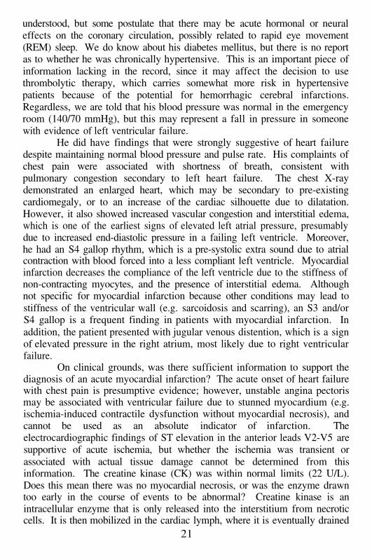
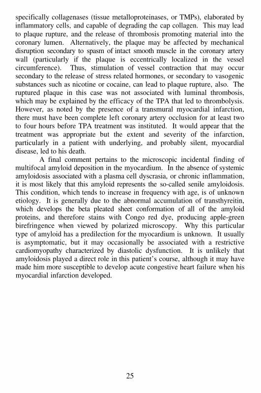
The patient’s medical history was significant for hypertension,hypercholesterolemia and severe coronary artery disease (80-90% stenosis ofleft anterior descending coronary artery and 50-60% occlusion of rightcoronary artery). Eight months prior to her death, the patient underwentpercutaneous transluminal coronary angioplasty (PTCA) of the left anteriordescending coronary artery (LAD). Four months later, because of post-angioplasty re-stenosis, she underwent coronary artery bypass graft (CABG)using the left internal mammary artery to the LAD (Figure 3).
Gross and Microscopic Description
The patient measured 154 cm and weighed 64 kg. The heart weighed450 g. The pericardium was thickened. The left ventricle revealed concentrichypertrophy consistent with long-standing hypertension (wall thickness 1.9cm). The lateral wall of the left ventricle had an area of discoloration, whichon histologic examination consisted of multiple foci of fibrosis andgranulation tissue consistent with subacute and remote myocardial infarcts.The right ventricle was of normal thickness (0.5 cm). Both atria were dilated.The aortic and mitral valves were thickened and calcified, but were relativelypliable. All coronary arteries showed severe stenosis with up to 90%occlusion. Sectioning of the right coronary artery revealed 100 % occlusionby an organized thrombus. The left circumflex also had fresh thrombi withinthe lumen, and the distal left anterior descending showed evidence of re-stenosis, proximal to the bypass graft. Severe atherosclerosis involved theaorta and main branches, and a 3 cm fusiform aneurysm was found in theabdominal portion. The lungs, liver and spleen were congested. The rightkidney was small and weighed 35 g. The left kidney was enlarged andweighed 100 g. The surface of both was granular. Moderate to severeatherosclerosis was also noted in the circle of Willis with up to 60% occlusionin the right middle cerebral artery. Multiple old infarcts were found in thelower thoracic/upper lumbar spinal cord (watershed area).
29
Case Analysis
This woman, with a known history of significant coronary arterydisease, died suddenly at home. In general, because of the possibility ofillegal circumstances, the determination of the cause of death for someonewho dies outside of a medical facility would be the responsibility of theMedical Examiner’s Office. However, in this case, the history ofcardiovascular disease was sufficiently strong to suggest that death was due tonatural causes.
Sudden cardiac death (SCD) is virtually always arrhythmic, and isusually caused by a so-called malignant ventricular rhythm such as ventricularfibrillation (VF, often following episodes of ventricular tachycardia or runs ofpremature supraventricular or ventricular contractions). Asystole also mayoccur, either as the initial event of cardiac arrest, or following VF. Electro-mechanical dissociation (EMD) is another pattern, in which a normal or non-fatal electrical pattern is maintained, but there is an uncoupling of electricalactivity and muscular contraction. Although this may occur following VFwith resuscitation attempts including defibrillation, it can often be seen in thesetting of hemopericardium due to ventricular rupture (as a result ofmyocardial infarction) or aortic rupture (due to aortic dissection). In this case,the patient was initially asystolic, followed by VF shortly thereafter, withunsuccessful resuscitation attempts.
She had a number of risk factors for SCD, all of which are positively andindependently associated with this outcome. It is noteworthy that SCD is byfar the most common cause of death due to heart disease, and it accounts for500,000 to one million deaths per year, with the majority occurring outsidethe hospital. Only a small percentage of individuals experiencing anarrhythmic cardiac arrest will have resuscitation attempted on them, and ofthose, less than 5-10% are resuscitated successfully to reach a medicalfacility. Many of the survivors have significant anoxic brain injury thatsubsequently leads to death or disability. Of the risk factors predisposing toSCD, the following are the most important:
1.2.3.4.5.6.7.
left ventricular hypertrophyocclusive coronary artery diseaseacute coronary artery thrombosismyocardial scarringacute or organizing myocardial infarctioncardiomyopathymyocarditis
30
Of these risk factors, this patient had all except #6 and #7: She hadsignificant cardiac hypertrophy with a gross heart weight of 450 g, and a leftventricular wall measuring 1.9 cm in greatest thickness (normal up to 1.5 cm).The estimated cardiac mass for a woman who weighed 63 kg is between 250-300 g. The calculation is estimated by taking the weight of the individual inkilograms and multiplying by 0.4%. For males, with somewhat greater leanmuscle mass, the multiplier is 0.5%. More precise estimates can be obtainedby using the body surface mass-index, but for practical purposes, a 0.4-0.5%multiplier gives a reasonable approximation. The upper limits are equallysignificant, and are considered pathological, regardless of body size, if morethan 350-400 gm in a woman, or more than 400-450 gm in a man. Theproviso that the hypertrophy is non-physiologic is also an importantconsideration. In a well-trained athlete, particularly one performing isometricexercise (e.g. weight lifting), cardiac hypertrophy may ensue; however, it is aphysiologic response not associated with myocardial scarring. In general, thedifference between pathologic hypertrophy and physiologic hypertrophy isdependent on the presence or absence of fibrosis.
The etiology of this patient’s hypertrophy is most likely secondary tolongstanding hypertension, although compensatory hypertrophy of the non-infarcted heart may have played a role. Even without the medical history ofhypertension, it would be possible to deduce that she was hypertensive basedon her kidney pathology. The kidneys were discrepant in size (35 gm and 100gm), and they had granular cortices. The discordant renal mass was mostlikely the result of a “Goldblatt kidney” phenomenon. The latter occurs whenone kidney is chronically ischemic (usually as a result of renal arterystenosis), and produces excess renin with increases of circulating angiotensin.The angiotensin causes an elevation of the systemic blood pressure that leadsto arterionephrosclerosis of the contralateral kidney (e.g. damage and sclerosisof intrarenal arteries and arterioles with secondary localized ischemia andfibrosis of the glomeruli and tubules). The ischemic kidney is “protected”from the effects of hypertension by the renal artery stenosis. Thus, in aclassic Goldblatt kidney, the diffusely ischemic kidney is small and generallyhas a smooth cortex, with the other kidney showing features of a granularcortex consistent with nephrosclerosis. In this case, although one kidney wassmaller than the other, even the larger of the two was moderately shrunken(normal kidney weight is approximately 150-200 gm), and both weregranular. This suggests that the patient was chronically hypertensive therebycausing nephrosclerosis of both kidneys, and then subsequently she developedrenal artery stenosis leading to superimposed ischemia. Thus, she did nothave a classic Goldblatt kidney phenomenon, though she did havehypertensive renal disease.
This patient had severe chronic coronary artery disease secondary toatherosclerosis, with predominant involvement of the LAD (e.g. single vessel
31
disease), although her right coronary artery was also affected. The 50-60%narrowing of the RCA is below the critical level of stenosis (defined asgreater than 65%), because that level of narrowing at rest will have anassociated pressure drop across the stenotic segment. Sub-critical narrowingcan certainly be significant during exercise, or if there is an acutely unstablevessel. Furthermore, such sub-critical vessels can progress to critical stenosisover time, or they can be associated with thrombosis.
The autopsy revealed obvious disease progression in the RCA (e.g.90% occlusion), and it showed changes of LAD re-stenosis proximal to theCABG. Re-stenosis is a commonly recognized complication of balloonangioplasty that occurs in up to 40% of patients within 3-4 months followingthe procedure. The precise explanation of why it occurs preferentially insome patients, while sparing others, is not understood; nor are there predictorsof who will re-stenose. The process of re-stenosis is an exuberantproliferation of smooth muscle cells and myxomatous stroma in the intimallayer of the ballooned vessel, leading to a neo-intima that impinges on thelumen. The pathogenesis of this tissue proliferation is complex. It is partiallydependent on the injury to endothelial cells following the balloon damage tothe vessel wall that generally causes a local compression and dissection of theoccluding plaque. The balloon injury leads to an “uncovering” ofsubendothelial connective tissue with adherence of platelets that release theprostaglandin thromboxane (TXa, a smooth muscle vasoconstricting agent),and platelet-derived growth factor (PDGF, which is a mitogen for smoothmuscle cell proliferation), thereby stimulating smooth muscle cell migrationinto the intima. Additionally, there is release of endothelin-1, a potentvasoconstrictor, and other hormones and cytokines, which can also stimulatesmooth muscle and connective tissue proliferation.
In this case, the patient did re-stenose following the balloonangioplasty, and then she underwent an internal mammary artery to LADgraft. The internal mammary artery is a useful arterial graft that is relativelydevoid of atherosclerosis, and does not develop atherosclerosis wheninterposed in the coronary circulation. Its major limitation is that it can onlybe grafted to the anterior heart surface, because of its location and length;therefore it is primarily used for LAD grafts. At autopsy, the native LAD wasseverely narrowed proximal to the patent graft, and it showed evidence of re-stenosis.
Prior to her death, this woman had several other significant conditionsthat made her susceptible to a sudden fatal arrhythmia. She had evidence of ahealed scar in lateral left ventricle, which was at least 3 months old (it takes aminimum of 3 months for scar tissue to mature). In the same geographic areaof the ventricle as the scar tissue, she had granulation tissue, which representsa healing response to myocardial necrosis composed of endothelial cell and
32
fibroblastic proliferation. Granulation tissue generally begins to form at 7-10days after tissue injury, and then it progressively matures for several weeksuntil immature scar tissue develops. Thus, this region represents a subacuteinfarction that presumably developed silently (e.g. without clinical signs andsymptoms) at least several weeks prior to her death. The temporal changeslink this area of necrosis to the organizing right coronary artery thrombus,which was also approximately 1-2 weeks in age. Individuals with myocardialscarring are at risk to develop cardiac arrhythmias; however, those withhealing infarction are at markedly higher risk. The myocardial tissueimmediately adjacent to a healing infarction has increased irritability as aresult of inflammation, interstitial edema, and slight hypoxemia, and istherefore more likely to cause a generalized arrhythmic event.
It is most probable that the sudden arrhythmia that led to thiswoman’s death was a direct result of the acute event that was temporarilyrelated to her cardiac arrest. We are referring to the acute thrombosis of theleft circumflex coronary artery. Sudden occlusion of this vessel, together withprior complete occlusion in the right coronary artery, made her ventricleischemic, and susceptible to generate fatal electrical rhythms. It is noteworthythat she developed an acute thrombosis of her circumflex artery within a fewweeks of an RCA thrombus. It is not that rare for patients to develop thrombiin multiple vessels, sometimes concurrently. This suggests an increasedtendency for thrombus formation in already diseased atherosclerotic arteries,possibly as the result of enhanced plaque instability and/or systemic or localhormonal factors. In this case, no specific etiology could be determined.
33
Suggested Readings
2.
3.
4.
5.
6.
7.
8.
Levine GN, Ali MN. The role of percutaneous revascularization in thetreatment of ischemic heart disease: insights from published reports andrandomized clinical trials. Chest. 1997; 112:805-21.Kawasuji M, Sakakibara N, Takemura H, Tedoriya T, Ushijima T,Watanabe Y. Is internal thoracic artery grafting suitable for a moderatelystenotic coronary artery? J Thorac Cardiovasc Surg. 1996; 112:253-9.Jensen J, Eriksson SV, Lindvall B, Lundin P, Sylven C. Women react withmore myocardial ischemia and angina pectoris during elective percutaneoustransluminal coronary angioplasty. Coron Artery Dis. 2000; 11:527-35.Friesinger GC, Ryan TJ. Coronary heart disease. Stable and unstablesyndromes. Cardiol Clin. 1999; 17:93-122.Cishek MB, Gershony G. Roles of percutaneous transluminal coronaryangioplasty and bypass graft surgery for the treatment of coronary arterydisease. Am Heart J. 1996; 131:1012-7.Tennant M, McGeachie JK. A biological basis for re-stenosis afterpercutaneous transluminal angioplasty: possible underlying mechanisms.Aust N Z J Surg. 1992; 62:135-4.Weiner DA. Significance of silent myocardial ischemia after coronaryartery bypass surgery. Am J Cardiol. 1992; 70:35F-38F.Factor SM, Minase T, Cho S, Fein F, Capasso JM, Sonneblick EH.Coronary microvascular abnormalities in the hypertensive-diabetic rat. Acause of cardiomyopathy? Am J Pathol 1984; 116:9-20.
34
1.
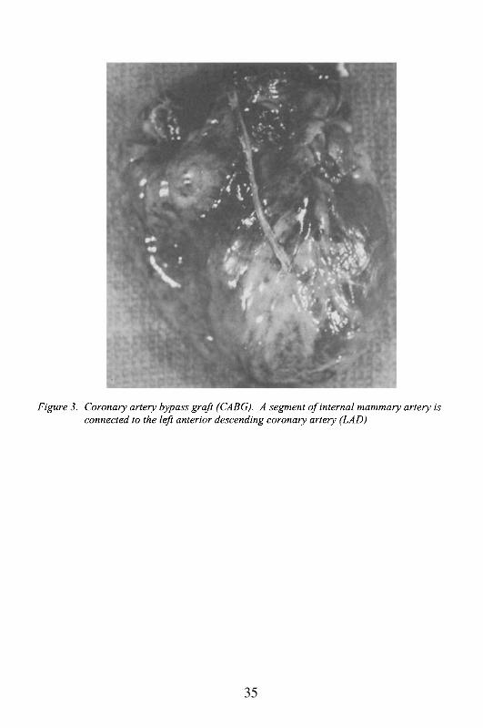
35
Case # 3
Clinical Summary
The patient was a 77 year old obese, Hispanic man who presented tothe emergency room with a 2 hour history of shortness of breath. The patienthad a past medical history significant for hypertension and a myocardialinfarction 10 years previously. The day prior to his current admission, he wasseen by a physician, who diagnosed bronchitis and prescribed antibiotics aswell as a non-steroidal anti-inflammatory medication for his back pain.
On admission to the ER, his vital signs were BP 100/64 mmHg, HR116/min, and RR 24/min. Shortly after his arrival to the hospital, hedeveloped severe chest pain radiating to his upper right back. A chest-x-rayshowed widened mediastinum with interstitial markings. A CT of the thoraxwith contrast revealed a thoracic aortic dissection with questionable leak.
The patient developed respiratory distress and hypotension, whichprogressed to a cardiorespiratory arrest. He was successfully resuscitated andtransferred to the CCU where vasopressors and fluids were administered. Inaddition, the patient required a transfusion with a unit of packed red bloodcells (PRBC). The attending cardiologist suggested immediate surgicalintervention; however, the patient deteriorated rapidly, had a secondcardiorespiratory arrest and died.
Gross Description
Permission was granted for a complete autopsy. The heart weighed600 g. The pericardial sac had minimal amount of serous fluid. Thepericardium and epicardium were smooth and glistening. The atria were ofnormal size and free of thrombi. The foramen ovale was closed. The leftventricle was hypertrophic (wall thickness 2cm) and the right ventricleshowed severe dilatation and hypertrophy. The valves were partiallycalcified. The tricuspid valve was hemorrhagic. The postero-lateral papillarymuscle of the left ventricle was ruptured with consequent prolapse of themitral valve (diameter of the ring 11.5 cm). (Figure 4) The chordae tendineaewere focally thickened. The myocardium was red-brown with averageconsistency, and the endocardium was thin and shiny.
Examination of the coronary arteries showed complete occlusion ofthe right coronary artery as well as acute thrombosis of the left circumflexartery. The aorta showed severe atherosclerosis with thrombi and ulceration,especially in the thoracic and abdominal portions. The major branches wereunremarkable except for aneurysmal dilatation of the right iliac artery.
36
The lungs were heavy (2250 g combined), but no discrete lesionswere identified. The liver was congested (1530 g). Other significant findingsincluded severe nephrosclerosis with cyst formation, and hemorrhagicadrenals, pancreas and intestine. The brain weighed 1255 g and was grosslyunremarkable except for severe atherosclerosis of the circle of Willis.
Microscopic Description
Sections from the heart revealed a recent apical-lateral myocardialinfarction involving the papillary muscle. Remote infarcts in both anteriorand posterior walls were also noted. Sections from the left circumflex showedplaque hemorrhage with associated inflammation. Significant findings in thelungs included moderate emphysematous change, early focalbronchopneumonia and intimal vascular proliferation consistent withpulmonary hypertension. The kidneys, adrenals, pancreas, intestines andbladder revealed diffuse hemorrhage in keeping with shock. The brain wasremarkable for acute anoxic changes and multiple, old areas ofencephalomalacia involving cerebellum, cortex and white matter.
Case Analysis
Although this patient does not fit the classical definition of suddencardiac death (sudden collapse without symptoms, or death within one hour ofsymptom onset), essentially this is a sudden death case. He had acuterespiratory distress of only a few hours duration. In retrospect, his complaintsthe day prior to admission were almost certainly related to the events that ledto his death. When he presented to the hospital he had a low blood pressure(even if he was adequately treated for his longstanding hypertension, 100/64mmHg is hypotensive), tachycardia, and tachypnea. This suggests significantacute compromise of his cardiopulmonary system. He then developed severe,acute chest pain radiating to his upper back. Together with a widenedmediastinum on chest X-ray, the physicians evaluating him in the emergencydepartment suspected an aortic dissection. A CT scan of the thorax withcontrast was ordered which confirmed the diagnosis. Although this was theappropriate diagnostic modality to evaluate a suspected aortic dissection, theinterpretation of the scan was inaccurate, thereby providing a false positiveresult. Soon thereafter, he deteriorated and developed shock and cardiacarrest. He died in a short period of time despite resuscitative measures. Inretrospect, the inaccurate diagnosis of aortic dissection, and the treatment thatmay or may not have been instituted as a result of the diagnosis, did not play arole in his death. Before addressing the actual disease process that led to this
37
patient’s demise, it is worthwhile to address whether the suspicion of aorticdissection was reasonable.
Dissection of the aorta has multiple etiologies. One group of entitiesis the congenital or inherited abnormalities of the aortic connective tissue,most commonly represented by Marfan’s syndrome. The latter is anautosomal dominant disease, with high penetrance, that results from amutation in the fibrillin-1 gene on chromosome 15. Although the penetranceis high, the phenotypic features of Marfan’s syndrome are variable, related tothe greater than 50 different mutations of the fibrillin gene. Fibrillin is a 10-15 nm microfibrillar protein component of elastic tissue. Abnormal fibrillinaffects connective tissue throughout the body, thereby leading to elongatedskeleton, subluxation of the lens, annuloectasia of the aortic valve ring, andfibromyxomatous cardiac valve degeneration, among other conditions.Classically, Marfan associated aortic dissection occurs in the to thedecades of life. In contrast, non-Marfan’s dissection is a disease of unknownetiology, often associated with hypertension, and occurring most often inindividuals in the through the decades. There is no known hereditarypredisposition, and no mutations of connective tissue have been identified.However, it is noteworthy that aortic tissue from Marfan’s syndrome and non-Marfan’s dissections has an increased friability and increased ease inseparating the tissue layers (e.g. split the media). This suggests that even non-Marfan’s syndrome dissections are secondary to some type of connectivetissue defect.
Although the patient had hypertension by history, the likelihood of aspontaneous dissection leading to his cardiovascular collapse is significantlyless likely due to his age. More specifically, someone 77 years old, withrecognized atherosclerotic cardiovascular disease (a myocardial infarction 10years before admission), is virtually precluded from developing dissection.This broad statement is predicated on the fact that such a patient is almostcertain to have aortic atherosclerosis; and, this condition, even if moderatelysevere, leads to intimal and medial plaque formation. The atheroscleroticplaque destroys the normal lamellar structure of the media, and replaces itwith scar and plaque material. Intact elastic lamellae are a necessaryrequirement for dissection, as the dissecting channel of blood splits apart thelayers comprising the media. Although localized plaque hemorrhage andfocal dissection may occur in an atherosclerotic aorta, spread of the dissectionis stopped when the channel of blood reaches an adjacent scarred region. In asimilar way, aortitis with scarring (e.g. syphilitic aortitis) precludes dissection.Rupture of an atherosclerotic aneurysm is far more likely in a patient in hisdecade, than a ruptured aortic dissection. Thus, on clinical and pathologicalgrounds, the consideration of aortic dissection as a possible diagnosis in thispatient should not have been high on the differential diagnosis list.
38
The rapid downhill course of this patient with cardiogenic shock andpulmonary edema is explained by the postmortem findings of acute coronaryocclusion, acute posterolateral wall myocardial infarction, and a rupturedpapillary muscle. Statistically, the two most likely causes of cardiogenicshock in this age group would be a massive myocardial infarction withextensive damage of the myocardium, and a myocardial infarction with tissuedisruption (e.g. acquired ventricular septal defect or papillary muscle rupture).With tissue disruption, the infarction may be relatively small, yet havedisproportionate effects on ventricular function. In the current case, that isprecisely what occurred, with a rupture of the posterolateral papillary muscle.This event accounted for the acute mitral regurgitation (new holosystolic 3/6murmur) followed by rapid development of cardiogenic shock, andrespiratory distress due to pulmonary edema. It is not clear why the clinicalstaff considered the diagnosis of acute aortic dissection with this constellationof findings. An aortic dissection, even if it led to aortic valve insufficiencydue to annular disruption, would be associated with a diastolic murmur.Furthermore, aortic dissection is only rarely associated with cardiogenicshock, and pulmonary edema. It is most often associated with sudden deathdue to rupture and pericardial tamponade. If it extends retrograde towards theaortic annulus, it can dissect or occlude a main coronary artery at the ostium,and under those circumstances it could cause massive myocardial injury andcardiogenic shock.
Rupture of the myocardium can affect 3 main sites: free leftventricular wall, interventricular septum, and papillary muscle. Ruptures tendto occur more frequently in patients in their to decades, in a backgroundof hypertension, and often with no prior history of myocardial infarction.Ruptures of the free wall lead to rapid death with pericardial tamponade.Either the interventricular septum or papillary muscle rupture can lead tocardiogenic shock. The latter, can affect the entire papillary muscle body (theso-called ‘belly’), any of the separate muscular heads (there are often as manyas six separate heads), or at the myotendon junction where it can lead todisruption of a primary chorda tendineae. With complete disruption of theentire papillary muscle, massive mitral valve regurgitation and overwhelmingpulmonary edema ensues rapidly, often leading to death before surgicalintervention. Rupture of a single head or of a chord may permit surgicalrepair. In such conditions, either replacement of the entire mitral valve withprosthesis, or more recently, chordal plication or repair in order to salvage thenative valve, may be attempted.
In this case, the rupture of the papillary muscle was not diagnosed,but even if it had been, it is unlikely that there would have been sufficienttime to permit surgical intervention. An echocardiogram, if it had beenperformed rather than a CT scan, would have revealed segmental leftventricular wall motion abnormalities, and either papillary muscle dysfunction
39
or rupture with a freely moving papillary muscle fragment in the ventricularchamber. In addition, severe mitral valve regurgitation would have beendetected. Papillary muscle rupture, as is true with other forms of ventricularrupture associated with myocardial infarction, frequently occurs between thefirst to day after infarct inception. True to form, the infarction in this casewas approximately 2 days old, based on the presence of well-definedcoagulative necrosis and a heavy infiltration of neutrophils.
40
Suggested Readings
1.
2.
3.
4.
5.
6.
7.
8.
Nienaber CA, Von Kodolitsch Y. Therapeutic management of patientswith Marfan syndrome: focus on cardiovascular involvement. Cardiol Rev.1999; 7:332-41.Libby P, Sukhova G, Lee RT, Liao JK Molecular biology ofatherosclerosis. Int J Cardiol. 1997; 62 (Suppl 2):S23-9.Leopold JA, Loscalzo J. Clinical importance of understanding vascularbiology Cardiol Rev. 2000; 8:115-23.Samman B, Korr KS, Katz AS, Parisi AF. Pitfalls in the diagnosis andmanagement of papillary muscle rupture: a study of four cases and reviewof the literature. Clin Cardiol. 1995; 18:591-6.Reeder GS. Identification and treatment of complications of myocardialinfarction. Mayo Clin Proc. 1995; 70:880-4.Buda AJ. The role of echocardiography in the evaluation of mechanicalcomplications of acute myocardial infarction. Circulation. 1991; 84 (3Suppl):I109-21.Prieto A, Eisenberg J, Thakur RK. Nonarrhythmic complications of acutemyocardial infarction. Emerg Med Clin North Am. 2001; 19: 397-415.Robinson TF, Geraci MA, Sonneblick EH, Factor SM. Coiled perimysialfibers of papillary muscle in rat heart; morphology, distribution, andchanges in configuration. Circ Res 1988; 63:577-92.
41
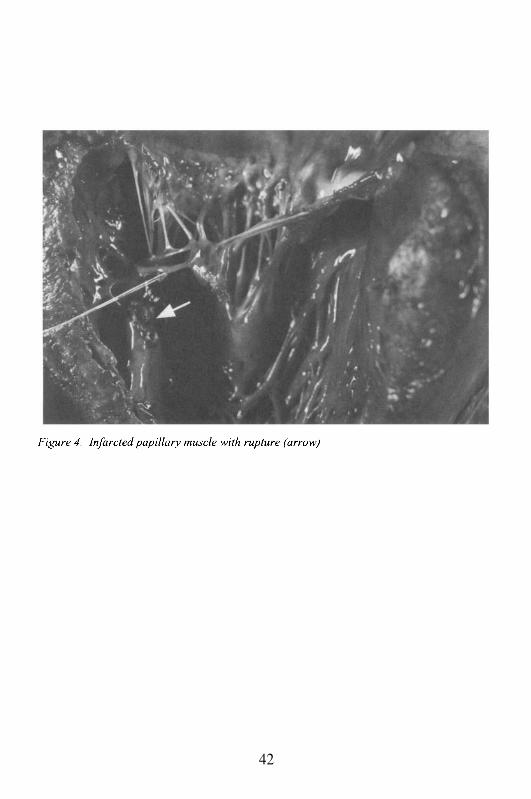
42
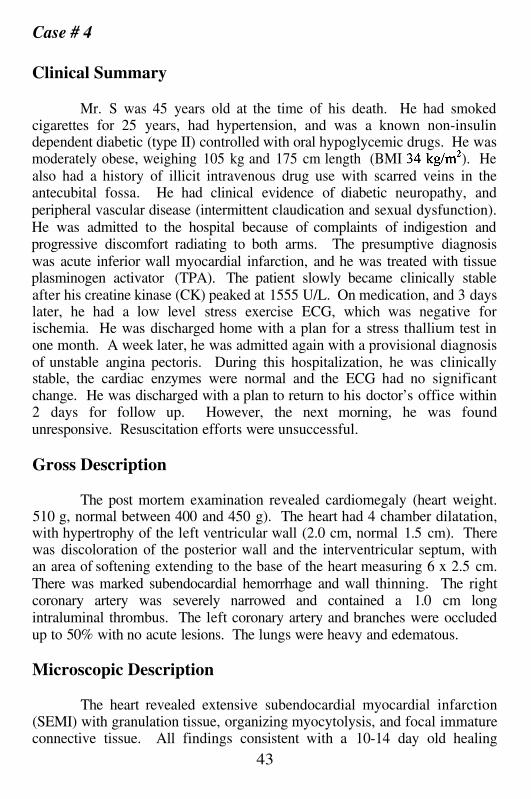
Case # 4
Clinical Summary
Mr. S was 45 years old at the time of his death. He had smokedcigarettes for 25 years, had hypertension, and was a known non-insulindependent diabetic (type II) controlled with oral hypoglycemic drugs. He wasmoderately obese, weighing 105 kg and 175 cm length (BMI ). Healso had a history of illicit intravenous drug use with scarred veins in theantecubital fossa. He had clinical evidence of diabetic neuropathy, andperipheral vascular disease (intermittent claudication and sexual dysfunction).He was admitted to the hospital because of complaints of indigestion andprogressive discomfort radiating to both arms. The presumptive diagnosiswas acute inferior wall myocardial infarction, and he was treated with tissueplasminogen activator (TPA). The patient slowly became clinically stableafter his creatine kinase (CK) peaked at 1555 U/L. On medication, and 3 dayslater, he had a low level stress exercise ECG, which was negative forischemia. He was discharged home with a plan for a stress thallium test inone month. A week later, he was admitted again with a provisional diagnosisof unstable angina pectoris. During this hospitalization, he was clinicallystable, the cardiac enzymes were normal and the ECG had no significantchange. He was discharged with a plan to return to his doctor’s office within2 days for follow up. However, the next morning, he was foundunresponsive. Resuscitation efforts were unsuccessful.
Gross Description
The post mortem examination revealed cardiomegaly (heart weight.510 g, normal between 400 and 450 g). The heart had 4 chamber dilatation,with hypertrophy of the left ventricular wall (2.0 cm, normal 1.5 cm). Therewas discoloration of the posterior wall and the interventricular septum, withan area of softening extending to the base of the heart measuring 6 x 2.5 cm.There was marked subendocardial hemorrhage and wall thinning. The rightcoronary artery was severely narrowed and contained a 1.0 cm longintraluminal thrombus. The left coronary artery and branches were occludedup to 50% with no acute lesions. The lungs were heavy and edematous.
Microscopic Description
The heart revealed extensive subendocardial myocardial infarction(SEMI) with granulation tissue, organizing myocytolysis, and focal immatureconnective tissue. All findings consistent with a 10-14 day old healing
43
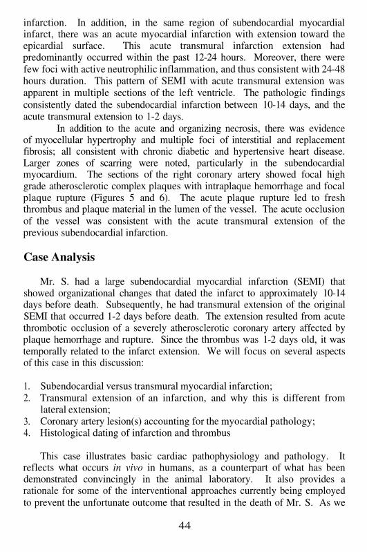
infarction. In addition, in the same region of subendocardial myocardialinfarct, there was an acute myocardial infarction with extension toward theepicardial surface. This acute transmural infarction extension hadpredominantly occurred within the past 12-24 hours. Moreover, there werefew foci with active neutrophilic inflammation, and thus consistent with 24-48hours duration. This pattern of SEMI with acute transmural extension wasapparent in multiple sections of the left ventricle. The pathologic findingsconsistently dated the subendocardial infarction between 10-14 days, and theacute transmural extension to 1-2 days.
In addition to the acute and organizing necrosis, there was evidenceof myocellular hypertrophy and multiple foci of interstitial and replacementfibrosis; all consistent with chronic diabetic and hypertensive heart disease.Larger zones of scarring were noted, particularly in the subendocardialmyocardium. The sections of the right coronary artery showed focal highgrade atherosclerotic complex plaques with intraplaque hemorrhage and focalplaque rupture (Figures 5 and 6). The acute plaque rupture led to freshthrombus and plaque material in the lumen of the vessel. The acute occlusionof the vessel was consistent with the acute transmural extension of theprevious subendocardial infarction.
Case Analysis
Mr. S. had a large subendocardial myocardial infarction (SEMI) thatshowed organizational changes that dated the infarct to approximately 10-14days before death. Subsequently, he had transmural extension of the originalSEMI that occurred 1-2 days before death. The extension resulted from acutethrombotic occlusion of a severely atherosclerotic coronary artery affected byplaque hemorrhage and rupture. Since the thrombus was 1-2 days old, it wastemporally related to the infarct extension. We will focus on several aspectsof this case in this discussion:
1.2.
3.4.
Subendocardial versus transmural myocardial infarction;Transmural extension of an infarction, and why this is different fromlateral extension;Coronary artery lesion(s) accounting for the myocardial pathology;Histological dating of infarction and thrombus
This case illustrates basic cardiac pathophysiology and pathology. Itreflects what occurs in vivo in humans, as a counterpart of what has beendemonstrated convincingly in the animal laboratory. It also provides arationale for some of the interventional approaches currently being employedto prevent the unfortunate outcome that resulted in the death of Mr. S. As we
44
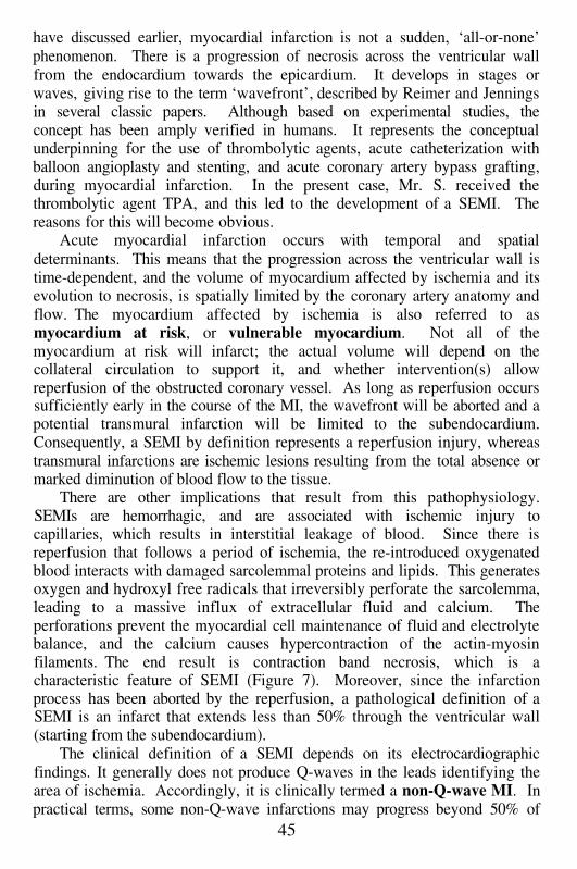
have discussed earlier, myocardial infarction is not a sudden, ‘all-or-none’phenomenon. There is a progression of necrosis across the ventricular wallfrom the endocardium towards the epicardium. It develops in stages orwaves, giving rise to the term ‘wavefront’, described by Reimer and Jenningsin several classic papers. Although based on experimental studies, theconcept has been amply verified in humans. It represents the conceptualunderpinning for the use of thrombolytic agents, acute catheterization withballoon angioplasty and stenting, and acute coronary artery bypass grafting,during myocardial infarction. In the present case, Mr. S. received thethrombolytic agent TPA, and this led to the development of a SEMI. Thereasons for this will become obvious.
Acute myocardial infarction occurs with temporal and spatialdeterminants. This means that the progression across the ventricular wall istime-dependent, and the volume of myocardium affected by ischemia and itsevolution to necrosis, is spatially limited by the coronary artery anatomy andflow. The myocardium affected by ischemia is also referred to asmyocardium at risk, or vulnerable myocardium. Not all of themyocardium at risk will infarct; the actual volume will depend on thecollateral circulation to support it, and whether intervention(s) allowreperfusion of the obstructed coronary vessel. As long as reperfusion occurssufficiently early in the course of the MI, the wavefront will be aborted and apotential transmural infarction will be limited to the subendocardium.Consequently, a SEMI by definition represents a reperfusion injury, whereastransmural infarctions are ischemic lesions resulting from the total absence ormarked diminution of blood flow to the tissue.
There are other implications that result from this pathophysiology.SEMIs are hemorrhagic, and are associated with ischemic injury tocapillaries, which results in interstitial leakage of blood. Since there isreperfusion that follows a period of ischemia, the re-introduced oxygenatedblood interacts with damaged sarcolemmal proteins and lipids. This generatesoxygen and hydroxyl free radicals that irreversibly perforate the sarcolemma,leading to a massive influx of extracellular fluid and calcium. Theperforations prevent the myocardial cell maintenance of fluid and electrolytebalance, and the calcium causes hypercontraction of the actin-myosinfilaments. The end result is contraction band necrosis, which is acharacteristic feature of SEMI (Figure 7). Moreover, since the infarctionprocess has been aborted by the reperfusion, a pathological definition of aSEMI is an infarct that extends less than 50% through the ventricular wall(starting from the subendocardium).
The clinical definition of a SEMI depends on its electrocardiographicfindings. It generally does not produce Q-waves in the leads identifying thearea of ischemia. Accordingly, it is clinically termed a non-Q-wave MI. Inpractical terms, some non-Q-wave infarctions may progress beyond 50% of
45
the ventricular wall. Consequently, there may not be an absolute correlationbetween the clinical and pathological terminology. The explanation for theabsence of Q-waves is of significant interest: the Q-wave represents thenegative deflection below the iso-electric line on the ECG. Necrotic muscledoes not generate any electrical impulses, positive or negative (the deadmuscle is electrically invisible). It allows the ECG leads to ‘see through’ thenecrotic tissue to the viable myocardium on the opposite side of theventricular wall, which is able to generate upward positive deflections, or R-waves, in the ECG leads directly facing this zone. In the opposite leads, andfacing the electrically negative infarct zone, the deflections are downwardnegative, or Q-waves. Since a SEMI has a viable zone of myocardiumoverlying the infarct, it is not electrically neutral. Therefore, no Q-waves areseen.
The SEMI is hemorrhagic and initially is composed of myocardium withcontraction band necrosis. Time-dependent necrosis of the ventricular layersextending towards the epicardium is associated with time-dependent survivalof the immediate subendocardial layers. Normally, if one examines themyocardium just under the endocardium from a heart with a transmuralinfarction, there will be 3-5 myocardial cell layers that survive. They aremaintained viable by direct diffusion of oxygen across the endocardium intothe tissue (oxygen can diffuse approximately 30-50 microns, or about thedistance of 3-5 myocardial cell diameters). If the myocytes are hypertrophied,the endocardium is fibrosed, or the zone underlying the infarction is blockedby a mural thrombus, there is no myocardial cell survival. Although thesecells probably have no functional role, the immediate subendocardial zone isalso where the Purkinje cells are found; thus, the survival of this tissuethrough oxygen diffusion may permit normal ventricular conduction. Thesame zone, in a SEMI, is initially much larger, representing as many as 10-20myocardial cells, which are potentially ischemic and represent vulnerablemyocardium. Furthermore, this subendocardial myocardium is most likely‘stunned’, is non-contractile, and with time, may even undergo necrosis.However, it can generate electrical activity, and as such may be a source ofmalignant and possibly fatal arrhythmias. Hence, the association of SEMIand lethal rhythm disturbances.
The other consequence of the different pathophysiology between a SEMIand a transmural myocardial infarction (TMMI), is that the TMMI occursfollowing complete occlusion of a coronary artery. In contrast to contractionbands characteristic of SEMI, the myocardium undergoes coagulativenecrosis, which is a denaturing process of the cellular proteins secondary tooxygen deprivation. This leads to a smudgy ‘cooked’ appearance of themyocardium, with intense red staining with the dye eosin, orhypereosinophilia. Since this may take as long as 4-6 hours to become
46
evident, it may be very difficult to ascertain whether any patient dyingsuddenly actually had a myocardial infarction as the precipitating event.
Clearly, then, there are pathophysiological, pathological, andfunctional differences between a SEMI and a TMMI. We will next turn to theissue of infarct extension:
Ordinarily, coronary vessels in humans have limited collateralbranches. This is particularly true when the obstruction is more distal in thecoronary artery, thereby providing fewer branches to bridge the zone ofischemia. However, the prediction as to whether any patient will or will nothave adequate collaterals to support the myocardium is not possible prior toobstruction. Generally, collaterals are more likely to be present in theimmediate subepicardial myocardium, decreasing from epicardium toendocardium. Accordingly, the myocardium at risk following coronaryocclusion is spatially determined in an inside-outside dimension, and issharply delimited along the lateral margins. The infarction is broader alongthe endocardium, and angles inward toward the epicardium. There areminimal collaterals that maintain the lateral margins. This means that there isno significant lateral border zone. The latter observation was a source ofmajor controversy 20 years ago, but now is a widely accepted concept.
The myocardium at risk for infarction remains vulnerable even if theinitiating coronary artery occlusion is transformed into a patent vessel. Evenafter reperfusion, the surviving subepicardial myocardium remains vulnerableto necrosis. If the vessel re-occludes at the same level, the survivingmyocardium will then infarct as if the coronary artery was initially blocked inits entirety, and remained so for many hours. This is what is known astransmural extension; and it is precisely what occurred in the present case.Mr. S. had re-establishment of flow with TPA, and then he re-occluded manydays later leading to a TMMI found at autopsy. The vulnerable infarct zonedoes not extend laterally, since distinct and separate vessels or branchessupply that myocardium. If the myocardium lateral to an infarct undergoesnecrosis, it would represent an extension to a new vascular territory. It is notunusual for additional vessels to become thrombosed or occluded by spasm inthe setting of a TMMI. This would lead to new lateral or distant lesions fromthe initial focus.
As a final point, there is one concept that relates to post-occlusionmodification of an MI and that is the term infarct expansion. It refers to themodification of the ventricular wall affected by a TMMI that undergoesremodeling. This leads to mural thinning and aneurysmal bulging of the wall.This process has significant effects on ventricular function, susceptibility toendocardial thrombus, and the potential for ventricular rupture. Expansion isusually associated with a large TMMI, but not a SEMI. When it occurs, it hasa relatively poor prognosis.
47
The coronary lesion that accounted for the events affecting thispatient is fairly common. He had a high-grade atherosclerotic plaque, whichpresumably led to an acute luminal thrombosis. The thrombus was then lysedwith TPA, re-establishing adequate coronary flow and aborting the infarctprogression across the ventricular wall. Subsequently, the same plaqueruptured leading to re-thrombosis of the vessel at the same level, extendinghis SEMI into a TMMI. This was a clinically silent event, most likelybecause of his diabetes mellitus. If these events had occurred today, he wouldhave undergone coronary catheterization following the development of theSEMI. An angiogram would have demonstrated persistent high-gradecoronary occlusion, and he would have had balloon angioplasty and stentplacement, if clinically feasible. If not, he might have had coronary arterybypass grafting. With either approach, the vulnerable myocardium wouldhave been well-perfused, and he would not have extended his SEMI.
The pathology of the initial infarction and the subsequent extensioncould be dated histologically. This is possible because of the use of well-established criteria that define the time course for the inflammatory response,the development of granulation tissue, and the deposition of connective tissueuntil the entire infarct is replaced by mature scar tissue. Similar dating of thethrombi can be done, as documented in this case with the coronary arterythrombus that correlated with the TMMI. Although the dating is not preciseto the minute and hour (in contrast to the frequent impression one gets fromtelevision or fictional depictions of pathological investigation, where the fatalevent is identified to the minute and hour), it can provide a rough 6-12 hourtime frame for most of the cellular events affecting the infarctingmyocardium. It is beyond the scope of this book to provide the details of thedating procedure, although they are adequately described in numerouspathology and cardiology texts. In most cases, such an evaluation is ofacademic interest, with some clinical implications. It is often of greatimportance in the medico-legal sphere where the determination of the precisetime of an infarct and correlation with clinical signs and symptoms may besignificant. For instance, this approach may help establish whether a fatalmotor vehicle accident occurred due to a MI and fatal arrhythmia, or traumadirectly caused the death of the driver.
In summary, the extension of a SEMI into a TMMI in this patient,serves as clinical verification for many of the major insights into myocardialischemia and coronary thrombosis, which have been developed over the past2 decades with animal studies. These insights have provided a firm scientificgrounding for the interventional cardiology and cardiac surgery treatments ofcoronary ischemia and myocardial infarction. Over the last years, thesetreatment options have had a tremendous impact on disease prognosis, qualityof life and survival of patients with ischemic coronary artery disease.
48
Suggested Readings
2.
3.
4.
5.
6.
7.
8.
9.
10.
Villablanca AC, McDonald JM; Rutledge JC. Smoking and cardiovasculardisease. Clin Chest Med. 2000; 21:159-72.Reimer KA, Jennings RB. Biologic basis for limitation of infarct size.Adv Exp Med Biol. 1986; 194:315-30.Ravn HB, Falk E. Histopathology of plaque rupture. Cardiol Clin. 1999;17:263-70.Schroeder AP, Falk E. Pathophysiology and inflammatory aspects ofplaque rupture. Cardiol Clin. 1996; 14:211-20.Becker RC, Bovill EG, Seghatchian MJ, Samama MM. Pathobiology ofthrombin in acute coronary syndromes. Am Heart J. 1998; 136:S19-31Stern S, Cohn PF, Pepine CJ. Silent myocardial ischemia. Curr ProblCardiol. 1993; 18:301-59.Okun EM, Factor SM, Kirk ES. End-capillary loops in the heart: Anexplanation for discrete myocardial infarctions without border zones.Science 1979; 206:565-67.Factor SM, Okun EM, Kirk ES. The histological lateral border of acutecanine myocardial infarction: a function of the microcirculation. Circ Res1981; 48:640-49.Factor SM, Okun EM, Minase T, Kirk ES. The microcirculation of thehuman heart: end capillary loops with discrete perfusion fields. Circulation1982; 66:1241-48.Forman R, Cho S, Factor SM, Kirk ES. Acute myocardial infarct extensioninto a previously preserved subendocardial region at risk in dogs andpatients. Circulation 1982; 67:117-24.
49
1.
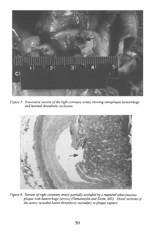
50
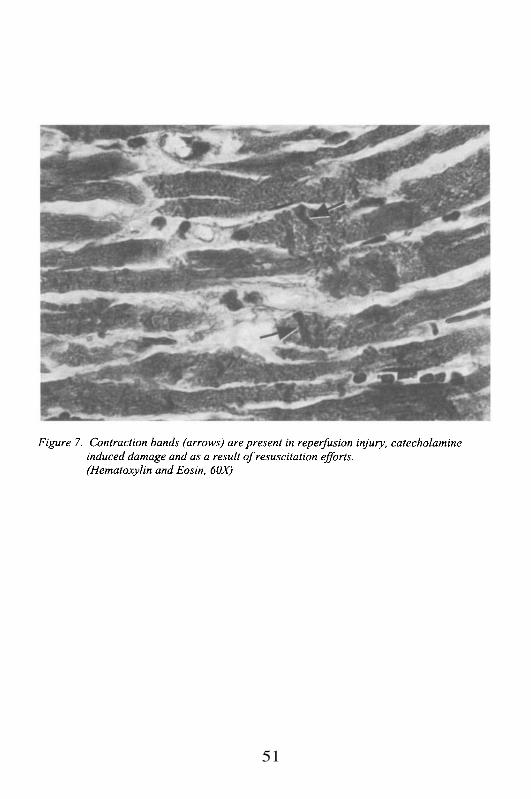
51
Case #5
Clinical Summary
The patient was a 69 year old man with a history of hypertension andchronic obstructive pulmonary disease, who three days prior to admission tothe hospital experienced exertional chest pain relieved by rest. The patientpresented to the emergency room with complaints of substernal chest pain ofa few hours duration that radiated to the shoulder, and was not relieved bynitroglycerin.
Laboratory data and other tests
Laboratory: WBC 10.3 (S 53%, L 39%, M 7%, E 1%), Hb/Hct 16/51, Plt 293;SGOT 384 U/L, LDH 751 U/L, CK 1875 U/L (CK-MB 11%). ECG: sinustachycardia at 107 and ST elevation V1-V5.
The diagnosis of acute myocardial infarction was made. The patientwas given thrombolytic therapy (tissue plasminogen activator, TPA) in the ERwith resolution of the chest pain and clearance of the cardiac enzymes overthe following 3 days. On the hospital day, the WBC was noted to beincreasing to 14 (S 77% and Bands 9%). He was afebrile and the blood andurine cultures were negative. A chest X-ray was done which showed a rightupper lobe infiltrate, which resolved in few days. The patient was feelingbetter and he was scheduled for a cardiac catheterization. The procedure waspostponed due to worsening abdominal and left hip pain. Within the next twodays the hematocrit dropped to 29% and subsequently to 17%. On theday of hospitalization, the patient suddenly developed shortness of breath andhypotension. Resuscitation efforts were unsuccessful and the patient waspronounced dead.
Gross Description
The patient measured 157 cm and weighed 59 kg. Externalexamination revealed no clubbing, cyanosis or edema. The heart weighed 450g and the shape was normal. The pericardium was smooth. The atria wereunremarkable. Both ventricular chambers were dilated and hypertrophic. Themyocardium showed a hemorrhagic subendocardial and focally transmuralinfarct of the antero-septal wall, approximately 2 weeks old. Other foci ofscarring were also noted. The coronaries showed severe atherosclerosis with100% occlusion of the left anterior descending artery and right coronaryartery, and up to 70% stenosis of the left main and left circumflex arteries.
52
No acute thrombosis was identified. The lungs weighed 1050 g combined.Pleural plaques were noted in the left upper lobe. Emphysematous changes aswell as anthracosis were moderate to severe. The liver, weighing 1200 g, wascongested. The kidneys were unremarkable; however, a large leftretroperitoneal hematoma was noted, enveloping the left kidney andextending to the psoas muscle and left diaphragmatic region.
Microscopic Description
The heart showed transmural myocardial necrosis of the anteroseptalwall with hemorrhage, fibrosis and granulation tissue consistent with a 12-14days old infarct. There was severe calcific atherosclerosis of the coronarieswith 100% occlusion of the left anterior descending and right coronaryarteries. Another significant finding was the large retroperitoneal hematomainvolving the psoas muscle (Figure 8). The lungs showed mild congestion,severe emphysematous change, chronic bronchitis, focal scarring andgranulomatous reaction with calcification consistent with old pulmonarytuberculosis. The pulmonary vessels were thickened consistent withpulmonary hypertension. The liver showed fatty infiltration, congestion andportal inflammation with bridging fibrosis.
Case Analysis
This case illustrates the major complication of thrombolytic therapy,which is bleeding. The patient was admitted with the diagnosis of amyocardial infarction based on the clinical symptoms, ECG changes, andelevated cardiac enzymes. ST elevations are often seen as the earliestexpression of a transmural myocardial infarction, and are followed by thedevelopment of abnormal Q waves. Since most transmural myocardialinfarctions (~ 90%) are the result of coronary thrombosis, administration ofTPA was indicated to avoid further myocardial damage. TPA convertsplasminogen to plasmin, which in turn is responsible for the lysis of fibrin andfibrinogen, the main components of an acute thrombus. The use of TPAfollowed by intravenous heparin has been successful in restoring blood flowafter an acute coronary occlusion. To maximize results, TPA must beadministered within a period of less than 6 hours after the onset of symptoms,as was done in this case. Reperfusion was successful since the chest paindisappeared and the enzymes normalized. Adjuvant therapy with heparin wasstarted to prevent re-occlusion.
Sudden onset of abdominal pain in a patient that is receivinganticoagulant therapy should alert the clinician of the possibility of bleeding.Potential sources include a gastric or duodenal peptic ulcer, hemorrhagic
53
pancreatitis, and a dissecting or ruptured abdominal aortic aneurysm. UpperGI bleeding was unlikely in the absence of hematemesis or melena. Acutepancreatitis is associated with leukocytosis, but there was no evidence ofincreased levels of serum amylase and lipase. Abdominal aortic aneurysmsare common in patients with atherosclerosis and hypertension. These patientsoften develop saccular dilatations below the renal and above the iliac arteries.Aortic dissection can occur in patients with a history of hypertension, but arerare in the context of severe atherosclerosis due to the protective effect ofsubstantial medial scarring and fibrosis. Rupture of an aortic aneurysm cancause excruciating pain, but this is a catastrophic event with massive bloodloss and rapid death. There is no time for development of progressive painwith subsequent radiation to the hip and dropping hematocrit within a 48hours period, unless the aneurysm was leaking slowly. Bleeding within athird space (pleura, pericardium and retroperitoneum) has been reportedfollowing anticoagulant therapy, although fatal bleeding is rare. Pleural andpericardial hemorrhages usually cause chest pain. Patients with a largepericardial effusion present with symptoms of cardiac tamponade. Dysphagiaand dyspnea may result from esophageal or lung compression due tomediastinal or pleural hemorrhage.
Retroperitoneal hematomas are more likely to cause abdominal painthat radiates to the extremities. Pain in the hip and leg are the result ofentrapment of the lumbar plexus and femoral nerve, and the dissection of theplanes of the psoas muscle. The retroperitoneum is a relatively large andexpansible space that allows a gradual accumulation of blood and changes inthe hematocrit values. Retroperitoneal hematomas may occur even withcontrolled prothrombin time (PT) and partial thromboplastin time (PTT)values; therefore a CT scan is indicated if the patient develops abdominal painassociated with progressive normochromic, normocytic anemia. In this case,the bleeding was most likely caused by heparin rather than by TPA. Theadverse effects of TPA are evident early after administration and includestroke, intracerebral hemorrhages and less often, bleeding from vascularaccess sites. Therapeutic heparin dose has been associated with an estimatedbleeding risk of 5%. The risk increases if the dose is given intermittentlyrather than continuously, and in patients with chronic illnesses such aspulmonary or liver diseases. Interestingly, our patient was a heavy smokerwith both severe COPD and evolving cirrhosis. Nonetheless, he had enoughspared and functional liver parenchyma to maintain normal coagulation andunremarkable levels of bilirrubin and alkaline phosphatase. The sudden onsetof shortness of breath was due to the extension of the hemorrhage into the leftdiaphragmatic region. The cause of death in this case was most likelyhypoxemia followed by a cardiac arrhythmia.
54
Suggested Readings
2.3.4.
Hirsh J. Coronary thrombolysis: hemorrhagic complications. Can JCardiol 1993; 9:505-11.Sleight P. Thrombolysis: state of the art. Eur Heart J 1993; Suppl G:41-7.Simoons ML. Risk-benefit of thrombolysis. Cardiol Clin 1995; 13:339-45.Jafri SM, Walters BL, Borzak S. Medical therapy of acute myocardialinfarction: Part I. Role of thrombolytic and antithrombotic therapy. JIntensive Care Med 1995; 10:54-63.Indications for fibrinolytic therapy in suspected acute myocardialinfarction: collaborative overview of early mortality and major morbidityresults from all randomised trials of more than 1000 patients. FibrinolyticTherapy Trialists' (FTT) Collaborative Group. Lancet 1994; 343:311-22.
55
5.
1.
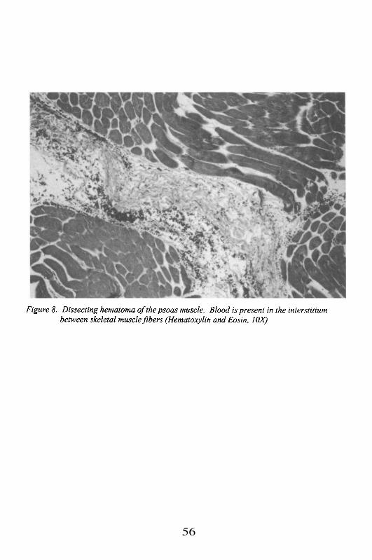
56
NON-ATHEROSCLEROTIC ISCHEMIA
Case # 6
Clinical Summary
This is the case of a 45 year old Hispanic woman with a 30 pack/yearsmoking history, and a six year history of Hepatitis B seropositivity. She hadsuffered from migraines for the past 11 years, and had had several syncopalepisodes. She presented to the hospital after acute onset of left-sided chestpain of 15 minutes duration. It was described as sharp with radiation to theleft arm, which felt numb and weak with associated paresthesias (‘tingling’).She had no shortness of breath, diaphoresis, dizziness, nausea, vomiting ordiarrhea. She did not have a history of hypertension, diabetes mellitus, anginaor myocardial infarction. Prior to her arrival to the hospital she had a briefperiod of arm weakness and unconsciousness.
Her past medical history was also remarkable for sinus problems, andhay fever. Both parents were alive and healthy. She had two sisters, one ofwhom had a history of coronary artery disease and myocardial infarction. Herbrother, age 50 y, had colon cancer.
On admission her vital signs were BP 112/52, P 85, T 37°C, and RR20. The patient was hydrated, alert, with normal mental exam, and in nosignificant distress. The neurological exam was grossly normal. Her skin hadno lesions. There was no mouth erythema or exudate. The neck was supple,with a diffusely enlarged thyroid, and no bruits or jugular venous distension(JVD). The heart had regular rhythm, and the lungs were clear toauscultation. The abdomen had normal bowel sounds, was soft, non-tender,non-distended, with no hepatosplenomegaly or masses. The extremities hadno clubbing, cyanosis or edema, and the peripheral pulses were 2+ bilaterally.
Laboratory data and other tests
Laboratory: WBC 5.9 (S 35%, L 49%, M 10%, E 5%), Hb/Hct 12/35,Plt 348; SGOT/SGPT 25/18 U/L, Cholesterol 108, PT/PTT 13/26. CXRnormal. ECG: sinus rhythm at 82, normal axis and ST elevation in II, III andavF.
The patient was admitted to rule out myocardial infarction and towork up her syncopal episodes. The day after admission she complained ofchest pain, which subsided with sublingual nitroglycerin. Her ECG at thetime showed ST elevations in many leads. Soon thereafter, she began gaspingand became pulseless. Resuscitation efforts were unsuccessful
57
Pre-Autopsy Evaluation and Analysis
Crucial clinical information can be derived from the first sentence of thehistory: 1) a 45 year old woman, 2) a smoker, 3) seropositive for hepatitis B,4) affected by migraine headaches, and 5) someone who had experiencedmultiple episodes of syncope (although the time frame is not indicated). Herpresentation was suggestive for acute ischemic heart disease. The quality andcharacteristics described are very specific for coronary pain (~95 %specificity, and a high positive predictive value). Electrocardiograms at firstrevealed ST segment elevations in II, III, and avF; and then diffuse STelevations across the precordium. Her death was rapid, and it was notassociated with cardiac enzyme increases. Thus, we are trying to establish thecause of apparent ischemic heart disease in a relatively young woman.Several questions about this case can easily be raised:
1.2.3.4.
Is this really ischemic heart disease?If so, is it more likely to be typical or atypical?If it is typical, what is the differential diagnosis?If it is atypical, what is the differential diagnosis?
Ischemia vs. Non-ischemia
It would certainly appear that the presentation is most consistent withischemic heart disease. The acute onset of chest pain with radiation to the leftarm is strongly suggestive of this conclusion. She did not have diaphoresis,nausea, or vomiting, all signs and symptoms of ischemic heart disease, butvery variable in their presence. Furthermore, as described below, theischemia may not have been persistent. Although one must consider othercauses of chest pain, it is difficult to explain the nature of these complaintswith chest wall disease (e.g. costochondritis, or myositis), muscle strain,pericarditis, pleuritis, or parenchymal pulmonary disease (includingpulmonary embolism). When one adds the electrocardiographic findings tothe clinical complaints, it strengthens the association even further withischemia. The fact that the cardiac enzymes were within normal limits,simply indicates that the ischemia was transient and did not persist forsufficient time to irreversibly injure the myocardium. It is possible that today,with the use of the more sensitive early marker of myocardial damage,troponin I, that there would have been evidence of myocardial injury, but thismarker was not in general use in 1994 when this case occurred. Additionally,the clinical history of multiple episodes of syncope, and the brief period ofunconsciousness associated with the acute chest pain events leading to herhospitalization, suggests that ischemia may have been intermittent and
58

causing transient cardiac arrhythmia. Thus, it appears entirely reasonable toassume that this patient had ischemic disease based on her presentation to thehospital; in fact, she was admitted with the working diagnosis of rule outmyocardial infarction (ROMI).
Typical vs. Atypical Ischemia
Once we consider it more likely than not that this patient hadischemic disease, the question of whether it was typical or atypical issomewhat more difficult to ascertain. First, it is necessary to define these twoterms. Typical ischemic heart disease is generally considered secondary tocoronary atherosclerosis, with atherosclerotic plaque (s) narrowing the lumenand causing damage to the coronary artery wall. The presence ofatherosclerosis may increase the possibility of superimposed coronary arterylesions including acute plaque hemorrhage or rupture, luminal thrombosis,and/or coronary artery spasm. The presence of atherosclerosis does notprovide information concerning the pathogenesis of the plaque, whether it isidiopathic but associated with cigarette smoking, hypercholesterolemia,hypertension, or diabetes mellitus (or combinations of all of these), or whetherit may be secondary to less common risk factors such ashyperhomocysteinemia, cocaine use, or radiotherapy (chest irradiation forneoplastic disease).
Atypical ischemia is discriminated from typical angina pectoris,because it is considered to be non-atherosclerotic coronary artery disease.This implies different pathophysiological mechanisms, primarily secondary tocoronary artery spasm, which may be spontaneous or caused bypharmacological agents including drugs such as cocaine. Other causesinclude hypercoagulable states, leading to coronary artery thrombosis; director indirect coronary artery infection (e.g. herpes virus or cytomegalovirusinfection in immunosuppressed patients; salmonella infection of endothelialcells; or direct extension of bacterial infection from active endocarditis orpericarditis, leading to mycotic coronary artery aneurysm); mechanicaldisruption of the coronary artery, such as spontaneous or post-traumaticdissection (e.g. the trauma may be related to blunt force chest wall injury, oriatrogenic injury during interventions such as coronary angiogram orangioplasty); and finally, inflammatory diseases such as vasculitis. It is ofparticular importance to recognize that combinations of these variousetiologies may occur simultaneously, making the assessment of the situationeven more difficult.
Based on the previous definitions, did this patient have typical oratypical coronary artery ischemia? She was a relatively young woman.Atherosclerosis begins to increase in women in their fifth decade, but it is
59
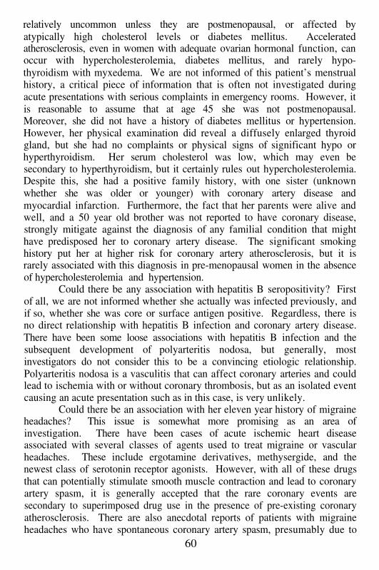
relatively uncommon unless they are postmenopausal, or affected byatypically high cholesterol levels or diabetes mellitus. Acceleratedatherosclerosis, even in women with adequate ovarian hormonal function, canoccur with hypercholesterolemia, diabetes mellitus, and rarely hypo-thyroidism with myxedema. We are not informed of this patient’s menstrualhistory, a critical piece of information that is often not investigated duringacute presentations with serious complaints in emergency rooms. However, itis reasonable to assume that at age 45 she was not postmenopausal.Moreover, she did not have a history of diabetes mellitus or hypertension.However, her physical examination did reveal a diffusely enlarged thyroidgland, but she had no complaints or physical signs of significant hypo orhyperthyroidism. Her serum cholesterol was low, which may even besecondary to hyperthyroidism, but it certainly rules out hypercholesterolemia.Despite this, she had a positive family history, with one sister (unknownwhether she was older or younger) with coronary artery disease andmyocardial infarction. Furthermore, the fact that her parents were alive andwell, and a 50 year old brother was not reported to have coronary disease,strongly mitigate against the diagnosis of any familial condition that mighthave predisposed her to coronary artery disease. The significant smokinghistory put her at higher risk for coronary artery atherosclerosis, but it israrely associated with this diagnosis in pre-menopausal women in the absenceof hypercholesterolemia and hypertension.
Could there be any association with hepatitis B seropositivity? Firstof all, we are not informed whether she actually was infected previously, andif so, whether she was core or surface antigen positive. Regardless, there isno direct relationship with hepatitis B infection and coronary artery disease.There have been some loose associations with hepatitis B infection and thesubsequent development of polyarteritis nodosa, but generally, mostinvestigators do not consider this to be a convincing etiologic relationship.Polyarteritis nodosa is a vasculitis that can affect coronary arteries and couldlead to ischemia with or without coronary thrombosis, but as an isolated eventcausing an acute presentation such as in this case, is very unlikely.
Could there be an association with her eleven year history of migraineheadaches? This issue is somewhat more promising as an area ofinvestigation. There have been cases of acute ischemic heart diseaseassociated with several classes of agents used to treat migraine or vascularheadaches. These include ergotamine derivatives, methysergide, and thenewest class of serotonin receptor agonists. However, with all of these drugsthat can potentially stimulate smooth muscle contraction and lead to coronaryartery spasm, it is generally accepted that the rare coronary events aresecondary to superimposed drug use in the presence of pre-existing coronaryatherosclerosis. There are also anecdotal reports of patients with migraineheadaches who have spontaneous coronary artery spasm, presumably due to
60
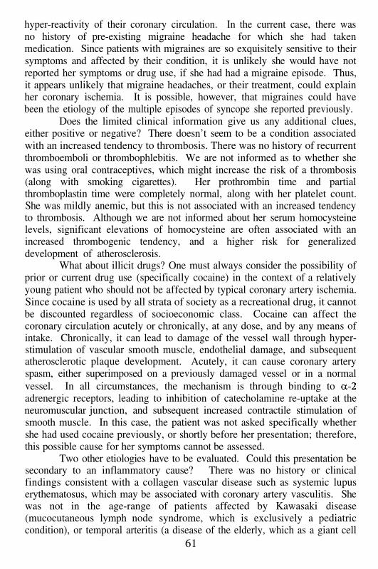
hyper-reactivity of their coronary circulation. In the current case, there wasno history of pre-existing migraine headache for which she had takenmedication. Since patients with migraines are so exquisitely sensitive to theirsymptoms and affected by their condition, it is unlikely she would have notreported her symptoms or drug use, if she had had a migraine episode. Thus,it appears unlikely that migraine headaches, or their treatment, could explainher coronary ischemia. It is possible, however, that migraines could havebeen the etiology of the multiple episodes of syncope she reported previously.
Does the limited clinical information give us any additional clues,either positive or negative? There doesn’t seem to be a condition associatedwith an increased tendency to thrombosis. There was no history of recurrentthromboemboli or thrombophlebitis. We are not informed as to whether shewas using oral contraceptives, which might increase the risk of a thrombosis(along with smoking cigarettes). Her prothrombin time and partialthromboplastin time were completely normal, along with her platelet count.She was mildly anemic, but this is not associated with an increased tendencyto thrombosis. Although we are not informed about her serum homocysteinelevels, significant elevations of homocysteine are often associated with anincreased thrombogenic tendency, and a higher risk for generalizeddevelopment of atherosclerosis.
What about illicit drugs? One must always consider the possibility ofprior or current drug use (specifically cocaine) in the context of a relativelyyoung patient who should not be affected by typical coronary artery ischemia.Since cocaine is used by all strata of society as a recreational drug, it cannotbe discounted regardless of socioeconomic class. Cocaine can affect thecoronary circulation acutely or chronically, at any dose, and by any means ofintake. Chronically, it can lead to damage of the vessel wall through hyper-stimulation of vascular smooth muscle, endothelial damage, and subsequentatherosclerotic plaque development. Acutely, it can cause coronary arteryspasm, either superimposed on a previously damaged vessel or in a normalvessel. In all circumstances, the mechanism is through binding toadrenergic receptors, leading to inhibition of catecholamine re-uptake at theneuromuscular junction, and subsequent increased contractile stimulation ofsmooth muscle. In this case, the patient was not asked specifically whethershe had used cocaine previously, or shortly before her presentation; therefore,this possible cause for her symptoms cannot be assessed.
Two other etiologies have to be evaluated. Could this presentation besecondary to an inflammatory cause? There was no history or clinicalfindings consistent with a collagen vascular disease such as systemic lupuserythematosus, which may be associated with coronary artery vasculitis. Shewas not in the age-range of patients affected by Kawasaki disease(mucocutaneous lymph node syndrome, which is exclusively a pediatriccondition), or temporal arteritis (a disease of the elderly, which as a giant cell
61
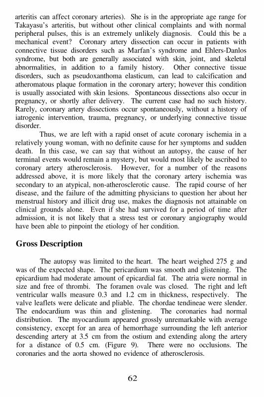
arteritis can affect coronary arteries). She is in the appropriate age range forTakayasu’s arteritis, but without other clinical complaints and with normalperipheral pulses, this is an extremely unlikely diagnosis. Could this be amechanical event? Coronary artery dissection can occur in patients withconnective tissue disorders such as Marfan’s syndrome and Ehlers-Danlossyndrome, but both are generally associated with skin, joint, and skeletalabnormalities, in addition to a family history. Other connective tissuedisorders, such as pseudoxanthoma elasticum, can lead to calcification andatheromatous plaque formation in the coronary artery; however this conditionis usually associated with skin lesions. Spontaneous dissections also occur inpregnancy, or shortly after delivery. The current case had no such history.Rarely, coronary artery dissections occur spontaneously, without a history ofiatrogenic intervention, trauma, pregnancy, or underlying connective tissuedisorder.
Thus, we are left with a rapid onset of acute coronary ischemia in arelatively young woman, with no definite cause for her symptoms and suddendeath. In this case, we can say that without an autopsy, the cause of herterminal events would remain a mystery, but would most likely be ascribed tocoronary artery atherosclerosis. However, for a number of the reasonsaddressed above, it is more likely that the coronary artery ischemia wassecondary to an atypical, non-atherosclerotic cause. The rapid course of herdisease, and the failure of the admitting physicians to question her about hermenstrual history and illicit drug use, makes the diagnosis not attainable onclinical grounds alone. Even if she had survived for a period of time afteradmission, it is not likely that a stress test or coronary angiography wouldhave been able to pinpoint the etiology of her condition.
Gross Description
The autopsy was limited to the heart. The heart weighed 275 g andwas of the expected shape. The pericardium was smooth and glistening. Theepicardium had moderate amount of epicardial fat. The atria were normal insize and free of thrombi. The foramen ovale was closed. The right and leftventricular walls measure 0.3 and 1.2 cm in thickness, respectively. Thevalve leaflets were delicate and pliable. The chordae tendineae were slender.The endocardium was thin and glistening. The coronaries had normaldistribution. The myocardium appeared grossly unremarkable with averageconsistency, except for an area of hemorrhage surrounding the left anteriordescending artery at 3.5 cm from the ostium and extending along the arteryfor a distance of 0.5 cm. (Figure 9). There were no occlusions. Thecoronaries and the aorta showed no evidence of atherosclerosis.
62
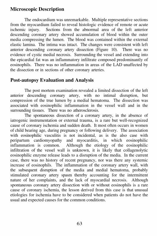
Microscopic Description
The endocardium was unremarkable. Multiple representative sectionsfrom the myocardium failed to reveal histologic evidence of remote or acuteischemic injury. Sections from the abnormal area of the left anteriordescending coronary artery showed accumulation of blood within the outermedia compressing the lumen. The blood was contained within the externalelastic lamina. The intima was intact. The changes were consistent with leftanterior descending coronary artery dissection (Figure 10). There was noevidence of cystic medial necrosis. Surrounding the vessel and extending intothe epicardial fat was an inflammatory infiltrate composed predominantly ofeosinophils. There was no inflammation in areas of the LAD unaffected bythe dissection or in sections of other coronary arteries.
Post-autopsy Evaluation and Analysis
The post mortem examination revealed a limited dissection of the leftanterior descending coronary artery, with no intimal disruption, butcompression of the true lumen by a medial hematoma. The dissection wasassociated with eosinophilic inflammation in the vessel wall and in thesurrounding tissues. There was no atherosclerosis.
The spontaneous dissection of a coronary artery, in the absence ofiatrogenic instrumentation or external trauma, is a rare but well-recognizedcause of coronary ischemia and sudden death. It most often occurs in womenof child bearing age, during pregnancy or following delivery. The associationwith eosinophilic vasculitis is not incidental, as is the also case withperipartum cardiomyopathy and myocarditis, in which eosinophilicinflammation is common. Although the etiology of the eosinophilicinfiltration of the vessel wall is unknown, it is likely that collagenolyticeosinophilic enzyme release leads to a disruption of the media. In the currentcase, there was no history of recent pregnancy, nor was there any systemicincrease of eosinophils. The inflammation of the coronary artery wall, withthe subsequent disruption of the media and medial hematoma, probablystimulated coronary artery spasm thereby accounting for the intermittentnature of her complaints, and the lack of myocardial necrosis. Althoughspontaneous coronary artery dissection with or without eosinophils is a rarecause of coronary ischemia, the lesson derived from this case is that unusualetiologies for ischemia have to be considered when patients do not have theusual and expected causes for the common conditions.
63
Suggested Readings
2.
3.
4.
5.
6.
7.
Elming H, Kober L. Spontaneous coronary artery dissection. Case reportand literature review. Scand Cardiovasc J. 1999; 33:175-9.Atay Y, Yagdi T, Turkoglu C, Altintig A, Buket S. Spontaneous dissectionof the left main coronary artery: a case report and review of the literature. JCard Surg. 1996; 11:371-5.Waller BF, Fry ET, Hermiller JB, Peters T, Slack JD. Nonatheroscleroticcauses of coronary artery narrowing--Part II. Clin Cardiol. 1996; 19:587-91.Jouriles NJ. Atypical chest pain. Emerg Med Clin North Am. 1998 Nov;16:717-40.Borczuk AC; van Hoeven KH; Factor SM. Review and hypothesis: theeosinophil and peripartum heart disease (myocarditis and coronary arterydissection)--coincidence or pathogenetic significance? Cardiovasc Res.1997; 33:527-32.Hahn IH, Hoffman RS. Cocaine use and acute myocardial infarction.Emerg Med Clin North Am. 2001; 19:493-510.Rump AF, Theisohn M, Klaus W. The pathophysiology of cocainecardiotoxicity. Forensic Sci Int. 1995; 71:103-15.
64
1.
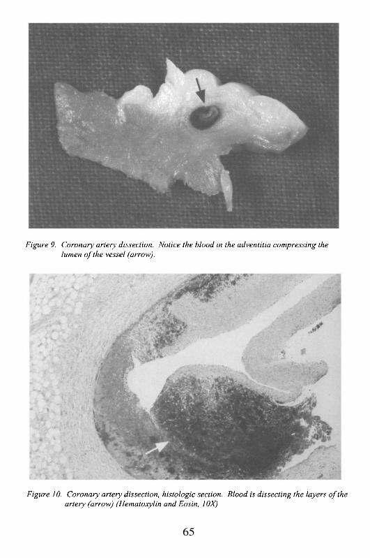
65

This Page Intentionally Left Blank
Chapter 3
DIABETES AND THE HEART
Case # 7
Clinical Summary
This is the case of a 43 year old Hispanic woman with more than a 20years history of diabetes mellitus, hypertension, congestive heart failure, andgastritis associated with Helicobacter pylori infection. She developednephropathy, neuropathy and retinopathy secondary to her diabetes, and waslegally blind and wheelchair bound. She was brought to the hospital after 24hours of localized colic-type epigastric pain, nausea and vomiting of “coffee-ground” material. Her medications before admission included NPH insulin,furosemide and atenolol.
Significant findings on her physical exam were: Vital signs BP168/94, HR 86, RR 12, T 37°C. Heart: normal S1S2 and an S3 with nomurmurs. Clear lungs. Abdomen: bowel sounds present; distended and softwith mild tenderness, no guarding and no rebound. Positive stool test forblood. Extremities: bilateral pretibial edema, left foot with healing ulcer onplantar surface, and the right first toe tip had evidence of dry gangrene.Neurologic: blind; decreased sensation on both lower extremities knee to footdistribution (stocking).
Laboratory data and other tests
Laboratory: WBC 19 (S 90%, L 5%), Hct 37, Plt 270; PT/PTT 10/29;amylase 6.1; glucose 320; BUN/creat 37/1.3; UA pH 7, protein 300, glucose1000, WBC/RBC 3/heavy, bacteria 5-10 and epithelial cells 10-20/HPF.
The patient continued to vomit, the bloody diarrhea persisted and thebowel sounds diminished. Infection or ischemic bowel disease were theclinical diagnoses considered. An abdominal CT-scan showed occlusionconsistent with thrombus in the superior mesenteric artery (SMA), which wasconfirmed by angiography. Thrombectomy of the SMA and Goretex patchangioplasty were performed. In addition, the patient was transfused. Soonafter surgery, acute renal failure developed. Hemodialysis andanticoagulation therapy were started. However, 10 days later she had anepisode of gastrointestinal bleeding and the heparin was discontinued. Thefollowing day the blood pressure suddenly dropped and the patient expired.
Gross Description
The heart weighed 500 g and showed dilatation of the right and leftatria, and hypertrophy of the left ventricle (1.6 cm in thickness). Thetricuspid valve measured 12.6 cm in length. The coronary arteries werepartially occluded (60–90%) by atherosclerosis with calcification. The aortahad moderate atherosclerosis. The lungs were congested (850 g combined).Two ulcers were noted in the body of the stomach, each measuring 1 cm indiameter. The distal portion of the jejunum and the proximal portion of theileum had serosal purple discoloration consistent with ischemic boweldisease. The luminal content was bloody. The vascular graft of the superiormesenteric artery was partially occluded by a thromboembolus. The kidneysweighed 450 g combined and had a finely granular surface. The cut surfacewas unremarkable.
Microscopic Description
The cardiovascular examination confirmed right and left ventricularhypertrophy with mild perivascular fibrosis and multifocal interstitial andreplacement fibrosis. Sections from the superior mesenteric artery and itsbranches revealed medial calcific sclerosis (Monckeberg’s) with recenthemorrhage in the portion where fragments of synthetic graft were identified.The lumen was 60% occluded by a hyaline plaque showing signs of remoteand recent hemorrhage.
Sections from the lung revealed congestion as well as numeroushemosiderin-laden macrophages, the so called ‘heart failure cells’, and signsof mild pulmonary hypertension. The small bowel had segments oftransmural ischemic and hemorrhagic infarction with focal areas of mucosalregeneration. The kidneys exhibited areas of nodular and diffuseglomerulosclerosis (Kimmelstiel-Wilson). The central nervous systemrevealed acute hippocampal hypoxic and degenerative changes. Theremaining organs were unremarkable; except for the liver which showed mildsteatosis and centrilobular congestion.
Case Analysis
This case classically depicts a number of features of diabeticcardiovascular disease. The patient had Type I, insulin-dependent diabetesmellitus (IDDM, juvenile onset) for more than 20 years, with hypertension,and complications affecting multiple organ systems. Primarily, she haddiffuse vascular disease affecting a wide range of different caliber bloodvessels, from the smallest to the largest. This is a crucial feature of
68
longstanding diabetes for 10 or more years, whether it is insulin dependent ornot (types I and II, respectively), and particularly when it is associated withsystemic hypertension. Even the autonomic and peripheral diabeticneuropathy, which has been associated with metabolic derangements thataffect neural transmission, may have an ischemic component mediated bymicrovascular damage to nerve fibers.
The classification of blood vessel calibers by size as macrovascular,small, and microvascular is an arbitrary one, and varies between investigators.Nevertheless, it is useful because pathological injury is generally limited toone class of vessel (e.g. atherosclerosis affects the macrovasculature), andtissue damage may depend on the size of the vessel affected in the process. Ingeneral, macrovascular blood vessels are the major elastic capacitance vesselsincluding the aorta and the iliac arteries (these have a media composed ofmultiple lamellae or layers of elastic tissue and smooth muscle cells), and themuscular arteries including the aortic branch vessels (e.g. coronary, carotid,femoral, popliteal, renal, celiac, superior mesenteric, etc.). The musculararteries have an intima separated from the media by an internal elasticlamella, and the media is composed of smooth muscle tissue. Themacrovasculature is primarily affected by atherosclerosis. Microvascularvessels are the small arteries below (micrometers or microns),although some investigators would consider vessels beginning atand smaller. The microvasculature includes vessels identified as arterioles,capillaries, and venules. These vessels are affected by smooth musclehypertrophy, and increased connective tissue deposition (fibrillar and non-fibrillar collagen).
In diabetes mellitus, it is common to find increased basementmembrane material surrounding capillaries, and intercalated around smoothmuscle cells in the media of small vessels. In addition, the microvasculaturemay have endothelial cell dysfunction, and hyper-reactivity (e.g. spasm) ofthe endothelial and smooth muscle cells, which may lead to tissue damage.The small vessels are arteries with calibers ranging between and
These are muscular vessels, easily identified by microscopic tissueexamination, that are generally affected by “sclerosis” not atherosclerosis.This means that they are ‘stiffer’ or hardened (literal definition of sclerosis)due to the deposition of connective tissue and smooth muscle hyperplasia.The end-result is an increase in resistance to blood flow, and with severeproliferative changes, there is decreased luminal caliber, thereby causingchronic obstruction and tissue ischemia. Essentially, diabetic vascular injuryis ubiquitous and diffuse, eventually affecting all blood vessels, leading toextensive tissue and multiorgan damage.
The patient in this case had macrovascular disease with significantatherosclerosis of coronary arteries, the aorta and its branches. It was thedisease of the superior mesenteric artery that precipitated the ischemic bowel
69
disease, requiring surgical intervention. Histologically, there was anatherosclerotic plaque with remote and recent hemorrhage, which can triggervascular instability or dynamic changes in the vessel, leading to acute luminalocclusion (thrombosis) and subsequent ischemia. There were also changes ofsclerosis, with features of Monckeberg’s medial sclerosis and calcification.This is a degenerative change that involves the internal elastic lamella withspread of calcium and scar tissue into the media. Monckeberg’s medialsclerosis is a common finding in diabetic patients, particularly in the lowerextremities where it may lead to vascular insufficiency and gangrene.
She had evidence of small vessel disease, with thickened smallarteries in the heart, and in the kidneys. The latter are associated witharterionephrosclerosis secondary to hypertension, which leads to chroniccortical ischemia, scarring, and renal dysfunction. Although her renaldysfunction was also related to microvascular disease, it is not uncommon indiabetic patients to develop ischemia due to vascular pathology affecting allsizes of vessels (e.g. renal artery, small arteries, arterioles, and capillaries). Inthe heart, small artery disease may lead to focal areas of ischemia withsubsequent scarring, which may contribute to the hypertensive and diabeticcardiomyopathy (see below). The ulceration and dry gangrene on herextremities may also have been secondary to disease of small arteries in themuscle and skin.
There was also evidence of microvascular disease in different organs.She had retinopathy leading to blindness. This is often secondary to ruptureof capillary microaneurysms with retinal and vitreous hemorrhage andproliferation of new vessels and scar tissue obliterating the retina (e.g.proliferative retinopathy). Ischemic damage may also result fromhypertensive sclerosis and narrowing of arterioles. In the kidney, in additionto hypertensive arterionephrosclerosis, she had nodular diabeticglomerulosclerosis or Kimmelstiel-Wilson disease. The latter causes markednodular thickening and ultimate obliteration of glomerular capillary loops.Only recently has it been shown to be secondary to damage to the glomerularcapillaries leading to microaneurysms with ultimate thrombosis andreorganization. These microaneurysms, which occur due to localized injuryto the capillary endothelial cells with outpouching of the lumen, are similar tothose found in the eye and in the heart.
The patient had a clinical history of congestive heart failure, and herheart was hypertrophied (500 g, normal up to 350 g), with microscopicchanges of interstitial and replacement fibrosis. The left and right ventricleswere hypertrophied. These findings were characteristic of hypertensive anddiabetic cardiomyopathy, with thick-walled chambers and microscopicscarring. Pathophysiologically, the chamber dimensions are small (e.g.decreased end-diastolic volume), and generally the contractility of theventricle is normal or super-normal. The congestive heart failure symptoms
70
result from diastolic dysfunction. The ventricles are stiff with poorcompliance; they pump adequately but fill poorly. As seen in the presentcase, the description of the ventricular chamber sizes, and the dilation of bothatria are typical of a heart with diastolic dysfunction (the atria dilate becauseof the poor compliance of the ventricles).
The pathogenesis of hypertensive and diabetic cardiomyopathy iscomplex and multiple factors contribute to its development. Though thediabetic heart is often affected by significant coronary artery atherosclerosis,large vessel ischemia may not be sufficient to account for the disease. In thispatient, there was no evidence of large areas of scarring secondary to coronaryartery disease. However, small vessel sclerosis certainly contributes tomicroscopic areas of scarring. These hearts have significant microvasculardisease, with microaneurysms present in arterioles and capillaries (Figure 11).In addition, the microvasculature is hyper-reactive (e.g. spasm) and leads tomultiple areas of reperfusion damage in the myocardium, with eventualreplacement by scar tissue (Figures 12 and 13). Finally, the effects ofhypertension also lead to hypertrophy of myocardial cells. The combinationof scar tissue and hypertrophy contributes the major component of thediastolic dysfunction, although metabolic cellular considerations (e.g. poorrelaxation of cells due to abnormal calcium flux) also play a role.
Despite the fact that the immediate cause of death in this patient couldbe directly attributed to the mesenteric artery thrombosis, and bowel ischemia,there was significant evidence for systemic disease affecting multiple organs.This is a very typical scenario in diabetic patients, because the disease affectsvessels of all calibers, leading to tissue damage throughout the body.
71
Suggested Readings
1.
2.
3.
4.
5.
6.
7.
8.
9.
10.
Drouet L. Atherothrombosis in diabetes--its evolution and management.Diabetes Obes Metab. 1999; 1 (Suppl 2):S37-47.Calles-Escandon J, Cipolla M. Diabetes and endothelial dysfunction: aclinical perspective. Endocr Rev. 2001; 22:36-52.Mazzone T. Current concepts and controversies in the pathogenesis,prevention, and treatment of the macrovascular complications of diabetes.J Lab Clin Med. 2000; 135:437-43.Donnelly R, Emslie-Smith AM, Gardner ID, Morris AD. ABC of arterialand venous disease: vascular complications of diabetes. BMJ. 2000;320:1062-6.Goldberg RB. Cardiovascular disease in diabetic patients. Med Clin NorthAm. 2000; 84:81-93.Modena MG, Barbieri A. Diabetes mellitus and cardiovascularcomplications: pathophysiological peculiarities and therapeuticimplications. Cardiologia. 1999; 44:865-77.Factor SM, Okun EM, Minase T. Capillary microaneurysms in the humandiabetic heart. .N Eng J Med 1980; 302:384-8.Factor SM, Bhan R, Minase T, Wolinsky H, Sonneblick EH.Hypertensive-diabetic cardiomyopathy in the rat: an experimental model ofhuman disease. .Am J Pathol 1981; 102:219-28.Factor SM, Sonneblick EH. Microvascular spasm as a cause ofcardiomyopathies. Cardiovasc Rev Rep 1983; 4:1177-82.Eng C, Cho S, Factor SM, Sonneblick EH, Kirk ES. Myocardialmicronecrosis produced by microsphere embolization. Role of an alphaadrenergic tonic influence of the coronary microcirculation. Circ Res1984; 54:74-82.
72
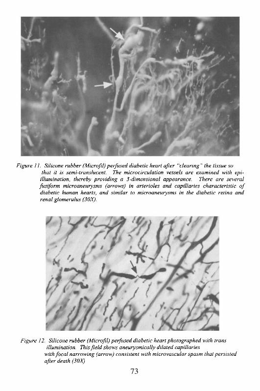
73
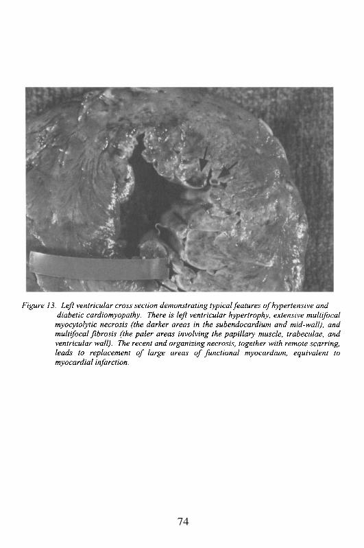
74
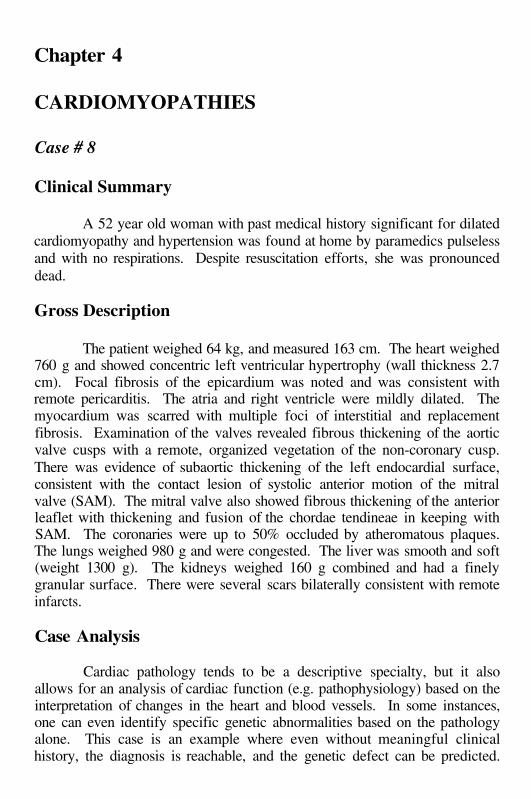
Chapter 4
CARDIOMYOPATHIES
Case # 8
Clinical Summary
A 52 year old woman with past medical history significant for dilatedcardiomyopathy and hypertension was found at home by paramedics pulselessand with no respirations. Despite resuscitation efforts, she was pronounceddead.
Gross Description
The patient weighed 64 kg, and measured 163 cm. The heart weighed760 g and showed concentric left ventricular hypertrophy (wall thickness 2.7cm). Focal fibrosis of the epicardium was noted and was consistent withremote pericarditis. The atria and right ventricle were mildly dilated. Themyocardium was scarred with multiple foci of interstitial and replacementfibrosis. Examination of the valves revealed fibrous thickening of the aorticvalve cusps with a remote, organized vegetation of the non-coronary cusp.There was evidence of subaortic thickening of the left endocardial surface,consistent with the contact lesion of systolic anterior motion of the mitralvalve (SAM). The mitral valve also showed fibrous thickening of the anteriorleaflet with thickening and fusion of the chordae tendineae in keeping withSAM. The coronaries were up to 50% occluded by atheromatous plaques.The lungs weighed 980 g and were congested. The liver was smooth and soft(weight 1300 g). The kidneys weighed 160 g combined and had a finelygranular surface. There were several scars bilaterally consistent with remoteinfarcts.
Case Analysis
Cardiac pathology tends to be a descriptive specialty, but it alsoallows for an analysis of cardiac function (e.g. pathophysiology) based on theinterpretation of changes in the heart and blood vessels. In some instances,one can even identify specific genetic abnormalities based on the pathologyalone. This case is an example where even without meaningful clinicalhistory, the diagnosis is reachable, and the genetic defect can be predicted.
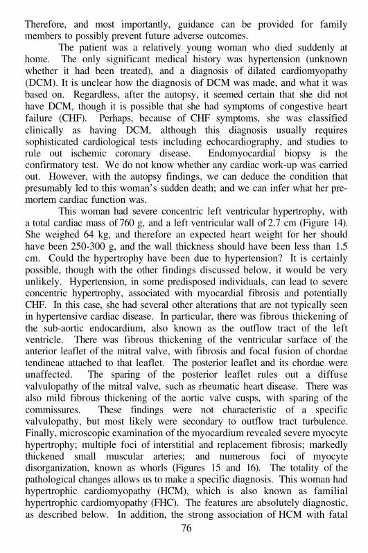
Therefore, and most importantly, guidance can be provided for familymembers to possibly prevent future adverse outcomes.
The patient was a relatively young woman who died suddenly athome. The only significant medical history was hypertension (unknownwhether it had been treated), and a diagnosis of dilated cardiomyopathy(DCM). It is unclear how the diagnosis of DCM was made, and what it wasbased on. Regardless, after the autopsy, it seemed certain that she did nothave DCM, though it is possible that she had symptoms of congestive heartfailure (CHF). Perhaps, because of CHF symptoms, she was classifiedclinically as having DCM, although this diagnosis usually requiressophisticated cardiological tests including echocardiography, and studies torule out ischemic coronary disease. Endomyocardial biopsy is theconfirmatory test. We do not know whether any cardiac work-up was carriedout. However, with the autopsy findings, we can deduce the condition thatpresumably led to this woman’s sudden death; and we can infer what her pre-mortem cardiac function was.
This woman had severe concentric left ventricular hypertrophy, witha total cardiac mass of 760 g, and a left ventricular wall of 2.7 cm (Figure 14).She weighed 64 kg, and therefore an expected heart weight for her shouldhave been 250-300 g, and the wall thickness should have been less than 1.5cm. Could the hypertrophy have been due to hypertension? It is certainlypossible, though with the other findings discussed below, it would be veryunlikely. Hypertension, in some predisposed individuals, can lead to severeconcentric hypertrophy, associated with myocardial fibrosis and potentiallyCHF. In this case, she had several other alterations that are not typically seenin hypertensive cardiac disease. In particular, there was fibrous thickening ofthe sub-aortic endocardium, also known as the outflow tract of the leftventricle. There was fibrous thickening of the ventricular surface of theanterior leaflet of the mitral valve, with fibrosis and focal fusion of chordaetendineae attached to that leaflet. The posterior leaflet and its chordae wereunaffected. The sparing of the posterior leaflet rules out a diffusevalvulopathy of the mitral valve, such as rheumatic heart disease. There wasalso mild fibrous thickening of the aortic valve cusps, with sparing of thecommissures. These findings were not characteristic of a specificvalvulopathy, but most likely were secondary to outflow tract turbulence.Finally, microscopic examination of the myocardium revealed severe myocytehypertrophy; multiple foci of interstitial and replacement fibrosis; markedlythickened small muscular arteries; and numerous foci of myocytedisorganization, known as whorls (Figures 15 and 16). The totality of thepathological changes allows us to make a specific diagnosis. This woman hadhypertrophic cardiomyopathy (HCM), which is also known as familialhypertrophic cardiomyopathy (FHC). The features are absolutely diagnostic,as described below. In addition, the strong association of HCM with fatal
76
arrhythmia, provides an explanation for this woman’s sudden death.The findings of massive left ventricular hypertrophy in association
with a subaortic endocardial plaque and anterior mitral valve leafletthickening, indicate that this woman had obstructive HCM. This means thatduring systolic contraction, there was a rapid expulsion of blood across theprominent subaortic bulging muscle, which led to two related phenomena.The anterior leaflet of the mitral valve was literally swept or sucked into theoutflow tract where it came in contact with the outflow tract endocardium.This is called systolic anterior motion of the mitral valve, or SAM. Themarkers of this process are the fibrous thickening of the outflow tract(endocardial plaque), and the fibrosis of the anterior leaflet. The pathologicaldegeneration results from the trauma of the leaflet striking the endocardiumduring each systolic contraction, but particularly during periods of increasedheart rate and more forceful contraction (e.g. during stress or exercise). Whydoes the anterior leaflet, which normally moves in a posterior direction tocoapt with the posterior leaflet occluding the atrial-ventricular orifice, movein the opposite direction to strike the septum? Although there is stillcontroversy over the explanation, most investigators believe that the rapidexpulsion of blood over the convex bulging septum leads to a pressure dropover the curved surface (Venturi effect, similar to the decreased pressure overthe curved upper surface of an airplane wing). The movement of the leaflet inthe anterior direction, together with the hypertrophied muscle that narrows theoutflow tract, leads to late systolic obstruction. The obstruction, together withturbulence causes a late systolic ejection murmur (the obstruction occurs latein systole because sufficient acceleration of the blood has to occur before theleaflet is sucked anteriorly).
The obstruction and the murmur can be accentuated by tachycardia,increased contractility, and decreased end-diastolic volume. Increasing thevelocity of the flow leads to a greater pressure fall across the septum, anddecreasing the chamber dimensions shortens the distance the valve leafletmust traverse to strike the septum. End-diastolic volume can fall if there is adecrease in circulating blood volume, which may result from the inappropriateuse of diuretics in a patient with congestive symptoms who is not identified ashaving HCM. End-diastolic volume will also decrease transiently during theValsalva maneuver (increasing intra-thoracic pressure against a closedglottis), which is a good way to accentuate the murmur when auscultating theheart in a patient suspected of having HCM. In contrast, a patient withstructural aortic stenosis may have a diminution of murmur intensity with afall in end-diastolic volume. Any process that increases end-diastolic volume,thereby increasing the left ventricular dimensions and enhancing theseparation of the anterior mitral valve leaflet and the septal endocardium, willlead to a decrease of the subaortic obstruction and the late systolic murmur.Similarly, decreasing contractility or slowing the heart rate (as with calcium
77
channel blocking agents or beta adrenergic blockers) will decrease theobstruction and the murmur. The concomitant effect of the anterior mitralvalve leaflet being swept into the outflow tract, is that it does not coapt withthe posterior leaflet. Thus, individuals with SAM, also have mitralregurgitation, which may further compromise cardiac function and mayenhance pulmonary congestion.
Usually, individuals with HCM who have CHF, do not havesignificant symptomatology due to the subaortic obstruction or the mitralvalve regurgitation. Rather, they generally have severe diastolic dysfunctionas a result of the marked ventricular hypertrophy, in association withextensive scarring in the myocardium. These hearts are also affected by anincreased interstitial connective tissue matrix, although the pathogenesis ofthis process is not understood. The result is a markedly stiffened ventricle,with small ventricular volumes. The ventricle fills poorly, and there is anelevation of left atrial pressure, which is increased even further if there issignificant mitral valve regurgitation. Thus, congestive symptoms aresecondary to diastolic dysfunction. In fact, systolic function is relatively wellmaintained and even super-normal, in most patients with HCM. Thepathophysiologic problem is one of inadequate ventricular filling. It is likelythat the diagnosis of DCM in this case was based on congestive symptomsthat she had secondary to diastolic dysfunction. Had an echocardiogram beenperformed, it would have been obvious immediately that she did not haveDCM, but she had HCM.
Microscopically, patients with HCM have extensive areas ofdisorganized muscle fibers called whorls. This pattern of disorganization isnot pathognomonic of HCM, as it can be seen in normal hearts and in areasaround myocardial scars; but its extent in HCM is greater than in any othercondition. Ordinarily, myocardial fibers are aligned in parallel within the 3layers of the ventricular wall (each of the layers is oriented obliquely to theadjacent layer, but within the layers, the myocytes line-up in parallel).However, in myocyte whorls, the fibers are arranged at random, and aretherefore unlikely to be capable of generating meaningful forcefulcontraction. The etiology for this fiber disposition is unknown, but onepossible explanation is that it develops as a result of localized abnormalcontraction patterns in the myocardium, secondary to the basic defect in thisdisease (see below).
The other microscopic finding typical of HCM, is that these patientsgenerally have extensive myocardial scarring with areas of interstitial fibrosis,and infarct-like replacement fibrosis. Some of the scarring may in fact berelated to myocardial ischemia, as the increased stiffness and thickness of theventricular wall, and the enhanced contractility, lead to increased wall stress,which in turn may diminish myocardial perfusion. In addition, theintramyocardial muscular arteries are abnormally thickened and sclerotic,
78
thereby causing a structural limitation to tissue perfusion. Patients with HCMmay have complaints of atypical chest pain, and may haveelectrocardiographic abnormalities consistent with ischemia. However,although ischemic injury may be significant, and may even provokearrhythmias, the most significant complication of this condition is suddenfatal arrhythmia. As in this patient, sudden death can be entirely unexpected,and may be the very first manifestation of the disease. Despite the relativerarity of HCM in the general population, it attains great notoriety because of itstrong association with sudden death, often in otherwise healthy youngpeople. Sudden death is also very predictable in individuals from familieswith known genetic mutations that have been shown to be particularly lethal.
Hypertrophic cardiomyopathy is an autosomal dominant hereditarydisease. It can occur sporadically, presumably due to a unique mutation, butit occurs in families, often with variable penetrance. Prior to the last decade,HCM was thought to be due to a single mutation of the gene that codes formyosin heavy chain (MHC, a structural component of myosin, which is partof the contractile apparatus of the heart cell) in cardiac myocytes. A numberof single amino acid mutations of MHC have now been identified innumerous families. Of interest, a particular mutation leads to similarphenotypic expression, including enhanced tendency to sudden death (therebyallowing for prediction and potential treatment in affected family members).The disease, however, has become far more complex than originally thought.Affected patients or families that did not have mutation of the MHC gene onchromosome 14 were identified. However, linkages to mutations on otherchromosomes were found. This suggested that HCM is not a disease, but it isa syndrome.
In fact, 7 different forms of the condition have been found, often withphenotypic variability, and not all associated with marked ventricularhypertrophy, outflow tract obstruction, or other related findings. All 7 defectsinvolve the sarcomere, thereby leading to the concept that HCM is asarcomeric cardiomyopathy. The two most significant, and apparently mostfrequent in the population, are those that affect MHC, and troponin T. Thelatter is interesting because it does not lead to significant ventricularhypertrophy and obstruction, but it is associated with a very high prevalenceof sudden death. Six of the 7 different abnormal proteins that cause HCMhave been identified, with their specific mutant gene. The seventh that leadsto HCM in association with a conduction defect known as the Wolf-Parkinson-White (WPW) syndrome is most likely the one that affected thepatient in this case. This form of HCM has been linked to a gene onchromosome 7. Recently, an abnormal protein kinase encoded by this genewas identified.
At the completion of this autopsy, the family was contacted due to thefact that HCM was the final diagnosis, and because of the concern that other
79
family members unknowingly might be affected. The patient had no siblings,and no other close relatives who had either been diagnosed with HCM, or haddied unexpectedly and suddenly as a result of heart disease. However, she didhave a single 29 year old daughter. Discussion with the daughter led to theinformation that she had been diagnosed with WPW as a child. AlthoughWPW can occur sporadically, the association of HCM and WPW in firstdegree relatives did not appear to be a coincidence. Based on the likelihoodof both mother and daughter having HCM, it was strongly suggested to thedaughter that she have a cardiac work-up, including echocardiography. If thediagnosis was confirmed, then appropriate monitoring and treatment would beinstituted. Pharmacologic treatment has been shown to be beneficial insymptomatic patients, although it may not prevent sudden arrhythmic death.To prevent the latter complication, an automated implantable defibrillator hasbeen used with some success.
As a final note, WPW is an arrhythmic condition that can occurindependently of HCM. It represents accelerated transmission of electricalactivation from the atrium to the ventricle through an abnormal bypass(accessory) pathway. Thus, the ventricle is activated faster than normal,which may lead to disruption of the atrio-ventricular coordination.Electrophysiologically, the result is a shortened PR interval, and a slurring ofthe upstroke of the QRS complex leading to the formation of a delta wave.This delta wave is thought to represent the combined rapid transmissionthrough the accessory bundle, and the normal transmission from the sino-atrial node through the atrial wall into the atrio-ventricular node. Drugtreatment of WPW is available, along with surgical or interventionalcardiologic ablation of the accessory bundle, if it can be identified. Suddendeath may be a rare but recognized complication of WPW. It is not knownwhether sudden death in patients with HCM and WPW results from the WPWalone.
80
Suggested Readings
1.
2.
3.
4.
5.
6.7.
8.
9.
Wigle ED. Novel insights into the clinical manifestations and treatment ofhypertrophic cardiomyopathy. Curr Opin Cardiol 1995; 10:299-305.Wigle ED, Rakowski H, Kimball BP, Williams WG. Hypertrophiccardiomyopathy: clinical spectrum and treatment. Circulation 1995;92:1680-92.Posma JL, van der Wall EE, Blanksma PK, van der Wall E; Lie KI. Newdiagnostic options in hypertrophic cardiomyopathy. Am Heart J. 1996;132:1031-41.Lakkis N. New treatment methods for patients with hypertrophicobstructive cardiomyopathy. Curr Opin Cardiol. 2000; 15:172-7.Basso C, Corrado D, Thiene G. Cardiovascular causes of sudden death inyoung individuals including athletes. Cardiol Rev. 1999; 7:127-35.Maron BJ. Hypertrophic cardiomyopathy. Lancet. 1997; 350:127-33.Davies MJ, McKenna WJ. Hypertrophic cardiomyopathy--pathology andpathogenesis. Histopathology. 1995; 26:493-500.Tardiff JC, Factor SM, Tompkins BD, Hewett TE, Palmer BM, Moore RL,Schwartz S, Robbins J, Leinwand LA. A truncated cardiac troponin Tmolecule in transgenic mice suggests multiple cellular mechanisms forfamilial hypertrophic cardiomyopathy. J Clin Invest 1998; 101:2800-11.Gollob MH, Green MS, Tang ASL, Gollob T, Karibe A, Hassan A, AhmadF, Lozado R, Shah G, Fananapazir L, Bachinski LL, Roberts R.Identification of a gene responsible for familial Wolff-Parkinson-Whitesyndrome. N Engl J Med 2001; 344:1823-31.
81
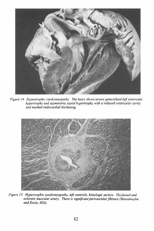
82

83
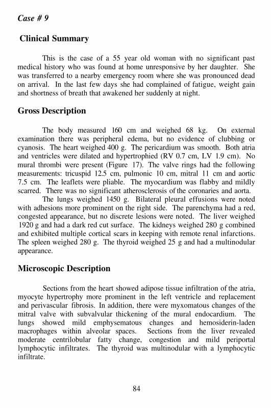
Case # 9
Clinical Summary
This is the case of a 55 year old woman with no significant pastmedical history who was found at home unresponsive by her daughter. Shewas transferred to a nearby emergency room where she was pronounced deadon arrival. In the last few days she had complained of fatigue, weight gainand shortness of breath that awakened her suddenly at night.
Gross Description
The body measured 160 cm and weighed 68 kg. On externalexamination there was peripheral edema, but no evidence of clubbing orcyanosis. The heart weighed 400 g. The pericardium was smooth. Both atriaand ventricles were dilated and hypertrophied (RV 0.7 cm, LV 1.9 cm). Nomural thrombi were present (Figure 17). The valve rings had the followingmeasurements: tricuspid 12.5 cm, pulmonic 10 cm, mitral 11 cm and aortic7.5 cm. The leaflets were pliable. The myocardium was flabby and mildlyscarred. There was no significant atherosclerosis of the coronaries and aorta.
The lungs weighed 1450 g. Bilateral pleural effusions were notedwith adhesions more prominent on the right side. The parenchyma had a red,congested appearance, but no discrete lesions were noted. The liver weighed1920 g and had a dark red cut surface. The kidneys weighed 280 g combinedand exhibited multiple cortical scars in keeping with remote renal infarctions.The spleen weighed 280 g. The thyroid weighed 25 g and had a multinodularappearance.
Microscopic Description
Sections from the heart showed adipose tissue infiltration of the atria,myocyte hypertrophy more prominent in the left ventricle and replacementand perivascular fibrosis. In addition, there were myxomatous changes of themitral valve with subvalvular thickening of the mural endocardium. Thelungs showed mild emphysematous changes and hemosiderin-ladenmacrophages within alveolar spaces. Sections from the liver revealedmoderate centrilobular fatty change, congestion and mild periportallymphocytic infiltrates. The thyroid was multinodular with a lymphocyticinfiltrate.
84
Case Analysis
As previously discussed, sudden cardiac death is virtually alwaysarrhythmic and has several predisposing factors. The post-mortem examexcluded all of them (occlusive coronary disease, myocardial infarction,myocardial fibrosis) except for one, cardiomyopathy. This is defined as adisease of the myocardium associated with cardiac dysfunction. Specifccardiomyopathies refer to heart muscle diseases related to particular cardiac(e.g. ischemic, valvular, or hypertensive) or systemic disorders.
The finding of cardiomegaly (400 g vs. normal 270 g), and fourchamber dilatation associated with mild ventricular hypertrophy, ischaracteristic of one type of cardiomyopathy known as dilatedcardiomyopathy (Table 1, 1995 WHO/IFSC classification ofcardiomyopathies). Dilated cardiomyopathy (DCM) has numerous anddifferent etiologies that include: ischemia, myocarditis, sarcoidosis,connective tissue disorders, alcohol, drugs, metabolic and neuromusculardiseases. In a significant number of patients there is no obvious cause, hencethe term “idiopathic dilated cardiomyopathy”. DCM is characterized by thegradual development of cardiac failure associated with heart enlargement.Patients with this condition may be asymptomatic for years due to thecompensatory mechanisms of the heart. This patient, for example, had nosignificant past medical history until she presented with fatigue, weight gaindue to accumulation of fluid in third space and paroxysmal nocturnal dyspnea,which are all symptoms of heart failure. The autopsy findings of peripheraledema, pleural effusions and chronic passive congestion of lungs, liver andspleen were also in keeping with cardiac decompensation.
The progressive myocardial thinning and dilatation in DCM decreasesthe contractile function of the ventricles and therefore the cardiac output(CO). Compensation is achieved in several ways. One of the most importantmechanisms is the Frank-Starling law of the heart, in which CO is maintainedby increasing the stroke force. In addition, reduced CO activates theadrenergic and renin-angiotensin system. In low CO situations, the stimulatedadrenergic system leads to increased heart rate and contractility; and theincreased production of renin and angiotensin results in systemicvasoconstriction. Unfortunately, the outcome is enhanced wall stress andoxygen consumption in an already impaired myocardium. Thus, furthermyocyte damage ensues leading to failure. It is unknown what causes themyocardial dysfunction in the first place. Possible explanations includeabnormal expression of genes responsible for the contractile function of themyocyte, loss of myocytes due to apoptosis (individual pre-programmed celldeath), and abnormal cellular and chamber remodeling. In the latter, there isdegradation of the connective tissue matrix and slippage of the cell layers
85
with the myocytes aligning in series rather than in their normal oblique andoverlapping fashion. The heart assumes a less elliptical shape and becomesmore round, which augments the wall tension and makes the myocyte lesseffective in managing increases in oxygen demands. Enlargement of thechambers also results in dilatation of the valve rings leading to insufficiency.Both mitral and tricuspid valves may become incompetent. This patient hadan increased mitral valve diameter of 11 cm and a tricuspid valve diameter of12.5 cm. In addition, she had thickening of the mural endocardium and amitral valve jet lesion that resulted from the turbulence of the regurgitantblood. Although we do not know if she was ever diagnosed with a murmur,we can be certain, based on the autopsy findings that she had a holosystolicmurmur. In addition, she must have had a third heart sound (S3) of leftventricular failure. The ECG would have shown cardiomegaly, conductionabnormalities, repolarization defects, and perhaps Q waves due to theinterstitial and replacement fibrosis noted in her myocardium.
Despite the dilatation, she had significant hypertrophy of both sides,likely the result of the sustained increase in systemic pressure from the renin-angiotensin compensatory mechanism. We are unaware of a prior history ofhypertension.
A common complication of patients with DCM is intracavitarythrombosis due to blood stasis. These thrombi may reach the systemic andpulmonary circulations depending on the cavity where they form, and reachthe brain, kidneys and lungs. Although we did not find mural thrombi in thiscase, there is evidence that she was embolizing since the renal corticesshowed scarring of prior infarctions.
The main question in this case is: What is the etiology of her DCM?Is it idiopathic or does it have a specific cause? As mentioned before, thereare numerous causes of dilated cardiomyopathy. We can definitely excludeischemia and myocarditis because she did not have significant coronary arterydisease, inflammatory infiltration or myocardial necrosis. Other causes suchas drugs, AIDS and alcohol are unlikely. Familial predisposition has beendocumented in some cases (the genetic abnormality is generally unknown),but she lacked a positive family history for heart disease. An interestinghypothesis, however, may be obtained from the autopsy findings. She had anodular thyroid with a lymphocytic infiltrate. Lymphocytic thyroiditis isconsidered an autoimmune disease that can cause hypothyroidism, one of theendocrinologic disorders that is known to cause DCM. She had some of thefeatures of Hashimoto’s disease with gross lobulation of the gland, scantcolloid, Hurthle cell changes and lymphoplasmocytic infiltrate. Thedifferential diagnosis is “nonspecific” thyroiditis, an entity frequentlydiagnosed on post-mortem examination that occurs in older patients withoutsymptoms and low levels of antithyroid antibodies. This condition is moreprevalent in females and has increased in the United States population after
86
the addition of iodine in the water. Thyroid tests were never performed in thispatient, but it has been described that clinical manifestations ofhypothyroidism may be subtle and insidious in elderly patients.Microscopically, changes associated with hypothyroidism are basophilicdegeneration of the myofibers, and accumulation of interstitialmucopolysaccharide-rich fluid (myxedema). These changes occur inadvanced hypothyroidism, and were not seen in this case. It is unlikely thatshe had hypothyroidism, and if she did it was not severe enough to cause acardiomyopathy. Patients with hypothyroidism also have hypercholesterol-emia and hypertriglyceridemia. It would be unusual to have DCM secondaryto hypothyroidism in the absence of coronary atherosclerosis. At the otherend of the spectrum, apathic hyperthyroidism occurs in elderly patients, isusually silent and is frequently associated with atrial arrhythmias.Furthermore, these patients are at a high risk for thromboembolic eventsassociated with atrial fibrillation.
In summary, this patient died of an arrhythmic event secondary to adilated cardiomyopathy of unknown etiology. Treatment of her conditionwould have included diuretics for her CHF, angiotensin-converting enzymeinhibitors and arteriolar as well as venous vasodilators. Cardiac transplant is aconsideration in patients with severe DCM.
87
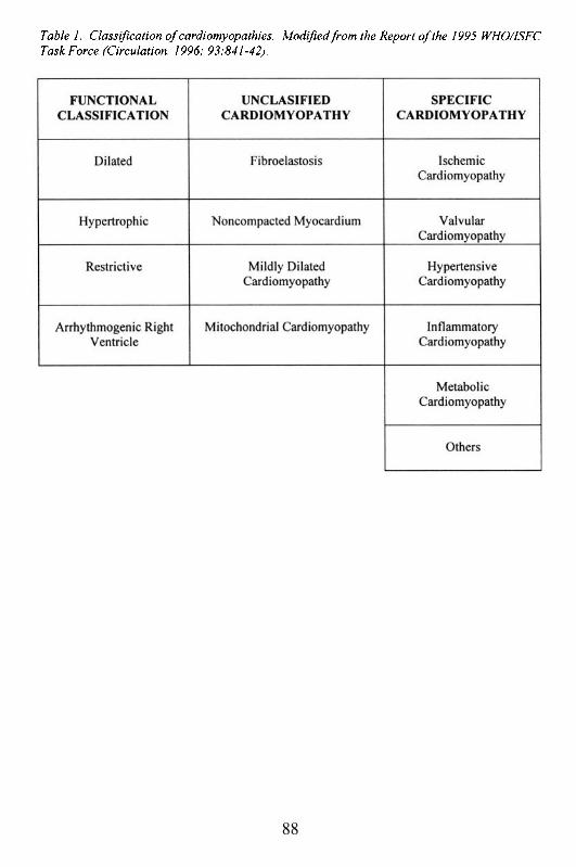
88
Suggested Readings
1.
2.
3.
4.
Pathak SK, Kukreja RC, Hess M. Molecular Pathology of DilatedCardiomyopathies. Curr Probl Cardiol. 1996; 21:99-144.Thiene G, Angelini A, Basso C, Calabrese F, Valente M. The newdefinition and classification of cardiomyopathies. Adv Clin Path. 2000;4:53-7.Richardson P, McKenna W, Bristow M; Maisch B, Mautner B; O’ConnellJ, et al. Report of the 1995 WHO/ISFC Task Force on the definition andclassification of cardiomyopathies. Circulation. 1996; 93:841-42.Bristow MR, Bohlmeyer TJ, Gilbert EM. “Dilated Cardiomyopathy”. InHurt’s The Heart. Ninth Edition. Alexander WR, Schlant RC, and FusterV eds. McGraw-Hill Co, Inc., 1998.
89
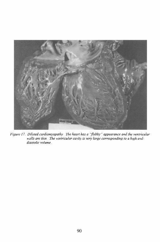
90
Case # 10
Clinical Summary
This is the case of a 30 year old woman who was 7 months post-partum with no significant past medical history. The present illness began 6weeks after delivery, when tachycardia, palpitations and exophtalmos led tothe diagnosis of Grave’s disease. Treatment with propylthiouracil (PTU) andpropranolol was prescribed. Her palpitations decreased initially, but after afew months her symptoms worsened. She suffered 3 syncopal episodeswithin the 3 months prior to admission, each lasting a few seconds. Her liverenzymes increased mildly, and the PTU was discontinued. She developedorthopnea, fever of 39°C, weight loss, diarrhea and decreased urine output.
The physical exam on admission revealed a young woman inmoderate respiratory distress. Vital signs: P 210, RR 32, BP 90/palpation,and T 37.5°C. HEENT: exophtalmos, hyperemic throat without exudate.Neck: jugular venous distension up to mandible and diffuse thyromegalywithout stridor, carotid bruit or lymphadenopathy. Chest: right lower thirddull to percussion with decreased breath sounds, but no egophony; left basewith rales. Heart: S1S2, no murmurs. Abdomen: distended with normalbowel sounds, ascites present, and liver span approximately 16 cm.Extremities: 3+ pitting edema up to distal thigh, bilaterally. Sacral edema.Neuro: normal mental status; grossly non-focal; hand tremor bilaterally.
Laboratory data and other tests
Laboratory: WBC 11.5 (S 67%, L 22%); Hb/Hct 10/31, Plt 236;bilirubin T/D 4.9/3.7, SGOT/SGPT 41/27 U/L, glucose 110, cholesterol 63,PT/PTT 19/27; free thyroxine 4.7 (normal 0.7-2), T3 RIA 476 (normal 90-180), TSH 0.03 (normal 4-4.6). CXR: enlarged cardiac silhouette, large rightpleural effusion and/or infiltrate. ECG: SVT at 264/min, regular (atrialflutter).
In the ER, her heart rate was slowed to 140/min by test doses ofadenosine followed by diltiazem in 3 divided doses. She was admitted to theCCU with a heart rate of 136 and a BP 102/65, fully oriented and receivingoxygen by nasal cannula. She was given furosemide IV and propranolol P.O.One and a half-hours after admission, the patient complained of severeshortness of breath and became restless and agitated, and shortly thereafter,hypotensive and unresponsive. CPR and fluids were given and she wasintubated. Cardioversion and administration of glucagon elevated her bloodpressure to 100 mmHg, but still she remained unresponsive. Vasopressor
91
therapy with norepinephrine and dopamine was started. Her serum potassiumwas 7.1 mEq/L, which was treated with CaC12, NAHCO3, insulin and glucoseIV. An echocardiogram showed global hypokinesis with all chambersmarkedly enlarged and dilated, moderate to severe mitral and tricuspidregurgitation, large pleural effusion, but no pericardial effusion. The Swan-Ganz catheter showed the following pressures RA 10 mmHg, PCWP 20mmHg and PA 38/20. Supraventricular tachycardia (SVT) and hypotensionrecurred several times and resolved every time after cardioversion andadjustment of pressor therapy. Follow-up neurologic examination revealedabsence of brainstem reflexes. A CT scan of the head was consistent withanoxic encephalopathy. The WBC rose to 19,700 with 89% granulocytes.Cultures of blood, sputum, CSF, pleural fluid and urine were all negative.Urine output was scant. Apnea test showed absence of spontaneousrespiration. Nine days after admission, bradycardia developed, her bloodpressure fell and her heart stopped. Resuscitation efforts were unsuccessful.
Gross Description
The body was that of a well-nourished, well-developed black womanappearing the stated age of 30 years. The heart weighed 420 g and was ofexpected shape. The pericardium was smooth and glistening. The epicardiumwas shiny with moderate amount of subepicardial fat. The atria and ventricleswere dilated, more significantly in the right side. The right and leftventricular walls measured 0.3 and 1.2 cm in thickness, respectively. Theright atrium contained 2 organizing mural thrombi measuring 0.5 to 1.0 cm indiameter. The foramen ovale was probe-patent. The valve ringsmeasurements were as follows: tricuspid valve 12 cm, pulmonic valve 9 cm,mitral valve 10 cm, and aortic valve 7 cm. The valve leaflets were delicateand pliable. The chordae tendineae were slender. The endocardium was thinand glistening. The myocardium was brown-red and mildly softened. Thecoronary arteries were of normal caliber and distribution, with no sclerosis orocclusion. The superior and inferior caval veins and major tributaries weregrossly unremarkable. The aorta showed no atherosclerosis. The lungsweighed 1400 g and were essentially similar in appearance with patchy aswell as confluent areas of increased firmness. The tracheobronchial tree, aswell as the pulmonary vessels, were unremarkable. The liver weighed 1400 gwith a smooth surface. The cut surface revealed centrilobular congestionwithout necrosis. The thyroid weighed 70 g and was of average shape. Onsection, the parenchyma was uniformly tan-brown with no apparent colloidcysts. The right ovary contained a 4 cm tumor, which on section showedmany cysts containing gelatinous yellow-brown material.
92
Microscopic Description
Sections from the heart revealed myocyte hypertrophy, mildinterstitial and replacement fibrosis, and organizing thrombi in the rightatrium (Figures 18 and 19). The lungs showed bilateral acutebronchopneumonia with areas of intra-alveolar hemorrhage.
Sections from the thyroid showed diffuse thyroid hyperplasia withcolloid depletion consistent with Grave’s disease. The tumor found in theright ovary consisted of thyroid tissue consistent with the diagnosis of strumaovarii. No colloid depletion was noted in the tumor.
Case Analysis
This 30 year old woman had a sudden arrhythmic cardiopulmonaryarrest occurring within 7 months post-partum, and in a setting stronglysuggestive of hyperthyroid disease. The question to be addressed is whetherher presentation and death were the result of peri-partum cardiomyopathy, orwhether she had an additional element of hyperthyroid cardiomyopathy.
Peri-partum cardiomyopathy (PPC) is a syndrome that, by definition,occurs between the last trimester of pregnancy and the first 6 monthsfollowing delivery. However, the timing of the disease is somewhat variable,since the acute symptomatic presentation may extend beyond the 6 monthspost-partum. In other words, there may be damage to the myocardium in theimmediate peri-partum period, but ventricular dysfunction may not ensue formany months later. The reason that PPC represents a syndrome rather than aspecific disease, is because there may be several different pathogenicmechanisms that lead to cardiac damage. In approximately one half of cases,endomyocardial biopsies taken during the peri-partum period reveal thepresence of active myocarditis, often characterized by an abundantmyocardial eosinophilic infiltrate (Figure 20). This suggests that themyocarditis is allergic in etiology. It remains unknown if the antigenicstimulation derives from the fetus, or represents cross-reactivity againstmyocardial tissue from the gestational uterine tissue. There are no animalmodels of the disease, and our knowledge is based on limited human tissuefrom biopsy material or autopsy. In the rest of the cases without myocarditis,there are features of cardiomyopathy with myocellular hypertrophy andfibrosis. Further studies are needed to determine whether these degenerativechanges develop during pregnancy, or are pre-existing and only becomemanifest because of the hemodynamic and hormonal stresses of pregnancy.Logically, one would assume that if there was cardiomyopathic damage priorto the pregnancy, then the disease would become manifest during pregnancyat the maximal hemodynamic stress period, when the intravascular volume
93
and cardiac output are the greatest (second trimester). Yet, as noted above,the disease is defined by its presentation during the third trimester, suggestingthat myocardial damage before the onset of pregnancy is an insufficientexplanation for most cases.
In our patient, although she presented to the hospital 7 months afterdelivery, her symptoms began 6 weeks post-partum. Accordingly, her diseasecould be secondary to PPC. The question is whether PPC can present withtachycardia and palpitations? The answer is that these are usually notcommon manifestations; however, tachyarrhythmias and paroxysmal atrialfibrillation may be secondary to cardiomyopathy. Therefore, PPC cannot beruled out on clinical grounds alone as a cause of this young woman’scomplaints and death.
There is an association of thyroid disease in the peri-partum period(although generally with hypothyroidism). In this case, it would difficult tooverlook the likelihood of hyperthyroidism. Her complaints were typical withtachycardia, palpitations, and exophthalmos. Weight loss might not be ahelpful sign post-partum, as she would be expected to lose weight; however,she continued to lose weight 4 months after delivery. She also had fever anddiarrhea, which may be secondary to hyperthyroidism. The episodes ofsyncope are suggestive of sudden ventricular arrhythmias. Her shortness ofbreath and complaints of orthopnea were symptoms of CHF. It is noteworthythat when she presented to the hospital she had a pulse of 260 andhypotension (BP 90/palpation). On physical examination, she had jugularvenous distension, fluid overload, and signs of pulmonary congestion withrales. She had features of both left heart failure (hypotension and pulmonarycongestion), and right heart failure (jugular venous distension, pleuraleffusion, ascites, liver congestion, and peripheral edema). Her laboratorystudies were confirmatory of severe hyperthyroidism, with elevated freethyroxine and T3, and markedly reduced thyroid stimulating hormone. Herelectrocardiogram revealed significant SVT at 220/min, and atrial flutter.Despite slowing of her heart rate to 140 with adenosine and calcium channelblocker (diltiazem), she had a sudden cardiac arrest. Subsequently, shedeveloped several more episodes of SVT and hypotension, and despiteresuscitation and slower heart rate, she died secondary to anoxicencephalopathy due to her initial cardiorespiratory arrest.
The post-mortem findings were consistent with hyperthyroid heartdisease with bi-ventricular failure. The heart was enlarged (420 g, normal300-350 g), both ventricular walls were thinned, and all 4 chambers weredilated. She had mural thrombi in the right atrium, most likely secondary toatrial flutter and stasis in the chamber, but also consistent withcardiomyopathy. Microscopically, the myocytes were hypertrophied, andthere was evidence of interstitial and replacement fibrosis. These are non-specific features of cardiomyopathy; but in the absence of inflammation and
94
with evidence of a hypertrophied and dilated heart, the findings are muchmore consistent with hyperthyroidism than peri-partum cardiomyopathy.
Hyperthyroidism causes a cardiomyopathy through several relatedmechanisms. It leads to the so-called high output heart failure, similar to theeffects of an arteriovenous malformation or severe anemia, where the cardiacoutput is markedly increased. However, in hyperthyroidism there ishypermetabolism with increased circulating blood volume, low peripheralvascular resistance, and tachycardia. Another characteristic feature is thedevelopment of a hypermetabolic state, which has direct and indirect thyroidhormone effects on the myocardium. Furthermore, thyroid hormonestimulates myocyte hypertrophy through its actions as a growth hormone onmyosin synthesis; and hyperthyroidism can also lead to hypertension, whichcan cause secondary hypertrophy of the heart. Moreover, catecholaminelevels are high in hyperthyroidism, which can further stimulate hypertrophyand tachycardia.
The pathogenesis of the hypertrophied and dilated heart (signs ofcardiac remodeling) has only become clear recently with experimental studieson prolonged tachycardia. Thyroid hormone stimulates rapid ventricular andatrial rhythm, leading to supraventricular tachycardia (e.g. atrial flutter orfibrillation). Animals with electrical pacing-induced tachycardia develop ahigh-output dilated cardiomyopathy over several weeks, even with normalthyroid hormone levels. The tachycardia causes global hypertrophy, chamberdilatation, mural thinning, and microscopic features of myocyte hypertrophyand focal fibrosis. There is evidence of remodeling of the ventricularchamber mediated through damage to the interstitial connective tissue matrix,(via activated metalloproteinases or collagenolytic enzymes) that allows for“slippage” of myocytes and mural myocardial layers. The result is a heartwith morphological features of cardiomyopathy, and an animal with clinicalsigns and symptoms of CHF.
Derived from all the above observations, hyperthyroid heart diseaseresults from a combination of direct cellular effects on the heart tissue by thethyroid hormone, hemodynamic effects of the systemic hypermetabolic state,and secondary effects of tachycardia that lead to cardiac remodeling anddamage. Together with the known association of cardiomyopathy and suddenmalignant ventricular arrhythmias (e.g. ventricular fibrillation), particularly inthe setting of increased catecholamine levels, it is not unexpected that apatient such as this one would experience a sudden cardiac arrest leading toanoxic encephalopathy. What is unusual in this modern medical era, is thatanyone would experience such severe hyperthyroidism. However, non-adherence with her medical treatment may explain the rarity of thisunfortunate complication of hyperthyroid disease.
95
Suggested Readings
1.
2.
3.
4.
5.
6.
Brown CS, Bertolet BD. Peripartum cardiomyopathy: a comprehensivereview. Am J Obstet Gynecol. 1998; 178:409-14.Toft AD, Boon NA. Thyroid disease and the heart. Heart. 2000; 84:455-60.Fadel BM, Ellahham S, Ringel MD, Lindsay J Jr, Wartofsky L, BurmanKD. Hyperthyroid heart disease. Clin Cardiol. 2000; 23:402-8.Klein I, Ojamaa K. Thyroid hormone and the cardiovascular system. NEngl J Med. 2001; 344:501-9.Gomberg-Maitland M, Frishman WH. Thyroid hormone andcardiovascular disease. Am Heart J. 1998; 135(2 Pt 1): 187-96.Borczuk AC, van Hoeven KH, Factor SM. Review and hypothesis: theeosinophil and peripartum heart disease (myocarditis and coronary arterydissection) coincidence or pathogenetic significance? Cardiovasc Res.1997; 33:527-32.
96
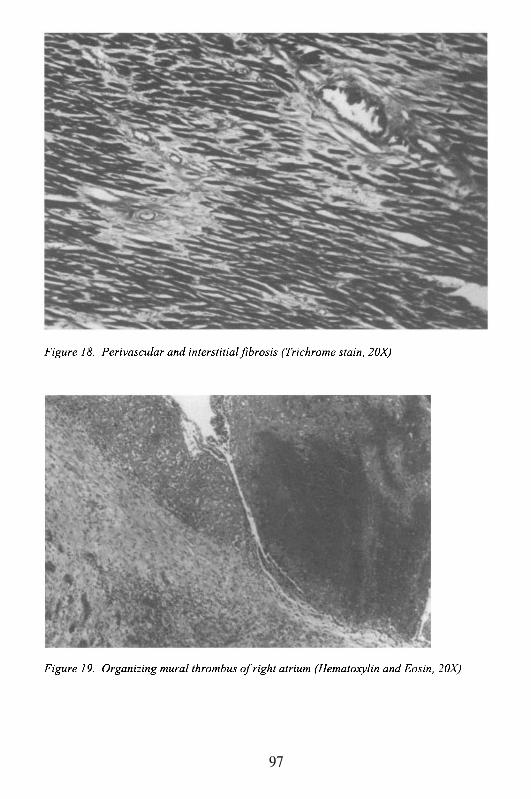
97
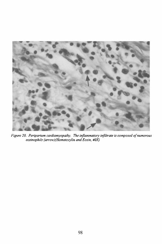
98
Case # 11
Clinical Summary
This was the first hospital admission for this 56 year old man, formeralcohol abuser, non-diabetic, with history of hypertension; with one weekhistory of shortness of breath, fever, shaking chills, productive cough andpleuritic chest pain. The patient had been a heavy drinker of 8–10 cans ofbeer a day for many years. He also had been diagnosed with gout, and had atwo year history of congestive heart failure treated with digoxin andfurosemide. The physical exam on admission was remarkable for atemperature of 40°C, scleral icterus and dull breath sounds in both lung bases.In addition, auscultation of the heart revealed a S3 and S4 gallop.Examination of the abdomen was significant for normal bowel sounds apalpable, moderately tender hepatomegaly (liver span 16 cm midclavicularline), and no splenomegaly. A 1+ bilateral ankle edema was noted. Theneurological examination was within normal limits.
Laboratory data and other tests
Laboratory: WBC 8.2; Hb/Hct 14/45; bilirubin T/D 3.4/1.2, SGOT/SGPT30/29 U/L, AP 75, LD 970, PT/PTT 11.4/18; CK 62. Urine analysis: pH 6,bilirrubin 1+, protein 3+. Urine culture: no growth. Sputum culture: alpha-streptococcus. CXR: bilateral haziness and cardiomegaly. ECG: sinustachycardia at 140, left bundle branch block. Echocardiogram: diminishedejection fraction and 1+ mitral regurgitation
The patient was admitted with the diagnosis of bronchopneumoniaand started on antibiotic therapy. After a week of treatment, even though thepneumonic process was considered resolved, the patient developedprogressive pedal edema. On day 12 after admission, he became febrile witha temperature of 39°C, his white count rose to 23,000 and his PT increased to23. He became hypotensive, the SGOT and SGPT were over 3,000 and theLDH over 6,000. His bilirubin was 2.4 and the uric acid was 17.0. He wasdiagnosed as having septic shock and transferred to the intensive care unitwhere he was placed on assisted ventilation. A Swan-Ganz catheter wasinserted, revealing a pulmonary artery systolic pressure of 40 and a capillarywedge pressure of 40, with a cardiac output of 4.1 liters. He improvedsignificantly on vasopressor therapy. However, three days later, the patientremained febrile with persistent leukocytosis and began to develop abdominaldistension (ileus). Although apparently hemodynamically stable, hecontinued to depend on vasopressors as his serum BUN/creatinine continued
99
to climb. He also suffered recurrent episodes of tachypnea, tachycardia anddiaphoresis interpreted as recurrent pulmonary emboli, so he was started on ashort course of anticoagulation therapy. About 1 week after he went intoshock, the patient had a major episode of gastrointestinal bleeding, which waseventually stabilized (blood transfusion was required). Thirty six days post-admission and 4 days after the GI bleed, the patient had a cardiopulmonaryarrest and expired.
Gross Description
The body measured 194 cm in length and weighed 62 kg. The heartweighed 630 g. The pericardium was smooth without adhesions. Thesubepicardial fat was present. The epicardium showed areas of fibrousthickening. Both atrial cavities were markedly dilated. The left atrialappendage contained an organized thrombus. The left ventricular wallmeasured 1.2 cm in thickness and the right ventricle measured 0.4 cm inthickness. The valve leaflets showed focal nonspecific nodularities and anon-bacterial thrombotic vegetation (also known as marantic) in one of theaortic valve cusps. The chordae tendineae were thin and pliable. Thepapillary muscles of the right and left ventricles appeared moderatelyhypertrophied. The myocardium had a flabby consistency. There was noevidence of ischemia. The topography of the coronaries revealed a rightdominant system with up to 40% occlusion of the lumen by atheromatousplaques. The aorta was of normal caliber. The elasticity of the wall wasfocally impaired past the aortic arch where two friable and ulcerated, calcificatherosclerotic plaques were present. The lungs weighed 1300 g combined.The pleural surfaces were shiny and the cut surface of the parenchymarevealed bilateral congestion without areas of consolidation. The small boweland right colon showed segmental discoloration with transmural thinning ofthe wall. No perforations were noted. The pattern was compatible withchronic ischemic enterocolitis. The liver weighed 1600 g. The capsuleshowed scattered areas of wrinkling consistent with cyclic episodes of passivecongestion and decongestion. The cut surface revealed a preserved lobularsinusoidal pattern with no increased fibrosis, but marked cholestasis asdepicted by the yellow-green coloration of the liver.
Microscopic Description
Sections of the right and left ventricle myocardium revealed diffusemyocardial hypertrophy with scattered areas of interstitial and perivascularfibrosis. The subendocardium of the right ventricle displayed myocytolyticchanges, and prominent basophilic degeneration of the myofibers was seen in
100
the left ventricle. No areas of coagulative necrosis were noted. Sections ofthe lungs showed moderate centrilobular emphysema and marked congestion.There was moderate hypertrophy of the walls of small and medium-sizedvessels. Examination of both small and large bowel resulted in similarhistologic findings. There were areas of acute mucosal ulceration andnecrosis alternating with areas of fibrosis of the lamina propria andhypertrophy of the muscular layers and neural elements. Sections of the liverconfirmed the macroscopic impression of passive congestion and cholestasis.
Case Analysis
This middle-aged man was admitted to the hospital with pneumonia,and then he developed secondary complications of septicemia due toenterocolitis; however, his underlying heart disease directly led to his demise.In the absence of significant coronary artery disease, in conjunction with theclinical history, the physical findings, and the gross and microscopicpathology of the heart, we can be very confident that this patient had analcoholic cardiomyopathy. Worldwide, alcoholic cardiomyopathy representsone of the most common causes of secondary (acquired) dilated, congestivecardiomyopathy. In the following discussion we will address thepathogenesis, pathology and clinical findings of alcoholic cardiomyopathy,and attempt to show how this patient had many of the typical features of thecondition.
Alcohol in moderation is generally recognized to be beneficial in adults(other than for pregnant women), with apparent protective effects againstatherosclerosis. It is only apparent because the scientific evidence forprotection is primarily epidemiological and anecdotal. Pathologists have beenaware for decades that chronic high dose alcohol abusers tend to have lessatherosclerosis than in comparably aged non-alcoholics, but there are alsomore recent epidemiologic data that suggest benefit (other than mental well-being) from a few drinks of wine or spirits daily. It has also been recognizedfor decades that chronic alcoholism is associated with heart damage, andcongestive heart failure. The absence of an animal model of alcoholiccardiomyopathy has limited our understanding of the syndrome. However,clinical observations have provided significant clues about pathogenesis.There are 3 potential mechanisms that have been proposed for alcoholiccardiomyopathy:1.2.3.
nutritional deficiency leading to myocardial damage;direct toxic effect of alcohol or its metabolites on the myocardium;toxic effects of chemicals associated with alcohol.
101
Nutritional
In the United States, nutritional deficiency has not been a major causeof alcoholic cardiomyopathy since before World War II. Diets deficient inthiamine (vitamin B-l) and protein lead to a particularly virulent form ofalcoholic cardiomyopathy, known as beri-beri heart disease. In thissyndrome, the heart is markedly dilated and flabby, with very poor contractilefunction. The affected individual is edematous and in overt congestive heartfailure. Malnutrition in the poorer areas of the inner cities, and in thecountryside particularly during the Great Depression was associated withnutritional alcoholic heart disease. Today, this entity is rare because intake ofadequate calories with a balanced diet is prevalent even among the homelessdrinker (e.g. ‘wino’). Additionally, commercial foods, such as breads andcereals, are fortified with vitamins, including thiamine, so that sufficientintake is maintained, even when significant caloric substitution is derivedfrom alcohol.
Although beri-beri is hardly ever considered as a diagnosticpossibility, it should be considered in another context associated withprofound malnutrition: patients with severe chronic bowel disease, eitherinflammatory or infectious. Malnutrition may affect patients with significantsmall and large intestinal disease, such as those with Crohn’s disease, or thosewith AIDS-associated bowel disease. Particularly in some patients withAIDS, some of whom may be street people with abuse of alcohol and illicitdrugs, malnutrition may contribute to cardiomyopathy. On occasions, thecardiomyopathy that develops may have features of beri-beri, with edema ofthe myocardial interstitium, and profound heart dysfunction.
Alcohol Toxicity
Direct effects of alcohol on the myocardium may lead to acutedepression of ventricular function presumably due to toxicity (e.g. metabolic‘poisoning’). Acute binge drinking may cause mild to moderate depression ofventricular contractility, which is generally reversible following cessation ofalcohol intake. Therefore, this reversibility suggests that there has not beendamage to myocytes, blood vessels or interstitium. Acute alcohol intoxicationalso may cause arrhythmia, usually supraventricular tachycardia. Onoccasions, ventricular arrhythmias may develop. The latter may account forthose relatively rare cases of sudden death associated with acute alcoholintoxication. Alcohol withdrawal in chronic alcoholics, often causingneurological symptoms characterized as delirium tremens (‘DT’s’), isassociated with serum electrolyte abnormalities and ventricular arrhythmiascausing sudden death, more than in acute alcohol intoxication. However, in
102
neither case is there significant damage to the myocardium. In fact,experimental chronic alcohol use in laboratory animals, as in humans, doesnot cause major pathological damage to the heart. At most, slight increases ininterstitial connective tissue, and myocyte abnormalities characterized ascellular edema and mild mitochondrial swelling have been described. Thelack of marked myocardial damage, and the absence of congestive heartfailure in animal models, suggests that the duration of experimental alcoholuse is insufficient to produce major pathological changes, or that chronicalcoholic cardiomyopathy is species specific. It also raises the questionwhether there are contributing mechanisms such as hypertension; or whethercigarette smoking (a common associated habit in alcoholics), plays anadditive role. Thus, there has been uncertainty over the mechanism(s) ofalcoholic damage of the myocardium.
The agent that has been implicated most prominently in chronicalcoholic heart disease is acetaldehyde, a chemical that results from alcoholmetabolism. Acetaldehyde is toxic, leading to myocellular and vasculardamage. Elevated levels of acetaldehyde may directly cause myocellularnecrosis, or through intramyocardial blood vessel injury. Thus, there may bea component of intramyocardial, localized ischemia that could lead to foci ofmyocytolysis. A further contribution to microvascular-induced myocardialnecrosis may result from systemic elevation of catecholamine levels, which iscommon in chronic alcoholics. The elevated catecholamines also causehypertension, a subject discussed below. The role of acetaldehyde isappealing, but it is still unproven as the primary mechanism leading tocardiomyopathy.
Indirect toxicity
Ethyl alcohol, in all of the forms in which it is imbibed, is not a purechemical (even vodka, which is the closest to pure distilled spirits, has anumber of additives). The possibility that one or multiple chemical additivescontribute to cardiac damage is attractive; however, the variety of spirits thatare associated with alcoholic cardiomyopathy suggests that this cannot be anall encompassing hypothesis. The only additive that has been directlyimplicated is cobalt. Cobalt salts were used as a chemical additive in beer tostabilize the foam (head), more than 40 years ago in several areas in NorthAmerica. The result was an epidemic of ‘beer-drinkers cardiomyopathy’ inseveral cities (e.g. Kansas City and Quebec City). The affected individualshad significant myocardial damage, and marked ventricular dysfunctionleading to severe congestive heart failure. After an association with beerdrinking was proposed, it was demonstrated that the beer involved containedthe cobalt additive. Subsequent removal of cobalt, prevented the epidemicform of alcoholic cardiomyopathy, although the sporadic form still exists
103
secondary to excessive beer drinking. The patient in this case drank 8-10 cansof beer daily for many years.
Whether direct or indirect toxicity is the sole pathogenesis ofalcoholic cardiomyopathy is unknown; however, it is noteworthy that at leasttwo other mechanisms may contribute. One is hypertension; and the other isthe effect of alcohol-induced liver disease, which may lead to the release ofother toxic substances into the bloodstream that may secondarily damage themyocardium. As an example, it has been proposed that pulmonary vasculardamage in cirrhosis is mediated by either an abnormal liver metabolism, or bythe failure of the liver to detoxify potential injurious substances derived fromthe gut, which are present in the portal circulation. We will briefly discussthese mechanisms below.
Hypertension
The prevalence of hypertension in chronic alcoholics is remarkablyhigh, and even approaches the frequency observed in diabetic patients. Themechanism is probably caused by multiple factors, and is linked to theelevated levels of circulating catecholamines, and fluid retention withincreased circulating blood volume. The hypertension is even moresignificant in that it occurs despite the marked peripheral vasodilatationinduced by alcohol. The combination of alcoholic toxicity and the injuriouseffects of hypertension could conceivably lead to exacerbated ventriculardamage similar to that observed in hypertensive and diabetic cardiomyopathy.The majority of patients with alcoholic cardiomyopathy are hypertensive, aswas the case with the patient discussed in this chapter. Hypertension couldlead to cardiomegaly, multifocal fibrosis and vascular sclerosis, which arecharacteristic features of alcoholic cardiomyopathy. However, the alcoholicheart is typically dilated and flabby, whereas the hypertensive heart istypically concentrically hypertrophied with thick ventricular walls. It ispossible that the toxic effects of alcohol contribute to the marked dilatationsuperimposed on the hypertensive heart. Whether this combination is asufficient explanation remains unknown, but it is likely that hypertension doescontribute significantly, in at least some patients.
Liver Disease
It is interesting and it remains an unexplained observation, that mostpatients with alcoholic cardiomyopathy do not have alcoholic cirrhosis.Whether this reflects some unknown genetic protection of one organ versusanother, or whether there are some fundamental pathophysiological effects inthis relationship, cannot be answered at this time. This is also not to implythat patients with alcoholic cardiomyopathy never have cirrhosis, because
104
some do. Even patients without cirrhosis have chronic alcoholic liverdamage, and perturbed hepatic function. Therefore, it is possible that theaffected liver does not metabolize adequately, leading to indirect toxicdamage to the heart.
Pathology and Pathophysiology
The pathology of alcoholic cardiomyopathy does not differsignificantly from other types of dilated, congestive cardiomyopathy,although there are some subtle distinct features. Grossly, there iscardiomegaly, generalized chamber dilatation, and often mural thrombi in anyof the chambers. The left ventricular wall is thinned, in keeping with theprofound ventricular dysfunction and remodeling that occurs in late stagecardiomyopathy. The heart tends to be even flabbier that in other types ofdilated cardiomyopathy, and it may have a ‘greasy’ feel due to increased lipidaccumulation in myocardial cells. Fibrosis may be visible in the myocardium,and the endocardium is often thickened, focally opaque and gray. Thecoronary arteries may or may not have significant atherosclerosis, although asdiscussed previously there is often a protective affect against atherosclerosisassociated with chronic alcohol abuse. There is an interesting lesion ofchordae tendineae first described about a decade ago, apparently related tochronic alcohol intake. Focal individual or several chords of the mitral valveare thickened significantly compared to the adjacent thin, delicate chords.The appearance is different from the chordal involvement in rheumatic heartdisease where all of the mitral valve chordae are involved. The localizedchordal thickening is caused by concentric new connective tissue depositedaround the normal chord that persists as a core (Figure 21). Earlier in theprocess, the chord is involved by mononuclear inflammation. Although thepathogenesis of this process is unknown, it may be related to the proliferativefibrosis associated with chronic alcoholism, which leads to such lesions asDupuytren’s contracture of the hand. There may also be some relationship tothe interstitial changes in the alcoholic heart discussed below.
Microscopically, there is myocellular hypertrophy, diffuse interstitialedema and fibrosis, multiple foci of replacement fibrosis, and small vesseldisease (Figures 22 and 23). None of these features are distinct from otherforms of dilated cardiomyopathy. However, as noted above, there isincreased neutral fat in the myocardial cell as a consequence of the metabolicinjury to the myocyte. This can be demonstrated by staining frozen sectionsof myocardium (so that the lipid is not dissolved by organic solvents routinelyused for processing paraffin-embedded tissue) with special lipid stains (e.g.oil red O). In addition, lipid deposition associated with mitochondria can bedemonstrated with electron microscopy. Two relatively characteristic featuresof alcoholic cardiomyopathy relate to the proliferation of inflammatory cells
105
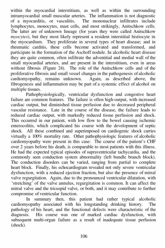
within the myocardial interstitium, as well as within the surroundingintramyocardial small muscular arteries. The inflammation is not diagnosticof a myocarditis, or vasculitis. The mononuclear infiltrates includelymphocytes, monocytes, mast cells, and most strikingly, Anitschkow cells.The latter are of unknown lineage (for years they were called Anitschkowmyocytes), but they most likely represent a resident interstitial histiocyte inthe myocardium. They proliferate in several types of heart disease; and inrheumatic carditis, these cells become activated and transformed, andparticipate in the formation of the Aschoff nodule. In alcoholic heart diseasethey are quite common, often infiltrate the adventitial and medial wall of thesmall myocardial arteries, and are present in the interstitium, even in areaswithout fibrosis (Figure 24). The role of the Anitschkow cells and of theproliferative fibrosis and small vessel changes in the pathogenesis of alcoholiccardiomyopathy, remains unknown. Again, as described above, thefibrogenesis and inflammation may be part of a systemic effect of alcohol onmultiple tissues.
Pathophysiologically, ventricular dysfunction and congestive heartfailure are common features. The failure is often high-output, with increasedcardiac output, but diminished tissue perfusion due to decreased peripheralvascular resistance. Late in the course of the disease, the failure leads toreduced cardiac output, with markedly reduced tissue perfusion and shock.This occurred in our patient, with low flow to the bowel causing ischemicenterocolitis, which complicated his course with the development of septicshock. All these combined and superimposed on cardiogenic shock carriesvirtually a 100% mortality rate. Other pathophysiologic features of alcoholiccardiomyopathy were present in this case: The course of the patient’s CHFover 2 years before his death, is comparable to most patients with this illness.He had the expected typical episodes of supraventricular tachycardia, and thecommonly seen conduction system abnormality (left bundle branch block).The conduction disorders can be varied, ranging from partial to completeheart block. Finally, his echocardiogram revealed not only severe ventriculardysfunction, with a reduced ejection fraction, but also the presence of mitralvalve regurgitation. Again, due to the pronounced ventricular dilatation, with‘stretching’ of the valve annulus, regurgitation is common. It can affect themitral valve and the tricuspid valve, or both, and it may contribute to furthercompromise of ventricular function.
In summary then, this patient had rather typical alcoholiccardiomyopathy associated with his longstanding drinking history. Thepathology of his heart, and the functional defects were characteristic of thediagnosis. His course was one of marked cardiac dysfunction, withsubsequent multi-organ failure as a result of inadequate tissue perfusion(shock).
106
Suggested Readings
1.
2.
3.4.
5.
Patel VB, Why HJ, Richardson PJ, Preedy VR. The effects of alcohol onthe heart. Adverse Drug React Toxicol Rev. 1997; 16:15-43.Preedy VR, Richardson PJ. Alcoholic cardiomyopathy: clinical andexperimental pathological changes. Herz. 1996; 21:241-7.Maisch B. Alcohol and the heart. Herz. 1996; 21:207-12.Piano MR, Schwertz DW. Alcoholic heart disease: a review. Heart Lung.1994; 23:3-17.Djoenaidi W, Notermans SL, Dunda G. Beriberi cardiomyopathy. Eur JClin Nutr. 1992; 46:227-34.
107
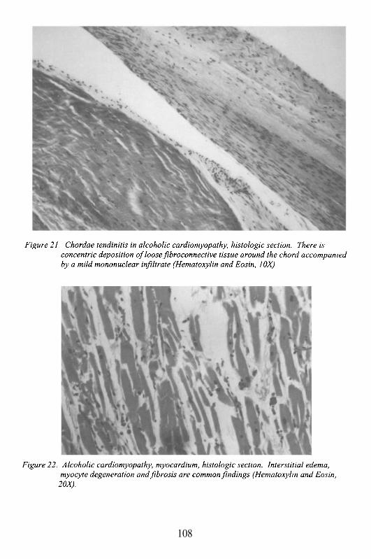
108
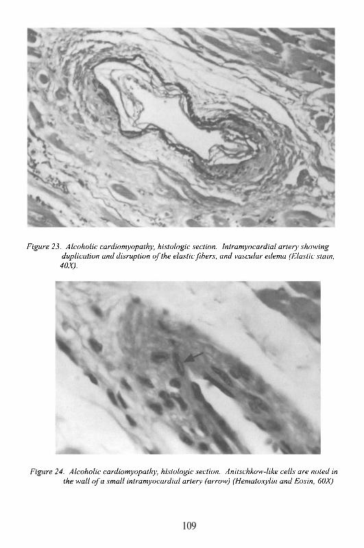
109
Case # 12
Clinical Summary
A 40 year old Hispanic woman, known HlV-infected for the past 3years and followed by a specialty HIV/AIDS clinic, had a routine chest-x-raywhere cardiomegaly was noted. Her most recent CD4 T lymphocyte countwas indicating severe immunosuppression. An echocardiogramwas performed, which showed a small pericardial effusion, biventricularhypertrophy, mild to moderate mitral regurgitation, moderate to severetricuspid regurgitation, diffuse left ventricular hypokinesis with diminishedleft ventricular systolic function, and an ejection fraction of 40%. Fourmonths later she was hospitalized with severe respiratory distress. Heartcatheterization revealed right ventricular and pulmonary artery pressures of58/18 and 38/18 mm Hg, respectively. The pulmonary capillary wedgepressure was 35 mmHg. The diagnosis of cardiogenic shock was made.Renal dysfunction was also present evidenced by a creatinine of 3.3 mg/dLand BUN of 60 mg/dL. She improved and was discharged, but returned to thehospital two months later with shortness of breath, cough and hemoptysis. Onthis admission her vital signs were: P 120, R 24, T 37°C, BP 78/palpable.Her physical exam was significant for bilateral rales with diminished air entryon the right side, tachycardia and gallop rhythm. The arterial gases were pH7.17, pCO2 26, pO2 72, O2 sat 87%, BE –18. The chest-x-ray showedbilateral lower lobe consolidation with effusion. The patient was intubatedand started on 100% FiO2 and PEEP (>5). Nonetheless, she became severelyhypoxic and acidotic, and expired two hours after admission.
Gross Description
The autopsy was performed 3 hours after death. The body length was160 cm and the body weight was 45 kg. The heart weighed 410 g (heart/bodyweight ratio of 0.009, normal 0.004-0.005). The pericardium was smooth andglistening. The epicardium was shiny with a moderate amount ofsubepicardial fat. The atria were of normal size and free of thrombi. Theforamen ovale was closed. The right and left ventricular walls werehypertrophied measuring 2.5 and 0.7 cm, respectively (Figure 25). The valverings measured as follows: TV 10 cm, PV 7 cm, MV 11 cm, AV 7 cm. Thevalve leaflets were delicate and pliable except for some myxomatous changes.The chordae tendineae were slender. The endocardium was thin andglistening. The myocardium showed multiple foci of myocytolysis. Theanterior wall and the interventricular septum had areas of pallor due toischemia. Subendocardial necrosis was also present. The coronary arteries
110
were unremarkable. The lungs weighed 1550 g, were voluminous, lookedessentially identical, and did not collapse upon entering the thoracic cavity.The pleural surfaces had fibrous adhesions. On section, marked pulmonaryedema was noted. The left lower lobe showed patchy areas of consolidationand hemorrhage. The right lower lobe showed diffuse acute hemorrhage.Chronic bronchitis was noted. The pulmonary artery and its major brancheswere unremarkable. The kidneys weighed 200 g combined and had nosignificant gross abnormalities.
Microscopic Description
Myocardial fibrosis and multifocal myocytolysis were observed(Figure 26). There were focal interstitial and perivascular deposits of a lightlyeosinophilic homogeneous material, resembling amyloid. However, Congo-red and crystal violet stains were negative. The elastic stain was only positivein the endocardium and in vessels, but not in the interstitium.Microscopically, sections from both lungs showed extensive interstitial andintraalveolar hemorrhage and a necrotizing intraalveolar exudate. Gram stainrevealed clusters of gram negative rods that in culture were identified asKlebsiella pneumoniae. No interstitial amyloid-like material was present inany tissue other than the myocardium.
Sections from the heart were submitted for electron microscopy. Theepoxy-resin-embedded sections showed diffuse interstitial and perivascularaggregates of fibrillar materials, which were morphologically distinct fromamyloid or collagen fibrils. The fibrils were slightly curvilinear with adiameter of 15-20 nm. A suggestion of helical configurations and periodicitywere noted at higher magnifications. These fibrils were loosely distributed inthe interstitial space and did not have the close relationship with the myocytesarcolemma typical of amyloid deposition (Figure 27).
Case Analysis
Diverse pathological manifestations of heart disease have beendescribed in patients with HIV/AIDS. These include a variety ofopportunistic infections, drug toxicity, malignancies and idiopathicendocardial and myocardial diseases. Regardless of the etiology, the mostcommon clinical syndromes of cardiac dysfunction seen in these patientsinclude dilated cardiomyopathy, pericardial effusion with or withouttamponade, ventricular tachycardia and thromboembolic disease as a result ofinfectious or non-infectious endocarditis.
Opportunistic infections and malignancies are secondary toimmunosuppression, but the ultimate cardiac damage is multifactorial and
111
includes lymphokines and growth factors released by activated lymphocytesand other cellular immune mediators.
Infections
A variety of opportunistic and non-opportunistic infections have beendescribed in the heart of HIV/AIDS patients. These include fungal(Cryptococcus species, Candida species and Aspergillus organisms), bacterial,mycobacterial (tuberculous and atypical mycobacteria), viruses (CMV, HSV),protozoans (Toxoplasma gondii), Pneumocystis carinii infection, and parasitic(Microsporidium, Pentastomiasis). Myocarditis and endocarditis are moreoften secondary to infection than idiopathic. As in non-AIDS patients, adultmyocarditis in an HIV-infected individual may be immunological in originwhen it occurs in the absence of an identifiable infectious organism. With theadvent of highly active antiretroviral therapy (HAART), the rate ofopportunistic infections has decreased significantly compared to those seenpreviously to the HAART era.
Malignancies
Primary Kaposi sarcoma and lymphomas compromising the heart,have been described, but are extremely rare.
HIV infection of the myocytes
Recognition of HIV-1 DNA sequences by PCR and in-situhybridization in the heart of HIV/AIDS patients, supports the possibility ofselective infection of the myocardium by HIV virions. However, there areseveral authors that believe that these results are due to HIV contamination ofthe heart by the patient’s infected blood. The proliferation of multilamellarmembrane bodies within myocytes with subsequent mitochondrialdysfunction also has been described in HIV infection as a possibleexplanation of cardiac disturbances, but there is no evidence that these bodiesare the result of direct HIV toxicity to the myocyte. CD4 receptors are absentin cardiac myocytes, but their absence does not entirely exclude thepossibility of myocardial infection by HIV. Recent studies suggest thatperhaps the infection occurs in endothelial cells rather than myocytes, whichin this case might explain the multiple foci of chronic ischemia(myocytolysis). If in fact, HIV damages the heart directly, it is notaccompanied by a significant inflammatory response.
112
Dilated Cardiomyopathy
The exact pathogenic mechanism of dilated cardiomyopathyassociated with HIV/AIDS is unknown, but diverse factors may play a role.Nutritional deficiencies are known to cause dilated cardiomyopathy (seeDCM chapter), and HIV/AIDS wasting syndrome is characterized bypersistent poor appetite and generalized cachexia. In addition, gastrointestinaldisease with malabsorption and protein loss is a common feature of HIV, butin many cases has no detectable cause. Our patient was thin but not cachectic.In addition, with profound malnutrition and wasting, the adipose tissuedeposits are lost, and one of the last ones to disappear is the subepicardial fat(Figure 28). However, in our case, the subepicardial fat was present innormal amounts, indicating that she did not have significant nutritionalcompromise.
Several drugs such as interferon alpha and nucleoside analogues havebeen associated with the development of cardiomyopathy. Zidovudine (AZT)in particular is known to cause a striated muscle myopathy that is dosedependent and requires discontinuation for reversal of the effect. Thepostulated mechanism is through mitochondrial structural and geneticalterations in the myocyte, with abnormal expression of mitochondrial geneproducts.
Myocardial ischemia due to spasm of the microvasculature, whetherassociated or not with cocaine use, and excess in catecholamine productionhave also been described as possible causes of cardiomyopathy in HIV/AIDSpatients.
Metabolic alterations
HIV infection and some of its treatment drugs (protease inhibitors),are associated with a decrease in the levels of high density lipoproteins (HDL)and increase levels of LDL and triglycerides; thus increasing susceptibility toatherosclerosis. The benefits of therapy outweigh the risks; however, the riskincreases if other factors for cardiovascular disease are present, such assmoking, hypertension, diabetes, and cocaine abuse. Insulin resistance iscommon in HIV-infected patients, but the prevalence of diabetes is notincreased as compared to the general population. There is no known directassociation between hypertension and HIV.
Interstitial myocardial disease
Interstitial heart disease, other than myocardial fibrosis, does notappear to be a commonly recognized feature of patients with HIV/AIDS.
113
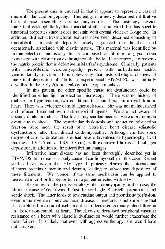
The present case is unusual in that it appears to represent a case ofmicrofibrillar cardiomyopathy. This entity is a newly described infiltrativeheart disease resembling cardiac amyloidosis. The histology revealsinterstitial eosinophilic hyaline material similar to amyloid, but has specifictinctorial properties since it does not stain with crystal violet or Congo red. Inaddition, distinct ultrastructural features have been described consisting ofmicrofibrillar interstitial deposits loosely organized into bundles, andoccasionally associated with elastic matrix. This material was identified byimmunoelectron microscopy to be composed of fibrillin, a glycoproteinassociated with elastic tissues throughout the body. Furthermore, it representsthe matrix protein that is defective in Marfan’s syndrome. Clinically, patientswith microfibrillar cardiomyopathy present with arrhythmias and/orventricular dysfunction. It is noteworthy that histopathologic changes ofinterstitial deposition of fibrils in experimental HIV/AIDS, was initiallydescribed in the early 80s in a colony of macaque monkeys.
In this patient, no other specific cause for dysfunction could beidentified on either light or electron microscopy. There was no history ofdiabetes or hypertension, two conditions that could explain a rigid, fibroticheart. There was evidence of mild atherosclerosis. She was not malnourishedand refused treatment with anti-retroviral agents. We are not aware ofcocaine or alcohol abuse. The foci of myocardial necrosis were a pre-mortemevent due to shock. The ventricular dyskinesis and reduction of ejectionfraction were more the result of a restrictive heart disease (diastolicdysfunction), rather than dilated cardiomyopathy. Although she had somedegree of cardiac dilatation, she had severe biventricular hypertrophy (wallthickness: LV 2.5 cm and RV 0.7 cm), with extensive fibrosis and collagendeposition, in addition to the microfibrillar changes.
Infiltrative heart disease has not been thoroughly described yet inHIV/AIDS, but remains a likely cause of cardiomyopathy in this case. Recentstudies have proven that HIV type 1 protease cleaves the intermediatefilament proteins vimentin and desmin, leading to subsequent deposition ofthese filaments. We wonder if the same mechanism can be applied toincreased microfibrillar deposition in a patient infected with HIV.
Regardless of the precise etiology of cardiomyopathy in this case, theultimate cause of death was diffuse hemorrhagic Klebsiella pneumonia andseptic shock. The latter leads to low cardiac output and poor tissue perfusion,even in the absence of previous heart disease. Therefore, is not surprising thatshe developed myocardial ischemia due to decreased coronary blood flow inan already non-compliant heart. The effects of decreased peripheral vascularresistance on a heart with diastolic dysfunction would further exacerbate theheart failure. It is likely that even with aggressive therapy, she would havenot survived.
114
Suggested Readings
1.
2.
3.
4.
5.
6.
7.
8.
Lewis W. Cardiomyopathy in AIDS: a pathophysiological perspective.Prog Cardiovasc Dis. 2000; 43:151-70.Rerkpattanapipat P, Wongpraparut N, Jacobs LE, Kotler MN. Cardiacmanifestations of acquired immunodeficiency syndrome. Arch Intern Med.2000; 160:602-8.Milei J, Grana D, Fernandez Alonso G, Matturri L. Cardiac involvement inacquired immunodeficiency syndrome--a review to push action. TheCommittee for the Study of Cardiac Involvement in AIDS. Clin Cardiol.1998; 21:465-72.Yunis NA, Stone VE. Cardiac manifestations of HIV/AIDS: a review ofdisease spectrum and clinical management. J Acquir Immune Defic SyndrHum Retrovirol. 1998; 18:145-54.Herskowitz A. Cardiomyopathy and other symptomatic heart diseasesassociated with HIV infection. Curr Opin Cardiol. 1996; 11:325-31.Flomenbaum M, Soeiro R, Udem SA, Kress Y, Factor SM. Proliferativemembranopathy and human immunodeficiency virus in AIDS hearts. JAcq Imm Def Synd. 1989; 2:129-35.van Hoeven KH, Segal B, Factor SM. AIDS Cardiomyopathy: first rule outother myocardial risk factors. Int J Cardiol 1990; 67:780-3.Factor SM, Menegus MA, Kress Y, Cho S, Fisher JD, Sakai LY,Goldfischer S. Microfibrillar cardiomyopathy: A new entity resemblingcardiac amyloidosis. Cardiovasc Pathol 1992; 1:307-16.
115

116
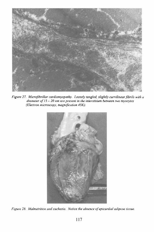
117

This Page Intentionally Left Blank
Chapter # 5
VALVULOPATHIES
Case # 13
Clinical Summary
A 78 year old woman was brought to the ER with a 3 day history ofrespiratory distress. In route to the ER, she was hypertensive, tachycardic andtachypneic (BP 200-220/130 mmHg, P 135, R 28). The ECG showed atrialfibrillation with premature contractions. Upon arrival, she was found to bepale and diaphoretic and she began to experience mild chest pain. Her vitalsigns at that time were: BP 170/130 mmHg, P 176, R 34 and T 37.8°C. Shewas placed on a cardiac monitor and on 100% FiO2 by nasal cannula. Inaddition, sublingual nitroglycerine, aspirin, furosemide, diltiazem, clonidineand digoxin were started. After her admission to the cardiac care unit, hercondition improved. There were no complaints of respiratory distress or chestpain, and the cardiac monitor showed occasional premature contractions.Through the day she was occasionally confused, and at one point requiredhand restraints to prevent her from pulling her IV lines. Her physical examwas significant for scattered bilateral pulmonary rales and lower extremityedema. The chest X ray was suggestive of congestive heart failure andcardiomegaly. The ECG showed atrial fibrillation, left ventricularhypertrophy, right bundle branch block pattern, but no ST-T wave changes.There were no enzymatic changes (CK, LDH or troponin I) consistent withacute myocardial infarction. A repeat chest-x ray was ordered and the patienthad a cardiac arrest while in radiology. Resuscitation was unsuccessfullyattempted.
Gross Description
The patient weighed 68 kg and measured 147 cm. The heart weighed430 g. The pericardium was smooth. The left ventricle was hypertrophic(wall thickness 2.7 cm) (Figure 29). The right atrium was also hypertrophic,but the left atrium and right ventricle were unremarkable. The valves had thefollowing measurements: tricuspid 13.5 cm, pulmonic 10.0 cm, mitral 6.5cm, and aortic 4.0 cm. Stenosis of the mitral valve was mild. The aorticvalve was also stenotic with fusion of the cusps (Figure 30). Other findingsincluded valvular and endocardial calcified vegetations, endocardial
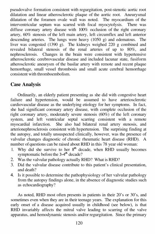
pseudovalve formation consistent with regurgitation, post-stenotic aortic rootdilatation and linear atherosclerotic plaque of the aortic root. Aneurysmaldilatation of the foramen ovale wall was noted. The myocardium of theinterventricular septum was scarred with focal myocytolysis. There wasdiffuse coronary artery disease with 100% occlusion of the right coronaryartery, 60% stenosis of the left main artery, left circumflex and left anteriordescending arteries. The lungs were heavy (1050 g) and edematous. Theliver was congested (1390 g). The kidneys weighed 220 g combined andrevealed bilateral stenosis of the renal arteries of up to 80%, andnephrosclerosis. Changes in the brain were consistent with hypertensiveatherosclerotic cerebrovascular disease and included lacunar state, fusiformatherosclerotic aneurysm of the basilar artery with remote and recent plaquehemorrhage, small vessel thrombosis and small acute cerebral hemorrhageconsistent with thromboembolism.
Case Analysis
Ordinarily, an elderly patient presenting as she did with congestive heartfailure and hypertension, would be assumed to have arterioscleroticcardiovascular disease as the underlying etiology for her symptoms. In fact,she had significant coronary artery disease, with complete occlusion of theright coronary artery, moderately severe stenosis (60%) of the left coronarysystem, and left ventricular septal scarring consistent with a remotemyocardial infarction. She also had bilateral renal artery stenosis, andarterionephrosclerosis consistent with hypertension. The surprising finding atthe autopsy, and totally unsuspected clinically, however, was the presence ofvalvular changes diagnostic of chronic rheumatic heart disease (RHD). Anumber of questions can be raised about RHD in this 78 year old woman:1.
2.3.
4.
Why did she survive to her decade, when RHD usually becomessymptomatic before the decade?Was the valvular pathology actually RHD? What is RHD?Did the valvular disease contribute to this patient’s clinical presentation,and death?Is it possible to determine the pathophysiology of her valvular pathologyfrom the autopsy findings alone, in the absence of diagnostic studies suchas echocardiography?
As noted, RHD most often presents in patients in their 20’s or 30’s, andsometimes even when they are in their teenage years. The explanation for thisearly onset of a disease acquired usually in childhood (see below), is thatRHD invariably affects the mitral valve leading to scarring of the valveapparatus, and hemodynamic stenosis and/or regurgitation. Since the primary
120
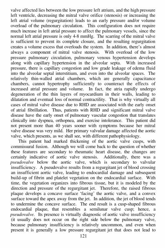
valve affected lies between the low pressure left atrium, and the high pressureleft ventricle, decreasing the mitral valve orifice (stenosis) or increasing theleft atrial volume (regurgitation) leads to an early pressure and/or volumeoverload of the pulmonary circulation. This configuration does not requiremuch increase in left atrial pressure to affect the pulmonary vessels, since thenormal left atrial pressure is only 4-8 mmHg. The scarring of the mitral valveis sufficient to prevent its complete closure, and the resulting regurgitationcreates a volume excess that overloads the system. In addition, there’s almostalways a component of mitral valve stenosis. With overload of the lowpressure pulmonary circulation, pulmonary venous hypertension developsalong with capillary hypertension in the alveolar septa. With increasedpressure, there is capillary congestion and low grade leakage of edema fluidinto the alveolar septal interstitium, and even into the alveolar spaces. Therelatively thin-walled atrial chambers, which are generally capacitancechambers, cannot hypertrophy sufficiently to counteract the effects ofincreased atrial pressure and volume. In fact, the atria rapidly undergodegeneration of the thin layers of myocardium in their walls, leading todilatation and eventual loss of normal contractility. That is why virtually allcases of mitral valve disease due to RHD are associated with the early onsetof atrial fibrillation. Thus, patients with RHD and significant mitral valvedisease have the early onset of pulmonary vascular congestion that translatesclinically into dyspnea, orthopnea, and exercise intolerance. This patient didnot present more than 40 years sooner with symptoms because her mitralvalve disease was very mild. Her primary valvular damage affected the aorticvalve, which presents, as we shall see, with different pathophysiology.
This patient had marked thickening of the aortic valve cusps, withcommissural fusion. Although we will come back to the question of whetherthese features are secondary to rheumatic heart disease, the changes arecertainly indicative of aortic valve stenosis. Additionally, there was apseudovalve below the aortic valve, which is secondary to valvularinsufficiency. A pseudovalve results from a regurgitant jet of blood crossingan insufficient aortic valve, leading to endocardial damage and subsequentbuild-up of fibrin and platelet vegetation on the endocardial surface. Withtime, the vegetation organizes into fibrous tissue, but it is modeled by thedirection and pressure of the regurgitant jet. Therefore, the raised fibrousplaque develops a concave surface ‘facing’ the aortic valve, and a convexsurface toward the apex away from the jet. In addition, the jet of blood tendsto undermine the concave surface. The end result is a cusp-shaped fibrousendocardial plaque, that resembles a semilunar valve cusp; hence, apseudovalve. Its presence is virtually diagnostic of aortic valve insufficiency(it usually does not occur on the right side below the pulmonary valve,because pulmonary insufficiency is relatively uncommon, and even whenpresent it is generally a low pressure regurgitant jet that does not lead to
121
significant endocardial damage). Two other findings suggest that she hadhemodynamically significant aortic valve disease: she had prominent leftventricular hypertrophy (wall thickness 2.7 cm, normal 1.5 cm), and post-stenotic dilatation of the aortic root above the valve. Concentric leftventricular hypertrophy (LVH) could have been partially the result of chronichypertension, but valvular outflow tract obstruction generally produces amore severe LVH, consistent with the range of what this patient had. Thepost-stenotic dilatation of the aortic root is a characteristic result of a jet ofblood progressing through the narrowed valve orifice, and leading to damageto the aortic root wall, with degeneration of the elastic tissue and secondaryaneurysm formation. Thus, this patient had gross morphologic evidence ofaortic valve stenosis and insufficiency. Even without an echocardiogram, thediagnosis can be made with absolute certainty. The fact that clinical murmurswere not appreciated (mid-systolic ejection murmur for aortic stenosis, anddiastolic murmur for aortic insufficiency heard at the base; diastolic rumblewith an opening snap heard at the apex and radiating to the left axilla formitral stenosis - note that there would be no pre-systolic accentuation due tothe atrial fibrillation), can be explained on the basis of her presentation withcongestive heart failure with probable decreased stroke volume. There mustbe adequate flow across the valves in order to produce the fluid turbulencethat causes the murmurs.
Aortic valve disease, regardless of etiology, is much better tolerated thanmitral valve disease. The latter, as described above, affects the low pressurepulmonary circulation, and may do so early in its course, leading to exerciseintolerance and progressive respiratory distress. In contrast, aortic valvedisease, particularly stenosis, results in compensatory left ventricularhypertrophy that maintains stroke volume for a prolonged period. Thus,patients with aortic stenosis may have the condition in a well-compensatedstate well into their decade, and sometime even later, with maintenanceof their exercise tolerance. Ultimately, there is a cost. There is chronicdamage to the subendocardium due to the high end-systolic pressures.Consequently, the newly generated wall stress causes myocyte injury; and theremodeling of the wall and chamber eventually results in elevated end-diastolic pressures. The remodeling is accentuated by aortic insufficiency(volume overload), and coronary ischemia. Although, both were present inthis patient, significant remodeling had not yet developed. Regardless,ventricular failure may develop very rapidly in patients with aortic stenosis,leading to the precipitous onset of congestive heart failure, even beforeremodeling ensues. Thus, in contrast to mitral stenosis, which leads to anearly presentation, the course of aortic stenosis is one of prolongedcompensation often for many years, and rapid decompensation. The latter,often results in death in weeks or a few months, usually secondary to fatalarrhythmia, or left ventricular failure. This patient’s presentation with
122
fulminant congestive heart failure is consistent with decompensated aorticvalve disease, even though mitral valve disease, ischemia, and hypertensioncontributed significantly.
Was this patient’s valvular disease rheumatic in origin? It is far morecommon to have degenerative valve disease affecting patients in theirdecade, than to have RHD as the etiology. Calcification of the mitral annulus(mitral annular calcification), and calcification and fibrosis of the aortic valve(calcific aortic stenosis of the elderly) are two degenerative conditions oftenoccurring simultaneously, that can cause mitral regurgitation and aorticstenosis in older patients. Mitral annular calcification, when it affects thevalve leaflets, tends to damage the posterior leaflet and focal posteriorchordae preferentially, generally sparing the anterior leaflet and chords.Calcific aortic stenosis, by its description, tends to have heavy calcificationthat often compromises the sinuses of Valsalva. It often affects only mildlythe commissures; and, it is usually not associated with aortic insufficiency.In contrast, rheumatic mitral valve disease affects the entire valve, tends tospare the annulus unless very severe, causes commissural fusion, and leads tothickening, shortening and fusion of chordae tendineae (Figures 31 and 32).Rheumatic aortic valve disease causes fibrosis of the cusps (with or withoutcalcification), leads to fusion of commissures, and frequently is associatedwith insufficiency. Based on these criteria, this patient had rheumaticvalvular disease. It is also noteworthy that rheumatic disease always involvesthe mitral valve, with the aortic valve being the second most affected;however, aortic valve disease without mitral involvement, is not rheumatic inetiology. The relative severity of the two valve lesions may be variable(usually, when there is one valve more affected than another, it is the mitralvalve that is more severe; but, on occasion as in this patient, the relationship isinverted).
As a final point, a brief discussion of the pathogenesis of RHD and how itaffects the valves, is in order. The clinical manifestations of RHD arevirtually always the chronic effects of an inflammatory process that occurredseveral decades before. The condition results from a childhood infection withrheumatogenic strains of group A, beta-hemolytic streptococcus, oftencausing pharyngitis (‘strep throat’). Then, in a small proportion of susceptiblechildren (less than 3%) an auto-immune reaction develops several weeks afterthe infection. The auto-immunity is thought to result from cross-reactivitybetween one or several glycoprotein(s) in the streptococcal capsule (Mprotein), and antigens expressed in the myocardium, pericardium, and heartvalves. During the active inflammatory phase of the disease, a cellularinfiltrate involves the interstitium of the heart (myocarditis), the pericardium(fibrinous pericarditis), and the valve tissues including the leaflets,commissures, and chordae tendineae (atrio-ventricular valves). Theinflammatory infiltrate, particularly in the myocardium, consists of a
123
granulomatous collection of T lymphocytes, plasma cells, and activatedmacrophages known as Aschoff cells, often surrounding a focus of fibrinoidcollagen necrosis. The entire granuloma is known as an Aschoff body, and itis pathognomonic for rheumatic heart disease (Figures 33 and 34). TheAschoff body may persist for years (its persistence does not necessarilyindicate that the inflammatory disease is active), but it often goes on to heal asa fibrous scar. In the valvular tissues, true Aschoff bodies are uncommon, butthe inflammatory infiltrate is similar. Scarring then results in fibrousthickening and secondary calcification of the leaflets and cusps withdecreased pliability and inability to coapt properly. Furthermore, fusion ofcommissures leads to narrowing of the valve orifice; and the narrowing,shortening and fusion of chordae, limit the normal excursion of the mitral andless commonly, the tricuspid valves. With time, secondary non-rheumaticvegetations may develop on these valves that organize into additional scartissue that further distorts and deforms the valve structure. The leaflet andcusp fibrosis, the commissural fusion, and the chordal thickening were thechanges identified in this patient, indicating the presence of chronic rheumaticdamage that occurred years before. Despite extensive search, no Aschoffbodies were identified, but the diagnosis of chronic rheumatic valvulitis doesnot require their presence.
124
Suggested Readings
1.2.
3.
4.
5.6.
7.
FeldmanT. Rheumatic heart disease. Curr Opin Cardiol. 1996; 11:126-30.Shulman ST. Complications of streptococcal pharyngitis. Pediatr InfectDis J. 1994; 13(1 Suppl l):S70-4; discussion S78-9.Veasy LG, Hill HR. Immunologic and clinical correlations in rheumaticfever and rheumatic heart disease. Pediatr Infect Dis J. 1997; 16:400-7.Rose AG. Etiology of valvular heart disease. Curr Opin Cardiol. 1996;11:98-113.BaxleyWA. Aortic valve disease. Curr Opin Cardiol. 1994; 9:152-7.Stollerman GH. Rheumatogenic streptococci and autoimmunity. ClinImmunol Immunopathol. 1991; 61:131-42.Adler Y, Fink N, Spector D, Wiser I, Sagie A. Mitral annulus calcification-- a window to diffuse atherosclerosis of the vascular system.Atherosclerosis. 2001; 155:1-8.
125
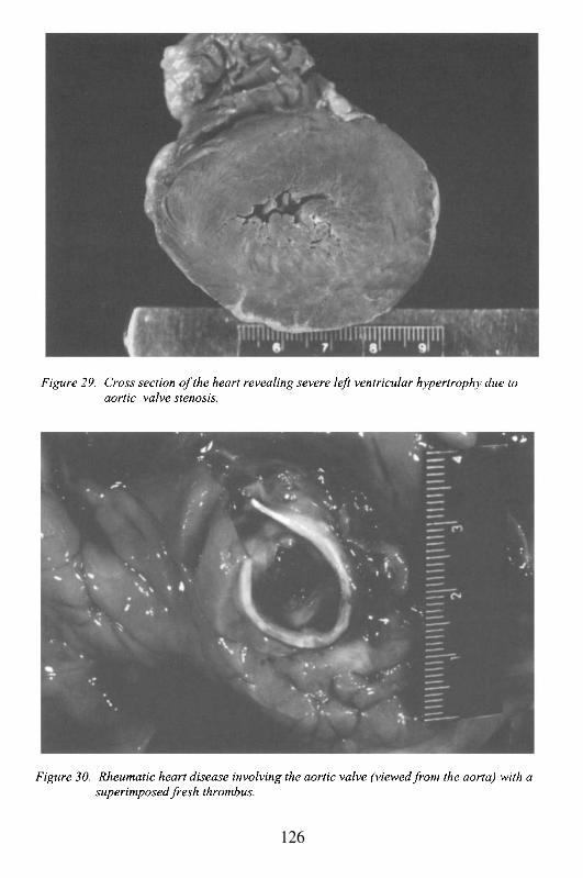
126
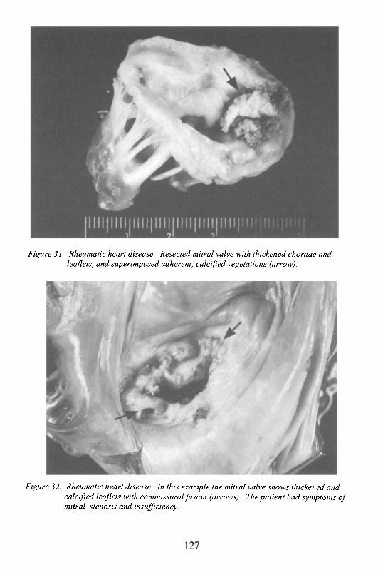
127
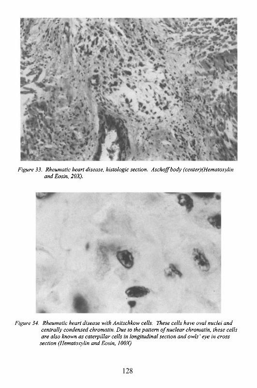
128
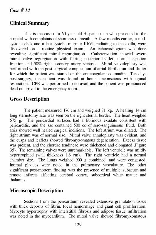
Case # 14
Clinical Summary
This is the case of a 60 year old Hispanic man who presented to thehospital with complaints of shortness of breath. A few months earlier, a mid-systolic click and a late systolic murmur III/VI, radiating to the axilla, werediscovered on a routine physical exam. An echocardiogram was donerevealing significant mitral regurgitation. Catheterization showed severemitral valve regurgitation with flaring posterior leaflet, normal ejectionfraction and 50% right coronary artery stenosis. Mitral valvuloplasty wasperformed with the post-surgical complication of atrial fibrillation and flutterfor which the patient was started on the anticoagulant coumadin. Ten dayspost-surgery, the patient was found at home unconscious with agonalrespiration. CPR was performed to no avail and the patient was pronounceddead on arrival to the emergency room.
Gross Description
The patient measured 176 cm and weighed 81 kg. A healing 14 cmlong sternotomy scar was seen on the right sternal border. The heart weighed575 g. The pericardial surfaces had a fibrinous exudate consistent withpericarditis, and the sac contained 500 cc of sero-sanguineous fluid. Bothatria showed well healed surgical incisions. The left atrium was dilated. Theright atrium was of normal size. Mitral valve annuloplasty was evident, andthe cusps and leaflets showed fibromyxomatous degeneration. Excess tissuewas present, and the chordae tendineae were thickened and elongated (Figure35). The remaining valves were unremarkable. The left ventricle was mildlyhypertrophied (wall thickness 1.6 cm). The right ventricle had a normalchamber size. The lungs weighed 900 g combined, and were congested.Intimal plaques were noted in the pulmonary vasculature. The othersignificant post-mortem finding was the presence of multiple subacute andremote infarcts affecting cerebral cortex, subcortical white matter andthalamus.
Microscopic Description
Sections from the pericardium revealed extensive granulation tissuewith thick deposits of fibrin, focal hemorrhage and giant cell proliferation.Myocyte hypertrophy with interstitial fibrosis and adipose tissue infiltrationwas noted in the myocardium. The mitral valve showed fibromyxomatous
129
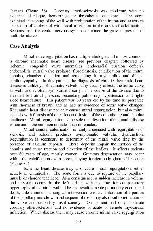
changes (Figure 36). Coronary arteriosclerosis was moderate with noevidence of plaque, hemorrhage or thrombotic occlusions. The aortaexhibited thickening of the wall with proliferation of the intima and extensivedeposition of cholesterol with focal ulceration in the areas of calcification.Sections from the central nervous system confirmed the gross impression ofmultiple infarcts.
Case Analysis
Mitral valve regurgitation has multiple etiologies. The most commonis chronic rheumatic heart disease (see previous chapter) followed byischemia, congenital valve anomalies (endocardial cushion defects),endocarditis, mitral valve prolapse, fibroelastosis, calcification of the mitralannulus, chamber dilatation and remodeling in myocarditis and dilatedcardiomyopathy. In this patient, the diagnosis of chronic rheumatic heartdisease is unlikely. Rheumatic valvulopathy usually affects the aortic valveas well, and is often symptomatic early in the course of the disease due toelevated left atrial pressure, secondary pulmonary hypertension and right-sided heart failure. This patient was 60 years old by the time he presentedwith shortness of breath, and he had no evidence of aortic valve changes.Rheumatic heart disease not only causes mitral regurgitation, but also mitralstenosis with fibrosis of the leaflets and fusion of the commissure and chordaetendineae. Mitral regurgitation as the sole manifestation of rheumatic diseaseis rare and more common in males than in females.
Mitral annular calcification is rarely associated with regurgitation orstenosis, and seldom produces symptomatic valvular dysfunction.Regurgitation is secondary to deformity of the mitral valve ring by thepresence of calcium deposits. These deposits impair the motion of theannulus and cause traction and elevation of the leaflets. It affects patientsover 60 years of age, mostly women. Grumous degeneration may occurwithin the calcifications with accompanying foreign body giant cell reaction(Figure 37).
Ischemic heart disease may also cause mitral regurgitation, eitheracutely or chronically. The acute form is due to rupture of the papillarymuscle or chordae tendineae. As a consequence, a sudden increase in volumeand pressure occurs in the left atrium with no time for compensatoryhypertrophy of the atrial wall. The end result is acute pulmonary edema anddeath, unless immediate surgical intervention ensues. Infarction of a portionof the papillary muscle with subsequent fibrosis may also lead to retraction ofthe valve and secondary insufficiency. Our patient had only moderatecoronary atherosclerosis and no evidence of acute or remote myocardialinfarction. Which disease then, may cause chronic mitral valve regurgitation
130
that is often asymptomatic for years, and incidentally discovered duringphysical exam? The answer is mitral valve prolapse or MVP.
MVP, also known as Barlow’s syndrome or “floppy” valve disease,occurs in 3-5% of the general population. It affects females more than malesand may be associated with connective tissue disorders such as Marfan andEhlers-Danlos syndromes. MVP may also be inherited as an autosomaldominant condition with variable penetrance. Occasionally, MVP may leadto severe mitral regurgitation with elevation of the mean left atrial pressureand resultant dilatation of the left atrium. This pathologic dilatation is apredisposing factor for the development of atrial fibrillation and flutter.
In order to understand the pathophysiology of this type ofvalvulopathy, it is necessary that we review the physiology of the normalvalve. The mitral valve consists of the following structures: the annulus, 2leaflets (anterior and posterior), the chordae tendineae and 2 papillary musclegroups, one antero-medial and the other postero-lateral. The annulus is anasymmetric ring where the leaflets attach. The posterior leaflet, despite thefact that it is the smaller of the two, occupies most of the annuluscircumference. Both leaflets are composed of fibrous tissue lined byendothelium. They are attached to the anterior and posterior papillarymuscles by delicate thin bands of tissue known as chordae tendineae. Thereare a total of 24 main chordae and 120 branches that insert not only at the freeedge of the leaflets, but also at the undersurface (rough zone). The nature ofthis insertion assures proper closure and prevents eversion of the leaflets intothe atrium during systole. Abnormal valvular movements are also preventedby overlapping of the leaflets during their closed position and by the fact thatthe anterior leaflet runs parallel to the blood flow. During diastole, thevalve’s leaflets are forced into a semi-closed position by the passive stretchingof the papillary muscles during ventricular filling. In mitral valve prolapse,regurgitation occurs when the valvular tissue protrudes into the left atriumduring systole. The leaflets prolapse due to two main reasons: 1) the tissue isexcessive and 2) the leaflets are no longer fibrous, but rather myxomatous(rich in mucopolysaccharides) which makes them thin, soft and highlymobile. In addition, the chordae tendineae may be thin and elongated.
The heart murmur of this patient was characteristic of MVP. Themidsystolic click occurs when the redundant leaflet enters into the left atriumand is suddenly pulled back to its original position. The murmur is the resultof turbulence caused by the regurgitant blood flow. In our case, despite theannuloplasty, the post-mortem mitral valve findings are consistent with thediagnosis of MVP. The leaflets had marked fibromyxomatous degenerationand the chordae tendineae were long and only mildly thickened. Followingsurgery, this patient developed arrhythmia and was anticoagulated to avoidmicrothrombi that could embolize in the systemic circulation. However, theautopsy revealed subacute and remote infarcts that occurred prior to surgery.
131
The thrombi originated either in the dilated atrium or beneath the redundantleaflets of the mitral valve. In some cases, superimposed bacterial infection(e.g. endocarditis) may develop. MVP is managed clinically unless itprogresses to severe mitral regurgitation; and when that happens, as in thiscase, surgical repair is required. Valve replacement for this condition is not ascommon, and carries a higher risk of post-surgical complications.Furthermore, patients who undergo cardiac surgery are always at risk forthromboembolic events, bleeding, infections and arrhythmias.
A well described complication of open heart surgery is the post-cardiotomy syndrome. This entity develops a week to ten days after theprocedure and is clinically undistinguishable from Dressier’s syndrome, acomplication of myocardial infarction. Dressier’s syndrome is an idiopathicform of acute pericarditis manifested as chest pain, pleural and pericardialeffusion, fever and increased white blood cell count. A pericardial frictionrub is present on auscultation. Possible causes include activation of a latentviral infection and/or an immunologic response with the formation of anti-heart antibodies. As in Dressier’s, post-cardiotomy syndrome has also beenassociated with viral illness.
Post-cardiotomy syndrome may present as cardiac tamponade, as inthis case, with significant accumulation of sero-sanguineous fluid andassociated fibrinous pericarditis. Presumably, the blood comes from thenumerous new vessels that comprise the granulation tissue that is part of thehealing process of the pericardium. Increased vascular permeability added tothe effects of coumadin therapy (anticoagulant), contributed to the continuousbleeding into the pericardial sac, leading to compression of the heart(tamponade) and the patient’s death. The microscopic exam of thepericardium revealed extensive granulation tissue and hemorrhage in bothvisceral and parietal layers. The lungs had evidence of pleuritis withadhesions, chronic congestion with numerous intraalveolar hemosiderin-ladenmacrophages and thickened vessels consistent with pulmonary hypertension.It appears that the mitral regurgitation had just recently become severe. Thechanges in the pulmonary vasculature were mild and focal, the left atrium wasonly slightly dilated, the ejection fraction was normal and significant changesin the right side of the heart were absent. Interestingly, the ring of thetricuspid valve was dilated (13 cm) and similar histologic changes to thoseseen in the mitral valve were noted in the tricuspid leaflets (myxomatousdegeneration). These findings suggest a mild form of tricuspid valve prolapsethat can be seen in approximately 40% of patients with mitral valve prolapse.Similar changes can affect the pulmonic and aortic valves, but less frequently(10% of cases). Unfortunately, the patient’s clinical course deteriorated sorapidly that treatment could not be instituted. Patients with cardiactamponade may experience pulsus paradoxus (decline in systemic arterialpressure of more than 10mm of Hg during inspiration). Pulsus paradoxus is
132
an exaggeration of what occurs normally when a person inhales. Duringinspiration, the arterial pressure diminishes and the venous pressure increases.Increased venous return produces a septal shift from the right ventricle intothe left ventricle that is restored when the patient exhales. These changes areenhanced by the external, mainly atrial compression exerted by the fluid in thepericardial sac with severe compromise of the cardiac output. The diagnosiscan be made by examining the jugular vein pulsations. Echocardiography isconfirmatory. Pulsus paradoxus does not occur in the setting of significantleft ventricular hypertrophy. When the left ventricle is hypertrophic with noassociated dilatation, the rigidity of the wall precludes the inspiratory effect ofseptal displacement.
Treatment of cardiac tamponade varies and includes pericardio-centesis and open surgical drainage. In cases of post-cardiotomy syndrome,less severe than in the present case, anti-inflammatory treatment appears to beeffective, which further supports the notion that this entity has animmunologic basis.
In addition to surgical trauma and anticoagulant therapy, otherconditions should be considered as etiologic or precipitating factors of cardiactamponade such as ruptured myocardial infarction, ruptured aortic dissection,renal failure, metastatic malignancies and sepsis.
133
Suggested Readings
1.
2.
3.
4.
Chambers J. The clinical and diagnostic features of mitral valve disease.HospMed. 2001; 62:72-8.Carabello BA. The pathophysiology of mitral regurgitation. J Heart ValveDis. 2000; 9:600-8.Kronick-Mest C. Postpericardiotomy syndrome: etiology, manifestations,and interventions. Heart Lung. 1989; 18:192-8Engle MA. Humoral immunity and heart disease: postpericardiotomysyndrome. Adv Exp Med Biol. 1983; 161:471-8
134
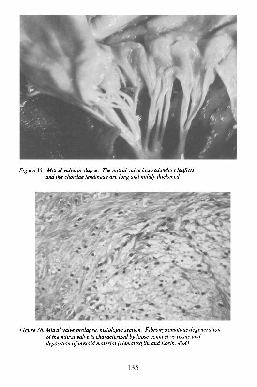
135
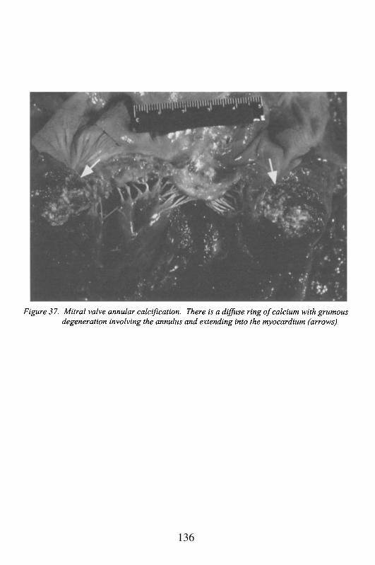
136
Chapter 6
CONGENITAL HEART DISEASE
Case # 15
Clinical Summary
The patient was a 28 year old Hispanic woman with mild mentalretardation, and a known congenital heart defect. She lived with a fosterfamily and always had decreased activity or exercise intolerance withepisodes of dyspnea.
The current illness began a few days prior to her admission with thedevelopment of diarrhea, vomiting, abdominal pain and fever. Her fosterfamily took her to the hospital, where she was rehydrated and sent home withthe diagnosis of a viral syndrome. At home she had a seizure manifested bystiffness, turning of the head and cyanosis. The next morning she waslethargic, had poor respiration and weak pulses. The paramedics were calledand on their arrival they found the patient pulseless, without detectable bloodpressure or respiration. She was immediately intubated and CPR was started.Her post-resuscitation physical exam revealed BP 70/palpable, P 134/min, T37°C; evident peripheral cyanosis and clubbing. There was nolymphadenopathy, or edema. Neurologically her pupils were dilated, fixed,and non-reactive. There were no doll’s eyes or caloric and pain responses.The cardiovascular exam showed no jugular distension; an S1 present,diminished S2 and no murmurs; peripheral pulses were present. The lungshad equal breath sounds bilaterally.
Laboratory data and other tests
Laboratory: WBC 11, Hb/Hct 22/65, Plt 203; BUN/creat 6/1.2, Na 133, K 3.3,Cl 98, CO2 16, glucose 505, lactate (nl 0.3-1.3) 2.6, bilirubin T 0.2,SGOT/SGPT 48/50, Protein T/alb 4.2/2.5, AP 64, LD 316, CK 260; PT/PTT14.7/41.5. Arterial blood gases (pre/post-resuscitation): pH 6.83/7.24, pCO283/31, pO2 54/41, O2 sat % 60/68. CXR: right middle lobe consolidation andcongestive heart failure. ECG: normal sinus rhythm, right ventricularhypertrophy, and T-wave inversion in III.
The patient was admitted in the intensive care unit where she waspersistently hypotensive. An echocardiogram was done showing right
ventricular hypertrophy, overriding aorta and a ventricular septal defect. Thepatient became ventilator dependent and died the day after admission.
Gross Description
The body measured 160 cm and weighed 55 kg. The external examwas significant for clubbing of fingers and toes. There was no evidence ofpast thoracic surgery.
The heart weighed 460 g and had right ventricular prominence with adouble apex. The pericardium was smooth and glistening. There was amoderate amount of subepicardial fat. The right atrium was hypertrophied,but the left was normal in size and free of thrombi. The foramen ovale wasclosed. The right and left ventricular wall thickness was 1.1 and 1.5 cm,respectively. Both ventricles were hypertrophied and the right ventricle wasdilated as well. The membranous interventricular septum was absent, andthus there was a large high ventricular septal defect measuring 2 x 1.5 cm(Figure 38). The aortic valve, which had three normal cusps, was shifted tothe right and opened over the right ventricle, with a small area overriding theventricular septal defect. Therefore, the left ventricular outflow was throughthe ventricular septal defect and into the aorta. The ductus arteriosus wasclosed. The pulmonary artery trunk and left and right pulmonary arterybranches were atrophic and fibrotic with no identifiable lumen (Figure 39).There was no associated identifiable pulmonary valve. The lungs receivedtheir arterial supply from two large bronchial arteries arising from thedescending aorta. These arteries arose at the level of the aortic valve. Thelungs drained via four normal pulmonary veins into the left atrium (two fromeach lung). The valve leaflets were delicate and pliable except for mildmyxomatous change of the mitral valve. The chordae tendineae were slender.The endocardium was thin and glistening. The myocardium was brown-redof average consistency. The coronary arteries were of normal caliber anddistribution with right dominance. There were no occlusions. The superiorand inferior caval veins were unremarkable. The aorta showed extensive fattystreaking, especially in the abdominal portion. A non-adherent thrombus waspresent in the right carotid artery.
The lungs weighed 1360 g combined. The visceral and parietalpleural surfaces were adherent. Dilated pleural lymphatic vessels were seen.On section, the parenchyma showed confluent areas of firmness in the upperlobes, apices of the lower lobes and to a lesser extent in the lung bases.Within the upper lobes there were foci of necrosis.
The brain weighed 1120 g. The calvarium was unremarkable. Thedural sinuses were free of thrombi. The leptomeninges over the base of thebrain were focally opaque and contained exudate consistent with basal
138
meningitis. The cerebral hemispheres were asymmetrical. The right frontallobe was soft and replaced by an abscess (Figure 40). There was evidence ofcerebral herniation with bilateral uncal grooving and necrotic tonsils. Thepons was soft. The spinal cord was grossly normal.
Microscopic Description
Sections of the heart showed bilateral myocyte hypertrophy. Theaorta had focal fibrous plaque and subendothelial degeneration with increasedbasophilic ground substance. All lobes of the lungs showed extensivebronchopneumonia with hemorrhage and associated bronchiolitis. Fungalstain revealed pseudohyphal forms in the upper lobes consistent with Candidaspecies. The most significant finding was present in the pulmonary arteries.The small pulmonary arteries showed variable degrees of intimal hyperplasiawith thickening of the media. Some vessels had evidence of complete luminalocclusion with recanalization (Figure 41). Many vessels had reduplication oftheir elastic lamellae making it very difficult to distinguish bronchial andpulmonary arteries. No bronchopulmonary anastomoses were identified onthe sections examined.
The brain abscess showed necrotic tissue surrounded by mild gliosis.No capsule was present. Within the areas of necrosis, many gram positivecocci, some in chains, were seen associated with neutrophils andmacrophages. The hippocampus had many hypoxic-ischemic neurons. In thearea of the temporal horn, there was focal ventriculitis. In addition, thechoroid plexus was denuded of epithelium and revealed numerous bacterialcolonies as well as focal necrosis.
Case Analysis
In 1888, Professor Etienne-Louis Arthur Fallot authored a paperentitled, “Contribution ‘a L’Anatomic Pathologique de la Maladie Bleue(Cyanose Cardiaque)”. It is the blue sickness, or cyanotic heart disease, thatis the crux of this case; the particular entity known as Tetralogy of Fallot(TOF), which represents the most common of the cyanotic congenital heartdiseases. Although Fallot’s paper followed prior descriptions of the syndromeby more than 200 years, it is his name that has been associated with thecondition. One does not always have to be first to win the battle of theeponyms! It helps to write well and to provide a detailed report; it also helpsto have your predecessors forgotten in obscurity.
Fallot detailed all of the characteristics of the syndrome at a period inmedical history when interest in heart disease was attaining a new level ofsophistication. He described four components (hence, tetralogy): 1.
139
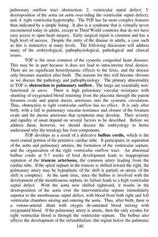
pulmonary outflow tract obstruction; 2. ventricular septal defect; 3.dextroposition of the aorta (or aorta over-riding the ventricular septal defect);and, 4. right ventricular hypertrophy. The TOF has far more complex featuresthan indicated by a simple listing. It also is a syndrome that is virtually neverencountered today in adults, except in Third World countries that do not haveeasy access to open heart surgery. Early surgical repair is common and has ahigh level of success. Despite the rarity of the disease in adults, a case suchas this is instructive at many levels. The following discussion will addressmany of the embryological, pathophysiological, pathological and clinicalissues.
TOF is the most common of the cyanotic congenital heart diseases.This may be in part because it does not lead to intra-uterine fetal demise.There are no significant hemodynamic effects to the fetus, and the diseaseonly becomes manifest after birth. The reasons for this will become obviousas we discuss the pathology and pathophysiology. The primary abnormalityin TOF is obstruction to pulmonary outflow. The lungs are essentially non-functional in utero. There is high pulmonary vascular resistance withshunting of oxygenated blood returning from the placenta through the patentforamen ovale and patent ductus arteriosus into the systemic circulation.Thus, obstruction to right ventricular outflow has no effect. It is only afterbirth, with a fall in pulmonary vascular resistance and closure of the foramenovale and the ductus arteriosus that symptoms may develop. Their severityand rapidity of onset depend on several factors to be described. Before weaddress them, however, we should discuss the embryology to betterunderstand why the tetralogy has four components.
TOF develops as a result of a defective bulbus cordis, which is themost cranial portion of the primitive cardiac tube. It participates in separationof the aorta and pulmonary arteries, the formation of the ventricular septum,and the organization of the right ventricular outflow tract. An abnormalbulbus cordis at 5-7 weeks of fetal development leads to inappropriateseptation of the truncus arteriosus, the common artery leading from thecardiac tube. If the spiral septum in the truncus is shifted toward the right, thepulmonary artery may be hypoplastic (if the shift is partial) or atretic (if theshift is complete). At the same time, since the bulbus is involved with thedevelopment of the membranous septum, its failure leads to a high ventricularseptal defect. With the aorta now shifted rightward, it results in thedextroposition of the aorta over the interventricular septum immediatelysuperior to the membranous septal defect, with blood from both right and leftventricular chambers mixing and entering the aorta. Thus, after birth, there isa venous-arterial shunt with oxygen de-saturated blood mixing withoxygenated blood. If the pulmonary artery is atretic, then the only exodus forright ventricular blood is through the ventricular septum. The bulbus alsoaffects the development of the infundibulum (the region below the pulmonic
140
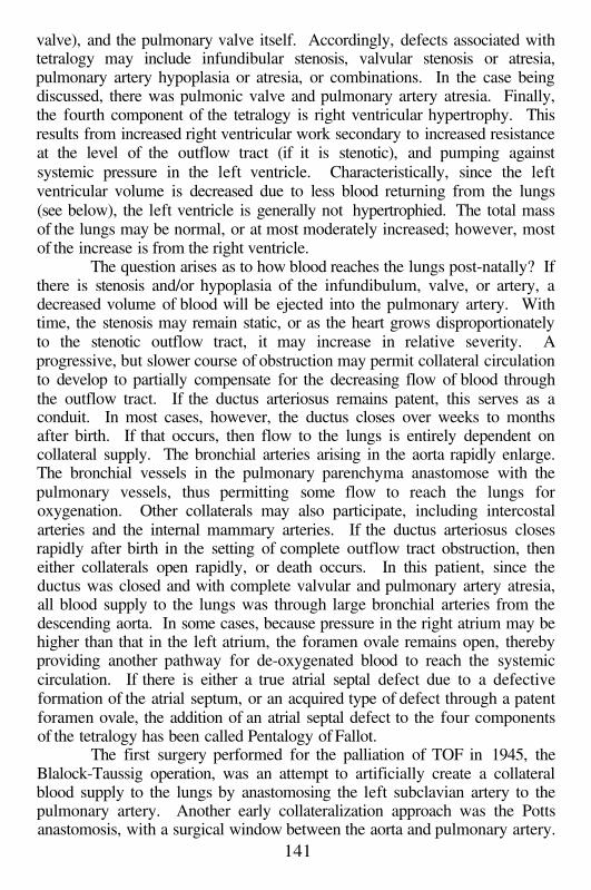
valve), and the pulmonary valve itself. Accordingly, defects associated withtetralogy may include infundibular stenosis, valvular stenosis or atresia,pulmonary artery hypoplasia or atresia, or combinations. In the case beingdiscussed, there was pulmonic valve and pulmonary artery atresia. Finally,the fourth component of the tetralogy is right ventricular hypertrophy. Thisresults from increased right ventricular work secondary to increased resistanceat the level of the outflow tract (if it is stenotic), and pumping againstsystemic pressure in the left ventricle. Characteristically, since the leftventricular volume is decreased due to less blood returning from the lungs(see below), the left ventricle is generally not hypertrophied. The total massof the lungs may be normal, or at most moderately increased; however, mostof the increase is from the right ventricle.
The question arises as to how blood reaches the lungs post-natally? Ifthere is stenosis and/or hypoplasia of the infundibulum, valve, or artery, adecreased volume of blood will be ejected into the pulmonary artery. Withtime, the stenosis may remain static, or as the heart grows disproportionatelyto the stenotic outflow tract, it may increase in relative severity. Aprogressive, but slower course of obstruction may permit collateral circulationto develop to partially compensate for the decreasing flow of blood throughthe outflow tract. If the ductus arteriosus remains patent, this serves as aconduit. In most cases, however, the ductus closes over weeks to monthsafter birth. If that occurs, then flow to the lungs is entirely dependent oncollateral supply. The bronchial arteries arising in the aorta rapidly enlarge.The bronchial vessels in the pulmonary parenchyma anastomose with thepulmonary vessels, thus permitting some flow to reach the lungs foroxygenation. Other collaterals may also participate, including intercostalarteries and the internal mammary arteries. If the ductus arteriosus closesrapidly after birth in the setting of complete outflow tract obstruction, theneither collaterals open rapidly, or death occurs. In this patient, since theductus was closed and with complete valvular and pulmonary artery atresia,all blood supply to the lungs was through large bronchial arteries from thedescending aorta. In some cases, because pressure in the right atrium may behigher than that in the left atrium, the foramen ovale remains open, therebyproviding another pathway for de-oxygenated blood to reach the systemiccirculation. If there is either a true atrial septal defect due to a defectiveformation of the atrial septum, or an acquired type of defect through a patentforamen ovale, the addition of an atrial septal defect to the four componentsof the tetralogy has been called Pentalogy of Fallot.
The first surgery performed for the palliation of TOF in 1945, theBlalock-Taussig operation, was an attempt to artificially create a collateralblood supply to the lungs by anastomosing the left subclavian artery to thepulmonary artery. Another early collateralization approach was the Pottsanastomosis, with a surgical window between the aorta and pulmonary artery.
141
Until the advent of open heart surgery in the 1950’s, these procedurespermitted prolonged survival until the and decades. These operationsare still employed as temporizing measures until the baby reaches a sufficientsize to allow for definitive surgical repair without significant increasingoperative mortality. Alternatively, in cases where there is only partial outflowtract obstruction, immediate definitive repair can be undertaken.
1.
2.
3.
The signs and symptoms of TOF are dependent on the severity of theanatomical outflow tract obstruction. There are 3 groups of patients:Babies who are cyanotic at birth, usually associated with the mostsevere obstruction of the right ventricular outflow. Because there isgenerally complete atresia or marked hypoplasia of the valve and/orartery, there is no systolic murmur, or if there is one, it is of lowintensity. These babies suffer anoxic spells, and they die within ashort time, unless the ductus arteriosus remains open and/or collateralcirculation develops rapidly, or there is surgical intervention. Ascollateral flow increases, there may be a continuous murmurauscultated over the left chest.The most common type of TOF is the one in which cyanosis developsafter one month of age, and increases in intensity during the first yearof life. These babies generally have outflow tract stenosis.Accordingly, they have a loud systolic murmur, and they may nothave continuous murmur of collateral flow. Anoxic spells may notoccur until months after birth. Other signs and symptoms develop asthey begin to ambulate. Without surgery, these infants survived untilthe or decades, and their death was usually secondary to strokeor congestive heart failure. Today, the natural history has beenmarkedly improved with surgical intervention. However, withincreasing numbers of adults living into their and decades andbeyond, after TOF surgical repair in infancy or childhood; there havebeen reports of significant late cardiac dysfunction secondary to acardiomyopathy. The cardiomyopathy may even occur at an earlyage. Late deaths and cardiac transplantations for ventricular failurehave occurred.The type of tetralogy generally does not become cyanotic untilafter one year of age. These have been referred to as ‘acyanotic orpink TOF’. They have less severe stenosis of the outflow tract, andwith reduced pulmonary vascular resistance the pulmonary bloodflow is adequate to prevent immediate cyanosis. They may not haveanoxic episodes. Symptoms, however, do increase with age, andparticularly with exercise. At rest they may not be dyspneic, but withexercise they may develop shortness of breath and cyanosis. Thismay also be exacerbated with cold weather. They do not have
142
significant polycythemia. Without surgery they may survive until theto the decade of life.
The general signs and symptoms include paroxysmal episodes ofdyspnea on exertion, and syncope particularly associated with exercise. Theinfants and children have central cyanosis. They characteristically oftensquat, following minimal exercise. This posture is spontaneous; and it isthought to result from an attempt to maximize central blood flow to the lungsby decreasing blood flow to the legs and increasing venous return to the heart.With age and more severe cyanosis, the red blood cell mass increases, leadingto secondary polycythemia. Hematocrits of 65 to 70 vol.% or higher arepossible, although there are consequences to this compensatory mechanism(discussed below). Also secondary to the oxygen de-saturation (and possiblythe intra-pulmonary collateral circulation) is the development of clubbing.Clubbing is a growth of connective tissue and thickening of periosteal bone atthe junction between the nail and soft tissue of the fingers and toes. It is notunique to TOF, but occurs in other cyanotic heart diseases, as well as inassociation with lung disease (particularly carcinoma) and cirrhosis.Although hormonal stimulation and blood flow phenomena have beenimplicated, the etiology remains unknown.
There are signs of the condition that may be evident. Today, thediagnosis is easily made with echocardiogram. In the past, cliniciansdepended on chest X-ray, electrocardiogram, and ultimately ventricularangiography. The chest X-ray typically demonstrates hypoperfused lungfields. The heart may not be enlarged, but if so it is the right ventricle thatincreases the cardiac silhouette. The pulmonary artery segment, below theaortic knob, is either absent or decreased in prominence; depending onwhether the artery is hypoplastic or atretic. The result of a prominent rightventricle and an absent pulmonary artery segment gives rise to a boot-shapedheart, or coeur en sabot. The ECG shows right axis deviation, and enlargedR waves in the right precordial leads. The degree of right ventricularhypertrophy is dependent on the degree of stenosis. On auscultation, inaddition to the murmurs described above, there is a decreased second heartsound without splitting.
An unusual physical finding, virtually never seen any more becauseof surgical intervention, is the development of hemi-atrophy of the body. Theatrophy occurs opposite the side in which the aorta arches. In other words,the normal side would be the left, if there were a normal left-sided aortic arch.Since another associated finding seen in TOF, which we have not discussed,is the presence of a right aortic arch (up to 30% of cases), the hemi-atrophywould be on the left side, with the right side normal. The condition can bediagnosed clinically with this finding alone. Obviously, on chest X-ray or
143
echocardiography the anomaly can be confirmed. The etiology is notunderstood.
The complications of tetralogy have been altered by surgicalintervention, but now include the late-onset cardiomyopathy and heart failureas described above. Complications include sudden death secondary toprolonged anoxic spells. Other complications are directly related to the flowacross the ventricular septal defect, and flow across a stenotic pulmonaryoutflow tract. Thus, endocarditis may occur, particularly in regions ofturbulent high velocity flow. A particular complication associated with rightto left shunt, characteristic of tetralogy but not unique, is the development ofcerebral abscess. This was the cause of death in our patient. Cerebral abscessis a result of flow from the right-sided circulation with organisms from theintestinal tract and elsewhere, which cross the membranous septum and gainaccess to the systemic circulation. It has been suggested that the decreasedfiltering of organisms by the hypoperfused lungs contributes to thiscomplication. It is also possible that infected emboli may pass paradoxicallyacross the septum and directly travel to the brain. Cerebral infection may alsooccur as a result of secondary bacterial seeding of bland cerebral thrombi.
Cerebral and other organ thrombi are common in tetralogy as a resultof the polycythemia. Thus, thrombotic, non-infectious strokes may occur.The increased red blood cell mass enhances thrombogenesis, particularlywhen the hematocrit is greater than 70 vol.%. At this level, there is sludgingof flow, and also decreased capillary flow with secondary local tissueischemia. It is also important to recognize that there may be relative anemiain these children, even with hematocrits of 40 vol.%, and hemoglobin of 10-13 g/dL. These levels, though normal for children without cyanotic heartdisease, are distinctly abnormal for children with TOF. Iron deficiency maybe an important contributing factor.
As discussed earlier, this case is extremely unusual in this era ofaggressive surgical intervention. It is unknown why she was not surgicallyrepaired, although there was speculation that it had to do with her mentalretardation and placement in foster homes. She had severe pulmonaryoutflow tract obstruction with atresia of the valve and the artery. The ductusarteriosus was closed. Thus, the only blood supply to her lungs was throughthe very large bronchial arteries that extended to both sides. She did not havea murmur upon admission, although admittedly since she was in a terminalstate secondary to her cerebral infection and septicemia, the cardiaccontraction may not have been sufficient to generate one. She had no splittingof the second heart sound. She had clubbing, cyanosis, and polycythemia.There was moderate cardiac hypertrophy, with both right ventricularhypertrophy and mild dilatation. She died secondary to a cerebral abscess,with changes that were subacute consistent with a duration of days to severalweeks. The abscess demonstrated gram-positive cocci that eventually grew
144
gamma-hemolytic streptococcus. In addition, she had ventriculitis andmeningitis, and the extensive cerebral infection led to cerebral edema andherniation. She also had bronchopneumonia. The lungs demonstratedmultiple thrombi, with organization and recanalization. The latter, is anothercommon finding in tetralogy that may contribute to late deterioration of rightventricular function due to increasing pulmonary vascular resistance, even inthose patients without severe obstruction. Her survival to age 28 is fairlycharacteristic of TOF as an overall group (without surgery), but distinctly rarewith the type of complete outflow obstruction she had. The survival can onlybe attributed to the large size of the bronchial collateral vessels that produceda spontaneous shunt comparable to a Blalock-Taussig anastomosis.
145
Suggested Readings
1.
2.
3.
4.
Friedman WF, Silverman N. “Congenital Heart Disease in Infancy andChildhood”. In Braunwald: Heart Disease: A Textbook of CardiovascularMedicine. Sixth ed. Braunwald E, Zipes DP, and Libby P eds. W. B.Saunders Company, 2001Deanfield JE, Gersh BJ, Warnes CA, Mair DD. “Congenital heart diseasein adults”. In Hurt’s The Heart. Ninth Edition. Alexander WR, SchlantRC, and Fuster V eds. McGraw-Hill Co, Inc., 1998Hokanson JS, Moller JH. Adults with tetralogy of Fallot: long-termfollow-up. Cardiol Rev. 1999; 7:149-55.McNamara DG. The adult with congenital heart disease. Curr ProblCardiol. 1989; 14:57-114.
146

147
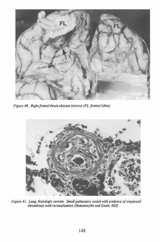
148
Case # 16
Clinical Summary
This was a 74 year old female with several medical problemsincluding adult onset diabetes mellitus, hypothyroidism treated withthyroxine, congestive hear failure and one transient cerebral ischemic attackwithout residual deficit. She was partially deaf and required a left hearing aid.Her body habitus and primary amenorrhea suggested Turner’s syndrome, anda buccal smear performed several years before demonstrated 4% Barr bodiesconsistent with Turner’s mosaicism. The patient was admitted to the hospitalwith signs and symptoms of CHF. She experienced somnolence, increasingdyspnea and pedal edema for several weeks. On admission, her vital signswere BP 100/86mm Hg, P 80, irregular; RR 20, and T 37°C. Significantfindings on her physical exam included webbed neck, jugular venousdistension, bilateral carotid bruits and delayed upstroke of the carotidpulsations, fullness in the anterior neck; wide-spaced nipples; normal S1 andS2, S4, a III/VI systolic ejection murmur at the aortic area, and a III/VIholosystolic murmur at the apex and left sternal border radiating to the axilla.The extremities showed 3+ edema and 2+ pulses throughout. The abdominaland neurologic exams were normal.
Laboratory data and other tests
Laboratory: WBC 14.1, Hb/Hct 13/38, Plt 240. CXR: cardiomegaly, bilateralpleural effusions and congestive changes. ECG: 120/min, paroxysmal atrialtachycardia (PAT), axis at 90°, right bundle branch block (RBBB).
The patient was treated with furosemide and digoxin, and remainedrelatively stable until the fourth day post-admission, when she developedagonal respirations and died.
Gross Description
The body measured 122 cm in length and weighed 52 kg. In additionto short stature, the external exam revealed webbed neck, wide spacednipples, underdeveloped clitoris, sacral and lower extremity edema andkyphoscoliosis. The pleural cavities contained 1.0 L each of straw-coloredfluid. The pericardial sac contained also 30 cc of clear yellow fluid. Theheart weighed 400 g with concentric hypertrophy and dilatation of the leftventricle (wall thickness 1.8 cm, normal up to 1.5 cm). The right ventriclewall measured 0.5 cm. The papillary muscles were hypertrophied and the
149
myocardium showed fibrosis of the left ventricular wall with no acutechanges. The aortic valve was bicuspid, and the aortic cusps were thickenedwith calcifications (Figure 42). The chordae tendineae were slightlythickened in the mitral valve. The coronary arteries showed focalatherosclerosis with less than 50% luminal narrowing. There was acircumferential ridge in the aortic wall, just distal to the ligamentumarteriosus, with a large ulcerative plaque distal to the ridge. The intercostalarteries were not dilated. The lungs and liver were congested. The only othersignificant autopsy finding was the absence of normal thyroid tissue. Acurved band of fibrous tissue was in the normal position of the gland with themajor portion occupying the midline (isthmus) region.
Microscopic Description
The left ventricle showed marked myocellular hypertrophy, focalmyocardial fibrosis and myocytolysis. Sections from the lungs and liverconfirmed the presence of acute and chronic congestive changes. The thyroidwas markedly fibrotic with scattered nests of atrophic follicles and aprominent interstitial lymphocytic infiltrate.
Case Analysis
This complex patient presented with what appeared to be fairlyrapidly progressive congestive heart failure (CHF) over several months, ofunclear etiology. She was admitted to the hospital, but then suffered a suddenand unexpected cardiac arrest. The questions are: 1. Based on the clinicalfindings, can we develop a list of differential diagnoses, any one or acombination that might explain her course?; and 2. With the addition of thepathology results, can we pin down a specific etiology for her CHF anddemise?
What do we know objectively? She was an older woman who hadpresented initially with dyspnea and pulmonary congestion. Dyspnea, or anincreased sense of discomfort breathing (likely associated with exercise), andpulmonary congestion (either rales or radiographic findings of increasedvascular markings), are signs and symptoms of left-sided heart failure. Tocause pulmonary venous and capillary congestion, she probably had increasedend-diastolic pressure in the left ventricle, and elevated filling pressures in theleft atrium. If this was the result of ischemic heart disease, which would bethe most likely etiology in a woman in her decade of life? Although shedid not have any complaints that might suggest ischemia, she also wasdiabetic and may have had ‘silent’ ischemia. In addition, there was a historyof one event 4 years prior consistent with a transient cerebral ischemic attack
150
(TIA), which often is associated with platelet aggregates developing in thecarotid or cerebral vessels, generally secondary to atherosclerosis. Thus, shealmost certainly had some element of systemic atherosclerotic disease,possibly affecting the coronary arteries. On the other hand, a relatively rapidprogression of congestive symptoms, in the absence of an acute myocardialinfarction, would not be explained entirely by coronary artery ischemia.Moreover, we have no evidence of such an event; however, she might havehad prior myocardial damage with decompensation of an ischemic heart. Yet,we do not know what her cardiac function was, and specifically as to whethershe had a poorly contracting dilated heart. But certainly, cardiomyopathywith decompensation, and coronary artery disease with atherosclerosis, mustbe considered in the differential diagnosis.
In regard to cardiomyopathy, she progressed from apparent left-sidedto right-sided heart failure, when pedal edema developed. The most frequentcause of right-sided heart failure is left-sided compromise. A dysfunctionaland poorly contracting left heart, leads to pulmonary congestion, and volumeand pressure overload of the right ventricle. In spite of this, cardiomyopathyof various etiologies may directly affect right heart function, secondary to thegeneralized damage of the right ventricular myocardium, and right atrial wall.Ischemia is usually not a common cause of such damage. Based on thepatient’s history, are there other possible causes for cardiomyopathy? Thereare at least two. She was diabetic, and she was hypothyroid.
Diabetic cardiomyopathy must be considered, but it does not appearto be a likely explanation. It has been discussed in case #7. For one thing,she had had adult-onset Type II diabetes for only 6 years, and she was noteven being treated with medication. Secondly, cardiomyopathy usuallydevelops in diabetic patients of more than 10 years, and generally in thosewho are not controlled solely by diet. Lastly, it is most commonly seen indiabetics who are also hypertensive, and there was no evidence ofhypertension during her prior evaluations. Therefore, diabeticcardiomyopathy should not be a diagnosis high on our list.
What about hypothyroidism? Does it cause cardiomyopathy? Theanswer is yes. In fact, both hyperthyroidism and hypothyroidism can causecardiac dysfunction and congestive heart failure, although through twodistinctly different mechanisms. Hyperthyroid disease is associated withtachycardia and elevated catecholamines, which may lead to multiple foci ofmyocardial cell necrosis (e.g. myocytolysis). Clinically, hyperthyroidismmay cause high-output heart failure, but mostly in patients with pre-existingheart disease. Atrial fibrillation is common, usually with a rapid ventricularresponse. The high-output state is secondary to the tachycardia, whichtogether with peripheral vasodilatation and decreased afterload, increasesstroke volume but results in poor organ perfusion (similar to an arteriovenousfistula). Tachycardia may reduce diastolic filling time, and compromise
151
coronary blood flow, thus leading to angina pectoris. Furthermore, persistenttachycardia can lead to a remodeling of the left ventricle, with mural thinningand ventricular cavity dilation. However, hyperthyroid patients usuallypresent because of the rapid heart rate or chest pain, before the left ventriclechanges occur. An additional effect of increased thyroid hormone, is itsfunction as a protein synthesis promoter that may stimulate myocytehypertrophy, and ultimately ventricular hypertrophy, independent ofhypertension. However, this effect alone is not sufficient to account for thedevelopment of heart failure.
Hypothyroidism leads to cardiomyopathy through differentmechanisms. There is often associated hypertension and left ventricularhypertrophy. The hypothyroid state elevates low density lipoproteins (LDL)cholesterol, and may lead to accelerated atherosclerosis. Thus, in addition tothe metabolic effects of hypothyroidism, there may be an element of coronaryartery ischemia. The hypothyroid heart has increased sensitivity tocatecholamines, which may lead to small areas of myocyte necrosis andsubsequent fibrosis. The pathogenesis of hypothyroid cardiomyopathy is theresult of leakage of myxoid glycoprotein into vessel walls and into theinterstitium of the heart, similar to the development of myxedema in the skinand subcutaneous tissue. There is also a metabolic intracellular effect, withthe accumulation of myocyte sarcoplasmic inclusions. This condition hasbeen termed ‘basophilic degeneration of the heart’, because the material stainsblue with hematoxylin (Figure 43). The inclusions are a glycoprotein,identical to the interstitial and cutaneous material. It stains strongly withperiodic acid-Schiff (PAS stain), but it is not digested with diastase, therebyindicating that it is not glycogen. Although its metabolic effects areunknown, it is thought to damage the contractile apparatus of the cell. Oneadditional non-cardiomyopathic consequence of hypothyroidism that maylead to heart failure, is the leakage of proteinaceous fluid into the pericardialsac which may progress to tamponade. The fluid leaks as a result of alteredpermeability of lymphatic and venular vessels, and increased venous pressuresecondary to heart failure. The pericardial effusion is unique, and post-percardiocentesis, its gross examination can be used to diagnosehypothyroidism: The fluid has gold flecks floating in it, which representcholesterol crystals. This is one of those increasingly rare situations inmedicine, where a complex disease can be diagnosed by simple observation(with no requirement for CT scan, MRI, or echocardiogram!!).
This patient was hypothyroid, a common association with Turner’ssyndrome (a subject to which we will return shortly), as a result ofautoimmune thyroiditis (Hashimoto’s disease), present in up to 20% ofaffected individuals. Although she was chronically treated with thyroidreplacement hormone (thyroxine), she may have been non-adherent andstopped taking her thyroid medication along with diuretic and digitalis. Also,
152
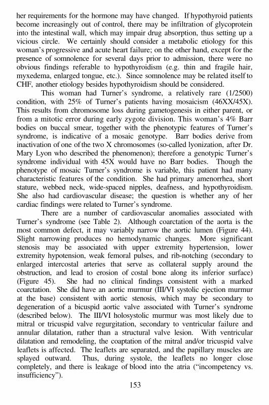
her requirements for the hormone may have changed. If hypothyroid patientsbecome increasingly out of control, there may be infiltration of glycoproteininto the intestinal wall, which may impair drug absorption, thus setting up avicious circle. We certainly should consider a metabolic etiology for thiswoman’s progressive and acute heart failure; on the other hand, except for thepresence of somnolence for several days prior to admission, there were noobvious findings referable to hypothyroidism (e.g. thin and fragile hair,myxedema, enlarged tongue, etc.). Since somnolence may be related itself toCHF, another etiology besides hypothyroidism should be considered.
This woman had Turner’s syndrome, a relatively rare (1/2500)condition, with 25% of Turner’s patients having mosaicism (46XX/45X).This results from chromosome loss during gametogenesis in either parent, orfrom a mitotic error during early zygote division. This woman’s 4% Barrbodies on buccal smear, together with the phenotypic features of Turner’ssyndrome, is indicative of a mosaic genotype. Barr bodies derive frominactivation of one of the two X chromosomes (so-called lyonization, after Dr.Mary Lyon who described the phenomenon); therefore a genotypic Turner’ssyndrome individual with 45X would have no Barr bodies. Though thephenotype of mosaic Turner’s syndrome is variable, this patient had manycharacteristic features of the condition. She had primary amenorrhea, shortstature, webbed neck, wide-spaced nipples, deafness, and hypothyroidism.She also had cardiovascular disease; the question is whether any of hercardiac findings were related to Turner’s syndrome.
There are a number of cardiovascular anomalies associated withTurner’s syndrome (see Table 2). Although coarctation of the aorta is themost common defect, it may variably narrow the aortic lumen (Figure 44).Slight narrowing produces no hemodynamic changes. More significantstenosis may be associated with upper extremity hypertension, lowerextremity hypotension, weak femoral pulses, and rib-notching (secondary toenlarged intercostal arteries that serve as collateral supply around theobstruction, and lead to erosion of costal bone along its inferior surface)(Figure 45). She had no clinical findings consistent with a markedcoarctation. She did have an aortic murmur (III/VI systolic ejection murmurat the base) consistent with aortic stenosis, which may be secondary todegeneration of a bicuspid aortic valve associated with Turner’s syndrome(described below). The III/VI holosystolic murmur was most likely due tomitral or tricuspid valve regurgitation, secondary to ventricular failure andannular dilatation, rather than a structural valve lesion. With ventriculardilatation and remodeling, the coaptation of the mitral and/or tricuspid valveleaflets is affected. The leaflets are separated, and the papillary muscles aresplayed outward. Thus, during systole, the leaflets no longer closecompletely, and there is leakage of blood into the atria (“incompetency vs.insufficiency”).
153
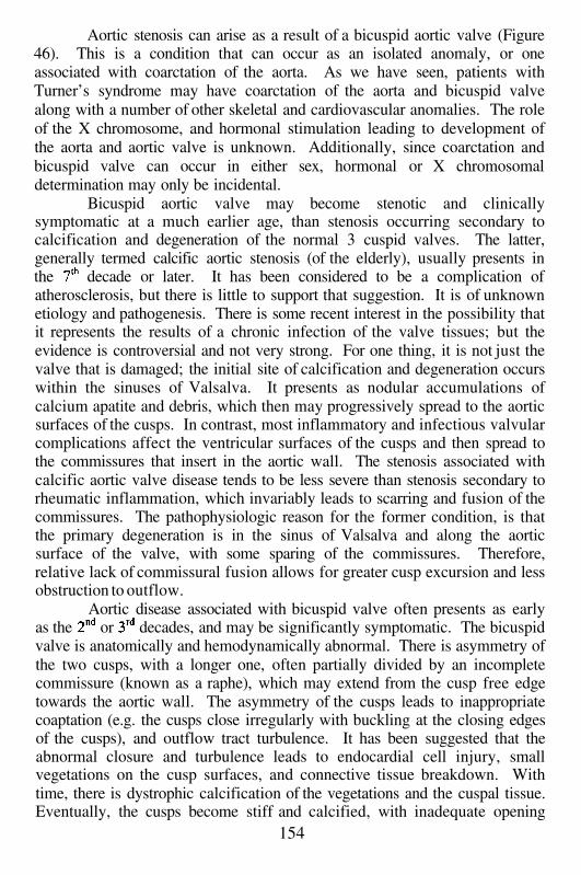
Aortic stenosis can arise as a result of a bicuspid aortic valve (Figure46). This is a condition that can occur as an isolated anomaly, or oneassociated with coarctation of the aorta. As we have seen, patients withTurner’s syndrome may have coarctation of the aorta and bicuspid valvealong with a number of other skeletal and cardiovascular anomalies. The roleof the X chromosome, and hormonal stimulation leading to development ofthe aorta and aortic valve is unknown. Additionally, since coarctation andbicuspid valve can occur in either sex, hormonal or X chromosomaldetermination may only be incidental.
Bicuspid aortic valve may become stenotic and clinicallysymptomatic at a much earlier age, than stenosis occurring secondary tocalcification and degeneration of the normal 3 cuspid valves. The latter,generally termed calcific aortic stenosis (of the elderly), usually presents inthe decade or later. It has been considered to be a complication ofatherosclerosis, but there is little to support that suggestion. It is of unknownetiology and pathogenesis. There is some recent interest in the possibility thatit represents the results of a chronic infection of the valve tissues; but theevidence is controversial and not very strong. For one thing, it is not just thevalve that is damaged; the initial site of calcification and degeneration occurswithin the sinuses of Valsalva. It presents as nodular accumulations ofcalcium apatite and debris, which then may progressively spread to the aorticsurfaces of the cusps. In contrast, most inflammatory and infectious valvularcomplications affect the ventricular surfaces of the cusps and then spread tothe commissures that insert in the aortic wall. The stenosis associated withcalcific aortic valve disease tends to be less severe than stenosis secondary torheumatic inflammation, which invariably leads to scarring and fusion of thecommissures. The pathophysiologic reason for the former condition, is thatthe primary degeneration is in the sinus of Valsalva and along the aorticsurface of the valve, with some sparing of the commissures. Therefore,relative lack of commissural fusion allows for greater cusp excursion and lessobstruction to outflow.
Aortic disease associated with bicuspid valve often presents as earlyas the or decades, and may be significantly symptomatic. The bicuspidvalve is anatomically and hemodynamically abnormal. There is asymmetry ofthe two cusps, with a longer one, often partially divided by an incompletecommissure (known as a raphe), which may extend from the cusp free edgetowards the aortic wall. The asymmetry of the cusps leads to inappropriatecoaptation (e.g. the cusps close irregularly with buckling at the closing edgesof the cusps), and outflow tract turbulence. It has been suggested that theabnormal closure and turbulence leads to endocardial cell injury, smallvegetations on the cusp surfaces, and connective tissue breakdown. Withtime, there is dystrophic calcification of the vegetations and the cuspal tissue.Eventually, the cusps become stiff and calcified, with inadequate opening
154
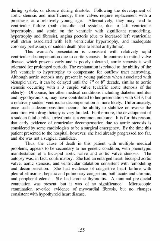
during systole, or closure during diastole. Following the development ofaortic stenosis and insufficiency, these valves require replacement with aprosthesis at a relatively young age. Alternatively, they may lead toventricular failure (both diastolic and systolic, due to left ventricularhypertrophy, and strain on the ventricle with significant remodeling,hypertrophy and fibrosis), angina pectoris (due to increased left ventricularwall strain associated with left ventricular hypertrophy, and inadequatecoronary perfusion), or sudden death (due to lethal arrhythmia).
This woman’s presentation is consistent with relatively rapidventricular decompensation due to aortic stenosis. In contrast to mitral valvedisease, which presents early and is poorly tolerated, aortic stenosis is welltolerated for prolonged periods. The explanation is related to the ability of theleft ventricle to hypertrophy to compensate for outflow tract narrowing.Although aortic stenosis may present in young patients when associated withbicuspid valve, it can be delayed until the or decade, similar to aorticstenosis occurring with a 3 cuspid valve (calcific aortic stenosis of theelderly). Of course, her other medical conditions including diabetes mellitusand hypothyroidism, may have contributed to her presentation with CHF, buta relatively sudden ventricular decompensation is more likely. Unfortunately,once such a decompensation occurs, the ability to stabilize or reverse thecondition with drug therapy is very limited. Furthermore, the development ofa sudden fatal cardiac arrhythmia is a common outcome. It is for this reason,that early evidence of ventricular decompensation due to aortic stenosis isconsidered by some cardiologists to be a surgical emergency. By the time thispatient presented to the hospital, however, she had already progressed too far,and she was not a surgical candidate.
Thus, the cause of death in this patient with multiple medicalproblems, appears to be secondary to her genetic condition, with phenotypicmanifestation of a bicuspid aortic valve and aortic valve stenosis. Theautopsy was, in fact, confirmatory. She had an enlarged heart, bicuspid aorticvalve, aortic stenosis, and ventricular dilatation consistent with remodelingand decompensation. She had evidence of congestive heart failure withpleural effusions, hepatic and pulmonary congestion, both acute and chronic,and peripheral edema. She had chronic thyroiditis. A minimal pre-ductalcoarctation was present, but it was of no significance. Microscopicexamination revealed evidence of myocardial fibrosis, but no changesconsistent with hypothyroid heart disease.
155
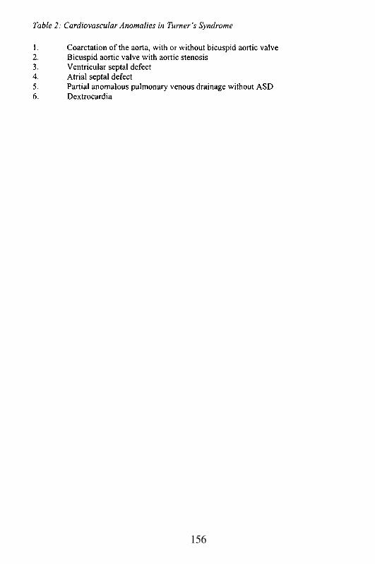
156
Suggested Readings
1.
2.
3.4.
Elsheikh M, Conway GS, Wass JA. Medical problems in adult womenwith Turner's syndrome. Ann Med. 1999; 31:99-105.Ward C. Clinical significance of the bicuspid aortic valve. Heart. 2000;83:81-5.Saenger P. Turner's syndrome. N Engl J Med. 1996; 335:1749-54.Van der Hauwaert LG, Fryns JM, Dumoulin M, Logghe N. CardiovascularMalformations in Turner’s and Noonan’s Syndrome. Br Heart J. 1978;40:500-9.
157
158
159
160
Case # 17
Clinical Summary
The patient was a 52 year old woman with past medical historysignificant for morbid obesity (205 kg), cigarette smoking with chronicobstructive pulmonary disease (COPD), and cor pulmonale. She was onoxygen therapy at home for the past 7 months. She presented to the hospitalwith complaints of increasing weight gain, abdominal and lower extremityedema with pruritus and skin ulcers. She had no fever, chills, cough, dysuria,chest pain or palpitations. She denied any trauma to her legs. Her breathingwas at baseline and she denied any acute shortness of breath.
Her physical exam on admission revealed vital signs: BP 100/70, HR72, RR 16, T 37°C. Morbidly obese woman, with oxygen, restingcomfortably in bed. Skin: bilateral lower extremities erythema with multiplevesicular lesions (<1 cm in diameter). HEENT: unremarkable, no evidence ofjaundice or conjunctival pallor. Neck: jugular venous distension (JVD), butno bruit. Heart: regular rhythm, S1S2 no S3 or S4, no murmurs appreciated.Lungs: expiratory wheeze without crackles or rales. Abdomen: globulous,positive bowel sounds, abdominal wall edema, skin dimpling, soft, and non-tender. Extremities: bilateral pitting edema (+++).
Laboratory data and other tests
Laboratory: WBC 7.7, Hb/Hct 17/55, Plt 186; bilirubin T/D 9.1/5.5, LD 378,BUN/creat 20/1.3, glucose 88, cholesterol 173, CO2 37, Ca 9.7; PT/PTT14/29. ECG: NSR, right axis deviation, tachycardia (107), and RVH. CXR:cardiomegaly. An echocardiogram showed no pericardial effusion, normalLV systolic function, moderate to severe tricuspid regurgitation, severepulmonary hypertension (RSVP 70 mmHg), dilated RV and RA.
She was aggressively diuresed with IV furosemide to treat her rightheart failure. She was also treated for skin and soft tissue infection with IVantibiotics. Despite the therapy, the patient continued to have signs ofworsening cor pulmonale with increased shortness of breath and lethargy.Ten days after admission, she was found apneic and pulseless.
Gross Description
The body weighed 205 kg, and measured 2 m (BMI > 50, indicatingmassive obesity). The heart weighed 760 g. The pericardium was smoothand contained 100 cc of serous fluid. The right atrium was significantly
161
dilated. Both right and left ventricles were dilated and hypertrophied (wallthickness 0.7 and 1.3 cm, respectively). The valve ring measurements were asfollows: tricuspid 13, mitral 10, pulmonic 8.5, and aortic 7 cm. The cusps andchordae tendineae were slender and pliable. The myocardium was notdiscolored or scarred. The coronaries showed mild atherosclerosis. The mostsignificant finding in the heart was the presence of a patent ductusarteriosus (Figures 47 and 48)
Each of the pleural cavities contained 200 cc of serous fluid. Thelungs weighed 1600 g combined. Their cut surfaces showed diffusecongestion but no discrete lesions were noted. Intimal plaques were seen inthe pulmonary vessels. Upon entering the abdominal cavity, 4 L of serousascitic fluid were obtained. The liver was congested and fatty (2800 g). Thekidneys weighed 400 g combined, with no identifiable lesions. The brain wasunremarkable.
Microscopic Description
H&E stained sections from the cardiovascular system revealedperivascular fibrosis, fat infiltration of the myocardium, myocyte hypertrophy,and mild atherosclerosis of the aorta and coronary arteries. Severe plexogenicarteriopathy was noted in the pulmonary vessels. Numerous hemosiderin-laden macrophages consistent with chronic alveolar hemorrhage, were presentin association with the damaged vessels. The plexogenic changes were betterappreciated with the elastin stain. These changes included medialhypertrophy, intimal proliferation and fibrosis, obliteration of the lumen andbranching of the vessels (Figures 49 and 50).
Case Analysis
This is a complex clinical presentation, with several differentialdiagnoses to consider as the etiology of this patient’s severe pulmonaryhypertension and right ventricular heart failure. This middle-aged womanwas massively obese, and a cigarette smoker with chronic obstructivepulmonary disease (COPD) and oxygen dependence. She had signs of severehypoxia (pO2 60 mmHg and O2 saturation 85%), hypercapnia (pCO2 60mmHg), and polycythemia (Hgb 17.4, Hct 55 vol%) indicating that thearterial blood gas findings were chronic. There was no description ofcyanosis or peripheral clubbing, but both findings may have been preventedby chronic oxygen therapy. She had elevation of bilirubin and LDH, possiblysecondary to liver congestion. There were no findings of pleural effusion orascites during life, but these may have been obscured by her massive obesity.The latter, and the thickened chest wall, could also have accounted for the
162
lack of appreciation of any cardiac murmurs, as well as the expansion of thechest cavity due to COPD. The question to be addressed is whether eitherCOPD and/or obesity are sufficient explanation for pulmonary hypertensionof such severity (mean systolic pressure 70 mmHg), which ultimately led to asudden cardiac arrhythmia and death.
COPD can certainly cause cor pulmonale, which is right heart failuresecondary to pulmonary hypertension. In general, chronic bronchitis, whichis associated with bronchiolar and pulmonary vascular sclerosis, leads topulmonary hypertension more than emphysema. However, since patients withCOPD typically have both emphysema and chronic bronchitis, this distinctionis not that meaningful. The pulmonary hypertension secondary to COPD isgenerally moderate in severity. However, it would be very unusual to havepulmonary pressures above 30-40 mmHg, independent of any other condition.Pathologically, this would generally be associated with vascular sclerosis nomore than grade II-III in the Heath and Edwards classification (see table 3 andbelow for a further discussion). We know from the clinical presentation thatthis patient had virtually systemic levels of pulmonary artery pressure, whichmakes it very unlikely to be secondary to COPD alone.
Could the massive obesity have been the additional contributingfactor to cause such marked pulmonary hypertension? Although it is possible,it is unlikely. Furthermore, the subsequent post-mortem finding of plexogeniclesions in the pulmonary circulation virtually rules out obesity as the etiology.Obesity may have several effects on the cardiovascular system. It has beenassociated with a dilated congestive cardiomyopathy of uncertain etiology.Partly, the chronic stress of supplying an increased body mass, together withthe known association with hypertension, may lead to pathologicalhypertrophy, and eventual remodeling of the ventricular wall. Contributionsfrom chronic tissue hypoxia may also result in tissue damage. The morbidlyobese patient is chronically hypoxic, a condition known as “Pickwickiansyndrome”, derived from an obese and somnolent character in Dickens’“Pickwick Papers.” Due to the massive panniculus there is difficulty inexpanding the chest wall, leading to inadequate lung volumes. Moreover,there is increased pulmonary vascular resistance due to localized areas ofatelectasis, and eventual pathologic damage to blood vessels with sclerosis.Again, the changes are rarely more than grade II-III, and the pulmonary arterypressures generally do not reach systemic levels. With time, these stressescan progress into marked pressure overload of the right ventricle, with thedevelopment of right ventricular hypertrophy, eventual dilatation and right-sided heart failure. However, even with severe COPD combined with morbidobesity, it would be extremely unusual to find pulmonary artery pressures tothe level attained by this patient.
The autopsy, unexpectedly, revealed the etiology of the severepulmonary hypertension. This patient had a patent ductus arteriosus (PDA),
163
which accounted for the plexogenic pulmonary hypertension (PPH, see table4). This congenital heart defect is a primary cause of pulmonary hypertensionat systemic levels. Moreover, plexogenic lesions in the pulmonaryvasculature occur in small to medium size vessels, often in association withatherosclerosis in larger vessels (the pulmonary artery is virtually immunefrom atherosclerosis, except in the presence of severe pulmonaryhypertension). The plexogenic lesion is a cluster or conglomeration of smallvascular channels, connective tissue, and smooth muscle cell proliferation,which develops in an area of vascular wall damage leading to apsuedoaneurysm. The latter, is an area of vessel wall destruction partiallywalled-off by surrounding tissue, or by reactive fibrosis if it develops slowly
ove r time. In contrast, a true aneurysm represents a bulge in the actual vesselwall. The plexogenic lesions are often associated with localized tissuehemorrhage (as a result of vessel wall disruption), and they also can beadjacent to diffuse angiomatous dilatation of surrounding thin-walled bloodvessels. In the Heath and Edwards classification of pulmonary vascularinjury, they represent lesions ranging from grade IV to VI (Table 3).
There are multiple etiologies of plexogenic lesions (Table 4);however, COPD and morbid obesity, even combined, are not usual causes.Of course, one must consider the possibility that a morbidly obese patient mayhave taken anorexigenic drugs such as fenfluramine-phentermine (Fen-Phen).However, in the presence of a known cause of PPH, such as a PDA, it is notnecessary to postulate another etiology: Left-to-right shunts (systemic topulmonary) are known causes of PPH, due to an increased volume andpressure overloads on the pulmonary vasculature. These lead to increasedpulmonary vascular constriction, and eventual damage to the vessel wall.Early in childhood, the functional constriction of the vasculature istransformed into anatomic constriction, with sclerosis and plexogenic lesionformation. The degree and extent of the pathologic damage to the vessels isdetermined by the size of the left-to-right shunt. Once the plexogenic lesionsdevelop, the condition is irreversible, and it will result in marked rightventricular hypertrophy, and eventually right ventricular failure (e.g. corpulmonale). With time, the hemodynamic load on the right heart progresses,and a reversal of the shunt may occur, either intermittently or continually.This takes place when the right-sided pressures in the pulmonary artery areequal to or higher than the left-sided pressures in the aorta. This condition isknown as “Eisenmenger’s syndrome”. It is a cyanotic heart condition due tothe reversal of blood flow, mixing de-oxygenated blood in the systemiccirculation, and is often associated with clubbing (bulbous overgrowth anddeformation of the distal phalangeal bone associated with marked chronic de-oxygenation). Based on the absence of cyanosis and clubbing in this patient,it is likely that she had not yet developed the reversed flow through her PDA.She probably had a balanced shunt, where the actual flow through the PDA
164
was limited by the equalization of pressures in the aorta and pulmonary artery,possibly explaining the absence of a murmur. When this occurs,demonstration of a PDA may be difficult by auscultation, since the continuousmurmur disappears, and it may be non-visualized with diagnostic techniquessuch as echocardiogram with Doppler flow evaluation. Altering thoracicvascular resistance through techniques such as the Valsalva maneuver, mayelicit a change in flow through the PDA.
The autopsy certainly revealed the severity of her cardiac andpulmonary disease. She had marked cardiomegaly (760 g, normal 350-400 gmaximum, even with her obesity), right ventricular hypertrophy, and leftventricular dilatation. In fact, both ventricles were described as dilated andhypertrophied, consistent with the chronic pathology, as well as with theventricular failure. As a sign of right-sided failure, she had pleural effusions,ascites, and liver congestion. The gross examination of the lungs revealedatherosclerotic plaques in the large pulmonary vessels; however, microscopicexamination demonstrated plexogenic lesions in small vessels (these can onlybe demonstrated microscopically; thus, in suspected clinical cases of PPH,open lung biopsy is required for diagnosis). These lesions are almostcertainly the result of the PDA and right-to-left shunt, although there mayhave been a contribution from her COPD and morbid obesity. It isnoteworthy, that PPH is virtually only seen at levels of mean arterialpulmonary pressure of 60-70 mmHg or above. Her sudden death should nothave been unexpected; particularly if her physicians knew that she had PPH,which can be an extremely lethal condition with a high prevalence of suddenventricular arrhythmia. Unfortunately, there is no long-term treatment asvascular dilating agents (e.g. calcium channel blocking drugs and nitric oxide)have only been used to decrease pulmonary vascular resistance acutely. Thesingle accepted therapy when there is severe pulmonary hypertension and corpulmonale is a heart-lung transplant. Mean survival after this transplant isapproximately 2-3 years, although longer survivals are becoming morefrequent. The transplantation was not available to the patient because of hermorbid obesity, nor was any medical therapy likely to prevent her suddenfatal outcome.
165
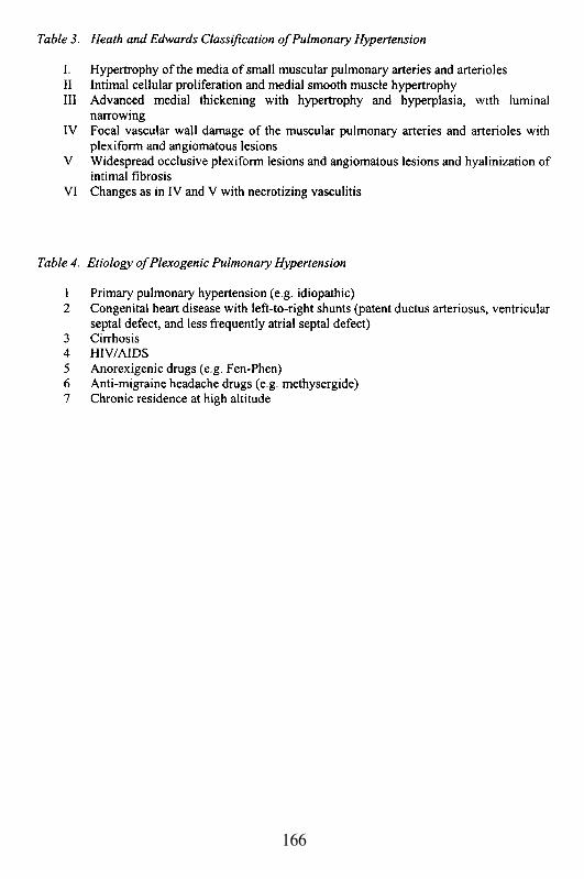
166
Suggested Readings
1.
2.
3.
4.
5.
6.
7.8.
Rich S. Pulmonary Hypertension. In Braunwald: Heart Disease: ATextbook of Cardiovascular Medicine. Sixth ed. Braunwald E, Zipes DP,and Libby P eds. W. B. Saunders Company, 2001.Voelkel NF, Tuder RM. Severe pulmonary hypertensive diseases: aperspective. Eur Respir J. 1999; 14:1246-50.Brickner ME, Hillis LD, Lange RA. Congenital heart disease in adults.First of two parts. N Engl J Med. 2000; 342:256-63.Hang CL, Sullebarger JT. Patent ductus arteriosus presenting in old age.Cathet Cardiovasc Diagn. 1993; 28:228-30.Alpert MA. Obesity cardiomyopathy: pathophysiology and evolution ofthe clinical syndrome. Am J Med Sci. 2001; 321:225-36.Anderson RC, Fagan MJ, Sebastian J. Teaching students the art andscience of physical diagnosis. Am J Med. 2001; 110:419-23.Kopelman PG. Obesity as a medical problem. Nature. 2000; 404:635-43.Heath D, Edwards JE: The pathology of hypertensive pulmonary vasculardisease: A description of six grades of structural changes in the pulmonaryarteries with special reference to congenital cardiac septal defects.Circulation 18:533-547, 1958.
167
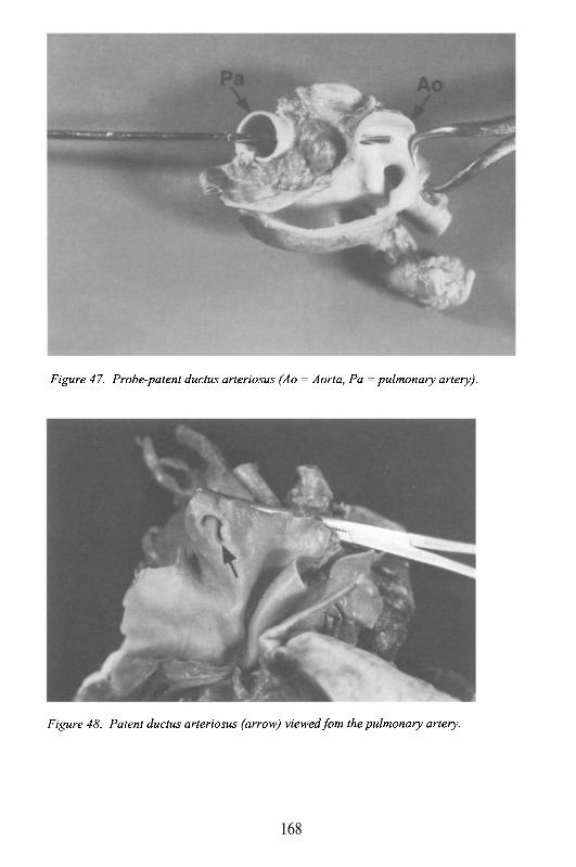
168

169

This Page Intentionally Left Blank
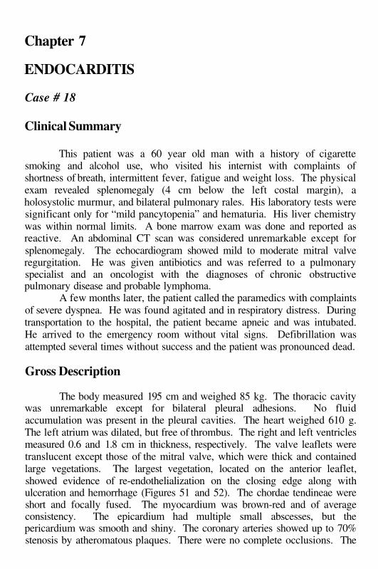
Chapter 7
ENDOCARDITIS
Case # 18
Clinical Summary
This patient was a 60 year old man with a history of cigarettesmoking and alcohol use, who visited his internist with complaints ofshortness of breath, intermittent fever, fatigue and weight loss. The physicalexam revealed splenomegaly (4 cm below the left costal margin), aholosystolic murmur, and bilateral pulmonary rales. His laboratory tests weresignificant only for “mild pancytopenia” and hematuria. His liver chemistrywas within normal limits. A bone marrow exam was done and reported asreactive. An abdominal CT scan was considered unremarkable except forsplenomegaly. The echocardiogram showed mild to moderate mitral valveregurgitation. He was given antibiotics and was referred to a pulmonaryspecialist and an oncologist with the diagnoses of chronic obstructivepulmonary disease and probable lymphoma.
A few months later, the patient called the paramedics with complaintsof severe dyspnea. He was found agitated and in respiratory distress. Duringtransportation to the hospital, the patient became apneic and was intubated.He arrived to the emergency room without vital signs. Defibrillation wasattempted several times without success and the patient was pronounced dead.
Gross Description
The body measured 195 cm and weighed 85 kg. The thoracic cavitywas unremarkable except for bilateral pleural adhesions. No fluidaccumulation was present in the pleural cavities. The heart weighed 610 g.The left atrium was dilated, but free of thrombus. The right and left ventriclesmeasured 0.6 and 1.8 cm in thickness, respectively. The valve leaflets weretranslucent except those of the mitral valve, which were thick and containedlarge vegetations. The largest vegetation, located on the anterior leaflet,showed evidence of re-endothelialization on the closing edge along withulceration and hemorrhage (Figures 51 and 52). The chordae tendineae wereshort and focally fused. The myocardium was brown-red and of averageconsistency. The epicardium had multiple small abscesses, but thepericardium was smooth and shiny. The coronary arteries showed up to 70%stenosis by atheromatous plaques. There were no complete occlusions. The
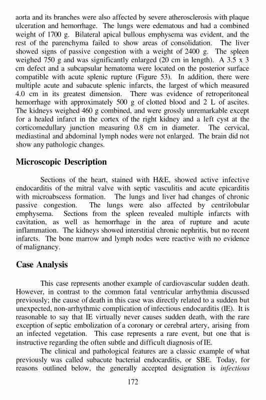
aorta and its branches were also affected by severe atherosclerosis with plaqueulceration and hemorrhage. The lungs were edematous and had a combinedweight of 1700 g. Bilateral apical bullous emphysema was evident, and therest of the parenchyma failed to show areas of consolidation. The livershowed signs of passive congestion with a weight of 2400 g. The spleenweighed 750 g and was significantly enlarged (20 cm in length). A 3.5 x 3cm defect and a subcapsular hematoma were located on the posterior surfacecompatible with acute splenic rupture (Figure 53). In addition, there weremultiple acute and subacute splenic infarcts, the largest of which measured4.0 cm in its greatest dimension. There was evidence of retroperitonealhemorrhage with approximately 500 g of clotted blood and 2 L of ascites.The kidneys weighed 460 g combined, and were grossly unremarkable exceptfor a healed infarct in the cortex of the right kidney and a left cyst at thecorticomedullary junction measuring 0.8 cm in diameter. The cervical,mediastinal and abdominal lymph nodes were not enlarged. The brain did notshow any pathologic changes.
Microscopic Description
Sections of the heart, stained with H&E, showed active infectiveendocarditis of the mitral valve with septic vasculitis and acute epicarditiswith microabscess formation. The lungs and liver had changes of chronicpassive congestion. The lungs were also affected by centrilobularemphysema. Sections from the spleen revealed multiple infarcts withcavitation, as well as hemorrhage in the area of rupture and acuteinflammation. The kidneys showed interstitial chronic nephritis, but no recentinfarcts. The bone marrow and lymph nodes were reactive with no evidenceof malignancy.
Case Analysis
This case represents another example of cardiovascular sudden death.However, in contrast to the common fatal ventricular arrhythmia discussedpreviously; the cause of death in this case was directly related to a sudden butunexpected, non-arrhythmic complication of infectious endocarditis (IE). It isreasonable to say that IE virtually never causes sudden death, with the rareexception of septic embolization of a coronary or cerebral artery, arising froman infected vegetation. This case represents a rare event, but one that isinstructive regarding the often subtle and difficult diagnosis of IE.
The clinical and pathological features are a classic example of whatpreviously was called subacute bacterial endocarditis, or SBE. Today, forreasons outlined below, the generally accepted designation is infectious
172
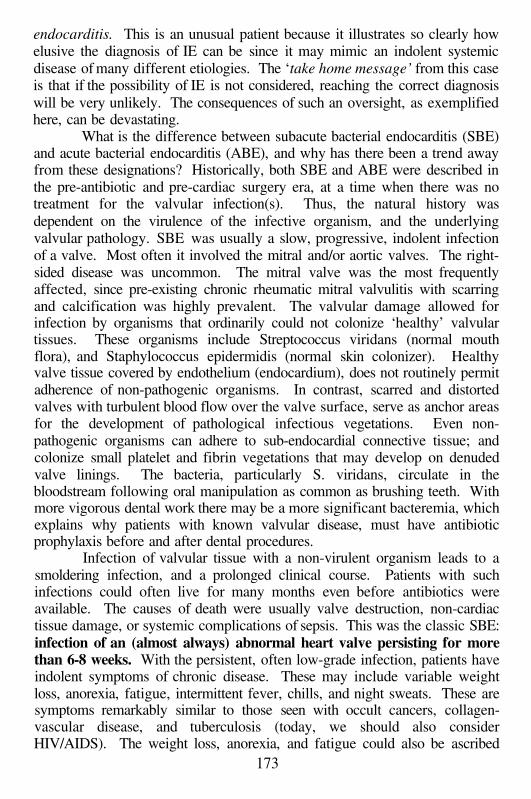
endocarditis. This is an unusual patient because it illustrates so clearly howelusive the diagnosis of IE can be since it may mimic an indolent systemicdisease of many different etiologies. The ‘take home message’ from this caseis that if the possibility of IE is not considered, reaching the correct diagnosiswill be very unlikely. The consequences of such an oversight, as exemplifiedhere, can be devastating.
What is the difference between subacute bacterial endocarditis (SBE)and acute bacterial endocarditis (ABE), and why has there been a trend awayfrom these designations? Historically, both SBE and ABE were described inthe pre-antibiotic and pre-cardiac surgery era, at a time when there was notreatment for the valvular infection(s). Thus, the natural history wasdependent on the virulence of the infective organism, and the underlyingvalvular pathology. SBE was usually a slow, progressive, indolent infectionof a valve. Most often it involved the mitral and/or aortic valves. The right-sided disease was uncommon. The mitral valve was the most frequentlyaffected, since pre-existing chronic rheumatic mitral valvulitis with scarringand calcification was highly prevalent. The valvular damage allowed forinfection by organisms that ordinarily could not colonize ‘healthy’ valvulartissues. These organisms include Streptococcus viridans (normal mouthflora), and Staphylococcus epidermidis (normal skin colonizer). Healthyvalve tissue covered by endothelium (endocardium), does not routinely permitadherence of non-pathogenic organisms. In contrast, scarred and distortedvalves with turbulent blood flow over the valve surface, serve as anchor areasfor the development of pathological infectious vegetations. Even non-pathogenic organisms can adhere to sub-endocardial connective tissue; andcolonize small platelet and fibrin vegetations that may develop on denudedvalve linings. The bacteria, particularly S. viridans, circulate in thebloodstream following oral manipulation as common as brushing teeth. Withmore vigorous dental work there may be a more significant bacteremia, whichexplains why patients with known valvular disease, must have antibioticprophylaxis before and after dental procedures.
Infection of valvular tissue with a non-virulent organism leads to asmoldering infection, and a prolonged clinical course. Patients with suchinfections could often live for many months even before antibiotics wereavailable. The causes of death were usually valve destruction, non-cardiactissue damage, or systemic complications of sepsis. This was the classic SBE:infection of an (almost always) abnormal heart valve persisting for morethan 6-8 weeks. With the persistent, often low-grade infection, patients haveindolent symptoms of chronic disease. These may include variable weightloss, anorexia, fatigue, intermittent fever, chills, and night sweats. These aresymptoms remarkably similar to those seen with occult cancers, collagen-vascular disease, and tuberculosis (today, we should also considerHIV/AIDS). The weight loss, anorexia, and fatigue could also be ascribed
173
among others, to depression, diabetes mellitus, and adrenal corticalinsufficiency. The clinical presentation might suggest SBE if there weretypical findings of a heart murmur that was changing in intensity at differenttimes. Additionally, patients with SBE may have small mucocutaneoushemorrhages peripherally, particularly in their nail beds or the so-called‘splinter hemorrhages’, and cutaneous petechiae. These lesions are generallythought to be secondary to small non-infected emboli from the valvevegetation, although infected material or immune mediation may play a role.Since the spleen is frequently enlarged, patients may present with left upperquadrant pain and tenderness. Splenic involvement includes changesassociated with sepsis (‘acute splenitis’), infarction and/or abscess formation(septic emboli). Moreover, when the infection has persisted for some time,the valves leaflets or cusps are destroyed, which results in valvularinsufficiency, and ultimately in ventricular failure. Often, patients succumbsecondary to systemic embolization, particularly to the brain, where theymight develop cerebral infarctions, abscesses, and meningitis. Patients mayalso develop systemic disease related to the prolonged indolent course of theirinfection. Specifically, the subacute infection may lead to the development ofantigen-antibody complexes, with the antigen being bacterial membraneprotein. These complexes can circulate in the bloodstream, and ultimatelydeposit in small skeletal muscle and skin vessels, and in glomerular capillariesof the kidney. In the former sites, they may lead to a necrotizing vasculitis,with tissue inflammation and necrosis. When present in the palms, fingerpulp, or soles of the feet, they are known as Osler’s nodes. In the kidney, afocal, necrotizing glomerulonephritis may develop, which is not secondary toembolism of vegetations (even though classically it has been called focalembolic glomerulonephritis). Other manifestations of immune complexdisease include arthralgias and arthritis.
Despite the relative ease with which the diagnosis of SBE should bemade in patients who present with these clinical signs and symptoms; the factremains that, even in this modern era of echocardiography, it is still a diseaseassociated with a high number of mis-diagnoses. The primary reason, otherthan physicians not considering it, is that many patients do not present with allof these classic features. Since the infection is often a subtle one, and giventhe intermittent nature of active bacterial release into the blood stream,negative blood cultures may be common, unless taken frequently.Furthermore, once the bacteria extend into the valve tissue, or into the peri-valvular annulus, they are not released into the bloodstream. Clinically, theremay be no changing murmur; petechiae may not be present, or in dark-skinned patients, may be difficult to appreciate; and complications associatedwith immune complexes may not develop. However, non-specific clinicalsymptoms may suggest cancer, or systemic diseases like lupus erythematosus.Laboratory signs such as low-grade leukocytosis (with white blood cell counts
174
between 10,000-15,000), and elevated erythrocyte sedimentation rate (ESR)are relatively non-specific, and may not be present. So again, as statedpreviously, if you don’t think of the diagnosis, you won't make it!
In contrast to SBE, acute bacterial endocarditis (ABE) has differentfeatures and pathogenesis. It is generally associated with infection by virulentorganisms, particularly Staphylococcus aureus, Streptococcus pneumoniae(pneumococcus), enterococci, and a number of Gram-negative bacilli. As aresult of the virulence of these or other pathogens, pre-existing valve damagemay or may not be present. With aggressive, rapidly growing, and destructiveorganisms, there is an acute and progressive tissue disruption associated withcollagenolysis of valvular and annular connective tissue. Intra-valvular andannular abscesses may occur. Large platelet and fibrin vegetations develop,which are very susceptible to fragmentation and embolization. The timecourse of the disease is rapid, often measured in days or few weeks.Moreover, signs and symptoms of valvular insufficiency are frequent andoccur early. Systemic embolization to other organs, with tissue infarction andabscess formation, is common. Because of the acuteness of the disease, thereis generally not sufficient time for the immune complexes to develop. Thus,the immune complications characteristic of SBE are usually absent.Historically, death ensues in a relatively short time, usually secondary tocardiac failure with valve destruction, or septicemia. With these features,ABE has been defined as: infection of a normal or abnormal heart valve bya virulent organism, with disease generally less than 4 weeks in duration.
With such a clear distinction between SBE and ABE, why are thesedesignations inappropriate today? Why is the preferred terminology: activeinfectious endocarditis? The simple answer is because antibiotics, andearlier diagnoses with the advent of echocardiography, have modified thenatural history of the disease. In addition, surgical intervention has had amajor impact in the course of this disease. Prolonged survival is now possiblewith non-virulent and virulent organisms, allowing for the possibility ofimmune-mediated complications to develop. Finally, as a result ofintravenous drug use and the HIV/AIDS epidemic, infections with fungi(aspergillus and Candida species, in particular), rather than bacteria, havegained tremendous attention. Although it is still true, that infection withStaphylococcus aureus and pneumococcus is more severe and moredestructive than infection with S. viridans, medical intervention has changedthe classic features of SBE and ABE.
This patient presented in a terminal state, and had a cardio-pulmonaryarrest during transportation to the hospital. Death was a result of septicemia,cardiac failure, and acute splenic rupture. It is unlikely that he hadhypovolemia, as only 500 cc of blood were found in the retroperitoneal space.The presence of 2 liters of ascites in the peritoneum was most probably fromcongestive heart failure (there was no evidence of cirrhosis). His heart was
175
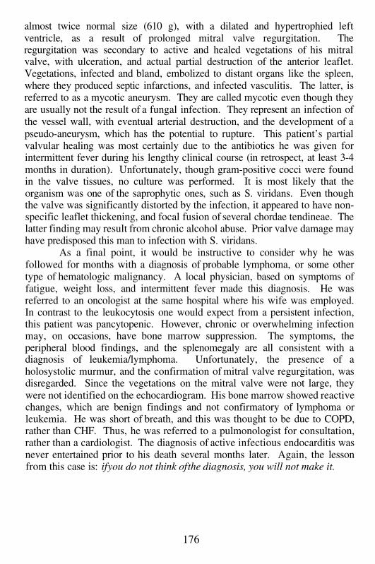
almost twice normal size (610 g), with a dilated and hypertrophied leftventricle, as a result of prolonged mitral valve regurgitation. Theregurgitation was secondary to active and healed vegetations of his mitralvalve, with ulceration, and actual partial destruction of the anterior leaflet.Vegetations, infected and bland, embolized to distant organs like the spleen,where they produced septic infarctions, and infected vasculitis. The latter, isreferred to as a mycotic aneurysm. They are called mycotic even though theyare usually not the result of a fungal infection. They represent an infection ofthe vessel wall, with eventual arterial destruction, and the development of apseudo-aneurysm, which has the potential to rupture. This patient’s partialvalvular healing was most certainly due to the antibiotics he was given forintermittent fever during his lengthy clinical course (in retrospect, at least 3-4months in duration). Unfortunately, though gram-positive cocci were foundin the valve tissues, no culture was performed. It is most likely that theorganism was one of the saprophytic ones, such as S. viridans. Even thoughthe valve was significantly distorted by the infection, it appeared to have non-specific leaflet thickening, and focal fusion of several chordae tendineae. Thelatter finding may result from chronic alcohol abuse. Prior valve damage mayhave predisposed this man to infection with S. viridans.
As a final point, it would be instructive to consider why he wasfollowed for months with a diagnosis of probable lymphoma, or some othertype of hematologic malignancy. A local physician, based on symptoms offatigue, weight loss, and intermittent fever made this diagnosis. He wasreferred to an oncologist at the same hospital where his wife was employed.In contrast to the leukocytosis one would expect from a persistent infection,this patient was pancytopenic. However, chronic or overwhelming infectionmay, on occasions, have bone marrow suppression. The symptoms, theperipheral blood findings, and the splenomegaly are all consistent with adiagnosis of leukemia/lymphoma. Unfortunately, the presence of aholosystolic murmur, and the confirmation of mitral valve regurgitation, wasdisregarded. Since the vegetations on the mitral valve were not large, theywere not identified on the echocardiogram. His bone marrow showed reactivechanges, which are benign findings and not confirmatory of lymphoma orleukemia. He was short of breath, and this was thought to be due to COPD,rather than CHF. Thus, he was referred to a pulmonologist for consultation,rather than a cardiologist. The diagnosis of active infectious endocarditis wasnever entertained prior to his death several months later. Again, the lessonfrom this case is: if you do not think of the diagnosis, you will not make it.
176
Suggested Readings
1.
2.3.
4.
5.
6.
Hoesley CJ, Cobbs CG. Endocarditis at the millennium. J Infect Dis.1999; 179 (Suppl 2):S360-5.Farmer JA, Torre G. Endocarditis. Curr Opin Cardiol. 1997; 12:123-30.Cunha BA, Gill MV, Lazar JM. Acute infective endocarditis. Diagnosticand therapeutic approach. Infect Dis Clin North Am. 1996; 10:811-34.Harris PS, Cobbs CG. Cardiac, cerebral, and vascular complications ofinfective endocarditis. Cardiol Clin. 1996; 14:437-50.Keys TF. Infective endocarditis: prevention, diagnosis, treatment, referral.Cleve Clin J Med. 2000; 67:353-60.Burke AP, Kalra P, Li L, Smialek J, Virmani R. Infectious endocarditisand sudden unexpected death: incidence and morphology of lesions inintravenous addicts and non-drug abusers. J Heart Valve Dis. 1997; 6:198-203.
177
178
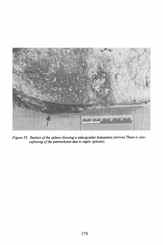
179
Case # 19
Clinical Summary
This 51 year old African American homosexual man, with a historyof intravenous drug abuse, was admitted to the hospital with a chief complaintof increasing dyspnea on exertion, fever and left facial droop. He also had ahistory of non-productive cough and a 23 kg weight loss over the past fewmonths. He had been seen by a cardiologist who performed anechocardiogram, told the patient he had congestive heart failure (CHF), andstarted him on furosemide and digoxin.
On admission his vital signs were T 39.4°C, Pulse 105/min, R 20/min,BP 140/60 mmHg. His physical exam was remarkable for anterior cervicaland inguinal lymphadenopathy. His cardiac exam revealed a split S1, a gradeII/VI holosystolic murmur best heard at the apex and radiating to the axilla,and another II/VI systolic murmur best heard along the lower left sternalborder. Pistol shot pulses with strong upstrokes and fast downstrokes werenoted. Pedal edema was present bilaterally. The neurologic exam revealedleft facial weakness, confusion, weaker left grip, and left pronator drift.Asterixis was noted on the left.
Laboratory data and other tests
Laboratory: WBC 13.2, Hb/Hct 8.6/26, Plt 320; bilirubin T/D 9.1/5.5, LD378, BUN/creat 26/1.6, glucose 89, CO2 20. ECG: Sinus tachycardia, Qwaves and decreased T waves in lead III. CXR: bilateral interstitialinfiltrates. Head CT: remote left temporal lobe infarct.
The patient was admitted with the presumptive diagnosis ofendocarditis and AIDS-related pneumonia, and was started on ampicillin,nafcillin, cefoxitin and trimethoprim-sulfamethoxazole (TMP/SMX). Bloodcultures grew pneumococci and enterococci. The antibiotics were changed topenicillin, and gentamicin. A lumbar puncture was performed whichdemonstrated RBC 690, WBC 170 (90% polymorphonuclear leukocytes),protein 14, and glucose 41 (serum glucose 87). India ink and gram stains ofthe cerebrospinal fluid were negative. Echocardiography revealed prominentaortic insufficiency, with aortic and mitral valve vegetations. During hishospital stay he developed left hemiparesis with flat affect. A repeat head CTwith contrast demonstrated no ring enhancement of his existent left fronto-temporal lobe lesion. During his 5th hospital day, he developed a pericardialfriction rub and was started on captopril to reduce his cardiac afterload. Onday 9 post-admission he was found in the bathroom pulseless and
180
unresponsive. Resuscitation efforts, which included pacemaker insertion,were unsuccessful.
Gross Description
The body was that of a well-developed, well nourished black manmeasuring 162 cm in length and weighing 63 kg. The heart weighed 490 gand was of the expected shape. The atria were normal in size and free ofthrombi. The foramen ovale was closed. The right and left ventricular wallsmeasured 0.6 cm and 1.8 cm in thickness, respectively. There were multiplelarge vegetations on the aortic, mitral and tricuspid valves with hyperemicmargins. The aortic valve vegetations hung free in the ventricle (Figures 54and 55). Just distal to the coronary arteria ostia and above the non coronarycusp there was a pseudo-aneurysm with rupture into the pericardium causinghemopericardium (200 cc). The pericardium showed fibrinous pericarditis.The endocardium was thin and glistening. The chordae tendineae werethickened. The myocardium was grossly unremarkable. The aorta and itsbranches showed moderate calcific atherosclerosis, especially in theabdominal portion. The lungs weighed 1800 g combined and were affectedby centrilobular emphysema. The kidneys weighed 400 g combined. Therenal parenchyma showed multiple small abscesses (<0.5 cm diameter). Thespleen was grossly unremarkable (weight 250 g). The brain weighed 1480 gand had evidence of an old left fronto-temporal lobe and recent right fronto-parietal and insular cortex infarcts.
Microscopic Description
Sections from the heart stained with hematoxylin and eosin revealedmild myocellular hypertrophy with scattered areas of old myocytolysis. Themitral and aortic valves showed active infective endocarditis. In addition,sections from the aortic tear and pericardium showed acute and chronicaortitis and fibrinous pericarditis, respectively. The kidneys demonstratedacute necrotizing glomerulitis. Sections from the upper lobe of the right lungrevealed a solitary infiltrating moderately differentiated adenocarcinoma witha bronchioalveolar pattern. The non-neoplastic lung parenchyma showedacute and organizing interstitial pneumonitis. Sections of the right fronto-parietal area of the brain confirmed the presence of subacute infarcts. Themiddle cerebral artery had an occluded branch containing a septicthromboembolus with colonies of bacteria and numerous neutrophils andmononuclear cells within and around the vessel.
181
Case Analysis
Fulminant, acute bacterial endocarditis (ABE) or subacuteendocarditis (SBE)? This case illustrates the confusion in the terminologydiscussed previously. A rapidly progressive disease leading to death withinone week involving virulent organisms (pneumococcus and enterococcus), noobvious pre-existing valve pathology, extensive valvular and blood vesseldestruction, and widespread systemic complications of embolization. Itcertainly fits with the features of ABE, yet he also had significant weight lossover several months, and glomerular inflammation (e.g. glomerulitis), whichindicated an immune complication of a more indolent disease. Of course,with his history of continuing intravenous drug abuse, and homosexualcontact, one could explain at least the weight loss as HIV/AIDS-related. Itwas only at autopsy, however, that his HIV status became known as negative.It was also evident that the glomerular inflammation resulted frommicroscopic emboli of infected vegetations, and not from the development ofantigen-antibody complexes in the glomerular basement membranes. Withthe uncertainty, it is preferable to designate cases such as this as active IE.But as discussed previously, such designation should not be used to obscurethe unique features of someone with a fulminant, aggressive infectionsuperimposed on normal heart valves.
The other important lesson in this case, as in the previous discussionof endocarditis, is the failure of several clinicians to consider the diagnosis ofactive IE, even in the face of some very obvious clues. A local physician hadseen this patient, probably for complaints of shortness of breath and exerciseintolerance. He had an echocardiogram, which led to a diagnosis of CHF.Presumably, even though we do not have access to this echocardiogram, itmost likely showed ventricular contractile dysfunction, and possibly mitralvalvular insufficiency. As we have seen previously in patients with dilatedcardiomyopathy, ventricular dysfunction and valvular insufficiency mayresult from ventricular remodeling and annular dilatation. If vegetations arenot appreciated or looked for on the valves, the etiology may be missed, andascribed to myocardial disease. Certainly, with his personal history and hisweight loss, the assumption that he had HIV/AIDS cardiomyopathy, orcocaine-induced cardiomyopathy, is not unreasonable. On the other hand,with intravenous drug use and acute onset CHF, it is equally reasonable toinclude IE in the list of possible differential diagnoses. Regardless, thispatient was diagnosed as having CHF, and he was started on diuretic anddigitalis therapy. Shortly after the initial visit with a local physician, thepatient presented to an affiliated hospital complaining of weakness.Unfortunately, the work-up did not demonstrate a cause, and he wasdischarged.
182
The failure to diagnose this patient with IE in the days before his finalpresentation to our hospital had devastating consequences. By the time hewas admitted with fever, increasing dyspnea on exertion, and a left facialdroop, his disease was already advanced. He had signs and symptoms of atleast two compromised valves, even before confirmation byechocardiography. Specifically, he had aortic insufficiency, with a wide pulsepressure (140/60 mmHg), and a low diastolic pressure secondary to the rapiddiastolic backflow into the left ventricle through an incompetent aortic valve.Normally, the diastolic pressure results at least partially from the recoil of theaorta as it are distended during systole. With stretching of the elastic fibers inthe aortic media, the vessel normally ‘snaps-back’, providing the coronaryarteries with blood flow during diastole. If the aortic valve is insufficient, theelastic recoil is attenuated, and the diastolic pressure falls. This also accountsfor the II/VI diastolic murmur heard along the left sternal border, althoughone would expect such a murmur to be heard loudest at the heart base. As acorrelate of the aortic insufficiency, he also had pistol-shot pulses (strongupstroke followed by a rapid downstroke). The upstroke is a sign of avigorous ventricular contraction, while the rapid downstroke is a result of thevalve being anatomically open during diastole, comparable to the lowdiastolic pressure. The pistol-shot sound, which can be auscultated, alsooccurs because of the diastolic collapse. It is typically a loud sound that canbe heard over the femoral arteries. It is one of classical pulsationabnormalities reported in aortic insufficiency (these physical examinationfindings were all described by physicians in the century, during a periodin medicine when aortic insufficiency due to infectious endocarditis, chronicrheumatic heart disease, and syphilitic aortic valve disease were common).Other signs include: 1. Corrigan’s pulse, a ‘water-hammer’ sound due to arapid collapse of pressure late in systole and during diastole. This sound isthe banging in pipes associated with rapid sudden flow, or gas bubbles in thefluid; 2. Duroziez’s sign, a to-and-fro sound heard over the femoral arteries;and, 3. Quincke’s pulse, which refers to the filling and emptying of nail-bedcapillaries, seen when firm pressure is applied to the fingertip. All of thesephenomena result from the movement of blood back through an insufficientvalve, and its association with diastolic collapse.
This patient also had evidence of mitral valve disease. He was notedto have a grade II/VI holosystolic murmur at the apex radiating to the leftaxilla. This is a typical location of a mitral valve murmur, and theholosystolic nature of the murmur is consistent with mitral regurgitation. TheII/VI systolic murmur heard along the left sternal border is less easilycharacterized as to site of origin. It may represent turbulent flow across adistorted and damaged aortic valve, or ventricular cavity turbulence resultingfrom the aortic and mitral valve insufficiency. Ultimately, specific aorticvalve and mitral valve disease was confirmed on echocardiography, with large
183
vegetations on both valves, and prominent aortic insufficiency. It isnoteworthy that the intensity of the murmurs was not as loud as one wouldexpect from the degree of damage appreciated on echocardiography, oreventually found at autopsy. This is not an unusual observation, as theintensity of the murmur may vary with cardiac function, size of vegetation,and degree of valvular pathology. If, for instance, there is a perforation of theleaflet or cusp, this may intermittently be covered by vegetation with no flowthrough the hole. In the case of a torn cusp or ruptured group of chordaetendineae, the flail tissue may not move consistently with each cardiac cycle,thereby making the murmur variable in intensity. This anatomical variabilityis the explanation for the so-called ‘changing murmur’, which can occur in IE.Other manifestations of anatomic distortion of valvular tissues includes themusical or high-pitched cooing murmurs due to rapidly vibrating loose tissueor ruptured chordae; and the Austin Flint murmur (mid-diastolic rumbling),which is thought to result from the impingement of blood coming through aninsufficient aortic valve, striking the ventricular surface of the mitral valve,and causing it to vibrate. At this point, you have probably realized that noother area in medicine is as rife with eponyms as valvular heart disease andendocarditis.
As noted above, the failure to diagnosis this patient earlier had majorconsequences that contributed to his death. By the time of presentation, henot only had fever and CHF, but also dysarthria, a left facial droop, anddrooling. These findings were presumptive evidence of intracerebral emboli,and the presence of brain infarction or abscess. Confirmation came from thelumbar puncture, which demonstrated pleocytosis with polymorphonuclearpredominance and decreased CSF glucose (CSF to blood ratio less than 2/3),suggesting meningitis. Cerebral abscess from embolized fragments ofvegetation can often extend to the meningeal surface with the development ofmeningitis. Small fragments of vegetation preferentially embolize to themicrocirculation at the cortico-medullary junction in the brain; then a micro-abscess develops and extends to the brain surface, where it secondarily infectsthe meninges. Alternatively, there may be direct seeding of meningeal bloodvessels by the infective material. Cerebral abscess and meningitis areparticularly common with pneumococcal endocarditis, which was one of thetwo organisms isolated from his blood culture (another eponym associatedwith endocarditis is the Austrian’s triad: pneumococcal endocarditis,pneumonia, and meningitis). Often, by the time IE embolizes systemically,the process is too far advanced for any meaningful intervention. In this case,shortly after confirmation by echocardiography of the aortic and mitral valveinvolvement, the patient developed left hemiparesis, and mental statuschanges. Despite CT scan findings of an old left temporal lobe infarctionwith no new lesions (it may take several days for new cerebral infarctions tobecome visible), clinically, he had obvious findings consistent with acute
184
cerebral embolization. Although surgical intervention was considered, theclinicians caring for this patient considered his cardiac function stable, andbefore attempting valve replacement surgery, wanted to sterilize his valveswith antibiotics.
Several days before his death he developed a new, and potentiallyominous complication: a pericardial friction rub. This new physical findingshould have led to additional diagnostic work-up, including a repeatechocardiogram and possibly a pericardiocentesis with culture of the aspiratedmaterial. No such studies were carried out. This finding should have beenconsidered a serious new complication because it almost certainly representedspread of infection to the pericardial sac, with the development offibrinopurulent exudate on the parietal and visceral pericardial surfaces. It isthe contact of these surfaces during the cardiac cycle that leads to theauscultatory finding of friction rub (similar to the sound of two sheets of finesandpaper rubbed together). If there is sufficient fluid or pus in thepericardial sac to separate the two layers of pericardium, the friction rub maynot occur, or it may disappear if it was previously heard. There are multiplemechanisms leading to a pericardial friction rub in IE. None of them isbenign:
1. There may be spread of infection from the valve surface to thefibrous annulus of the valve, where it produces an annular (or valve ring)abscess. This is a very difficult lesion to treat, because the annulus is virtuallyavascular, and bactericidal levels of antibiotic may not reach this tissue.Annular abscess is often a cause of a smoldering, persistent infection. It isalso difficult to diagnose because blood cultures may not be positive.Additionally, it is common in patients with infection of prosthetic heartvalves, particularly mechanical valves, which rarely become infected on thesurface components of the valve, but are susceptible to bacterial growth in thearea of the cloth-covered sewing ring (where it can spread to the annulus).The valve annulus is immediately adjacent to epicardial adipose tissue, andthe epicardial surface. Thus, infection can easily spread to the pericardial sac.
2. Other mechanisms for purulent pericarditis include direct spreadfrom the vegetation to epicardial blood vessels through the blood stream.There may also be embolization of coronary vessels leading to myocardialabscess. The abscesses that develop in the heart tend to be preferentiallylocated in the subepicardium and mid-wall myocardium due to the anatomicalorganization of the microcirculation (similar to the anatomy of the cerebralmicrocirculation, which predominantly leads to the development of abscessesat the cortico-medullary junction). A subepicardial abscess can extenddirectly to the epicardium, where it causes infective pericarditis.
3. A final pathogenic mechanism is the one that affected the patient inthis case. There was spread of infection from the aortic valve cusps to theaortic wall immediately superior to the valve, in the aortic root. Aggressive
185
organisms can adhere to endothelial tissue triggering a local vascular infection(infective vasculitis). They can also spread from the annulus to the adventitiaof the aorta where external invasion of the vessel may occur. In this case, thevirulent infection resulted in the formation of a vascular abscess, and vesselwall destruction. A pseudo-aneurysm developed in the region, known as amycotic aneurysm. With spread of infection through the vessel into theadventitia and beyond, pericarditis developed, since the pericardium envelopsnot just the heart but extends around the ascending aorta up to the aortic arch.Ultimately, the pseudo-aneurysm ruptured leading to hemopericardium. Theblood in the pericardial sac (200 cc), together with the fibrinous pericarditis,was sufficient to cause pericardial tamponade, and his sudden death. Clinicalrecognition of fibrinous pericarditis in a patient with aortic and mitral valveendocarditis, might have led to urgent surgical intervention with valvereplacement, and a vascular graft repair of the aortic root. On the other hand,at the time when patients have such widespread embolization, with cerebraland meningeal involvement, surgical cure is extremely unlikely.
Several closing remarks are worth mentioning. The renal findingswere, in fact, infective glomerulitis due to micro-emboli, and not immune-complex glomerulonephritis. In addition, multiple micro-abscesses,secondary to embolization, were evident. Surprisingly, there were noinfarctions or abscesses in the spleen, an organ that is commonly involved inpatients with active IE. Moreover, neuropathology examination revealedinfected thromboemboli associated with organizing right fronto-parietal andinsular infarcts. The valvular involvement (e.g. more than one valve) is oftenobserved in patients with fulminant endocarditis. Vegetations can spreadrelatively easily from the aortic valve to the anterior leaflet of the mitralvalve. If there is extension into the aortic valve annulus, or to the sinuses ofValsalva above the valve, infection can cross to the tricuspid valve, which liesadjacent to these structures. In our case, the severe damage to the aortic andthe mitral valves did spread secondarily to the tricuspid valve. However, as isusually the case, the pulmonic valve was not involved (the aortic, mitral, andtricuspid valve annuli are anatomically adjacent, and potentially at risk forinfectious spread; the pulmonic valve is embryologically and anatomicallyseparate). Furthermore, the pulmonic valve is relatively resistant toendocarditis because of the low-pressure flow across it, and the closure of thevalve with pulmonary diastolic pressure. In contrast, the tricuspid valve,which is also affected by low pressure flow, closes with higher rightventricular systolic pressure.
The partial destruction of aortic and mitral valve tissues led to thevalvular insufficiency, which is characteristic of IE. The bacterial organismscoupled with the inflammatory response, triggers the synthesis and release oflocal collagenases that destroy the collagen framework of the cusps andleaflets, along with the chordae tendineae. Even when endocarditis heals, it
186
generally leaves evidence of tissue loss, with perforations, irregular tears, andflail leaflets associated with chordal rupture. Valve stenosis virtually neveroccurs, except when vegetations obstruct the function of a mechanical valve,or form very bulky vegetations that occlude the orifice of a native valve.
As an incidental finding, this patient had a moderately large,previously undiagnosed, adenocarcinoma of the right upper lobe of the lung.As it extended to the pleural surface, it was potentially a fatal neoplasm.
187
Suggested Readings.
1.
2.
3.
4.
5.
Alestig K, Hogevik H, Olaison L. Infective endocarditis: a diagnostic andtherapeutic challenge for the new millennium. Scand J Infect Dis. 2000;32:343-56.Murphy JG, Steckelberg JM. New developments in infective endocarditis.Curr Opin Cardiol. 1995; 10:150-4.Bayer AS, Bolger AF, Taubert KA, Wilson W, Steckelberg J, KarchmerAW, Levison M, Chambers HF, Dajani AS, Gewitz MH, Newburger JW,Gerber MA, Shulman ST, Pallasch TJ, Gage TW, Ferrieri P. Diagnosis andmanagement of infective endocarditis and its complications. Circulation.1998; 98:2936-48.Pawsat DE. Inflammatory disorders of the heart. Pericarditis, myocarditis,and endocarditis. Emerg Med Clin North Am. 1998; 16: 665-81.Taylor SN, Sanders CV. Unusual manifestations of invasive pneumococcalinfection. Am J Med. 1999; 107(1 A):12S-27S.
188
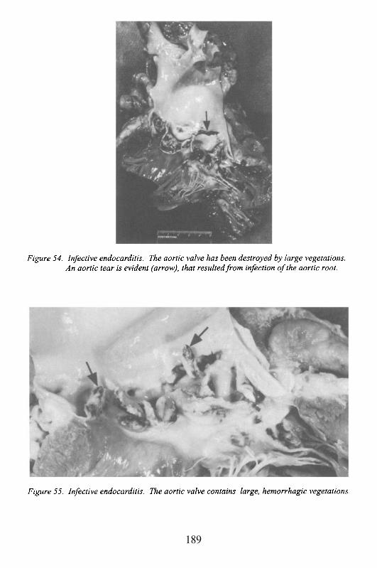
189

This Page Intentionally Left Blank
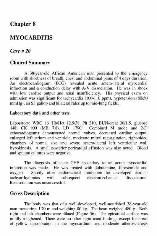
Chapter 8
MYOCARDITIS
Case # 20
Clinical Summary
A 38-year-old African American man presented to the emergencyroom with shortness of breath, chest and abdominal pains of 4 days duration.An electrocardiogram (ECG) revealed acute antero-lateral myocardialinfarction and a conduction delay with A-V dissociation. He was in shockwith low cardiac output and renal insufficiency. His physical exam onadmission was significant for tachycardia (100-110 ppm), hypotension (80/50mmHg), an S3 gallop and bilateral rales up to mid-lung fields.
Laboratory data and other tests
Laboratory: WBC 16, Hb/Hct 12.5/38, Plt 210; BUN/creat 30/1.5, glucose148; CK 900 (MB 7.8), LD 1790. Combined M mode and 2-Dechocardiograms demonstrated normal valves, decreased cardiac output,enlarged left atrium and ventricle, moderate mitral regurgitation, right-sidedchambers of normal size and severe antero-lateral left ventricular wallhypokinesis. A small posterior pericardial effusion was also noted. Bloodand sputum cultures were negative.
The diagnosis of acute CHF secondary to an acute myocardialinfarction was made. He was treated with dobutamine, furosemide andoxygen. Shortly after endotracheal intubation he developed cardiactachyarrhythmias with subsequent electromechanical dissociation.Resuscitation was unsuccessful.
Gross Description
The body was that of a well-developed, well-nourished 38-year-oldman measuring 1.70 m and weighing 80 kg. The heart weighed 480 g. Bothright and left chambers were dilated (Figure 56). The epicardial surface wasmildly roughened. There were no other significant findings except for areasof yellow discoloration in the myocardium and moderate atherosclerosis
involving the left anterior descending coronary artery. The lungs werecongested, edematous and weighed 2150 g.
Microscopic Description
H&E stained sections of the heart revealed extensive diffusemyocarditis. The infiltrate was predominantly lymphocytic and involved allchambers including the conduction system at the base of the heart, as well asthe papillary muscles. The lymphocytes appeared reactive with enlargedirregular nuclei (Figure 57). Extensive confluent and focal transmuralmyocardial necrosis was noted. Inflammation and necrosis were seenadjacent to the atrio-ventricular conduction system, which explains theconduction block on presentation. Histologic features of organization andgranulation tissue formation were consistent with an interval of 10-14 days.Other areas were less than a week old and many foci showed acute activeinflammation (24 hours). Active inflammatory pericarditis was also noted.Additional findings included congestion of the lungs, spleen and liver,consistent with the clinical diagnosis of acute congestive heart failure.
Case Analysis
Inflammation of the human myocardium due to infectious and non-infectious agents is collectively termed as myocarditis. This case representsan atypical presentation of myocarditis in an adult, but would be thecommonest one in children. The fulminant presentation is usually secondaryto a viral infection of the cardiac tissues, and with appropriate cultures thevirus may be grown. The onset and progression tend to be relatively rapidand the outcome is usually fatal over a period of days to weeks. The atypicallymphocytes infiltrating this patient’s heart are consistent with an acutereaction to a viral agent. The adult often presents with a more indolent courseand the pathogenesis is invariably immunologic.
The extensive multifocal, and in some areas, confluent myocardialdamage seen in this patient, can resemble clinically a transmural myocardialinfarction. Both the ECG and the cardiac enzyme elevations were indicativeof myocardial necrosis. However, in contrast to the global injury seen infulminant myocarditis, the damage seen in a myocardial infarction isgenerally limited to the territory of a coronary artery. Although the autopsyrevealed moderate atherosclerosis of the left anterior descending coronaryartery, ischemia played no role in the myocardial damage.
The histology demonstrated that extensive damage was at least 2weeks old at the time of the patient’s demise, although he allegedly was notsymptomatic until 4 days prior to admission. It is not unusual for myocarditis
192
to be clinically “silent” until sufficient myocardium is damaged to lead to thedevelopment of CHF, arrhythmia, chest pain, or sudden death. In this case,even if the patient had been admitted several days prior to the onset of hissymptoms, the outcome would have probably been the same. The damagewas so extensive that supportive therapy with vasopressors and/or intra-aorticballoon pump, would have been only of short term benefit.
Other non-infectious etiologies to consider in this patient includeimmune-mediated diseases, and toxic and drug related injury. Although, thereis no clinical history to support either of these possibilities, they may havefulminant presentations as well. However, the pathology in such acute casesmay be of significant value, since it can identify features associated withspecific etiologies (e.g. rheumatoid nodules in rheumatoid arthritis involvingthe conduction system).
In Europe and the United States of America the most importantinfectious cause of myocarditis are viruses, of which Coxsackie A and B,ECHO and Influenza A and B are the major types. In this patient, even if theviral agent had been identified, specific antiviral treatments are not currentlyavailable. In addition, immunosuppressive therapy is of no value in fulminantmyocarditis, and may even exacerbate the already severe condition. In Southand Central America parasitic infections of the heart account for a significantnumber of myocarditis cases with Chaga’s disease one of its main causes,along with chronic cardiomyopathy. Furthermore, with increasing numbers ofimmunocompromised patients, disseminated infections (HIV/AIDS,toxoplasmosis, aspergillosis, candidiasis) will continue to be associated withmyocardial damage and dysfunction.
The clinical course of myocarditis depends not only in the causativeagent, but also in the type, degree and duration of the damage. The host playsa pivotal role because of the genetic determinants that define its inherentsusceptibility and the response to the injury. The various infectious agentsmay directly affect the myocytes, exert a cytotoxic effect (toxin-mediated),cause injury via an immune-mediated mechanism or cause myocardialdysfunction as it spreads from adjacent inflammatory foci. In addition, theseagents can induce a vascular damage that in turn will affect the myocardium.
The specific immune alterations associated with myocarditis areunknown. The expression of major histocompatibility complex (MHC)antigens class I and II during the course of human viral myocarditis supportsthe possibility of this process having an autoimmune component. Messengerribonucleic acid of perform, the pore-forming protein that mediatescytotoxicity, has been identified in the cytoplasmic granules of inflammatorycells. Interleukins 1, 6 and 8 as well as tumor necrosis factor also appear toplay a role in myocardial damage. Recently, myosin-specific antibodies havebeen identified in the serum of a large percentage of adult patients withmyocarditis. These antibodies may arise as a result of tissue injury or may
193
induce actual tissue damage. Cytomegalovirus (CMV) induced cross-reactingantibodies to myosin have also been described. In certain mouse strains(DBA/2) there is an increased susceptibility to myocarditis following in vivoadministration of myosin-specific antibodies. It appears that autoimmunemyocarditis mediated by myosin-specific antibodies has a genetic basis,which is complex and has yet to be defined.
Endomyocardial biopsies to evaluate the presence of myocarditis areobtained from the right side of the ventricular septum. The DallasClassification was developed to standardize the histopathological diagnosis ofactive and chronic myocarditis. In the acute cases, in addition to theinflammatory infiltrate, there is necrosis and degeneration of myocytes.However, clinical improvement usually precedes histologic recovery. Inchronic myocarditis, necrosis is not a required feature, but interstitial fibrosisand loss of myofibers will indicate the degree of severity of the injury. Otherterms such as ongoing, resolving and healed myocarditis can be used insubsequent biopsies.
In this case, the outcome was determined by the diffuse and extensiveacute inflammatory damage to the myocardium. Although some authors haveproposed that fulminant myocarditis can have a good long-term prognosis, themajority of patients die as a result of severe hemodynamic alterations.
194
Suggested Readings
1.
2.3.
4.
5.
6.
7.
8.
Feldman AM, McNamara D. Myocarditis. N Engl J Med. 2000; 343:1388-98.Mason JW. Myocarditis. Adv Intern Med. 1999; 44:293-310.Kearney MT, Cotton JM, Richardson PJ, Shah AM. Viral myocarditis anddilated cardiomyopathy: mechanisms, manifestations, and management.Postgrad Med J. 2001; 77:4-10.Klingel K, Selinka HC, Huber M, Sauter M, Leube M, Kandolf R.Molecular pathology and structural features of enteroviral replication.Toward understanding the pathogenesis of viral heart disease. Herz. 2000;25:216-20.Aretz HT, Billingham ME, Edwards WD, Factor SM, Fallon JT, FenoglioJJ Jr, Olsen EG, Schoen FJ. Myocarditis. A histopathologic definition andclassification. Am J Cardiovasc Pathol. 1987; 1:3-14.Liao L, Sindhwani R, Leinwand L, Diamond B, Factor SM. Cardiac alpha-myosin heavy chains differ in their induction of myocarditis. Identificationof pathogenic epitopes. J Clin Invest 1993; 90:2877-82.Liao L, Sindhwani R, Rojkind M, Factor SM, Leinwand L, Diamond B.Antibody-mediated autoimmune myocarditis depends on geneticallydetermined target organ sensitivity. J Exp Med 1996; 181:1123-31.Kuan AP, Chamberlain W, Malkiel S, Lieu HD, Factor SM, Diamond B,Kotzin BL. Genetic control of autoimmune myocarditis mediated bymyosin-specific antibodies. Immunogenet 1999; 49:79-85.
195
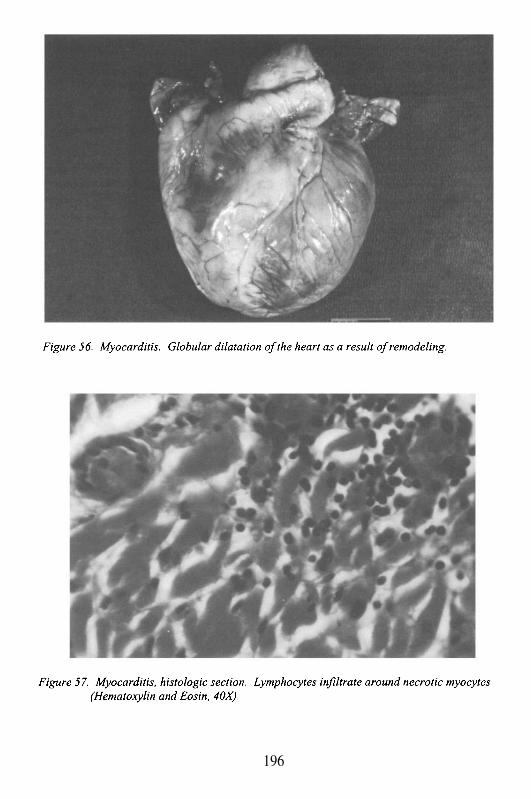
196
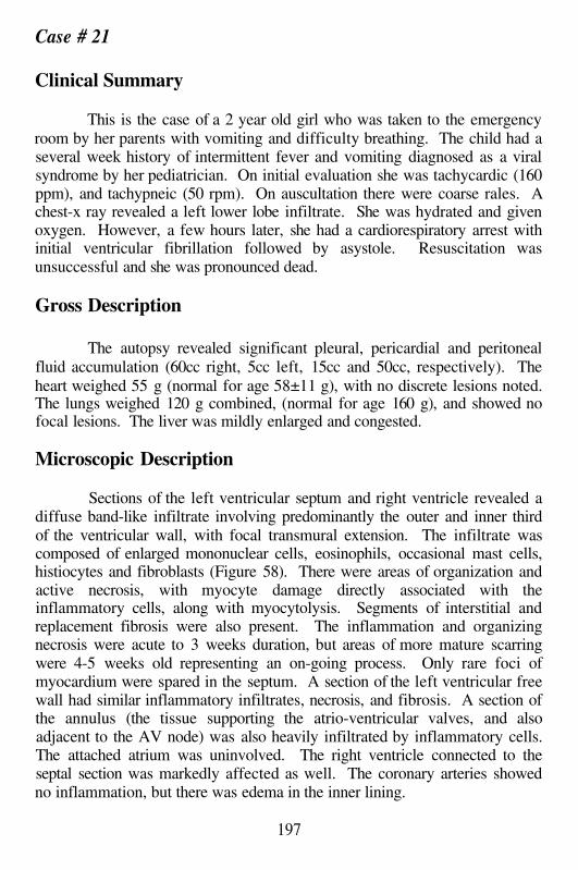
Case # 21
Clinical Summary
This is the case of a 2 year old girl who was taken to the emergencyroom by her parents with vomiting and difficulty breathing. The child had aseveral week history of intermittent fever and vomiting diagnosed as a viralsyndrome by her pediatrician. On initial evaluation she was tachycardic (160ppm), and tachypneic (50 rpm). On auscultation there were coarse rales. Achest-x ray revealed a left lower lobe infiltrate. She was hydrated and givenoxygen. However, a few hours later, she had a cardiorespiratory arrest withinitial ventricular fibrillation followed by asystole. Resuscitation wasunsuccessful and she was pronounced dead.
Gross Description
The autopsy revealed significant pleural, pericardial and peritonealfluid accumulation (60cc right, 5cc left, 15cc and 50cc, respectively). Theheart weighed 55 g (normal for age 58±11 g), with no discrete lesions noted.The lungs weighed 120 g combined, (normal for age 160 g), and showed nofocal lesions. The liver was mildly enlarged and congested.
Microscopic Description
Sections of the left ventricular septum and right ventricle revealed adiffuse band-like infiltrate involving predominantly the outer and inner thirdof the ventricular wall, with focal transmural extension. The infiltrate wascomposed of enlarged mononuclear cells, eosinophils, occasional mast cells,histiocytes and fibroblasts (Figure 58). There were areas of organization andactive necrosis, with myocyte damage directly associated with theinflammatory cells, along with myocytolysis. Segments of interstitial andreplacement fibrosis were also present. The inflammation and organizingnecrosis were acute to 3 weeks duration, but areas of more mature scarringwere 4-5 weeks old representing an on-going process. Only rare foci ofmyocardium were spared in the septum. A section of the left ventricular freewall had similar inflammatory infiltrates, necrosis, and fibrosis. A section ofthe annulus (the tissue supporting the atrio-ventricular valves, and alsoadjacent to the AV node) was also heavily infiltrated by inflammatory cells.The attached atrium was uninvolved. The right ventricle connected to theseptal section was markedly affected as well. The coronary arteries showedno inflammation, but there was edema in the inner lining.
197
A section of a cervical lymph node revealed follicular hyperplasiawith reactive germinal centers. The thymus had expansion of the medullarytissue into the cortex with reactive and atypical lymphocytes. Both lungsshowed peribronchial and interstitial aggregates of mononuclear cellsassociated with eosinophils. Intranuclear viral inclusions consistent withadenovirus were noted in the bronchial epithelium. Additionally, there wasbronchial epithelial hyperplasia and mucosal necrosis. Alveolar spaces werefilled with histiocytes and numerous inflammatory cells infiltrated theinterstitium. A section of the liver had centrilobular acute and chroniccongestive changes consistent with cardiac failure. The central veins weredilated and fibrosed indicating longstanding congestive heart failure (CHF).
Case Analysis
The cause of death in this case was overwhelming viral pneumonitisand fulminant viral myocarditis, causing CHF and terminal fatal arrhythmia.The damage in the heart had been present for at least a month prior to thischild’s demise, as indicated by the extensive fibrosis in areas of persistentactive inflammation. The signs of CHF were evident from the fluidaccumulation in the pleural spaces, pericardial and abdominal cavities, andthe acute and chronic dilatation of the central veins in the liver. The presenceof large atypical mononuclear cells in the heart and lung, as well as theadenoviral intranuclear inclusions, confirms the viral etiology of this illness.
Myocarditis in infants and young children is almost always due to aspecific viral infection of the heart, which leads to inflammatory injury andmyocardial cell necrosis. In, contrast myocarditis in older immunocompetentchildren and adults, is frequently an immune-mediated disease. Myocarditisis usually suspected in cases of unexplained onset of CHF and arrhythmia,after a respiratory (upper respiratory infection or URI) or systemic febrileillness. The viral etiology may be suggested by epidemiologic clustering ofcases during seasonal epidemics of certain viruses (enteroviruses), diagnosticserologic studies, and the application of molecular techniques, like in situhibridization and polymerase chain reaction (PCR), to detect genomicsequences in tissue samples such as endomyocardial biopsies. Other rarecauses of myocarditis include, bacterial infections of the heart, which resultfrom direct extension to myocardial tissue with microabscesses, or fromtoxin-mediated injury (e.g. diphtheria). Occasionally, contiguous spread fromthe endocardium or pericardium can occur, most commonly from pneumonicfoci.
This girl presented with fever, episodes of vomiting and respiratorydistress. Her pediatrician ascribed these non-specific symptoms to a viralsyndrome, and decided just to follow her clinically. This approach is fairly
198
common in pediatric medicine. Even if a child is symptomatic, but is overallstable with adequate cardiovascular and neurologic function, he or she will bemonitored over time. Still, consideration of several facts may be warranted.First, the issue of intermittent fevers may be of importance. Fever representsa non-specific host response to an on-going process, which can be significantor trivial. What define the importance of the fever are concomitant symptomsand their progression and/or resolution. In this case, if the fever had beendaily or frequent, it should have prompted a more thorough physical exam andlaboratory analyses to detect potentially treatable conditions. The crucialelements to evaluate include the height of the fever, associated signs andsymptoms (particularly rashes, change in mental status, respiratory signs andhemodynamic stability), and response to therapy (e.g. antibiotics, antipyreticdrugs).
In the beginning of the illness, her main symptom was vomiting(there is no mention of change in bowel habits, or rashes). Enterovirusespredominantly cause fever, gastrointestinal, respiratory and skinmanifestations. Therefore, it is conceivable that her symptoms were due to anenteroviral infection. The turning point came when she began developingrespiratory complaints, and on physical exam she had signs of pulmonarycompromise characterized by coarse breath sounds and rales. Clues that thisillness perhaps represented more than a self-limited viral syndrome includethe progressive involvement of new organ systems. Other diagnoses toconsider are progressive lung involvement such as significant pneumonia withor without effusion, diffuse interstitial lung disease, sepsis syndrome,hereditary cardiomyopathies or congenital heart disease.
The clinical history, physical examination and pathologic findings ofthe heart definitely excluded the possibility of a common pediatric illness ofunknown etiology, Kawasaki’s disease. This entity is a non-infectious,immune-mediated disorder, which afflicts infants and young children. It is asystemic vasculitis that affects the small and medium muscular arteries,resulting in fever, rash, mucositis, lymphadenopathy, and the hallmarkcoronary artery inflammation. Other potential fatal conditions in children,that may present subacutely, are those related to toxic ingestions or toxin-mediated illnesses. Again, in the absence of epidemiologic and clinicalhistory, and with the strong pathologic evidence of myocarditis, it is veryunlikely that there was a different cause that may have resulted in this child’sdeath.
Even though the majority of myocarditis episodes end up beingidiopathic, of those identified with a specific etiology, viruses account for asignificant number of cases. Coxsackie B viruses, are small stranded RNAviruses, which have a natural affinity for the myocardium by interacting withspecific receptors on the myocardial cell surface, and can be identified in 25-40% of acute cases (mostly types B2-B5). The determinant for host
199
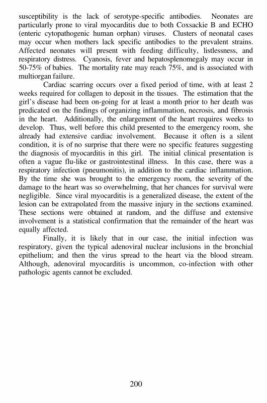
susceptibility is the lack of serotype-specific antibodies. Neonates areparticularly prone to viral myocarditis due to both Coxsackie B and ECHO(enteric cytopathogenic human orphan) viruses. Clusters of neonatal casesmay occur when mothers lack specific antibodies to the prevalent strains.Affected neonates will present with feeding difficulty, listlessness, andrespiratory distress. Cyanosis, fever and hepatosplenomegaly may occur in50-75% of babies. The mortality rate may reach 75%, and is associated withmultiorgan failure.
Cardiac scarring occurs over a fixed period of time, with at least 2weeks required for collagen to deposit in the tissues. The estimation that thegirl’s disease had been on-going for at least a month prior to her death waspredicated on the findings of organizing inflammation, necrosis, and fibrosisin the heart. Additionally, the enlargement of the heart requires weeks todevelop. Thus, well before this child presented to the emergency room, shealready had extensive cardiac involvement. Because it often is a silentcondition, it is of no surprise that there were no specific features suggestingthe diagnosis of myocarditis in this girl. The initial clinical presentation isoften a vague flu-like or gastrointestinal illness. In this case, there was arespiratory infection (pneumonitis), in addition to the cardiac inflammation.By the time she was brought to the emergency room, the severity of thedamage to the heart was so overwhelming, that her chances for survival werenegligible. Since viral myocarditis is a generalized disease, the extent of thelesion can be extrapolated from the massive injury in the sections examined.These sections were obtained at random, and the diffuse and extensiveinvolvement is a statistical confirmation that the remainder of the heart wasequally affected.
Finally, it is likely that in our case, the initial infection wasrespiratory, given the typical adenoviral nuclear inclusions in the bronchialepithelium; and then the virus spread to the heart via the blood stream.Although, adenoviral myocarditis is uncommon, co-infection with otherpathologic agents cannot be excluded.
200
Suggested Readings
1
2
3
Penninger JM, Bachmaier K. Review of microbial infections and theimmune response to cardiac antigens. J Infect Dis. 2000; 181 Suppl3:S498-504.Huber SA, Gauntt CJ, Sakkinen P. Enteroviruses and myocarditis: viralpathogenesis through replication, cytokine induction, andimmunopathogenicity. Adv Virus Res. 1998; 51:35-80.Bowles NE, Towbin JA. Molecular aspects of myocarditis. Curr OpinCardiol. 1998; 13:179-84.
201
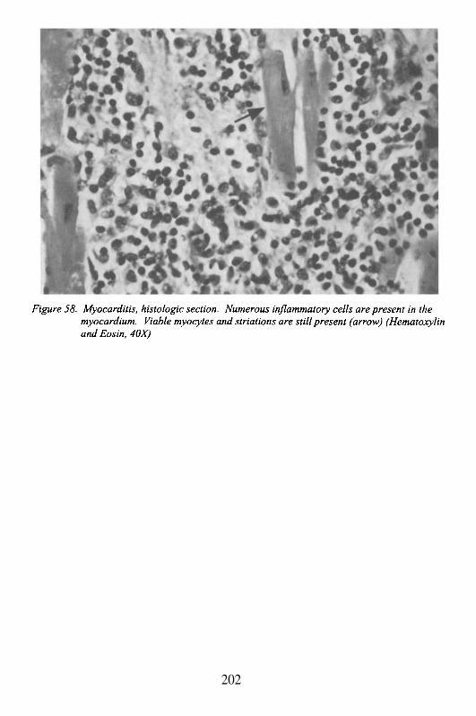
202
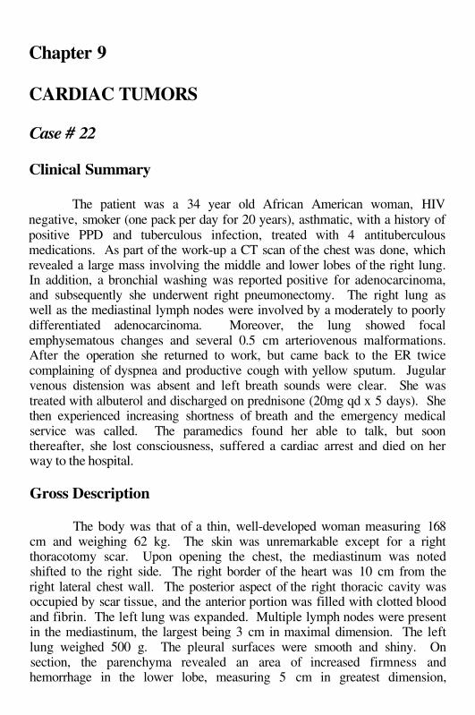
Chapter 9
CARDIAC TUMORS
Case # 22
Clinical Summary
The patient was a 34 year old African American woman, HIVnegative, smoker (one pack per day for 20 years), asthmatic, with a history ofpositive PPD and tuberculous infection, treated with 4 antituberculousmedications. As part of the work-up a CT scan of the chest was done, whichrevealed a large mass involving the middle and lower lobes of the right lung.In addition, a bronchial washing was reported positive for adenocarcinoma,and subsequently she underwent right pneumonectomy. The right lung aswell as the mediastinal lymph nodes were involved by a moderately to poorlydifferentiated adenocarcinoma. Moreover, the lung showed focalemphysematous changes and several 0.5 cm arteriovenous malformations.After the operation she returned to work, but came back to the ER twicecomplaining of dyspnea and productive cough with yellow sputum. Jugularvenous distension was absent and left breath sounds were clear. She wastreated with albuterol and discharged on prednisone (20mg qd x 5 days). Shethen experienced increasing shortness of breath and the emergency medicalservice was called. The paramedics found her able to talk, but soonthereafter, she lost consciousness, suffered a cardiac arrest and died on herway to the hospital.
Gross Description
The body was that of a thin, well-developed woman measuring 168cm and weighing 62 kg. The skin was unremarkable except for a rightthoracotomy scar. Upon opening the chest, the mediastinum was notedshifted to the right side. The right border of the heart was 10 cm from theright lateral chest wall. The posterior aspect of the right thoracic cavity wasoccupied by scar tissue, and the anterior portion was filled with clotted bloodand fibrin. The left lung was expanded. Multiple lymph nodes were presentin the mediastinum, the largest being 3 cm in maximal dimension. The leftlung weighed 500 g. The pleural surfaces were smooth and shiny. Onsection, the parenchyma revealed an area of increased firmness andhemorrhage in the lower lobe, measuring 5 cm in greatest dimension,
extending to the visceral pleura. In addition, there was a moderate loss ofparenchyma with formation of smooth-lined cysts of up to 0.5 cm in diameter.These cysts were present in both lobes throughout. The larynx, trachea, majorand segmental bronchi were of average caliber. Organizing emboli measuringup to 3 mm occluded small branches of the pulmonary artery.
The heart was 240 g and of the expected shape. The parietalpericardium was smooth and glistening. The sac contained approximately500 cc of serosanguineous fluid. The epicardium was thickened, dull, firmand white in a few areas with multiple hemorrhagic foci of up to 0.2 cm(Figure 59). The atria were normal and free of thrombi. The foramen ovalewas closed. The right and left ventricular walls measured 0.4 cm and 1.2 cmin thickness, respectively. The valve rings measured as follows: tricuspidvalve 12 cm, pulmonic valve 7 cm, mitral valve 8.5 cm, and aortic valve 6cm. The valve leaflets were pliable and the chordae tendineae were thin andslender. The myocardium and endocardium were unremarkable. Thecoronary arteries were of normal caliber and distribution. The aorta showedmild atherosclerosis in the abdominal portion.
The liver weighed 1400 g and was of normal size and shape. The cutsurface showed marked acute centrilobular congestion and multiple tan, firmnodules measuring 0.2 to 0.4 cm. The lobular pattern was preservedotherwise. The remaining organs had no significant pathologic changes.
Microscopic Description
Sections from the epicardium showed many single and small clustersof poorly differentiated carcinoma cells, near the epicardial surface. Some ofthese cells contained vacuoles consistent with mucin droplets ofadenocarcinoma. The malignant cells were large with pleomorphic nuclei andprominent nucleoli. Some were present within lymphatics. Metastaticcarcinoma was present bilaterally in hilar lymph nodes. The firm,hemorrhagic areas in both lobes of the left lung corresponded to foci ofhemorrhagic infarcts. Surrounding arteries contained numerous acute,organizing and recanalized thrombi. The tan nodules present in the liver didnot represent metastatic adenocarcinoma but rather foci of adenomatosis.They were composed of thickened cords of hepatocytes separated bysinusoids and devoid of portal tracts, bile ductules or scars.
Case Analysis
The heart can be involved by primary as well as metastatic tumors.Primary tumors of the heart are rare (< 0.3 % in postmortem series), withmyxoma the most frequent. Metastases from neoplasms arising in other
204
organs are far more common and include carcinomas, sarcomas, hematologicmalignancies and melanoma. Nonetheless, cardiac metastases are infrequent.Why is the heart relatively protected from metastasis? Possible explanationsinclude the continuous forceful stroke of the heart, rapid blood flow andafferent lymphatic circulation. If these hypotheses were true, cardiac tumorswould be present almost invariably in patients with previous cardiac damagewith reduced pump action and blood flow, but there is no such confirmatorydata in the literature. Regarding afferent lymphatics, metastatic disseminationin the heart occurs most commonly through lymphatic invasion. Overall, theheart is probably protected as a result of the combination of all these and otherunknown factors. Moreover, tumor type, metabolic properties of the heart,and adhesion molecules among others, are under consideration and requirefurther investigation.
Secondary tumors may involve the heart by direct extension,hematogenous spread or lymphatic dissemination. Direct extension occursfrom tumors arising in the lungs or mediastinum (i.e. bronchogenic andesophageal carcinoma, malignant thymoma, thymic carcinoma).Hematogenous spread is often seen with sarcomas (e.g. uterine.leiomyosarcomas can reach the heart through the inferior vena cava),leukemia, multiple myeloma, and with certain types of carcinoma (e.g.hepatocellular carcinoma). However, the lymphatic pathway appears to bethe most significant. This is demonstrated by the frequent metastaticinvolvement of the pericardium, richly supplied by lymphatic channels thatdrain in the anterior and posterior mediastinum. In the present case, thepatient had a history of adenocarcinoma of the lung involving mediastinallymph nodes; therefore, it is not surprising that she presented with apericardial effusion and associated cardiac tamponade. Pericardialinvolvement in a patient with a known primary malignancy, should besuspected in the setting of pericardial effusion, intractable heart failure orhepatomegaly in the absence of metastatic lesions. The differential diagnosisincludes congestive heart failure in a patient with a cardiomyopathy, and non-malignant pericardial effusions (infectious and non-infectious). This patientwas young, with no history of heart disease. Although she had asthma in thepast and evidence of chronic obstructive pulmonary disease as per the autopsyexam, she did not have pulmonary hypertension or cor-pulmonale.
Tuberculous (TB) pericarditis is a consideration based on her priorhistory of PPD positivity and antituberculous treatment. TB pericarditis maydevelop insidiously, or acutely with a large effusion and cardiac tamponade,particularly in immunosuppressed patients. She was HIV negative and hadnot been treated with chemotherapeutic agents. If she had lived, the diagnosisof malignant pericardial effusion could have been made with apericardiocentesis and cytologic examination of the fluid. Adenocarcinomacells are readily identified in pericardial fluid by their tri-dimensional
205
arrangement, distinct cell borders, high nuclear-cytoplasmic ratio andprominent nucleoli (Figure 60). They must be differentiated from reactivemesothelial cells, which is not often easy. Reactive mesothelial cells,especially in long-standing effusions can also exhibit a glandulararrangement, prominent nucleoli, mitotic figures and even vacuolization thatmimic adenocarcinoma. Immunohistochemistry is useful in distinguishing thetwo. Both cell types are immunoreactive to cytokeratin, but onlyadenocarcinoma stains with the adenocarcinoma marker B72.3 andcarcinoembryonic antigen (CEA).
Pulmonary adenocarcinoma arises preferentially in the periphery ofthe lung parenchyma. Central lesions may occur, but are less frequent. Thistype of tumor can develop in smokers as well as in non-smokers, de novo orin areas of pre-existing scarring. Our patient was a smoker for 20 years.Although she had a risk factor for lung cancer, the aggressive behavior of thistumor (stage T2, N2 at initial diagnosis) suggests a genetic predisposition.
The advent of cardiovascular imaging studies has improved thediagnosis of cardiac involvement by metastatic disease, but the prognosisremains poor. In a recent study done at the Tottori University School ofMedicine in Japan, 46 of 161 patients had secondary cardiac tumorsdiagnosed by echocardiography. The most common primary site was the lungfollowed by mediastinum, liver, uterus and testis. Forty seven percent of thecases had pericardial involvement, 32% involved the right side of the heartand 14 % involved the left. Metastases on both sides of the heart were notedin 7% of cases. It is unknown why intracavitary lesions are less common, butthere is evidence that certain tumors have predilection for this pattern ofgrowth (Figures 61 and 62). In our experience, squamous carcinoma arisingin different sites has the tendency to invade and seed cardiac valves,endocardium and myocardium. Prior valve damage or endothelial damageappears to be a sine qua non in such cases. As a result, death can occur due toarrhythmia with or without conduction system involvement and embolization.Changes in the electrocardiogram such as prolonged elevation of the STsegment are indicative of myocardial invasion.
In the present case, the patient died of cardiac tamponade due to amalignant pericardial effusion. In addition, she was in a hypercoagulablestate with numerous acute and organizing pulmonary emboli leading to areasof hemorrhagic infarction. Her symptoms of dyspnea and shortness of breathwere related to both the pulmonary embolism and evolving cardiactamponade. Of note, dyspnea is the most common manifestation of cardiactumors in general, and is suggestive of cardiac decompensation. Treatment ispalliative, aimed at controlling effusions, arrhythmia and heart failure.Radiotherapy is the treatment of choice to manage malignant effusions. Insome instances, a pericardial window is required to treat tamponade.Chemotherapy has been used to prevent further cardiac involvement, but its
206
use is limited by its cardiotoxicity. In rare instances, surgical debulking isindicated to alleviate obstructive cardiac masses.
207

Suggested Readings
1.
2.
3.
4.
Ghosh K, Dusenbery KE, Twiggs LB. Cardiac metastasis of cloacageniccarcinoma of the vagina: a case and review of gynecologic malignancieswith cardiac metastasis. Gynecol Oncol. 2000; 76:208-12.Majano-Lainez RA. Cardiac tumors: a current clinical and pathologicalperspective. Crit Rev Oncog. 1997; 8:293-303.Raaf HN, Raaf JH. Sarcomas related to the heart and vasculature. SeminSurg Oncol. 1994; 10:374-82.Vohra A, Saiz E, Davila E, Burkle J. Metastatic germ cell tumor to theheart presenting with syncope. Clin Cardiol. 1999; 22:429-33.
208
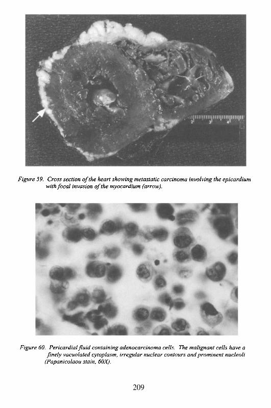
209
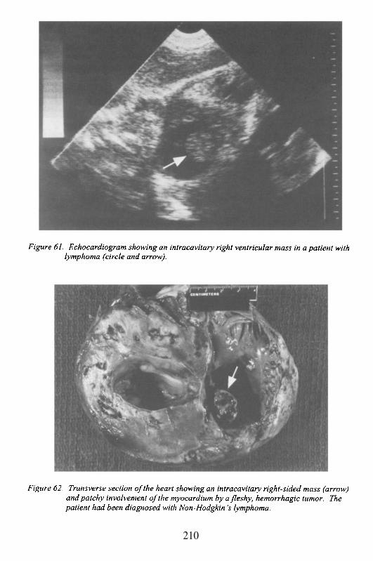
210
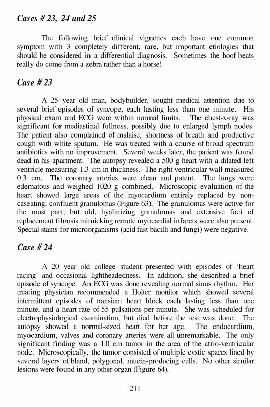
Cases # 23, 24 and 25
The following brief clinical vignettes each have one commonsymptom with 3 completely different, rare, but important etiologies thatshould be considered in a differential diagnosis. Sometimes the hoof beatsreally do come from a zebra rather than a horse!
Case # 23
A 25 year old man, bodybuilder, sought medical attention due toseveral brief episodes of syncope, each lasting less than one minute. Hisphysical exam and ECG were within normal limits. The chest-x-ray wassignificant for mediastinal fullness, possibly due to enlarged lymph nodes.The patient also complained of malaise, shortness of breath and productivecough with white sputum. He was treated with a course of broad spectrumantibiotics with no improvement. Several weeks later, the patient was founddead in his apartment. The autopsy revealed a 500 g heart with a dilated leftventricle measuring 1.3 cm in thickness. The right ventricular wall measured0.3 cm. The coronary arteries were clean and patent. The lungs wereedematous and weighed 1020 g combined. Microscopic evaluation of theheart showed large areas of the myocardium entirely replaced by non-caseating, confluent granulomas (Figure 63). The granulomas were active forthe most part, but old, hyalinizing granulomas and extensive foci ofreplacement fibrosis mimicking remote myocardial infarcts were also present.Special stains for microorganisms (acid fast bacilli and fungi) were negative.
Case # 24
A 20 year old college student presented with episodes of ‘heartracing’ and occasional lightheadedness. In addition, she described a briefepisode of syncope. An ECG was done revealing normal sinus rhythm. Hertreating physician recommended a Holter monitor which showed severalintermittent episodes of transient heart block each lasting less than oneminute, and a heart rate of 55 pulsations per minute. She was scheduled forelectrophysiological examination, but died before the test was done. Theautopsy showed a normal-sized heart for her age. The endocardium,myocardium, valves and coronary arteries were all unremarkable. The onlysignificant finding was a 1.0 cm tumor in the area of the atrio-ventricularnode. Microscopically, the tumor consisted of multiple cystic spaces lined byseveral layers of bland, polygonal, mucin-producing cells. No other similarlesions were found in any other organ (Figure 64).
211
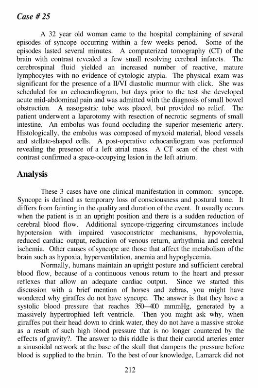
Case # 25
A 32 year old woman came to the hospital complaining of severalepisodes of syncope occurring within a few weeks period. Some of theepisodes lasted several minutes. A computerized tomography (CT) of thebrain with contrast revealed a few small resolving cerebral infarcts. Thecerebrospinal fluid yielded an increased number of reactive, maturelymphocytes with no evidence of cytologic atypia. The physical exam wassignificant for the presence of a II/VI diastolic murmur with click. She wasscheduled for an echocardiogram, but days prior to the test she developedacute mid-abdominal pain and was admitted with the diagnosis of small bowelobstruction. A nasogastric tube was placed, but provided no relief. Thepatient underwent a laparotomy with resection of necrotic segments of smallintestine. An embolus was found occluding the superior mesenteric artery.Histologically, the embolus was composed of myxoid material, blood vesselsand stellate-shaped cells. A post-operative echocardiogram was performedrevealing the presence of a left atrial mass. A CT scan of the chest withcontrast confirmed a space-occupying lesion in the left atrium.
Analysis
These 3 cases have one clinical manifestation in common: syncope.Syncope is defined as temporary loss of consciousness and postural tone. Itdiffers from fainting in the quality and duration of the event. It usually occurswhen the patient is in an upright position and there is a sudden reduction ofcerebral blood flow. Additional syncope-triggering circumstances includehypotension with impaired vasoconstrictor mechanisms, hypovolemia,reduced cardiac output, reduction of venous return, arrhythmia and cerebralischemia. Other causes of syncope are those that affect the metabolism of thebrain such as hypoxia, hyperventilation, anemia and hypoglycemia.
Normally, humans maintain an upright posture and sufficient cerebralblood flow, because of a continuous venous return to the heart and pressorreflexes that allow an adequate cardiac output. Since we started thisdiscussion with a brief mention of horses and zebras, you might havewondered why giraffes do not have syncope. The answer is that they have asystolic blood pressure that reaches 350—400 mmmHg, generated by amassively hypertrophied left ventricle. Then you might ask why, whengiraffes put their head down to drink water, they do not have a massive strokeas a result of such high blood pressure that is no longer countered by theeffects of gravity?. The answer to this riddle is that their carotid arteries entera sinusoidal network at the base of the skull that dampens the pressure beforeblood is supplied to the brain. To the best of our knowledge, Lamarck did not
212
know this when he proposed that giraffes developed long necks by reaching toeat leaves from high tree branches.
Not uncommonly, healthy individuals may experience syncope due toincreased vagal activity. These episodes of loss of consciousness andgeneralized muscle weakness are triggered by stress, high temperatures, lackof ventilation, mild blood loss and physical pain, and are preceded by nausea,tachypnea, tachycardia and sweating. Sudden change of posture may alsoresult in syncope. In vasovagal syncope, hypotension occurs, but is notfollowed by a compensatory increase in cardiac output. Instead, bradycardiaand reduced arterial pressure ensue, which exacerbates the patient’s condition.Strength is regained by placing the patient in supine posture and elevating thelegs to facilitate venous return.
In case # 23, syncope was the result of reduced cardiac output andconduction abnormalities due to severe myocardial damage. The presence ofmultiple, confluent, non-necrotizing granulomas replacing the myocardiumand involving the mediastinal lymph nodes, is diagnostic of sarcoidosis.Sarcoidosis is a systemic disease that commonly affects individuals youngerthan 40 years of age. It is more common in females. The etiology isunknown, but the manifestations are the result of an exaggerated response ofT-helper lymphocytes leading to activation and recruitment of mononuclearphagocytes and granuloma formation. The lymph nodes and lungs arevirtually always involved, but the heart is affected in only 5% of the patients.In this case, the involvement was so extensive that it caused cardiac failure.The flu-like symptoms interpreted by the patient and his physician as a viralor possibly a bacterial infection were no more than symptoms of congestiveheart failure (CHF), and explain why the empiric treatment with antibioticsfailed. CHF was confirmed by the autopsy findings of an enlarged, dilatedheart and pulmonary edema. In addition to pump failure, the patient had acomponent of restrictive heart disease due to the presence of granulomas andextensive areas of replacement and interstitial fibrosis. In this setting,arrhythmogenic events resulting in death are likely to occur. Sarcoidosis inmost instances is a diagnosis of exclusion. When a young patient presentswith syncope, other heart conditions causing arrhythmia and reduced cardiacoutput should be considered such as Stokes-Adams-Morgagni syndrome(complete atrio-ventricular block), Wolff-Parkinson-White syndrome, sicksinus syndrome, hypertrophic as well as other types of cardiomyopathy andmyocarditis. Massive myocardial infarction must also be excluded.
In case # 24, we are also dealing with a young patient whoexperienced syncope as well as lightheadedness and tachycardia. However,this case, although interesting, was unlikely to be diagnosed pre-mortem. Thecharacteristics of this patient’s tumor (multicystic and multilayered bypolygonal cells) are those of a cystic tumor of the atrio-ventricular node (AVnode), formerly known as mesothelioma of the AV node. Cystic tumors are
213
located in the atrial septum in the region of the AV node. The cell of origin isunknown. These neoplasms were previously considered mesothelial innature, but with the advent of immunohistochemical markers, endodermalderivation has been suggested. Although considered a “benign” tumor interms of its growth rate and potential for metastatic spread, its location islethal. Cystic tumors of the AV node are rare, and incidentally found inpatients that die suddenly of complete heart block or ventricular fibrillation.Clinically, the differential diagnosis in a patient with syncope due toventricular fibrillation includes long Q-T syndrome, a condition that may befamilial and associated with deafness. An important lesson in this case is tothink about all the possibilities in a patient that dies of an arrhythmia.Sections of the AV node are not standard in an autopsy. The pathologist hasto think about this diagnosis as a possibility in order to take sections of theAV node. The block of tissue for histologic examination should be obtainedfrom the right atrium, near the coronary sinus. The section must include thetricuspid valve leaflet in continuity with the aortic valve leaflet and theatrioventricular septum. The latter contains the AV node immediatelybeneath the endocardium. Examination of the entire conduction system is notroutinely carried out unless structural abnormalities are suspected. Mostpathologists are not familiar with the histologic examination of conductiontissues and prefer to refer these cases to cardiovascular pathologists.Nonetheless, it is important to be aware of primary tumors that can potentiallycause arrhythmogenic events in addition to cystic tumors of the AV node.These include fibromas, rhabdomyomas, hemangiomas, lymphangiomas andtheir malignant counterparts fibrosarcoma, rhabdomyosarcoma andangiosarcoma. Other primary tumors of the heart are teratomas and granularcell tumors. These are rare neoplasms and their description is beyond thescope of this discussion. The most primary cardiac tumor of all is illustratedin case # 25.
This young woman presented with episodes of syncope that lastedseveral minutes and was found to have multiple cerebral lesions that on CTscan were suggestive of infarcts. Weeks later she developed ischemic boweldisease caused by an embolic lesion of the superior mesenteric artery that washistologically consistent with a myxoma. The ischemic infarcts of the brainand small bowel were the result of arterial occlusion by embolic fragments ofa left atrial myxoma diagnosed on a post-surgical echocardiogram. Atrialmyxomas are benign, pedunculated or sessile tumors composed of amucopolysaccharide matrix in which polygonal and stellate-shaped cells areembedded (Figures 65 and 66). Occasionally, these cells may have aglandular arrangement and can exhibit tinctorial properties similar toglandular epithelial cells by staining positive for mucin. The majority of thesetumors arise in the left atrium (75% of cases) and in middle-aged women.They are often solitary, but a familial, autosomal dominant condition has been
214
described in which the myxomas are multicentric and are associated withendocrine abnormalities and pigmented lesions of the skin (Carney’ssyndrome).
The clinical presentation of cardiac myxomas depends on the site oforigin. In this case, the patient presented with embolic phenomena. Althoughher cerebral infarcts appeared to have no sequelae, she required surgicalintervention to remove a segment of necrotic bowel. Syncope was due tomechanical reduction of venous return, similarly to what occurs when theValsalva maneuver is applied as the patient strains against a closed glottis.The tumor was pedunculated and acted as a ball valve obstructing the mitralvalve outflow. The obstruction was responsible for the diastolic murmur andthe click. Both, the diastolic murmur and the click can be mistaken for thoseof mitral valve stenosis, and in fact, patients with left atrial myxoma were inthe past erroneously diagnosed as having rheumatic heart disease. Thediastolic murmur caused by atrial myxoma and the diastolic murmur of mitralstenosis can be discriminated on the basis of positional change. The murmurcaused by the obstructing tumor diminishes or disappears when the patientchanges positions. Similarly, the click (also known as a tumor plop)auscultated in patients with left atrial myxoma can be distinguished from theopening click of mitral stenosis in that the former occurs during ventricularfilling when the valve is already open. Right atrial myxomas are less frequentand may be more difficult to diagnose clinically since they can exhibitvariable symptomatology. Some of the clinical manifestations includepulmonary emboli, arrhythmias and congestive heart failure. Presumptivediagnosis is made with imaging studies (Figure 67) and the treatment issurgical removal of the tumor. In this case, following the diagnosis, thepatient underwent open heart surgery with a curative resection of the tumor.
215
Suggested Readings
1.
2.3.
4.
5.
6.
7.
8.
9.10.
Arthur W, Kaye GC. The pathophysiology of common causes of syncope.Postgrad Med J. 2000; 76:750-3.Kapoor WN. Syncope. N Engl J Med. 2000; 343:1856-62.Meyer MD, Handler J. Evaluation of the patient with syncope: an evidencebased approach. Emerg Med Clin North Am. 1999; 17:189-201.Shammas RL, Movahed A. Sarcoidosis of the heart. Clin Cardiol. 1993;16:462-72.Sekiguchi M, Yazaki Y, Isobe M, Hiroe M. Cardiac sarcoidosis:diagnostic, prognostic, and therapeutic considerations. Cardiovasc DrugsTher. 1996; 10:495-510.Cina SJ, Smialek JE, Burke AP, Virmani R, Hutchins GM. Primarycardiac tumors causing sudden death: a review of the literature. Am JForensic Med Pathol. 1996; 17:271-81.MsAllister HA Jr. Primary tumors and cysts of the heart and pericardium.In: Current Problems in Cardiology. Harvey WP, ed. Chicago; Year Book;1979.Shapiro LM. Cardiac tumours: diagnosis and management. Heart. 2001;85:218-22.Reynen K. Cardiac myxomas. N Engl J Med. 1995; 333:1610-7.Pinede L, Duhaut P, Loire R. Clinical presentation of left atrial cardiacmyxoma. A series of 112 consecutive cases. Medicine (Baltimore). 2001;80:159-72.
216
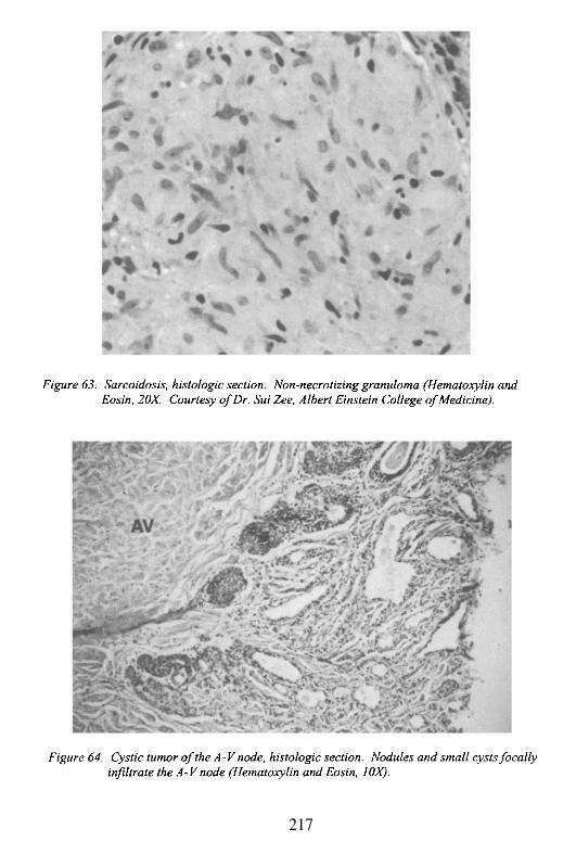
217
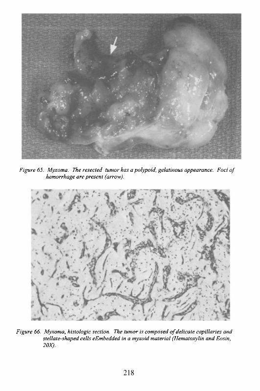
218
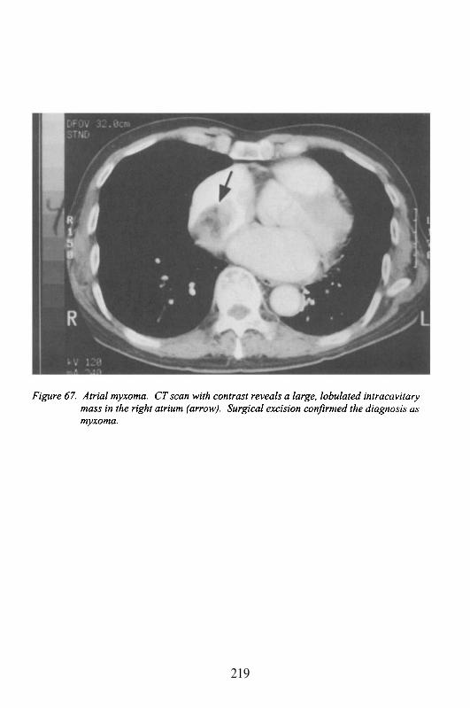
219

This Page Intentionally Left Blank
Chapter 10
THE PERICARDIUM
Case # 26
Clinical Summary
This 43 year old African American woman, former intravenous druguser with unknown HIV status (spouse was HIV-infected), presented to theemergency room with a 5 month history of weight loss, night sweats, coughwith occasional hemoptysis, and 2 weeks of severe pleuritic chest pain andshortness of breath. On initial evaluation in the emergency department shehad a normal mental status, but was in significant respiratory distress. Therewas no jaundice, edema or cyanosis. Head and neck exam was remarkable fororal thrush and no significant lymphadenopathy. The upper extremities hadfibrotic areas in the antecubital areas and forearms consistent with old healedscars. Auscultation of the heart revealed S1 and S2, tachycardic, regularrhythm, and a friction rub. No murmurs were reported. There was dullnesson percussion at the right lung base, and diffuse crackles over both lungs.
Laboratory data and other tests
Laboratory: WBC 4.6 (S 93, L 4, M 1, E 2), Hb/Hct 7/22, P1t 133;BUN/creat 37/1.4, SGOT 180, protein T/A 7.2/2.2, bilirrubin T/D 2.1/1.5,glucose 124; CK 307, LD 1288. The ECG showed sinus tachycardia at 127ppm, and low voltage complexes. The chest x ray revealed diffuse interstitialnodular infiltrates with a miliary pattern.
Hospital course: the patient was admitted and started ontrimethoprim-sulfamethoxazole (TMP-SMX) for possible Pneumocystiscarinii pneumonia and anti-tuberculous drugs (isoniazid, rifampin, ethambutoland pyrazinamide). The following day, she was found without vital signs.Resuscitation efforts were unsuccessful.
Gross Description
The body was that of a thin, well developed African Americanwoman appearing her stated age. It measured 158 cm and weighed 60 kg.The heart weighed 410 g and was of normal shape. However, the pericardialsac was thickened with numerous adhesions. The pericardium and
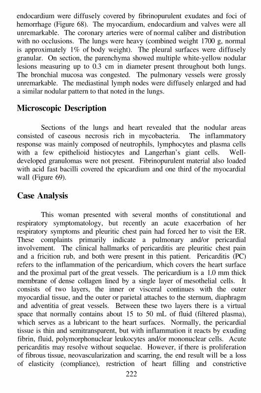
endocardium were diffusely covered by fibrinopurulent exudates and foci ofhemorrhage (Figure 68). The myocardium, endocardium and valves were allunremarkable. The coronary arteries were of normal caliber and distributionwith no occlusions. The lungs were heavy (combined weight 1700 g, normalis approximately 1% of body weight). The pleural surfaces were diffuselygranular. On section, the parenchyma showed multiple white-yellow nodularlesions measuring up to 0.3 cm in diameter present throughout both lungs.The bronchial mucosa was congested. The pulmonary vessels were grosslyunremarkable. The mediastinal lymph nodes were diffusely enlarged and hada similar nodular pattern to that noted in the lungs.
Microscopic Description
Sections of the lungs and heart revealed that the nodular areasconsisted of caseous necrosis rich in mycobacteria. The inflammatoryresponse was mainly composed of neutrophils, lymphocytes and plasma cellswith a few epithelioid histiocytes and Langerhan’s giant cells. Well-developed granulomas were not present. Fibrinopurulent material also loadedwith acid fast bacilli covered the epicardium and one third of the myocardialwall (Figure 69).
Case Analysis
This woman presented with several months of constitutional andrespiratory symptomatology, but recently an acute exacerbation of herrespiratory symptoms and pleuritic chest pain had forced her to visit the ER.These complaints primarily indicate a pulmonary and/or pericardialinvolvement. The clinical hallmarks of pericarditis are pleuritic chest painand a fricition rub, and both were present in this patient. Pericarditis (PC)refers to the inflammation of the pericardium, which covers the heart surfaceand the proximal part of the great vessels. The pericardium is a 1.0 mm thickmembrane of dense collagen lined by a single layer of mesothelial cells. Itconsists of two layers, the inner or visceral continues with the outermyocardial tissue, and the outer or parietal attaches to the sternum, diaphragmand adventitia of great vessels. Between these two layers there is a virtualspace that normally contains about 15 to 50 mL of fluid (filtered plasma),which serves as a lubricant to the heart surfaces. Normally, the pericardialtissue is thin and semitransparent, but with inflammation it reacts by exudingfibrin, fluid, polymorphonuclear leukocytes and/or mononuclear cells. Acutepericarditis may resolve without sequelae. However, if there is proliferationof fibrous tissue, neovascularization and scarring, the end result will be a lossof elasticity (compliance), restriction of heart filling and constrictive
222
pericarditis. The pleuritic pain is classically described as a retrosternal sharp,stabbing, knife-like quality pain, which worsens with inspiration, lying downand swallowing, and improves sitting up and leaning forward. The reasonsfor the exacerbation and ease of the pain have to do with the position of theheart relative to the rest of the mediastinal structures, which impact on howthe pericardium is pulled and stretched, or relaxed. The radiation of the painrelates to the visceral reflections of the pericardium, and is mostly to theshoulder and neck (trapezius muscle). The pain is common in acute infectiouspericarditis, whereas insidious accumulation of fluid in the pericardial sac(usually due to non-infectious etiologies) may go unnoticed. The ability ofthe pericardium to accommodate fluid subacutely or chronically depends onthe ratio between production, reabsorption and pericardial distensibilityproperties. The latter, are a function of the pericardial connective tissuecomponents, which allow for progressive remodeling. With slowaccumulation of fluid, or progressive cardiac hypertrophy, the volume of thepericardial sac increases by slippage of the collagen fibers in the parietallayer. With extreme increases of volume, disruption of the collagen fibers canoccur. If the increase in pericardial fluid develops too rapidly for the stiffpericardium to respond, tamponade may ensue (see below).
The other critical finding on physical examination was the presenceof a friction rub. This variably coarse, scratchy, velcro-like sound, is bestheard over the left sternal border, and when the patient is not breathing. It hasthree components: the atrial systole, the ventricular systole and the rapidventricular filling. The ventricular systolic component is the loudest and mostcommonly appreciated. The sound is made when the two roughened andinflamed pericardial layers slide against each other during the cardiac cycle.A disappearing pericardial rub initially heard in a patient who continues todeteriorate clinically, is reason for concern. It implies that the pericardialfluid is increasing and the layers are not in contact at all. The continuedaccumulation of fluid in the pericardial sac results in interference with heartchamber filling, which leads to increased intracardiac pressures, decreasedvenous return and eventually a decrease in stroke volume and cardiac output(if untreated, these will eventually lead to death). All thesepathophysiological events translate in to the classic Beck’s triad ofhypotension, jugular venous distension, and low intensity or muffled heartsounds, characteristic of cardiac tamponade. Normally, during inspiration,there is a decrease in intrathoracic pressure, which increases venous return.When the heart pressures are higher, the pulmonary venous blood is preventedfrom easily entering the left atrium. This causes a decrease in arterial pulsepressure and pulsus paradoxus becomes evident. The latter, is a decrease insystolic blood pressure by more than 10 mm Hg (rarely > 15) duringinspiration, because of the decreased venous return. The progressiverestriction of ventricular filling triggers a series of compensatory mechanisms
223
similar to those observed in congestive heart failure (CHF). These include anincrease in the heart rate, since the stroke volume becomes fixed (there istachycardia with an abbreviated filling period); peripheral vasoconstriction;and the secretion of humoral or hormonal and renal factors (due to restrictionof atrial distensibility there is no significant secretion of the atrial natriureticfactor). In our patient this was evident in the signs and symptoms resemblingCHF (e.g. progressive dyspnea, hypoxemia, weakness, passive livercongestion).
Her clinical history had two epidemiologic facts that were extremelyimportant to the overall approach to her illness. First, her immunecompetence was in question because of the significant risk factors of pastintravenous drug use, her spouse being HIV-infected, and the presence of oralthrush. Thus, the index of suspicion for HIV-infection was extremely high asher HIV status was unknown. Secondly, her daughter, who was HIV-infected, had been diagnosed with pulmonary tuberculosis (TB) that did notrequire hospitalization, and was convalescing in the patient’s house. All theseelements make our patient a special host, not only because of her possibleincreased exposure to TB, but also due to a probable dysfunctional immunesystem which would render her abnormally susceptible to TB (HIV-infectionincreases both primary TB infection and reactivation). In addition to HIValtering the natural history and epidemiology of TB, there appears to be anequally important influence of TB on the course of HIV-infection. TB cancause CD4 lymphopenia, which reverses after treatment; and TB-relatedimmune activation may increase HIV replication as well. Even more startlingis the observation that TB may accelerate the course of HIV disease. Patientsdually infected with HIV and TB have worse outcomes (development ofopportunistic infections and death) than their counterparts without TB. Froma global perspective, TB may be one of the most common HIV-relatedopportunistic infections.
The cause of death in this patient was a combination of disseminatedtuberculous infection, predominantly in the heart (pericardium) and lungs,with a likely underlying acquired immunodeficiency. The pericardial findingsdemonstrated acute severe fibrino-purulent pericarditis, with adhesions andthickening of the pericardium. The hallmark of her immunodeficiency wasthe almost complete absence of granuloma formation. This observationsuggests that the normal initial mechanisms to contain mycobacterial infectionwere not in place, and were probably due to cellular immune defects coupledwith a lack of immune activation which coordinates the cellular and tissueinteraction. The TB pericarditis is rare but a serious complication oftuberculosis. It can lead, in about 30-50% of the cases, to constricitivepericarditis (even if treated), with a particularly higher prevalence inimmunosuppressed individuals (especially those with HIV infection).Mycobacterium tuberculosis (MTB) reaches the pericardium via lymphatic or
224
contiguous spread from a lung or a lymph node (peritracheal, peribronchial ormediastinal) focus; or hematogenously from a distant site. Initial stageinvolves fibrin deposition and multiple granulomas formation with abundantviable MTB. This is followed by a slow accumulation of a serous or sero-sanguineous pericardial effusion, usually without symptoms. Once theeffusion is absorbed, further granuloma formation occurs and the pericardiumthickens with dense fibrous tissue and collagen deposits. The final stageoccurs when the pericardial space is obliterated by dense adhesions, theparietal pericardium thickens and most granulomas are replaced by fibroustissue and calcification. All these changes result in a markedly constructivepericardium, where small increments of fluid will produce significantincreases in intracardiac pressures, leading to decreased cardiac output. Othercauses of pericarditis that must be included in the differential diagnosis arethe idiopathic and viral (usually benign, self-limited), purulent pericarditis(fatal if untreated), and the non-infectious group which includes collagen-vascular disease, drugs, post-trauma, malignancies, and uremia.
The laboratory and other diagnostic tests for pericarditis may be non-specific and may reflect the nature of the underlying disease. In general, theremay be mild CK-MB elevation (our patient had a modest CK andtransaminase increase), which suggests subepicardial myocarditis (contiguousspread). In this case the local invasion to a third of the myocardial thicknessreflects the total inability to contain and control the TB infection. Again thisis a manifestation of her severely compromised cellular immune response. Inaddition, abnormal liver function tests may indicate chronic congestion, asseen in cases of CHF. Although, there usually are no systolic abnormalities(except with coexistent myocardial lesions), the constricition of thepericardium resembles the clinical features of CHF (there is diminishedfunctional heart volume, with decreased stroke volume and low cardiacoutput). The chest x ray may be normal, since only pericardial volumes ofabove 200-250 mL may present as cardiomegaly. Therefore, a normal cardiacsilhouette on CXR does not exclude acute or small pericardial effusions.Although the pericardium does not produce electrical activity, the ECG isabnormal in about 90% of the cases of pericarditis, probably reflectingsubepicardial inflammation. The usual changes involve diffuse ST elevation,and after recovery, T wave abnormalities may persist. In addition, the typicallow voltage complexes result from the damping of electrical activity due tothe pericardial effusion. The echocardiogram may be the most important testto perform in someone suspected of having a pericardial effusion. It not onlycan accurately detect small volumes, but it can also estimate the pericardialthickness and overall cardiac function. Furthermore, it may even be used todo an ultrasound guided pericardiocentesis both for therapeutic and diagnostic(fluid examination) purposes.
225
The autopsy findings in this woman were specific for the significantdisseminated TB infection, and evident cardiovascular failure. By the time ofher presentation, both the TB and her immunodeficiency were at such anadvanced stage, that it would have been extremely difficult to reverse theeffects of any of them. Her death, under these circumstances wasunavoidable.
226
Suggested Readings
1.
2.
3.
4.
5.
6.
Chin DP, Hopewell PC. Mycobacterial complications of HIV infection.Clin Chest Med. N Amer. 1996; 17:697-711.Kardon D, Borczuk AC, Factor SM. Mechanism of pericardial dilatationassociated with cardiac hypertrophy and pericardial effusion. CardiovascPathol. 2000; 9:9-15Afzal A, Keohane M, Keeley E, Borzak S, Callender CW, Iannuzzi M.Myocarditis and pericarditis with tamponade associated with disseminatedtuberculosis. Can J Cardiol. 2000; 16:519-21.Chen Y, Brennessel D, Walters J, Johnson M, Rosner F, Raza M. Humanimmunodeficiency virus-associated pericardial effusion: report of 40 casesand review of the literature. Am Heart J. 1999; 137:516-21.Alvarez S, McCabe WR. Extrapulmonary tuberculosis revisited: a reviewof experience at Boston City and other hospitals. Medicine (Baltimore).1984; 63:25-55.Myers RB; Spodick DH. Constrictive pericarditis: clinical andpathophysiologic characteristics. Am Heart J. 1999; 138:219-32
227
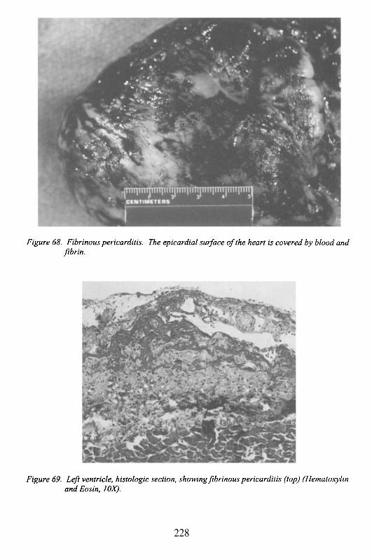
228
Chapter 11
AORTIC ANEURYSMS
Case # 27
Clinical Summary
This 61 year old hypertensive African American man was brought tothe emergency room with severe shortness of breath, pallor, and leftabdominal and epigastric pain.
On admission, his vital signs were: BP 188/110, P 138, RR 30 and T36.8°C. His physical exam was remarkable for a firm pulsatile mass felt inthe abdominal lower left quadrant, and decreased femoral pulses. A CT ofabdomen and chest revealed an abdominal aortic aneurysm andretroperitoneal blood. In the emergency room, he became bradycardic, apneicand unresponsive, but responded to resuscitation and then was transferred tothe operating room. The abdomen was opened and the aorta was clampedabove the renal arteries. The retroperitoneum was filled with blood and themargins of the aneurysm were difficult to establish. After five units of bloodand 5 liters of crystalloids were given, the patient arrested on the table andwas pronounced dead after all resuscitation efforts failed.
Gross Description
The body measured 158 cm in length and weighed 80 kg. The heartweighed 600 g. The pericardium was smooth and shiny. The epicardium hadfocal petechial hemorrhages on the anterior surface consistent withresuscitation maneuvers. The atria were of normal size without thrombi. Theright and left ventricular walls measure 0.6 and 1.6 cm in thickness,respectively. The valve leaflets were pliable and the chordae tendineae werethin. The endocardium had foci of hemorrhage. The myocardium wasgrossly normal. The coronaries revealed mild atherosclerosis with noocclusions. The aorta showed aneurysmal dilatation extending from the celiacartery to the iliac bifurcation. The aneurysm measured 19 cm in length by 8cm in diameter and had a large defect with ragged edges measuring up to 15cm in length. The wall of the aneurysm was very thin and covered withatherosclerotic plaques and areas of necrosis. There was an aneurysmaldilatation of the aortic arch measuring 4 cm in greatest diameter. The
brachiocephalic artery was also dilated, and was occluded by a recentthrombus.
Case Analysis
This man presented to the hospital with shortness of breath and severeabdominal pain of acute onset. Although he had tachycardia (138/min)suggesting hypovolemia, he was hypertensive (188/110 mmmHg), most likelydue to peripheral vasoconstriction. On physical examination, a pulsatile massin the left lower quadrant was evident. A CT scan of the abdomen and chestrevealed an aortic aneurysm from the thorax to the common iliac arteriesassociated with retroperitoneal blood clot. He became unresponsive and had acardiopulmonary arrest, after attempting stabilization he was taken to theoperating room with the presumptive diagnosis of a ruptured aneurysm. Thesurgeons used a combined thoraco-abdominal incision because of the lengthof the aneurysm. Despite blood and fluid replacement, he had a cardiac arreston the operating table, and could not be resuscitated.
The mortality rate of a ruptured abdominal aortic aneurysm that leadsto emergency surgery can be as high as 90%. The outlook has improvedrecently, but still the mortality remains greater than 50%. In contrast, electivesurgical intervention performed prior to rupture has a completely differentresult, with greater than 90% survival. Therefore, a primary goal in dealingwith patients at risk for abdominal aortic aneurysm is the early diagnosis, andappropriate intervention. The issues we will discuss include the risk profile ofpatients, the etiology, anatomy and pathophysiology of aneurysms with andwithout rupture, and the complications beyond free rupture and hypovolemicshock.
An aneurysm is an outward ‘bulge’ of a vascular structure involvingthe full-thickness of the vascular wall. For instance, one can have ananeurysm of the left ventricle (a muscular vascular tube) following amyocardial infarction, with replacement of the full thickness of ventricularmyocardium by scar tissue. Aneurysm development in arteries can involvethe entire circumference of the vessel, in which case there is a diffuse orfusiform (cylindrical) aneurysm (Figures 70 and 71); or a localized bulge of asegment of the vessel known as a saccular aneurysm. Either type issusceptible to rupture or the other complications we will discuss. The termaneurysm is often applied to dissection of a vessel, but this is incorrect. Adissection is a partial tear or separation of layers within the vessel wall. Itresults in a pseudo-aneurysm, where there is a localized vascular rupturepartially enclosed by surrounding tissue. Pseudo-aneurysm also can developwith a complete rupture of a vascular structure, and the tear site andhematoma are maintained within the surrounding tissues and organs (e.g.
230
pleura, pericardium, peritoneum). By this definition, there could be a ruptureof a true aneurysm of the aorta, with a secondary pseudo-aneurysm, if therupture site was walled-off by peritoneum, mesentery, or the vertebral column(Figure 72). The other use of the term aneurysm is applied to mycoticaneurysm (Figure 73). Mycotic aneurysm is a pseudo-aneurysm secondary toinfection of the vascular wall. In general, the rupture site is an abscess withorganizing inflammation. Mycotic aneurysm also can develop within a trueaneurysm, following secondary infection. Any of these aneurysms, orpseudo-aneurysms has the potential to rupture.
A somewhat subjective aspect of aneurysms is how much dilatationof the vessel wall is required before it is called an aneurysm. With a focalbulge it is relatively straightforward; a saccular outpouching is easy torecognize and discriminate from the remaining vessel wall. In contrast, adiffuse or fusiform aneurysm may involve a segment of the vessel, or it maybe a continuation of a generalized dilatation. The latter, known as ectasia, isa common phenomenon in elderly patients over the age of 75 years. The aortadilates diffusely, and may even become tortuous. As a rule, it is preferred toretain the term aneurysm for focal bulges, whether saccular or fusiform, thatare more than 25% over the adjacent vessel circumference.
Although aneurysms can occur anywhere in the vascular tree (e.g.intracerebral arteries, thoracic aorta, etc), abdominal aortic aneurysms havethe highest incidence, and are the most likely to have a fatal rupture. Even thefamiliar shorthand term for abdominal aortic aneurysms, AAA or ‘triple A’,reflects how common it is. The location of the aneurysm, and the profile ofthe patient often provides significant clues as to the etiology of the condition.AAAs are predominantly the result of severe atherosclerosis of the aorta.Thus, the risk factors for AAA should be the same of those foratherosclerosis. If this is the case, why is it that some individuals with severeatherosclerosis develop coronary or cerebral complications, while othersdevelop aneurysms of the aorta? Are the risk factors for atherosclerosis asufficient explanation for the development of AAA? Recent informationstrongly suggests the answer is a resounding no!
Although aneurysms can develop anywhere in the aorta, the ones dueto atherosclerosis are far more common in the abdominal aorta, particularlydistal to the renal arteries. They may extend into the iliac arteries as acontinuous lesion, or there may form separate iliac aneurysms.Atherosclerosis of the abdominal aorta tends to increase in severity fromproximal to distal. The explanation for this is uncertain, although it has beensuggested that it is a phenomenon of human upright posture and gravity. Theincreased vascular wall injury develops secondary to the effects of thepressurized column of blood within the aorta (until the time when humanscould live permanently in low gravity conditions, this hypothesis will not betested). However, what is definitely known, is that there is a significant
231
association of AAA with systemic hypertension, smoking, diabetes mellitusand older age (aneurysms are rare before the decade, and are moscommon between the and decades). Even though these are all riskfactors for atherosclerosis, there is a disproportionate ratio of male gender,and a known familial predisposition. The presence of an AAA in a sibling orparent, significantly increases the risk for that individual. This familialtendency and male predominance strongly suggests that additionalunrecognized entities are interacting with the more common and generallymodifiable risk factors, which contribute to the development ofatherosclerotic AAA.
Molecular defects that impact on the structural integrity of the vesselwall might explain the known link between aneurysms and several relativelyrare genetic disorders. Osteogenesis imperfecta, a disease of the skeleton dueto a defect in collagen type I (the primary fibrillar component of collagen), isassociated with aortic aneurysms. The basis for Ehlers-Danlos type IVsyndrome is a defect of collagen type III (the reticular component ofcollagen), and it presents with skin, connective tissue and cardiovascularsystem manifestations, including aortic aneurysms. On the other hand,defective collagen resulting from inadequate cross-linking, due to the effectsof (the active ingredient in sweat-pea that has beenassociated with a clinical and experimentally-induced condition known aslathyrism), typically leads to aortic dissection in man and laboratory animals.Therefore, although collagen abnormalities may be associated withaneurysms, it is unclear whether genetic, acquired or both types of collagendefects explain the pathogenesis of aortic aneurysms. It is instructive to recallthat for more than half a century the etiology of Marfan’s syndrome and aorticdissection was thought to be defective collagen, until the defect was identifiedin a microfibrillar glycoprotein component of elastic tissue, called fibrillin.
There are two other pathogenic considerations that have receivedinterest recently, but before we discuss them we should address thepathological changes that occur within aneurysms. The media of the aorta isan elastic structure composed of approximately 40 stacked layers of elastictissue fibers, smooth muscle cells, and collagen, called lamellae. There arealso vessels in the outer third of the tissue (vasa vasorum), that provide bloodsupply to the inner media (diffusion of oxygen from the lumen through thetissue is insufficient). Atherosclerosis is initially an intimal disease.However, with increasing severity, it extends into the media where it replacesthe lamellar structure with plaque, debris, fibrous tissue, calcification, andinflammation. The loss of elastic lamellae leads to a weakening of the tensilestrength of the aorta. If there is only localized atherosclerosis, there may beno adverse consequences; but when it becomes diffuse and involves the fullaortic wall thickness, there may be aneurysm formation. The disruption of thetissue by plaque, can also affect the vasa vasorum and their ability to supply
232
blood to the media, thereby causing further damage to the wall. Thedegenerative process resulting from atherosclerosis is the primary cause ofaneurysms; but as we have seen, there appear to be other elements thatcontribute to the breakdown of the media, and the weakening of the aorticwall.
One potential pathogenic mechanism may be related to the activationof lytic enzymes in the aortic atherosclerotic plaque. Similar to thetransformation of a stable coronary artery atherosclerotic plaque into anunstable one, with plaque rupture and coronary thrombosis, the atheroscleroticAAA tissue breakdown can be associated with enzyme activity derived frominflammatory cells in the plaque. In particular, collagenases and elastasesmay play a significant role in media destruction and mural weakening. Thereappears to be variability in the degree of inflammation in atherosclerosisregardless of site. Some patients seem to have more inflamed plaques thanothers, making them more susceptible to atherosclerotic complications.Whether this inflammatory activity is genetically determined is unknown.Another potential etiology is related to the possibility that atheroscleroticplaques may become sites of secondary infection. Bacteria can lead to aninflammatory response and a secondary release of collagenases and elastasesfrom inflammatory cells. Furthermore, they can also elaborate their owncollagenases with direct degradation of tissue, which may lead to bacterialspread and abscess formation. Gram negative bacteria, and particularlySalmonella species, have a predilection for the debris and surface thrombusassociated with atherosclerotic plaques in the aorta (Salmonella have beencultured from surgically repaired aortic aneurysms). Whether, any or all ofthese potential etiologies is working separately or together to cause aneurysmsis unknown. But, since a number of these mechanisms may be prevented ormodified by treatment, medical intervention to avoid aneurysm expansion andpossible rupture may be feasible in the future.
Size and expansion is the critical issue with abdominal aorticaneurysms. The diameter of the aneurysm is directly proportional to itslikelihood of rupture. Also, whether it is stable or progressing over time,determines its rupture potential. Since both of these parameters can beassessed non-invasively with ultrasound, CT scan, or magnetic resonanceimaging (MRI), patients can be followed longitudinally to determine when, orif, it is appropriate to intervene therapeutically. What is the critical size? Thenormal aorta has a diameter of 2.5-3.0 cm. An aneurysm of 4.0-6.0 cm is at acritical size; beyond that, the potential for rupture increases significantly eachyear. Furthermore, an unstable aneurysm that expands progressively at a rategreater than 0.5 cm per year is at risk for rupture. A 4.0-6.0 cm aneurysm isgenerally palpable in most individuals; therefore physical examination is animportant screening test. The size of the aneurysm and its growth aredetermined by the mechanisms discussed above. However, there are also
233
physical factors that impact on the likelihood of rupture. As the aneurysmexpands, the increased diameter affects the wall stress and potential forrupture. The relationship is explained by LaPlace’s law:
Wall Stress = Pressure X Radius2 X Wall Thickness
Thus, the radius or diameter of the aneurysm is directly proportionalto the wall stress (a wall that is already weakened by atheroscleroticdegeneration). Essentially, the larger the balloon, the more likely it is toburst. It is critically important to intervene before this occurs, for reasons thatbecame evident in this case.
There are complications of aneurysm in addition to fatal rupture.Aneurysms frequently thrombose and the thrombus may fragment andembolize into the distal circulation of the lower extremities leading to acuteischemia. If the aneurysm is fusiform, and the thrombus continues toaggregate, there may be partial or complete obstruction of the lowerabdominal aorta. This can cause lower extremity ischemia, intermittentclaudication, or gangrene. When there is lower extremity ischemia involvingthe thigh and below, in association with impotence, the Leriche syndrome(obstruction of the distal aorta) must be considered. In the absence ofsignificant thrombosis, the ulcerative, atheromatous surface of an aneurysmmay be the source of cholesterol and plaque emboli. These usually affectsmaller vessels in the distal extremities, but may lead to toe and footgangrene, or the so-called ‘trash foot’.
There are several other complications associated with localizedrupture. With progressive atherosclerosis, the abdominal aortic aneurysmsoften develop inflammation and fibrosis of the adventitia. The inflammatoryresponse may induce the aorta to adhere to the surrounding tissues or organs,which include the intestine, vena cava, and vertebral column. The abdominalaorta is a retroperitoneal structure. It lies immediately posterior to the and
portions of the duodenum, proximal to the ligament of Treitz. Theduodenum in this location crosses the aorta at the level of the aorticbifurcation into the iliac arteries. Accordingly, this site, which is the mostprevalent for atherosclerotic aneurysms, is susceptible to the rupture of ananeurysm into the intestine. This is known as an aorto-enteric fistula. It isvirtually always fatal, with exsanguinating gastrointestinal hemorrhage.Rarely, it may produce a slow leak, and be a cause of chronic anemia. If theaortic aneurysm becomes adherent along its lateral wall, it can rupture into thevena cava, causing an arteriovenous fistula, and high output congestive heartfailure. Finally, if the aneurysm extends posteriorly, either with or withoutrupture, it can lead to vertebral column erosion and severe back pain.
234
Several comments about diagnosis and treatment. As noted above,aneurysms at a critical size (over 5.0 cm) can be palpated with carefulexamination of the abdomen at the level of the umbilicus. Palpation of amass, with characteristic lateral pulsations, is strongly supportive of thediagnosis. It also is noteworthy that aneurysms susceptible to rupture oftenhave slow leakage over days to weeks prior to massive rupture. This can leadto inflammation and irritation of nerves in adventitial tissues, thereby causingback or flank pain. When aneurysms do rupture, the blood generally tracks inthe retroperitoneal tissues, causing flank ecchymoses, scrotal hematoma, andinguinal hematoma. Most often, patients with these signs will also be inshock. If the aneurysm ruptures anteriorly, it can ‘break through’ theperitoneal membrane, and cause massive hemoperitoneum. This is rapidlyfatal.
Treatment is dependent on the size and the rate of growth of theaneurysm, and the presence of complications such as thrombosis andembolization. The approach for the last 5 decades has been prosthetic graftbypass of the aneurysm with interposition of a woven, artificial vesselbetween the aorta and the iliac or femoral arteries. The graft material isusually Dacron, and it functions very effectively with minimal risk ofthrombosis. Even though it is a prosthetic material, which is potentiallythrombogenic, its large diameter, and the ingrowth of the patient’s own cellsthrough the porous spaces (a de novo endothelial lining) between the weavefibers prevent thrombosis. For many years the aneurysm was actuallyresected; currently, the aneurysm is opened and the graft is placed over theaneurysm bed. The major complications of surgery are related to the timerequired to cross-clamp the aorta. Since most aneurysms occur below therenal arteries, the potential for renal ischemia is minimized. Clamping abovethe renal vessels puts the kidney at risk for ischemic injury. Other rareischemic complications include spinal cord damage with hemiplegia due tointerruption of vessels supplying the cord, and left colon ischemia due to lackof flow in the inferior mesenteric artery. Both are unusual, because ofcollateral blood supply that efficiently develops over time, triggered by thelongstanding atherosclerotic occlusion of the larger vessel. Other standardsurgical complications are bleeding and infection. Moreover, a major causeof morbidity and mortality, is related to cardiac and cerebral ischemia, sincepatients with AAA, invariably have systemic atherosclerosis.
As a final point, it should be mentioned that a new approach toaneurysm ‘repair’ is becoming prevalent in selected patients. Aneurysmswithout significant atherosclerotic stenosis of iliac arteries can be treated withendovascular grafting. Tubular, telescoped grafts can be introduced into theaorta from the femoral artery under radiographic control, where they can beextended from the normal aorta superior to the aneurysm to the iliac arteries.They function as a conduit across the aneurysm, thereby eliminating the risk
235

of aneurysm rupture, and any of the surgical complications associated withrepair. It is likely that as with the growing use of stents to maintain coronarypatency following angioplasty, endovascular aortic grafting will rapidlybecome a popular approach to dealing with the common, and dangerousAAA.
236
Suggested Readings
1.
2.
3.
4.
5.
6.
7.
8.
9.
Blanchard JF. Epidemiology of abdominal aortic aneurysms. EpidemiolRev. 1999; 21:207-21.Dietz HC. New insights into the genetic basis of aortic aneurysms.MonogrPathol. 1995; 37:144-55.Vorp DA, Trachtenberg JD, Webster MW. Arterial hemodynamics andwall mechanics. Semin Vase Surg. 1998; 11:169-80.Grange JJ, Davis V, Baxter BT. Pathogenesis of abdominal aorticaneurysm: an update and look toward the future. Cardiovasc Surg. 1997;5:256-65.Wills A, Thompson MM, Crowther M, Sayers RD, Bell PR. Pathogenesisof abdominal aortic aneurysms--cellular and biochemical mechanisms. EurJ Vasc Endovasc Surg. 1996; 12:391-400.Pearce WH, Koch AE. Cellular components and features of immuneresponse in abdominal aortic aneurysms. Ann N Y Acad Sci. 1996;800:175-85.Thompson RW, Parks WC. Role of matrix metalloproteinases inabdominal aortic aneurysms. Ann N Y Acad Sci. 1996; 800:157-74.Lindsay J Jr. Diagnosis and treatment of diseases of the aorta. Curr ProblCardiol. 1997; 22:485-542.Fillinger MF. Imaging of the thoracic and thoracoabdominal aorta. SeminVase Surg. 2000; 13:247-63.
237
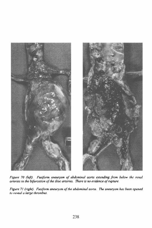
238
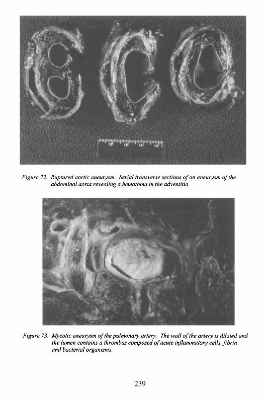
239

This Page Intentionally Left Blank
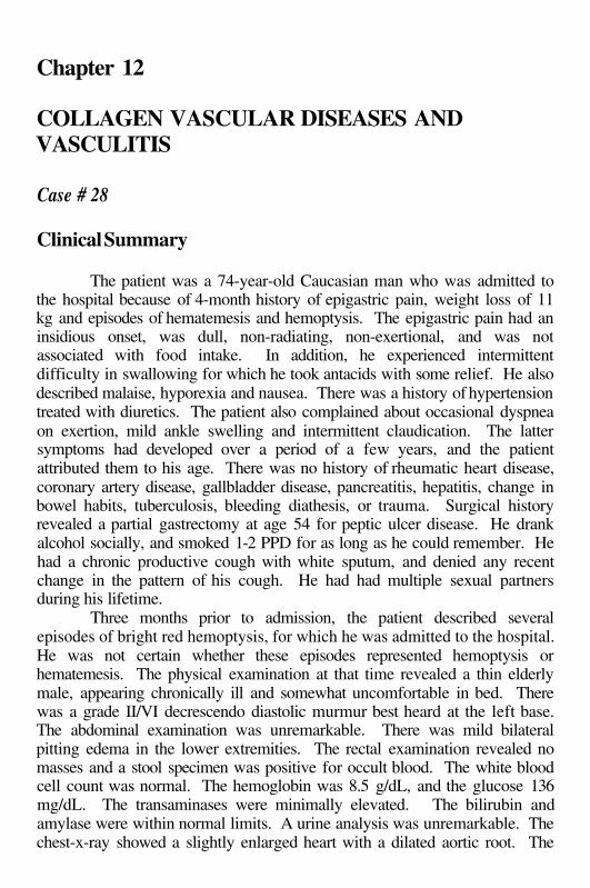
Chapter 12
COLLAGEN VASCULAR DISEASES ANDVASCULITIS
Case # 28
Clinical Summary
The patient was a 74-year-old Caucasian man who was admitted tothe hospital because of 4-month history of epigastric pain, weight loss of 11kg and episodes of hematemesis and hemoptysis. The epigastric pain had aninsidious onset, was dull, non-radiating, non-exertional, and was notassociated with food intake. In addition, he experienced intermittentdifficulty in swallowing for which he took antacids with some relief. He alsodescribed malaise, hyporexia and nausea. There was a history of hypertensiontreated with diuretics. The patient also complained about occasional dyspneaon exertion, mild ankle swelling and intermittent claudication. The lattersymptoms had developed over a period of a few years, and the patientattributed them to his age. There was no history of rheumatic heart disease,coronary artery disease, gallbladder disease, pancreatitis, hepatitis, change inbowel habits, tuberculosis, bleeding diathesis, or trauma. Surgical historyrevealed a partial gastrectomy at age 54 for peptic ulcer disease. He drankalcohol socially, and smoked 1-2 PPD for as long as he could remember. Hehad a chronic productive cough with white sputum, and denied any recentchange in the pattern of his cough. He had had multiple sexual partnersduring his lifetime.
Three months prior to admission, the patient described severalepisodes of bright red hemoptysis, for which he was admitted to the hospital.He was not certain whether these episodes represented hemoptysis orhematemesis. The physical examination at that time revealed a thin elderlymale, appearing chronically ill and somewhat uncomfortable in bed. Therewas a grade II/VI decrescendo diastolic murmur best heard at the left base.The abdominal examination was unremarkable. There was mild bilateralpitting edema in the lower extremities. The rectal examination revealed nomasses and a stool specimen was positive for occult blood. The white bloodcell count was normal. The hemoglobin was 8.5 g/dL, and the glucose 136mg/dL. The transaminases were minimally elevated. The bilirubin andamylase were within normal limits. A urine analysis was unremarkable. Thechest-x-ray showed a slightly enlarged heart with a dilated aortic root. The
electrocardiogram revealed a pattern compatible with left ventricularhypertrophy. Extensive work-up including GI series, celiac angiogram, andpancreatic scan did not uncover any specific lesion. He was dischargedwithout a definitive diagnosis.
Three days after his release from the hospital, the patient againdeveloped hemoptysis. Meanwhile, his epigastric pain had increased, and hehad an overall deterioration of his health with increasing fatigue and anorexia.The temperature was 37°C rectally; pulse 108, respirations 22 and the bloodpressure was 140/72 mmHg, without orthostatic changes. The neurologic andHEENT examination was unremarkable. There was no lymphadenopathy.The patient was non-icteric. The carotid pulse was 2+ bilaterally withoutbruits. The jugular venous pressure was estimated to be within normal limits.The chest was clear to percussion and auscultation. The cardiac examinationrevealed that the PMI was slightly displaced to the left laterally. Again, therewas a grade II/VI diastolic decrescendo murmur that radiated to the left lowersternal border, which was best auscultated at the left base with the patientsitting up and leaning forward. The femoral and popliteal pulses were notpalpable bilaterally; however, the pulses of the upper extremities were withinnormal limits. The abdomen was mildly distended with normal bowel sounds,there was no guarding, rebound tenderness or hepatosplenomegaly. Thelower extremities were cool to the touch and had 1+ pitting edema. The rectalexamination revealed no masses. A stool sample remained positive for occultblood.
Laboratory data and other tests
Laboratory: WBC 7.0, Hb/Hct 8.4/26, MCV 76, Plt 260 (peripheral bloodsmear confirmed a microcytic anemia, but was otherwise unremarkable);normal electrolytes, SGOT 48, protein T/A 6.5/3.2, bilirubin T 0.6, LD 378,BUN/creat 28/1.2, glucose 110. The urine analysis was unremarkable. ECG:normal PR and QRS intervals and left ventricular hypertrophy. The CXRshowed a moderately enlarged heart with a dilated aortic root. An uprightplain film of the abdomen revealed no free air.
While in the hospital, the patient had several more episodes ofhemoptysis of about 50 cc each time and one episode of massive bleeding,bringing up an estimate of 1000-1500 cc of bright red blood. Meanwhile, abronchoscopy failed to reveal any pathology. The bleeding episode recurredand a nasogastric tube was placed revealing fresh blood in the stomach.Irrigation with iced saline, transfusion of 6 units of blood and placement of aSengstaken-Blakemore tube failed to control the bleeding. The patient wastaken to the operating room for a laparotomy and the source of bleeding waslocated above the stomach. A thoracotomy was performed and a presumptive
242
diagnosis was made. The patient was placed on a partial cardiopulmonarybypass, but had a cardiac arrest and could not be resuscitated.
Gross Description
The body was that of an elderly white male weighing 54 kg. andmeasuring 167 cm. The heart weighed 400 g, with hypertrophy of the leftventricle. Marked dilatation of the aortic arch was evident. A 10 cm segmentof the thoracic aorta below the arch was replaced by prosthesis. Suture lineswere intact. The abdominal aorta and iliac vessels showed a moderate degreeof atherosclerosis with some calcification. The intimal surface of the aorticroot and arch showed changes consistent with a “tree bark” appearance withpearly-grey plaques and sclerosis (Figure 74). The lungs were edematouswith centrilobular emphysema. The distal esophagus was adherent to bothpleural surfaces and to a massive blood clot associated with fibrous tissue.On opening, a large defect was noted in the esophageal wall approximately 10cm from the gastric cardia. The defect was consistent with an oval ulcermeasuring 3 cm in its greatest dimension that extended through the fullthickness of the wall (Figure 75). The edges were sharp, not raised andclearly demarcated. A major portion of the stomach was absent and there wasevidence of a Billroth type II anastomosis. The remaining organs showed nosignificant pathology.
Microscopic Description
The myocardium was essentially normal. There was no evidence ofexcessive deposition of fibrous tissue. At the site where the aortic root wasinserted into the left ventricle there was necrosis of the heart muscle andinterstitial hemorrhage. Hemorrhage was also present in the pericardium, thelatter finding probably a reaction to the trauma of surgery.
Sections from the aorta showed subintimal sclerosis with depositionof cholesterol and calcium. The adventitia showed a marked perivascularinflammatory cell infiltrate composed primarily of plasma cells. Sectionstaken from the esophageal ulcer revealed fragments of aortic wall displayingcholesterol and fibrosis, which was adherent to the esophageal wall. Thesefindings were consistent with an aorto-esophageal fistula.
Case Analysis
The two most significant symptoms that this patient experienced werehemoptysis and epigastric pain. The causes of epigastric pain are numerousand include: peptic ulcer disease, pancreatitis, myocardial infarction, aortic
243
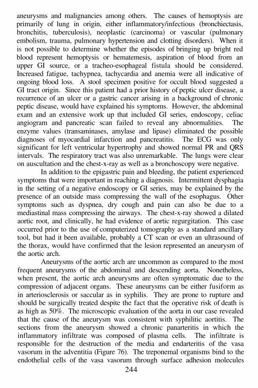
aneurysms and malignancies among others. The causes of hemoptysis areprimarily of lung in origin, either inflammatory/infectious (bronchiectasis,bronchitis, tuberculosis), neoplastic (carcinoma) or vascular (pulmonaryembolism, trauma, pulmonary hypertension and clotting disorders). When itis not possible to determine whether the episodes of bringing up bright redblood represent hemoptysis or hematemesis, aspiration of blood from anupper GI source, or a tracheo-esophageal fistula should be considered.Increased fatigue, tachypnea, tachycardia and anemia were all indicative ofongoing blood loss. A stool specimen positive for occult blood suggested aGI tract origin. Since this patient had a prior history of peptic ulcer disease, arecurrence of an ulcer or a gastric cancer arising in a background of chronicpeptic disease, would have explained his symptoms. However, the abdominalexam and an extensive work up that included GI series, endoscopy, celiacangiogram and pancreatic scan failed to reveal any abnormalities. Theenzyme values (transaminases, amylase and lipase) eliminated the possiblediagnoses of myocardial infarction and pancreatitis. The ECG was onlysignificant for left ventricular hypertrophy and showed normal PR and QRSintervals. The respiratory tract was also unremarkable. The lungs were clearon auscultation and the chest-x-ray as well as a bronchoscopy were negative.
In addition to the epigastric pain and bleeding, the patient experiencedsymptoms that were important in reaching a diagnosis. Intermittent dysphagiain the setting of a negative endoscopy or GI series, may be explained by thepresence of an outside mass compressing the wall of the esophagus. Othersymptoms such as dyspnea, dry cough and pain can also be due to amediastinal mass compressing the airways. The chest-x-ray showed a dilatedaortic root, and clinically, he had evidence of aortic regurgitation. This caseoccurred prior to the use of computerized tomography as a standard ancillarytool, but had it been available, probably a CT scan or even an ultrasound ofthe thorax, would have confirmed that the lesion represented an aneurysm ofthe aortic arch.
Aneurysms of the aortic arch are uncommon as compared to the mostfrequent aneurysms of the abdominal and descending aorta. Nonetheless,when present, the aortic arch aneurysms are often symptomatic due to thecompression of adjacent organs. These aneurysms can be either fusiform asin arteriosclerosis or saccular as in syphilis. They are prone to rupture andshould be surgically treated despite the fact that the operative risk of death isas high as 50%. The microscopic evaluation of the aorta in our case revealedthat the cause of the aneurysm was consistent with syphilitic aortitis. Thesections from the aneurysm showed a chronic panarteritis in which theinflammatory infiltrate was composed of plasma cells. The infiltrate isresponsible for the destruction of the media and endarteritis of the vasavasorum in the adventitia (Figure 76). The treponemal organisms bind to theendothelial cells of the vasa vasorum through surface adhesion molecules
244
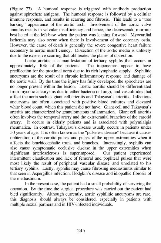
(Figure 77). A humoral response is triggered with antibody productionagainst spirochete antigens. The humoral response is followed by a cellularimmune response, and results in scarring and fibrosis. This leads to a “treebarking” appearance of the aortic arch. Involvement of the aortic valveannulus results in valvular insufficiency and hence, the decrescendo murmurbest heard at the left base when the patient was leaning forward. Myocardialischemia may also occur when there is involvement of the coronary ostia.However, the cause of death is generally the severe congestive heart failuresecondary to aortic insufficiency. Dissection of the aortic media is unlikelydue to the extensive scarring that obliterates the planes of dissection.
Luetic aortitis is a manifestation of tertiary syphilis that occurs inapproximately 10% of the patients. The treponemas appear to havepredilection for the proximal aorta due to its rich lymphatic supply. Syphiliticaneurysms are the result of a chronic inflammatory response and damage ofthe aortic wall. By the time the injury has fully developed, the spirochetes areno longer present within the lesion. Luetic aortitis should be differentiatedfrom mycotic aneurysms due to either bacteria or fungi, and vasculitides thataffect the aorta such as giant cell arteritis and Takayasu’s arteritis. Infectiousaneurysms are often associated with positive blood cultures and elevatedwhite blood count, which this patient did not have. Giant cell and Takayasu’sarteritis are characterized by granulomatous inflammation. Giant cell arteritisoften involves the temporal artery and the extracranial branches of the carotidartery. It occurs in elderly patients and is associated with polymialgiarheumatica. In contrast, Takayasu’s disease usually occurs in patients under50 years of age. It is often known as the “pulseless disease” because it causesobliteration of the carotid pulses and pulses of the upper extremities when itaffects the brachiocephalic trunk and branches. Interestingly, syphilis canalso cause symptomatic occlusive disease in the upper extremities whensignificant arteriosclerosis is superimposed. Our patient experiencedintermittent claudication and lack of femoral and popliteal pulses that weremost likely the result of peripheral vascular disease and unrelated to histertiary syphilis. Lastly, syphilis may cause fibrosing mediastinitis similar tothat seen in Aspergillus infection, Hodgkin’s disease and idiopathic fibrosis ofthe mediastinum.
In the present case, the patient had a small probability of surviving theoperation. By the time the surgical procedure was carried out the patient hadbled significantly. Although currently, aortic syphilitic aneurysms are rare,this diagnosis should always be considered, especially in patients withmultiple sexual partners and in HIV-infected individuals.
245
Suggested Readings
1.
2.
3.
4.
Dossa CD, Pipinos II, Shepard AD, Ernst CB. Primary aortoenteric fistula:Part I.Ann Vasc Surg. 1994; 8:113-20.Grey TC, Mittleman RE, Wetli CV, Horowitz S. Aortoesophageal fistulaand sudden death. A report of two cases and review of the literature. Am JForensic Med Pathol. 1988; 9:19-22.Coady MA, Rizzo JA, Goldsstein LJ, Elefteriades JA. Natural history,pathogenesis, and etiology of thoracic aortic aneurysms and dissections.Cardiol Clin. 1999; 17:615-635.Amin S. Aortoesophageal fistula: case report and review of the literature.Dig Dis Sci. 1998; 43:1665-1671.
246
247
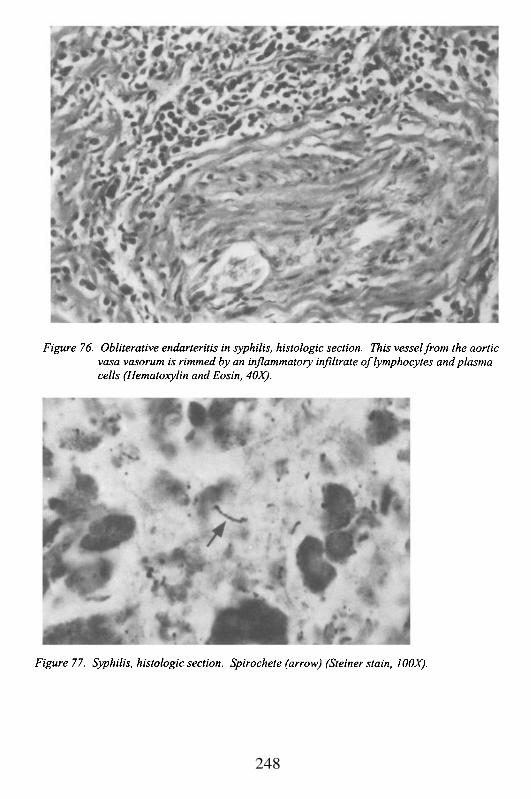
248
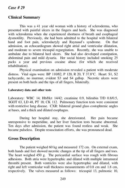
Case # 29
Clinical Summary
This was a 41 year old woman with a history of scleroderma, whopresented with painful ulcers in the fingers and heels. She was diagnosedwith scleroderma when she experienced shortness of breath and esophagealdysmotility. Previously, she had been admitted to the hospital with bilateralhand and foot pain, sclerodactyly and Raynaud’s syndrome. On thatadmission, an echocardiogram showed right atrial and ventricular dilatation,and moderate to severe tricuspid regurgitation. Recently, she was unable toambulate due to bilateral heel ulcers. She had also developed constipation,abdominal pain and mild dysuria. Her social history included smoking 25packs a year and previous cocaine abuse (for which she receivedrehabilitation).
Physical examination on admission revealed a patient with moderatedistress. Vital signs were: BP 110/82, P 120, R 20, T 37.8°C. Heart: S1, S2tachycardic, no murmur, evident S3 and S4 gallop. Necrotic ulcers werepresent on both heels and the tips of all fingers, bilaterally.
Laboratory data and other tests
Laboratory: WBC 14; Hb/Hct 14/42; creatinine 0.9, bilirubin T/D 0.8/0.5,SGOT 63, LD 48, PT 18; CK 112. Pulmonary function tests were consistentwith restrictive lung disease. CXR: bilateral ground glass costophrenic anglesat bases, and a thick and dilated esophagus.
During her hospital stay, she deteriorated. Her pain becameunresponsive to meperidine, and her liver function tests became abnormal.Ten days after admission, the patient was found restless and weak. Shebecame pulseless. Despite resuscitation efforts, she was pronounced dead.
Gross Description
The patient weighed 60 kg and measured 172 cm. On external exam,both hands and feet showed necrotic changes at the tip of all fingers and toes.The heart weighed 450 g. The pericardial surface was rough with fibrousadhesions. Both atria were hypertrophic and dilated with multiple intramuralthrombi present. Both ventricles were also hypertrophic and dilated, withright and left ventricular wall thickness measurements of 0.8 cm and 1.8 cm,respectively. The valves measured as follows: tricuspid 13, pulmonic 10,
249
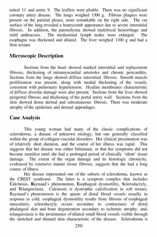
mitral 11 and aortic 9. The leaflets were pliable. There was no significantcoronary artery disease. The lungs weighed 1300 g. Fibrous plaques werepresent on the parietal pleura, more remarkable on the right side. The cutsurface of the lung revealed a honeycomb appearance due to severe interstitialfibrosis. In addition, the parenchyma showed multifocal hemorrhage andmild anthracosis. The mediastinal lymph nodes were enlarged. Theesophagus was thickened and dilated. The liver weighed 1100 g and had afirm texture
Microscopic Description
Sections from the heart showed marked interstitial and replacementfibrosis, thickening of intramyocardial arterioles and chronic pericarditis.Sections from the lungs showed diffuse interstitial fibrosis. Smooth muscleproliferation was present, along with medial thickening of the vessels,consistent with pulmonary hypertension. Hyaline membranes characteristicof diffuse alveolar damage were also present. Sections from the liver showedperiportal fibrosis and thickening of the portal artery wall. Sections from theskin showed dense dermal and subcutaneous fibrosis. There was moderateatrophy of the epidermis and dermal appendages.
Case Analysis
This young woman had many of the classic complications ofscleroderma, a disease of unknown etiology, but one generally classifiedwithin the group of collagen-vascular disorders. Her clinical presentation wasof relatively short duration, and the course of her illness was rapid. Thissuggests that her disease was either fulminant, or that her symptoms did notbecome manifest until she had a prolonged period of clinically ‘silent’ tissuedamage. The extent of the organ damage and its histologic chronicity,evidenced by extensive mature tissue fibrosis, suggests that she had a longcourse of illness.
Her disease represented one of the subsets of scleroderma, known asthe CREST syndrome. The latter is a symptom complex that includesCalcinosis, Raynaud’s phenomenon, Esophageal dysmotility, Sclerodactyly,and Telangiectasia. Calcinosis is dystrophic calcification in soft tissues;Raynaud’s phenomenon is the spasm of distal blood vessels usually inresponse to cold; esophageal dysmotility results from fibrosis of esophagealmuscularis; sclerodactyly occurs secondary to contractures of distalphalangeal skin and bone degeneration secondary to ischemic atrophy; and,telangiectasia is the prominence of dilated small blood vessels visible throughthe stretched and thinned skin characteristic of the disease. Scleroderma is
250
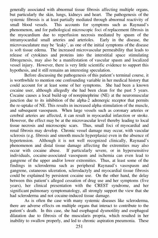
generally associated with abnormal tissue fibrosis affecting multiple organs,but particularly the skin, lungs, kidneys and heart. The pathogenesis of thesystemic fibrosis is at least partially mediated through abnormal reactivity ofsmall blood vessels. This accounts for symptoms such as Raynaud’sphenomenon, and for pathological microscopic foci of replacement fibrosis inthe myocardium due to reperfusion necrosis mediated by spasm of theintramyocardial small arteries and arterioles. Early in the disease, themicrovasculature may be ‘leaky’, as one of the initial symptoms of the diseaseis soft tissue edema. The increased microvascular permeability that leads torelease of cytokines and proteins into the interstitial space stimulatingfibrogenesis, may also be a manifestation of vascular spasm and localizedvessel injury. However, there is very little scientific evidence to support thishypothesis, and it still remains an interesting speculation.
Before discussing the pathogenesis of this patient’s terminal course, itis worthwhile to mention one confounding variable in her medical history thatcould account for at least some of her symptoms. She had been a knowncocaine user, although allegedly she had been clean for the past 5 years.Cocaine causes a local build-up of norepinephrine (NE) at the neuromuscularjunction due to its inhibition of the alpha-2 adrenergic receptor that permitsthe re-uptake of NE. This results in increased alpha stimulation of the muscle,and subsequent vasospasm. When large vessels such as coronary arteries orcerebral arteries are affected, it can result in myocardial infarction or stroke.However, the effect may be at the microvascular level thereby leading to localtissue necrosis and microscopic scars. Thus, small foci of myocardial andrenal fibrosis may develop. Chronic vessel damage may occur, with vascularsclerosis (e.g. fibrosis and smooth muscle hyperplasia) even in the absence ofhypertension. Although it is not well recognized clinically, Raynaud’sphenomenon and distal tissue damage affecting the extremities may alsooccur with cocaine abuse. If particularly severe, or in hypersensitiveindividuals, cocaine-associated vasospasm and ischemia can even lead togangrene of the upper and/or lower extremities. Thus, at least some of thefindings in scleroderma such as peripheral Raynaud’s vasospasm andgangrene, cutaneous ulceration, sclerodactyly and myocardial tissue fibrosiscould be explained by persistent cocaine use. On the other hand, the delaybetween this patient’s alleged cessation of drug use and her symptoms (fiveyears), her clinical presentation with the CREST syndrome, and hersignificant pulmonary symptomatology, all strongly support the view that shehad scleroderma and not cocaine-induced tissue damage.
As is often the case with many systemic diseases like scleroderma,there are adverse effects on multiple organs that interact to contribute to thepatient’s death. In this case, she had esophageal dysmotility and esophagealdilatation due to fibrosis of the muscularis propria, which resulted in herinability to swallow properly, and led to chronic aspiration pneumonia. These
251
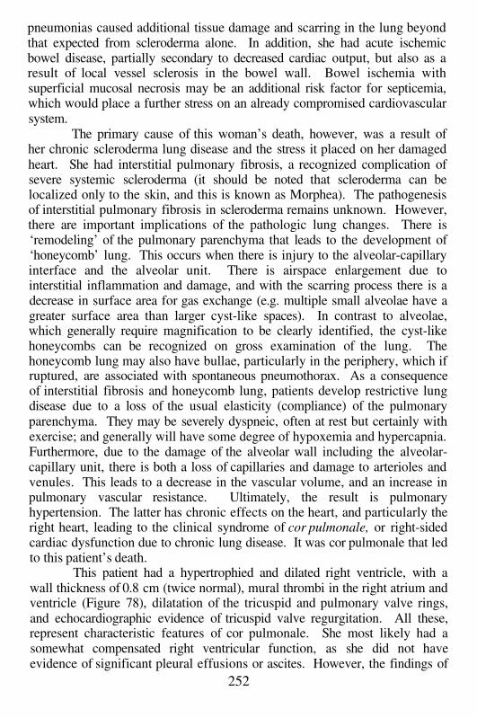
pneumonias caused additional tissue damage and scarring in the lung beyondthat expected from scleroderma alone. In addition, she had acute ischemicbowel disease, partially secondary to decreased cardiac output, but also as aresult of local vessel sclerosis in the bowel wall. Bowel ischemia withsuperficial mucosal necrosis may be an additional risk factor for septicemia,which would place a further stress on an already compromised cardiovascularsystem.
The primary cause of this woman’s death, however, was a result ofher chronic scleroderma lung disease and the stress it placed on her damagedheart. She had interstitial pulmonary fibrosis, a recognized complication ofsevere systemic scleroderma (it should be noted that scleroderma can belocalized only to the skin, and this is known as Morphea). The pathogenesisof interstitial pulmonary fibrosis in scleroderma remains unknown. However,there are important implications of the pathologic lung changes. There is‘remodeling’ of the pulmonary parenchyma that leads to the development of‘honeycomb’ lung. This occurs when there is injury to the alveolar-capillaryinterface and the alveolar unit. There is airspace enlargement due tointerstitial inflammation and damage, and with the scarring process there is adecrease in surface area for gas exchange (e.g. multiple small alveolae have agreater surface area than larger cyst-like spaces). In contrast to alveolae,which generally require magnification to be clearly identified, the cyst-likehoneycombs can be recognized on gross examination of the lung. Thehoneycomb lung may also have bullae, particularly in the periphery, which ifruptured, are associated with spontaneous pneumothorax. As a consequenceof interstitial fibrosis and honeycomb lung, patients develop restrictive lungdisease due to a loss of the usual elasticity (compliance) of the pulmonaryparenchyma. They may be severely dyspneic, often at rest but certainly withexercise; and generally will have some degree of hypoxemia and hypercapnia.Furthermore, due to the damage of the alveolar wall including the alveolar-capillary unit, there is both a loss of capillaries and damage to arterioles andvenules. This leads to a decrease in the vascular volume, and an increase inpulmonary vascular resistance. Ultimately, the result is pulmonaryhypertension. The latter has chronic effects on the heart, and particularly theright heart, leading to the clinical syndrome of cor pulmonale, or right-sidedcardiac dysfunction due to chronic lung disease. It was cor pulmonale that ledto this patient’s death.
This patient had a hypertrophied and dilated right ventricle, with awall thickness of 0.8 cm (twice normal), mural thrombi in the right atrium andventricle (Figure 78), dilatation of the tricuspid and pulmonary valve rings,and echocardiographic evidence of tricuspid valve regurgitation. All these,represent characteristic features of cor pulmonale. She most likely had asomewhat compensated right ventricular function, as she did not haveevidence of significant pleural effusions or ascites. However, the findings of
252

mural thrombi in the right atrium and ventricle do indicate abnormalcontractile function of these chambers, and turbulent blood flow causingendocardial damage with platelet and fibrin aggregation and adherence to thetissue. She also had chronic congestion in the liver, with central venoussclerosis due to transmitted elevated right atrium pressure to the hepatic veins.Chronic congestion accounted for the elevation of her liver function testsduring her last admission.
Patients with cor pulmonale are highly susceptible to develop heartfailure, as well as sudden death due to cardiac arrhythmia. In this case, shealso had damage to her left ventricular myocardium as a result of small vesseldisease. This led to microscopic foci of replacement fibrosis. Myocardiumaround such foci is electrically unstable and may be arrhythmogenic.Furthermore, the basic underlying cause of the replacement fibrosis - vascularhyper-reactivity - can persist, and lead to sudden development of microscopicareas of reperfusion necrosis characterized by contraction bands. Reperfusionnecrosis is highly arrhythmogenic, as well.
The terminal event of a sudden cardiac arrest resulted from acombination of cardiac and pulmonary compromise. The major effects wereon the right side of the heart, but there was also left ventricular injury. Herdisease was progressive, and only partially controlled by steroid therapy.Even if entirely controlled, however, once the pulmonary and cardiac damagedevelops it is irreversible. When the effects of chronic aspiration pneumoniadue to esophageal dysmotility, and bowel necrosis due to local intestinalischemia are superimposed on the primary lung and heart damage, it is notsurprising that she developed a sudden fatal cardiac arrhythmia.
253
Suggested Readings
1.
2.
3.
4.
5.
Gurubhagavatula I; Palevsky HI. Pulmonary hypertension in systemicautoimmune disease. Rheum Dis Clin North Am. 1997; 23:365-94.LeRoy EC. Systemic sclerosis. A vascular perspective. Rheum Dis ClinNorth Am. 1996; 22:675-94.Rump AF, Theisohn M, Klaus W. The pathophysiology of cocainecardiotoxicity. Forensic Sci Int. 1995; 71:103-15.Deswal A; Follansbee WP. Cardiac involvement in scleroderma. RheumDis Clin North Am. 1996; 22:841-60.Bolster MB; Silver RM. Lung disease in systemic sclerosis (scleroderma).Baillieres Clin Rheumatol. 1993; 7:79-97.
254
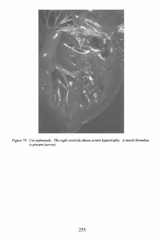
255
Case # 30
Clinical Summary
This 20 year old Caucasian woman, diagnosed at 8 years of age withsystemic lupus erythematosus (SLE), presented to the hospital with a onemonth history of non-traumatic low back pain. She was significantlyincapacitated and required crutches for ambulation. The pain had a suddenonset and woke her up from her sleep. There was no history of recentchanges in her urinary or gastrointestinal functions. Prior to admission, shehad been seen by a physician who prescribed ibuprofen (400 mg q6h), butwith no relief. Other medications at the time of admission were prednisolone(60 mg/day) and furosemide (20 mg QD). She had increased her steroid doseon her own because of worsening arthralgias. She had not visited herrheumatologist for a considerable period of time and admitted poorcompliance regarding scheduled medical appointments. Her knowledge ofher medical condition was limited. The initial presentation during childhoodwas that of epistaxis secondary to thrombocytopenia. She had primaryamenorrhea, with some breast development and minimal growth of axillaryand pubic hair. In addition, the patient had a history of avascular necrosis ofthe hips and she had also developed cataracts.
The physical examination on admission revealed truncal obesity andcushingoid facies. The vital signs were T 37.8°C, P 104/min, R 20/min, BP120/80 mmHg. She had a dry oral mucosa and supple neck. The lungs wereclear. On auscultation she had normal S1 and S2, regular rhythm and nomurmurs. The abdomen had striae. No organomegaly was noted. She hadlarge hematomas on both shins. Her sensation and reflexes were intact, buther motor functions could not be explored due to pain.
Laboratory data and other tests
Laboratory: WBC 8.5 (S 96%, L 3%), Hb/Hct 9.6/28, MCV 91, Plt 75,PT/PTT 14/36; Calcium 6.6, SGOT/SGPT 92/30, protein T/A 4.1/2.1,bilirubin T 0.3, BUN/creat 21/1.1, glucose 133. Arterial blood gas: pH 7.38,pCO2 32, pO2 80, HCO3 19, and base excess –4.1. In addition, her serologywas positive for anti-double stranded DNA and anti-cardiolipin antibodies.The urine analysis showed trace protein, many bacteria, and amorphouscrystals. ECG: sinus tachycardia, otherwise unremarkable. The chest-x rayhad evidence for increased pulmonary vascular congestion. An x-ray of thespine demonstrated diffuse demineralization and loss of height of vertebralbodies.
256
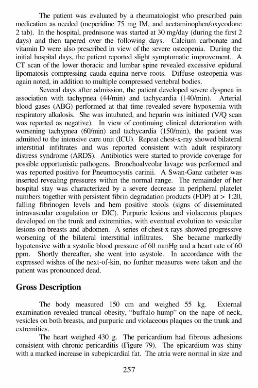
The patient was evaluated by a rheumatologist who prescribed painmedication as needed (meperidine 75 mg IM, and acetaminophen/oxycodone2 tab). In the hospital, prednisone was started at 30 mg/day (during the first 2days) and then tapered over the following days. Calcium carbonate andvitamin D were also prescribed in view of the severe osteopenia. During theinitial hospital days, the patient reported slight symptomatic improvement. ACT scan of the lower thoracic and lumbar spine revealed excessive epidurallipomatosis compressing cauda equina nerve roots. Diffuse osteopenia wasagain noted, in addition to multiple compressed vertebral bodies.
Several days after admission, the patient developed severe dyspnea inassociation with tachypnea (44/min) and tachycardia (140/min). Arterialblood gases (ABG) performed at that time revealed severe hypoxemia withrespiratory alkalosis. She was intubated, and heparin was initiated (V/Q scanwas reported as negative). In view of continuing clinical deterioration withworsening tachypnea (60/min) and tachycardia (150/min), the patient wasadmitted to the intensive care unit (ICU). Repeat chest-x-ray showed bilateralinterstitial infiltrates and was reported consistent with adult respiratorydistress syndrome (ARDS). Antibiotics were started to provide coverage forpossible opportunistic pathogens. Bronchoalveolar lavage was performed andwas reported positive for Pneumocystis carinii. A Swan-Ganz catheter wasinserted revealing pressures within the normal range. The remainder of herhospital stay was characterized by a severe decrease in peripheral plateletnumbers together with persistent fibrin degradation products (FDP) at > 1:20,falling fibrinogen levels and hem positive stools (signs of disseminatedintravascular coagulation or DIC). Purpuric lesions and violaceous plaquesdeveloped on the trunk and extremities, with eventual evolution to vesicularlesions on breasts and abdomen. A series of chest-x-rays showed progressiveworsening of the bilateral interstitial infiltrates. She became markedlyhypotensive with a systolic blood pressure of 60 mmHg and a heart rate of 60ppm. Shortly thereafter, she went into asystole. In accordance with theexpressed wishes of the next-of-kin, no further measures were taken and thepatient was pronounced dead.
Gross Description
The body measured 150 cm and weighed 55 kg. Externalexamination revealed truncal obesity, “buffalo hump” on the nape of neck,vesicles on both breasts, and purpuric and violaceous plaques on the trunk andextremities.
The heart weighed 430 g. The pericardium had fibrous adhesionsconsistent with chronic pericarditis (Figure 79). The epicardium was shinywith a marked increase in subepicardial fat. The atria were normal in size and
257
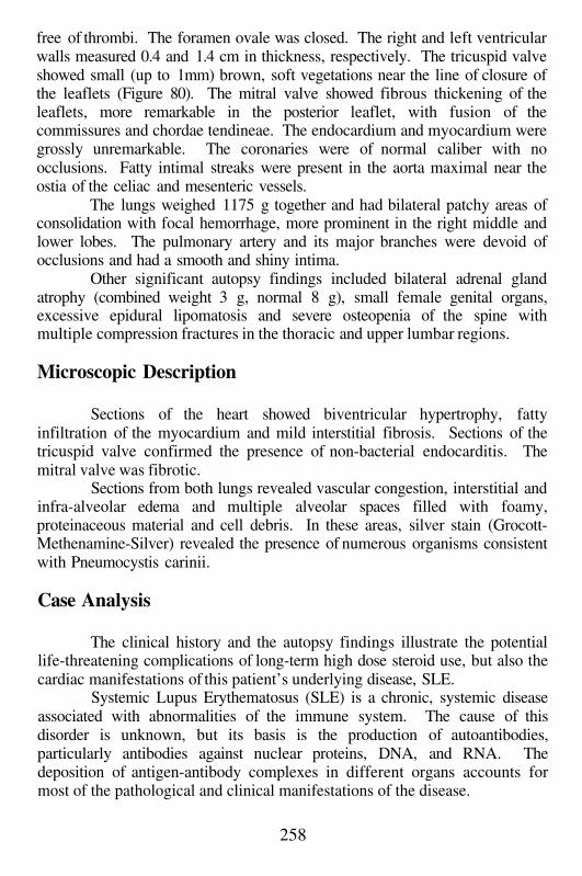
free of thrombi. The foramen ovale was closed. The right and left ventricularwalls measured 0.4 and 1.4 cm in thickness, respectively. The tricuspid valveshowed small (up to 1mm) brown, soft vegetations near the line of closure ofthe leaflets (Figure 80). The mitral valve showed fibrous thickening of theleaflets, more remarkable in the posterior leaflet, with fusion of thecommissures and chordae tendineae. The endocardium and myocardium weregrossly unremarkable. The coronaries were of normal caliber with noocclusions. Fatty intimal streaks were present in the aorta maximal near theostia of the celiac and mesenteric vessels.
The lungs weighed 1175 g together and had bilateral patchy areas ofconsolidation with focal hemorrhage, more prominent in the right middle andlower lobes. The pulmonary artery and its major branches were devoid ofocclusions and had a smooth and shiny intima.
Other significant autopsy findings included bilateral adrenal glandatrophy (combined weight 3 g, normal 8 g), small female genital organs,excessive epidural lipomatosis and severe osteopenia of the spine withmultiple compression fractures in the thoracic and upper lumbar regions.
Microscopic Description
Sections of the heart showed biventricular hypertrophy, fattyinfiltration of the myocardium and mild interstitial fibrosis. Sections of thetricuspid valve confirmed the presence of non-bacterial endocarditis. Themitral valve was fibrotic.
Sections from both lungs revealed vascular congestion, interstitial andinfra-alveolar edema and multiple alveolar spaces filled with foamy,proteinaceous material and cell debris. In these areas, silver stain (Grocott-Methenamine-Silver) revealed the presence of numerous organisms consistentwith Pneumocystis carinii.
Case Analysis
The clinical history and the autopsy findings illustrate the potentiallife-threatening complications of long-term high dose steroid use, but also thecardiac manifestations of this patient’s underlying disease, SLE.
Systemic Lupus Erythematosus (SLE) is a chronic, systemic diseaseassociated with abnormalities of the immune system. The cause of thisdisorder is unknown, but its basis is the production of autoantibodies,particularly antibodies against nuclear proteins, DNA, and RNA. Thedeposition of antigen-antibody complexes in different organs accounts formost of the pathological and clinical manifestations of the disease.
258
The hallmark of the therapeutic approach to SLE in the past decadeshas been the use of corticosteroids, and recently adjuvant cytotoxic andantimetabolite drugs have been added successfully. Therefore, thecardiovascular manifestations in patients with SLE are a combination of theinflammatory processes characteristic of the disease itself, and the secondaryeffects of corticosteroid/immunosuppressive therapy. In the heart, the mostaffected sites are the pericardium and endocardium. Patients with SLE oftenpresent with pericarditis (approximately 80%), and it can be acute (fibrinouspericarditis) or chronic. Recurrent or persistent inflammation of themesothelial lining of the pericardium may lead to mesothelial hyperplasia andfibrosis with the development of adhesions and subsequent obliteration of thepericardial cavity (fibrous pericarditis). Occasionally, examination of pleuralfluid can reveal the presence of lupus erythematosus (LE) cells. These aremacrophages or neutrophils that have phagocytized nuclei denatured bycirculating antinuclear antibodies (ANA). Similar cells, known as LE orhematoxylin bodies can also be identified in tissue samples (Figure 81). Lesscommonly, myocarditis with a non-specific mononuclear infiltrate can occur.It is usually subtle, causing spotty myocardial necrosis, and is diagnosed whenthere is a high level of suspicion leading to an endomyocardial biopsy. Theclinical presentation varies from tachycardia to complete heart block.Moreover, inflammation with subsequent scarring may affect the sinus andatrioventricular nodes as well as the bundle of His. Newborns of motherswith SLE and anti-Ro (Sjögren Syndrome A or SS-A) antibodies may developheart block secondary to transient myocarditis. The latter are anti-nuclearantibodies directed against 52 and 60 kilo-Dalton (kDa) proteins, and arepresent in approximately 35% of patients with SLE and in patients withSjögren syndrome. Anti-Ro IgG can cross the placenta and may cause aneonatal lupus-like syndrome. These maternal autoantibodies disappearwithin a 6-month period; however, long lasting conduction abnormalities inthe offspring have been reported.
Valvular endocardial damage was more dramatic in the pre-steroidera. The classic valvular lesion of SLE is known as Libman-Sacksendocarditis (described in a necropsy series by Drs. Libman and Sacks fromMount Sinai Hospital, New York in 1924). It is a non-infective endocarditismanifested by the presence of small verrucous lesions on any valvular surfaceof the heart. The vegetations are composed of a mixture of immunecomplexes, inflammatory cells and fibrinoid necrosis. Significant valvedysfunction requiring valvular replacement is rare unless superimposedbacterial infection ensues. Aortic and mitral valvulitis are common in SLE,particularly in patients with antiphospholipid antibodies. The clinicalmanifestations in patients with the antiphospholipid syndrome (e. g.phospholipid or cardiolipin antibodies), which include venous and arterial
259
thrombosis, recurrent miscarriages and thrombocytopenia, are secondary to ahypercoagulable state, and to the cellular damage caused by the antibodies.
In our patient, the valvular abnormalities were most likely the resultof her independent, high-dose steroid abuse rather than her positiveanticardiolipin status. Recently, prolonged use of corticosteroids has beenlinked to the development of a serious form of valvular pathologycharacterized by rigid and thickened valvular leaflets. The patient hadfibrosis of the mitral valve leaflets with fusion of the valve commissure andchordae tendineae similar to that seen in patients with rheumatic heart disease(RHD). Could these changes be the result of chronic rheumatic heart disease?They could, but the clinical history and presence of other elements of cardiacinvolvement favor SLE and steroid use as etiologic factors for hervalvulopathy. The vegetations present on the tricuspid valve were moreconsistent with non-bacterial thrombotic (marantic) endocarditis than withLibman-Sacks endocarditis. Marantic endocarditis occurs in chronically ill ordebilitated patients and is manifested by the presence of small, brownvegetations situated close to the line of closure of the leaflets.Microscopically, they consist of non-infectious and non-inflammatory fibrinaggregates. These small thrombi may detach and embolize causing infarcts inthe brain or other organs. They differ from Libman-Sacks vegetations bytheir location, and absence of inflammation and valvular damage. However,in some cases, they may be indistinguishable from each other.
Other cardiovascular manifestations of SLE include coronary arterydisease and acute necrotizing vasculitis. Patients with long-standing SLE,especially those diagnosed at a young age, are prone to develop acceleratedcoronary atherosclerosis. Risk factors for coronary artery disease andischemia such as early onset hypertension, glucose intolerance, obesity andhyperlipidemia are strongly associated with long term glucocorticoidtreatment (e.g. cushingoid features). Atherosclerosis is also more common inpatients taking 10 mg or more of prednisone a day. Our patient had earlyatherosclerotic changes in the coronary arteries, and in the aorta near the ostiaof the celiac and mesenteric vessels. Despite the fact that her cholesterollevels were not elevated, the presence of multiple abdominal lipomas, fattyinfiltration of the myocardium and excessive epidural lipomatosis reflectedthe alterations of this young woman’s lipid metabolism. The fat distributionaffected by steroid treatment not only involves visceral organs and softtissues, but bone as well. She had a history of avascular necrosis of the hipsprobably due to fat emboli involving osseous arterioles, and microvesselocclusion secondary to hypertrophy of bone marrow adipocytes.
Acute necrotizing vasculitis can occur in any tissue and involvessmall arterioles and capillaries. Histologically, it is characterized byinflammation of the vessel wall and fibrinoid necrosis (Figure 82).Endothelial damage causes extravasation of red blood cells, thrombosis and
260
ischemia. The pathogenesis of vasculitis is complex and not completelyunderstood. Potential causal mechanisms include deposition of immunecomplexes in the vessel wall as well as direct damage of the endothelium byantiendothelial cell antibodies.
In this case, the cause of death as well as the clinical signs andsymptoms were the result of her dependence on corticosteroids. Thecushingoid facies, cataracts, centripetal obesity, bilateral adrenal atrophy,myopathy and severe osteopenia were all adverse effects of steroid therapy.Corticosteroids cause osteopenia by inhibiting osteoblastic function andintestinal calcium absorption, and by increasing bone turnover andparathyroid activity. Steroid therapy is effective in suppressing the acuteinflammatory manifestations of SLE, but carries a significant risk since itinhibits the host’s response to infections. Infectious complications in animmunocompromised host, like this patient, can be due to the usual organismsaffecting the normal general population. However, what makes these hostsunique is their susceptibility to infections caused by opportunistic pathogens(e.g. Pneumocystis carinii, Cryptococcus neoformans). The latter group ofagents can be difficult to diagnose, since they would not normally pose athreat to the normal host. Therefore, the medical personnel taking care ofimmunosuppressed individuals must have a high index of suspicion whenapproaching acute, subacute or chronic infections in these patients. Thiswoman died as a result of overwhelming infection with Pneumocystis cariniileading to adult respiratory distress syndrome, disseminated intravascularcoagulation and fibrino lysis. Unfortunately, she attributed her back pain toher underlying disease when in fact it was caused by her self-prescribedtreatment. Even though steroids and other immunosuppressive agents haveproven to be very effective in controlling autoimmune diseases, the patientshave to be carefully monitored to prevent or closely follow any significantdeleterious effects that may arise as a consequence of these medications useor abuse.
261
Suggested Readings
1 .
2.
3.
4.
5.
6.
7.
Roberts WC, High ST. The heart in systemic lupus erythematosus. CurrProbl Cardiol. 1999; 24:1-56.Nesher G, Ilany J, Rosenmann D, Abraham AS. Valvular dysfunction inantiphospholipid syndrome: prevalence, clinical features, and treatment.Semin Arthritis Rheum. 1997; 27:27-35.Moroni G, La Marchesina U, Banfi G, Nador F, Vigano E, Marconi M,Lotto A, Ponticelli C. Cardiologic abnormalities in patients with long-termlupus nephritis. Clin Nephrol. 1995; 43:20-8.Ben-Chetrit E. The molecular basis of the SSA/Ro antigens and theclinical significance of their autoantibodies. Br J Rheumatol. 1993;32:396-402.Bruce IN; Gladman DD; Urowitz MB. Premature atherosclerosis insystemic lupus erythematosus. Rheum Dis Clin North Am. 2000; 26:257-78.Cunnane G; Lane NE. Steroid-induced osteoporosis in systemic lupuserythematosus. Rheum Dis Clin North Am. 2000; 26:311 -29.Stuck A, Minder C, Frey F. Risk of infectious complications in patientstaking glucocorticoids. Rev Infect Dis. 1989; 11:954-963.
262
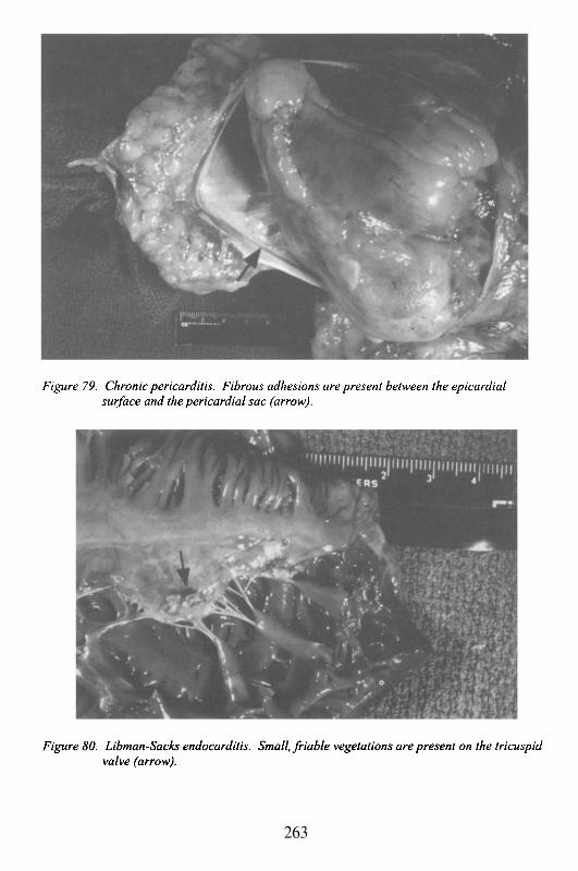
263
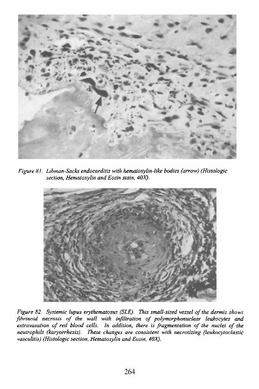
264
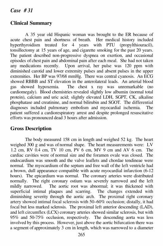
Case # 31
Clinical Summary
A 35 year old Hispanic woman was brought to the ER because ofacute chest pain and shortness of breath. Her medical history includedhyperthyroidism treated for 4 years with PTU (propylthiouracil),tonsillectomy at 15 years of age, and cigarette smoking for the past 20 years.The patient described non-progressive dyspnea on exertion, and occasionalepisodes of chest pain and abdominal pain after each meal. She had not takenany medications recently. Upon arrival, her pulse was 120 ppm withdiminished carotid and lower extremity pulses and absent pulses in the upperextremities. Her BP was 97/68 mmHg. There was central cyanosis. An ECGshowed RBBB and ST elevation in the anterolateral leads. An arterial bloodgas showed hypoxemia. The chest x ray was unremarkable (nocardiomegaly). Blood chemistries revealed slightly low albumin (normal totalprotein), calcium and uric acid; slightly elevated LDH, SGPT, CK, alkalinephosphatase and creatinine, and normal bilirubin and SGOT. The differentialdiagnoses included pulmonary embolism and myocardial ischemia. Thepatient suffered a cardiorespiratory arrest and despite prolonged resuscitativeefforts was pronounced dead 3 hours after admission.
Gross Description
The body measured 158 cm in length and weighed 52 kg. The heartweighed 300 g and was of normal shape. The heart measurements were: LV1.2 cm, RV 0.4 cm, TV 10 cm, PV 6 cm, MV 9 cm and AV 6 cm. Thecardiac cavities were of normal size and the foramen ovale was closed. Theendocardium was smooth and the valve leaflets and chordae tendineae werenormal. The myocardium of the septum and free wall of the left ventricle hada brown, dull appearance compatible with acute myocardial infarction (6-12hours). The epicardium was normal. The coronary arteries were distributednormally. The right coronary ostium was severely narrowed and the leftmildly narrowed. The aortic root was abnormal; it was thickened withsuperficial intimal plaques and scarring. The changes extended withdiminishing severity through the aortic arch. The proximal right coronaryartery showed intimal focal sclerosis with 50–60% occlusion; distally, it hadfocal but less marked sclerosis. The proximal left anterior descending (LAD),and left circumflex (LCX) coronary arteries showed similar sclerosis, but with95% and 50-75% occlusion, respectively. The descending aorta was lessinvolved by this process. However, just above the aortic bifurcation there wasa segment of approximately 3 cm in length, which was narrowed to a diameter
265
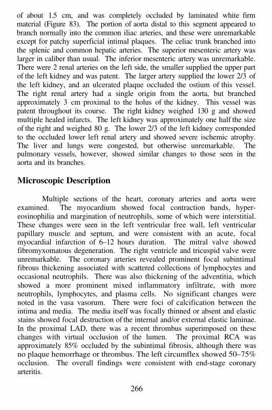
of about 1.5 cm, and was completely occluded by laminated white firmmaterial (Figure 83). The portion of aorta distal to this segment appeared tobranch normally into the common iliac arteries, and these were unremarkableexcept for patchy superficial intimal plaques. The celiac trunk branched intothe splenic and common hepatic arteries. The superior mesenteric artery waslarger in caliber than usual. The inferior mesenteric artery was unremarkable.There were 2 renal arteries on the left side, the smaller supplied the upper partof the left kidney and was patent. The larger artery supplied the lower 2/3 ofthe left kidney, and an ulcerated plaque occluded the ostium of this vessel.The right renal artery had a single origin from the aorta, but branchedapproximately 3 cm proximal to the holus of the kidney. This vessel waspatent throughout its course. The right kidney weighed 130 g and showedmultiple healed infarcts. The left kidney was approximately one half the sizeof the right and weighed 80 g. The lower 2/3 of the left kidney correspondedto the occluded lower left renal artery and showed severe ischemic atrophy.The liver and lungs were congested, but otherwise unremarkable. Thepulmonary vessels, however, showed similar changes to those seen in theaorta and its branches.
Microscopic Description
Multiple sections of the heart, coronary arteries and aorta wereexamined. The myocardium showed focal contraction bands, hyper-eosinophilia and margination of neutrophils, some of which were interstitial.These changes were seen in the left ventricular free wall, left ventricularpapillary muscle and septum, and were consistent with an acute, focalmyocardial infarction of 6–12 hours duration. The mitral valve showedfibromyxomatous degeneration. The right ventricle and tricuspid valve wereunremarkable. The coronary arteries revealed prominent focal subintimalfibrous thickening associated with scattered collections of lymphocytes andoccasional neutrophils. There was also thickening of the adventitia, whichshowed a more prominent mixed inflammatory infiltrate, with moreneutrophils, lymphocytes, and plasma cells. No significant changes werenoted in the vasa vasorum. There were foci of calcification between theintima and media. The media itself was focally thinned or absent and elasticstains showed focal destruction of the internal and/or external elastic laminae.In the proximal LAD, there was a recent thrombus superimposed on thesechanges with virtual occlusion of the lumen. The proximal RCA wasapproximately 85% occluded by the subintimal fibrosis, although there wasno plaque hemorrhage or thrombus. The left circumflex showed 50–75%occlusion. The overall findings were consistent with end-stage coronaryarteritis.
266
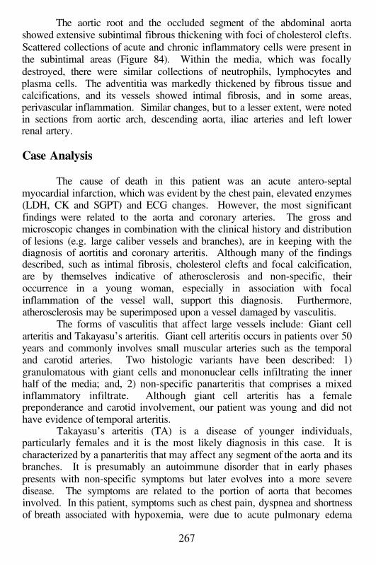
The aortic root and the occluded segment of the abdominal aortashowed extensive subintimal fibrous thickening with foci of cholesterol clefts.Scattered collections of acute and chronic inflammatory cells were present inthe subintimal areas (Figure 84). Within the media, which was focallydestroyed, there were similar collections of neutrophils, lymphocytes andplasma cells. The adventitia was markedly thickened by fibrous tissue andcalcifications, and its vessels showed intimal fibrosis, and in some areas,perivascular inflammation. Similar changes, but to a lesser extent, were notedin sections from aortic arch, descending aorta, iliac arteries and left lowerrenal artery.
Case Analysis
The cause of death in this patient was an acute antero-septalmyocardial infarction, which was evident by the chest pain, elevated enzymes(LDH, CK and SGPT) and ECG changes. However, the most significantfindings were related to the aorta and coronary arteries. The gross andmicroscopic changes in combination with the clinical history and distributionof lesions (e.g. large caliber vessels and branches), are in keeping with thediagnosis of aortitis and coronary arteritis. Although many of the findingsdescribed, such as intimal fibrosis, cholesterol clefts and focal calcification,are by themselves indicative of atherosclerosis and non-specific, theiroccurrence in a young woman, especially in association with focalinflammation of the vessel wall, support this diagnosis. Furthermore,atherosclerosis may be superimposed upon a vessel damaged by vasculitis.
The forms of vasculitis that affect large vessels include: Giant cellarteritis and Takayasu’s arteritis. Giant cell arteritis occurs in patients over 50years and commonly involves small muscular arteries such as the temporaland carotid arteries. Two histologic variants have been described: 1)granulomatous with giant cells and mononuclear cells infiltrating the innerhalf of the media; and, 2) non-specific panarteritis that comprises a mixedinflammatory infiltrate. Although giant cell arteritis has a femalepreponderance and carotid involvement, our patient was young and did nothave evidence of temporal arteritis.
Takayasu’s arteritis (TA) is a disease of younger individuals,particularly females and it is the most likely diagnosis in this case. It ischaracterized by a panarteritis that may affect any segment of the aorta and itsbranches. It is presumably an autoimmune disorder that in early phasespresents with non-specific symptoms but later evolves into a more severedisease. The symptoms are related to the portion of aorta that becomesinvolved. In this patient, symptoms such as chest pain, dyspnea and shortnessof breath associated with hypoxemia, were due to acute pulmonary edema
267
secondary to myocardial infarction caused by the inflammation andsubsequent thrombotic occlusion of the coronary vessels. This last event waspreceded by episodes of angina pectoris and angina intestinalis (the post-pyramidal abdominal pain was related to mesenteric involvement). She washypotensive due to decreased peripheral vascular resistance. However, it issignificant that there was no history of hypertension, which is a commonsymptom in TA as it is related to renal artery stenosis. The patient had noinvolvement of the right renal artery, and in the left side, one of the twovessels was patent and maintained the function of the upper 1/3 of the leftkidney; therefore, sparing her of renovascular hypertension. The leftpulmonary artery showed intimal proliferation and thickened adventitialvessels with perivascular infiltrates. These changes were focal with nosignificant occlusion; thus, the absence of pulmonary hypertension and rightventricular hypertrophy. It is worth mentioning that TA may present with adistinct pulmonary arteriopathy that consists of outer media defects withcapillary ingrowth in this layer and in the thickened and fibrosed intima.
The occlusion of the subclavian and iliac arteries was responsible forthe diminished or absent peripheral pulses. The asymmetry as well as theabsence of the peripheral pulses is quite characteristic of this entity, hence theterm “pulseless” disease. TA is not a common disorder; therefore, othercauses of reduced peripheral pulses should be considered such asarteriosclerotic occlusive disease, coarctation of the aorta, syphilis andneoplasias.
Coarctation of the aorta is more common in males and in patientswith gonadal dysgenesis. It is also associated with other congenital cardiacabnormalities in approximately 10% of cases. It presents in early childhoodunless the coarctation is distal to the ductus arteriosus, in which case the onsetof symptoms occurs much later in life. This could have been a considerationin this case, because coarctation of the aorta can present with diminishedpulses in the lower extremities. Additionally, it is associated withcardiomegaly, hypertension in the arms and engorged collateral vessels in theintercostal and subscapular areas. Both the clinical history and autopsy didnot reveal any of these findings in our patient. Coarctation of the aorta has acharacteristic chest X ray with a “3” sign made by the stenotic area and the 2adjacent dilated segments. The ECG and echocardiogram invariably showleft ventricular hypertrophy, but other findings depend on the location of thecoarctation and presence of other heart anomalies.
Syphilis is another form of arteritis that might be considered in thiscase, but it is usually seen in older patients and most often spares theabdominal aorta. The characteristics features of syphilitic arteritis were notpresent in our patient. Although there was perivascular inflammationinvolving the vasa vasorum of the adventitia, it was not prominent and theinfiltrate did not consist predominantly of plasma cells.
268
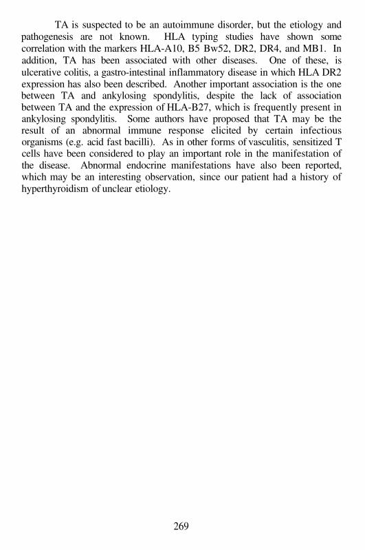
TA is suspected to be an autoimmune disorder, but the etiology andpathogenesis are not known. HLA typing studies have shown somecorrelation with the markers HLA-A10, B5 Bw52, DR2, DR4, and MB1. Inaddition, TA has been associated with other diseases. One of these, isulcerative colitis, a gastro-intestinal inflammatory disease in which HLA DR2expression has also been described. Another important association is the onebetween TA and ankylosing spondylitis, despite the lack of associationbetween TA and the expression of HLA-B27, which is frequently present inankylosing spondylitis. Some authors have proposed that TA may be theresult of an abnormal immune response elicited by certain infectiousorganisms (e.g. acid fast bacilli). As in other forms of vasculitis, sensitized Tcells have been considered to play an important role in the manifestation ofthe disease. Abnormal endocrine manifestations have also been reported,which may be an interesting observation, since our patient had a history ofhyperthyroidism of unclear etiology.
269
Suggested Readings
1.2.
3.
4.
5.
Kerr G. Takayasu's arteritis. Curr Opin Rheumatol. 1994; 6: 32-8.Matsubara O; Kuwata T; Nemoto T; Kasuga T; Numano F. Coronaryartery lesions in Takayasu arteritis: pathological considerations. HeartVessels Suppl. 1992; 7:26-31.Hall S; Buchbinder R. Takayasu's arteritis. Rheum Dis Clin North Am.1990; 16:411-22.Rizzi R, Bruno S, Stellacci C, Dammacco R. Takayasu's arteritis: a cell-mediated large-vessel vasculitis. Int J Clin Lab Res. 1999; 29:8-13.Morita Y, Yamamura M, Suwaki K, Mima A, Ishizu T, Hirohata M,Kashihara N, Makino H, Ota Z. Takayasu's arteritis associated withulcerative colitis; genetic factors in this association. Intern Med. 1996;35:574-8.
270
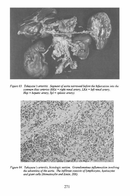
271
Case # 32
Clinical Summary
This is the case of a 31 month old girl who was admitted to thehospital because of increasing respiratory distress and wheezing. She was afull-term baby after an uncomplicated pregnancy, vaginal delivery and post-natal period. Past medical history was remarkable for: 1) mild intermittentasthma diagnosed at 8 m of age; 2) hospitalization at 20 m of age for anepisode of intussusception which resolved uneventfully with medicaltreatment; 3) since her birthday she complained of intermittent arthralgias(mostly lower extremities) to the point of sometimes refusing to walk. At 30m she was given a tentative diagnosis of juvenile rheumatoid arthritis becauseof these arthralgias and a new onset conjunctival injection that was diagnosedas uveitis (of note is that all her clinical work-up was unremarkable, exceptfor mild hypergammaglobulinemia). She was given naproxen PO twice a dayand a steroid based ophthalmic solution for her eyes, which significantlyimproved her symptoms. The day of her admission, she had a 2-day historyof worsening respiratory distress and wheezing with minimal response afterthe usual home treatments with nebulized albuterol. She was admitted withan acute asthma exacerbation and probable pneumonia. Over the first hoursshe remained stable, her physical exam was remarkable for Temp 38.2 °C, HR174, RR 42, and pulse oxymetry on room air 97 %. She seemed alert andinteractive with moderate respiratory distress (increased expiratory phase,mild nasal flaring, mild to moderate intercostals, and subcostal retractions).The lungs had decreased air movement with diffuse wheezing; there wastachycardia with a regular rhythm and no reported heart murmur. There washepatomegaly (10 cm liver span) and no splenomegaly or abdominal masses.The laboratory results (CBC and chemistries) were unremarkable. The chestx-ray showed diffuse infiltrates bilaterally. The respiratory signs progressedand she turned restless, agitated and developed perioral cyanosis.Concomitantly, her blood gasses deteriorated. As her cyanosis progressed,she became lethargic and was intubated and transferred to the pediatricintensive care unit. There she had a cardiopulmonary arrest with goodresponse to resuscitative maneuvers, and was stabilized. Two hours later,despite vasopressors, her blood pressure dropped, and again she becamepulseless. This time there was no response and she was pronounced deadabout 20 hours after admission.
272
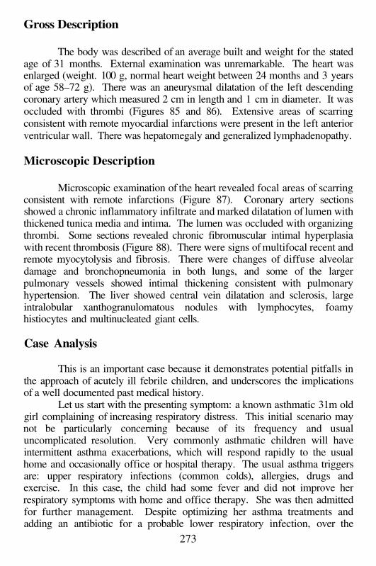
Gross Description
The body was described of an average built and weight for the statedage of 31 months. External examination was unremarkable. The heart wasenlarged (weight. 100 g, normal heart weight between 24 months and 3 yearsof age 58–72 g). There was an aneurysmal dilatation of the left descendingcoronary artery which measured 2 cm in length and 1 cm in diameter. It wasoccluded with thrombi (Figures 85 and 86). Extensive areas of scarringconsistent with remote myocardial infarctions were present in the left anteriorventricular wall. There was hepatomegaly and generalized lymphadenopathy.
Microscopic Description
Microscopic examination of the heart revealed focal areas of scarringconsistent with remote infarctions (Figure 87). Coronary artery sectionsshowed a chronic inflammatory infiltrate and marked dilatation of lumen withthickened tunica media and intima. The lumen was occluded with organizingthrombi. Some sections revealed chronic fibromuscular intimal hyperplasiawith recent thrombosis (Figure 88). There were signs of multifocal recent andremote myocytolysis and fibrosis. There were changes of diffuse alveolardamage and bronchopneumonia in both lungs, and some of the largerpulmonary vessels showed intimal thickening consistent with pulmonaryhypertension. The liver showed central vein dilatation and sclerosis, largeintralobular xanthogranulomatous nodules with lymphocytes, foamyhistiocytes and multinucleated giant cells.
Case Analysis
This is an important case because it demonstrates potential pitfalls inthe approach of acutely ill febrile children, and underscores the implicationsof a well documented past medical history.
Let us start with the presenting symptom: a known asthmatic 31m oldgirl complaining of increasing respiratory distress. This initial scenario maynot be particularly concerning because of its frequency and usualuncomplicated resolution. Very commonly asthmatic children will haveintermittent asthma exacerbations, which will respond rapidly to the usualhome and occasionally office or hospital therapy. The usual asthma triggersare: upper respiratory infections (common colds), allergies, drugs andexercise. In this case, the child had some fever and did not improve herrespiratory symptoms with home and office therapy. She was then admittedfor further management. Despite optimizing her asthma treatments andadding an antibiotic for a probable lower respiratory infection, over the
273
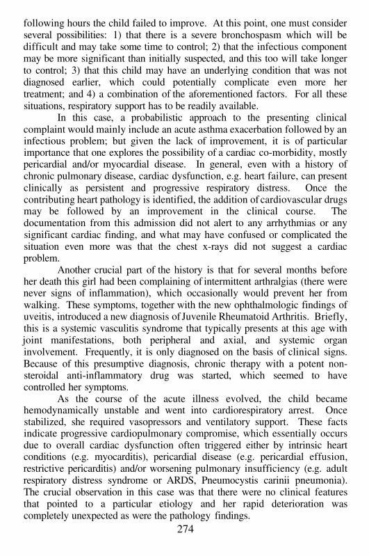
following hours the child failed to improve. At this point, one must considerseveral possibilities: 1) that there is a severe bronchospasm which will bedifficult and may take some time to control; 2) that the infectious componentmay be more significant than initially suspected, and this too will take longerto control; 3) that this child may have an underlying condition that was notdiagnosed earlier, which could potentially complicate even more hertreatment; and 4) a combination of the aforementioned factors. For all thesesituations, respiratory support has to be readily available.
In this case, a probabilistic approach to the presenting clinicalcomplaint would mainly include an acute asthma exacerbation followed by aninfectious problem; but given the lack of improvement, it is of particularimportance that one explores the possibility of a cardiac co-morbidity, mostlypericardial and/or myocardial disease. In general, even with a history ofchronic pulmonary disease, cardiac dysfunction, e.g. heart failure, can presentclinically as persistent and progressive respiratory distress. Once thecontributing heart pathology is identified, the addition of cardiovascular drugsmay be followed by an improvement in the clinical course. Thedocumentation from this admission did not alert to any arrhythmias or anysignificant cardiac finding, and what may have confused or complicated thesituation even more was that the chest x-rays did not suggest a cardiacproblem.
Another crucial part of the history is that for several months beforeher death this girl had been complaining of intermittent arthralgias (there werenever signs of inflammation), which occasionally would prevent her fromwalking. These symptoms, together with the new ophthalmologic findings ofuveitis, introduced a new diagnosis of Juvenile Rheumatoid Arthritis. Briefly,this is a systemic vasculitis syndrome that typically presents at this age withjoint manifestations, both peripheral and axial, and systemic organinvolvement. Frequently, it is only diagnosed on the basis of clinical signs.Because of this presumptive diagnosis, chronic therapy with a potent non-steroidal anti-inflammatory drug was started, which seemed to havecontrolled her symptoms.
As the course of the acute illness evolved, the child becamehemodynamically unstable and went into cardiorespiratory arrest. Oncestabilized, she required vasopressors and ventilatory support. These factsindicate progressive cardiopulmonary compromise, which essentially occursdue to overall cardiac dysfunction often triggered either by intrinsic heartconditions (e.g. myocarditis), pericardial disease (e.g. pericardial effusion,restrictive pericarditis) and/or worsening pulmonary insufficiency (e.g. adultrespiratory distress syndrome or ARDS, Pneumocystis carinii pneumonia).The crucial observation in this case was that there were no clinical featuresthat pointed to a particular etiology and her rapid deterioration wascompletely unexpected as were the pathology findings.
274
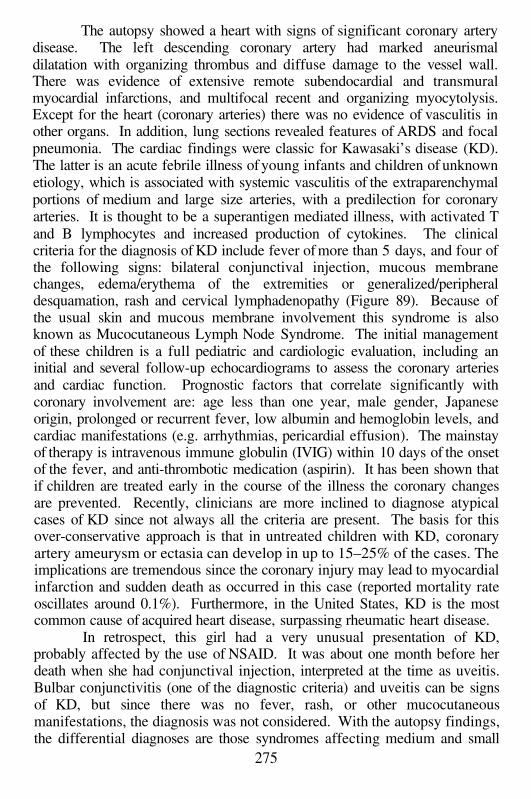
The autopsy showed a heart with signs of significant coronary arterydisease. The left descending coronary artery had marked aneurismaldilatation with organizing thrombus and diffuse damage to the vessel wall.There was evidence of extensive remote subendocardial and transmuralmyocardial infarctions, and multifocal recent and organizing myocytolysis.Except for the heart (coronary arteries) there was no evidence of vasculitis inother organs. In addition, lung sections revealed features of ARDS and focalpneumonia. The cardiac findings were classic for Kawasaki’s disease (KD).The latter is an acute febrile illness of young infants and children of unknownetiology, which is associated with systemic vasculitis of the extraparenchymalportions of medium and large size arteries, with a predilection for coronaryarteries. It is thought to be a superantigen mediated illness, with activated Tand B lymphocytes and increased production of cytokines. The clinicalcriteria for the diagnosis of KD include fever of more than 5 days, and four ofthe following signs: bilateral conjunctival injection, mucous membranechanges, edema/erythema of the extremities or generalized/peripheraldesquamation, rash and cervical lymphadenopathy (Figure 89). Because ofthe usual skin and mucous membrane involvement this syndrome is alsoknown as Mucocutaneous Lymph Node Syndrome. The initial managementof these children is a full pediatric and cardiologic evaluation, including aninitial and several follow-up echocardiograms to assess the coronary arteriesand cardiac function. Prognostic factors that correlate significantly withcoronary involvement are: age less than one year, male gender, Japaneseorigin, prolonged or recurrent fever, low albumin and hemoglobin levels, andcardiac manifestations (e.g. arrhythmias, pericardial effusion). The mainstayof therapy is intravenous immune globulin (IVIG) within 10 days of the onsetof the fever, and anti-thrombotic medication (aspirin). It has been shown thatif children are treated early in the course of the illness the coronary changesare prevented. Recently, clinicians are more inclined to diagnose atypicalcases of KD since not always all the criteria are present. The basis for thisover-conservative approach is that in untreated children with KD, coronaryartery ameurysm or ectasia can develop in up to 15–25% of the cases. Theimplications are tremendous since the coronary injury may lead to myocardialinfarction and sudden death as occurred in this case (reported mortality rateoscillates around 0.1%). Furthermore, in the United States, KD is the mostcommon cause of acquired heart disease, surpassing rheumatic heart disease.
In retrospect, this girl had a very unusual presentation of KD,probably affected by the use of NSAID. It was about one month before herdeath when she had conjunctival injection, interpreted at the time as uveitis.Bulbar conjunctivitis (one of the diagnostic criteria) and uveitis can be signsof KD, but since there was no fever, rash, or other mucocutaneousmanifestations, the diagnosis was not considered. With the autopsy findings,the differential diagnoses are those syndromes affecting medium and small
275
arteries. The main one is Polyarteritis Nodosa (PAN). The onset of PAN isinsidious and frequently presents with constitutional symptoms, fever andweight loss. In contrast with this case, PAN usually affects not only thecardiac, but also the renal and nervous systems (peripheral and central).There is significant organ involvement and abnormal laboratory findings arethe rule (CBC, ESR, UA, serum immunoglobulins). Pathologically, the twoconditions may be undistinguishable. Other important conditions that wereexcluded clinically or based on the autopsy findings were streptococcal andstaphylococcal-related syndromes (toxin-mediated), drug toxicity andhypersensitivity reactions.
In summary, given the severe coronary involvement found in this girl,and the fact that it went undetected for a long period of time, it is highlyunlikely that she would have survived. It is possible that the use of NSAIDprevented the expression of the full inflammatory syndrome, and thus thediagnosis of KD was never considered.
276
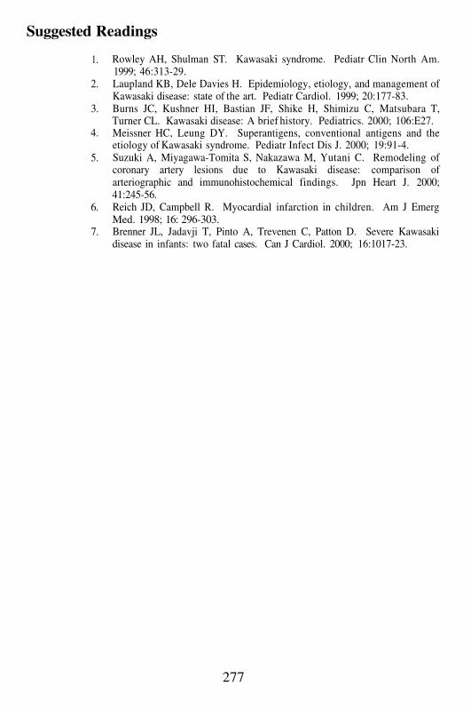
Suggested Readings
1.
2.
3.
4.
5.
6.
7.
Rowley AH, Shulman ST. Kawasaki syndrome. Pediatr Clin North Am.1999; 46:313-29.Laupland KB, Dele Davies H. Epidemiology, etiology, and management ofKawasaki disease: state of the art. Pediatr Cardiol. 1999; 20:177-83.Burns JC, Kushner HI, Bastian JF, Shike H, Shimizu C, Matsubara T,Turner CL. Kawasaki disease: A brief history. Pediatrics. 2000; 106:E27.Meissner HC, Leung DY. Superantigens, conventional antigens and theetiology of Kawasaki syndrome. Pediatr Infect Dis J. 2000; 19:91-4.Suzuki A, Miyagawa-Tomita S, Nakazawa M, Yutani C. Remodeling ofcoronary artery lesions due to Kawasaki disease: comparison ofarteriographic and immunohistochemical findings. Jpn Heart J. 2000;41:245-56.Reich JD, Campbell R. Myocardial infarction in children. Am J EmergMed. 1998; 16: 296-303.Brenner JL, Jadavji T, Pinto A, Trevenen C, Patton D. Severe Kawasakidisease in infants: two fatal cases. Can J Cardiol. 2000; 16:1017-23.
277
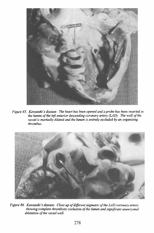
278
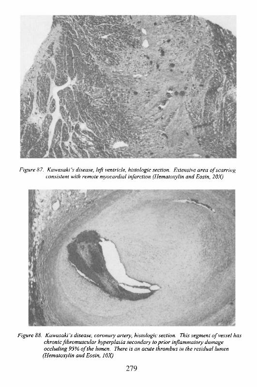
279
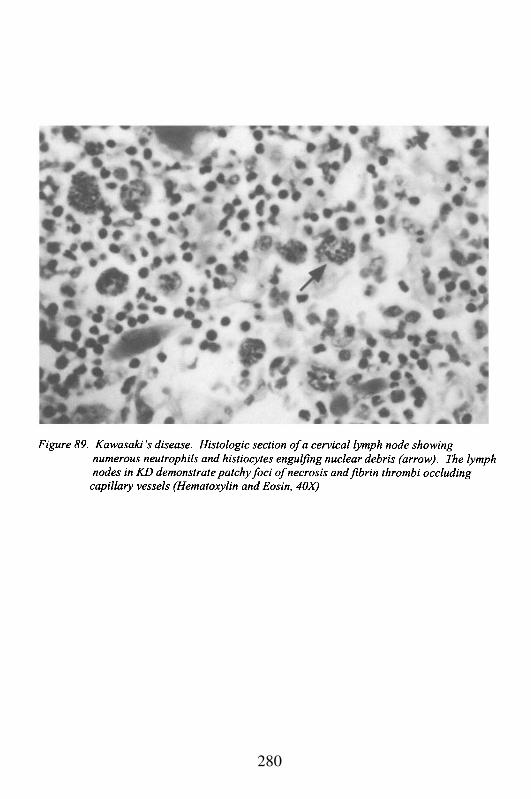
280
Chapter 13
MEDICINE, LAW AND THE HEART
Cardiovascular Disease in the Medico-Legal Era:An Introduction
The United States is a litigious society, far more so than other regionsof the world with sophisticated healthcare systems and economies comparableto ours, such as Canada, Western Europe and Great Britain, and Japan. Amajor focus of this litigation deals with medical malpractice, with patients orsurvivors suing healthcare providers for either a real or claimed injury.Cardiovascular disease represents a disproportionate number of these cases,along with failure or delay in the diagnosis of cancer, or complicationsfollowing surgery. The defense of these cardiovascular law suits oftenrequires knowledge of complex medicine, and a correlation with cardiacpathology and pathophysiology. Similarly, the plaintiff must be aware ofmechanisms of cardiovascular disease and the actual pathology in order tomake an effective argument to a jury.
Other types of litigation such as personal injury (e.g. automobileaccident), or product liability (e.g. adverse drug affects, or claims that cars ortires were defective leading to an accident) often also require knowledge ofcardiovascular disease. For instance, an automobile accident may apparentlylead to the death of the driver as a result of trauma; however, if the driveractually had an arrhythmic cardiac arrest prior to the accident (e.g. coronaryartery ischemia or cardiomyopathy) and then crashed, this would mitigate ifnot eliminate the liability of insurers or the automobile manufacturer. Inanother instance, a so-called drug adverse effect may represent the naturalconsequences of the underlying disease for which the drug was prescribed,rather than a complication of the drug. An example of this phenomenon issudden cardiac death during sexual activity of an individual with diabeticcardiomyopathy, who is taking Viagra® for erectile dysfunction secondary todiabetic neuropathy. Association is not the equivalent of causation; however,unless one is aware of diabetic cardiomyopathy as an entity, and its potentialfor causing sudden cardiac arrhythmias, a law suit against the manufacturer ofthe drug, or the prescribing doctor, might be successful.
Different types of cardiovascular disease, many of them discussed inthis book, present with atypical complaints (or no complaints at all, such as‘silent ischemia’). These are conditions that affect the heart and bloodvessels, and are often the source of medico-legal suits. Two frequent sourcesof suits are those dealing with ischemic heart disease (IHD), and infectious
endocarditis (IE). Failure to or delay in the diagnosis of IHD may lead to amyocardial infarction or death; and in the cases of IE, this may result infailure to perform a valve replacement or death. Typical scenarios are thoseof subtle electrocardiographic changes such as non-specific ST-T waveabnormalities, or atypical chest pain misconstrued as chest wall pain ordysphagia, which then result in death or a myocardial infarction. Anothercommon situation is a patient who presents with low-grade fever and malaisediagnosed as an upper respiratory infection, and the diagnosis of IE isoverlooked. Thus, it becomes crucial to be aware of these cardiovascularconditions, and to recognize the pathological changes that may have importantimplications for the plaintiff or the defense. For instance, the timing or datingof a coronary thrombus or plaque rupture, may help in determining whetherthe symptoms or signs ascribed by one party to IHD, in fact were present at oraround the time of the abnormal ECG or the atypical chest pain. Similarly,not only is the dating of the valvular endocarditis important to assess howlong it had been active, but it is important to determine how much valvedestruction had taken place. If the valve was sufficiently damaged, or if therewere other conditions such as an annular abscess that would require surgicalintervention, a delay in diagnosis might have no bearing on whether the valvecould have been salvaged with an earlier diagnosis. This is known as aproximate cause defense. In other words, the failure in timely diagnosis hadno bearing on the ultimate outcome.
The following 2 cases were selected because they represent unusualcardiovascular complications that led to a medico-legal suit. In one instance,the complication resulted from application of an organo-phosphateinsecticide; thus, this was a product liability case. In the other instance, thecomplications followed coronary artery intervention with atherectomy andangioplasty; and therefore, a medical malpractice case. It is not entirelysurprising that often medico-legal suits result from rare or unexpectedcomplications, or unusual disease manifestations. The usual and commonpresentations of disease are generally diagnosed with facility; it is the atypicalpresentation that leads to medical puzzlement, patient dissatisfaction, oradverse outcome.
Case # 33
Clinical Summary
This is the case of a 47 year old man who presented to the emergencyroom two weeks after a pesticide application in his home, with complaints ofitchy eyes, blurred vision, “foggy” sensorium, dizziness and headache. Hisvital signs were unremarkable, although his diastolic pressure of 96 mmHg
282
was higher than it had been five months ago (86 mmHg). He was treated withdiphenhydramine. The symptoms persisted. A laboratory test demonstrated ared blood cell cholinesterase of 5.6 u/mL (normal range 3.8-8.0 u/mL).
Two weeks later he presented again to the emergency room withacute complaints of stabbing chest pain for two days radiating to the back. Hehad a new murmur of aortic insufficiency, acute anuria, cyanosis of the rightlower extremity, and unstable blood pressure readings of 190/110 to 130/80mmHg. The diagnosis of aortic dissection was made. He was found to have atype I dissection with extension into the abdominal aorta, and with absentfilling of the superior mesenteric, celiac and right common iliac arteries. Hewas immediately taken to the operating room and placed on cardiopulmonarybypass. The intraoperative findings included a non-ruptured aorta with adissection beginning 1.5 cm above the aortic valve. An intimal flap extendedfrom above the valve to near the origin of the brachiocephalic artery. Theaortic valve was thick with fibrosis, and was bicuspid. The valve wasreplaced by a mechanical St. Jude prosthesis, and the ascending aorta wasrepaired with a graft. The patient tolerated the procedure well and was stable.He was immediately taken to angiography to assess the abdominal vessels.While the angiogram was being performed, he developed sudden bradycardiaand hypotension, and he arrested with ventricular fibrillation. Resuscitationefforts were unsuccessful.
Past medical history
The patient was 18 years old when a coarctation of the aorta wasdiagnosed by catheterization. This finding was confirmed byechocardiography 16 years later. He also had a mild aortic regurgitationmurmur of II/VI. In addition, he had mild hypertension for 3 years for whichhe was given antihypertensive medication. Subsequently, and until the acuteepisode of dissection, he did not have any recorded blood pressures in thehypertensive range nor was he taking any medication for blood pressurecontrol. Other pertinent findings included a chest-X-ray that demonstrated anectatic and prominent ascending aorta. He was also noted to have a mid-systolic click murmur compatible with a mitral valve prolapse syndrome. Hishabitus was average (180 cm, 95 kg) and he was not Marfanoid in appearance.An ECG suggested the presence of multiple old myocardial infarctions ofindeterminate age. He was also a 2 pack per day cigarette smoker.
Gross Description
Upon exam, the surgical anastomoses were intact, and the aorta hadnot ruptured. Infarction of the small intestine and liver were noted. The
283
dissection extended into both common iliac vessels. There was minimalatherosclerosis of the aorta and coronary arteries. The heart weighed 335 g(normal), but the left ventricular wall was concentrically hypertrophied at 2.2cm. No abnormality of the mitral valve was seen. The lungs weresignificantly heavy (2360 g, normal 685-1050 g) with pulmonary edema.Post-mortem pseudocholinesterase levels of 1151 u/mL (approximately 50%of normal) were measured.
Microscopic Description
Examination of the autopsy slides confirmed that the dissection wasacute, with the histological changes of no more than 24–48 hours in duration.The dissection occurred in the outer segment of the aortic media; and wasassociated with a fresh fibrin coagulum in the false channel along with focalneutrophils in the adventitia and media. The latter inflammatory cells mayrepresent a cellular response to the acute injury, and/or a reaction to surgicalmanipulation occurring during the attempted repair of the lesion. There wasno hemosiderin (iron) pigment in the adventitia, thus further confirming thatthe dissection was acute and had not caused any blood leakage from the falsechannel greater than 48 hours before death. In the aortic wall there wasinsignificant intimal atherosclerosis. More importantly, in the media therewere focal areas of elastic lamella and smooth muscle degeneration withconcomitant increase of basophilic mucoppolysaccharide. These are the typesof change commonly associated with aortic dissection. However, the medialdegeneration did not appear as severe as is often seen in those casesassociated with known connective tissue disease (e.g. Marfan’s syndrome) orhypertension.
Case Analysis
1)2)
Two cardiovascular aspects should be addressed in this case:The chronic underlying diseaseThe acute dissection with the potential role played by theorganophosphate toxicity
This patient appeared to have two distinct and probably unrelatedconditions: coarctation of the aorta associated with a bicuspid aortic valve,and chronic stable aortic degeneration suggestive of a connective tissueabnormality. The coarctation of the aorta and bicuspid aortic valve are well-recognized and relatively common congenital abnormalities that often occurtogether. The aortic constriction present at or just distal to the left subclavianartery and the ductus arteriosus varies in severity. It ranges from completely
284
asymptomatic and clinically silent to marked obstruction of the aortic flowand severe hypertension. The aortic valve disease may also be asymptomatic,or may be associated with clinically significant aortic insufficiency orstenosis. In this case, the coarctation and the aortic valve disease wereapparently mild. It is noteworthy that he was found to be hypertensive foronly 3 years; however, by the time his aorta was noted to be prominent onchest-X-ray, he no longer required antihypertensive medication, and with notherapy, he remained normotensive. This is an unusual and atypical patternfor hypertension associated with coarctation. This suggests that he “healed”or compensated for his coarctation by mildly dilating his aorta due to anunderlying tissue disorder. The aortic valvular disease was not particularlysevere, as judged by the murmur and the lack of physical limitations (e.g.absence of symptoms referable to congestive heart failure). The valvedegeneration, rather than the hypertension, is most likely the explanation forthe concentric left ventricular hypertrophy. However, it should be noted, thathypertension occurring several years previously, still could have contributedto chronic damage to the aortic media, thereby increasing its susceptibility todissection with the appropriate stimulus.
The suggestion that the patient had an underlying connective tissuedisorder is based on several findings:1)
2)3)
4)
The dilatation of the aorta as noted on the chest X ray in the absence ofatherosclerosis (confirmed by the autopsy).The dissection as a terminal eventThe findings of elastic tissue degeneration associated with increasedmucopolysaccharide material in the aortic mediaThe clinical findings strongly consistent with mitral valve prolapse
Although this patient did not have the habitus of a patient with Marfansyndrome, it is known that patients can have similar aortic pathology withdilatation and a susceptibility to dissection. In the absence of specific geneticor biochemical studies of the glycoprotein abnormality in Marfan’s syndrome(e.g. fibrillin), or of the collagen and elastin that provide the tensile strengthof the aorta, it cannot be established with certainty if any connective tissueabnormality existed in this patient.
In regard to the issue of organophosphate poisoning and its role ininducing aortic dissection, there are several pertinent observations:
Aortic dissection virtually never occurs in the presence of an anatomicallyand microscopically normal aorta. In young individuals ( thoughdecades) with overt Marfan’s syndrome or other recognized heritableconnective tissue disorders, it may occur spontaneously with no associatedhypertension. In older individuals without Marfan’s syndrome, it is stronglyassociated with hypertension (generally greater than 75% of those affected).In both situations, cardiac and blood vessel contractility is thought to play a
285
major pathogenic role. Increased cardiac contractility places abnormal stresson the ascending aorta (the commonest site for initiating dissection); whereasincreased smooth muscle contractility may be important in generating localdamage in the aortic wall, and may cause tears with hemorrhage in the vasavasorum that supply the media along the plane in the aortic wall wheredissection typically occurs (e.g. outer 2/3 of the media where the vasavasorum branch parallel to the intimal surface, thereby providing a potentialanatomic cleavage plane). Damage to the vasa vasorum may lead to focalhemorrhage that dissects through the tissue until it eventuates in an intimaltear, with subsequent distal propagation of the dissection. It also has beenshown that agents that cause relaxation of smooth muscle and vasodilatationcan be injurious to the media. Dysfunction or necrosis of smooth muscle is acorrelate of the focal lesions that occur as a predisposition to dissection. Thesmooth muscle cell not only serves as a contractile modulator of the aortictone, but it is also critical in the synthesis of the connective tissue componentsof the media. Smooth muscle damage, occurring either as a consequence ofincreased constriction (e.g. spasm) or increased relaxation, may be deleteriousto medial integrity. This is of particular relevance in the present case becauseof the known effects of undegraded acetylcholine (Ach) at the neuromuscularjunction that causes vasodilatation in certain vascular beds, andvasoconstriction in vessels with chronic pathological damage (e.g.atherosclerosis or endothelial injury). Underlying damage to previousepisodes of hypertension, increased turbulence secondary to the coarctation inthe proximal descending aorta, as well as in the ascending aorta due tobicuspid valvular dysfunction, is highly likely to have made the patient's aortasusceptible to the dilating and/or constricting effects of ACh. As notedpreviously, there was focal chronic damage of smooth muscle cells and elastictissue in the aortic media at a distance from the acute dissection. These arepathological changes that could increase the susceptibility of the aortic mediato the hemodynamic effects of ACh as a vasodilating or vasoconstrictingagent.
There is an obvious temporal relationship between the onset oforganophosphate poisoning and the development of the aortic dissection insomeone who was entirely stable and asymptomatic, approximately onemonth before. It is known that organophosphates, as inhibitors ofacetylcholinesterase (AChE) can lead to unrestrained effects of ACh atneuromuscular junctions and in the central nervous system. ACh can act as asmooth muscle relaxing agent in normal blood vessels, but it is known to actas a constrictor in pathological vessels. It is impossible to predict whicheffects it might have had in this patient’s aorta; but in addition to potentialmedial smooth muscle injury, either effect could have led to abnormalmotility of the vessel or of the vasa vasorum causing damage. ACh inhibitorstypically cause bradycardia, but under certain circumstances they may lead to
286
tachycardia, presumably as a result of central nervous system actions. Inaddition, there may be a reduction in peripheral vascular resistance (due to thevasodilating effects of ACh) that can cause increased systemic catecholaminesecretion leading to enhanced cardiac contractility, a major cause of aorticdissection in the setting of an abnormal vessel. It has been shown thatprophylactic treatment of patients with Marfan’s syndrome by using betaadrenergic blockers or calcium channel blocking drugs to decrease heart rateand cardiac contractility, can prevent or significantly delay the developmentof aortic dissection. Similarly, although AChE inhibitors are generallyassociated with hypotension, their central nervous system activity may lead tohypertension, specifically through the stimulation of the locus ceruleus, whichis involved in peripheral sympathetic tone control. Thus, the potentialcardiovascular effects of ACh inhibition by an organophosphate agent withincreased ACh activity are complex and difficult to predict; however, they areclearly consistent with the development of vascular and cardiac instability thatin the appropriate setting of underlying aortic disease could directly lead todissection.
The effects of AChE inhibition with a build-up of ACh at theneuromuscular junction and in the central nervous system (CNS), arecomparable in many ways to the effects of cocaine, crack, andmethamphetamines at the neuromuscular junction and in the CNS. However,the latter agents exert their effect through different receptors than AChEinhibition. Specifically, cocaine, crack and methamphetamines act to block areceptor on the nerve ending, the alpha-2 adrenergic receptor. This receptor isinvolved in the re-uptake of norepinephrine (NE) at the neuromuscularjunction. Receptor blockade leads to an increase of catecholamines at thejunction, thereby causing unrestrained stimulation of smooth muscle. This iscomparable to the build-up of ACh at the junction following AChE inhibitionby organophosphate agents, which can cause either relaxation or stimulationof the smooth muscle cell. With catecholamines, the vascular smooth muscleis constricted. This hypercontractile effect can then lead to muscle cellnecrosis, overlying endothelial damage with the development of vascularthrombosis and eventual atherosclerosis, intensifying the immediate effects oftissue injury (e.g. myocardial infarction or stroke). These agents have alsobeen associated with aortic dissection. If the effects of organophosphatepoisons, which produce irreversible inhibition of the AchE, are notcounteracted rapidly, they lead to a prolonged build-up of ACh at theneuromuscular junction. In contrast, the cocaine class of drugs does notirreversibly bind to the neuromuscular receptor, and they can be rapidlyreversed by alpha or beta adrenergic receptor antagonists, or by other drugsthat act as smooth muscle relaxants (e.g. calcium channel blockers or drugsthat act by releasing nitric oxide).
287
In regard to the patient’s terminal event, it is noticeable that he survivedthe surgery in apparent good condition and the decompensated suddenlyduring the post-surgical angiogram. Although he still had significantabdominal cardiovascular compromise that would have required furtherimmediate revascularization, the pattern of collapse is in keeping with anabrupt cardiovascular event possibly related to the autonomic dysfunctionsecondary to the organophosphate poisoning. Specifically, if he hadcardiovascular collapse secondary to the infarcted bowel, it would beanticipated that he might become hypotensive with tachycardia; yet his initialrhythm was bradycardia. It is very likely that he had an atypical reaction tothe angiographic agent due to persistently dysfunctional autonomicneuromuscular junctions. Unless AChE inhibitors are reversed with a specificantidote such as pralidoxime within the first 48 hours of exposure, the bindingto the junction becomes irreversible and requires de novo synthesis of AChEthat can take one to three months to be completed. Therefore, when thepatient was undergoing surgery, he still was in a state of relative AChEinhibition that could have made him overly sensitive to the normallyhypotensive effects of the angiographic dye. With the release of toxins fromthe necrotic small intestine, the AChE inhibition may also have contributed tofurthering its vasodilating effects. This combination of the angiographic dyeand the septic shock may have caused a loss of peripheral vascular resistancein a vascular bed blocked from normal constriction. The resultinghypotension then could have led to heart failure and arrhythmia as terminalevents.
288
Suggested Readings
1.2.
Marrs TC. Organophosphate poisoning. Pharmacol Ther. 1993; 58:51-66.Ward C. Clinical significance of the bicuspid aortic valve. Heart. 2000;83:81-5.
289
Case # 34
The death of this middle-aged man several weeks after aninterventional catheterization and coronary artery procedure resulted in a suitagainst the treating cardiologist. The claim against the physicians is based onthe performance of an unjustified procedure in lieu of recognizedinterventions with high clinical success and low morbidity or mortality, suchas undertaking a coronary artery bypass or a balloon angioplasty withstenting. It is also suggested that the failure to adequately follow the patientafter the procedure contributed to his death. The further claim was that theunjustified procedure not only did not improve the coronary stenosis, butactually directly contributed to his death through unexpected complications.The first claim may be countered by establishing that the decision to carry outthe procedure is a matter of medical judgment and not negligence, and thelack of follow-up may have been the result of patient uncooperation.However, it would be very difficult to argue that the improper procedure didnot negligently cause this patient’s death, as will be established below.
This 51 year-old man, with a strong family history of coronary arterydisease, complained of new onset chest pain. He had undergone a relativelyrecent screening procedure, ultrafast CT scanning, which revealedsignificant calcium scores in his coronary arteries. This test has receivedmuch hype in the medical community over the last 5 years, as a non-invasivetest to determine the presence of coronary disease. It has yet to be establishedprospectively that the claims for the test’s sensitivity or specificity arescientifically justified. There is evidence that coronary atherosclerosis(particularly vulnerable plaques with high susceptibility to rupture, associatedwith unstable angina or myocardial infarction) may have minimal or nocalcification; and calcification is not necessarily an indication of luminalocclusion or narrowing. Obviously, a positive test that then leads to a positiveconfirmatory finding is valuable; however, a negative test may not rule out thepotential for a serious coronary lesion. Ultimately, it will have to bedetermined that the benefit of the test justifies its cost.
Regardless, in this symptomatic patient the test was positive, and itled to coronary catheterization that demonstrated severe complex stenoses ofthe proximal left anterior descending coronary artery (LAD), and high gradestenosis of the mid-right coronary artery (RCA). The left circumflex artery(LCX) was apparently had minimal involvement. With two-vessel disease,particularly with involvement of the proximal LAD, it would have beenprudent to recommend coronary artery bypass grafting (CABG). The LADcould have been bypassed with an internal mammary artery with a very highguarantee of long-term success, since the internal mammary artery isminimally susceptible to atherosclerotic stenosis. The RCA would have
290
required either a vein-graft, or a free arterial graft with a radial artery. Thisprocedure, particularly in a young man with no prior cardiac damage, couldhave been performed with very low morbidity, and a mortality rate of at most1-2%. The survival of the grafts could be estimated in the range of 15-20years, or more.
A considerably riskier approach would be to perform a balloonangioplasty with stenting. This is potentially dangerous with proximallesions, particularly when there are multiple sites within the same vessel,along with high-grade disease in another major vessel. The placement ofstents would function to maintain vessel patency post-angioplasty.Angioplasty, however, does have the significant potential of re-stenosis, witha rate of 30-40% in the first 3-4 months after the procedure, and a slowlyprogressive increase thereafter. Although somewhat ameliorated with the useof stents, re-stenosis can occur, and occasionally even at an accelerated rate.Moreover, with multiple sites of stenosis in the LAD, the vessel would haverequired multiple stents. Although technically feasible, this may prolong theprocedure and make the left ventricle susceptible to ischemia or infarction.
The procedure that was employed known as atherectomy, is rarelyused today, since it was supplanted clinically by angioplasty and stenting inthe mid 1990’s. Atherectomy uses a variety of cutting catheters, includingsharp blades or rotating blades (and experimentally, lasers), to excise plaquematerial from the vessel wall and re-establish an adequate lumen. The plaquematerial is then suctioned through the coronary catheter, and removed fromthe patient. Atherectomies became valuable source of research tissue in anumber of centers investigating atherosclerosis and re-stenosis, since it wasthe only methodology that provided fresh atheroma for biochemical,histochemical, or molecular studies. The tissue fragments were composed ofall of the elements of atherosclerotic plaque including fibrofatty tissue, lipidcore, calcium, and inflammatory cells. Not infrequently, segments of medialsmooth muscle were included; yet, there was only a rare case of actualcoronary artery rupture that required surgical intervention. Complicationsother than vessel rupture include vessel collapse or spasm, coronarydissection, and lumen thrombosis. These complications are prevented bystenting, which can be used with atherectomy. In this case, stenting orremoval of plaque debris was not performed. There has been some concernabout this approach over the years, but there are few clinical data todemonstrate a potential adverse affect of allowing fragments of plaque (evenmicroscopic) to embolize downstream in the coronary circulation.
This patient had an apparently successful procedure carried out in theLAD and RCA on separate days. He allegedly had good coronary flow withwidely patent vessels and no residual stenosis. He did have bleedingcomplications at his arteriotomy site, but this is not unusual. He also hadminor elevations of creatine kinase (CK), with a significant increase of his
291
MB iso-enzyme after the LAD procedure. He was maintained on aspirinprophylactically. Approximately 3 weeks after the procedure, he returned tohis executive position overseas. On the following day, he collapsed suddenlyand died. The autopsy was carried out in a well-recognized academic centerwith sophisticated knowledge of cardiovascular disease.
Before actually turning to the specific autopsy findings and discussingthem in detail, it might be worthwhile to address this patient’s death from aclinical perspective. What are the likely cause(s) of his sudden cardiac arrest?Although some unrelated event only indirectly related to the coronary arteryprocedure could have occurred (e.g. stroke), the temporal proximity to thecardiac catheterization and intervention, strongly suggests an association.Acute coronary occlusion is a likely cause, with several etiologies possible.Neo-intima proliferation, thrombosis, and vessel trauma should be considered.As a general rule, neo-intimal proliferation leading to re-stenosis requiresmonths to develop sufficiently to cause coronary artery ischemia; although theprocess may have begun over 3 weeks, it is not likely to cause sufficientocclusion to lead to ischemia and arrhythmia. There could have been an acutethrombus developing at an atherectomy site with sudden myocardial ischemiaor infarction. Thrombus, however, is much more likely to develop shortlyafter the procedure, not weeks later. There could have been a ‘silent’complication of the atherectomy and balloon angioplasty such as a delayedrupture of a pseudo-aneurysm (e.g. disruption of the vessel during theprocedure leading to a localized hematoma that then ruptured weeks later withpericardial tamponade). There also could have been a dissection of thecoronary artery with a delayed rupture. If any of these complications hadactually been the cause of death, then the medico-legal aspects of this casewould have focused on the utilization of the atherectomy procedure ratherthan coronary artery bypass grafting.
What did happen? In fact, 3 things occurred that together contributedto his death:1.2.3.
Persistence of high-grade coronary stenosisFocal coronary artery dissection that contributed to the stenosisMulti-focal myocardial necrosis secondary to embolization of plaquedebris and other foreign material
We will address each of these processes.
High-grade Coronary Artery Stenosis: Despite the clinicalimpression that following the atherectomy and balloon angioplasty there waswide patency of all coronary arteries, the autopsy showed otherwise. The leftmain coronary artery (LM) was narrowed 50-75%, the LAD and RCA were90% occluded focally, and the LCX was 75% occluded. Although it ispossible that there was a complete mis-reading or inaccurate reporting of the
292
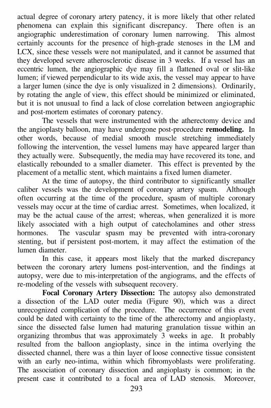
actual degree of coronary artery patency, it is more likely that other relatedphenomena can explain this significant discrepancy. There often is anangiographic underestimation of coronary lumen narrowing. This almostcertainly accounts for the presence of high-grade stenoses in the LM andLCX, since these vessels were not manipulated, and it cannot be assumed thatthey developed severe atherosclerotic disease in 3 weeks. If a vessel has aneccentric lumen, the angiographic dye may fill a flattened oval or slit-likelumen; if viewed perpendicular to its wide axis, the vessel may appear to havea larger lumen (since the dye is only visualized in 2 dimensions). Ordinarily,by rotating the angle of view, this effect should be minimized or eliminated,but it is not unusual to find a lack of close correlation between angiographicand post-mortem estimates of coronary patency.
The vessels that were instrumented with the atherectomy device andthe angioplasty balloon, may have undergone post-procedure remodeling. Inother words, because of medial smooth muscle stretching immediatelyfollowing the intervention, the vessel lumens may have appeared larger thanthey actually were. Subsequently, the media may have recovered its tone, andelastically rebounded to a smaller diameter. This effect is prevented by theplacement of a metallic stent, which maintains a fixed lumen diameter.
At the time of autopsy, the third contributor to significantly smallercaliber vessels was the development of coronary artery spasm. Althoughoften occurring at the time of the procedure, spasm of multiple coronaryvessels may occur at the time of cardiac arrest. Sometimes, when localized, itmay be the actual cause of the arrest; whereas, when generalized it is morelikely associated with a high output of catecholamines and other stresshormones. The vascular spasm may be prevented with intra-coronarystenting, but if persistent post-mortem, it may affect the estimation of thelumen diameter.
In this case, it appears most likely that the marked discrepancybetween the coronary artery lumens post-intervention, and the findings atautopsy, were due to mis-interpretation of the angiograms, and the effects ofre-modeling of the vessels with subsequent recovery.
Focal Coronary Artery Dissection: The autopsy also demonstrateda dissection of the LAD outer media (Figure 90), which was a directunrecognized complication of the procedure. The occurrence of this eventcould be dated with certainty to the time of the atherectomy and angioplasty,since the dissected false lumen had maturing granulation tissue within anorganizing thrombus that was approximately 3 weeks in age. It probablyresulted from the balloon angioplasty, since in the intima overlying thedissected channel, there was a thin layer of loose connective tissue consistentwith an early neo-intima, within which fibromyoblasts were proliferating.The association of coronary dissection and angioplasty is common; in thepresent case it contributed to a focal area of LAD stenosis. Moreover,
293
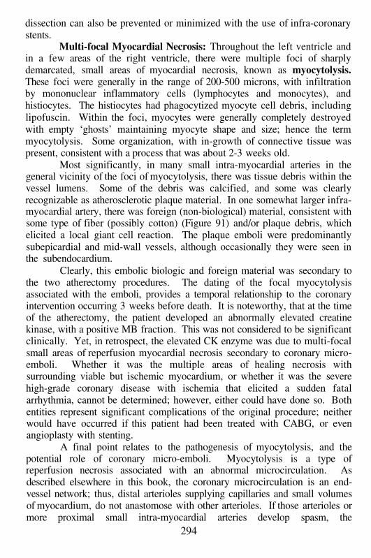
dissection can also be prevented or minimized with the use of infra-coronarystents.
Multi-focal Myocardial Necrosis: Throughout the left ventricle andin a few areas of the right ventricle, there were multiple foci of sharplydemarcated, small areas of myocardial necrosis, known as myocytolysis.These foci were generally in the range of 200-500 microns, with infiltrationby mononuclear inflammatory cells (lymphocytes and monocytes), andhistiocytes. The histiocytes had phagocytized myocyte cell debris, includinglipofuscin. Within the foci, myocytes were generally completely destroyedwith empty ‘ghosts’ maintaining myocyte shape and size; hence the termmyocytolysis. Some organization, with in-growth of connective tissue waspresent, consistent with a process that was about 2-3 weeks old.
Most significantly, in many small intra-myocardial arteries in thegeneral vicinity of the foci of myocytolysis, there was tissue debris within thevessel lumens. Some of the debris was calcified, and some was clearlyrecognizable as atherosclerotic plaque material. In one somewhat larger infra-myocardial artery, there was foreign (non-biological) material, consistent withsome type of fiber (possibly cotton) (Figure 91) and/or plaque debris, whichelicited a local giant cell reaction. The plaque emboli were predominantlysubepicardial and mid-wall vessels, although occasionally they were seen inthe subendocardium.
Clearly, this embolic biologic and foreign material was secondary tothe two atherectomy procedures. The dating of the focal myocytolysisassociated with the emboli, provides a temporal relationship to the coronaryintervention occurring 3 weeks before death. It is noteworthy, that at the timeof the atherectomy, the patient developed an abnormally elevated creatinekinase, with a positive MB fraction. This was not considered to be significantclinically. Yet, in retrospect, the elevated CK enzyme was due to multi-focalsmall areas of reperfusion myocardial necrosis secondary to coronary micro-emboli. Whether it was the multiple areas of healing necrosis withsurrounding viable but ischemic myocardium, or whether it was the severehigh-grade coronary disease with ischemia that elicited a sudden fatalarrhythmia, cannot be determined; however, either could have done so. Bothentities represent significant complications of the original procedure; neitherwould have occurred if this patient had been treated with CABG, or evenangioplasty with stenting.
A final point relates to the pathogenesis of myocytolysis, and thepotential role of coronary micro-emboli. Myocytolysis is a type ofreperfusion necrosis associated with an abnormal microcirculation. Asdescribed elsewhere in this book, the coronary microcirculation is an end-vessel network; thus, distal arterioles supplying capillaries and small volumesof myocardium, do not anastomose with other arterioles. If those arterioles ormore proximal small intra-myocardial arteries develop spasm, the
294
myocardium supplied by those vessels may develop a type of reperfusionnecrosis (e.g. there is initially ischemia, followed by blood flow). This causesfree radical damage to the myocyte, with rupture of the sarcolemma and aninflux of calcium into the cell causing an ‘explosive’ type of necrosis (hence,lysis). Most often, there are concurrent high-catecholamine states such asshock; however, other vasoactive substances can cause microvascularvasospasm. Myocytolysis is a very common finding in cardiomyopathies ofmany etiologies, myocarditis, and ischemic heart disease.
Although it is logical to assume that micro-embolization would causelocalized myocyte necrosis due to obstruction of the vessels, in fact, themorphology of the microscopic necrosis with areas of contraction bands (areperfusion lesion that precedes myocytolysis) or myocytolysis, suggests areperfusion process. Experimentally, embolization of 25 and 50 micron-sizedmicrospheres into the coronary circulation of animals led to identical areas ofmicroscopic necrosis as seen in this case. Interestingly, treatment of theanimals with drugs that prevent coronary artery or microvascular spasm(calcium channel blocking drugs, or alpha-adrenergic blocking drugs),significantly prevented the necrosis even with the microspheres in thecirculation. Thus, it is not obstruction, but spasm that leads to myocytolysis.The plaque emboli caused multiple areas of microvascular spasm andmyocyte necrosis in this patient. It is not known where the foreign fibermaterial came from, but it is likely to have been a contaminant of one of thecoronary catheters. Since the sections of myocardium that demonstrated thesechanges were randomly sampled (e.g. the pathologist performing the autopsydid not see any grossly visible areas in the myocardium to samplespecifically), then the plaque emboli, and probably the foreign material, wereprobably a generalized and widespread phenomenon throughout the heart.
In summary, this case has many interesting clinical and pathologicalfeatures, but it also clearly indicates why it led to a negligence suit against thecardiologists performing the coronary interventional procedure. The wrongtreatment, and its subsequent complications, was medical negligence thatunfortunately caused the death of this treatable patient.
295
Suggested Readings
1.
2.
3.
4.
Carroza JP Jr, Braim DS. Complications of directional coronaryatherectomy: incidence, causes, and management. Am J Cardiol. 1993; 72:47E-54E.Hinohara T, Robertson GC, Selmon MR, Vetter JW, McAuley BJ, SheehanDJ, Simpson JB. Directional coronary atherectomy complications andmanagement. Cather Cardiovasc Diagn. 1993; Suppl 1:61-71.Kaufmann UP, Meyer BJ. Atherectomy (directional, rotational,extractional) and its role in percutaneous revascularization. Curr OponCardiol. 1995; 10:412-9.Corcos T, Zimarino M, Tamburino C, Favereau X. Randomized trials ofdireccional atherectomy: implications for clinical practice and futureinvestigation. J Am Coll Cardiol. 1994; 24:431-9.
296
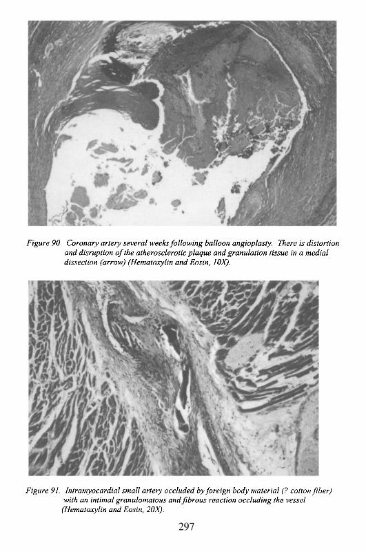
297

This Page Intentionally Left Blank
299
Appendix I. Heart Measurements in Adultts (modified fromSunderman FW, Boerner F: Normal values in clinicalmedicine. Philadelphia. W.B. Saunders Company, 1949).
300
Appendix II. Normal adult laboratory values
Appendix III. Abbreviations.
AAA abdominal aortic aneurysmABE acute bacterial endocarditisABG arterial blood gasesACh acetylcholineAChE acetylcholinesteraseAIDS acquired immunodeficiency syndromeANA antinuclear antibodiesARDS adult respiratory distress syndromeASA aspirinASD atrial septal defectATP adenosin triphosphateAV atrioventricularBMI body mass indexBP blood pressureCABG coronary artery bypass graftCAD coronary artery diseaseCCU coronary care unitCEA carcinoembryonic antigenCHF congestive heart failureCK creatin kinaseCM cardiomyopathyCMV cytomegalovirusCNS central nervous systemCO cardiac outputCOPD chronic obstructive pulmonary diseaseCPR cardiopulmonary resuscitationCREST calcinosis, Raynaud's phenomenom, esophageal
dysmotility, sclerodactyly and telangiectasia
CSF cerebrospinal fluidCT computarized tomographyCVA cerebrovascular accidentCXR chest x rayDCM dilated cardiomyopathyDIC disseminated intravascular coagulationDM diaebtes mellitusECG electrocardiogramECHO enteric cytopathogenic human orphan virusesEMD electro-mechanical dissociationER emergency roomESR erythrocyte sedimentation rate
301
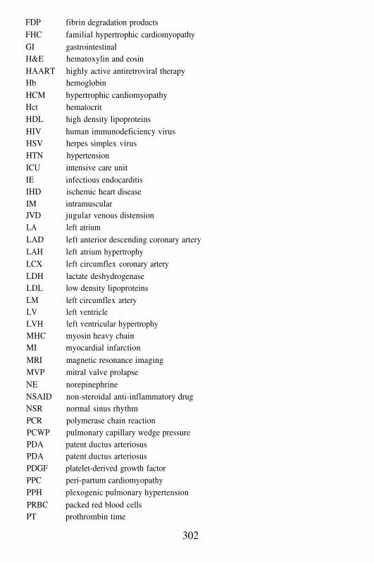
FDP fibrin degradation productsFHC familial hypertrophic cardiomyopathyGI gastrointestinalH&E hematoxylin and eosinHAART highly active antiretroviral therapyHb hemoglobinHCM hypertrophic cardiomyopathyHct hematocritHDL high density lipoproteinsHIV human immunodeficiency virusHSV herpes simplex virusHTN hypertensionICU intensive care unitIE infectious endocarditisIHD ischemic heart diseaseIM intramuscularJVD jugular venous distensionLA left atriumLAD left anterior descending coronary arteryLAH left atrium hypertrophyLCX left circumflex coronary arteryLDH lactate deshydrogenaseLDL low density lipoproteinsLM left circumflex arteryLV left ventricleLVH left ventricular hypertrophyMHC myosin heavy chainMI myocardial infarctionMRI magnetic resonance imagingMVP mitral valve prolapseNE norepinephrineNSAID non-steroidal anti-inflammatory drugNSR normal sinus rhythmPCR polymerase chain reactionPCWP pulmonary capillary wedge pressurePDA patent ductus arteriosusPDA patent ductus arteriosusPDGF platelet-derived growth factorPPC peri-partum cardiomyopathyPPH plexogenic pulmonary hypertensionPRBC packed red blood cellsPT prothrombin time
302
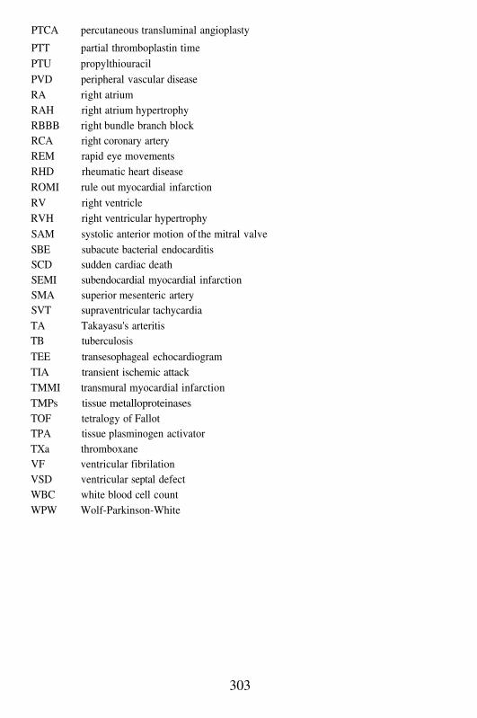
PTCA percutaneous transluminal angioplasty
PTT partial thromboplastin timePTU propylthiouracilPVD peripheral vascular diseaseRA right atriumRAH right atrium hypertrophyRBBB right bundle branch blockRCA right coronary arteryREM rapid eye movementsRHD rheumatic heart diseaseROMI rule out myocardial infarctionRV right ventricleRVH right ventricular hypertrophySAM systolic anterior motion of the mitral valveSBE subacute bacterial endocarditisSCD sudden cardiac deathSEMI subendocardial myocardial infarctionSMA superior mesenteric arterySVT supraventricular tachycardiaTA Takayasu's arteritisTB tuberculosisTEE transesophageal echocardiogramTIA transient ischemic attackTMMI transmural myocardial infarctionTMPs tissue metalloproteinasesTOF tetralogy of FallotTPA tissue plasminogen activatorTXa thromboxaneVF ventricular fibrilationVSD ventricular septal defectWBC white blood cell countWPW Wolf-Parkinson-White
303

This Page Intentionally Left Blank
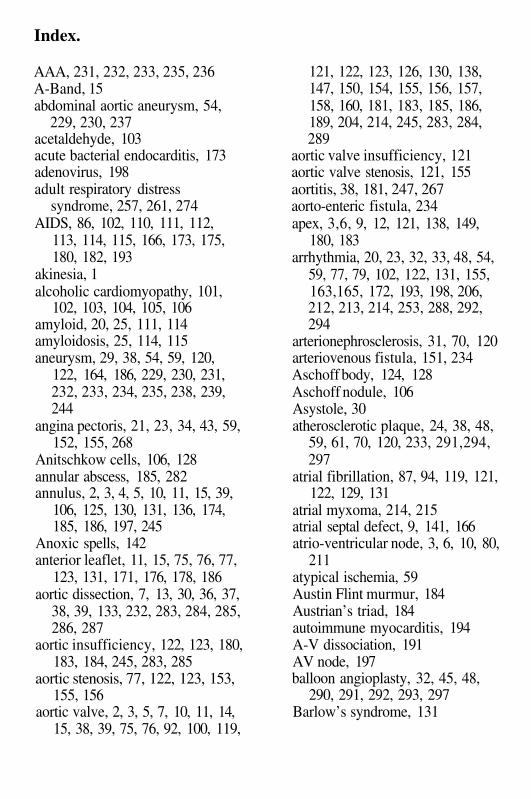
Index.
AAA, 231, 232, 233, 235, 236A-Band, 15abdominal aortic aneurysm, 54,
229, 230, 237acetaldehyde, 103acute bacterial endocarditis, 173adenovirus, 198adult respiratory distress
syndrome, 257, 261, 274AIDS, 86, 102, 110, 111, 112,
113, 114, 115, 166, 173, 175,180, 182, 193
akinesia, 1alcoholic cardiomyopathy, 101,
102, 103, 104, 105, 106amyloid, 20, 25, 111, 114amyloidosis, 25, 114, 115aneurysm, 29, 38, 54, 59, 120,
122, 164, 186, 229, 230, 231,232, 233, 234, 235, 238, 239,244
angina pectoris, 21, 23, 34, 43, 59,152, 155, 268
Anitschkow cells, 106, 128annular abscess, 185, 282annulus, 2, 3, 4, 5, 10, 11, 15, 39,
106, 125, 130, 131, 136, 174,185, 186, 197, 245
Anoxic spells, 142anterior leaflet, 11, 15, 75, 76, 77,
123, 131, 171, 176, 178, 186aortic dissection, 7, 13, 30, 36, 37,
38, 39, 133, 232, 283, 284, 285,286, 287
aortic insufficiency, 122, 123, 180,183, 184, 245, 283, 285
aortic stenosis, 77, 122, 123, 153,155, 156
aortic valve, 2, 3, 5, 7, 10, 11, 14,15, 38, 39, 75, 76, 92, 100, 119,
121, 122, 123, 126, 130, 138,147, 150, 154, 155, 156, 157,158, 160, 181, 183, 185, 186,189, 204, 214, 245, 283, 284,289
aortic valve insufficiency, 121aortic valve stenosis, 121, 155aortitis, 38, 181, 247, 267aorto-enteric fistula, 234apex, 3,6, 9, 12, 121, 138, 149,
180, 183arrhythmia, 20, 23, 32, 33, 48, 54,
59, 77, 79, 102, 122, 131, 155,163,165, 172, 193, 198, 206,212, 213, 214, 253, 288, 292,294
arterionephrosclerosis, 31, 70, 120arteriovenous fistula, 151, 234Aschoff body, 124, 128Aschoff nodule, 106Asystole, 30atherosclerotic plaque, 24, 38, 48,
59, 61, 70, 120, 233, 291,294,297
atrial fibrillation, 87, 94, 119, 121,122, 129, 131
atrial myxoma, 214, 215atrial septal defect, 9, 141, 166atrio-ventricular node, 3, 6, 10, 80,
211atypical ischemia, 59Austin Flint murmur, 184Austrian’s triad, 184autoimmune myocarditis, 194A-V dissociation, 191AV node, 197balloon angioplasty, 32, 45, 48,
290, 291, 292, 293, 297Barlow’s syndrome, 131
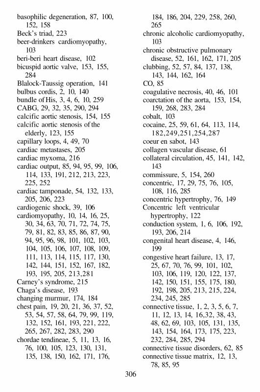
basophilic degeneration, 87, 100,152, 158
Beck’s triad, 223beer-drinkers cardiomyopathy,
103beri-beri heart disease, 102bicuspid aortic valve, 153, 155,
284Blalock-Taussig operation, 141bulbus cordis, 2, 10, 140bundle of His, 3, 4, 6, 10, 259CABG, 29, 32, 35, 290, 294calcific aortic stenosis, 154, 155calcific aortic stenosis of the
elderly, 123, 155capillary loops, 4, 49, 70cardiac metastases, 205cardiac myxoma, 216cardiac output, 85, 94, 95, 99, 106,
114, 133, 191, 212, 213, 223,225, 252
cardiac tamponade, 54, 132, 133,205, 206, 223
cardiogenic shock, 39, 106cardiomyopathy, 10, 14, 16, 25,
30, 34, 63, 70, 71, 72, 74, 75,79, 81, 82, 83, 85, 86, 87, 90,94, 95, 96, 98, 101, 102, 103,104, 105, 106, 107, 108, 109,111, 113, 114, 115, 117, 130,142, 144, 151, 152, 167, 182,193, 195, 205, 213,281
Carney’s syndrome, 215Chaga’s disease, 193changing murmur, 174, 184chest pain, 19, 20, 21, 36, 37, 52,
53, 54, 57, 58, 64, 79, 99, 119,132, 152, 161, 193, 221, 222,265, 267, 282, 283, 290
chordae tendineae, 5, 11, 13, 16,76, 100, 105, 123, 130, 131,135, 138, 150, 162, 171, 176,
184, 186, 204, 229, 258, 260,265
chronic alcoholic cardiomyopathy,103
chronic obstructive pulmonarydisease, 52, 161, 162, 171, 205
clubbing, 52, 57, 84, 137, 138,143, 144, 162, 164
CO, 85coagulative necrosis, 40, 46, 101coarctation of the aorta, 153, 154,
159, 268, 283, 284cobalt, 103cocaine, 25, 59, 61, 64, 113, 114,
182,249,251,254,287coeur en sabot, 143collagen vascular disease, 61collateral circulation, 45, 141, 142,
143commissure, 5, 154, 260concentric, 17, 29, 75, 76, 105,
108, 116, 285concentric hypertrophy, 76, 149Concentric left ventricular
hypertrophy, 122conduction system, 1, 6, 106, 192,
193, 206, 214congenital heart disease, 4, 146,
199congestive heart failure, 13, 17,
25, 67, 70, 76, 99, 101, 102,103, 106, 119, 120, 122, 137,142, 150, 151, 155, 175, 180,192, 198, 205, 213, 215, 224,234, 245, 285
connective tissue, 1, 2, 3, 5, 6, 7,11, 12, 13, 14, 16,32, 38, 43,48, 62, 69, 103, 105, 131, 135,143, 154, 164, 173, 175, 223,232, 284, 285, 294
connective tissue disorders, 62, 85connective tissue matrix, 12, 13,
78, 85, 95306
constricitive pericarditis, 224contraction band necrosis, 17, 45,
46cor pulmonale, 161, 163, 164, 165,
252, 253coronary arteries, 3, 4, 7, 13, 20,
29, 36, 53, 60, 62, 63, 68, 69,92, 105, 110, 138, 150, 151,162, 171, 183, 197,204,211,222, 251, 260, 265, 266, 267,275, 284, 290, 292
coronary artery ameurysm, 275coronary artery bypass graft, 29coronary artery disease, 7, 24, 29,
30, 31, 34, 48, 57, 59, 60, 71,86, 101, 120, 151,241,250,260, 275, 290
coronary artery dissection, 63, 64,96, 292
Coronary artery dissection, 62, 65coronary artery spasm, 23, 59, 60,
61, 63, 293coronary artery vasculitis, 61coronary ostia, 3, 245Corrigan’s pulse, 183Coxsackie, 193, 199creatine kinase, 21, 291, 294Crohn’s disease, 102cyanosis, 52, 57, 84, 137, 142,
143, 144, 162, 164, 221, 265,272, 283
cystic tumor of the atrio-ventricular node, 213
Cytomegalovirus, 194DCM, 76, 78, 85, 86, 87, 113degenerative valve disease, 123diabetes mellitus, 14, 19, 20, 21,
22, 24, 48, 57, 59, 60, 67, 68,69, 149, 155, 174, 232
diabetic cardiomyopathy, 151, 281diabetic neuropathy, 43, 69, 281diabetic vascular injury, 69
diastolic dysfunction, 7, 25, 71,78, 114
dilated cardiomyopathy, 76, 85,105, 113
dilated congestivecardiomyopathy, 163
dissection, 32, 37, 38, 39, 54, 59,63, 64, 230, 232, 245, 283, 284,285, 287, 291, 292, 293, 297
Dressler’s syndrome, 132ductus arteriosus, 8, 138, 140, 141,
142, 144, 166, 167, 168, 268,284
Duroziez’s sign, 183dyskinesia, 1eccentric, 17, 293ECHO, 193, 200ectasia , 231, 275Ehlers-Danlos syndrome, 16, 62Eisenmenger’s syndrome, 164electrocardiogram, 29, 94, 143,
191, 206, 242electro-mechanical dissociation,
30endarteritis, 244, 248endocardial cushion defects, 11,
130endocarditis, 2, 4, 5, 8, 11, 16, 59,
111, 112, 130, 132, 144, 172,175, 176, 177, 178, 180, 181,182, 183, 184, 186, 188, 189,258, 259, 260, 263, 264, 282
endocardium, 2, 4, 8, 14, 16, 36,45, 46, 47, 62, 63, 76, 77, 84,86, 92, 105, 110, 111, 138, 173,181, 198, 204, 206, 211, 214,222, 229, 258, 259, 265
Endomyocardial biopsies, 194endothelin-1, 32endovascular grafting, 235eosinophilic vasculitis, 63
307
epicardium, 14, 16, 36, 45, 46, 47,62, 75, 92, 100, 110, 171, 185,204, 209, 222, 229, 257, 265
epigastric pain, 67, 229, 241, 242,243, 244
familial hypertrophiccardiomyopathy, 6, 76, 81
fenfluramine-phentermine, 164fibrillin, 38, 114, 232, 285fibrinous pericarditis, 123, 132,
181, 186, 228, 259fibromyxomatous valve
degeneration, 16fibromyxomatous valve disease,
15floppy valve disease, 131foramen ovale, 8, 9, 36, 62, 92,
110, 120, 138, 140, 141, 181,204, 258, 265
fossa ovalis, 8gallop rhythm, 21, 110giant cell arteritis, 62, 245, 267glomerulosclerosis, 68, 70Goldblatt kidney, 31granulation tissue, 29, 32, 43, 48,
53, 129, 132, 192, 293, 297Grave’s disease, 91, 93group A, beta-hemolytic
streptococcus, 123Hashimoto’s disease, 86, 152HCM, 76, 77, 78, 79, 80heart orientation, 9hemopericardium, 30, 181, 186hemoptysis, 110, 221, 241, 242,
243highly active antiretroviral
therapy, 112HIV-infection, 224homocysteine, 61hypercholesterolemia, 29, 59, 60hypertension, 8, 9, 13, 14, 20, 22,
23, 24, 29, 31, 36, 37, 38, 39,43, 52, 54, 57, 59, 60, 67, 68,
70, 71, 75, 76, 86, 95, 99, 103,104, 113, 114, 120, 121, 122,123, 130, 132, 151, 152, 153,161, 163, 165, 166, 169, 205,232, 241, 244, 250, 251, 252,254, 260, 268, 273, 283, 284,285, 287
hypertensive cardiac disease, 76hyperthyroid cardiomyopathy, 93hyperthyroid heart disease, 94, 95hyperthyroidism, 60, 87, 94, 95,
151, 265, 269hypertrophic cardiomyopathy, 76hypertrophy, 12, 14, 16, 17, 29,
31, 36, 43, 44, 68, 69, 71, 76,79, 82, 84, 86, 93, 95, 101, 105,110, 114, 116, 121, 129, 130,139, 144, 150, 152, 155, 162,163, 166, 181, 243, 255, 258,260
hypokinesia, 1hypothyroidism, 86, 94, 149, 151,
152, 153, 155infarct expansion, 47infarct extension, 44, 47, 49infectious endocarditis, 172, 173Influenza, 193Interleukins, 193internal mammary artery, 29, 32,
35, 290interventricular septum, 10, 39, 43,
110, 120, 138, 140ischemic bowel disease, 67, 68,
70, 214, 252ischemic heart disease, 20, 34, 58,
59, 60, 150, 281, 295jugular venous distension, 57, 91,
94, 149, 161, 223jugular venous distention, 21Juvenile Rheumatoid Arthritis,
274Kaposi sarcoma, 112Kawasaki disease, 61, 277
308
Kawasaki’s disease, 199, 275, 278,279, 280
Kimmelstiel-Wilson, 68, 70Lambl’s excrescence, 11LaPlace’s law, 234lateral border zone, 47Leriche syndrome, 234long Q-T syndrome, 214Luetic aortitis, 245lymphoma, 112, 171, 176, 210macrovasculature, 69Marfan’s syndrome, 2, 5, 16, 38,
62, 114, 232, 284, 285, 287medial calcific sclerosis, 68membranous septal defects, 11membranous septum, 4, 11, 140,
144mesothelioma of the AV node, 213metalloproteinases, 95, 237metastatic tumors, 204MHC antigens, 193microaneurysms, 70, 71, 72, 73microfibrillar cardiomyopathy,
114microvascular disease, 70, 71microvasculature, 69, 71, 113, 251mitral annular calcification, 123,
130mitral regurgitation, 5, 39, 78, 99,
110, 123, 129, 130, 131, 132,134, 183, 191
mitral valve, 3, 4, 5, 9, 11, 13, 15,16, 36, 39, 40, 76, 77, 78, 84,86, 92, 105, 119, 120, 122, 127,129, 130, 131, 132, 134, 135,138, 150, 155, 171, 172, 173,176, 178, 180, 183, 184, 186,204, 215, 258, 260, 266, 283,284, 285
mitral valve prolapse, 5, 15, 131,132
mitral valve prolapse syndrome, 5,283
mitral valve regurgitation, 78, 106,130
Mitral valvuloplasty, 129Monckeberg’s, 68, 70Mucocutaneous Lymph Node
Syndrome, 275MVP, 131mycotic aneurysm, 176, 186, 231myocardial fibrosis, 76, 85, 113,
150, 155myocardial hypertrophy, 12, 20,
23, 100myocardial infarction, 11, 12, 19,
20, 21, 22, 23, 24, 25, 26, 30,36, 37, 38, 39, 40, 41, 43, 44,45, 46, 48, 49, 52, 53, 55, 57,59, 60, 64, 74, 85, 119, 120,130, 132, 133, 151, 191, 192,213, 230, 243, 251, 265, 266,267, 268, 275, 279, 282, 287,290
myocardial ischemia, 7, 20, 23, 26,34, 48, 49, 78, 114, 265, 292
myocarditis, 10, 30, 63, 64, 85, 86,93, 96, 106, 112, 123, 130, 188,192, 193, 194, 195, 198, 199,200, 201, 213, 225, 259, 274,295
myocardium, 4, 6, 7, 12, 14, 15,19, 22, 23, 25, 26, 36, 39, 44,45, 46, 47, 48, 52, 58, 62, 63,71, 74, 75, 76, 78, 84, 85, 92,93, 95, 100, 101, 102, 104, 105,108, 110, 111, 112, 120, 121,123, 129, 136, 138, 150, 151,162, 171, 181, 185, 191, 192,193, 194, 197, 199, 202, 204,206, 209, 210, 211, 213, 222,229, 230, 243, 251, 253, 258,260, 265, 266, 294, 295
myocardium at risk, 26, 45, 47myocyte hypertrophy, 76, 93, 95myocyte nuclei, 12
309
myocyte whorls, 78myocytolysis, 43, 103, 110, 111,
112, 120, 150, 151, 197, 273,275, 294, 295
myofibrils, 15myosin heavy chain, 79necrotizing glomerulonephritis,
174necrotizing vasculitis, 166, 174,
260neo-intima, 32, 293non-bacterial thrombotic
vegetation, 100non-Q-wave MI, 45non-steroidal anti-inflammatory
drug, 274obesity, 14, 161, 162, 163, 164,
165, 256, 257, 260, 261Osler’s nodes, 174ostium, 7, 11, 39, 62, 265papillary fibroelastoma, 10papillary muscle, 4, 5, 13, 36, 37,
39, 41, 42, 74, 130, 131, 266patent ductus arteriosus, 162, 163pathologic hypertrophy, 31Pentalogy of Fallot, 141percutaneous transluminal
coronary angioplasty, 29, 34perform, 193pericardial effusion, 19, 54, 92,
110, 111, 132, 152, 161, 191,205, 206, 225, 274, 275
pericardial friction rub, 132, 180,185
pericardial tamponade, 13, 39,186, 292
pericardiocentesis, 185, 205, 225pericarditis, 58, 59, 75, 129, 132,
185, 186, 192, 222, 225, 228,250, 257, 259, 263, 274
pericardium, 13, 19, 29, 36, 52,54, 62, 84, 92, 100, 110, 119,123, 129, 132, 138, 161, 171,
181, 185, 186, 198, 204, 205,216, 221, 222, 224, 225, 229,231, 243, 257, 259
peripartum cardiomyopathy, 63peri-partum cardiomyopathy, 93peri-partum cardiomyopathy, 95peripheral vascular disease, 19, 43,
245physiologic hypertrophy, 31Pickwickian syndrome, 163platelet-derived growth factor, 32pleuritic pain, 223plexogenic pulmonary
hypertension, 164pneumococcal endocarditis, 184Pneumocystis carinii pneumonia,
221, 274polyarteritis nodosa, 60Polyarteritis Nodosa, 276polycythemia, 143, 144, 162post-cardiotomy syndrome, 132,
133posterior leaflet, 11, 76, 77, 78,
123, 129, 131, 258Potts anastomosis, 141PPC, 93, 94primary tumors, 204, 214pseudo-aneurysm, 176, 181, 186,
230, 292pseudovalve, 120, 121pseudoxanthoma elasticum, 62PTCA, 29pulmonary adenocarcinoma, 206pulmonary hypertension, 53, 162,
163, 268pulmonary valve, 14, 121, 138,
141, 252pulmonic valve, 2, 5, 15, 92, 141,
186, 204pulsus paradoxus, 132, 223Purkinje cells, 46Purkinje fibers, 6Quincke’s pulse, 183
310
remodeling, 6, 7, 14, 17, 47, 85,95, 105, 122, 130, 152, 153,155, 163, 182, 196, 252, 293
reperfusion infarction, 22, 24reperfusion injury, 17, 45, 51, 116re-stenosis, 29, 32, 34, 291, 292restrictive heart disease, 114, 213retroperitoneal hematoma, 53RHD, 120, 123, 260rheumatic heart disease, 5, 11, 76,
105, 120, 121, 124, 125, 130,183, 215, 241, 260, 275
rheumatic mitral valve, 123rheumatoid arthritis, 3, 193, 272right ventricle, 4, 9, 12, 13, 14, 17,
19, 29, 36, 75, 100, 116, 119,129, 133, 138, 141, 143, 149,151, 163, 197, 252, 255, 266,294
rule out myocardial infarction, 59ruptured papillary muscle, 39ruptured plaque, 20, 25SAM, 75, 77, 78sarcoidosis, 21, 85, 213, 216sarcolemma, 15, 17, 45, 111, 295sarcomere, 15, 17, 79sarcomeric cardiomyopathy, 79sarcoplasmic reticulum, 15SEMI, 43, 44, 45, 46, 47, 48septum secundum, 8shock, 37, 39, 99, 106, 110, 114,
191, 230, 235, 288, 295sick sinus syndrome, 213sinoatrial node, 14sinus of Valsalva, 3, 7, 154sinuses of Valsalva, 14, 123, 154 ,
160, 186small vessel disease, 24, 70, 105,
253splinter hemorrhages, 174Stokes-Adams-Morgagni
syndrome, 213streptokinase, 22
stunned myocardium, 21subacute bacterial endocarditis,
172subendocardial infarction, 23, 44subendocardial myocardial
infarction, 43sudden cardiac death, 7, 14, 24,
37, 85, 281Sudden death, 79, 80superior mesenteric artery, 67, 68,
69, 212, 214, 266Swan-Ganz catheter, 92, 99, 257syncope, 58, 61, 94, 143, 208, 211,
212, 213, 214, 216syphilis, 2, 244, 245, 248, 268Syphilitic aneurysms, 245syphilitic aortitis, 38, 244systemic lupus erythematosus, 61,
256, 262systolic anterior motion of the
mitral valve, 75, 77Takayasu’s arteritis, 2, 62, 245,
267, 271Takayasu’s disease, 245TB pericarditis, 205, 224TEE, 9tetralogy of Fallot, 14, 146Tetralogy of Fallot, 139, 147The Dallas Classification, 194thrombolytic therapy, 21, 26, 52,
53thromboxane, 32tissue metalloproteinase, 25tissue plasminogen activator, 19,
43, 52TMMI, 46, 47, 48TPA, 19, 22, 23, 24, 25, 43, 45,
47, 48, 52, 53, 54trans-esophageal
echocardiography, 9transient cerebral ischemic attack,
149, 150transmural extension, 44, 47, 197
311
transmural infarction, 23, 24, 44,45, 46
transmural myocardial infarction,24, 25, 44, 46, 53
triangle of Koch, 10tricuspid valve, 3, 5, 10, 11, 15,
36, 68, 86, 92, 106, 132, 153,186, 204, 214, 252, 258, 260,266
troponin I, 22, 58, 119truncus arteriosus, 140T-tubule, 15tuberculosis, 53, 173, 224, 241,
244tuberculous pericarditis, 205tumor necrosis factor, 193tumor plop, 215Turner’s syndrome, 149, 152, 153,
154Valsalva maneuver, 77, 165, 215valve ring abscess, 2, 185valves, 1, 2, 3, 5, 8, 10, 15, 16, 19,
29, 36, 75, 86, 119, 122, 123,129, 132, 154, 155, 173, 174,181, 182, 183, 184, 185, 186,191, 197, 206, 211, 222, 249
vasa nervorum, 7vasa vasorum, 7, 232, 244, 248,
266, 268, 286ventricular dyskinesis, 114ventricular fibrillation, 29, 30, 95,
197, 214, 283ventricular hypertrophy, 12, 30,
68, 74, 75, 76, 77, 78, 79, 85,119, 122, 126, 133, 137, 138,140, 141, 143, 144, 152, 155,163, 164, 165, 242, 244, 268,285
ventricular rupture, 23, 30, 40, 47ventricular septal defect, 39, 138,
140, 144, 147, 166ventricular tachycardia, 23, 30,
111Venturi effect, 77vulnerable myocardium, 45, 46, 48water-hammer, 183wavefront, 26, 45wave-front phenomenon, 22whorls, 76, 83Wolf-Parkinson-White, 79WPW, 79, 80Z-band, 15, 17
312