Embed Size (px)
Citation preview
Cardiac Testing in Adolescents
Christian D. Nagy, M.D. and W. Reid Thompson, M.D.
Diagnostic testing in adolescents and young adults with known or suspected heart disease typically
involves the use of electrocardiography, various imaging modalities, and, in some cases, other
laboratory investigations. In this chapter the authors discuss common tests that may be ordered by the
generalist or cardiologist to evaluate the heart. The emphasis is on indications for ordering a specific
test, understanding the strengths and weaknesses of those tests, and basic interpretation of the results.
Heart disease in adolescents primarily includes previously diagnosed congenital lesions, undiagnosed
defects such as atrial septal defect (ASD) or aortic valve abnormalities that are often asymptomatic in
childhood, inherited latent conditions that may first become manifest during the teen years, such as
hypertrophic cardiomyopathy (HCM), and acquired disease such as myocarditis. Signs and symptoms of
possible heart disease, when present, may include a pathological murmur or heart sound(s), chest pain
and shortness of breath, especially when associated with exercise, palpitations or syncope, or signs of
heart failure such as a gallop, increased jugular venous distension, hepatomegaly, rales, and peripheral
edema. Often, a careful family history may yield important clues to the possibility of inherited cardiac
disease. An electrocardiogram is often ordered by the generalist or specialist to evalutate symptoms of
possible heart disease or to monitor potential side effects of medications. Most imaging studies,
including echocardiography, cardiac MRI and CT, as well as cardiac catheterizations are ordered or
performed by the cardiologist to diagnose specific defects or conditions, and catheterizations are
increasingly done primarily for intervention purposes.
1. ELECTROCARDIOGRAPHY
The electrocardiogram (ECG) remains an invaluable noninvasive tool to assess the electrical activity of
the heart in order to evaluate adolescents with known or suspected cardiovascular disease and may be
appropriately ordered by the generalist as part of the investigation of symptoms or monitoring of
therapy. The standard 12-lead ECG helps to identify abnormalities of impulse formation (sinus
bradycardia, isolated premature atrial, junctional, or ventricular beats, or atrial, junctional, or ventricular
arrhythmias) or impulse propagation, such as slowed conduction through the AV node (AV block) and
His-Purkinje system (high degree AV block or bundle branch blocks). It can identify those individuals
with a short PR interval and delta wave indicating ventricular preexcitation (Wolff-Parkinson-White
syndrome). Repolarization syndromes involving abnormal myocardial cell membrane ion channels (long
QT syndrome and Brugada syndrome) can also be diagnosed on the standard ECG. Additionally,
abnormalities of the ECG associated with myocardial hypertrophy (hypertrophic cardiomyopathy),
inflammation (myocarditis or pericarditis), myocardial ischemia (anomalous coronary arteries or
premature coronary artery disease) or injury (myocardial infarction) can be detected. Serial ECGs are
also commonly used to monitor potential cardiac affects of certain psychotropic medications.
Guidelines for the performance of electrocardiograms were published by the American College of
Cardiology and American Heart Association (ACC/AHA) in 19921
and have not changed in recent
years. These guidelines make recommendations for the use of ECGs in patients with and without
cardiovascular disease, which for the most part are applicable to the adolescent population.2
Cardiac Testing in Adolescents, Nagy and Thompson
2
Electrocardiography is a quick, inexpensive, and widely-available test that can be administered
accurately in a variety of clinical settings with a minimum of training. In addition, detailed automated
computer interpretation algorithms can assist in interpretation, though must always be carefully
confirmed for accuracy by an experienced reader. Data can be stored digitally and transmitted
electronically or by fax for rapid expert interpretation. Artifacts include those due to movement or faulty
connections, although many errors in connection can be easily detected (e.g., by checking for
consistency between lead I versus V6 pattern). The ECG is less helpful for diagnosing specific structural
abnormalities and has a high false positive rate for detecting left ventricular hypertrophy, particularly in
athletes or those with thin body habitus.
Interpretation of abnormal heart rate and rhythm
Sinus tachycardia, characterized by heart rate greater than the 98%tile for age (usually >120 but less
than ~200 beats per minute with P waves of normal axis (0-90 degrees) preceding each QRS complex, is
by far the most common tachyarrhythmia and is often due to an underlying hypersympathetic state such
as fever, pain, anxiety, anemia, dehydration, substance abuse or withdrawal, or hyperthyroidism. Other
forms of narrow complex tachycardia represent primary cardiac disorders of either increased
automaticity or reentry pathways. When the atrial activity (P wave) occurs shortly after the QRS
complex, an atrio-ventricular bypass tract is the most likely mechanism (Fig 1). The baseline (non
tachycardic) ECG in patients with Wolff-Parkinson-White syndrome (WPW) has the typical short PR
interval with an upstroke (delta wave) from the end of the P wave to the beginning of the QRS (Fig 2).
When a regular, narrow complex tachycardia is present with no visible P waves, simultaneous
depolarization of the atria and ventricles with the P wave “hidden” in the QRS complex is more likely,
indicating an AV nodal re-entry tachycardia. When P waves are prior to the QRS but have an abnormal
axis (i.e., other than 0-90 degrees), automatic or ectopic atrial tachycardia is more likely. When the 12-
lead ECG demonstrates a wide complex tachycardia (QRS duration > 120 msec), the differential
diagnosis includes ventricular tachycardia, supraventricular tachycardia with aberrant conduction
between the atria and ventricles, or a fixed bundle branch block.
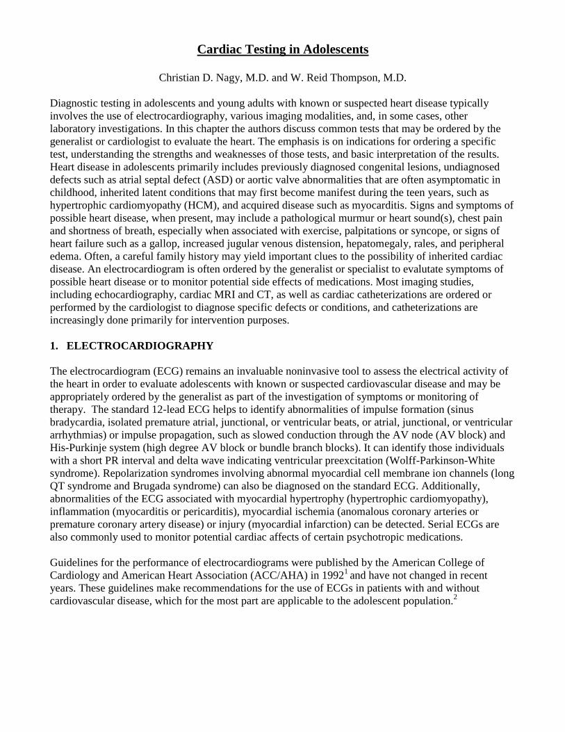
Figure 1. ECG of a 13 year old boy showing supraventricular tachycardia alternating with bradycardia.
Cardiac Testing in Adolescents, Nagy and Thompson
3
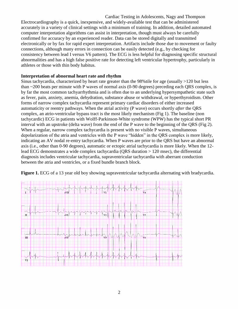
Figure 2. ECG of 20 year old female with WPW. Note the short PR interval and the upstroke (delta
wave) from the end of the P wave to the beginning of the QRS complex (arrow).
Bradycardia in the adolescent is encountered in competitive athletes or individuals with eating disorders
(Anorexia Nervosa) and most commonly manifested on ECG as sinus bradycardia (defined as sinus
rhythm with a heart rate <2%tile for age, usually < 60 beats per minute). Other causes of sinus
bradycardia include hypoxia, hypothyroidism, hypothermia, hypercalcemia, hyperkalemia,
hypoglycemia, and increased intracranial pressure. In addition, bradycardia may be seen in congenital
long QT syndrome. In patients with complete heart block (CHB), the ventricular rate is slower and
dissociated from the atrial rate (Fig 3). CHB occurs congenitally in infants born to mothers with lupus or
may be acquired following certain infections (e.g., Lyme disease).
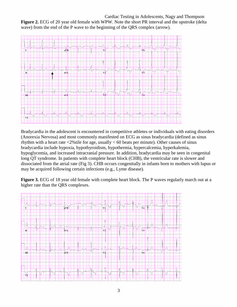
Figure 3. ECG of 18 year old female with complete heart block. The P waves regularly march out at a
higher rate than the QRS complexes.

Cardiac Testing in Adolescents, Nagy and Thompson
4
Adolescents evaluated for palpitations or syncope should have an ECG performed as a simple,
inexpensive, and non-invasive diagnostic tool. Ideally a 12-lead ECG should be recorded during an
episode of symptoms, though this is often not feasible. Roughly, up to 50% of patients evaluated for
syncope will have an abnormal but non-diagnostic ECG.3 The presence of ventricular pre-excitation,
ectopic atrial or ventricular beats, ventricular hypertrophy, long QT interval, bradycardia, or sustained
tachycardia suggest a plausible cause of syncope. Premature ventricular beats (PVCs) (Fig 4) are
common findings and usually do not require further investigation. If they occur with increased
frequency a Holter monitor aids in quantification, and an echocardiogram is useful to exclude
ventricular dysfunction.
Figure 4. ECG in a 14-year old adolescent demonstrating frequent PVCs.
Evaluation of chest pain
An ECG is indicated as part of the initial evaluation of adolescents with non reproducible, exertional
chest pain. Although chest pain is often of great concern to the patient and family, the majority of
adolescents with this symptom do not have a cardiac etiology. ECG changes that warrant further
investigation, such as ST segment deviation, T wave changes, ventricular hypertrophy, or conduction
abnormalities are easily detected. In patients evaluated for chest pain, deviations of the ST segment from
baseline may indicate myocardial ischemia, injury, other pathological processes, or represent a normal
variant. Comparison to an old ECG is often helpful, however not always available. The ST segment is
identified as the portion between the end of the QRS complex (ventricular depolarization) and beginning
of T wave (repolarization). The normal ST segment is usually isoelectric relative to the TP segment. The
end of the QRS complex is marked by the junction to the ST segment (J point) (Fig 5).
Cardiac Testing in Adolescents, Nagy and Thompson
5
Fig 5. Normal ECG
Variations occur in the ST pattern. It is important to recognize them because they can be mistaken for
abnormalities. ST-T patterns can be affected by changes in autonomic tone, variations in body position,
hyperventilation, drinking cold water, and performing Valsalva maneuvers. ST segment elevation in
adolescents may represent benign “early repolarization”, or be seen in pericarditis, acute myocardial
infarction (e.g., in the setting of cocaine use), commotio cordis, left ventricular hypertrophy, left bundle
branch block, hyperkalemia, and critical illness (including neurologic conditions such as subarachnoid
hemorrhage). Up to 90% of patients with hypertrophic cardiomyopathy (HCM) have some abnormality
of the resting ECG,4 often either ST segment abnormalities or T wave changes (Fig 6). Whereas
infarction and ischemia are uncommon in adolescents the term “early repolarization” is applied when it
is associated with ST elevation, most easily appreciated in the anterior and mid-precordial leads (V2-
V4) (Fig 7). The term is a misnomer (as repolarization normally begins before depolarization ends), the
associated elevation of the ST segment may be due to the normal age-dependent changes of ST segment
potentials. It can especially mimic changes seen in pericarditis. J point elevation due to early
repolarization often disappears with exercise. Table 1 summarizes common ECG features in myocardial
ischemia or injury, pericarditis, and early repolarization.
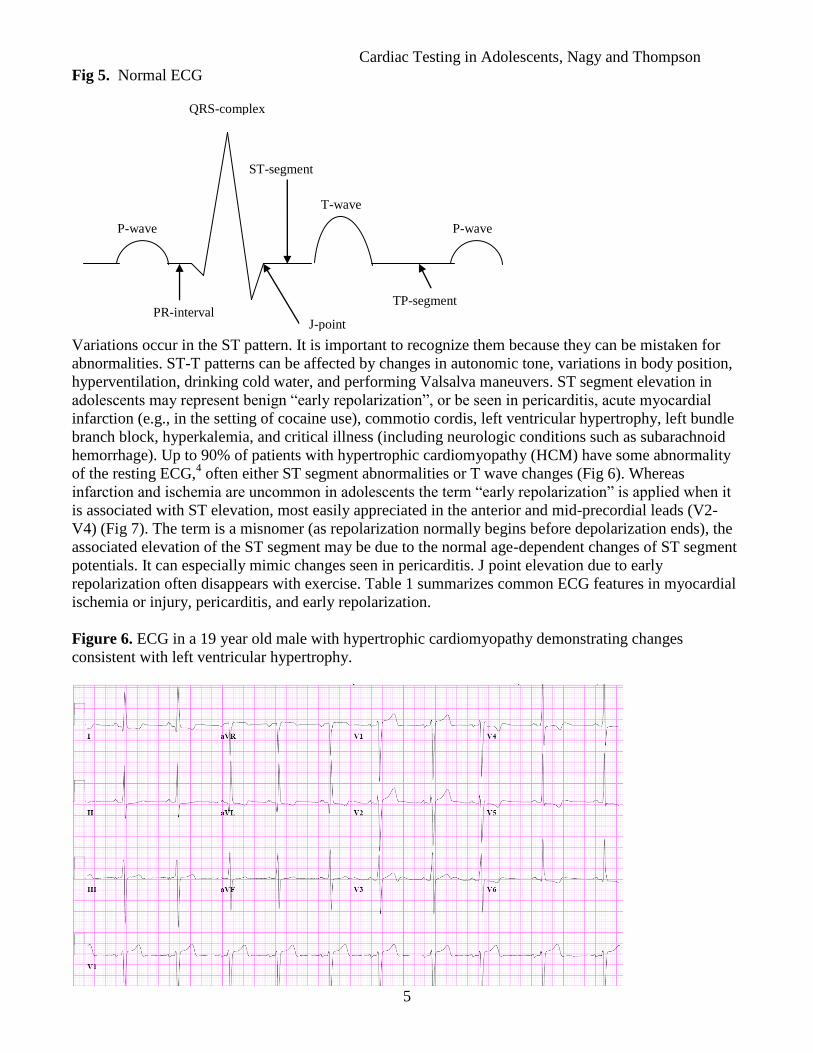
Figure 6. ECG in a 19 year old male with hypertrophic cardiomyopathy demonstrating changes
consistent with left ventricular hypertrophy.
J-point
P-wave
QRS-complex
T-wave
ST-segment
TP-segment PR-interval
P-wave
Cardiac Testing in Adolescents, Nagy and Thompson
6
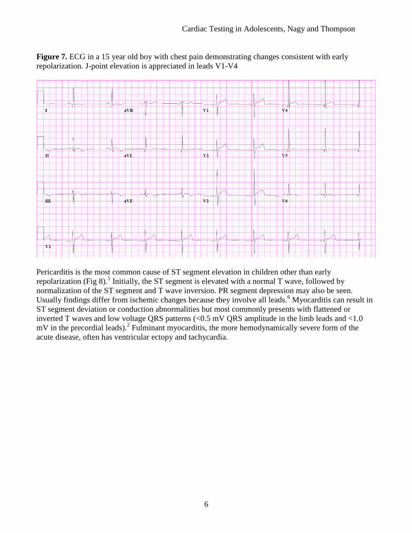
Figure 7. ECG in a 15 year old boy with chest pain demonstrating changes consistent with early
repolarization. J-point elevation is appreciated in leads V1-V4
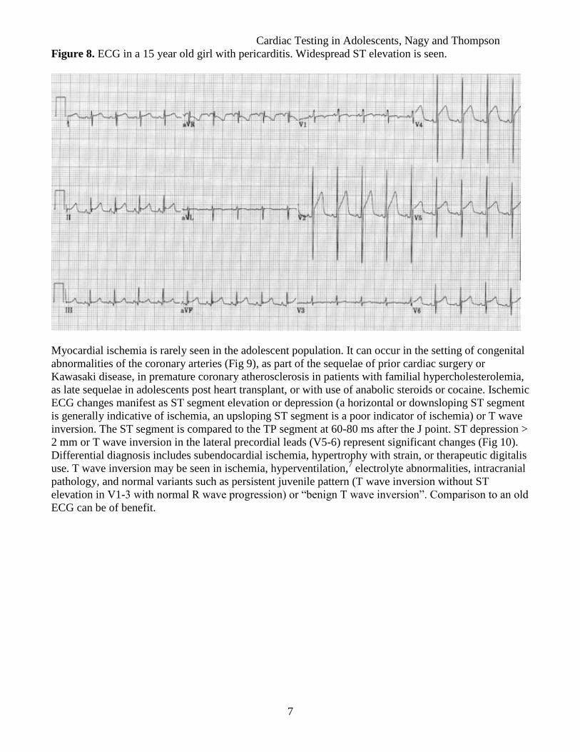
Pericarditis is the most common cause of ST segment elevation in children other than early
repolarization (Fig 8).5 Initially, the ST segment is elevated with a normal T wave, followed by
normalization of the ST segment and T wave inversion. PR segment depression may also be seen.
Usually findings differ from ischemic changes because they involve all leads.6 Myocarditis can result in
ST segment deviation or conduction abnormalities but most commonly presents with flattened or
inverted T waves and low voltage QRS patterns (<0.5 mV QRS amplitude in the limb leads and <1.0
mV in the precordial leads).2 Fulminant myocarditis, the more hemodynamically severe form of the
acute disease, often has ventricular ectopy and tachycardia.
Cardiac Testing in Adolescents, Nagy and Thompson
7
Figure 8. ECG in a 15 year old girl with pericarditis. Widespread ST elevation is seen.
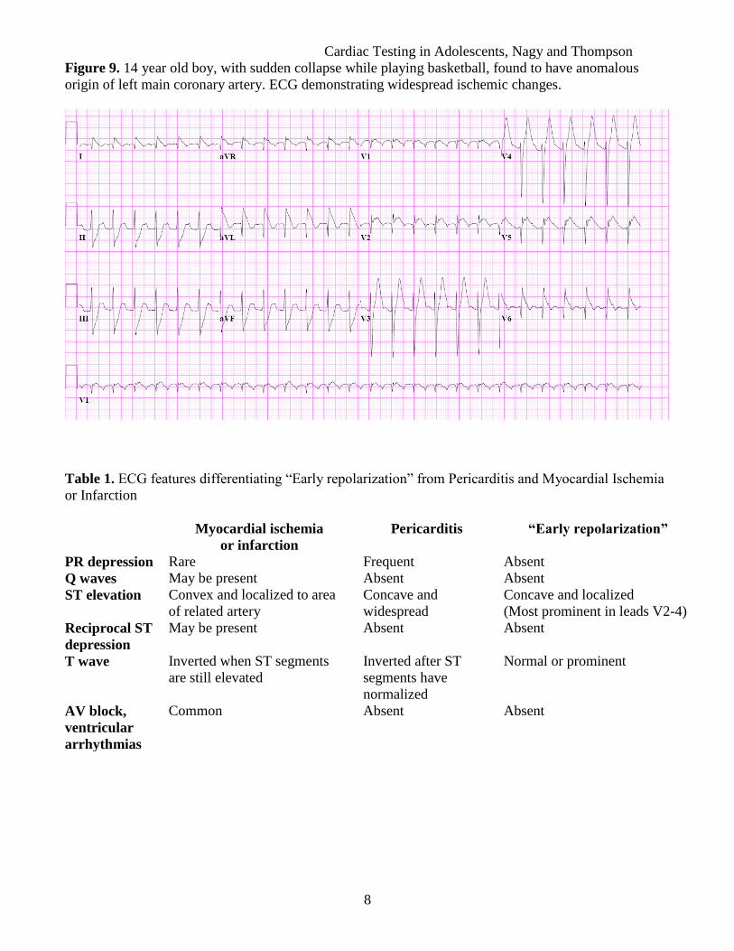
Myocardial ischemia is rarely seen in the adolescent population. It can occur in the setting of congenital
abnormalities of the coronary arteries (Fig 9), as part of the sequelae of prior cardiac surgery or
Kawasaki disease, in premature coronary atherosclerosis in patients with familial hypercholesterolemia,
as late sequelae in adolescents post heart transplant, or with use of anabolic steroids or cocaine. Ischemic
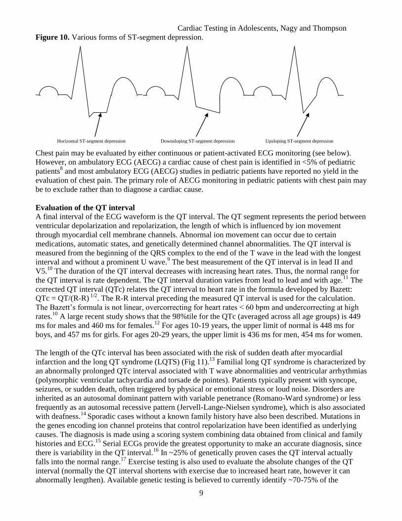
ECG changes manifest as ST segment elevation or depression (a horizontal or downsloping ST segment
is generally indicative of ischemia, an upsloping ST segment is a poor indicator of ischemia) or T wave
inversion. The ST segment is compared to the TP segment at 60-80 ms after the J point. ST depression >
2 mm or T wave inversion in the lateral precordial leads (V5-6) represent significant changes (Fig 10).
Differential diagnosis includes subendocardial ischemia, hypertrophy with strain, or therapeutic digitalis
use. T wave inversion may be seen in ischemia, hyperventilation,7 electrolyte abnormalities, intracranial
pathology, and normal variants such as persistent juvenile pattern (T wave inversion without ST
elevation in V1-3 with normal R wave progression) or “benign T wave inversion”. Comparison to an old
ECG can be of benefit.
Cardiac Testing in Adolescents, Nagy and Thompson
8
Figure 9. 14 year old boy, with sudden collapse while playing basketball, found to have anomalous
origin of left main coronary artery. ECG demonstrating widespread ischemic changes.
Table 1. ECG features differentiating “Early repolarization” from Pericarditis and Myocardial Ischemia
or Infarction
Myocardial ischemia
or infarction
Pericarditis “Early repolarization”
PR depression Rare Frequent Absent
Q waves May be present Absent Absent
ST elevation Convex and localized to area
of related artery
Concave and
widespread
Concave and localized
(Most prominent in leads V2-4)
Reciprocal ST
depression
May be present Absent Absent
T wave Inverted when ST segments
are still elevated
Inverted after ST
segments have
normalized
Normal or prominent
AV block,
ventricular
arrhythmias
Common Absent Absent
Cardiac Testing in Adolescents, Nagy and Thompson
9
Figure 10. Various forms of ST-segment depression.
Chest pain may be evaluated by either continuous or patient-activated ECG monitoring (see below).
However, on ambulatory ECG (AECG) a cardiac cause of chest pain is identified in <5% of pediatric
patients8 and most ambulatory ECG (AECG) studies in pediatric patients have reported no yield in the
evaluation of chest pain. The primary role of AECG monitoring in pediatric patients with chest pain may
be to exclude rather than to diagnose a cardiac cause.
Evaluation of the QT interval
A final interval of the ECG waveform is the QT interval. The QT segment represents the period between
ventricular depolarization and repolarization, the length of which is influenced by ion movement
through myocardial cell membrane channels. Abnormal ion movement can occur due to certain
medications, automatic states, and genetically determined channel abnormalities. The QT interval is
measured from the beginning of the QRS complex to the end of the T wave in the lead with the longest
interval and without a prominent U wave.9 The best measurement of the QT interval is in lead II and
V5.10
The duration of the QT interval decreases with increasing heart rates. Thus, the normal range for
the QT interval is rate dependent. The QT interval duration varies from lead to lead and with age.11
The
corrected QT interval (QTc) relates the QT interval to heart rate in the formula developed by Bazett:
QTc = QT/(R-R) 1/2
. The R-R interval preceding the measured QT interval is used for the calculation.
The Bazett’s formula is not linear, overcorrecting for heart rates < 60 bpm and undercorrecting at high
rates.10
A large recent study shows that the 98%tile for the QTc (averaged across all age groups) is 449
ms for males and 460 ms for females.12
For ages 10-19 years, the upper limit of normal is 448 ms for
boys, and 457 ms for girls. For ages 20-29 years, the upper limit is 436 ms for men, 454 ms for women.
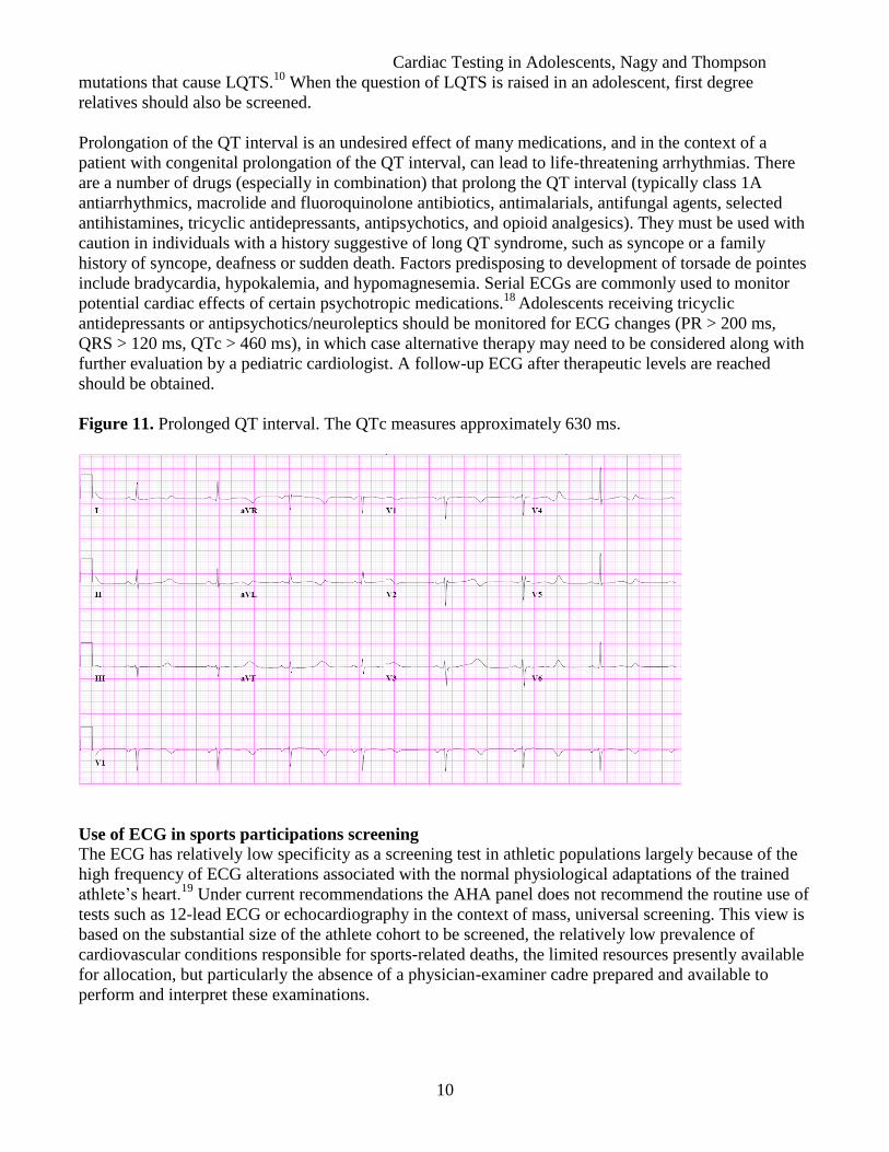
The length of the QTc interval has been associated with the risk of sudden death after myocardial
infarction and the long QT syndrome (LQTS) (Fig 11).13
Familial long QT syndrome is characterized by
an abnormally prolonged QTc interval associated with T wave abnormalities and ventricular arrhythmias
(polymorphic ventricular tachycardia and torsade de pointes). Patients typically present with syncope,
seizures, or sudden death, often triggered by physical or emotional stress or loud noise. Disorders are
inherited as an autosomal dominant pattern with variable penetrance (Romano-Ward syndrome) or less
frequently as an autosomal recessive pattern (Jervell-Lange-Nielsen syndrome), which is also associated
with deafness.14
Sporadic cases without a known family history have also been described. Mutations in
the genes encoding ion channel proteins that control repolarization have been identified as underlying
causes. The diagnosis is made using a scoring system combining data obtained from clinical and family
histories and ECG.15
Serial ECGs provide the greatest opportunity to make an accurate diagnosis, since
there is variability in the QT interval.16
In ~25% of genetically proven cases the QT interval actually
falls into the normal range.17
Exercise testing is also used to evaluate the absolute changes of the QT
interval (normally the QT interval shortens with exercise due to increased heart rate, however it can
abnormally lengthen). Available genetic testing is believed to currently identify ~70-75% of the
Downsloping ST-segment depression Upsloping ST-segment depression Horizontal ST-segment depression
Cardiac Testing in Adolescents, Nagy and Thompson
10
mutations that cause LQTS.10
When the question of LQTS is raised in an adolescent, first degree
relatives should also be screened.
Prolongation of the QT interval is an undesired effect of many medications, and in the context of a
patient with congenital prolongation of the QT interval, can lead to life-threatening arrhythmias. There
are a number of drugs (especially in combination) that prolong the QT interval (typically class 1A
antiarrhythmics, macrolide and fluoroquinolone antibiotics, antimalarials, antifungal agents, selected
antihistamines, tricyclic antidepressants, antipsychotics, and opioid analgesics). They must be used with
caution in individuals with a history suggestive of long QT syndrome, such as syncope or a family
history of syncope, deafness or sudden death. Factors predisposing to development of torsade de pointes
include bradycardia, hypokalemia, and hypomagnesemia. Serial ECGs are commonly used to monitor
potential cardiac effects of certain psychotropic medications.18
Adolescents receiving tricyclic
antidepressants or antipsychotics/neuroleptics should be monitored for ECG changes (PR > 200 ms,
QRS > 120 ms, QTc > 460 ms), in which case alternative therapy may need to be considered along with
further evaluation by a pediatric cardiologist. A follow-up ECG after therapeutic levels are reached
should be obtained.
Figure 11. Prolonged QT interval. The QTc measures approximately 630 ms.
Use of ECG in sports participations screening
The ECG has relatively low specificity as a screening test in athletic populations largely because of the
high frequency of ECG alterations associated with the normal physiological adaptations of the trained
athlete’s heart.19
Under current recommendations the AHA panel does not recommend the routine use of
tests such as 12-lead ECG or echocardiography in the context of mass, universal screening. This view is
based on the substantial size of the athlete cohort to be screened, the relatively low prevalence of
cardiovascular conditions responsible for sports-related deaths, the limited resources presently available
for allocation, but particularly the absence of a physician-examiner cadre prepared and available to
perform and interpret these examinations.
Cardiac Testing in Adolescents, Nagy and Thompson
11
2. HOLTER AND EVENT MONITORING
Holter, or ambulatory ECG (AECG) monitoring in the adolescent patient is indicated for evaluation of
symptoms that may be arrhythmia related, risk assessment in patients with cardiovascular disease, with
or without symptoms of an arrhythmia, and evaluation of cardiac rhythm after an intervention such as
drug therapy or device implantation.
In adolescents with frequent symptoms possibly related to an arrhythmia such as palpitations,
presyncope, syncope, dizziness, or chest pain, AECG is an excellent tool in helping to correlate an
arrhythmia with symptoms.20
In contrast to the standard 12-lead ECG, which captures a brief period of
time (a few seconds), the ambulatory monitor records the cardiac rhythm over a prolonged period of
time, usually 24-48 hours. It is an ideal test for patients with frequent (at least daily) arrhythmias and
provides information on the frequency of occurrence, related symptoms, and potential exacerbating
factors. An activity diary can aid in correlating symptoms to ECG findings. Between 25-50% of patients
will have complaints of symptoms during the time wearing a Holter monitor. Of these patients 2-15%
will have a causal arrhythmia.20,21
The second type of ambulatory monitoring is the event recorder.
There are multiple technologies available. They can be broadly categorized as event recorders, loop
recorders, or implantable long-term recorders. The event recorder typically records the electrogram on a
continuous tape. Only the last 30-90 seconds are available for playback. When symptoms occur, the
patient can stop the tape by pressing a button and transmit the information on the tape via telephone.
This type of testing is best ordered for relatively infrequent symptoms that have been difficult to
document. This monitor is carried for an extended period of time (usually several weeks), until a
symptomatic episode is captured. More sophisticated loop devices can store presymptomatic,
symptomatic, and postsymptomatic arrhythmias for a period of several minutes when activated. The
diagnostic yield of such recorders is up to 60% in individuals with intermittent symptoms. The third
option is an implantable rhythm monitor, a tiny micro-processor based device placed under the skin,
which has the ability to record a patient’s ECG for weeks to months. The information can be
downloaded periodically and reviewed. This type of device is usually ordered and implanted by
electrophysiologists.
A patient-activated recorder is generally recommended for the evaluation of palpitations because of the
paroxysmal nature of the symptom. An arrhythmia, usually supraventricular tachycardia, has been
reported to correlate with palpitation in 10-15% of young patients, whereas ventricular ectopy or
bradycardia is demonstrated in another 2-5%. Sinus tachycardia is identified in nearly 50% of young
patients with symptoms of palpitation during ambulatory monitoring, whereas 30-40% of patients have
no symptoms during monitoring.20
One of the primary uses of ambulatory ECG monitoring in
adolescents is to exclude an arrhythmia as the cause of palpitation. The intermittent nature of symptoms
results in a low efficacy of 24-48 hours of continuous ECG monitoring; conversely, temporary patient
incapacitation usually precludes patient-activated recording. Continuous ECG monitoring is primarily
indicated in pediatric patients with exertional symptoms or those with known heart disease, in whom the
presence and significance of an arrhythmia may be increased.
Ambulatory ECG monitoring is commonly used in the periodic evaluation of pediatric patients with
heart disease (Fig 12), with or without symptoms of an arrhythmia. The rationale for this testing is the
evolution of disease processes (such as long QT syndromes or hypertrophic cardiomyopathies), growth
of patients and the need to adjust medication dosages, and the progressive onset of late arrhythmias after
surgery for congenital heart defects. The use of AECG monitoring for periodic evaluation of patients
with prior surgical treatment of congenital heart disease must be based on consideration of the type of
defect, ventricular function, and risk of late postoperative arrhythmias. For example, uncomplicated
repairs of atrial or ventricular septal defects are associated with low incidence of late postoperative
Cardiac Testing in Adolescents, Nagy and Thompson
12
arrhythmias. Complex repairs or those with residual hemodynamic abnormalities have a higher
incidence of late-onset atrial and ventricular arrhythmias. Although the significance of arrhythmias in
these patients remains controversial, high-grade ambulatory ventricular ectopy associated with
ventricular dysfunction does appear to identify patients at an increased risk of late sudden death.
Complex arrhythmias detected in these patients by AECG may indicate the need for further investigation
or intervention, even in the absence of overt symptoms.
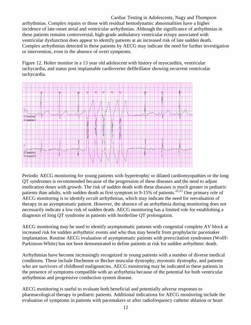
Figure 12. Holter monitor in a 13 year old adolescent with history of myocarditis, ventricular
tachycardia, and status post implantable cardioverter defibrillator showing recurrent ventricular
tachycardia.
Periodic AECG monitoring for young patients with hypertrophic or dilated cardiomyopathies or the long
QT syndromes is recommended because of the progression of these diseases and the need to adjust
medication doses with growth. The risk of sudden death with these diseases is much greater in pediatric
patients than adults, with sudden death as first symptom in 9-15% of patients.22,23
One primary role of
AECG monitoring is to identify occult arrhythmias, which may indicate the need for reevaluation of
therapy in an asymptomatic patient. However, the absence of an arrhythmia during monitoring does not
necessarily indicate a low risk of sudden death. AECG monitoring has a limited role for establishing a
diagnosis of long QT syndrome in patients with borderline QT prolongation.
AECG monitoring may be used to identify asymptomatic patients with congenital complete AV block at
increased risk for sudden arrhythmic events and who thus may benefit from prophylactic pacemaker
implantation. Routine AECG evaluation of asymptomatic patients with preexcitation syndromes (Wolff-
Parkinson-White) has not been demonstrated to define patients at risk for sudden arrhythmic death.
Arrhythmias have become increasingly recognized in young patients with a number of diverse medical
conditions. These include Duchenne or Becker muscular dystrophy, myotonic dystrophy, and patients
who are survivors of childhood malignancies. AECG monitoring may be indicated in these patients in
the presence of symptoms compatible with an arrhythmia because of the potential for both ventricular
arrhythmias and progressive conduction system disease.
AECG monitoring is useful to evaluate both beneficial and potentially adverse responses to
pharmacological therapy in pediatric patients. Additional indications for AECG monitoring include the
evaluation of symptoms in patients with pacemakers or after radiofrequency catheter ablation or heart

Cardiac Testing in Adolescents, Nagy and Thompson
13
surgery, particularly when complicated by transient AV block. AECG monitoring is also indicated for
the evaluation of cardiac rhythm after treatment of incessant tachyarrhythmias, which have been
associated with progressive ventricular dysfunction.
3. STRESS TESTING
Reasons for stress testing in the young include investigating exercise-related symptoms, evaluating the
stress of exercise on known cardiac conditions, and assessing the effectiveness of medications (e.g., beta
blockade effect on heart rate). In general, these studies are most appropriately ordered and supervised by
the cardiologist with expertise in adolescent heart disease. Exercise testing is rarely needed to look for
occult coronary obstructions in the pediatric population. Exercise capacity is diminished in some
adolescents with heart disease, and measurement is often useful in evaluating subjective limitations.24
Exercise testing of children and adolescents has a very low risk compared with testing of adults.
Complications of pediatric exercise testing are extremely infrequent, even when testing is done in
populations of children with congenital cardiac defects and arrhythmias. Indications for exercise stress
testing have been published in specific guidelines.24,25
Several different methods of stress testing are available including treadmill or bicycle exercise with
ECG monitoring, imaging by echocardiography during or just after exercise, or pharmacologic
simulation of stress (e.g., using dobutamine infusion) with imaging by echo or nuclear methods.
Treadmill protocols are relatively simple for the patient, simulate typical activities engaged in
(representing a more physiological stress), and provide important information about peak endurance,
respiratory health, overall fitness, blood pressure response, as well as cardiac electrical activity.
However, confounders may include lack of cooperation, poor coordination, or unfamiliarity with the
procedure, especially in younger patients. Stress tests should be ordered in consideration of the pretest
probability of the presence of a specific disease process, since sensitivity and specificity will need to be
interpreted in this context.
Evaluation of exercise-induced symptoms
Though chest pain is common in children and adolescents, the history and physical examination are
generally adequate to exclude serious pathology and provide reassurance to patient and family. Non
cardiac chest pain is usually described as a brief stabbing or shooting pain that occurs with or without
exercise. Pleuritic pain is common. Typical etiologies include gastroesophageal reflux, exercise-induced
reactive airways disease, costochondritis, or anxiety. Routine use of exercise testing for evaluation of
chest pain in children and adolescents is not required. Exercise testing is appropriate for evaluation of
the uncommon child with chest pain that is typical angina by description and consistently related to
exercise. Exercise-induced bronchospasm is best identified by pulmonary function tests.
Exercise testing is an important component of evaluation of adolescents who have unexplained syncope
related to physical exertion. Features of syncope that suggest a potentially life-threatening cardiac event
include an abrupt loss of consciousness (as opposed to a prodrome or aura), injury on impact (as
opposed to a gradual “slump”), and syncope related to physical activity. Evaluation for possible
arrhythmia, left ventricular outflow obstruction, and cardiomyopathy is appropriate. Neurally-mediated
hypotension may rarely manifest primarily as exercise-induced syncope, most often with symptoms
occurring immediately upon cessation of the activity.
Premature atrial contractions are common and benign in young persons. Exercise test evaluation of
premature atrial contractions in adolescents is not required. Exercise testing helps in evaluation of
selected cases in which the history suggests an exercise-related tachycardia. The majority of children
with supraventricular tachycardia however will not have exercise-induced tachycardia.
Cardiac Testing in Adolescents, Nagy and Thompson
14
Isolated premature ventricular depolarizations in asymptomatic children and adolescents usually
disappear with the higher heart rates associated with exercise. It is not necessary to perform formal
laboratory testing to demonstrate this. Exercise testing is often of value in diagnosing ventricular
tachycardia (VT) and assessing the efficacy of treatment. VT with exercise is often observed in children
with no structural heart abnormality as well as in children with myocarditis, cardiomyopathy, or a
congenital cardiac malformation. Arrhythmogenic right ventricular dysplasia (ARVD) is an inherited
cardiomyopathy particularly related to development of VT with exercise. Measurement of shortening or
prolongation of the corrected QT interval to exercise has been used as an adjunct in the diagnosis of long
QT syndromes.
4. TILT TABLE TESTING
Tilt table testing is used primarily to diagnose or confirm suspicion of neurally-mediated hypotension in
patients with syncope or near syncope. The test, while relatively simple to perform, is somewhat
uncomfortable for the patient and is thus usually reserved for situations in which the diagnosis is unclear
from history and physical exam alone, or in which response to empiric therapy has been limited.
Although protocols used in performing tilt table testing vary among centers, most laboratories tilt
patients for 15-45 minutes at an angle of 60-85 degrees. After baseline parameters are measured the
patient is secured to a table and tilted. Normally, individuals compensate for such a tilt by increasing
both α- and β-adrenergic tone as a result of baroreceptor stimulation, thus compensating for the decrease
in venous return. In susceptible individuals, these compensatory mechanisms eventually collapse, and
venous return is never completely compensated. As a result sympathetic tone increases producing
vigorous ventricular contractions of a relatively empty heart. This results in recruitment of cardiac C
fibers, which causes stimulation of the medullary vasodepressor region. The result is a sudden
withdrawal of sympathetic tone, a sudden increase in vagal tone, vasodilation, and syncope. If the test is
negative, the table is lowered in the original horizontal position, and an intravenous infusion of
isoproterenol started and the dose adjusted to increase the baseline heart rate to > 20%. The tilt table test
is repeated for 15-20 minutes. Syncopal episodes have been shown to be preceded by a catecholamine
surge. The addition of isoproterenol increases the sensitivity of the tilt table test.
Questions about the sensitivity, specificity, diagnostic yield, and day-to-day reproducibility of tilt table
testing have been raised in the most recent AHA/ACCF scientific statement on the evaluation of
syncope.26
The reported sensitivity and specificity of tilt table testing depend on the technique used.
Sensitivity ranges from 26-80%, and specificity is approximately 90%. The statement argues that in
patients with a negative evaluation, (i.e. no evidence of ischemia and a structurally normal heart), the
pretest probability that the diagnosis is neurocardiogenic syncope is high; hence head-up tilt table testing
contributes little to establishing the diagnosis.
Evaluation of syncope
Syncope is common in adolescents as up to 47% of college students report having fainted.27
Syncope is
a disabling condition that requires attention, but is generally not life-threatening. It can be frightening to
patients, families, and primary care providers. If cardiac or neurologic abnormalities are not apparent
after a thorough history and physical examination has been performed, and blood work, ECG, and
echocardiogram are normal, tilt table testing is often used as an aid in establishing the diagnosis of
neurally mediated hypotension (NMH) and postural orthostatic tachycardia syndrome (POTS).
NMH is defined by a drop in systolic blood pressure of > 25 mmHg (compared to the BP measured
when the person lies flat) during standing or upright tilt table testing.28
POTS is defined by an
exaggerated increase in heart rate with standing. A healthy teenager usually has a slight increase in heart
rate by about 10-15 beats per minute within the first 10 minutes of standing. POTS is considered present
Cardiac Testing in Adolescents, Nagy and Thompson
15
if the HR increases > 30 beats per minute (or if it reaches 120 beats per minute or higher) over the first
10 minutes of standing.29
Some patients with POTS in the first 10 minutes of standing or tilt testing will
go on to develop NMH if the test is continued. Different response patterns to tilt table testing in normal
individuals, as well as in NMH, POTS, dysautonomia, and psychogenic syncope are listed in Table 2
(Adapted form Feinberg).27
Table 2. Response Patterns to Tilt Table Test
Pattern Blood Pressure Pulse Response
Normal No change, or slight increase No change, or slight increase No symptoms
NMH Rapid decrease Decrease Presyncope/Syncope
POTS No change or decrease Increase Presyncope
Dysautonomia Gradual decrease No change or increase Presyncope/Syncope
Psychogenic No change, or slight increase No change, or slight increase Presyncope/Syncope
5. ECHOCARDIOGRAPHY
Echocardiography is the primary tool of the cardiologist for diagnosing structural heart disease and is
highly accurate when performed and interpreted in an experienced laboratory. However, screening for
heart disease, especially in the adolescent and young adult, is still more appropriately done by careful
history and physical examination. The more important role for echocardiography in an adolescent is in
fully characterizing a cardiac lesion once an abnormality is suspected. It also provides essential
information concerning the natural history of the abnormality and responses to medical and surgical
management. Transthoracic echocardiogram is a reliable and versatile tool for the assessment of cardiac
structure, function, and pathophysiology. It is associated with little if any patient discomfort, and no
risks. Because it depends on obtaining satisfactory examining windows from the body surface to the
cardiovascular structures, there may be limitations to its use. In the
obese adolescent, the interposition of
adipose tissue between body surface and the heart can limit image quality, and complete examination
may not be possible. Echocardiographic contrast agents (administered through a peripheral access line)
that can pass through the pulmonary circulation and opacify the left heart have been developed and
become a useful aid in the evaluation of obese patients. For specific indications (i.e. evaluation for
endocarditis or intracardiac thrombus), transesophageal echocardiography is an excellent tool for further
assessment. The echocardiographic transducer is mounted on a flexible endoscope and passed into the
esophagus and stomach. However, this technique is invasive and requires sedation and intubation.
Indications for the performance of an adolescent echocardiogram span a wide range of symptoms and
signs, including exercise induced chest pain or syncope, murmurs, respiratory distress, abnormal arterial
pulses, and cardiomegaly, which may suggest structural heart disease. An echocardiogram is indicated
for the evaluation of acquired heart diseases in children, including rheumatic fever and carditis, infective
endocarditis, HIV infection, myocarditis, pericarditis, follow-up for Kawasaki disease, all forms of
cardiomyopathies (Fig 13), systemic lupus erythematosus, renal disease, and connective tissue diseases
with known cardiovascular manifestations (Marfan’s syndrome, Loeys-Dietz syndrome). Patients
receiving anthracycline or other cardiotoxic agents should have baseline and reevaluation
follow-up
studies. Pediatric echocardiography is indicated in the assessment of potential cardiac or
cardiopulmonary transplant donors and transplant recipients. Echocardiography has been recommended
in all children who are newly diagnosed with systemic hypertension.30
Noncardiac disease states
affecting the heart such as pulmonary hypertension constitute an important indication for serial pediatric
echocardiograms. Echocardiography may also be indicated in adolescents with thromboembolic events
(i.e. sickle cell disease). Elevated right ventricular and pulmonary artery pressure, as estimated by echo-
Doppler evaluation of tricuspid regurgitation correlates with increase mortality risk in patients with
sickle cell disease.31
Children with arrhythmias may have previously undiagnosed structural cardiac
disease such as congenitally corrected transposition, Ebstein’s anomaly of the tricuspid valve, or
Cardiac Testing in Adolescents, Nagy and Thompson
16
cardiomyopathy, which may be associated with subtle clinical findings and are best evaluated using
echocardiography. Sustained arrhythmias or antiarrhythmic medications may lead to functional
impairment of the heart that may only be detectable by echocardiography and have important
implications for management.
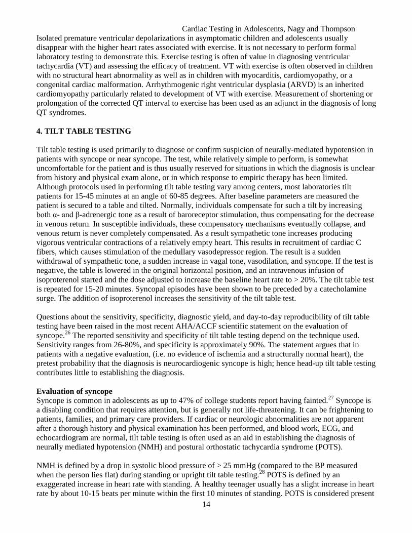
Figure 13. Echocardiogram of a 19 year young adult demonstrating hypertrophic cardiomyopathy (Ao =
aorta, LV=left ventricle).
Practice guidelines for the clinical application of echocardiography have been published by the
ACC/AHA.32,33,34,35
Cardiovascular disease in the adolescent includes anomalies of cardiac anatomy, function, and rhythm.
While problems often present as an asymptomatic heart murmur, the cardiac murmurs of this age group
are more commonly functional than pathological. The contribution of echocardiography to
the evaluation
of an asymptomatic patient with this finding on routine examination by an experienced clinician is
limited. History and skilled physical examination are usually sufficient to distinguish functional from
pathological murmurs and are more cost-effective than referral for an echocardiogram.
36 However, in the
presence of ambiguous clinical findings, echocardiography can demonstrate the presence or absence of
abnormalities such as an interatrial septal defect, bicuspid aortic valve, mildly
obstructive subaortic
stenosis, mitral valve prolapse, aortic aneurysm, or functionally occult cardiomyopathy.
6. CXR/MRI/CT/PET
The chest x-ray is an important clinical tool in the cardiovascular evaluation. In many cases heart
disease is associated with cardiomegaly. A quantitative estimate of heart size may be obtained by
determining the cardiothoracic ratio, which is calculated by dividing the maximal transverse diameter of
the heart in the postero-anterior view by the width of the thoracic cavity. As a general rule, the heart is
enlarged if the cardiothoracic ratio is greater then 0.5. Whereas the cardiothoracic ratio is useful in the
Cardiac Testing in Adolescents, Nagy and Thompson
17
detection of cardiomegaly in cases of left ventricular enlargement or pericardial effusion, it is not as
sensitive in the assessment of right ventricular enlargement.
Cardiovascular magnetic resonant imaging (MRI) is a well established diagnostic imaging technique
with emerging roles in the evaluation of cardiovascular disease. Its ability to acquire high-resolution
images in virtually any plane, to make accurate measurements of blood velocity flow and cardiac
volumes, to perform noninvasive angiography, and to assess myocardial mechanics and perfusion, all in
the absence of ionizing radiation, promotes its versatility. Indications include, but are not limited to
evaluation of congenital heart disease, evaluation of aortic disease, assessment of intrinsic non-ischemic
myocardial disease, assessment of ischemic heart disease, assessment of valvular heart disease,
evaluation of the pericardium, evaluation of cardiac masses, and assessment of pulmonary arteries.
Contraindications to the use of this technique are mainly related to electrically, magnetically, or
mechanically active implants in the patient’s body.
Indications for cardiovascular computed tomography (CT) have rapidly expanded over the last years,
mainly secondary to technical improvements allowing faster scanning with increased special resolution.
Clinical indications range from assessment of the great vessels to noninvasive imaging of the coronary
arteries (Fig 14). Cardiac CT is a non-invasive alternative test to cardiac catheterization. Current
technologies include electron beam CT (EBCT) and multiple detector CT (MDCT). EBCT involves the
use of a rapidly oscillating electron beam reflected onto a stationary target. It has a high temporal
resolution and the slice thickness is 1.5-3 mm, covering the entire heart in one or two breath-hold
periods. MDCT involves the use of a mechanically rotated x-ray tube at high speed. This type of scanner
is used for non-cardiac and cardiac imaging with minimal slice thickness of 0.75-1 mm, covering the
entire heart in a single breath-hold. Clinical indications include coronary artery assessment (calcium
scoring, CT angiography), cardiac chamber assessment, evaluation of the great vessels, evaluation of
congenital heart disease, and assessment of pericardial disease. The downside of this technique remains
the need for radiation. Whereas the average risk for radiation-induced cancer in the general population is
estimated at 5% per 1 Sv (Sievert), in children and adolescents the risk is predicted to be up to 2-3 times
higher than in adults (as high as 15% per 1 Sv).37
The typical effective radiation dose for a cardiac CT
angiography is estimated to be in the order of 10-25 mSv. In comparison, the typical effective radiation
dose for a chest x-ray equals 0.1-0.2 mSv.38
Cardiac Testing in Adolescents, Nagy and Thompson
18
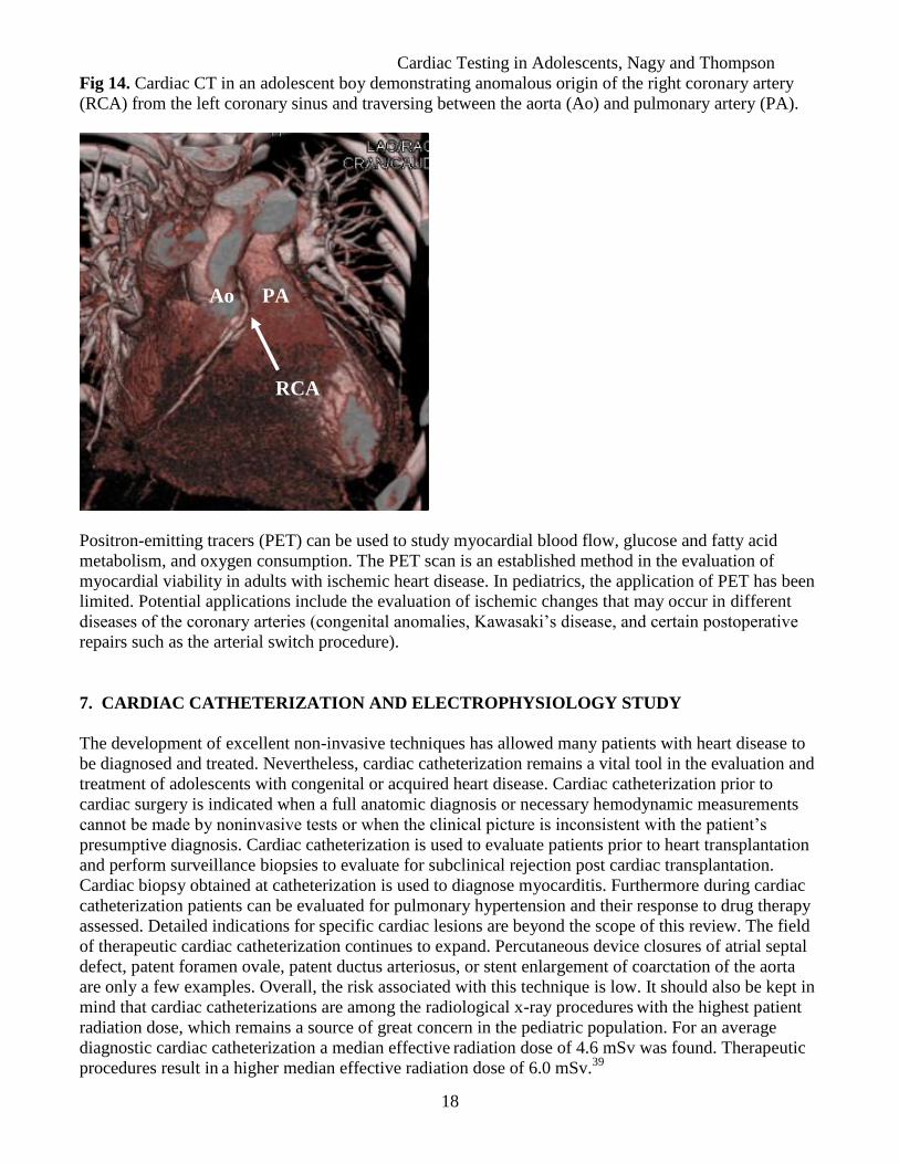
Fig 14. Cardiac CT in an adolescent boy demonstrating anomalous origin of the right coronary artery
(RCA) from the left coronary sinus and traversing between the aorta (Ao) and pulmonary artery (PA).
Positron-emitting tracers (PET) can be used to study myocardial blood flow, glucose and fatty acid
metabolism, and oxygen consumption. The PET scan is an established method in the evaluation of
myocardial viability in adults with ischemic heart disease. In pediatrics, the application of PET has been
limited. Potential applications include the evaluation of ischemic changes that may occur in different
diseases of the coronary arteries (congenital anomalies, Kawasaki’s disease, and certain postoperative
repairs such as the arterial switch procedure).
7. CARDIAC CATHETERIZATION AND ELECTROPHYSIOLOGY STUDY
The development of excellent non-invasive techniques has allowed many patients with heart disease to
be diagnosed and treated. Nevertheless, cardiac catheterization remains a vital tool in the evaluation and
treatment of adolescents with congenital or acquired heart disease. Cardiac catheterization prior to
cardiac surgery is indicated when a full anatomic diagnosis or necessary hemodynamic measurements
cannot be made by noninvasive tests or when the clinical picture is inconsistent with the patient’s
presumptive diagnosis. Cardiac catheterization is used to evaluate patients prior to heart transplantation
and perform surveillance biopsies to evaluate for subclinical rejection post cardiac transplantation.
Cardiac biopsy obtained at catheterization is used to diagnose myocarditis. Furthermore during cardiac
catheterization patients can be evaluated for pulmonary hypertension and their response to drug therapy
assessed. Detailed indications for specific cardiac lesions are beyond the scope of this review. The field
of therapeutic cardiac catheterization continues to expand. Percutaneous device closures of atrial septal
defect, patent foramen ovale, patent ductus arteriosus, or stent enlargement of coarctation of the aorta
are only a few examples. Overall, the risk associated with this technique is low. It should also be kept in
mind that cardiac catheterizations are among the radiological x-ray procedures with the highest patient
radiation dose, which remains a source of great concern in the pediatric population. For an average
diagnostic cardiac catheterization a median effective radiation dose of 4.6 mSv was found. Therapeutic
procedures result in a higher median effective radiation dose of 6.0 mSv.
39
PA Ao
RCA
Cardiac Testing in Adolescents, Nagy and Thompson
19
The electrophysiology study is a specialized form of cardiac catheterization that can be helpful in
evaluating a broad spectrum of cardiac arrhythmias. It can assess the function of the sinus node, the
atrioventricular node, and the His-Purkinje system. It can also determine the mechanism and
characteristics of tachyarrhythmias and locate reentrant circuits (accessory pathways). Finally, it can
evaluate the efficacy of antiarrhythmic medication and devices. During the procedure multiple
electrodes are placed in the heart recording the electrical signals from the atria, atrioventricular node,
and ventricles. Pacing from localized areas within the heart is used to induce the arrhythmia to be
studied. If an amenable tachycardia is identified, an ablation procedure can be performed, thus
interrupting the aberrant electrical pathways. Ablation techniques use heat (radio-frequency) or cold
(cryoablation) to produce thermal damage to the myocardial tissue. Procedures can be repeated for
recurrences. Specific guidelines for intracardiac electrophysiological and catheter ablation procedures
have been published.40
REFERENCES
1 Schlant RC, Adolph RJ, DiMarco JP, Dreifus LS, Dunn MI, Fisch C, Garson A Jr, Haywood LJ, Levine HJ and Murray JA .
Guidelines for electrocardiography. A report of the American College of Cardiology/American Heart Association Task Force
on Assessment of Diagnostic and Therapeutic Cardiovascular Procedures (Committee on Electrocardiography). Circulation
1992;85:1221-1228.
2 Kadish AH, Buxton AE, Kennedy HL, Knight BP, Mason JW, Schuger CD, Tracy CM, Winters WL, Boone AW, Elnicki
M, Hirshfeld JW Jr, Lorell BH, Rodgers GP, and Weitz HH. ACC/AHA clinical competence statement on
electrocardiography and ambulatory electrocardiography: A report of the American College of Cardiology/American Heart
Association/American College of Physicians-American Society of Internal Medicine Task Force on Clinical Competence
(ACC/AHA Committee to Develop a Clinical Competence Statement on Electrocardiography and Ambulatory
Electrocardiography); Circulation 2001;104:3169-3178.
3 Estes MNA. Diagnostic Techniques. In: Lewis RP, O’Gara PT, Hirsch G. Adult Clinical Cardiology Self-Assessment
Program 6. Bethesda: American College of Cardiology Foundation, 2005:15.11-15.23.
4 Maron BJ. The electrocardiogram as a diagnostic tool for hypertrophic cardiomyopathy: revisited. Ann Noninvasive
Electrocardiol. 2001;6:277-279.
5 Van Hare GF, Dubin A. The normal electrocardiogram. In: Allen HD, Gutgesell HP, Clark EB, Driscoll DJ, eds. Moss and
Adams’ heart disease in infants, children, and adolescents, 6th
ed. Philadelphia: Lippincott Williams & Wilkins, 2001:425-
442.
6 Spodick DH. Electrocardiogram in acute pericarditis. Am J Cardiol 1974;33:470-474.
7 Wasserburger RH, Alt WJ, Lloyd CJ. The normal RS-T segment elevation variant. Am J Cardiol 1961;8:184-192.
8 Selbst SM, Ruddy RM, Clark BJ, Henretig FM, Santulli T Jr. Pediatric chest pain: a prospective study. Pediatrics
1998;82:319-23.
9 Bednar MM, Harrigan EP, Anziano RJ, Camm AJ, Ruskin JN. The QT interval. Prog Cardiovasc Dis 2001;43(Suppl I):1-
45.
10
Vetter V. Clues or Miscues? How to Make the Right Interpretation and Correctly Diagnose Long-QT Syndrome.
Circulation. 2007;115:2595-2598.
11
Davignon A, Rautaharju PM, Boiselle E. Normal ECG standards for infants and children. Pediatr Cardiol 1979/80;1:123-
34.
Cardiac Testing in Adolescents, Nagy and Thompson
20
12
Mason JW, Ramseth DJ, Chanter DO, Moon TE, Goodman DB, Mendzelevski B. Electrocardiographic reference ranges
derived from 79,743 ambulatory subjects. J Electrocardiol 2007 Jul;40(3):228-34.
13
Moss AJ. Measurement of the QT interval and the risk associated with QTc interval prolongation: a review. Am J Cardiol
1993;72:23B-25B.
14
Camm AJ, Janse MJ, Roden DM, Rosen MR, Cinca J, Cobbe SM. Congenital and acquired long QT syndrome. Eur Heart J
2000;21:1232-7.
15
Schwartz PJ, Moss AJ, Vincent GM, Crampton RS. Diagnostic criteria for the long QT syndrome: An update. Circulation
1993;88:782-784.
16
Goldenberg I, Matthew I, Moss AJ, McNitt S, Peterson DR, Zareba W, Benhorin J, Zhang L, Vincent GM, Andrews ML,
Robinson JL, Morray B. Corrected QT variability in serial electrograms in long QT syndrome: the importance of the
maximum corrected QT for risk stratification. J Am Coll Cardiol. 2006;48:1047-1052.
17
Tester DJ, Will ML, Haglund CM, Ackerman MJ. Effect of clinical phenotype on yield of long QT syndrome genetic
testing. J Am Coll Cardiol 2006;47:764-68.
18
Gutgesell H, Atkins D, Barst R, Buck M, Franklin W, Humes R, Ringel R, Shaddy R, Taubert KA. Cardiovascular
monitoring of children and adolescents receiving psychotropic drugs: A statement for healthcare professionals from the
Committee on Congenital Cardiac Defects, Council on Cardiovascular Disease in the Young, American Heart Association.
Circulation. 1999 Feb 23;99(7):979-82.
19
Maron BJ, Thompson PD, Ackerman MJ, Balady G, Berger S, Cohen D, Dimeff R, Douglas PS, Glover DW, Hutter AM
Jr, Krauss MD, Maron MS, Mitten MJ, Roberts WO, Puffer JC; American Heart Association Council on Nutrition, Physical
Activity, and Metabolism. Recommendations and considerations related to preparticipation screening for cardiovascular
abnormalities in competitive athletes: 2007 update: a scientific statement from the American Heart Association Council on
Nutrition, Physical Activity, and Metabolism: endorsed by the American College of Cardiology Foundation. Circulation.
2007 Mar 27;115(12):1643-455.
20
Crawford MH, Bernstein SJ, Deedwania PC, DiMarco JP, Ferrick KJ, Garson A Jr, Green LA, Greene HL, Silka MJ, Stone
PH, Tracy CM, Gibbons RJ, Alpert JS, Eagle KA, Gardner TJ, Gregoratos G, Russell RO, Ryan TH, Smith SC Jr. ACC/AHA
Guidelines for Ambulatory Electrocardiography. A report of the American College of Cardiology/American Heart
Association Task Force on Practice Guidelines (Committee to Revise the Guidelines for Ambulatory Electrocardiography).
Developed in collaboration with the North American Society for Pacing and Electrophysiology. JACC 1999;34(3):912-48.
21
Watake J, Camm AJ. Holter and event recordings for arrhythmia detection. In: Zareba W, Maison-Blanche P, Locati E.
Noninvasive Electrocardiography in Clinical Practice. Armonk: Futura Publishing Co, Inc. 2001:3-30.
22
McKenna WJ, Franklin RC, Nihoyannopoulos P, Robinson KC, Deanfield JE. Arrhythmia and prognosis in infants,
children and adolescents with hypertrophic cardiomyopathy. J Am Coll Cardiol 1988;11:147–53.
23
Garson A Jr, Dick M 2nd
, Fournier A, Gilette PC, Hamilton R, Kugler JD, van Hare JF 3rd
, Vetter V, Vick GW 3rd
. The
long QT syndrome in children: an international study of 287 patients. Circulation 1993;87:1866 –72.
24
Gibbons RJ, Balady GJ, Beasley JW, Bricker JT, Duvernoy WF, Froehlicher VF, Mark DB, Marwick TH, McCallister BD,
Thompson PD Jr, Winters WL, Yanowitz FG, Ritchie JL, Gibbons RJ, Cheitlin MD, Eagle KA, Gardner TJ, Garson A Jr,
Lewis RP, O’Rourke RA, Ryan TJ. ACC/AHA guidelines for exercise testing: A report of the American College of
Cardiology/American Heart Association Task Force on Practice Guidelines (Committee on Exercise Testing). J Am Coll
Cardiol 1997;30:260-315.
25
Paridon SM, Alpert BS, Boas SR, Cabrera ME, Caldarera LL, Daniels SR, Kimball TR, Knilans TK, Nixon PA, Rhodes J,
Yetman AT. AHA Scientific Statement on Clinical Stress Testing in the Pediatric Age Group. A Statement from the
American Heart Association Council on Cardiovascular Disease in the Young, Committee on Atherosclerosis, Hypertension,
and Obesity in Youth. Circulation. 2006;113:1905-1920.

Cardiac Testing in Adolescents, Nagy and Thompson
21
26
Strickberger AS, Benson WD, Biaggioni I, Callans D, Cohen MI, Ellenbogen KA, Epstein AE, Friedman P, Goldberger J,
Heidenreich PA, Klein GJ, Knight BP, Morillo CA, Myerburg RJ, Sila CA. AHA/ACCF Scientific Statement on the
Evaluation of Syncope. JACC 2006;47(2):473-84.
27
Feinberg AN, Lane-Davies A. Syncope in the adolescent. Adolescent Med. 2002 Oct;13(3):553-67.
28
Rowe PC, Calkins H, DeBusk K, McKenzie R, Anand R, Sharma G, Cuccherini B, Soto N, Hohman P, Snader S, Lucas
KE, Wolff M, Strauss SE. Fludrocortisone acetate to treat neurally mediated hypotension in chronic fatigue syndrome: a
randomized controlled trial. JAMA. 2001 Jan 3;285(1):52-9.
29
Grubb BP, Yousuf K, Kosinski DJ. The Postural Tachycardia Syndrome: A Concise Guide to Diagnosis and Management.
J Cardiovasc Electrophysiol. 2006;17:108-112.
30
Falkner B, Daniels SR. Summary of the fourth report on the diagnosis, evaluation and treatment of high blood pressure in
children and adolescent. Hypertension 2004;44:387-8.
31
Gladwin MT, Sachdev V, Jison ML, Shizukuda Y, Plehn JF, Minter K, Brown B, Coles WA, Nichols JS, Ernst I, Hunter
LA, Blackwelder WC, Schechter AN, Rodgers GP, Castro O, Ognibene FP. Pulmonary Hypertension as a Risk Factor for
Death in Patients with Sickle Cell Disease. NEJM. 2004 Feb;350(9):886-895.
32
Cheitlin MD, Alpert JS, Armstrong WF, Aurigemma GP, Beller GA, Bierman FZ, Davidson TW, Davis JL, Douglas PS,
Gillam LD, Lewis RP, Pearlman AS, Pjilbrick JT, Shah PM, Williams RG. ACC/AHA Guidelines for the Clinical
Application of Echocardiography. Circulation 1997;95:1686-1744.
33
Cheitlin MD, Armstrong WF, Aurigemma GP, Beller GA, Bierman FZ, Davis JL, Douglas PS, Faxon DP, Gillam LD,
Kimball TR, Kussmaul WG, Pearlman AS, Philbrick JT, Rakowski H, Thys DM, Antman EM, Smith SC Jr, Alpert JS,
Gregoratos G, Anderson JL, Hiratzka LF, Hunt SA, Fuster V, Jacobs AK, Gibbons RJ, Russell RO. ACC/AHA/ASE 2003
Guideline Update for the Clinical Application of Echocardiography: Summary Article: A Report of the American College of
Cardiology/American Heart Association Task Force on Practice Guidelines (ACC/AHA/ASE Committee to Update the 1997
Guidelines for the Clinical Application of Echocardiography). Circulation 2003 Sep;108(9):1146-62.
34
Ayres NA, Miller-Hance W, Fyfe DA, Stevenson JG, Sahn DJ, Young LT, Minich LL, Kimball TR, Geva T, Smith FC,
Rychik J. Indications and Guidelines for Performance of Transesophageal Echocardiography in the Patient with Pediatric
Acquired or Congenital Heart Disease. A Report from the Task Force of the Pediatric Council of the American Society of
Echocardiography. J Am Soc Echocardiogr 2005 Jan;18(1):91-98.
35
Bonow RO, Carabello BA, Kanu C, de Leon AC Jr, Faxon DP, Freed MD, Gaasch WH, Lytle BW, Nishimura RA, O'Gara
PT, O'Rourke RA, Otto CM, Shah PM, Shanewise JS, Smith SC Jr, Jacobs AK, Adams CD, Anderson JL, Antman EM,
Faxon DP, Fuster V, Halperin JL, Hiratzka LF, Hunt SA, Lytle BW, Nishimura R, Page RL, Riegel B. ACC/AHA 2006
Guidelines for the Management of Patients with Valvular Heart Disease: A Report of the American College of
Cardiology/American Heart Association Task Force on Practice Guidelines (Writing Committee to Revise the 1998
Guidelines for the Management of Patients With Valvular Heart Disease). Developed in collaboration with the Society of
Cardiovascular Anesthesiologists. Endorsed by the Society for Cardiovascular Angiography and Interventions and the
Society of Thoracic Surgeons. Circulation 2006 Aug;114(5):e84-231. Erratum in: Circulation. 2007 Apr;115(15):e409.
36
Newburger JW, Rosenthal A, Williams RG, Fellows K, Miettinen OS. Noninvasive tests in the initial evaluation of heart
murmurs in children. N Engl J Med 1983;308:61-4.
37
Hall EJ. Lessons we have learned from our children: cancer risks from diagnostic radiology. Pediatr Radiol 2002
Oct;32(10):700-6.
38
Hunold P, Vogt FM, Schmermund A, Debatin JF, Kerkhoff G, Budde T, Erbel R, Ewen K, Barkhausen J. Radiation
exposure during cardiac CT: effective doses at multi-detector row CT and electron-beam CT. Radiology 2003
Jan;226(1):145-52.
39
Bacher K, Bogaert E, Lapere R, De Wolf D, Thierens H. Patient-specific dose and radiation risk estimation in pediatric
cardiac catheterization. Circulation 2005 Jan;111(1):83-9.

Cardiac Testing in Adolescents, Nagy and Thompson
22
40
Zipes DP, DiMarco JP, Gillette PC, Jackman WM, Myerburg RJ, Rahimtoola SH, Ritchie JL, Cheitlin MD, Garson A Jr,
Gibbons RJ. Guidelines for clinical intracardiac electrophysiological and catheter ablation procedures. A report of the
American College of Cardiology/American Heart Association Task Force on Practice Guidelines (Committee on Clinical
Intracardiac Electrophysiologic and Catheter Ablation Procedures), developed in collaboration with the North American
Society of Pacing and Electrophysiology. J Am Coll Cardiol 1995 Aug;26(2):555-73.


![Making Sense of Student Drug Testing · [of random student drug testing] cannot work in the way it is hoped to and will, for many adolescents, interfere with more sound prevention](https://img.pdfslide.us/doc/110x75/6111b7f491444b353739e243/making-sense-of-student-drug-testing-of-random-student-drug-testing-cannot-work.jpg)