Embed Size (px)
Citation preview
Guideline No: 2006-8137 v5 Guideline: Cardiac Pacing: Patient Management
CARDIAC PACING: PATIENT MANAGEMENT
PRACTICE GUIDELINE ©
DOCUMENT SUMMARY/KEY POINTS Focus of practice guideline is on temporary pacing but does include some information on permanent pacemakers at end of document.
Note: At all times, whenever possible, all patients with external pacing should be managed within a “cardiac protected” patient treatment area. A "cardiac protected" patient treatment area is one identified by a green sign with a white heart symbol in a white box.
At all times, when handling pacing wires, gloves should be worn to avoid the potential for static electricity to cause a micro-electrocution.
• Cardiac pacing is a means of delivering an electrical stimulus to the heart muscle to treat low cardiac output state (LCOS) caused by an arrhythmia to optimize cardiac output.
• Patients with temporary pacing devices require cardiac monitoring in Paediatric Intensive Care Unit (PICU), Grace Centre for Newborn Care (GCNC), Edgar Stephen Ward (ESW), Emergency Department (ED), or Operating Theatre (OT) at Westmead campus and Children’s’ intensive care unit (CICU), Children’s 1 South (C1S), ED, or OT at Randwick campus.
• A patient who is dependent on temporary pacing, with no underlying rhythm is unsuitable for transfer to the ward. The patient needs to have a safe underlying rhythm with adequate cardiac output prior to transfer.
• There are 4 types of temporary cardiac pacing:
1. Epicardial pacing 3. Transvenous pacing
2. Transthoracic pacing 4. Transoesophageal pacing
• A patient’s coagulation profile must be checked and reviewed by the cardiac team prior to removal of pacing wires.
• General guide is an INR of <1.6 and platelets >100 000 for removal of wires. Some patients who are receiving anticoagulant therapy may have a higher INR; consult with cardiac team.
This document reflects what is currently regarded as safe practice. However, as in any clinical situation, there may be factors which cannot be covered by a single set of guidelines. This document does not replace the need for the application of clinical judgement to each individual presentation. Approved by: Director, Clinical Governance Date effective: 1st April 2017 Review Period: 3 years Team Leader: Clinical Nurse Educators Area/Dept: Edgar Stephens Ward CHW
Date of Publishing: 9 May 2017 1:00 PM Date of Printing: Page 1 of 22 K:\CHW P&P\ePolicy\Apr 17\OOS\Cardiac Pacing_ Patient Management.docx This Guideline may be varied, withdrawn or replaced at any time.
Guideline No: 2006-8137 v5 Guideline: Cardiac Pacing: Patient Management
CHANGE SUMMARY
• Due for mandatory review –Network document created and name changed slightly.
• No major changes made however the entire document should be re-read as minor changes have been made throughout.
• A search of the literature was performed and no new evidence was found.
READ ACKNOWLEDGEMENT
• All registered nurses involved in caring for patients requiring pacing must be deemed competent in the nursing management of cardiac pacing. See local NE/CNE for appropriate accreditation process.
• All clinical staff in PICU, ESW, CICU, C1S, ED, and OT must read and acknowledge the practice guideline.
This document reflects what are currently regarded as safe practice. However, as in any clinical situation there may be factors that cannot be covered by a single set of guidelines. This document does not replace the need for the application of clinical judgement to each individual presentation.
Date of Publishing: 9 May 2017 1:00 PM Date of Printing: Page 2 of 22 K:\CHW P&P\ePolicy\Apr 17\OOS\Cardiac Pacing_ Patient Management.docx
This Guideline may be varied, withdrawn or replaced at any time.
Guideline No: 2006-8137 v5 Guideline: Cardiac Pacing: Patient Management
TABLE OF CONTENTS Introduction ......................................................................................................................... 4 Rationale for Cardiac Pacing .............................................................................................. 4 Temporary Pacing ............................................................................................................... 4
Indications 2 ........................................................................................................................... 4 Methods or Routes Used for temporary pacing ................................................................ 4
1. Epicardial pacing ............................................................................................................... 4 2. Transthoracic (non-invasive) pacing .................................................................................. 6
General Principles for Non-Invasive Transthoracic Pacing ................................................ 6 3. Transvenous pacing .......................................................................................................... 7
General Principles for Transvenous Pacing ...................................................................... 7 4. Transoesophageal pacing ................................................................................................. 7
General Principles for Transoesophageal Pacing ............................................................. 7 Components of pacing system .......................................................................................... 8 Modes of Pacing .................................................................................................................. 8 Pacemaker Nomenclature *NASPE/BPEG** GENERIC PACEMAKER (NBG) CODE ....... 9 Nursing Management of Temporarily Paced Children ...................................................... 9
Assessing & Recording Vital Signs ........................................................................................ 9 How to Connect Pacing Wires ...........................................................................................10
Assessing & Recording Pacemaker Function .......................................................................10 Method for Changing Battery ............................................................................................13 Method for Change of Device ............................................................................................14 Temporary Pacing Trouble Shooting Guide1,2,5,7 ..............................................................15 Removal of Epicardial Pacing Wires .................................................................................16
Procedure ........................................................................................................................17 Observations and Tests ........................................................................................................18 Complications associated with removal of epicardial pacing wires:.......................................18 Removal of Transvenous Pacing Lead .............................................................................19 Indications for Insertion of a Permanent Pacemaker .......................................................19 Permanent Pacemaker Nomenclature *NASPE/**BPEG GENERIC PACEMAKER (NBG) CODE ...................................................................................................................................19 Indications for Insertion of automated Implantable Cardioverter Defibrillator (ICD) .....20 Emergency Procedure for Malfunctioning automated Implantable Defibrillation Device (ICD)/ Permanent Pacemaker (PPM) ..................................................................................20
General Description .........................................................................................................20 Use of the Magnet ...........................................................................................................21 Care of the patient with an ICD or PPM ...........................................................................21 Warnings .........................................................................................................................21
References ..........................................................................................................................22
Date of Publishing: 9 May 2017 1:00 PM Date of Printing: Page 3 of 22 K:\CHW P&P\ePolicy\Apr 17\OOS\Cardiac Pacing_ Patient Management.docx This Guideline may be varied, withdrawn or replaced at any time.
Guideline No: 2006-8137 v5 Guideline: Cardiac Pacing: Patient Management
Introduction
The management of paediatric patients requiring temporary cardiac pacing is restricted to the following clinical areas:
• PICU, GCNC, ESW, CICU, C1S, ED, and OT
A patient who is dependent on temporary pacing, with no underlying rhythm is unsuitable for transfer to the ward. The patient needs to have a safe underlying rhythm with adequate cardiac output prior to transfer.
Rationale for Cardiac Pacing
• Cardiac pacing is a means of delivering an electrical stimulus to the heart muscle to treat low cardiac output state (LCOS) caused by an arrhythmia.1 The aim of this treatment is to optimise cardiac output.
Temporary Pacing
Indications 2 • Atrioventricular (AV) Block - second degree or third degree (complete heart block)
• As an adjunctive therapy to establish AV synchrony in postoperative arrhythmias such as junctional and Junctional Ectopic tachycardia (JET)
• Overdrive pacing may be indicated for the termination of tachyarrhythmias, such as atrial flutter and supraventricular tachycardia (SVT)
• Any bradycardia with reduced cardiac output
• Support management of a patient prior to permanent pacemaker implantation
Methods or Routes Used for temporary pacing
Four types of temporary pacing: 1. Epicardial pacing
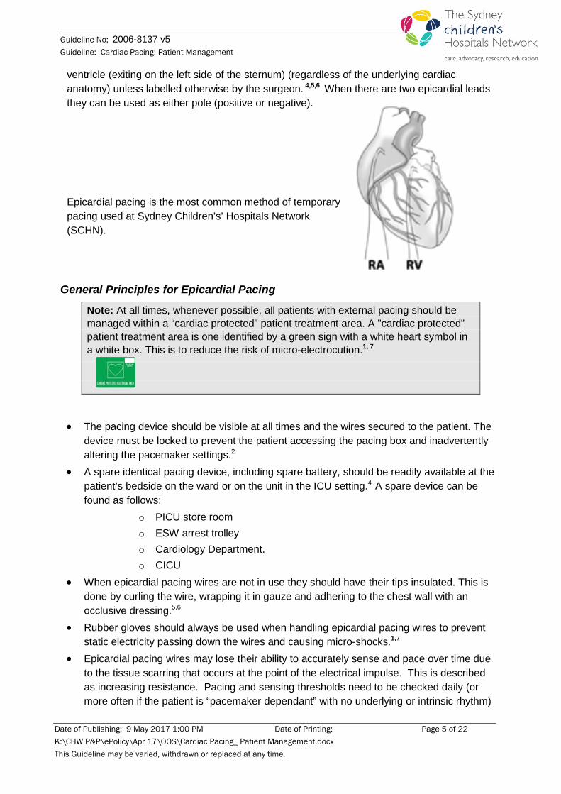
Arrhythmias are a common complication following cardiac surgery therefore some patients may require support from a temporary pacemaker.3,4 Epicardial wires are the preferred method if pacing is required post operatively and are routinely inserted during cardiac surgery. Epicardial pacing wires are insulated multifilament stainless steel wires that are stitched to the epicardium and brought through the chest wall before closing the sternotomy.5,6 Usually two pairs of wires are attached, one pair attached to the right atrium (exiting the thorax on the right side of the sternum) and one pair attached to the right
Date of Publishing: 9 May 2017 1:00 PM Date of Printing: Page 4 of 22 K:\CHW P&P\ePolicy\Apr 17\OOS\Cardiac Pacing_ Patient Management.docx This Guideline may be varied, withdrawn or replaced at any time.
Guideline No: 2006-8137 v5 Guideline: Cardiac Pacing: Patient Management
ventricle (exiting on the left side of the sternum) (regardless of the underlying cardiac anatomy) unless labelled otherwise by the surgeon. 4,5,6 When there are two epicardial leads they can be used as either pole (positive or negative).
Epicardial pacing is the most common method of temporary pacing used at Sydney Children’s’ Hospitals Network (SCHN).
General Principles for Epicardial Pacing
Note: At all times, whenever possible, all patients with external pacing should be managed within a “cardiac protected” patient treatment area. A "cardiac protected" patient treatment area is one identified by a green sign with a white heart symbol in a white box. This is to reduce the risk of micro-electrocution.1, 7
• The pacing device should be visible at all times and the wires secured to the patient. The device must be locked to prevent the patient accessing the pacing box and inadvertently altering the pacemaker settings.2
• A spare identical pacing device, including spare battery, should be readily available at the patient’s bedside on the ward or on the unit in the ICU setting.4 A spare device can be found as follows:
o PICU store room o ESW arrest trolley o Cardiology Department. o CICU
• When epicardial pacing wires are not in use they should have their tips insulated. This is done by curling the wire, wrapping it in gauze and adhering to the chest wall with an occlusive dressing.5,6
• Rubber gloves should always be used when handling epicardial pacing wires to prevent static electricity passing down the wires and causing micro-shocks.1,7
• Epicardial pacing wires may lose their ability to accurately sense and pace over time due to the tissue scarring that occurs at the point of the electrical impulse. This is described as increasing resistance. Pacing and sensing thresholds need to be checked daily (or more often if the patient is “pacemaker dependant” with no underlying or intrinsic rhythm)
Date of Publishing: 9 May 2017 1:00 PM Date of Printing: Page 5 of 22 K:\CHW P&P\ePolicy\Apr 17\OOS\Cardiac Pacing_ Patient Management.docx This Guideline may be varied, withdrawn or replaced at any time.
Guideline No: 2006-8137 v5 Guideline: Cardiac Pacing: Patient Management
and pacemaker settings adjusted accordingly by medical officer or accredited registered nurse in PICU/CICU.2,7
• Complication of epicardial wires include: o Infection o Myocardial damage o Arrhythmias o Perforation o Cardiac tamponade
• For a detailed table of potential complications post epicardial wire removal, see table on page 18.
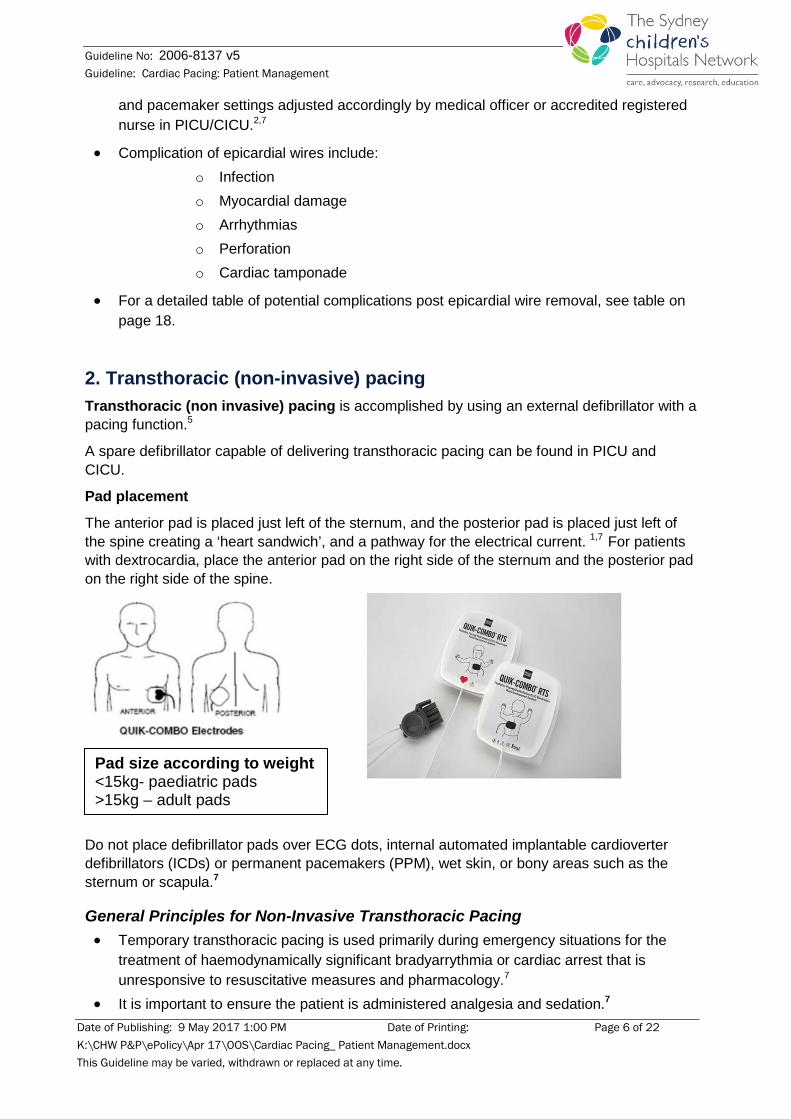
2. Transthoracic (non-invasive) pacing Transthoracic (non invasive) pacing is accomplished by using an external defibrillator with a pacing function.5
A spare defibrillator capable of delivering transthoracic pacing can be found in PICU and CICU.
Pad placement
The anterior pad is placed just left of the sternum, and the posterior pad is placed just left of the spine creating a ‘heart sandwich’, and a pathway for the electrical current. 1,7 For patients with dextrocardia, place the anterior pad on the right side of the sternum and the posterior pad on the right side of the spine.
Do not place defibrillator pads over ECG dots, internal automated implantable cardioverter defibrillators (ICDs) or permanent pacemakers (PPM), wet skin, or bony areas such as the sternum or scapula.7
General Principles for Non-Invasive Transthoracic Pacing • Temporary transthoracic pacing is used primarily during emergency situations for the
treatment of haemodynamically significant bradyarrythmia or cardiac arrest that is unresponsive to resuscitative measures and pharmacology.7
• It is important to ensure the patient is administered analgesia and sedation.7
Pad size according to weight <15kg- paediatric pads >15kg – adult pads
Date of Publishing: 9 May 2017 1:00 PM Date of Printing: Page 6 of 22 K:\CHW P&P\ePolicy\Apr 17\OOS\Cardiac Pacing_ Patient Management.docx This Guideline may be varied, withdrawn or replaced at any time.
Guideline No: 2006-8137 v5 Guideline: Cardiac Pacing: Patient Management
• As an effect of transthoracic pacing, the patient may experience muscular contraction of the chest and abdominal muscles.7
3. Transvenous pacing Transvenous pacing - an electrode catheter is threaded through a vein (normally femoral or jugular vein) into the patient’s right atrium or right ventricle.5,7 The pacing lead is a self contained bipolar system with two pins at the proximal end for connection to the bridging lead and pacing box.7
General Principles for Transvenous Pacing • The puncture site should be covered in a transparent dressing to facilitate assessment of
the site. The lead should be appropriately secured to the patient.7
• If the transvenous catheter is placed in the femoral vein the affected limb should be stabilised (kept as immobile and straight as comfortably possible). This is to avoid leg flexion, which may result in movement/dislodgement of the pacing lead. If there is any suspicion of lead movement, an xray must be performed.
• Circulation observations should also be performed on the affected limb. Refer to Post-Operative Management section in the ‘Cardiac Catheterisation: Interventional, Non-interventional and Electrophysiological Studies’ practice guideline.
• Transvenous pacing catheters positioned in the apex of the heart are prone to move or drift out of position.5 This can result in failure to capture (see Trouble-shooting guide).
• The pacing device should be visible at all times and the wires secured to the patient with occlusive dressing. Make sure the device is locked to prevent the patient accessing the pacing box and inadvertently altering the pacemaker settings.2
• A spare identical pacing device should be readily available at the patient’s bedside.7
4. Transoesophageal pacing Transoesophageal pacing is an electrode attached to an oesophageal probe inserted to the oesophagus just behind the left atrium to permit temporary atrial pacing. 6,7 The patient is to remain sedated for the duration of this type of pacing.7 This system may also be used in the diagnosis (electrophysiological study) and termination of arrhythmias including SVT.4 However, as this pacing only captures the atrium, it cannot usually be used to treat AV block.
General Principles for Transoesophageal Pacing • The bipolar lead is inserted by cardiologist via the nose or mouth, and is secured to the
face with tape. • The transoesophageal pacing lead is attached to an electrocardiographic (ECG)
machine. The lead is then advanced and positioned behind the left atrium. This is indicated by the largest atrial signal as recorded on the ECG trace.
• When pacing is discontinued, the lead can be removed after consultation with cardiac team.
• The lead should be placed in a plastic bag and returned for re-sterilisation.
Date of Publishing: 9 May 2017 1:00 PM Date of Printing: Page 7 of 22 K:\CHW P&P\ePolicy\Apr 17\OOS\Cardiac Pacing_ Patient Management.docx This Guideline may be varied, withdrawn or replaced at any time.
Guideline No: 2006-8137 v5 Guideline: Cardiac Pacing: Patient Management
• Oesophageal injury and burns can be caused by long term oesophageal pacing.
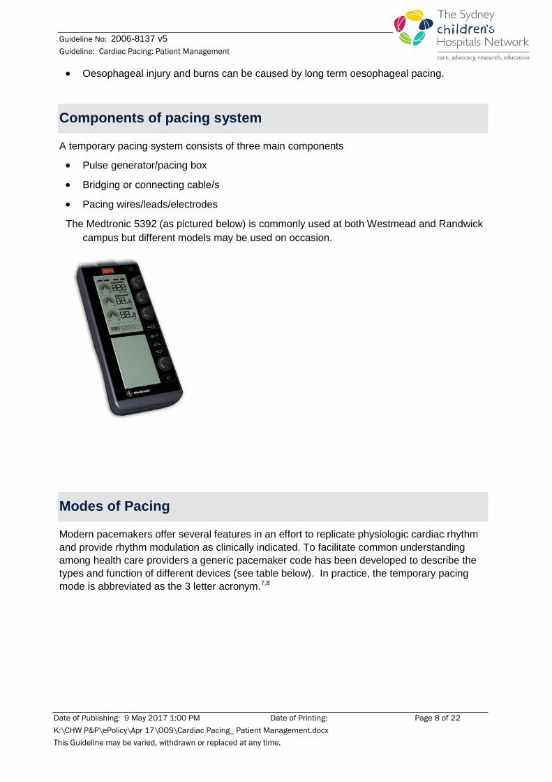
Components of pacing system
A temporary pacing system consists of three main components
• Pulse generator/pacing box
• Bridging or connecting cable/s
• Pacing wires/leads/electrodes
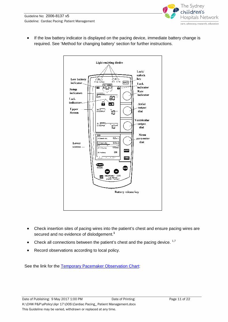
The Medtronic 5392 (as pictured below) is commonly used at both Westmead and Randwick campus but different models may be used on occasion.
Modes of Pacing
Modern pacemakers offer several features in an effort to replicate physiologic cardiac rhythm and provide rhythm modulation as clinically indicated. To facilitate common understanding among health care providers a generic pacemaker code has been developed to describe the types and function of different devices (see table below). In practice, the temporary pacing mode is abbreviated as the 3 letter acronym.7,8
Date of Publishing: 9 May 2017 1:00 PM Date of Printing: Page 8 of 22 K:\CHW P&P\ePolicy\Apr 17\OOS\Cardiac Pacing_ Patient Management.docx This Guideline may be varied, withdrawn or replaced at any time.
Guideline No: 2006-8137 v5 Guideline: Cardiac Pacing: Patient Management
Pacemaker Nomenclature *NASPE/BPEG** GENERIC
PACEMAKER (NBG) CODE
*North American Society of Pacing & Electrophysiology
**British Pacing & Electrophysiology Group
Temporary Pacemakers I II III
Chamber Paced Chamber Sensed Response to Sensing
O = None O = None
A = Atrium A = Atrium I = Inhibited
V = Ventricle V = Ventricle T = Triggered
D = Dual D = Dual D = Triggered & Inhibited
For example, AAI indicates that the atrium is paced, the atrium is sensed and the generator/pacemaker is inhibited if it senses intrinsic atrial activity. 5,7,8,9
Nursing Management of Temporarily Paced Children
Assessing & Recording Vital Signs All patients receiving temporary pacing therapy require continuous ECG monitoring; ensuring that the bedside monitor is set up appropriately for a paced patient. Regular assessment of hemodynamic stability is required, including the recording of hourly vital sign observations, or more frequently if required, including; 1
• Heart rate
• Blood pressure
• Perfusion (skin colour, warmth, capillary refill, peripheral pulse strength)
Print a rhythm strip from bedside monitoring once per shift and place in patient notes.
Date of Publishing: 9 May 2017 1:00 PM Date of Printing: Page 9 of 22 K:\CHW P&P\ePolicy\Apr 17\OOS\Cardiac Pacing_ Patient Management.docx This Guideline may be varied, withdrawn or replaced at any time.
Guideline No: 2006-8137 v5 Guideline: Cardiac Pacing: Patient Management
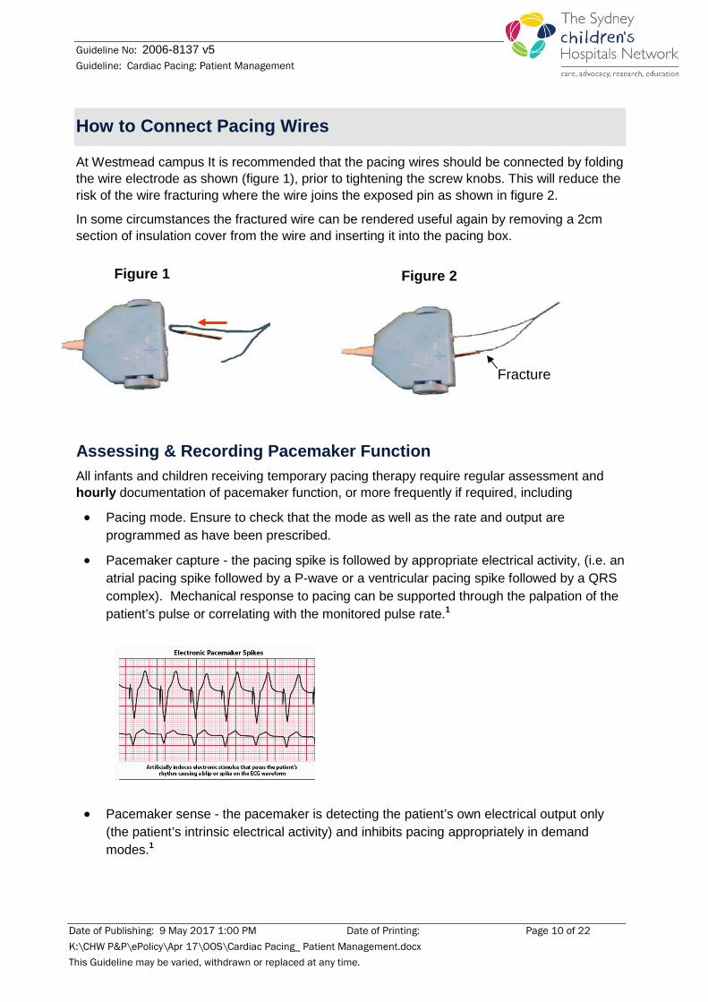
How to Connect Pacing Wires
At Westmead campus It is recommended that the pacing wires should be connected by folding the wire electrode as shown (figure 1), prior to tightening the screw knobs. This will reduce the risk of the wire fracturing where the wire joins the exposed pin as shown in figure 2.
In some circumstances the fractured wire can be rendered useful again by removing a 2cm section of insulation cover from the wire and inserting it into the pacing box.
Assessing & Recording Pacemaker Function All infants and children receiving temporary pacing therapy require regular assessment and hourly documentation of pacemaker function, or more frequently if required, including
• Pacing mode. Ensure to check that the mode as well as the rate and output are programmed as have been prescribed.
• Pacemaker capture - the pacing spike is followed by appropriate electrical activity, (i.e. an atrial pacing spike followed by a P-wave or a ventricular pacing spike followed by a QRS complex). Mechanical response to pacing can be supported through the palpation of the patient’s pulse or correlating with the monitored pulse rate.1
• Pacemaker sense - the pacemaker is detecting the patient’s own electrical output only (the patient’s intrinsic electrical activity) and inhibits pacing appropriately in demand modes.1
Figure 1 Figure 2
Fracture
Date of Publishing: 9 May 2017 1:00 PM Date of Printing: Page 10 of 22 K:\CHW P&P\ePolicy\Apr 17\OOS\Cardiac Pacing_ Patient Management.docx This Guideline may be varied, withdrawn or replaced at any time.
Guideline No: 2006-8137 v5 Guideline: Cardiac Pacing: Patient Management
• If the low battery indicator is displayed on the pacing device, immediate battery change is required. See ‘Method for changing battery’ section for further instructions.
• Check insertion sites of pacing wires into the patient’s chest and ensure pacing wires are secured and no evidence of dislodgement.1
• Check all connections between the patient’s chest and the pacing device. 1,7
• Record observations according to local policy.
See the link for the Temporary Pacemaker Observation Chart:
Date of Publishing: 9 May 2017 1:00 PM Date of Printing: Page 11 of 22 K:\CHW P&P\ePolicy\Apr 17\OOS\Cardiac Pacing_ Patient Management.docx This Guideline may be varied, withdrawn or replaced at any time.

Guideline No: 2006-8137 v5 Guideline: Cardiac Pacing: Patient Management
The following pacemaker checks need to be documented every 12-24 hours1, or as the patient’s condition warrants, and is to be performed by trained medical staff or in some circumstances by accredited RNs in the intensive care setting
• Threshold checks for capture • Sensitivity threshold • Determination of underlying rhythm • Correlation between pacemaker order and pacemaker settings 1,7 • Changes to the pacemaker order should be documented on each occasion a change
occurs (the original order will be documented on the commencement of pacing) • Additional pacemaker checking may be required if the patient has a breakthrough rhythm
(an intrinsic rate that is faster than the rate set by the pacing device). Unless indicated by the patient’s clinical condition, the underlying rate need not be checked more often that once per shift as this may cause a syncopal event if there is no underlying rhythm.7
The following section on output and sensitivity was taken from the Congenital heart disease: PICU Peri-operative management Practice Guideline – CHW. Refer to this document for further information:
Output 4,10
The threshold is the minimum current necessary to capture & stimulate the heart. To test for this:
• Atrial and ventricular output is generated in milliamperes (mA). The typical atrial output is 5 mA and typical ventricular output is 8-10 mA. Set pacer rate 10 ppm faster than patient’s heart rate
• Decrease mA until capture is lost (change in ECG)
• Increase output until capture is regained (threshold capture)
• Output setting to be 2mA above the threshold capture (example: set output at 7mA if capture was regained at 5mA)
Sensitivity 4,10
The sensitivity is the minimum level of intrinsic electric activity generated by the heart detectable by the pacemaker. ONLY test for sensitivity if cardiovascularly stable in their intrinsic rhythm. To test for this follow:
• Atrial and ventricular sensitivity is measured in millivolts (mV). The typical atrial sensitivity is 0.5 mV and typical ventricular sensitivity is 2.0 – 5.0mV
• Set pacer rate 10 ppm slower than patient’s HR • Increase sensitivity to chamber being tested to minimum level (0.4mV) • Decrease sensitivity of the pacer (↑mV) to the chamber being tested until pacer stops
sensing patient (orange light stops flashing) • Increase sensitivity of the pacer (↓mV) until the pacer senses the patient (orange light
begins flashing). This is the threshold for sensitivity.
Date of Publishing: 9 May 2017 1:00 PM Date of Printing: Page 12 of 22 K:\CHW P&P\ePolicy\Apr 17\OOS\Cardiac Pacing_ Patient Management.docx This Guideline may be varied, withdrawn or replaced at any time.
Guideline No: 2006-8137 v5 Guideline: Cardiac Pacing: Patient Management
• Set the sensitivity at ½ the threshold value (example: set sensitivity at 1mV if the threshold was 2mV)
Method for Changing Battery
• A battery should last about 3-7 days depending on the pacemaker settings.
• Change of battery is required when the low battery indicator is shown. It is recommended to change the battery as soon as the low battery indicator is noticed.
• Battery change to be performed by Registered nurse who has completed the pacemaker accreditation.
• The pacemaker will operate for approximately 15 seconds after the battery is removed. There is a diagram inside the battery drawer to show correct orientation of battery when changing it.
• A second pacemaker should be available.
• Check settings prior to changing battery.
• Always replace with a new battery/ batteries.
• Leave pacemaker operating.
• Press battery release button, pull out drawer, insert new battery/ batteries, close drawer.
• Check that the pacing is functional after changing battery. Low battery indicator should now be inactive.
• Assess the patient, their vital signs, and assess and record the pacemaker function.
Note: Depending on the pacemaker model, the pacemaker may require 2 AA batteries while others may require a 9volt battery. Ensure correct spare battery is ready before commencing battery change.
Date of Publishing: 9 May 2017 1:00 PM Date of Printing: Page 13 of 22 K:\CHW P&P\ePolicy\Apr 17\OOS\Cardiac Pacing_ Patient Management.docx This Guideline may be varied, withdrawn or replaced at any time.
Guideline No: 2006-8137 v5 Guideline: Cardiac Pacing: Patient Management
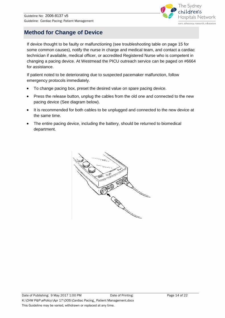
Method for Change of Device
If device thought to be faulty or malfunctioning (see troubleshooting table on page 15 for some common causes), notify the nurse in charge and medical team, and contact a cardiac technician if available, medical officer, or accredited Registered Nurse who is competent in changing a pacing device. At Westmead the PICU outreach service can be paged on #6664 for assistance.
If patient noted to be deteriorating due to suspected pacemaker malfunction, follow emergency protocols immediately.
• To change pacing box, preset the desired value on spare pacing device.
• Press the release button, unplug the cables from the old one and connected to the new pacing device (See diagram below).
• It is recommended for both cables to be unplugged and connected to the new device at the same time.
• The entire pacing device, including the battery, should be returned to biomedical department.
Date of Publishing: 9 May 2017 1:00 PM Date of Printing: Page 14 of 22 K:\CHW P&P\ePolicy\Apr 17\OOS\Cardiac Pacing_ Patient Management.docx This Guideline may be varied, withdrawn or replaced at any time.
Guideline No: 2006-8137 v5 Guideline: Cardiac Pacing: Patient Management
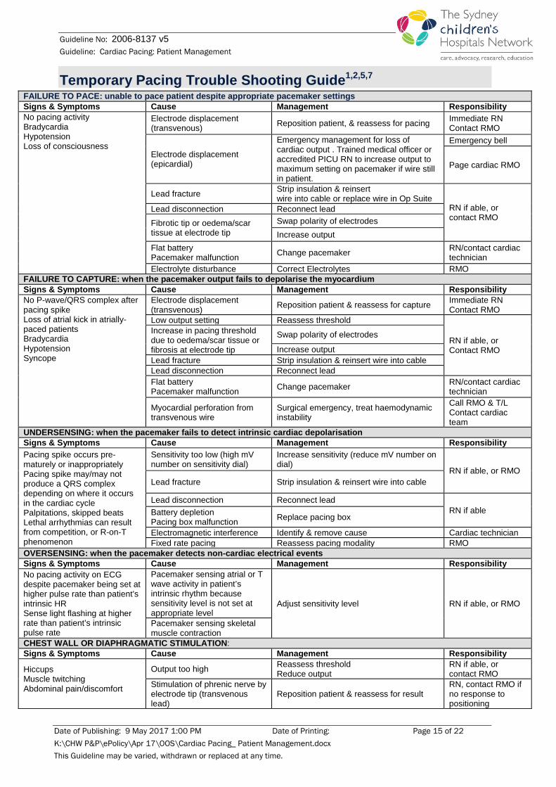
Temporary Pacing Trouble Shooting Guide1,2,5,7 FAILURE TO PACE: unable to pace patient despite appropriate pacemaker settings Signs & Symptoms Cause Management Responsibility No pacing activity Bradycardia Hypotension Loss of consciousness
Electrode displacement (transvenous) Reposition patient, & reassess for pacing Immediate RN
Contact RMO
Electrode displacement (epicardial)
Emergency management for loss of cardiac output . Trained medical officer or accredited PICU RN to increase output to maximum setting on pacemaker if wire still in patient.
Emergency bell
Page cardiac RMO
Lead fracture Strip insulation & reinsert wire into cable or replace wire in Op Suite
RN if able, or contact RMO
Lead disconnection Reconnect lead Fibrotic tip or oedema/scar tissue at electrode tip
Swap polarity of electrodes Increase output
Flat battery Pacemaker malfunction Change pacemaker RN/contact cardiac
technician Electrolyte disturbance Correct Electrolytes RMO
FAILURE TO CAPTURE: when the pacemaker output fails to depolarise the myocardium Signs & Symptoms Cause Management Responsibility No P-wave/QRS complex after pacing spike Loss of atrial kick in atrially-paced patients Bradycardia Hypotension Syncope
Electrode displacement (transvenous) Reposition patient & reassess for capture Immediate RN
Contact RMO Low output setting Reassess threshold
RN if able, or Contact RMO
Increase in pacing threshold due to oedema/scar tissue or fibrosis at electrode tip
Swap polarity of electrodes
Increase output Lead fracture Strip insulation & reinsert wire into cable Lead disconnection Reconnect lead Flat battery Pacemaker malfunction Change pacemaker RN/contact cardiac
technician
Myocardial perforation from transvenous wire
Surgical emergency, treat haemodynamic instability
Call RMO & T/L Contact cardiac team
UNDERSENSING: when the pacemaker fails to detect intrinsic cardiac depolarisation Signs & Symptoms Cause Management Responsibility Pacing spike occurs pre-maturely or inappropriately Pacing spike may/may not produce a QRS complex depending on where it occurs in the cardiac cycle Palpitations, skipped beats Lethal arrhythmias can result from competition, or R-on-T phenomenon
Sensitivity too low (high mV number on sensitivity dial)
Increase sensitivity (reduce mV number on dial)
RN if able, or RMO Lead fracture Strip insulation & reinsert wire into cable
Lead disconnection Reconnect lead RN if able Battery depletion
Pacing box malfunction Replace pacing box
Electromagnetic interference Identify & remove cause Cardiac technician Fixed rate pacing Reassess pacing modality RMO
OVERSENSING: when the pacemaker detects non-cardiac electrical events Signs & Symptoms Cause Management Responsibility No pacing activity on ECG despite pacemaker being set at higher pulse rate than patient’s intrinsic HR Sense light flashing at higher rate than patient’s intrinsic pulse rate
Pacemaker sensing atrial or T wave activity in patient’s intrinsic rhythm because sensitivity level is not set at appropriate level
Adjust sensitivity level RN if able, or RMO
Pacemaker sensing skeletal muscle contraction
CHEST WALL OR DIAPHRAGMATIC STIMULATION: Signs & Symptoms Cause Management Responsibility
Hiccups Muscle twitching Abdominal pain/discomfort
Output too high Reassess threshold Reduce output
RN if able, or contact RMO
Stimulation of phrenic nerve by electrode tip (transvenous lead)
Reposition patient & reassess for result RN, contact RMO if no response to positioning
Date of Publishing: 9 May 2017 1:00 PM Date of Printing: Page 15 of 22 K:\CHW P&P\ePolicy\Apr 17\OOS\Cardiac Pacing_ Patient Management.docx This Guideline may be varied, withdrawn or replaced at any time.
Guideline No: 2006-8137 v5 Guideline: Cardiac Pacing: Patient Management
Removal of Epicardial Pacing Wires
Note: A patient’s coagulation profile must be checked and reviewed by the cardiac team prior to removal of pacing wires. General guide is an INR of <1.6 and platelets >100 000 for removal of wires. Some patients who are receiving anticoagulant therapy may have a higher INR; consult with cardiac team.
Equipment:
• Dressing pack • Gloves (non-sterile) • Aqueous Chlorhexidine 0.5% / 0.9%
Sodium chloride
• Stitch cutter • Appropriate dressing
Timing of wire removal
• Pacing wires are only to be removed after discussion with Cardiologist and Cardiothoracic Surgeon as well as the intensivist if the patient is in the intensive care.
• The patient is to be in sinus rhythm for a minimum 24 hours prior to removal. • It is the responsibility of the person removing the wires to ensure that the nurse in charge
and a member of the cardiothoracic team are aware and available at the time of the wire removal.
Pre-procedural tests and documentation
• Most patients require a recent ECG or printed rhythm strip from bedside monitoring, and coagulation profile. INR< 1.6 and platelets >100 000.
• Consult with cardiac team for the need of establishment of IV access prior to wire removal
• It is the responsibility of the person performing the procedure to ensure all necessary tests are performed, that the results have been assessed by the medical staff and the decision to remove the wires documented.
Person performing the procedure
• The procedure will be performed by either a doctor or nurse with the relevant training and experience. Whoever undertakes the procedure must be competent or supervised in the procedure.
• Obtain assistance from another nurse or member of the medical team.
Preparation of the patient and carers
• Psychological preparation is to be given to the patient and/or parents at an appropriate level; a verbal explanation should be given of the possible complications of wires removal. The use of the child life therapist for preparation and/or distraction therapy is encouraged.
Date of Publishing: 9 May 2017 1:00 PM Date of Printing: Page 16 of 22 K:\CHW P&P\ePolicy\Apr 17\OOS\Cardiac Pacing_ Patient Management.docx This Guideline may be varied, withdrawn or replaced at any time.

Guideline No: 2006-8137 v5 Guideline: Cardiac Pacing: Patient Management
• It is not necessary to keep the patient fasted prior to pacing wire removal, but this will be determined by policy, especially where sedation is used. An assessment of the level of analgesia +/- sedation will be made on an individual patient basis
Chest Drains
• If chest drains are present they should be tapped/manipulated (in accordance with local policy for chest drain management), to ensure patency of drains thereby providing a pathway for bleeding and reducing potential for cardiac tamponade. Refer to the following SCHN chest drain practice guideline for further nursing management of patients with chest drains:
Important to remember:
• Notify the cardiothoracic registrar prior to procedure.
• The atrial pacing wire should be removed first as this allows for ventricular pacing to be undertaken should the patient’s clinical status become unstable.
• Pacing wires should be removed by constant gentle traction, allowing the motion of the heart to assist dislodgment from the epicardial surface. Excessive traction should not be applied. 2,3
• If the wires are unable to be removed due to excessive tension, stop and notify cardiothoracic nurse practitioner or fellow.
Procedure 1. Perform baseline vital signs and cardiac assessment and ensure patient is attached to a
cardiac monitor
2. Print pre-wire removal rhythm strip from bedside monitoring
3. Don gloves
4. Remove any dressings from pacing wires
5. Clean wires and skin around puncture site with Aqueous Chlorhexidine 0.5% / 0.9% Sodium chloride
6. Cut suture holding the atrial wire
7. Place folded sterile gauze swab over atrial puncture site and wire and pull wire gently until it slides from the chest
8. Check the wire and wire tip are intact
9. Assess rhythm on cardiac monitor and assess patient
10. Assess chest tube drainage if applicable. If there is a sudden increase in drainage that does not resolve, contact cardiothoracic team before removing ventricular wire.
11. If rhythm and patient is stable, repeat previous steps to remove ventricular wire
12. Cover pacing wire sites with appropriate dressings as necessary
Date of Publishing: 9 May 2017 1:00 PM Date of Printing: Page 17 of 22 K:\CHW P&P\ePolicy\Apr 17\OOS\Cardiac Pacing_ Patient Management.docx This Guideline may be varied, withdrawn or replaced at any time.
Guideline No: 2006-8137 v5 Guideline: Cardiac Pacing: Patient Management
13. Perform vital signs and patient cardiac assessment
14. Dispose of equipment appropriately
15. Print post-wire removal rhythm strip from bedside monitoring and paste both strips on appropriate form and placed in patient notes.
Alert: Be alert for tamponade acutely or slowly evolving. Notify cardiothoracic team and cardiology fellow (who should perform immediate echocardiogram) if concerned.
Observations and Tests
• The RN should check the vital signs and perform a cardiac assessment immediately post-procedure and again every 30 minutes for 2 hours, or as the patient’s condition warrants, to detect potential complications. Patient should remain on continuous cardiac monitoring until instructed to cease by team.
• Observe chest drain losses (if insitu) for signs of tamponade, eg: increased drain losses and haemoserous drainage.
• The patient should remain in the ward/unit for at least two hours post procedure. The patient (if age-appropriate) and parents should be aware of signs and symptoms of possible complications and to report any concerns to staff immediately.
• In some cases, an echocardiogram may be requested post-procedure.
Complications associated with removal of epicardial pacing wires: Complication Signs and Symptoms Management Cardiac tamponade2
Tachycardia, hypotension, cool peripheries with delayed capillary refill, reduced level of consciousness (LOC), arrhythmia, cardiac arrest
Notify RMO/Nursing team leader (TL) & Cardiac surgeon Tap chest drains vigorously (if insitu) Assess vital signs and perform cardiac assessment Prepare Chest Opening Tray Follow emergency protocol for campus
Excessive Bleeding
Obvious bleeding from site, tachycardia, hypotension, pallor, poor peripheral perfusion, reducing LOC, shock
Notify RMO & Nursing T/L Assess vital signs and perform cardiac assessment Measure blood loss Prepare IV fluids to replace losses (blood products/4% NSA as available)
Arrhythmias2 Evident on cardiac monitor with or without change in hemodynamic status Irregular pulse rate
Notify RMO/Nursing T/L & Consultant Assess vital signs and perform cardiac assessment
Wires become wedged in chest
Hard to pull the wires Do not use force to remove, Abandon the procedure and inform the cardiothoracic surgical registrar/cardiac team
Date of Publishing: 9 May 2017 1:00 PM Date of Printing: Page 18 of 22 K:\CHW P&P\ePolicy\Apr 17\OOS\Cardiac Pacing_ Patient Management.docx This Guideline may be varied, withdrawn or replaced at any time.
Guideline No: 2006-8137 v5 Guideline: Cardiac Pacing: Patient Management
Removal of Transvenous Pacing Lead
Transvenous Pacing Leads should be managed using the same principles as those for percutaneous Central Venous Access Devices (CVADs) outlined in the hospital guideline on CVADs. Once it has been deemed by the medical team that the patient’s underlying rhythm is safe and reliable, the transvenous pacing lead can be removed. The steps taken to remove a transvenous pacing lead should be the same as those taken to remove a non-tunnelled CVAD as per SCHN CVADs practice guideline:
Watch for arrhythmia when removing pacing lead as lead is intracardiac and can stimulate ectopic beats or arrhythmias.
Indications for Insertion of a Permanent Pacemaker
The following list is not exhaustive, but does include some typical indications for the insertion of a permanent pacemaker. The indications for permanent pacing are relative and not absolute. The overall clinical condition of the patient is considered rather than using this list as a definitive indication for insertion of a permanent pacemaker (for example, the rates specified for complete heart block in neonates).
• Second- or third-degree AV block with symptomatic bradycardia • Sinus node dysfunction with symptomatic bradycardia • An asymptomatic neonate with congenital third degree AV block and wide complex
escape rhythm • Asymptomatic patients after cardiac surgery with advanced second- or third-degree AV
block who’s conducted sinus rhythm did not spontaneously recover after ten days post operatively 11
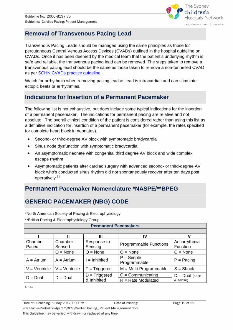
Permanent Pacemaker Nomenclature *NASPE/**BPEG
GENERIC PACEMAKER (NBG) CODE
*North American Society of Pacing & Electrophysiology **British Pacing & Electrophysiology Group
Permanent Pacemakers
I II III IV V Chamber Paced
Chamber Sensed
Response to Sensing Programmable Functions Antiarrythmia
Function O = None O = None O = None O = None
A = Atrium A = Atrium I = Inhibited P = Simple Programmable P = Pacing
V = Ventricle V = Ventricle T = Triggered M = Multi-Programmable S = Shock
D = Dual D = Dual D = Triggered & Inhibited
C = Communicating D = Dual (pace & sense) R = Rate Modulated
5,7,8,9
Date of Publishing: 9 May 2017 1:00 PM Date of Printing: Page 19 of 22 K:\CHW P&P\ePolicy\Apr 17\OOS\Cardiac Pacing_ Patient Management.docx This Guideline may be varied, withdrawn or replaced at any time.
Guideline No: 2006-8137 v5 Guideline: Cardiac Pacing: Patient Management
Indications for Insertion of automated Implantable Cardioverter
Defibrillator (ICD)
The American Heart Association (AHA) has guidelines on the indications for an ICD.12
Some typical indications for insertion of an implantable defibrillator include.12
• Cardiac arrest due to ventricular fibrillation (VF) or ventricular tachycardia (VT) not due to a transient or reversible cause
• Syncope of undetermined origin with clinically relevant, haemodynamically significant sustained VT or VF induced at electrophysiological study when drug therapy is ineffective, not tolerated or not preferred
For a full list of indications for an ICD, refer to the AHA guidelines.12
Alert - If patient with an ICD or PPM experiences cardiac arrest, the placement of external defibrillation pads/paddles in the anterior-posterior configuration is preferable to avoid damage to the device. Ensure pads are not placed directly over the ICD/PPM. Implantable cardioverter defibrillator shocks do not pose a danger to others during CPR; an unpleasant tingle has been described but can be prevented by wearing gloves while performing external cardiac compressions.13
Emergency Procedure for Malfunctioning automated
Implantable Defibrillation Device (ICD)/ Permanent Pacemaker
(PPM)
In the circumstance of an ICD delivering a shock inappropriately, the Medtronic® Pulse Generator Magnet can be used to disable the defibrillator mode. In the event of a malfunctioning PPM, the Medtronic® Pulse Generator Magnet can be used to remove the PPM’s sensing ability and places the PPM in an asynchronous pacing mode. The following information should be utilised in conjunction with the emergency protocol for each local area.
General Description The magnet is a blue-coated, ring-shaped magnet used to verify proper operation of Medtronic® pulse generators. They are available across the hospital as follows:
• PICU (resuscitation trolley opposite bed 13)
• ESW (resuscitation trolley and medication room)
• Cardiac department - Westmead
• CICU (resuscitation trolley)
• OT
• ED (mobile arrest trolley)
Date of Publishing: 9 May 2017 1:00 PM Date of Printing: Page 20 of 22 K:\CHW P&P\ePolicy\Apr 17\OOS\Cardiac Pacing_ Patient Management.docx This Guideline may be varied, withdrawn or replaced at any time.

Guideline No: 2006-8137 v5 Guideline: Cardiac Pacing: Patient Management
The following information is provided by Medtronic® in the Pulse Generator Magnet product information sheet.14
Use of the Magnet The patient needs to be ECG monitored when using the magnet as it will interfere with normal ICD function.
Holding the magnet against the skin directly over the implanted device will convert the device to asynchronous operation and interrupt automatic defibrillation. This means that the ICD pacing component will pace the heart at a preset heart rate as set in the pacemaker programming, regardless of the heart’s own activity (intrinsic activity). Fundamentally, it will inhibit the defibrillation function but not the pacing function.
Note: the magnet needs to be left against the skin for the defibrillation function to remain deactivated.
When the magnet is used on a patient to interrupt inappropriate ICD defibrillator activity, this should be treated as a medical emergency as inappropriate ICD therapy can precipitate fatal arrhythmias. Appropriate medical staff should be contacted urgently and the Medtronic representative (or relevant industry technician) should be contacted as soon as possible to run diagnostics on the implanted device.
Care of the patient with an ICD or PPM
Warnings • Magnetic conversion of an implanted device to asynchronous operation may result in
competitive pacing.
• MRI scans needs to be avoided.
• An implanted devicemay be inadvertently reprogrammed if the magnet is applied in the presence of pulsed energy sources (i.e. radio and radar transmitters, some electric razors and household appliances, ECG telemetry, arc welders, anti-theft devices and other spark-producing devices).
Date of Publishing: 9 May 2017 1:00 PM Date of Printing: Page 21 of 22 K:\CHW P&P\ePolicy\Apr 17\OOS\Cardiac Pacing_ Patient Management.docx This Guideline may be varied, withdrawn or replaced at any time.

Guideline No: 2006-8137 v5 Guideline: Cardiac Pacing: Patient Management
References 1. Overbay D, Criddle L. Mastering temporary invasive cardiac pacing. Critical Care Nurse 2004; 24 (3): 25-
32. 2. Reade MC. Temporary epicardial pacing after cardiac surgery: a practical review: part 1: General
considerations in the management of epicardial pacing. Anaesthesia 2007; 62 (3): 264-271. 3. Schoof S, Bertram H, Thommes J, Breymann T, Grosser U, Mesud Yelbuz T, Wessal A, Norozi K.
Removal of temporary pacemaker after cardiac surgery in infants: A harmless procedure? Journal of Pediatric Intensive Care 2012; 01 (02): 121-123.
4. Chiu-Man C, McGill-Lane S, Murphy C, Olen M, Daley E, St. George-Hyslop C. Care of the patient with temporary pacemaker in the neonatal and pediatric cardiac patient: What the nurse caring for a patient with congenital heart disease needs to know [Internet]. 2016 [cited 2017 February 6]. Available from: www.pcics.org/wp-content uploads/2016/09.
5. Skippen P, Sanatani S, Froese N, Gow RM. Pacemaker therapy of postoperative arrhythmias after pediatric cardiac surgery. Paediatric Critical Care Medicine 2010; 11 (1): 133-138.
6. Beattie S. Epicardial wires [Internet]. 2005 [cited 2013 June 18]. Available from: http://www.modernmedicine.com/modern-medicine/news/epicardial-wires
7. Craig J, Bloedel-Smith J, Fineman LD. Pacemaker Therapy. In: Curley M, Maloney-Harmon P, editors. Critical Care Nursing of Infants and Children. 2nd ed. Philadelphia. W.B. Saunders; 2001.
8. Bernstein AD, Daubert J, Fletcher RD, Hayes DL, Luderitz B, Reynolds DW, Schoenfeld MH, Sutton R. The revised NASPE/BPEG generic code for antibradycardia, adaptive-rate, and multisite pacing. Journal of Pacing and Clinical Electrophysiology 2002; 25 (2): 260-264.
9. Reade MC. Temporary epicardial pacing after cardiac surgery: a practical review: part 2: selection of epicardial pacing modes and troubleshooting. Anaesthesia 2007; 62: 364-373.
10. Congenital heart disease: PICU peri-operative management – CHW Practice guideline. Guideline No: 0/C/07:0051-01:02. The Children’s Hospital at Westmead [Internet]. 2016 [cited 2017 February 7]. Available from: http://chw.schn.health.nsw.gov.au/o/documents/policies/guidelines/2007-0051.pdf
11. Gregoratos G, Abrams J, Epstein AE, Freedman RA, Hayes DL, Hlatky MA, Kerber RE, Naccarelli GV, Schoenfeld MH, Silka MJ, Winters SL. ACC/AHA/NASPE: Guideline update for implantation of cardiac pacemakers and antiarrhythmia devices: summary article: a report of the American College of Cardiology/American Heart Association Task Force on Practice Guidelines (ACC/AHA/NASPE Committee to Update the 1998 Pacemaker Guidelines). Circulation 2002; 106: 2145-2161.
12. Epstein AE, DiMarco JP, Ellenbogen KA, Estes III NAM, Freedman RA, Gettes LS, Gillinov AM, Gregoratos G, Hammill SC, Hayes DL, Hlatky MA, Newby LK, Page RL, Schoenfeld MH, Silka MJ, Warner Stevenson L, Sweeny MO. ACC/ AHA/ HRS 2008 Guidelines for device-based therapy of cardiac rhythm abnormalities: Executive summary: A report of the American College of Cardiology/ American Heart Association Task Force on practice guidelines (Writing committee to revise the ACC/AHA/NASPE 2002 guideline update for implantations of cardiac pacemakers and antiarrhythmia devices). Circulation 2008; 117: 2820-2840.
13. McMullan J, Valento M, Attri M, Venkat A. Care of the pacemaker/implantable cardioverter defibrillator patient in the ED. The American Journal of Emergency Medicine 2007; 25: 812-822.
14. Medtronic®. Pulse generator magnet product information sheet. Medtronic® Resource Material 1997.
Copyright notice and disclaimer:
The use of this document outside Sydney Children's Hospitals Network (SCHN), or its reproduction in whole or in part, is subject to acknowledgement that it is the property of SCHN. SCHN has done everything practicable to make this document current, accurate and in accordance with accepted legislation and standards at the date of publication. SCHN is not responsible for consequences arising from the use of this document outside SCHN. A current version of this document is only available electronically from the Hospitals. If this document is printed, it is only valid to the date of printing.
Date of Publishing: 9 May 2017 1:00 PM Date of Printing: Page 22 of 22 K:\CHW P&P\ePolicy\Apr 17\OOS\Cardiac Pacing_ Patient Management.docx This Guideline may be varied, withdrawn or replaced at any time.