Embed Size (px)
Citation preview
Cardiac muscle physiologyJeremy Pinnell MB ChB FRCA
Simon Turner BSc MB ChB FRCA
Simon Howell MRCP FRCA MSc MD
The heart muscle is remarkable. At an average
heart rate of 70 beats min21, the heart needs to
contract and relax more than 100 000 times a
day without stopping or tiring. The rate and
strength of these contractions must vary to
meet physiological and pathological challenges.
This article provides an overview of cardiac
muscle physiology. We describe the structure
of the cardiac myocyte, the generation and
spread of the cardiac action potential, the
process of excitation-contraction coupling, and
the metabolism and energetics of the heart.
Finally, we discuss the mechanics of muscle
fibre contraction.
Structure of the cardiacmyocyte
Each cardiac myocyte is surrounded by a cell
membrane called the sarcolemma and contains
one nucleus. The cells are packed with mito-
chondria to provide the steady supply of ATP
required to sustain cardiac contraction. As with
skeletal muscle, cardiac myocytes contain the
contractile proteins actin (thin filaments) and
myosin (thick filaments) together with the regu-
latory proteins troponin and tropomyosin.
Cardiac muscle is striated, although the pattern
is not as ordered as in skeletal muscle.
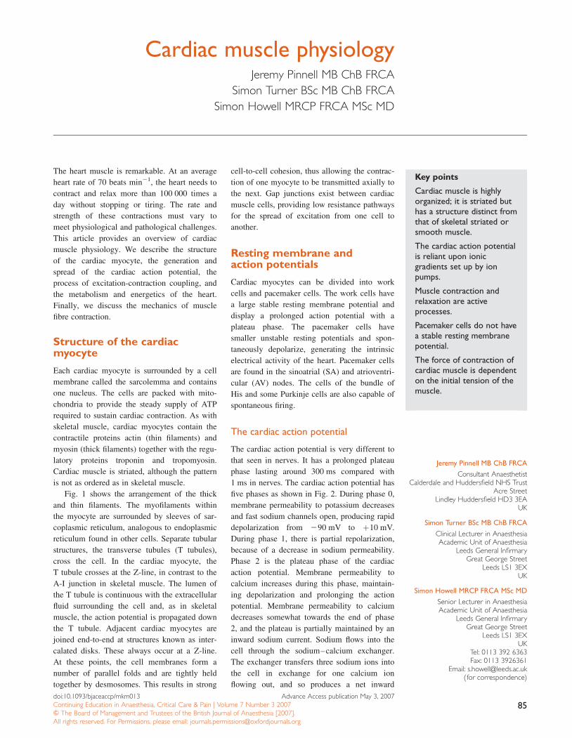
Fig. 1 shows the arrangement of the thick
and thin filaments. The myofilaments within
the myocyte are surrounded by sleeves of sar-
coplasmic reticulum, analogous to endoplasmic
reticulum found in other cells. Separate tubular
structures, the transverse tubules (T tubules),
cross the cell. In the cardiac myocyte, the
T tubule crosses at the Z-line, in contrast to the
A-I junction in skeletal muscle. The lumen of
the T tubule is continuous with the extracellular
fluid surrounding the cell and, as in skeletal
muscle, the action potential is propagated down
the T tubule. Adjacent cardiac myocytes are
joined end-to-end at structures known as inter-
calated disks. These always occur at a Z-line.
At these points, the cell membranes form a
number of parallel folds and are tightly held
together by desmosomes. This results in strong
cell-to-cell cohesion, thus allowing the contrac-
tion of one myocyte to be transmitted axially to
the next. Gap junctions exist between cardiac
muscle cells, providing low resistance pathways
for the spread of excitation from one cell to
another.
Resting membrane andaction potentials
Cardiac myocytes can be divided into work
cells and pacemaker cells. The work cells have
a large stable resting membrane potential and
display a prolonged action potential with a
plateau phase. The pacemaker cells have
smaller unstable resting potentials and spon-
taneously depolarize, generating the intrinsic
electrical activity of the heart. Pacemaker cells
are found in the sinoatrial (SA) and atrioventri-
cular (AV) nodes. The cells of the bundle of
His and some Purkinje cells are also capable of
spontaneous firing.
The cardiac action potential
The cardiac action potential is very different to
that seen in nerves. It has a prolonged plateau
phase lasting around 300 ms compared with
1 ms in nerves. The cardiac action potential has
five phases as shown in Fig. 2. During phase 0,
membrane permeability to potassium decreases
and fast sodium channels open, producing rapid
depolarization from 290 mV to þ10 mV.
During phase 1, there is partial repolarization,
because of a decrease in sodium permeability.
Phase 2 is the plateau phase of the cardiac
action potential. Membrane permeability to
calcium increases during this phase, maintain-
ing depolarization and prolonging the action
potential. Membrane permeability to calcium
decreases somewhat towards the end of phase
2, and the plateau is partially maintained by an
inward sodium current. Sodium flows into the
cell through the sodium–calcium exchanger.
The exchanger transfers three sodium ions into
the cell in exchange for one calcium ion
flowing out, and so produces a net inward
Key points
Cardiac muscle is highlyorganized; it is striated buthas a structure distinct fromthat of skeletal striated orsmooth muscle.
The cardiac action potentialis reliant upon ionicgradients set up by ionpumps.
Muscle contraction andrelaxation are activeprocesses.
Pacemaker cells do not havea stable resting membranepotential.
The force of contraction ofcardiac muscle is dependenton the initial tension of themuscle.
Jeremy Pinnell MB ChB FRCA
Consultant AnaesthetistCalderdale and Huddersfield NHS Trust
Acre StreetLindley Huddersfield HD3 3EA
UK
Simon Turner BSc MB ChB FRCA
Clinical Lecturer in AnaesthesiaAcademic Unit of Anaesthesia
Leeds General InfirmaryGreat George Street
Leeds LS1 3EXUK
Simon Howell MRCP FRCA MSc MD
Senior Lecturer in AnaesthesiaAcademic Unit of Anaesthesia
Leeds General InfirmaryGreat George Street
Leeds LS1 3EXUK
Tel: 0113 392 6363Fax: 0113 3926361
Email: [email protected](for correspondence)
85doi:10.1093/bjaceaccp/mkm013 Advance Access publication May 3, 2007Continuing Education in Anaesthesia, Critical Care & Pain | Volume 7 Number 3 2007& The Board of Management and Trustees of the British Journal of Anaesthesia [2007].All rights reserved. For Permissions, please email: [email protected]
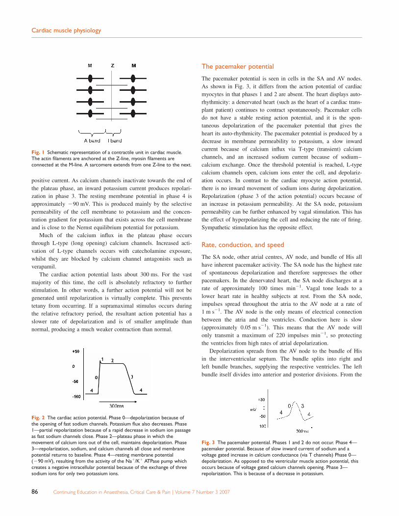
positive current. As calcium channels inactivate towards the end of
the plateau phase, an inward potassium current produces repolari-
zation in phase 3. The resting membrane potential in phase 4 is
approximately 290 mV. This is produced mainly by the selective
permeability of the cell membrane to potassium and the concen-
tration gradient for potassium that exists across the cell membrane
and is close to the Nernst equilibrium potential for potassium.
Much of the calcium influx in the plateau phase occurs
through L-type (long opening) calcium channels. Increased acti-
vation of L-type channels occurs with catecholamine exposure,
whilst they are blocked by calcium channel antagonists such as
verapamil.
The cardiac action potential lasts about 300 ms. For the vast
majority of this time, the cell is absolutely refractory to further
stimulation. In other words, a further action potential will not be
generated until repolarization is virtually complete. This prevents
tetany from occurring. If a supramaximal stimulus occurs during
the relative refractory period, the resultant action potential has a
slower rate of depolarization and is of smaller amplitude than
normal, producing a much weaker contraction than normal.
The pacemaker potential
The pacemaker potential is seen in cells in the SA and AV nodes.
As shown in Fig. 3, it differs from the action potential of cardiac
myocytes in that phases 1 and 2 are absent. The heart displays auto-
rhythmicity: a denervated heart (such as the heart of a cardiac trans-
plant patient) continues to contract spontaneously. Pacemaker cells
do not have a stable resting action potential, and it is the spon-
taneous depolarization of the pacemaker potential that gives the
heart its auto-rhythmicity. The pacemaker potential is produced by a
decrease in membrane permeability to potassium, a slow inward
current because of calcium influx via T-type (transient) calcium
channels, and an increased sodium current because of sodium–
calcium exchange. Once the threshold potential is reached, L-type
calcium channels open, calcium ions enter the cell, and depolariz-
ation occurs. In contrast to the cardiac myocyte action potential,
there is no inward movement of sodium ions during depolarization.
Repolarization (phase 3 of the action potential) occurs because of
an increase in potassium permeability. At the SA node, potassium
permeability can be further enhanced by vagal stimulation. This has
the effect of hyperpolarizing the cell and reducing the rate of firing.
Sympathetic stimulation has the opposite effect.
Rate, conduction, and speed
The SA node, other atrial centres, AV node, and bundle of His all
have inherent pacemaker activity. The SA node has the highest rate
of spontaneous depolarization and therefore suppresses the other
pacemakers. In the denervated heart, the SA node discharges at a
rate of approximately 100 times min21. Vagal tone leads to a
lower heart rate in healthy subjects at rest. From the SA node,
impulses spread throughout the atria to the AV node at a rate of
1 m s21. The AV node is the only means of electrical connection
between the atria and the ventricles. Conduction here is slow
(approximately 0.05 m s21). This means that the AV node will
only transmit a maximum of 220 impulses min21, so protecting
the ventricles from high rates of atrial depolarization.
Depolarization spreads from the AV node to the bundle of His
in the interventricular septum. The bundle splits into right and
left bundle branches, supplying the respective ventricles. The left
bundle itself divides into anterior and posterior divisions. From the
Fig. 1 Schematic representation of a contractile unit in cardiac muscle.The actin filaments are anchored at the Z-line, myosin filaments areconnected at the M-line. A sarcomere extends from one Z-line to the next.
Fig. 2 The cardiac action potential. Phase 0—depolarization because ofthe opening of fast sodium channels. Potassium flux also decreases. Phase1—partial repolarization because of a rapid decrease in sodium ion passageas fast sodium channels close. Phase 2—plateau phase in which themovement of calcium ions out of the cell, maintains depolarization. Phase3—repolarization, sodium, and calcium channels all close and membranepotential returns to baseline. Phase 4—resting membrane potential(290 mV), resulting from the activity of the Naþ/Kþ ATPase pump whichcreates a negative intracellular potential because of the exchange of threesodium ions for only two potassium ions.
Fig. 3 The pacemaker potential. Phases 1 and 2 do not occur. Phase 4—pacemaker potential. Because of slow inward current of sodium and avoltage gated increase in calcium conductance (via T channels) Phase 0—depolarization. As opposed to the ventricular muscle action potential, thisoccurs because of voltage gated calcium channels opening. Phase 3—repolarization. This is because of a decrease in potassium.
Cardiac muscle physiology
86 Continuing Education in Anaesthesia, Critical Care & Pain j Volume 7 Number 3 2007
bundle branches, impulses travel through the ventricular muscle
via a network of Purkinje fibres, at a velocity of 1–4 m s21. The
conducting system is arranged so that the apices of the ventricles
contract before the bases, propelling blood out of the chambers.
Excitation contraction coupling
This is the process linking electrical excitation to contraction.
Calcium has an essential role in this process; a raised intracellular
calcium concentration is the trigger that activates contraction. An
understanding of calcium handling is essential to understanding the
function of the heart. The intracellular calcium ion concentration in
the cardiac myocyte at rest is 0.0001 mM litre21 and that in the
extracellular fluid is 1.2 mM litre21. During the plateau phase of the
action potential, calcium ions flow down this steep concentration
gradient and enter the myocyte. Most of this calcium enters through
the L-type channels, located primarily at sarcolemmal/sarcoplasmic
reticulum junctions. The influx of calcium triggers the release of
further calcium from the sarcoplasmic reticulum via ryanodine recep-
tors. This calcium-triggered calcium release is in contrast to skeletal
muscle, where the action potential triggers calcium release directly.
Free intracellular calcium interacts with the C subunit of troponin.
This leads to a configuration change in the troponin/tropomyosin
complex, allowing actin to interact with myosin. Cross bridge cycling
occurs, leading to a shortening of the sarcomere and resultant muscu-
lar contraction. As intracellular calcium concentrations decrease
during repolarization, calcium dissociates from troponin as intracellu-
lar calcium concentration decreases, resulting in relaxation. Diastolic
relaxation is an active (ATP-dependent) process. Calcium transport
out of the cytosol occurs via a sarcoplasmic reticulum Ca2þ-ATPase,
through sarcolemmal Naþ/Ca2þ exchange, via a sarcolemmal
Ca2þ-ATPase, and finally by utilizing a mitochondrial Ca2þ uniport.
The strength of a contraction may be varied by increasing the
amount of free intracellular calcium, by altering the sensitivity of
the myofilaments to calcium, or both. The latter occurs during
stretching of the myofilaments and is responsible for the Frank–
Starling mechanism (discussed later). Myofilament calcium sensi-
tivity is reduced by acidosis. High concentrations of phosphate and
magnesium also impair cardiac function.
Catecholamines activate beta-adrenergic receptors in the heart
to produce a G-protein mediated increase in cAMP and enhanced
activity of a cAMP-dependent protein kinase. This leads to the
phosphorylation of calcium membrane channels, enhancing
calcium entry into the cell. Phosphorylation of myosin also occurs,
increasing the rate of cross bridge cycling. Catecholamines also
increase the rate of re-uptake of calcium into the sarcoplasmic reti-
culum, thus aiding relaxation.
Metabolism and energetics
The oxygen consumption of the beating heart is on average 9 ml
per 100 g min21 at rest. This increases during exercise. Oxygen
extraction from blood in the coronary circulation is high; therefore,
an increase in oxygen demand must be met by an increase in cor-
onary blood flow.
The heart is very versatile in its use of metabolic substrates.
Carbohydrate utilization accounts for 35–40% of total oxygen con-
sumption. Glucose and lactate are used in roughly equal pro-
portions. A small amount of energy may be derived from ketones;
however, 60% of the basal energy requirement is provided by fat.
The proportion of substrates utilized may vary depending on the
nutritional state of the person. After a large meal containing
glucose, more pyruvate and lactate are used. During periods of
starvation, more fat is utilized. Insulin enhances glucose uptake
into cardiac myocytes, and in untreated diabetes proportionally
more fat is utilized. Normally ,1% of the energy used by the
myocardium is produced by anaerobic metabolism. This proportion
increases during periods of hypoxaemia; however, lactic acidosis
impairs myocardial function and can ultimately lead to myocardial
cell death.
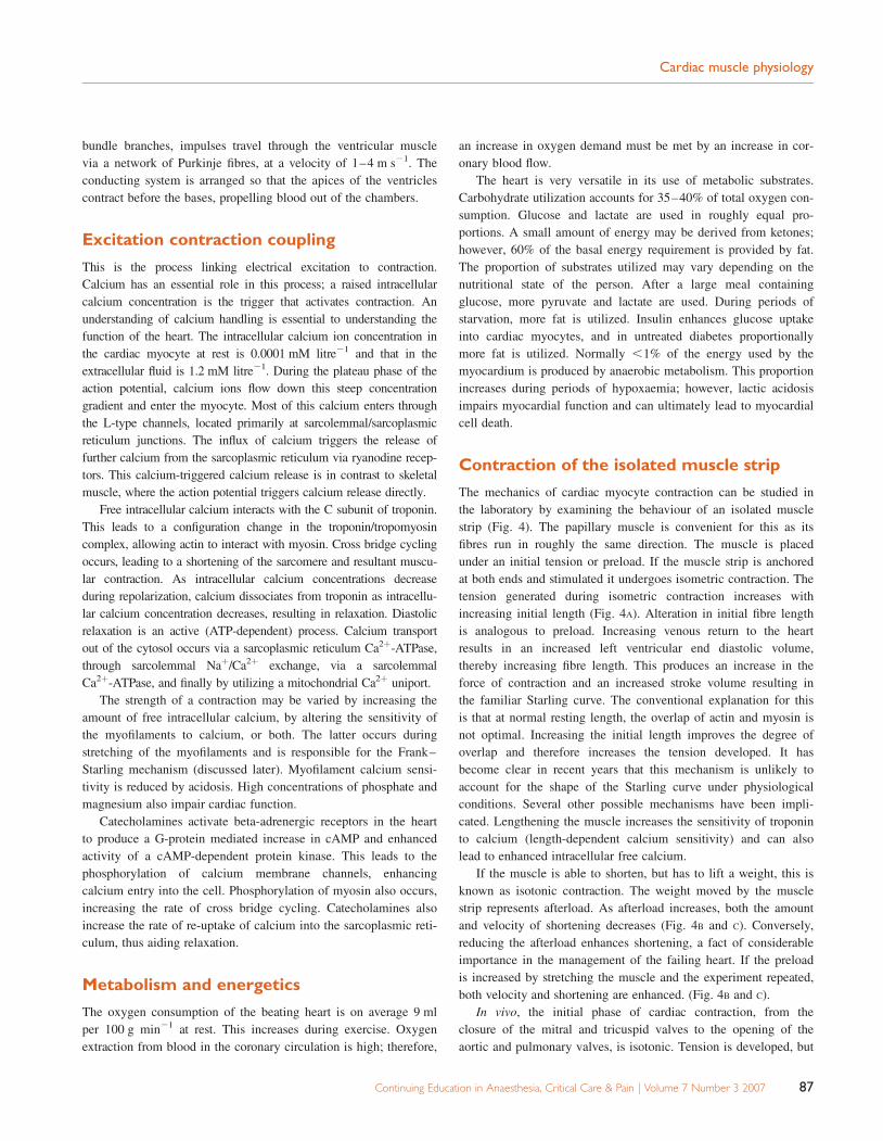
Contraction of the isolated muscle strip
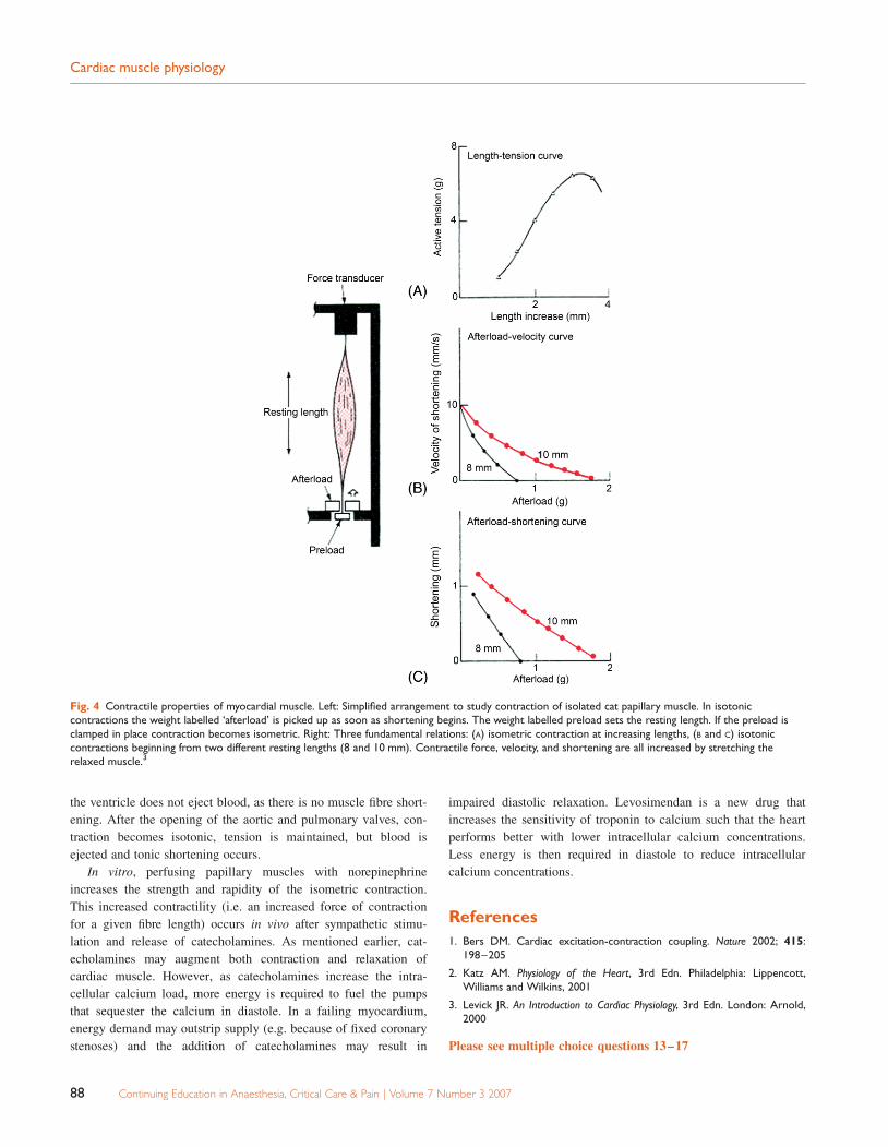
The mechanics of cardiac myocyte contraction can be studied in
the laboratory by examining the behaviour of an isolated muscle
strip (Fig. 4). The papillary muscle is convenient for this as its
fibres run in roughly the same direction. The muscle is placed
under an initial tension or preload. If the muscle strip is anchored
at both ends and stimulated it undergoes isometric contraction. The
tension generated during isometric contraction increases with
increasing initial length (Fig. 4A). Alteration in initial fibre length
is analogous to preload. Increasing venous return to the heart
results in an increased left ventricular end diastolic volume,
thereby increasing fibre length. This produces an increase in the
force of contraction and an increased stroke volume resulting in
the familiar Starling curve. The conventional explanation for this
is that at normal resting length, the overlap of actin and myosin is
not optimal. Increasing the initial length improves the degree of
overlap and therefore increases the tension developed. It has
become clear in recent years that this mechanism is unlikely to
account for the shape of the Starling curve under physiological
conditions. Several other possible mechanisms have been impli-
cated. Lengthening the muscle increases the sensitivity of troponin
to calcium (length-dependent calcium sensitivity) and can also
lead to enhanced intracellular free calcium.
If the muscle is able to shorten, but has to lift a weight, this is
known as isotonic contraction. The weight moved by the muscle
strip represents afterload. As afterload increases, both the amount
and velocity of shortening decreases (Fig. 4B and C). Conversely,
reducing the afterload enhances shortening, a fact of considerable
importance in the management of the failing heart. If the preload
is increased by stretching the muscle and the experiment repeated,
both velocity and shortening are enhanced. (Fig. 4B and C).
In vivo, the initial phase of cardiac contraction, from the
closure of the mitral and tricuspid valves to the opening of the
aortic and pulmonary valves, is isotonic. Tension is developed, but
Cardiac muscle physiology
Continuing Education in Anaesthesia, Critical Care & Pain j Volume 7 Number 3 2007 87
the ventricle does not eject blood, as there is no muscle fibre short-
ening. After the opening of the aortic and pulmonary valves, con-
traction becomes isotonic, tension is maintained, but blood is
ejected and tonic shortening occurs.
In vitro, perfusing papillary muscles with norepinephrine
increases the strength and rapidity of the isometric contraction.
This increased contractility (i.e. an increased force of contraction
for a given fibre length) occurs in vivo after sympathetic stimu-
lation and release of catecholamines. As mentioned earlier, cat-
echolamines may augment both contraction and relaxation of
cardiac muscle. However, as catecholamines increase the intra-
cellular calcium load, more energy is required to fuel the pumps
that sequester the calcium in diastole. In a failing myocardium,
energy demand may outstrip supply (e.g. because of fixed coronary
stenoses) and the addition of catecholamines may result in
impaired diastolic relaxation. Levosimendan is a new drug that
increases the sensitivity of troponin to calcium such that the heart
performs better with lower intracellular calcium concentrations.
Less energy is then required in diastole to reduce intracellular
calcium concentrations.
References
1. Bers DM. Cardiac excitation-contraction coupling. Nature 2002; 415:198–205
2. Katz AM. Physiology of the Heart, 3rd Edn. Philadelphia: Lippencott,Williams and Wilkins, 2001
3. Levick JR. An Introduction to Cardiac Physiology, 3rd Edn. London: Arnold,2000
Please see multiple choice questions 13–17
Fig. 4 Contractile properties of myocardial muscle. Left: Simplified arrangement to study contraction of isolated cat papillary muscle. In isotoniccontractions the weight labelled ‘afterload’ is picked up as soon as shortening begins. The weight labelled preload sets the resting length. If the preload isclamped in place contraction becomes isometric. Right: Three fundamental relations: (A) isometric contraction at increasing lengths, (B and C) isotoniccontractions beginning from two different resting lengths (8 and 10 mm). Contractile force, velocity, and shortening are all increased by stretching therelaxed muscle.3
Cardiac muscle physiology
88 Continuing Education in Anaesthesia, Critical Care & Pain j Volume 7 Number 3 2007