Embed Size (px)
Citation preview
Dmaeoia
FPsIr
2
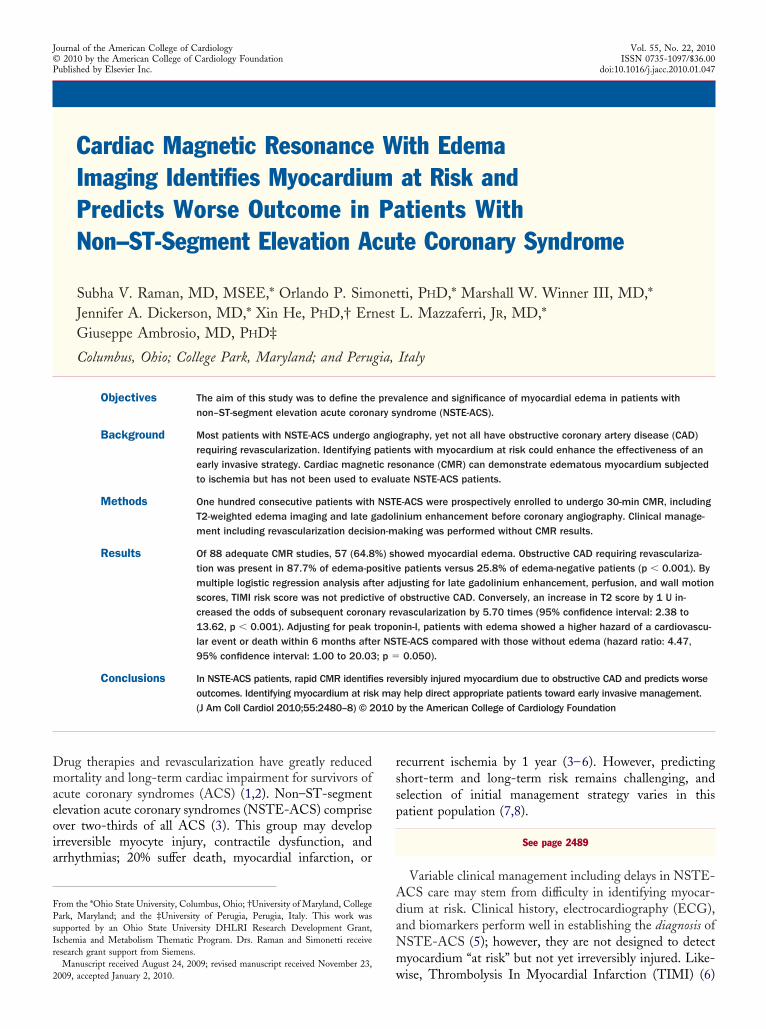
Journal of the American College of Cardiology Vol. 55, No. 22, 2010© 2010 by the American College of Cardiology Foundation ISSN 0735-1097/$36.00P
Cardiac Magnetic Resonance With EdemaImaging Identifies Myocardium at Risk andPredicts Worse Outcome in Patients WithNon–ST-Segment Elevation Acute Coronary Syndrome
Subha V. Raman, MD, MSEE,* Orlando P. Simonetti, PHD,* Marshall W. Winner III, MD,*Jennifer A. Dickerson, MD,* Xin He, PHD,† Ernest L. Mazzaferri, JR, MD,*Giuseppe Ambrosio, MD, PHD‡
Columbus, Ohio; College Park, Maryland; and Perugia, Italy
Objectives The aim of this study was to define the prevalence and significance of myocardial edema in patients withnon–ST-segment elevation acute coronary syndrome (NSTE-ACS).
Background Most patients with NSTE-ACS undergo angiography, yet not all have obstructive coronary artery disease (CAD)requiring revascularization. Identifying patients with myocardium at risk could enhance the effectiveness of anearly invasive strategy. Cardiac magnetic resonance (CMR) can demonstrate edematous myocardium subjectedto ischemia but has not been used to evaluate NSTE-ACS patients.
Methods One hundred consecutive patients with NSTE-ACS were prospectively enrolled to undergo 30-min CMR, includingT2-weighted edema imaging and late gadolinium enhancement before coronary angiography. Clinical manage-ment including revascularization decision-making was performed without CMR results.
Results Of 88 adequate CMR studies, 57 (64.8%) showed myocardial edema. Obstructive CAD requiring revasculariza-tion was present in 87.7% of edema-positive patients versus 25.8% of edema-negative patients (p � 0.001). Bymultiple logistic regression analysis after adjusting for late gadolinium enhancement, perfusion, and wall motionscores, TIMI risk score was not predictive of obstructive CAD. Conversely, an increase in T2 score by 1 U in-creased the odds of subsequent coronary revascularization by 5.70 times (95% confidence interval: 2.38 to13.62, p � 0.001). Adjusting for peak troponin-I, patients with edema showed a higher hazard of a cardiovascu-lar event or death within 6 months after NSTE-ACS compared with those without edema (hazard ratio: 4.47,95% confidence interval: 1.00 to 20.03; p � 0.050).
Conclusions In NSTE-ACS patients, rapid CMR identifies reversibly injured myocardium due to obstructive CAD and predicts worseoutcomes. Identifying myocardium at risk may help direct appropriate patients toward early invasive management.(J Am Coll Cardiol 2010;55:2480–8) © 2010 by the American College of Cardiology Foundation
ublished by Elsevier Inc. doi:10.1016/j.jacc.2010.01.047
rssp
AdaNm
rug therapies and revascularization have greatly reducedortality and long-term cardiac impairment for survivors of
cute coronary syndromes (ACS) (1,2). Non–ST-segmentlevation acute coronary syndromes (NSTE-ACS) comprisever two-thirds of all ACS (3). This group may developrreversible myocyte injury, contractile dysfunction, andrrhythmias; 20% suffer death, myocardial infarction, or
rom the *Ohio State University, Columbus, Ohio; †University of Maryland, Collegeark, Maryland; and the ‡University of Perugia, Perugia, Italy. This work wasupported by an Ohio State University DHLRI Research Development Grant,schemia and Metabolism Thematic Program. Drs. Raman and Simonetti receiveesearch grant support from Siemens.
wManuscript received August 24, 2009; revised manuscript received November 23,
009, accepted January 2, 2010.
ecurrent ischemia by 1 year (3–6). However, predictinghort-term and long-term risk remains challenging, andelection of initial management strategy varies in thisatient population (7,8).
See page 2489
Variable clinical management including delays in NSTE-CS care may stem from difficulty in identifying myocar-ium at risk. Clinical history, electrocardiography (ECG),nd biomarkers perform well in establishing the diagnosis ofSTE-ACS (5); however, they are not designed to detectyocardium “at risk” but not yet irreversibly injured. Like-
ise, Thrombolysis In Myocardial Infarction (TIMI) (6)
addmBiadimbracr
t((oiibfptpps
M
SNtbeEeenc
mv
EmAftaCtCpNiGmTcfqmffio(wbrbppbogapscnIb
C
3s
2481JACC Vol. 55, No. 22, 2010 Raman et al.June 1, 2010:2480–8 CMR in NSTE-ACS
nd other global risk scores on the basis of clinical and ECGata provide an average estimate of an individual’s risk ofeath or major ischemic events but do not specifically guideanagement decisions regarding timing of angiography.ecause myocardium at risk may be salvaged by revascular-
zation, an invasive strategy to identify obstructive coronaryrtery disease (CAD) is usually pursued once ACS isiagnosed (5). For patients with myocardium at risk, early
ntervention is invaluable (9); however, for patients withoutyocardium at risk, a costly invasive strategy confers no
enefit and may add unnecessary bleeding and proceduralisk (4). A diagnostic approach that identifies myocardiumt risk within the heterogeneous NSTE-ACS populationould facilitate timely revascularization and concentrateesource use to the most appropriate patients.
Extensive preclinical and human studies have establishedhat T2 signal hyperintensity by cardiac magnetic resonanceCMR) indicates increased myocardial water content10–14). T2 may increase within 30 min of ischemianset—before detectable injury by troponin or late gadolin-um enhancement (LGE) (15,16). T2-weighted CMR todentify myocardium that has recently suffered ischemia haseen employed to distinguish ACS from non-ACS and newrom old infarct scar in patients with undifferentiated chestain (14,17–19). We sought to extend this work to inves-igate whether CMR with edema imaging could stratifyatients admitted with NSTE-ACS to identify higher-riskatients who would warrant an early invasive managementtrategy.
ethods
tudy population. Consecutive patients hospitalized withSTE-ACS awaiting coronary angiography were prospec-
ively enrolled over a 20-month period. Diagnosis requiredoth suspected cardiac chest pain or anginal equivalent andither abnormal serum troponin-I (TnI) level or ischemicCG changes (20). Patients under age 30 years were
xcluded to minimize coronary events mediated by nonath-rosclerotic processes. Contraindication to magnetic reso-ance such as pacemaker or evidence of illicit drug ingestiononstituted additional exclusion criteria. Clinical decision-
ardiac Magnetic Resonance Scan ParametersTable 1 Cardiac Magnetic Resonance Scan Parameters
Sequence TypeParallel
Acceleration
Function Real-time SSFP TSENSE rate 3
Edema T2-weighted triple-inversion STIR segmentedturbo spin echo
GRAPPA rate 2 2
Perfusion Single-shot saturation recovery GRE-EPI TSENSE rate 2
Necrosis Single-shot inversion recovery steady-statefree-precession
GRAPPA rate 2
CH � 3-chamber; GRAPPA � generalized autocalibrating partially parallel acquisitions; GRE-EPI � grateady-state free-precession; STIR � short tau inversion recovery; TR/TE � repetition time/echo time; TS
aking was performed by pro-iders blinded to CMR results.
Medical history, clinical andCG findings, and serologicalarkers were recorded at entry.ll patients provided written in-
ormed consent to participate inhis Institutional Review Board-pproved protocol.MR examination. Examina-
ions were performed with a 1.5-TMR system and 12-element
hased-array cardiac coil (MAG-ETOM Avanto, Siemens Med-
cal Solutions, Inc., Erlangen,ermany). A physician providedonitoring throughout the study.he CMR protocol (Table 1) in-
luded 4 acquisition types in theollowing order (the first 2 ac-uired pre-contrast): real-timeulti-plane cine imaging suitable
or wall motion assessment, T2-weighted imaging, restingrst-pass perfusion imaging, and LGE. Cine images werebtained in horizontal long-axis (HLA), vertical long-axisVLA), 3-chamber, and contiguous short-axis (SAX) planesith non-breathhold, real-time acquisition (21). T2-weightedreath-hold (12 to 15 s) turbo spin echo short tau inversionecovery images of the myocardium were obtained in 10-mmasal, mid, and apical SAX, VLA, 3-chamber, and HLAlanes (12). Myocardial perfusion acquisition used an echo-lanar first-pass imaging technique in four 10-mm planes:asal/mid/apical SAX, and HLA (22). Perfusion images werebtained at rest during intravenous infusion of 0.1 mmol/kgadolinium diethylenetriamine penta-acetic acid. Ten minutesfter additional 0.1 mmol/kg gadolinium diethylenetriamineenta-acetic acid administration, multi-plane nonbreathholdingle-shot LGE images were obtained in the same planes asine imaging, with appropriate inversion time selection to nullormal myocardium (23).mage analysis. Two CMR experts (S.V.R., O.P.S.)linded to clinical information rated by consensus, after
Abbreviationsand Acronyms
CAD � coronary arterydisease
CMR � cardiac magneticresonance
ECG � electrocardiography
HLA � horizontal long-axis
IQR � interquartile range
LGE � late gadoliniumenhancement
LV � left ventricular
NSTE-ACS � non–ST-segment elevation acutecoronary syndrome
SAX � short axis
TIMI � Thrombolysis InMyocardial Infarction
TnI � troponin-I
VLA � vertical long-axis
Spatial ResolutionTemporal
Resolution (ms) Acquisition Planes
0 3.75 mm � 2.1 mm8-mm slice thickness
62 HLA, VLA, contiguous SAX(10–12 slices), 3CH
80 1.6 mm � 1.6 mm8-mm slice thickness
156 HLA, VLA, 3 SAX (base,mid, and apical), 3CH
2 3.1 mm � 2.5 mm10-mm slice thickness
70 3 SAX (base, mid, andapical), HLA
3 2.8 mm � 2.1 mm8-mm slice thickness
285 HLA, VLA, contiguous SAX(10–12 slices), 3CH
dient-echo, echo-planar hybrid imaging; HLA � horizontENSE � Time-adaptive SENSitivity Encoding; VLA � vertic
TR/TE(ms)
2.3/1.
� RR/
5.8/1.
2.8/1.
al long-axis; SAX � short-axis; SSFP �
al long-axis.

iriwT0f1sess
wavSaSbvmdo
rmeSancfiwvter
uaiLst6p
R
Sdvecpps
CotptfatCiMoMmni
pi
2482 Raman et al. JACC Vol. 55, No. 22, 2010CMR in NSTE-ACS June 1, 2010:2480–8
ndependent review, each patient’s CMR images byecording 17-segment (24) scores for each of the follow-ng: left ventricular (LV) myocardial T2 signal intensity,all motion, perfusion, and LGE. Each variable was scored:2: 0-normal, 1-intramyocardial hyperintensity; wall motion:-normal, 1-hypokinetic, 2-akinetic, 3-dyskinetic; per-usion: 0-normal, 1-abnormal; and LGE: 0-none,-hyperenhancement, 2-microvascular obstruction. Con-ensus scores were obtained for each study by 2 CMRxperts. Segmental T2, perfusion, wall motion, and LGEcores were summed to yield patient-level aggregatecores.
The LV volumes and ejection fraction were estimatedith the formula: volume � 0.85 � area^2/length, ml,
nd ejection fraction � (stroke volume)/end-diastolicolume, % (25).ubsequent clinical care and outcomes. Invasive coronaryngiography was done according to standard techniques.ignificant coronary artery stenosis (�70%) was identifiedy an independent interventional cardiologist (ELM) byisual assessment. Subsequent management decisions wereade by the clinical team on the basis of clinically available
ata, including findings at coronary angiography but with-ut knowledge of CMR results.Follow-up at 6 months by phone interview and chart
eview documented the occurrence of death, heart failure,ajor arrhythmia, or hospital stay for an acute coronary
vent.tatistical analysis. The mean values of continuous vari-bles were compared with the 2-sample t test, given theormal distribution of the variables, and correlation betweenontinuous variables was computed with the Pearson coef-cient. Otherwise, comparison of median values was doneith the 2-sample Wilcoxon rank-sum test. Prevalenceariables were compared with the 2-sample test for propor-ion. To test the relationship between presence/absence ofdema and normal/abnormal initial TnI, a tetrachoric cor-elation coefficient was calculated. Logistic regression was
Baseline Characteristics of Study PopulationTable 2 Baseline Characteristics of Study P
All Patients(n � 88)
Age, yrs 59.1 � 12.1
Male 57 (64.8)
Body mass index, kg/m2 29.4 (25.8–33.1)
Diabetes 38 (43.2)
Smoker 45 (51.1)
Hypertension 69 (78.4)
Prior CAD 47 (53.4)
Initial troponin-I, mg/dl 0.11 (0.01–0.99)
Peak troponin-I, mg/dl 8.2 � 16.2
TIMI risk score 4 (3–5)
Values are mean � SD, n (%), or median (interquartile range). *p � 0.05 conCAD � coronary artery disease; IQR � interquartile range; TIMI � Thrombo
sed to model the relationship between edema-positivitynd peak TnI as well as LGE score. Logistic regression ofntervention on T2 score was performed by adjusting forGE score, wall motion score, and myocardial perfusion
core. Cox proportional hazards regression model was usedo evaluate the composite occurrence of events within-month follow-up with a group predictor on the basis ofresence/absence of edema.
esults
tudy population. Six patients screened were not enrolledue to respiratory or hemodynamic instability (n � 5) orentricular tachyarrhythmia (n � 1). Of 100 patientsnrolled, 5 could not complete CMR examination due tolaustrophobia; T2 image quality was inadequate in 7atients. Patient characteristics for the remaining studyopulation (n � 88) are summarized in Table 2; all were ininus rhythm at the time of CMR examination.
Subject race was African-American in 11 (13%) andaucasian in the remainder. The majority of patients wereverweight (median body mass index 30.1 kg/m2, interquar-ile range [IQR] 25.8 to 33.0 kg/m2). Thirty-three (38%)atients in this study group were transferred to our institu-ion after initial presentation to another hospital. Peak TnIor patients who were troponin-positive (n � 77) occurredt admission in 9 (12%), after admission but before cathe-erization in 49 (64%), and after catheterization in 19 (25%).
MR findings. Global LV systolic function was preservedn this population: ejection fraction averaged 61 � 14%.
edian wall motion score was 4 (IQR 1 to 10), with 80%f patients having at least 1 dysfunctional LV segment.edian perfusion score was 1 (IQR 0 to 3). Similarly,edian LGE score was 1 (IQR 0 to 3); LGE score was
on-zero in 53 patients (60.2%). As expected, LGE scorencreased with increasing peak TnI (R � 0.28, p � 0.05).
T2-weighted and LGE imaging allowed assessment ofresence and extent of myocardial edema and irreversiblenjury (Fig. 1). Edema was detected in 57 patients (64.8%),
tion
ts Managededically
n � 30)
Patients ReceivingRevascularization
(n � 58) p Value
.6 � 11.3 59.4 � 12.5 0.78
(53.3) 41 (69.4) 0.11
(23.4–31.8) 30.2 (27.4–34.2) 0.02*
(40.0) 26 (44.8) 0.66
(40.0) 33 (56.9) 0.13
(86.7) 43 (74.1) 0.18
(60.0) 29 (50.0) 0.37
(0.01–0.30) 0.14 (0.02–1.13) 0.17
.3 � 14.7 7.9 � 18.9 0.89
(3–5) 4.5 (3–5) 0.47
opula
PatienM(
58
16
27.5
12
12
26
18
0.05
8
4
sidered significant.lysis In Myocardial Infarction.
wsrwCewtp7
mtnas1(
etIpdwisTs0pl2aAc
PM
V
2483JACC Vol. 55, No. 22, 2010 Raman et al.June 1, 2010:2480–8 CMR in NSTE-ACS
ith a median of 2 (IQR 0 to 3) segments per patienthowing edema. Clinical characteristics, including TIMIisk score, were similar between patients with versus thoseithout edema (Table 3). Time from initial admission toMR was also similar in patients with versus those without
dema (45.9 � 36.1 h vs. 53.9 � 52.3 h, p � 0.45). Thereas greater prevalence of myocardial edema in patients
ransferred in from an outside hospital versus patientsresenting directly to Ohio State University (41 of 55 or4% vs. 16 of 33 or 48%, p � 0.01).
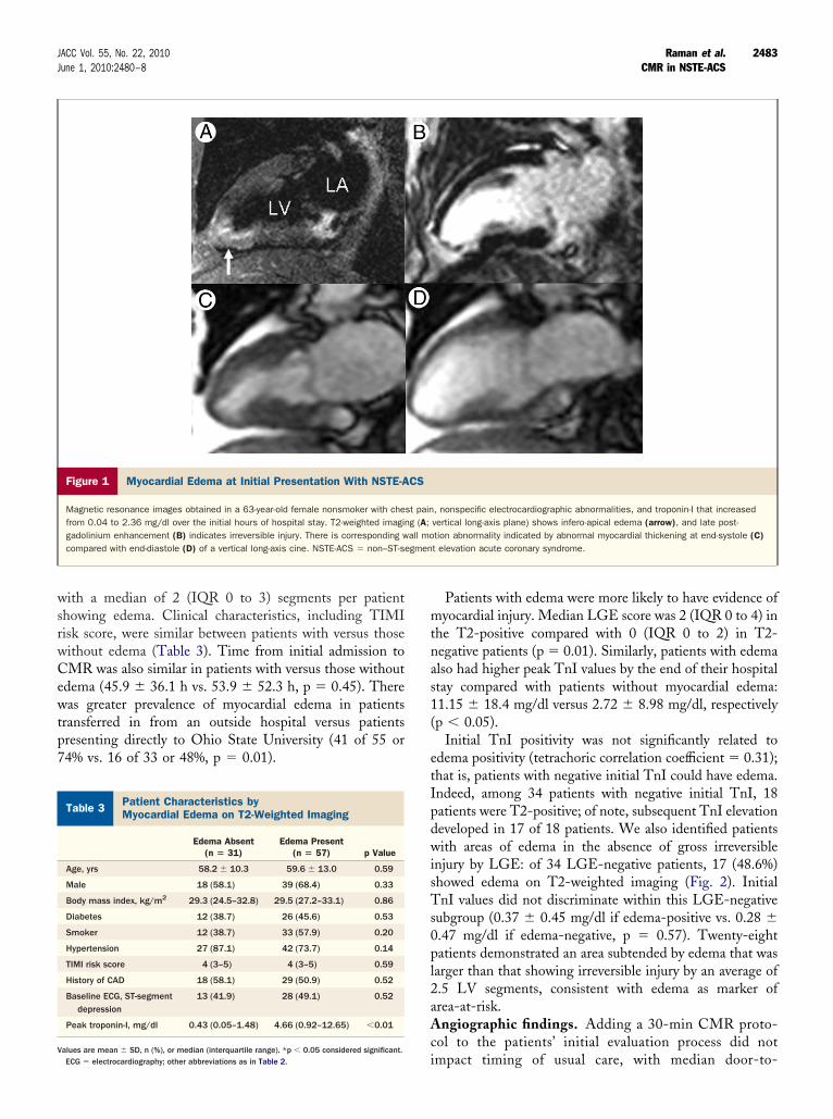
Figure 1 Myocardial Edema at Initial Presentation With NSTE-A
Magnetic resonance images obtained in a 63-year-old female nonsmoker with chefrom 0.04 to 2.36 mg/dl over the initial hours of hospital stay. T2-weighted imagingadolinium enhancement (B) indicates irreversible injury. There is corresponding wcompared with end-diastole (D) of a vertical long-axis cine. NSTE-ACS � non–ST-se
atient Characteristics byyocardial Edema on T2-Weighted ImagingTable 3 Patient Characteristics byMyocardial Edema on T2-Weighted Imaging
Edema Absent(n � 31)
Edema Present(n � 57) p Value
Age, yrs 58.2 � 10.3 59.6 � 13.0 0.59
Male 18 (58.1) 39 (68.4) 0.33
Body mass index, kg/m2 29.3 (24.5–32.8) 29.5 (27.2–33.1) 0.86
Diabetes 12 (38.7) 26 (45.6) 0.53
Smoker 12 (38.7) 33 (57.9) 0.20
Hypertension 27 (87.1) 42 (73.7) 0.14
TIMI risk score 4 (3–5) 4 (3–5) 0.59
History of CAD 18 (58.1) 29 (50.9) 0.52
Baseline ECG, ST-segmentdepression
13 (41.9) 28 (49.1) 0.52
Peak troponin-I, mg/dl 0.43 (0.05–1.48) 4.66 (0.92–12.65) �0.01
ialues are mean � SD, n (%), or median (interquartile range). *p � 0.05 considered significant.ECG � electrocardiography; other abbreviations as in Table 2.
Patients with edema were more likely to have evidence ofyocardial injury. Median LGE score was 2 (IQR 0 to 4) in
he T2-positive compared with 0 (IQR 0 to 2) in T2-egative patients (p � 0.01). Similarly, patients with edemalso had higher peak TnI values by the end of their hospitaltay compared with patients without myocardial edema:1.15 � 18.4 mg/dl versus 2.72 � 8.98 mg/dl, respectivelyp � 0.05).
Initial TnI positivity was not significantly related todema positivity (tetrachoric correlation coefficient � 0.31);hat is, patients with negative initial TnI could have edema.ndeed, among 34 patients with negative initial TnI, 18atients were T2-positive; of note, subsequent TnI elevationeveloped in 17 of 18 patients. We also identified patientsith areas of edema in the absence of gross irreversible
njury by LGE: of 34 LGE-negative patients, 17 (48.6%)howed edema on T2-weighted imaging (Fig. 2). InitialnI values did not discriminate within this LGE-negative
ubgroup (0.37 � 0.45 mg/dl if edema-positive vs. 0.28 �.47 mg/dl if edema-negative, p � 0.57). Twenty-eightatients demonstrated an area subtended by edema that was
arger than that showing irreversible injury by an average of.5 LV segments, consistent with edema as marker ofrea-at-risk.ngiographic findings. Adding a 30-min CMR proto-
ol to the patients’ initial evaluation process did not
, nonspecific electrocardiographic abnormalities, and troponin-I that increasedvertical long-axis plane) shows infero-apical edema (arrow), and late post-tion abnormality indicated by abnormal myocardial thickening at end-systole (C)t elevation acute coronary syndrome.
CS
st paing (A;all mogmen
mpact timing of usual care, with median door-to-
cttcpbbca
cCTlcdpt(pwoibp
bfiptdsp
2484 Raman et al. JACC Vol. 55, No. 22, 2010CMR in NSTE-ACS June 1, 2010:2480–8
atheterization time of 37 (IQR 22 to 63) h. Identifica-ion of �70% stenosis by angiography resulted in percu-aneous coronary intervention alone in 45 patients,oronary artery bypass graft surgery alone in 10, and bothercutaneous coronary intervention and coronary arteryypass graft surgery in 3 for a total of 58 (66%) patientseing revascularized versus 30 (34%) without significantoronary stenosis treated with medical managementlone.
Clinical and functional variables were largely nondis-riminating between patients with versus those withoutAD requiring revascularization (Table 2). MedianIMI risk score was 4.5 in patients who were revascu-
arized and 4.0 in those treated medically (p � 0.47). Inontrast, presence of edema was an extremely powerfuliscriminator: 50 of 57 (87.7%) patients showing T2-ositivity had obstructive CAD requiring revasculariza-ion, compared with 8 of 31 (25.8%) T2-negative patientsp � 0.001) (Fig. 3). After controlling for LGE score,erfusion score, and wall motion score, TIMI risk scoreas not predictive, whereas T2 score was predictive ofbstructive CAD requiring revascularization; an increasen T2 score by 1 U increased the odds of revascularizationy 5.70 times (95% confidence interval: 2.38 to 13.62,
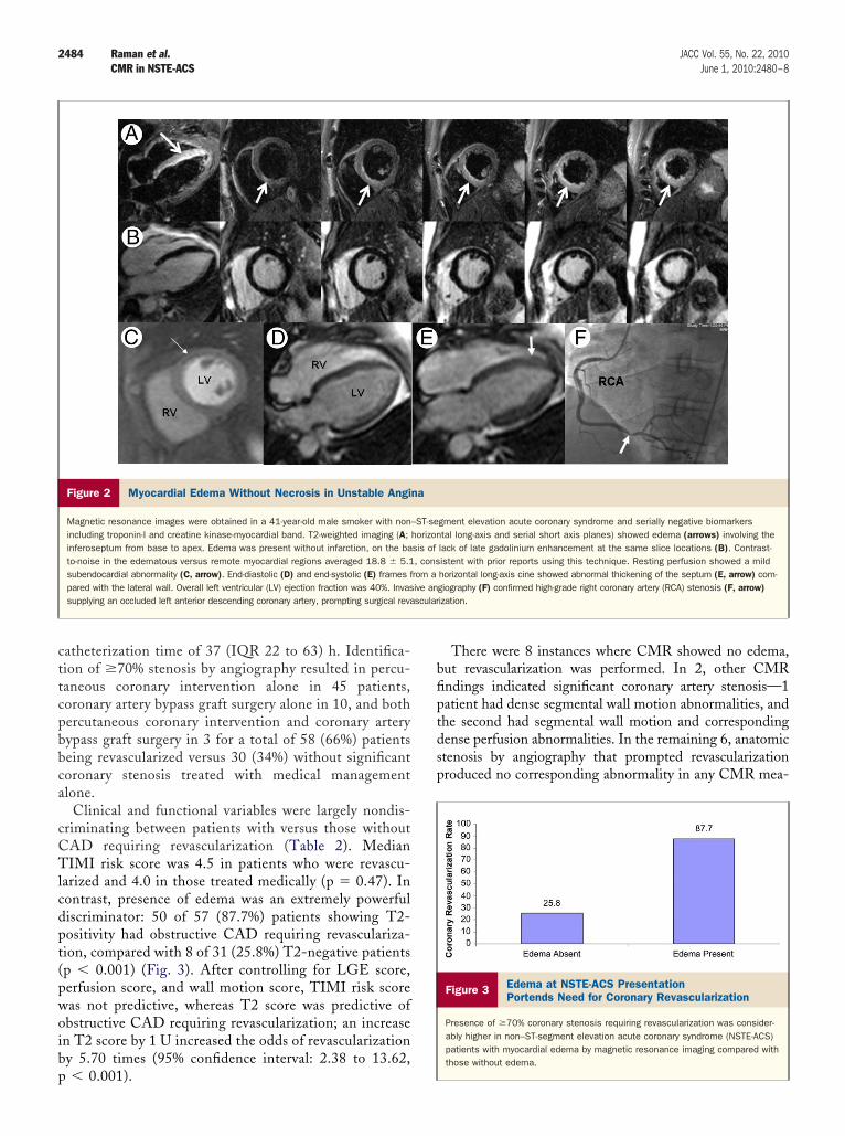
Figure 2 Myocardial Edema Without Necrosis in Unstable Angi
Magnetic resonance images were obtained in a 41-year-old male smoker with nonincluding troponin-I and creatine kinase-myocardial band. T2-weighted imaging (A;inferoseptum from base to apex. Edema was present without infarction, on the bato-noise in the edematous versus remote myocardial regions averaged 18.8 � 5.1subendocardial abnormality (C, arrow). End-diastolic (D) and end-systolic (E) frames fpared with the lateral wall. Overall left ventricular (LV) ejection fraction was 40%. Invassupplying an occluded left anterior descending coronary artery, prompting surgical reva
� 0.001).
There were 8 instances where CMR showed no edema,ut revascularization was performed. In 2, other CMRndings indicated significant coronary artery stenosis—1atient had dense segmental wall motion abnormalities, andhe second had segmental wall motion and correspondingense perfusion abnormalities. In the remaining 6, anatomictenosis by angiography that prompted revascularizationroduced no corresponding abnormality in any CMR mea-
gment elevation acute coronary syndrome and serially negative biomarkerstal long-axis and serial short axis planes) showed edema (arrows) involving thelack of late gadolinium enhancement at the same slice locations (B). Contrast-istent with prior reports using this technique. Resting perfusion showed a mildhorizontal long-axis cine showed abnormal thickening of the septum (E, arrow) com-giography (F) confirmed high-grade right coronary artery (RCA) stenosis (F, arrow)ization.
Figure 3 Edema at NSTE-ACS PresentationPortends Need for Coronary Revascularization
Presence of �70% coronary stenosis requiring revascularization was consider-ably higher in non–ST-segment elevation acute coronary syndrome (NSTE-ACS)patients with myocardial edema by magnetic resonance imaging compared withthose without edema.
na
–ST-sehorizonsis of, consrom aive anscular
swsw
rstflwCnoraNrfppo(
marame2
D
Iueplr
2485JACC Vol. 55, No. 22, 2010 Raman et al.June 1, 2010:2480–8 CMR in NSTE-ACS
ure (i.e., wall motion, perfusion, T2 imaging, and LGEere all normal). Further evaluation of coronary lesion
ignificance with techniques such as fractional flow reserveas not done.In 7 instances, CMR showed edema, but coronary
evascularization was not performed. All 7 patients hadignificant coronary atherosclerosis; however, 3 had noechnically suitable anatomic targets for revascularization. Aourth had angiographic findings suggestive of plaque thatikely produced distal embolization and myocardial sequelaeithout residual stenosis.linical outcomes. Five patients had disconnected phoneumbers without recurrent hospital stays at our institutionr the facility where they had initially presented. In theemaining patients, 16 events occurred during the 6 monthsfter initial NSTE-ACS admission: 12 (14%) had recurrentSTE-ACS (7 unstable angina, 5 subendocardial MI)
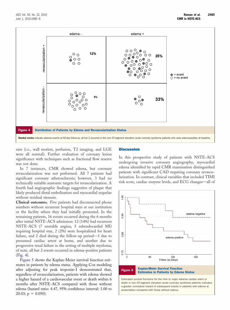
equiring hospital stay, 2 (2%) were hospitalized for heartailure, and 2 died during the follow-up period—1 due toresumed cardiac arrest at home, and another due torogressive renal failure in the setting of multiple myeloma;f note, all but 2 events occurred in edema-positive patientsFig. 4).
Figure 5 shows the Kaplan-Meier survival function esti-ates in patients by edema status. Applying Cox modeling
fter adjusting for peak troponin-I demonstrated that,egardless of revascularization, patients with edema showedhigher hazard of a cardiovascular event or death within 6onths after NSTE-ACS compared with those without
dema (hazard ratio: 4.47, 95% confidence interval: 1.00 to
Figure 4 Distribution of Patients by Edema and Revascularizati
Shaded circles indicate adverse events at 60-day follow-up; all but 2 occurred in the non–
0.03; p � 0.050).
iscussion
n this prospective study of patients with NSTE-ACSndergoing invasive coronary angiography, myocardialdema identified by rapid CMR examination distinguishedatients with significant CAD requiring coronary revascu-
arization. In contrast, clinical variables that included TIMIisk score, cardiac enzyme levels, and ECG changes—all of
atus
ent elevation acute coronary syndrome patients who were edema-positive at baseline.
Figure 5 Kaplan-Meier Survival FunctionEstimates in Patients by Edema Status
Estimated survival functions for the time to major adverse cardiac event ordeath in non–ST-segment elevation acute coronary syndrome patients indicatesa greater cumulative hazard of subsequent events in patients with edema atpresentation compared with those without edema.
on St
ST-segm

wdNrt
epblioenmwInhFNtdoTenrfi
pavtwcBcmieOCmtewwdrSwrAi
anupts
fmsnnlrc(CCnmraLscswvcisrC
tboFimiIwgtwrqNrp
C
R
2486 Raman et al. JACC Vol. 55, No. 22, 2010CMR in NSTE-ACS June 1, 2010:2480–8
hich are nonetheless essential to diagnose NSTE-ACS—id not discriminate among patients with establishedSTE-ACS as to who would go on to require subsequent
evascularization after coronary angiography versus thosehat would be treated with medical management alone.
Our findings indicate that demonstrating myocardialdema in vivo in patients with NSTE-ACS can be aowerful tool with major clinical implications. It has longeen known that myocardial ischemia of sufficient durationeaves behind an area of edema (26,27) that can be visual-zed by CMR (14,28,29). This can establish the occurrencef ischemic episodes in patients presenting with undiffer-ntiated chest pain (14,17). However, in this work we didot exploit T2-positivity to reveal a “history” of recentyocardial ischemia, because our study enrolled patients inhom the diagnosis of NSTE-ACS was already established.
nstead, we used edema imaging to identify myocardiumot irreversibly injured but at risk of further injury andence most likely to benefit from an early invasive strategy.urthermore, we found a sizable number of patients withSTE-ACS with only T2-positivity, negative initial
roponin-I, and no LGE, consistent with areas of myocar-ium where transient flow reduction with sufficient durationr severity has produced edema without irreversible injury.he implication in NSTE-ACS is that visualization of
dematous but not-yet-irreversibly injured myocardium sig-als the presence of a coronary artery lesion susceptible toepeated episodes of transient coronary artery thrombusormation, which requires aggressive treatment to minimizerreversible injury.
Review of revascularization strategy used in our series ofatients showed consistency with American Heart Associ-tion/American College of Cardiology guidelines that ad-ocate percutaneous coronary intervention or coronary ar-ery bypass graft surgery for NSTE-ACS patients identifiedith significant coronary artery stenosis (5); type of revas-
ularization strategy was not distinguished in our study.ecause of the reliance on coronary anatomic findings andurrent practice that emphasizes an early invasive strategy inanagement of NSTE-ACS, we maintained this paradigm
n our study by adding CMR with T2-weighted imaging ofdema in all subjects without affecting time to angiography.ur finding of the ability of CMR to predict flow-limitingAD suggests that edema imaging may, by demonstratingyocardium in jeopardy, demonstrate a myocardial signa-
ure of diverse pathophysiological processes and clinicalvents with relevance to management. Although our studyas not powered to assess outcomes, we identified a trend toorse prognosis in edema-positive patients that was presentespite the presumed benefits of greater frequency ofevascularization in this cohort.tudy limitations. We did not enroll low-risk patientsithout a definite plan to pursue invasive coronary angiog-
aphy. This population represents a subset of all NSTE-CS patients where the risk/benefit profile of an early
nvasive strategy is less certain. Lack of randomization is p
nother limitation of our study, as is the relatively smallumber of outcome events limiting the ability to define thenique prognostic utility of identifying edema. Given theromising results of this work, a prospective randomizedrial to assess the merits of CMR in guiding therapeutictrategy in this population is warranted.
Limitations of the short-TI inversion recovery techniqueor T2-weighted myocardial imaging include sensitivity tootion that may result in the false appearance of relative
ignal enhancement. Although we used the surface coilormalization algorithm provided by the vendor, we recog-ize use of a surface coil for T2-weighted imaging as a
imitation. Quantitative T2-mapping should offer a moreobust method of identifying regional myocardial edema,ompared with visual assessment of T2-weighted images30).
linical implications. Potential discrepancies betweenMR-derived myocardium at risk and angiography illumi-ate distinctions between the information provided by the 2odalities. In patients without edema who were felt to
equire coronary revascularization after angiography, mostctually had no CMR abnormalities by cine, perfusion, orGE imaging, that is, anatomic disease without myocardial
equelae. The long-term benefit of revascularization in suchases remains uncertain, particularly in light of recenttudies that suggest inconsistent improvement in outcomeshen adjusting for treatment selection bias (31,32). Con-ersely, CMR findings of edema without subsequent revas-ularization need not indicate “false-positives.” Our resultsndicated a substantial coronary atherosclerosis burden in alluch patients, albeit without suitable anatomic targets forevascularization and possible resolution of thrombus beforeMR because of intensive anticoagulant therapy.In this prospective study, we did not randomize patients
o early invasive versus selectively invasive strategy on theasis of CMR findings, nor were patients’ revascularizationr other management decisions affected by CMR results.urther randomized studies are warranted to study the
mpact of CMR with edema imaging on selection ofanagement strategies in patients with NSTE-ACS to
dentify approaches that reduce adverse events (20,32–35).f such studies confirm the predictive value suggested by ourork, CMR at regional medical centers could help distin-uish patients who would benefit from accelerated transfero facilities with interventional capabilities from those inhom invasive angiography is not likely to identify need for
evascularization. Cost-effectiveness analyses are also re-uired to evaluate adding CMR into the initial evaluation ofSTE-ACS versus current practice, which in many centers
outinely deploys even costlier invasive angiography in theseatients (36).
onclusions
apid CMR examination including imaging of edema,
erfusion, wall motion, and irreversible injury identifies
mtmifdi
ATBctfc
RO4E
R
1
1
1
1
1
1
1
1
1
1
2
2
2
2
2
2
2
2
2
2
3
3
2487JACC Vol. 55, No. 22, 2010 Raman et al.June 1, 2010:2480–8 CMR in NSTE-ACS
yocardium at risk in NSTE-ACS patients. This diagnos-ic strategy does not require stress, and may define ayocardial signature predictive of obstructive CAD requir-
ng revascularization. Use of rapid CMR upon admissionor NSTE-ACS warrants further evaluation through ran-omized trials as a tool to optimize selection of an early
nvasive strategy in these patients.
cknowledgmentshe authors are indebted to Beth McCarthy, RT, Michelleallinger, RN, and Tam Tran, BS for their assistance inoordinating all aspects of this study. They are also gratefulo Dr. David Verhaert for his editorial feedback and theaculty and staff of Ohio State University CMR for theirooperation with implementing this study.
eprint requests and correspondence: Dr. Subha V. Raman, Thehio State University, Davis Heart and Lung Research Institute,
73 West 12th Avenue, Suite 200, Columbus, Ohio 43210.-mail: [email protected].
EFERENCES
1. Faxon DP. Early reperfusion strategies after acute ST-segment eleva-tion myocardial infarction: the importance of timing. Nat Clin PractCardiovasc Med 2005;2:22–8.
2. Braunwald E. Application of current guidelines to the management ofunstable angina and non-ST-elevation myocardial infarction. Circu-lation 2003;108:III28–37.
3. Rosamond W, Flegal K, Furie K, et al. Heart Disease and StrokeStatistics 2008 Update. A report from the American Heart AssociationStatistics Committee and Stroke Statistics Subcommittee. Circulation2008;117:e25–146.
4. Hoenig MR, Doust JA, Aroney CN, Scott IA. Early invasive versusconservative strategies for unstable angina & non-ST-elevation myo-cardial infarction in the stent era. Cochrane Database Syst Rev2006;3:CD004815.
5. Anderson JL, Adams CD, Antman EM, et al. ACC/AHA 2007guidelines for the management of patients with unstable angina/non–ST-elevation myocardial infarction: a report of the American Collegeof Cardiology/American Heart Association Task Force on PracticeGuidelines (Writing Committee to Revise the 2002 Guidelines for theManagement of Patients With Unstable Angina/Non-ST-ElevationMyocardial Infarction). J Am Coll Cardiol 2007;50:e1–157.
6. Antman EM, Cohen M, Bernink PJ, et al. The TIMI risk score forunstable angina/non-ST elevation MI: a method for prognosticationand therapeutic decision making. JAMA 2000;284:835–42.
7. Hirsch A, Windhausen F, Tijssen JG, Verheugt FW, Cornel JH, deWinter RJ. Long-term outcome after an early invasive versus selectiveinvasive treatment strategy in patients with non-ST-elevation acutecoronary syndrome and elevated cardiac troponin T (the ICTUS trial):a follow-up study. Lancet 2007;369:827–35.
8. Giugliano RP, White JA, Bode C, et al. Early versus delayed,provisional eptifibatide in acute coronary syndromes. N Engl J Med2009;360:2176–90.
9. Terkelsen CJ, Lassen JF, Norgaard BL, et al. Mortality rates inpatients with ST-elevation vs. non-ST-elevation acute myocardialinfarction: observations from an unselected cohort. Eur Heart J2005;26:18–26.
0. Scholz TD, Martins JB, Skorton DJ. NMR relaxation times in acutemyocardial infarction: relative influence of changes in tissue water andfat content. Magn Reson Med 1992;23:89–95.
1. Karolle BL, Carlson RE, Aisen AM, Buda AJ. Transmural distribu-
tion of myocardial edema by NMR relaxometry following myocardialischemia and reperfusion. Am Heart J 1991;122:655–64.2. Simonetti OP, Finn JP, White RD, Laub G, Henry DA. “Blackblood” T2-weighted inversion-recovery MR imaging of the heart.Radiology 1996;199:49–57.
3. Aletras AH, Tilak GS, Natanzon A, et al. Retrospective determinationof the area at risk for reperfused acute myocardial infarction withT2-weighted cardiac magnetic resonance imaging: histopathologicaland displacement encoding with stimulated echoes (DENSE) func-tional validations. Circulation 2006;113:1865–70.
4. Abdel-Aty H, Simonetti O, Friedrich M. T2-weighted cardiovascularmagnetic resonance imaging. J Magn Reson Imaging 2007;26:452–9.
5. Abdel-Aty H, Cocker M, Meek C, Tyberg JV, Friedrich MG. Edemaas a very early marker for acute myocardial ischemia: a cardiovascularmagnetic resonance study. J Am Coll Cardiol 2009;53:1194–201.
6. Nilsson JC, Nielsen G, Groenning BA, et al. Sustained postinfarctionmyocardial oedema in humans visualised by magnetic resonanceimaging. Heart 2001;85:639–42.
7. Cury RC, Shash K, Nagurney JT, et al. Cardiac magnetic resonanceWith T2-weighted imaging improves detection of patients with acutecoronary syndrome in the emergency department. Circulation 2008;118:837–44.
8. Kwong RY, Arai AE. Detecting patients with acute coronarysyndrome in the chest pain center of the emergency departmentwith cardiac magnetic resonance imaging. Crit Pathw Cardiol2004;3:25–31.
9. Abdel-Aty H, Zagrosek A, Schulz-Menger J, et al. Delayed enhance-ment and T2-weighted cardiovascular magnetic resonance imagingdifferentiate acute from chronic myocardial infarction. Circulation2004;109:2411–6.
0. Anderson JL, Adams CD, Antman EM, et al. ACC/AHA 2007guidelines for the management of patients with unstable angina/non–ST-elevation myocardial infarction–executive summary: a report of theAmerican College of Cardiology/American Heart Association TaskForce on Practice Guidelines (Writing Committee to Revise the 2002Guidelines for the Management of Patients With Unstable Angina/Non-ST-Elevation Myocardial Infarction). J Am Coll Cardiol 2007;50:652–726.
1. Simonetti OP, Cook SC, Bello G, Raman SV. Detection of myocar-dial wall motion abnormalities using real-time TSENSE. Proceedingsof the International Society for Magnetic Resonance in Medicine 14thScientific Meeting 2006:3598.
2. Kellman P, Epstein FH, McVeigh ER. Adaptive sensitivity encodingincorporating temporal filtering (TSENSE). Magn Reson Med 2001;45:846–52.
3. Sievers B, Elliott MD, Hurwitz LM, et al. Rapid detection of myocardialinfarction by subsecond, free-breathing delayed contrast-enhancementcardiovascular magnetic resonance. Circulation 2007;115:236–44.
4. Cerqueira MD, Weissman NJ, Dilsizian V, et al. Standardizedmyocardial segmentation and nomenclature for tomographic imagingof the heart: a statement for healthcare professionals from the CardiacImaging Committee of the Council on Clinical Cardiology of theAmerican Heart Association. Circulation 2002;105:539–42.
5. Dell’Italia LJ, Blackwell GG, Pearce DJ, Thorn B, Pohost GM.Assessment of ventricular volumes using cine magnetic resonance inthe intact dog. A comparison of measurement methods. Invest Radiol1994;29:162–7.
6. Reimer KA, Jennings RB. The changing anatomic reference base ofevolving myocardial infarction. Underestimation of myocardial collat-eral blood flow and overestimation of experimental anatomic infarctsize due to tissue edema, hemorrhage and acute inflammation. Circu-lation 1979;60:866–76.
7. Desai KV, Laine GA, Stewart RH, et al. Mechanics of the leftventricular myocardial interstitium: effects of acute and chronic myo-cardial edema. Am J Physiol Heart Circ Physiol 2008;294:H2428–34.
8. Williams ES, Kaplan JI, Thatcher F, Zimmerman G, Knoebel SB.Prolongation of proton spin lattice relaxation times in regionallyischemic tissue from dog hearts. J Nucl Med 1980;21:449–53.
9. Foltz WD, Yang Y, Graham JJ, Detsky JS, Dick AJ, Wright GA. T2fluctuations in ischemic and post-ischemic viable porcine myocardiumin vivo. J Cardiovasc Magn Reson 2006;8:469–74.
0. Giri S, Chung YC, Merchant A, et al. T2 quantification for improveddetection of myocardial edema. J Cardiovasc Magn Res 2009;30:56.
1. Hirsch A, Windhausen F, Tijssen JG, et al. Diverging associations ofan intended early invasive strategy compared with actual revascular-
ization, and outcome in patients with non-ST-segment elevation acute
3
3
3
3
3
K
2488 Raman et al. JACC Vol. 55, No. 22, 2010CMR in NSTE-ACS June 1, 2010:2480–8
coronary syndrome: the problem of treatment selection bias. EurHeart J 2009;30:645–54.
2. O’Donoghue M, Boden WE, Braunwald E, et al. Early invasive vsconservative treatment strategies in women and men with unstableangina and non-ST-segment elevation myocardial infarction: a meta-analysis. JAMA 2008;300:71–80.
3. Ferreira-Gonzalez I, Permanyer-Miralda G, Heras M, et al. Patternsof use and effectiveness of early invasive strategy in non-ST-segmentelevation acute coronary syndromes: an assessment by propensity score.Am Heart J 2008;156:946–53, 953.e2.
4. Qayyum R, Khalid MR, Adomaityte J, Papadakos SP, Messineo
strategies for the acute coronary syndrome. Ann Intern Med2008;148:186 –96.
5. Mehta SR, Granger CB, Boden WE, et al. Early versus delayedinvasive intervention in acute coronary syndromes. N Engl J Med2009;360:2165–75.
6. Dijksman LM, Hirsch A, Windhausen F, et al. Cost-effectiveness ofearly versus selectively invasive strategy in patients with acute coronarysyndromes without ST-segment elevation. Int J Cardiol 2009;131:204–11.
ey Words: acute coronary syndrome y cardiac magnetic resonance
FC. Systematic review: comparing routine and selective invasive imaging y edema y ischemia y myocardium.


![Uveitic macular edema: a stepladder treatment paradigm€¦ · of macular edema [1,3–4], this review will focus on uveitic macular edema specifically. Uveitic macular edema Macular](https://img.pdfslide.us/doc/110x75/5ed770e44d676a3f4a7efe51/uveitic-macular-edema-a-stepladder-treatment-paradigm-of-macular-edema-13a4.jpg)