Embed Size (px)
Citation preview
Cardiac Function Tests
Neil J. Freedman, MD
Division of Cardiovascular Medicine
e-mail: [email protected]
5/6/11
A Body and Disease Workshop
Friday May 6, 2011 Notes by Anikia Tucker [email protected]
You will see this on the wards!
"If you want to know whether a patient with suspected or real CHD is ok for non-cardiac surgery, take the patient, the surgeon and the anesthesiologist the night before the surgery and have them all walk up a flight of stairs. If everybody make it, you're good to go!" - Functional capacity trumps everything else.
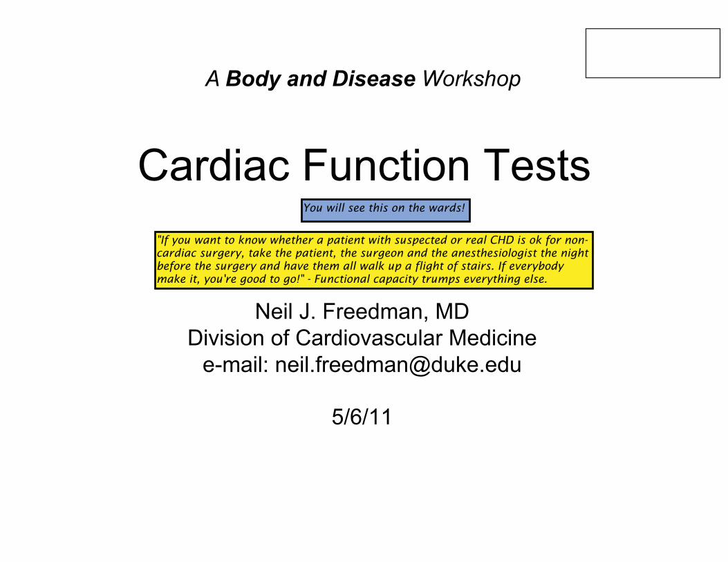
Cardiac Function Tests
Questions
(1) Does this pt have CAD?
(2) What is pt’s functional capacity?
(3) What is this pt’s prognosis?
(4) Is pt’s medical regimen effective?
Coronary Artery Disease
how much exercise the pt can do
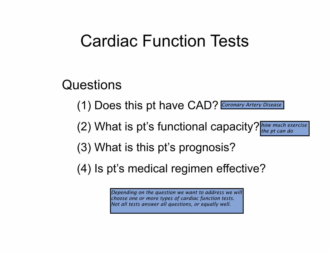
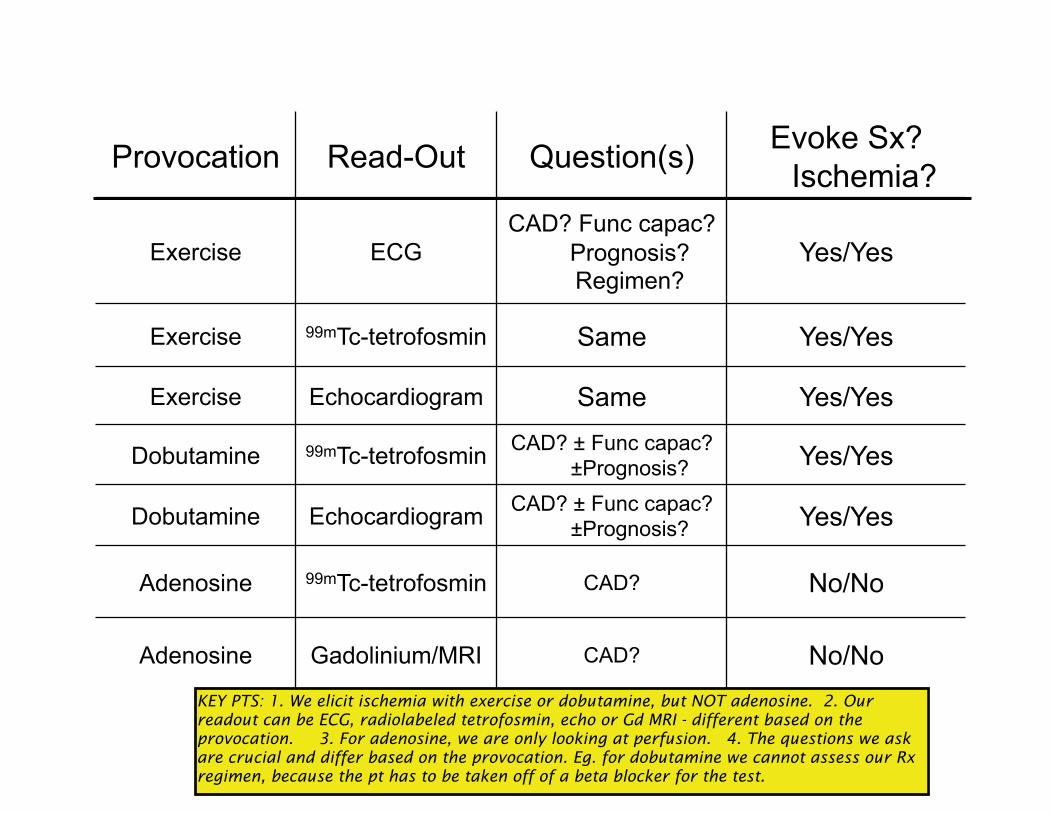
Depending on the question we want to address we will choose one or more types of cardiac function tests. Not all tests answer all questions, or equally well.
Provocation Read-Out Question(s) Evoke Sx?
Ischemia?
Exercise ECG
CAD? Func capac?
Prognosis? Regimen?
Yes/Yes
Exercise 99mTc-tetrofosmin Same Yes/Yes
Exercise Echocardiogram Same Yes/Yes
Dobutamine 99mTc-tetrofosmin CAD? ± Func capac?
±Prognosis? Yes/Yes
Dobutamine Echocardiogram CAD? ± Func capac?
±Prognosis? Yes/Yes
Adenosine 99mTc-tetrofosmin CAD? No/No
Adenosine Gadolinium/MRI CAD? No/No
1. Provocation = What we do to elicit signs/symptoms of insufficient blood supply to the heart (ischemia). If we want to elicit ischemia we use exercise or Dobutamine.
3. Dobutamine is a partial agonist of the beta-1 adrenergic receptor (myocardial stimulant). Increases heart rate and contractility.
2. Exercise increases HR and contractility (the force with which the muscle contracts)
4. If our question is whether the pt has ischemic heart disease, we are concerned with whether the heart muscle is getting enough blood. What determines how much blood the heart needs? - how hard it's working. We can quantitate this via HR and SYSTOLIC BP. Wall stress also is a determinant of oxygen demand - bigger heart = more wall stress = more O2 demand. But we can't measure wall stress.
5. Adenosine is used to allow us to look at myocardial perfusion - where does the blood flow in the heart? Adenosine binds to specific receptors (GPCRs). The A1 receptor is coupled to Gi, which reduces adenylyl cyclase activity, and reduces cAMP. The A2a receptor is coupled to Gs which stimulates AC and increases cAMP and engenders smooth muscle relaxation. Remember that anything that increases cAMP or cGMP in smooth muscle will cause relaxation and in platelets will inhibit aggregation. The reverse is also true. Adenosine is a vasodilator ONLY in coronary arteries. ONLY in coronary arteries. The coronary arteries are unique in their embryonic origin - they are the only smooth muscle cells that come from the pericardium. They have the highest density of adenosine A2a receptors, so they become maximally vasodilated with a small dose of Adenosine.
7. By the end of the lecture we will address all the things on this chart. This chart has all the info you need to knowledgably order cardiac function testing.
6. Adenosine is the only provocation that won't cause ISCHEMIA. It increases coronary vasodilation, BUT NOT HR or BP. Contraindication: bronchospastic lung disease (A1 receptors in the lungs cause bronchospasm). We use an A2a specific agonist (bregadenosine).With bronchospasm give theophylline (caffeine). You also need to be off of caffeine for 18hrs before any CFT using adenosine.
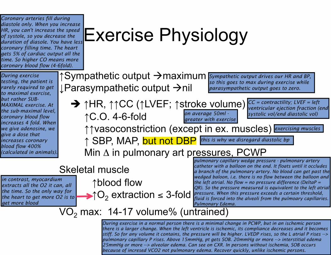
Exercise Physiology
�Sympathetic output �maximum
�Parasympathetic output �nil
� �HR, ��CC (�LVEF; �stroke volume)
�C.O. 4-6-fold
��vasoconstriction (except in ex. muscles)
� SBP, MAP, but not DBP Min � in pulmonary art pressures, PCWP
Skeletal muscle
�blood flow
�O2 extraction � 3-fold
VO2 max: 14-17 volume% (untrained) .
Sympathetic output drives our HR and BP, so this goes to max during exercise while parasympathetic output goes to zero.
CC = contractility; LVEF = left ventricular ejection fraction (end systolic vol/end diastolic vol)on average 50ml -
greater with exercise
Coronary arteries fill during diastole only. When you increase HR, you can't increase the speed of systole, so you decrease the duration of diasole. You have less coronary filling time. The heart gets 5% of cardiac output all the time. So higher CO means more coronary blood flow (4-6fold).
During exercise testing, the patient is rarely required to get to maximal exercise, but rather SUB-MAXIMAL exercise. At the sub-maximal level, coronary blood flow increases 4 fold. When we give adenosine, we give a dose that increases coronary blood flow 400% (calculated in animals).
but not DBP this is why we disregard diastolic bp
exercising muscles
pulmonary capillary wedge pressure - pulmonary artery catheter with a balloon on the end. It floats until it occludes a branch of the pulmonary artery. No blood can get past the wedged baloon, i.e. there is no flow between the balloon and the left atrial. No flow = no pressure difference (DeltaP = QR). So the pressure measured is equivalent to the left atrial pressure. When this pressure exceeds a certain threshold, fluid is forced into the alveoli from the pulmoary capillaries. Pulmonary Edema.
During exercise in a normal person there is a minimal change in PCWP, but in an ischemic person there is a larger change. When the left ventricle is ischemic, its compliance decreases and it becomes stiff. So for any volume it contains, the pressure will be higher. LVEDP rises, so the L atrial P rises --> pulmonary capillary P rises. Above 15mmHg, pt gets SOB. 20mmHg or more --> interstitial edema 25mmHg or more --> alveolar edema. Can see on CXR. In persons without ischemia, SOB occurs because of incresed VCO2 not pulmonary edema. Recover quickly, unlike ischemic persons.
in contrast, myocardium extracts all the O2 it can, all the time. So the only way for the heart to get more O2 is to get more blood
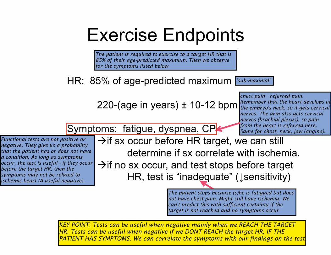
Exercise Endpoints
HR: 85% of age-predicted maximum
220-(age in years) ± 10-12 bpm
Symptoms: fatigue, dyspnea, CP
�if sx occur before HR target, we can still
determine if sx correlate with ischemia.
�if no sx occur, and test stops before target HR, test is “inadequate” (�sensitivity)
"sub-maximal"
The patient is required to exercise to a target HR that is 85% of their age-predicted maximum. Then we observe for the symptoms listed below
chest pain - referred pain. Remember that the heart develops in the embryo's neck, so it gets cervical nerves. The arm also gets cervical nerves (brachial plexus), so pain from the heart is referred here. Same for chest, neck, jaw (angina).
Functional tests are not positive or negative. They give us a probability that the patient has or does not have a condition. As long as symptoms occur, the test is useful - if they occur before the target HR, then the symptoms may not be related to ischemic heart (A useful negative).
The patient stops because (s)he is fatigued but does not have chest pain. Might still have ischemia. We can't predict this with sufficient certainty if the target is not reached and no symptoms occur
KEY POINT: Tests can be useful when negative mainly when we REACH THE TARGET HR. Tests can be useful when negative if we DONT REACH the target HR, IF THE PATIENT HAS SYMPTOMS. We can correlate the symptoms with our findings on the test
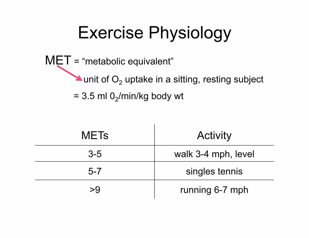
Exercise Physiology
MET = “metabolic equivalent”
unit of O2 uptake in a sitting, resting subject
= 3.5 ml 02/min/kg body wt
METs Activity
3-5 walk 3-4 mph, level
5-7 singles tennis
>9 running 6-7 mph
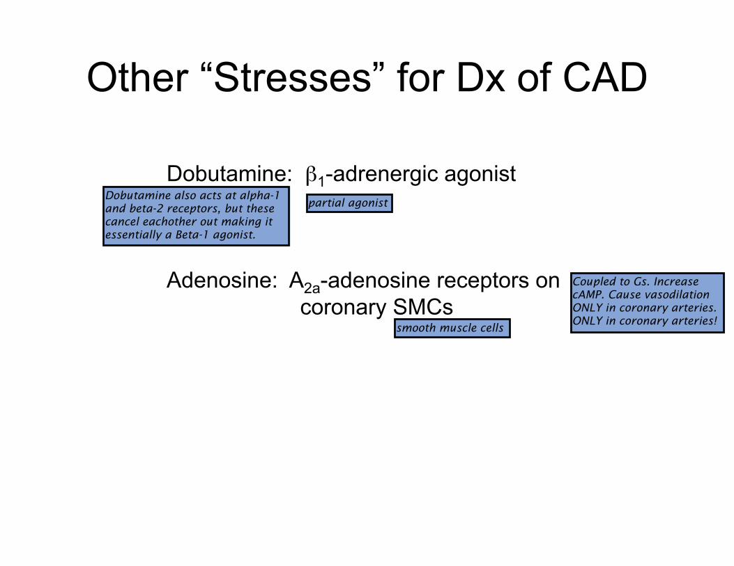
Other “Stresses” for Dx of CAD
Dobutamine: �1-adrenergic agonist
Adenosine: A2a-adenosine receptors on
coronary SMCs
Dobutamine also acts at alpha-1 and beta-2 receptors, but these cancel eachother out making it essentially a Beta-1 agonist.
smooth muscle cells
partial agonist
Coupled to Gs. Increase cAMP. Cause vasodilation ONLY in coronary arteries. ONLY in coronary arteries!
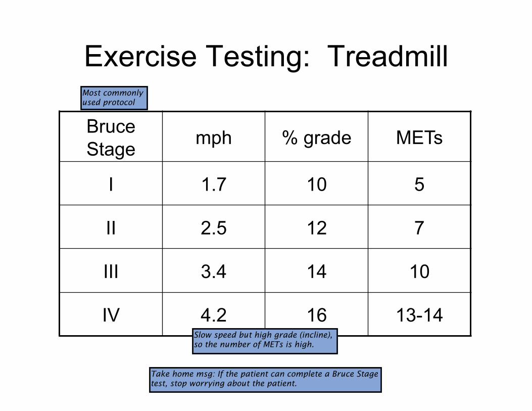
Exercise Testing: Treadmill
Bruce
Stage mph % grade METs
I 1.7 10 5
II 2.5 12 7
III 3.4 14 10
IV 4.2 16 13-14
Most commonly used protocol
Slow speed but high grade (incline), so the number of METs is high.
Take home msg: If the patient can complete a Bruce Stage test, stop worrying about the patient.
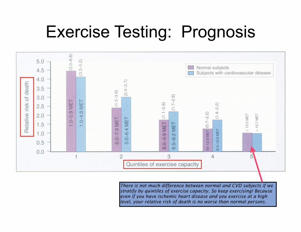
Exercise Testing: Prognosis
There is not much difference between normal and CVD subjects if we stratify by quintiles of exercise capacity. So keep exercising! Because even if you have ischemic heart disease and you exercise at a high level, your relative risk of death is no worse than normal persons.
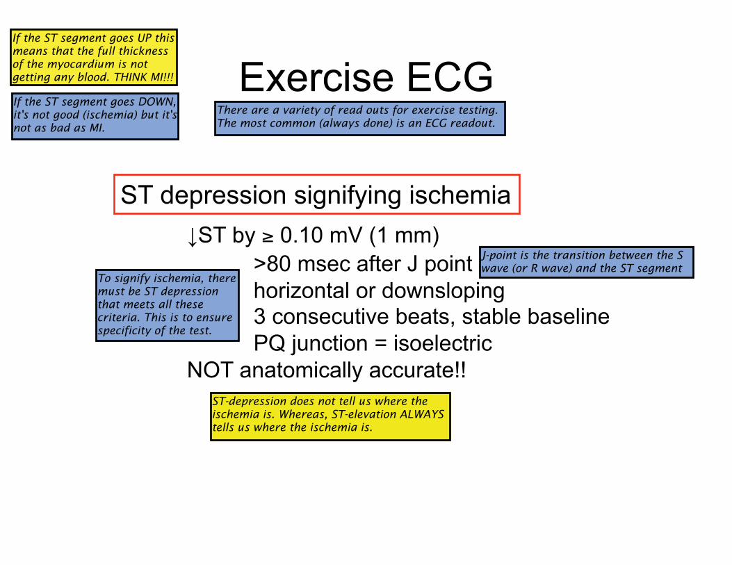
Exercise ECG
ST depression signifying ischemia
�ST by � 0.10 mV (1 mm)
>80 msec after J point
horizontal or downsloping 3 consecutive beats, stable baseline
PQ junction = isoelectric
NOT anatomically accurate!!
There are a variety of read outs for exercise testing. The most common (always done) is an ECG readout.
If the ST segment goes UP this means that the full thickness of the myocardium is not getting any blood. THINK MI!!!
If the ST segment goes DOWN, it's not good (ischemia) but it's not as bad as MI.
J-point is the transition between the S wave (or R wave) and the ST segment
To signify ischemia, there must be ST depression that meets all these criteria. This is to ensure specificity of the test.
ST-depression does not tell us where the ischemia is. Whereas, ST-elevation ALWAYS tells us where the ischemia is.
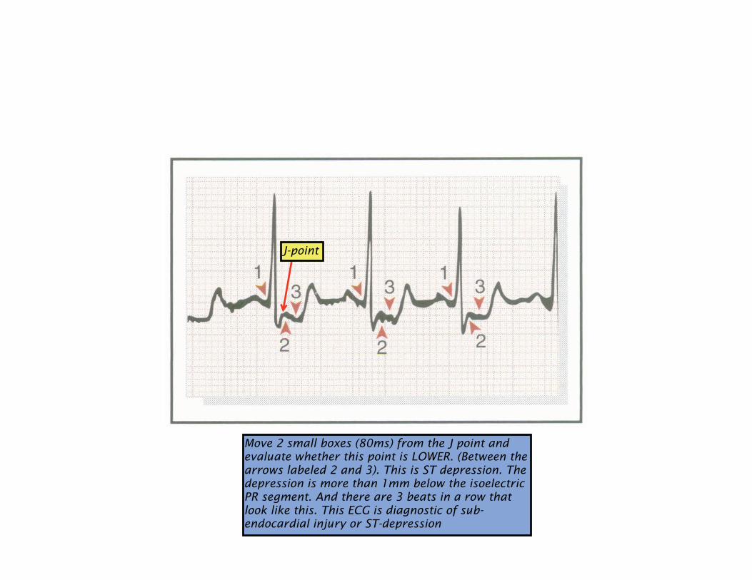
J-point
Move 2 small boxes (80ms) from the J point and evaluate whether this point is LOWER. (Between the arrows labeled 2 and 3). This is ST depression. The depression is more than 1mm below the isoelectric PR segment. And there are 3 beats in a row that look like this. This ECG is diagnostic of sub-endocardial injury or ST-depression
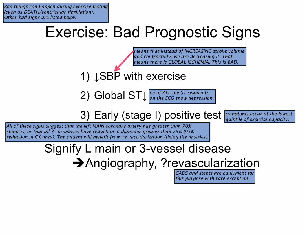
Exercise: Bad Prognostic Signs
1)� �SBP with exercise
2)� Global ST�
3)� Early (stage I) positive test
Signify L main or 3-vessel disease
�Angiography, ?revascularization
Bad things can happen during exercise testing (such as DEATH/ventricular fibrillation). Other bad signs are listed below
means that instead of INCREASING stroke volume and contractility, we are decreasing it. That means there is GLOBAL ISCHEMIA. This is BAD.
i.e. if ALL the ST segments on the ECG show depression.
symptoms occur at the lowest quintile of exercise capacity.
All of these signs suggest that the left MAIN coronary artery has greater than 70% stenosis, or that all 3 coronaries have reduction in diameter greater than 75% (95% reduction in CX area). The patient will benefit from re-vascularization (fixing the arteries).
CABG and stents are equivalent for this purpose with rare exception
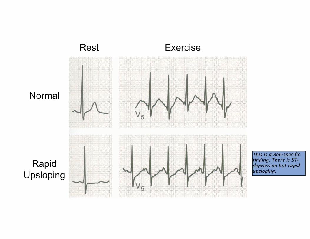
Rest Exercise
Normal
Rapid
Upsloping
This is a non-specific finding. There is ST-depression but rapid upsloping.
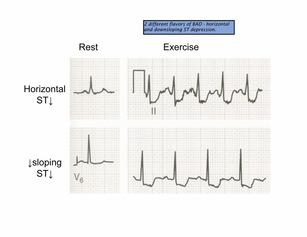
Rest Exercise
Horizontal
ST�
�sloping
ST�
2 different flavors of BAD - horizontal and downsloping ST depression.
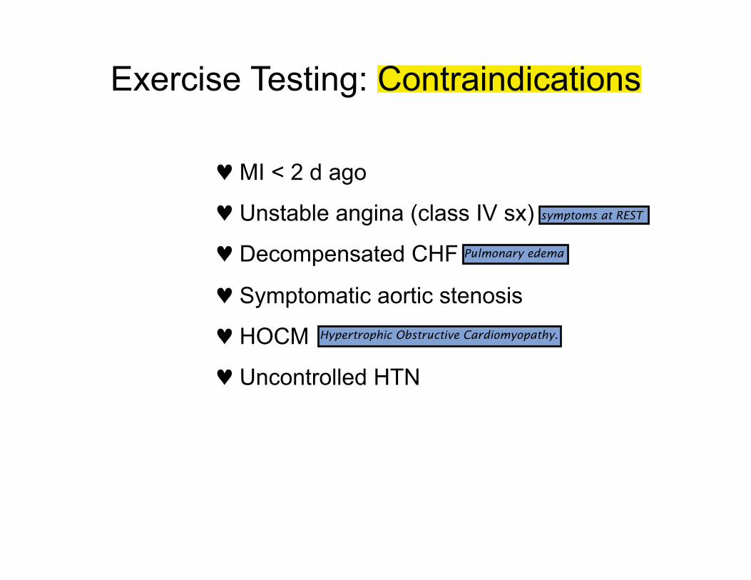
Exercise Testing: Contraindications
�� MI < 2 d ago
�� Unstable angina (class IV sx)
�� Decompensated CHF
�� Symptomatic aortic stenosis
�� HOCM
�� Uncontrolled HTN
Contraindications
symptoms at REST
Pulmonary edema
Hypertrophic Obstructive Cardiomyopathy.
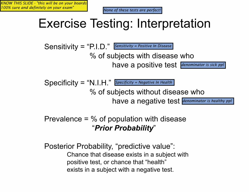
Exercise Testing: Interpretation
Sensitivity = “P.I.D.”
% of subjects with disease who
have a positive test
Specificity = “N.I.H.”
% of subjects without disease who
have a negative test
Prevalence = % of population with disease
“Prior Probability”
Posterior Probability, “predictive value”: Chance that disease exists in a subject with
positive test, or chance that “health”
exists in a subject with a negative test.
None of these tests are perfect!
Sensitivity = Positive In Disease
KNOW THIS SLIDE - "this will be on your boards 100% sure and definitely on your exam"
Specificity = Negative In Health
denominator is sick ppl
denominator is healthy ppl
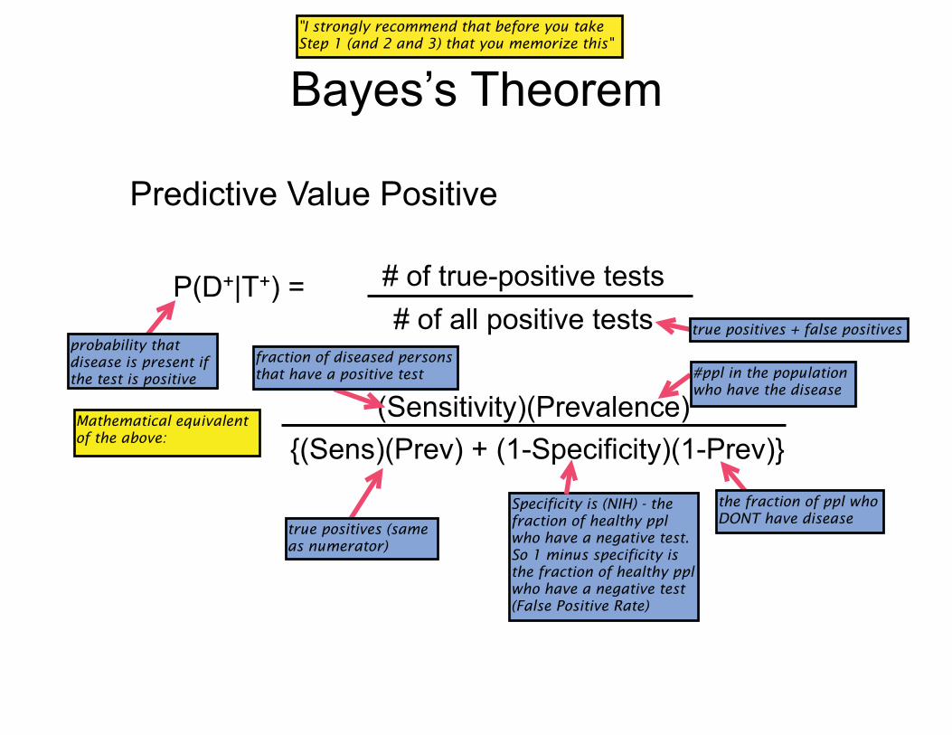
Bayes’s Theorem
P(D+|T+) = # of true-positive tests
# of all positive tests
(Sensitivity)(Prevalence)
{(Sens)(Prev) + (1-Specificity)(1-Prev)}
Predictive Value Positive
"I strongly recommend that before you take Step 1 (and 2 and 3) that you memorize this"
probability that disease is present if the test is positive
true positives + false positives
fraction of diseased persons that have a positive test #ppl in the population
who have the disease
the fraction of ppl who DONT have disease
Specificity is (NIH) - the fraction of healthy ppl who have a negative test. So 1 minus specificity is the fraction of healthy ppl who have a negative test (False Positive Rate)
true positives (same as numerator)
Mathematical equivalent of the above:
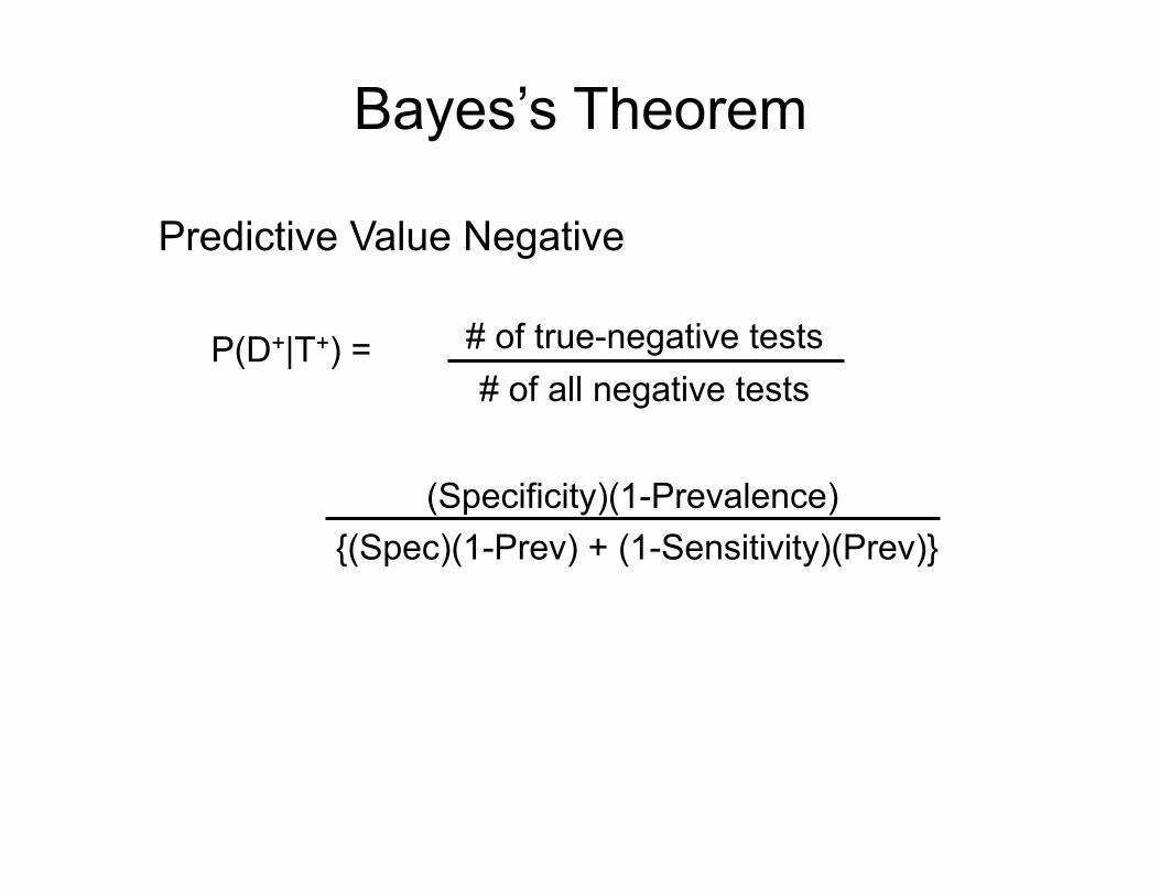
Bayes’s Theorem
P(D+|T+) = # of true-negative tests
# of all negative tests
(Specificity)(1-Prevalence)
{(Spec)(1-Prev) + (1-Sensitivity)(Prev)}
Predictive Value Negative
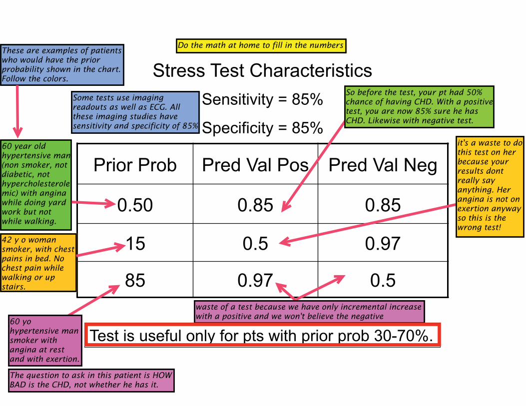
Prior Prob Pred Val Pos Pred Val Neg
0.50 0.85 0.85
15 0.5 0.97
85 0.97 0.5
Stress Test Characteristics
Sensitivity = 85%
Specificity = 85%
Test is useful only for pts with prior prob 30-70%.
Some tests use imaging readouts as well as ECG. All these imaging studies have sensitivity and specificity of 85%
Do the math at home to fill in the numbers
So before the test, your pt had 50% chance of having CHD. With a positive test, you are now 85% sure he has CHD. Likewise with negative test.
60 year old hypertensive man (non smoker, not diabetic, not hypercholesterolemic) with angina while doing yard work but not while walking.
42 y o woman smoker, with chest pains in bed. No chest pain while walking or up stairs.
it's a waste to do this test on her because your results dont really say anything. Her angina is not on exertion anyway so this is the wrong test!
60 yo hypertensive man smoker with angina at rest and with exertion.
waste of a test because we have only incremental increase with a positive and we won't believe the negative
The question to ask in this patient is HOW BAD is the CHD, not whether he has it.
These are examples of patients who would have the prior probability shown in the chart. Follow the colors.
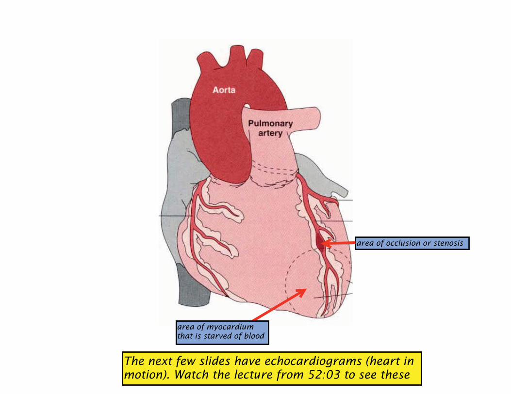
area of occlusion or stenosis
area of myocardium that is starved of blood
The next few slides have echocardiograms (heart in motion). Watch the lecture from 52:03 to see these
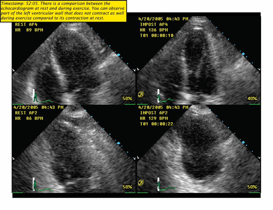
Timestamp: 52:05. There is a comparison between the echocardiogram at rest and during exercise. You can observe part of the left ventricular wall that does not contract as well during exercise compared to its contraction at rest.
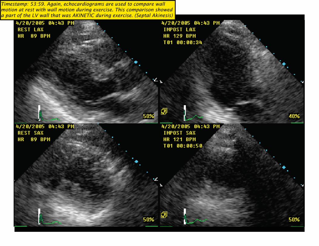
Timestamp: 53:59. Again, echocardiograms are used to compare wall motion at rest with wall motion during exercise. This comparison showed a part of the LV wall that was AKINETIC during exercise. (Septal Akinesis)
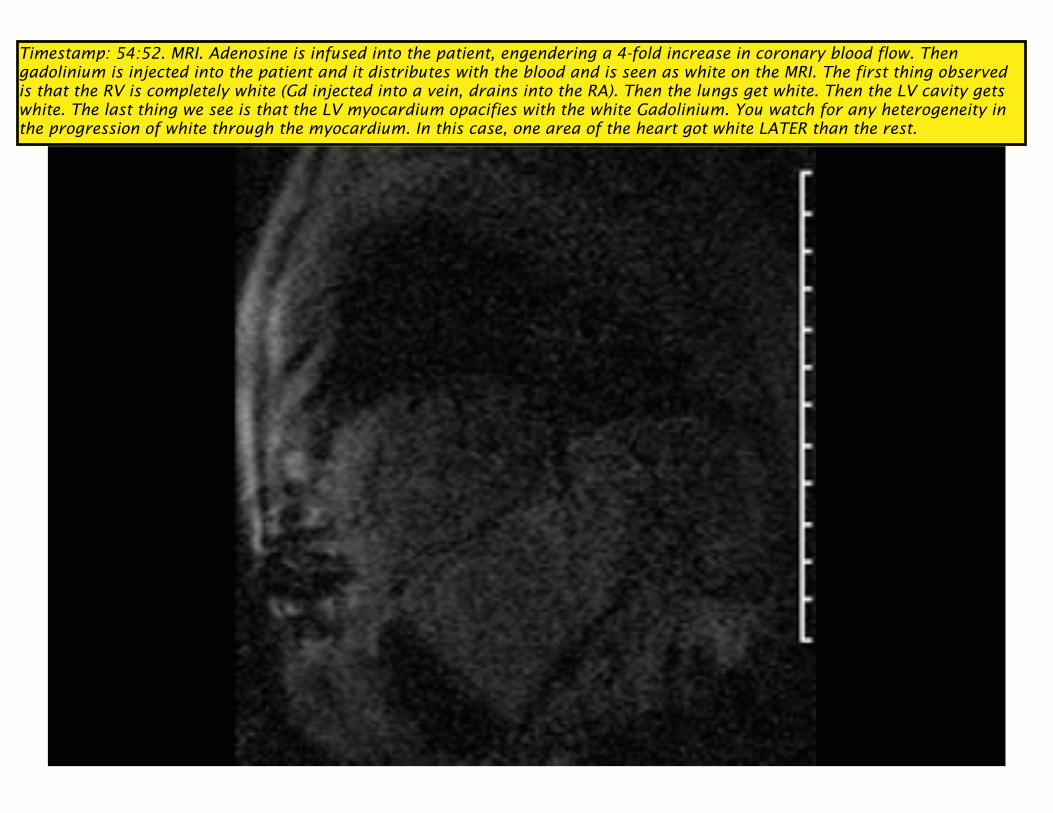
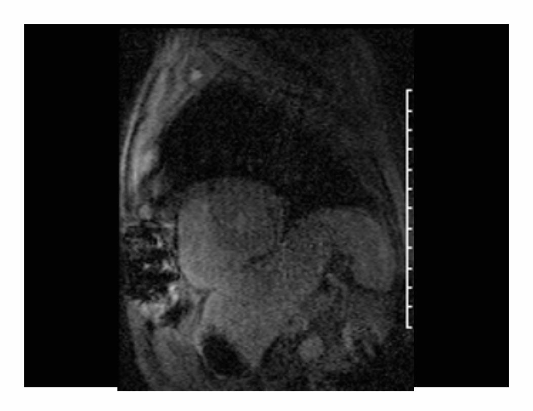
Timestamp: 54:52. MRI. Adenosine is infused into the patient, engendering a 4-fold increase in coronary blood flow. Then gadolinium is injected into the patient and it distributes with the blood and is seen as white on the MRI. The first thing observed is that the RV is completely white (Gd injected into a vein, drains into the RA). Then the lungs get white. Then the LV cavity gets white. The last thing we see is that the LV myocardium opacifies with the white Gadolinium. You watch for any heterogeneity in the progression of white through the myocardium. In this case, one area of the heart got white LATER than the rest.
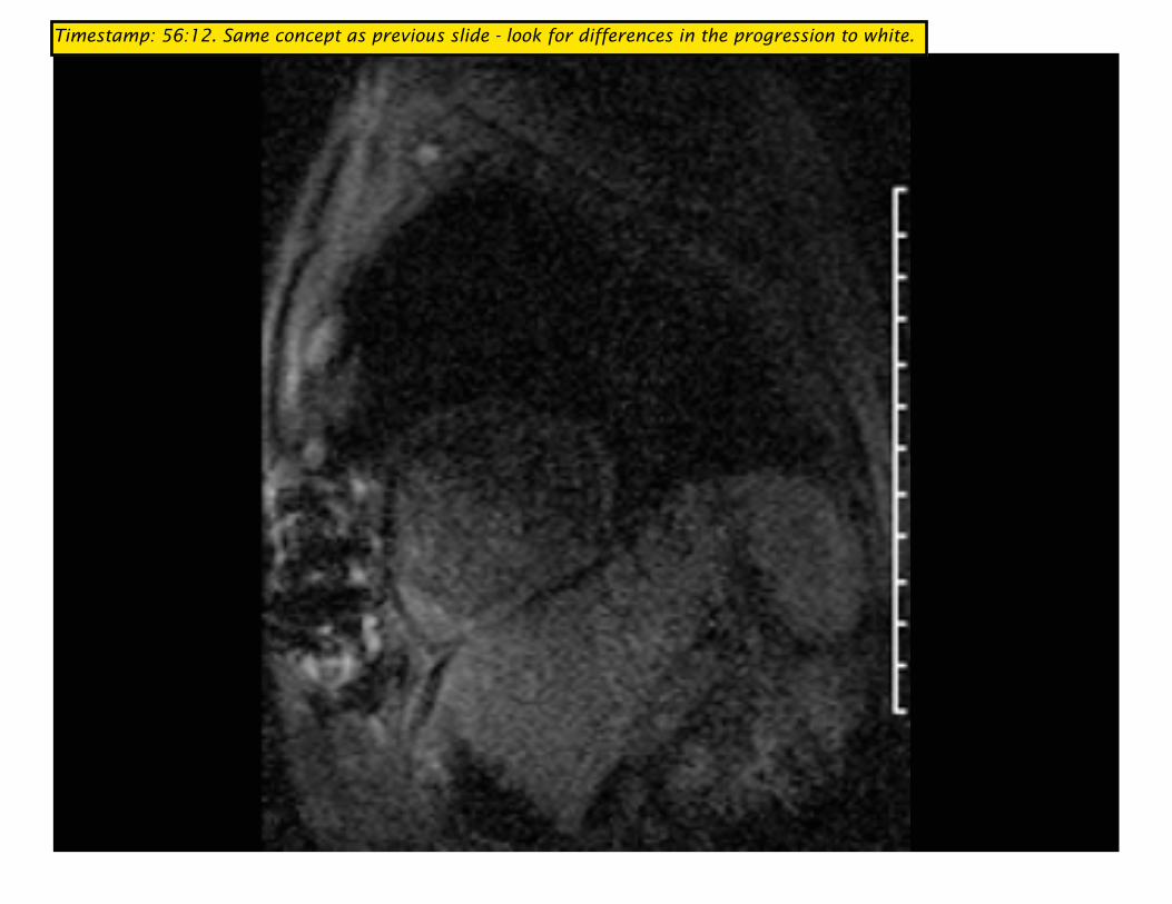
Timestamp: 56:12. Same concept as previous slide - look for differences in the progression to white.
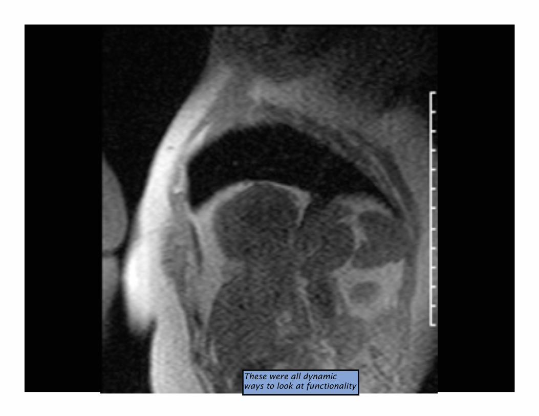
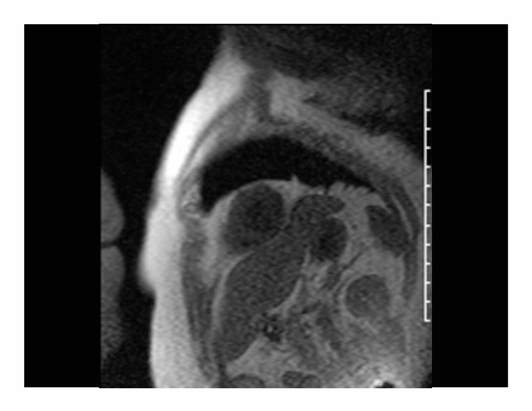
These were all dynamic ways to look at functionality
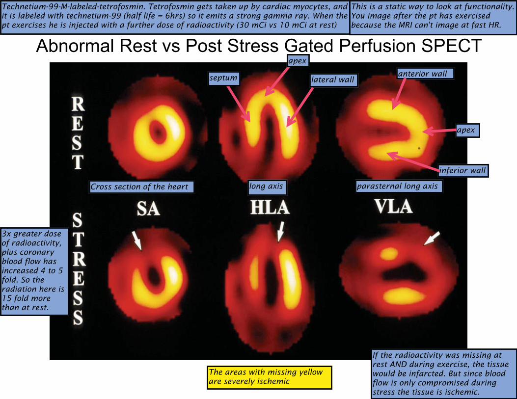
Abnormal Rest vs Post Stress Gated Perfusion SPECT
Technetium-99-M-labeled-tetrofosmin. Tetrofosmin gets taken up by cardiac myocytes, and it is labeled with technetium-99 (half life = 6hrs) so it emits a strong gamma ray. When the pt exercises he is injected with a further dose of radioactivity (30 mCi vs 10 mCi at rest)
Cross section of the heart
apex
lateral wallseptum
long axis parasternal long axis
anterior wall
apex
inferior wall
3x greater dose of radioactivity, plus coronary blood flow has increased 4 to 5 fold. So the radiation here is 15 fold more than at rest.
The areas with missing yellow are severely ischemic
If the radioactivity was missing at rest AND during exercise, the tissue would be infarcted. But since blood flow is only compromised during stress the tissue is ischemic.
This is a static way to look at functionality. You image after the pt has exercised because the MRI can't image at fast HR.
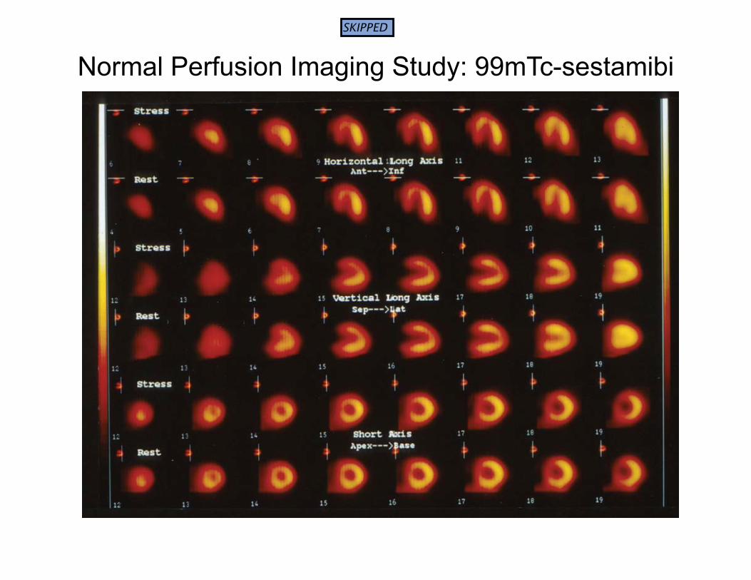
Normal Perfusion Imaging Study: 99mTc-sestamibi
SKIPPED
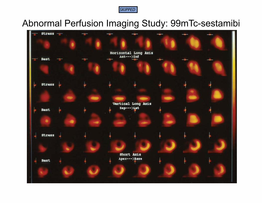
Abnormal Perfusion Imaging Study: 99mTc-sestamibi
SKIPPED
Provocation Read-Out Question(s) Evoke Sx?
Ischemia?
Exercise ECG
CAD? Func capac?
Prognosis? Regimen?
Yes/Yes
Exercise 99mTc-tetrofosmin Same Yes/Yes
Exercise Echocardiogram Same Yes/Yes
Dobutamine 99mTc-tetrofosmin CAD? ± Func capac?
±Prognosis? Yes/Yes
Dobutamine Echocardiogram CAD? ± Func capac?
±Prognosis? Yes/Yes
Adenosine 99mTc-tetrofosmin CAD? No/No
Adenosine Gadolinium/MRI CAD? No/No
KEY PTS: 1. We elicit ischemia with exercise or dobutamine, but NOT adenosine. 2. Our readout can be ECG, radiolabeled tetrofosmin, echo or Gd MRI - different based on the provocation. 3. For adenosine, we are only looking at perfusion. 4. The questions we ask are crucial and differ based on the provocation. Eg. for dobutamine we cannot assess our Rx regimen, because the pt has to be taken off of a beta blocker for the test.
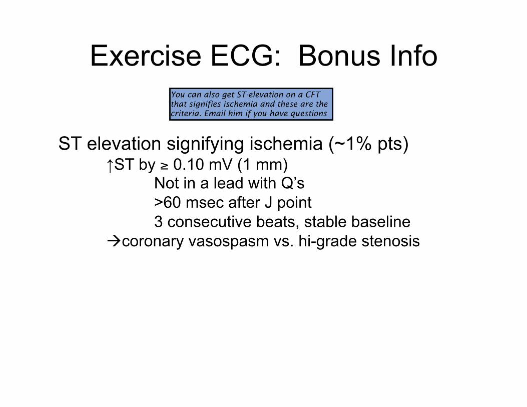
ST elevation signifying ischemia (~1% pts) �ST by � 0.10 mV (1 mm) Not in a lead with Q’s
>60 msec after J point
3 consecutive beats, stable baseline
�coronary vasospasm vs. hi-grade stenosis
Exercise ECG: Bonus Info You can also get ST-elevation on a CFT that signifies ischemia and these are the criteria. Email him if you have questions