Embed Size (px)
Citation preview
Cardiac Disease Cardiac Disease in Pregnancy in Pregnancy
Woman’s Hospital School of Woman’s Hospital School of Medicine Zhejing UniversityMedicine Zhejing University
He jin He jin
Physiological Changes in the Physiological Changes in the Cardiovascular System During Cardiovascular System During
PregnancyPregnancy
• A thorough knowledge – is essential
• In order to understand – the additional impact of cardiac disease
Physiological ChangesPhysiological Changes
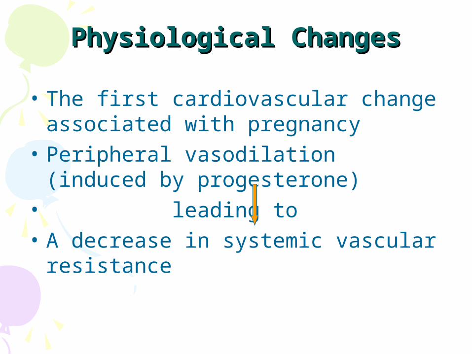
• The first cardiovascular change associated with pregnancy
• Peripheral vasodilation (induced by progesterone)
• leading to • A decrease in systemic vascular
resistance
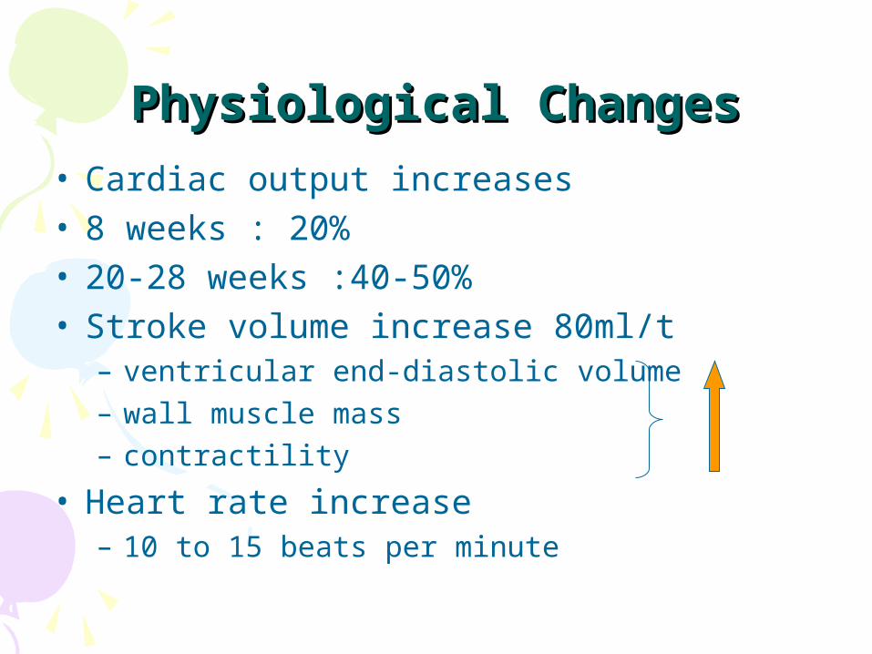
Physiological ChangesPhysiological Changes• Cardiac output increases • 8 weeks : 20%• 20-28 weeks :40-50% • Stroke volume increase 80ml/t
– ventricular end-diastolic volume– wall muscle mass– contractility
• Heart rate increase– 10 to 15 beats per minute
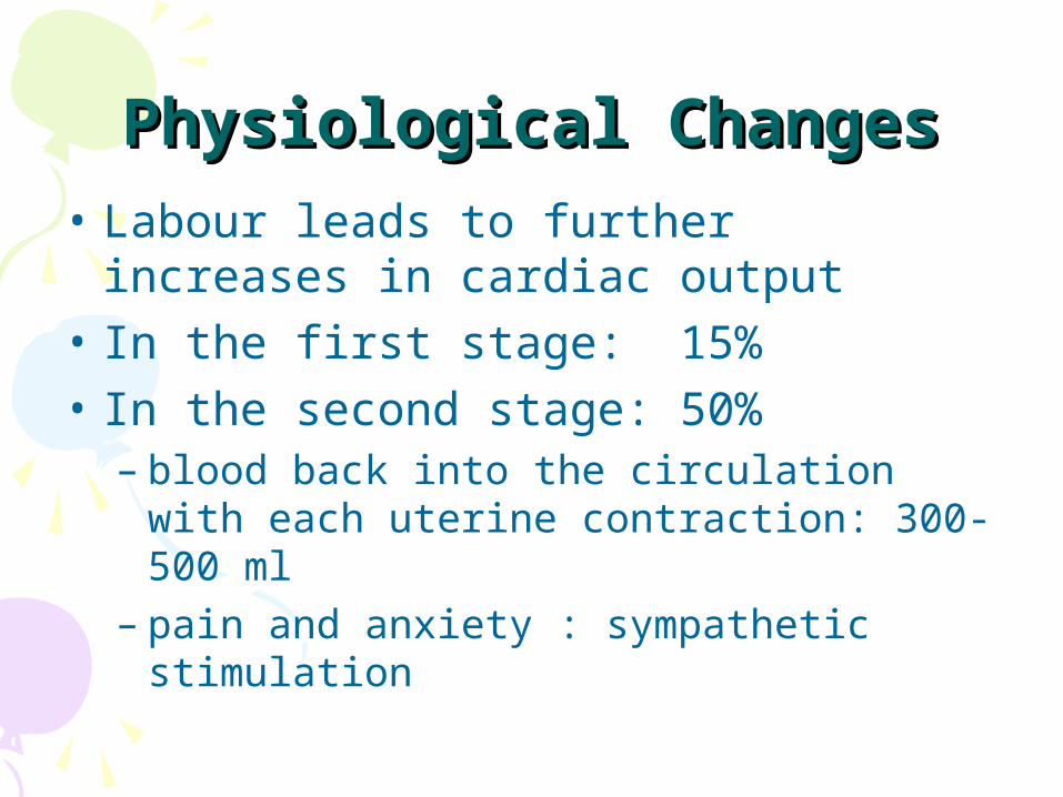
Physiological ChangesPhysiological Changes• Labour leads to further increases in
cardiac output • In the first stage: 15%• In the second stage: 50%
– blood back into the circulation with each uterine contraction: 300-500 ml
– pain and anxiety : sympathetic stimulation
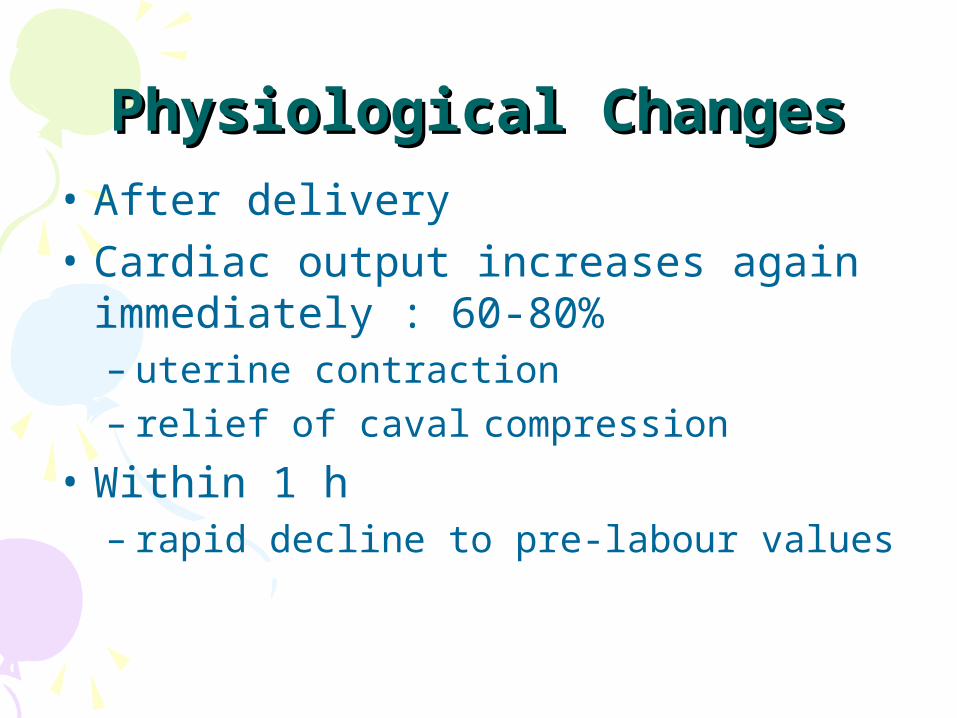
Physiological ChangesPhysiological Changes• After delivery• Cardiac output increases again
immediately : 60-80%– uterine contraction – relief of caval compression
• Within 1 h– rapid decline to pre-labour values
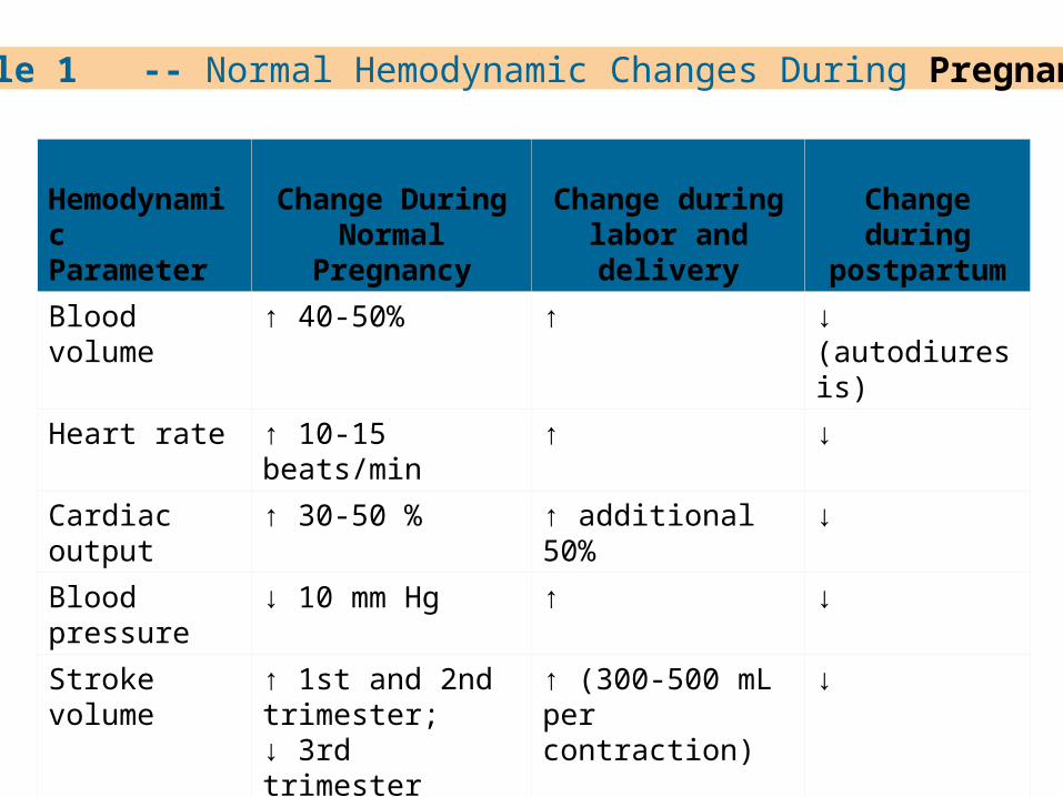
Table 1 -- Normal Hemodynamic Changes During Pregnancy
Hemodynamic Parameter
Change During Normal Pregnancy
Change during labor and delivery
Change during postpartum
Blood volume ↑ 40-50% ↑ ↓ (autodiuresis)
Heart rate ↑ 10-15 beats/min ↑ ↓
Cardiac output ↑ 30-50 % ↑ additional 50% ↓
Blood pressure ↓ 10 mm Hg ↑ ↓
Stroke volume ↑ 1st and 2nd trimester;↓ 3rd trimester
↑ (300-500 mL per contraction)
↓
Systemic vascular resistance
↓ ↑ ↓
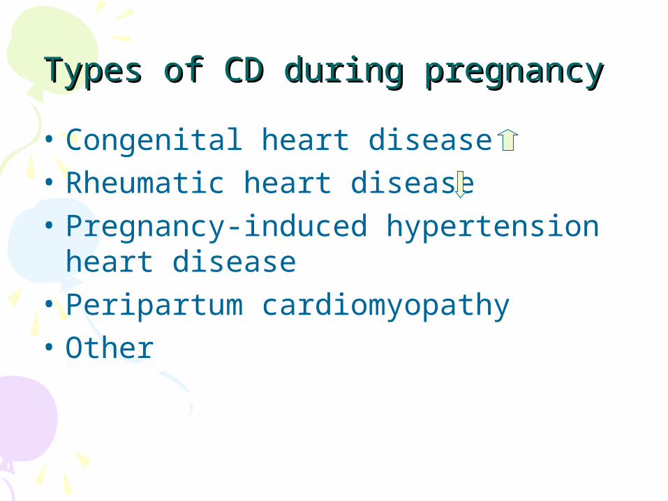
Types of CD during pregnancyTypes of CD during pregnancy
• Congenital heart disease• Rheumatic heart disease• Pregnancy-induced hypertension
heart disease• Peripartum cardiomyopathy• Other
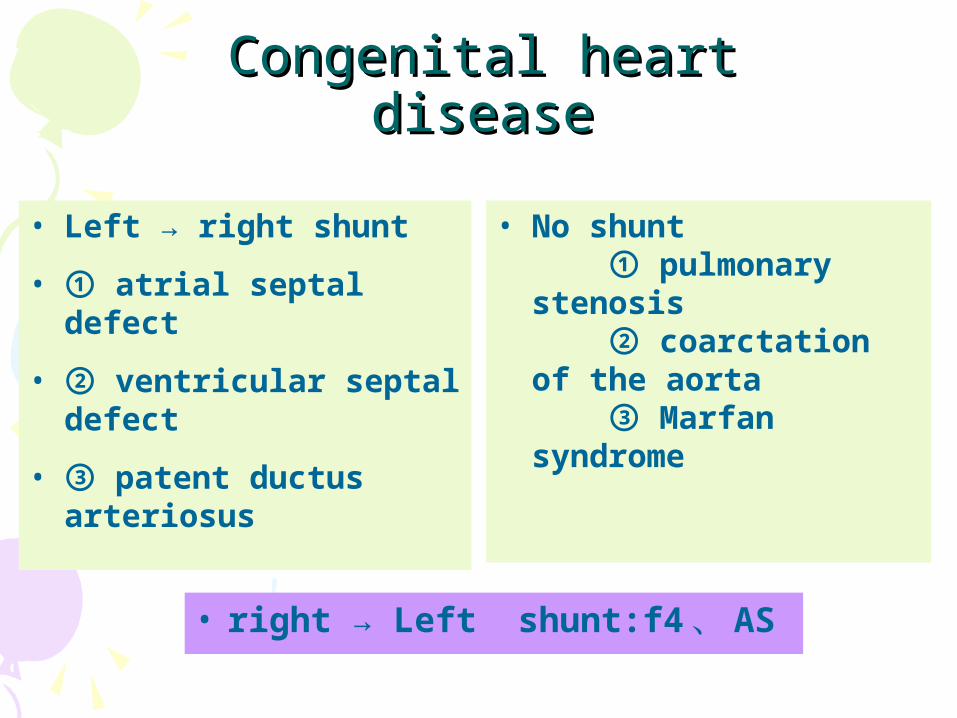
Congenital heart diseaseCongenital heart disease
• Left → right shunt
• ① atrial septal defect
• ② ventricular septal defect
• ③ patent ductus arteriosus
• No shunt ① pulmonary stenosis ② coarctation of the aorta ③ Marfan syndrome
• right → Left shunt:f4 、 AS
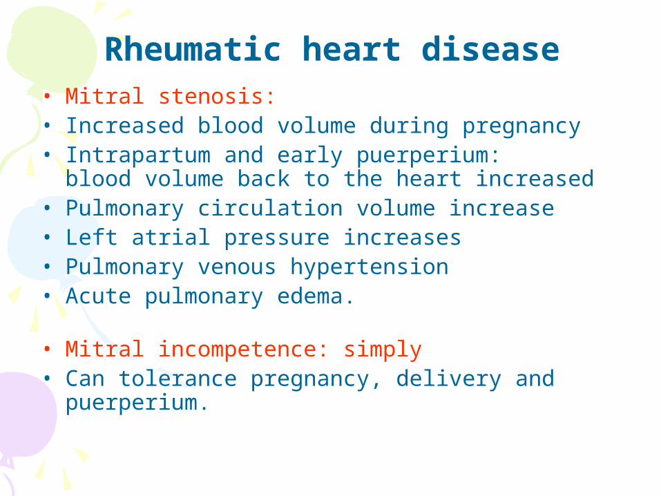
Rheumatic heart disease• Mitral stenosis: • Increased blood volume during pregnancy• Intrapartum and early puerperium:
blood volume back to the heart increased • Pulmonary circulation volume increase• Left atrial pressure increases• Pulmonary venous hypertension• Acute pulmonary edema.
• Mitral incompetence: simply • Can tolerance pregnancy, delivery and
puerperium.
Rheumatic heart disease
• Aortic stenosis: severe• Pulmonary edema • Low discharge capacity heart failure• Aortic incompetence : severe • Left ventricular failure • Combined with bacterial endocarditis
PIH heart diseasePIH heart disease
• No history of heart disease and signs over the past
• Sudden onset of systemic failure are dominated by left ventricular failure
• Misdiagnosed as the flu and bronchitis• Early diagnosis is important• After eliminate the cause, most can be
restored
PIH heart diseasePIH heart disease• Myocardial ischemia, interstitial
edema, hemorrhage and necrosis spots
• Blood viscosity increased to promote myocardial ischemia
• Combined with severe anemia• Heart failure occurs
Peripartum Cardiomyopathy Peripartum Cardiomyopathy (PPCM)(PPCM)
• Define: dilated cardiomyopathy
• Interval: between the last 3 month of pregnancy up to the first 6 months postpartum
• Women : without preexisting cardiac dysfunction
• Fetal death:10~30%
• Maternal mortality is approximately 9%
– heart failure, pulmonary infarction, arrhythmia
• These women should be counseled against subsequent pregnancies
PPCMPPCM• The exact etiology : unknown• Possible causes
– infection, immunity, multiple pregnancy, hypertension, malnutrition
– viral myocarditis– automimmune phenomena– specific genetic mutations
PPCMPPCM• Typical signs• Fatigue• Dyspnea on exertion, orthopnea• Nonspecific chest pain• Abdominal discomfort and distension• palpitations, cough, hemoptysis,
hepatomegaly, edema and other heart failure symptoms

PPCMPPCM• Saymptoms• Heart enlarged• Myocardial contractility reduce • Ejection function reduced
• ECG: • Arrhythmias, left ventricular hypertrophy,
ST segment and T wave abnormalities
CD main threat to pregnant CD main threat to pregnant women women
• Heart failure• Subacute infective endocarditis• Hypoxia and cyanosis• Venous thrombosis and pulmonary
embolism.
The impact of CD in The impact of CD in pregnant womenpregnant women
• Gestation period:• increased blood volume, heart burden• Delivery period: • uterine contractions• blood pressure↑• the blood flow increases• pulmonary artery pressure increased • sudden interruption of placental circulation• abdominal pressure plummeted
The impact of CD in The impact of CD in pregnant womenpregnant women
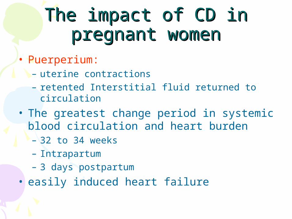
• Puerperium: – uterine contractions– retented Interstitial fluid returned to circulation
• The greatest change period in systemic blood circulation and heart burden– 32 to 34 weeks– Intrapartum– 3 days postpartum
• easily induced heart failure
The impact of CD in The impact of CD in pregnant womenpregnant women
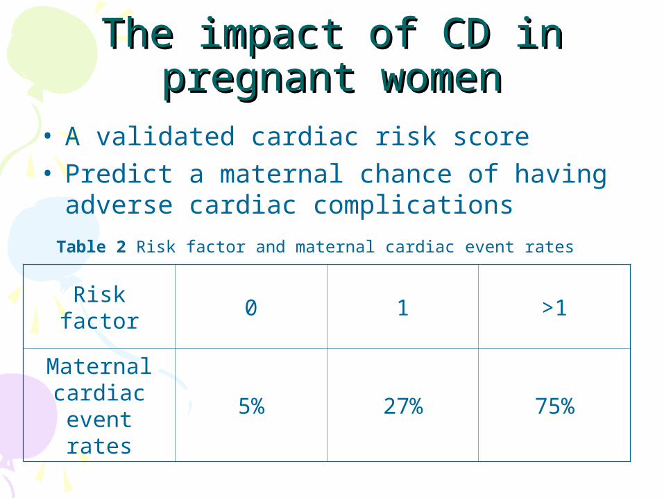
• A validated cardiac risk score • Predict a maternal chance of having
adverse cardiac complications
Risk factor 0 1 >1
Maternal cardiac
event rates5% 27% 75%
Table 2 Risk factor and maternal cardiac event rates
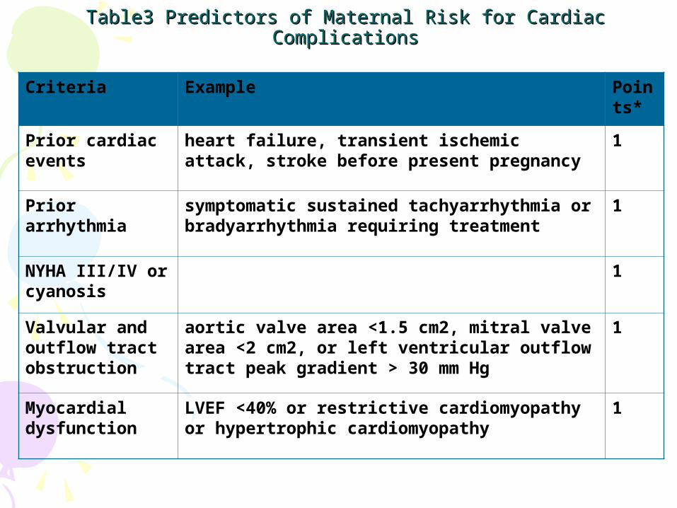
Table3 Predictors of Maternal Risk for Cardiac ComplicationsTable3 Predictors of Maternal Risk for Cardiac Complications
Criteria Example Points*
Prior cardiac events
heart failure, transient ischemic attack, stroke before present pregnancy
1
Prior arrhythmia
symptomatic sustained tachyarrhythmia or bradyarrhythmia requiring treatment
1
NYHA III/IV or cyanosis
1
Valvular and outflow tract obstruction
aortic valve area <1.5 cm2, mitral valve area <2 cm2, or left ventricular outflow tract peak gradient > 30 mm Hg
1
Myocardial dysfunction
LVEF <40% or restrictive cardiomyopathy or hypertrophic cardiomyopathy
1
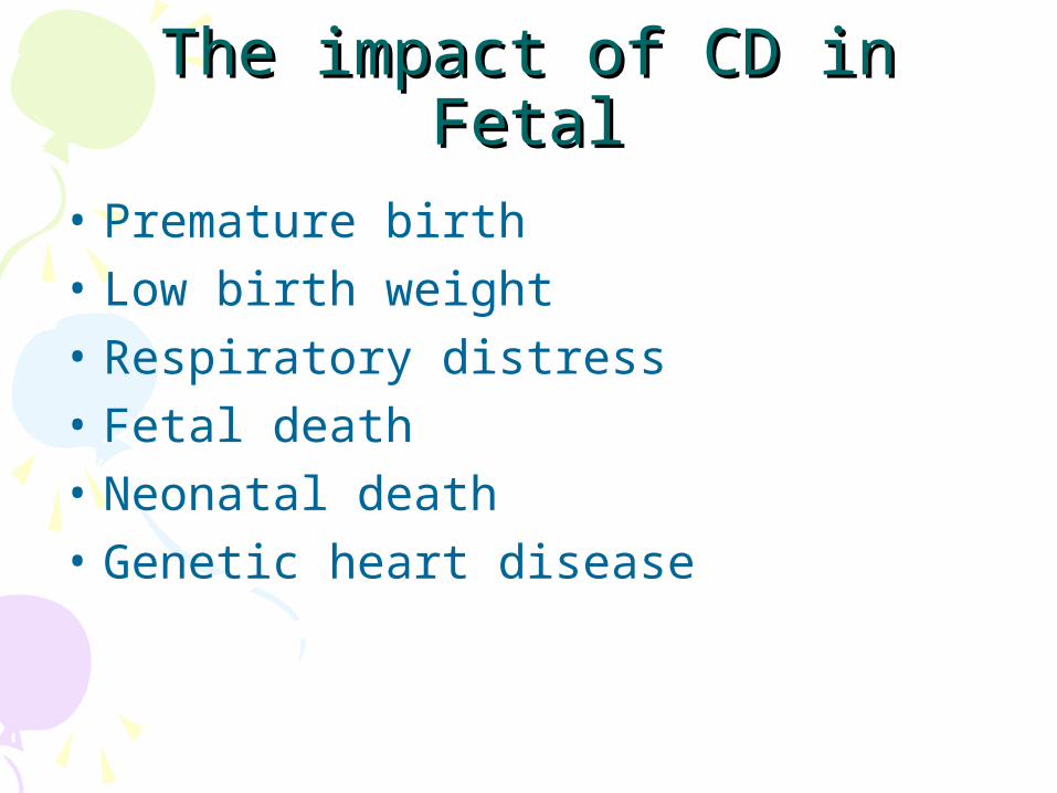
The impact of CD in The impact of CD in FetalFetal• Premature birth• Low birth weight• Respiratory distress• Fetal death• Neonatal death• Genetic heart disease
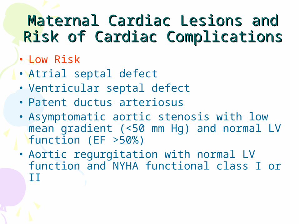
Maternal Cardiac Lesions and Risk Maternal Cardiac Lesions and Risk of Cardiac Complicationsof Cardiac Complications
• Low Risk • Atrial septal defect• Ventricular septal defect • Patent ductus arteriosus• Asymptomatic aortic stenosis with low
mean gradient (<50 mm Hg) and normal LV function (EF >50%)
• Aortic regurgitation with normal LV function and NYHA functional class I or II
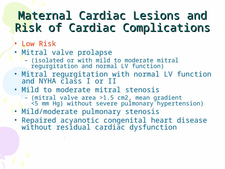
Maternal Cardiac Lesions and Risk Maternal Cardiac Lesions and Risk of Cardiac Complicationsof Cardiac Complications
• Low Risk • Mitral valve prolapse
– (isolated or with mild to moderate mitral regurgitation and normal LV function)
• Mitral regurgitation with normal LV function and NYHA class I or II
• Mild to moderate mitral stenosis – (mitral valve area >1.5 cm2, mean gradient <5 mm Hg)
without severe pulmonary hypertension)• Mild/moderate pulmonary stenosis• Repaired acyanotic congenital heart disease
without residual cardiac dysfunction
Maternal Cardiac Lesions and Risk Maternal Cardiac Lesions and Risk of Cardiac Complicationsof Cardiac Complications
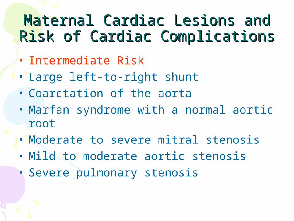
• Intermediate Risk • Large left-to-right shunt• Coarctation of the aorta• Marfan syndrome with a normal aortic root• Moderate to severe mitral stenosis• Mild to moderate aortic stenosis• Severe pulmonary stenosis
Maternal Cardiac Lesions and Risk Maternal Cardiac Lesions and Risk of Cardiac Complicationsof Cardiac Complications
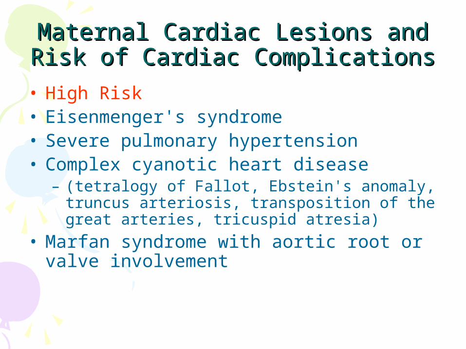
• High Risk • Eisenmenger's syndrome• Severe pulmonary hypertension• Complex cyanotic heart disease
– (tetralogy of Fallot, Ebstein's anomaly, truncus arteriosis, transposition of the great arteries, tricuspid atresia)
• Marfan syndrome with aortic root or valve involvement
Maternal Cardiac Lesions and Risk Maternal Cardiac Lesions and Risk of Cardiac Complicationsof Cardiac Complications
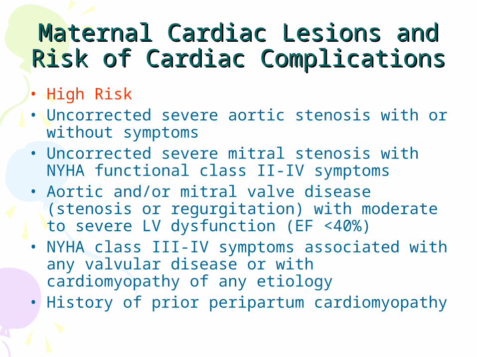
• High Risk• Uncorrected severe aortic stenosis with or
without symptoms• Uncorrected severe mitral stenosis with NYHA
functional class II-IV symptoms• Aortic and/or mitral valve disease (stenosis or
regurgitation) with moderate to severe LV dysfunction (EF <40%)
• NYHA class III-IV symptoms associated with any valvular disease or with cardiomyopathy of any etiology
• History of prior peripartum cardiomyopathy

DiagnosisDiagnosis• History:• Palpitations, difficulty breathing
or heart failure• Organic heart disease• Rheumatic fever
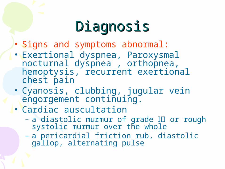
DiagnosisDiagnosis• Signs and symptoms abnormal: • Exertional dyspnea, Paroxysmal nocturnal
dyspnea , orthopnea, hemoptysis, recurrent exertional chest pain
• Cyanosis, clubbing, jugular vein engorgement continuing.
• Cardiac auscultation– a diastolic murmur of grade Ⅲ or rough systolic
murmur over the whole– a pericardial friction rub, diastolic gallop,
alternating pulse

Early signs of heart failureEarly signs of heart failure • Chest tightness, palpitations,
shortness of breath after mild activity
• Resting heart rate> 110 beats / min • Respiration> 20 times / min• Paroxysmal nocturnal dyspnea• The end of the lung wet rales
persisted
Diagnosis:Diagnosis:auxiliary examination
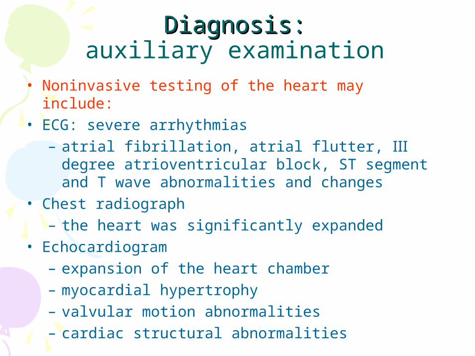
• Noninvasive testing of the heart may include:• ECG: severe arrhythmias
– atrial fibrillation, atrial flutter, Ⅲ degree atrioventricular block, ST segment and T wave abnormalities and changes
• Chest radiograph– the heart was significantly expanded
• Echocardiogram– expansion of the heart chamber– myocardial hypertrophy– valvular motion abnormalities– cardiac structural abnormalities
ManagementManagement
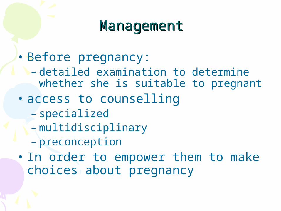
• Before pregnancy: – detailed examination to determine
whether she is suitable to pregnant• access to counselling
– specialized – multidisciplinary – preconception
• In order to empower them to make choices about pregnancy
Not suitable for pregnancyNot suitable for pregnancy !!• Cardiac function grade Ⅲ ~ Ⅳ• Those who previously had heart failure• A pulmonary hypertension, severe stenosis
the main A, Ⅲ atrioventricular block, atrial fibrillation, atrial flutter,diastolic gallop;
• Cyanotic heart disease• Active rheumatic or bacterial endocarditis
The main aims of The main aims of managementmanagement
• To optimize the mother's condition during the pregnancy– considering ß-blockers– Thromboprophylaxis– pulmonary arterial vasodilators
• To monitor for deterioration• Minimize any additional load on the
cardiovascular system
Pregnant womenPregnant women with CD with CD• Should be assessed clinically as soon as possible • A multidisciplinary team and appropriate
investigations undertaken• The core members of the team should include:• Suitably experienced obstetricians• Cardiologists• Anaesthetists• Midwives• Neonatologists• Intensivists
ManagementManagement of gestation period
• Regular prenatal care• Early prevention of heart failure
– adequate rest– appropriate weight limit– treatment the motivation of heart failure
: infection, anemia,PIH
• The treatment of heart failure – as same as those who are not pregnant
Mode of DeliveryMode of Delivery• Vaginal delivery:
– cardiac function Ⅰ ~ Ⅱ grade – not a fetal macrosomia– cervical conditions are good
• Cesarean section: – Marfan syndrome : expansion of the aortic
root> 45 mm – use warfarin during delivery– sudden hemodynamic deterioration– severe pulmonary hypertension and severe
aortic stenosis
ManagementManagement in intrapratumintrapratum• First stage of labor• Semi-recumbent position, oxygen
masks, attention Bp, R, P, heart rate,– cedilanid : 0.4mg +5% GS20ml iv slow
(when necessary)– antibiotics : during labor to 1 week after
postpartum
Vaginal delivery Vaginal delivery • Low-dose regional analgesia:usually
recommended • providing effective pain relief• reduce the further increases in
– cardiac output – myocardial oxygen demand
• Be careful not to inhibit the neonatal breathing
ManagementManagement in intrapratumintrapratum• Second stage of labor:
– episiotomy, facilitate instrumental delivery to shorten the stage
• Third stage of labor:– Ergot disabled to prevent venous pressure
increased– injection of morphine or pethidine immediately
postpartum – abdominal pressure sandbags – control the liquid velocity
ManagementManagement in puerperium• Monitoring heart rate, blood oxygen,
blood pressure during delivery 24 hours
• She could not breast-feeding – more than grade Ⅲ cardiac function
• Prophylactic antibiotics• High-level maternal surveillance

Thanks four your listening