Embed Size (px)
Citation preview
8
Cardiac Biomarkers in End-Stage Renal Disease
Leo Jacobs, Alma Mingels and Marja van Dieijen-Visser Department of Clinical Chemistry, Maastricht University Medical Centre (MUMC)
The Netherlands
1. Introduction
Patients with end-stage renal disease (ESRD) often suffer from cardiovascular complications and comorbidities. For example, 55% of ESRD patients suffer from congestive heart failure (CHF) and cardiovascular diseases account for the majority of deaths among ESRD patients (Herzog, Ma, & Collins, 1998; National Institutes of Health; National Institute of Diabetes and Digestive and Kidney Diseases; Bethesda, 2007). It is, therefore, of great importance to diagnose the underlying cardiac pathologies and to provide accurate risk stratification in ESRD patients. Over the years, a number of accurate and sensitive biochemical markers have been introduced that have greatly advanced the diagnosis and risk stratification of cardiovascular diseases. The most prominent of these biochemical markers are the cardiac troponins (cTn, either T or I) and the brain natriuretic peptides (BNPs) and their use has revolutionized the diagnosis and risk stratification of acute coronary syndromes (ACS) and CHF respectively (A. S. Maisel et al., 2002; Thygesen, Alpert, & White, 2007). However, in the setting of ESRD, cardiac troponin concentrations can be elevated in the absence of apparent cardiac damage or clinical symptoms (Apple, Murakami, Pearce, & Herzog, 2004; Aviles et al., 2002; C. deFilippi et al., 2003; Havekes et al., 2006; Sommerer, Beimler, et al., 2007). Similarly, BNP and N-terminal proBNP (NT-proBNP) concentrations are virtually always increased in ESRD patients (Apple et al., 2004; Madsen et al., 2007). The presence of such continuously elevated cardiac troponin, BNP and NT-proBNP concentrations can interfere with their diagnostic and prognostic potential in ESRD patients (David et al., 2008; Pimenta et al., 2009; Wu et al., 2007). In this chapter, we will elaborate on the frequency of these cardiac biomarker elevations and discuss the underlying mechanisms behind them. In addition, we discuss the diagnostic and prognostic impact of these elevations and present approaches to improve the usefulness of cTn, BNP and NT-proBNP measurements.
2. The cardiac troponins
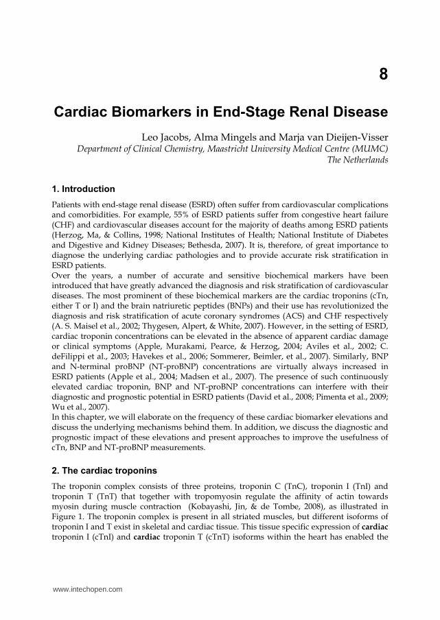
The troponin complex consists of three proteins, troponin C (TnC), troponin I (TnI) and troponin T (TnT) that together with tropomyosin regulate the affinity of actin towards myosin during muscle contraction (Kobayashi, Jin, & de Tombe, 2008), as illustrated in Figure 1. The troponin complex is present in all striated muscles, but different isoforms of troponin I and T exist in skeletal and cardiac tissue. This tissue specific expression of cardiac troponin I (cTnI) and cardiac troponin T (cTnT) isoforms within the heart has enabled the
www.intechopen.com
Chronic Kidney Disease and Renal Transplantation
148
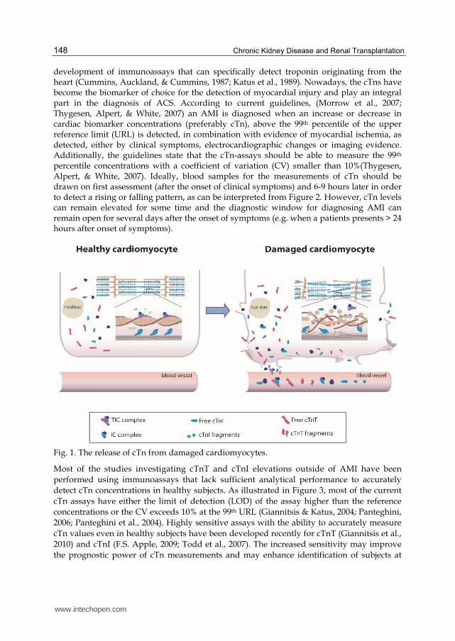
development of immunoassays that can specifically detect troponin originating from the heart (Cummins, Auckland, & Cummins, 1987; Katus et al., 1989). Nowadays, the cTns have become the biomarker of choice for the detection of myocardial injury and play an integral part in the diagnosis of ACS. According to current guidelines, (Morrow et al., 2007; Thygesen, Alpert, & White, 2007) an AMI is diagnosed when an increase or decrease in cardiac biomarker concentrations (preferably cTn), above the 99th percentile of the upper reference limit (URL) is detected, in combination with evidence of myocardial ischemia, as detected, either by clinical symptoms, electrocardiographic changes or imaging evidence. Additionally, the guidelines state that the cTn-assays should be able to measure the 99th percentile concentrations with a coefficient of variation (CV) smaller than 10%(Thygesen, Alpert, & White, 2007). Ideally, blood samples for the measurements of cTn should be drawn on first assessment (after the onset of clinical symptoms) and 6-9 hours later in order to detect a rising or falling pattern, as can be interpreted from Figure 2. However, cTn levels can remain elevated for some time and the diagnostic window for diagnosing AMI can remain open for several days after the onset of symptoms (e.g. when a patients presents > 24 hours after onset of symptoms).
Fig. 1. The release of cTn from damaged cardiomyocytes.
Most of the studies investigating cTnT and cTnI elevations outside of AMI have been
performed using immunoassays that lack sufficient analytical performance to accurately
detect cTn concentrations in healthy subjects. As illustrated in Figure 3, most of the current
cTn assays have either the limit of detection (LOD) of the assay higher than the reference
concentrations or the CV exceeds 10% at the 99th URL (Giannitsis & Katus, 2004; Panteghini,
2006; Panteghini et al., 2004). Highly sensitive assays with the ability to accurately measure
cTn values even in healthy subjects have been developed recently for cTnT (Giannitsis et al.,
2010) and cTnI (F.S. Apple, 2009; Todd et al., 2007). The increased sensitivity may improve
the prognostic power of cTn measurements and may enhance identification of subjects at
www.intechopen.com
Cardiac Biomarkers in End-Stage Renal Disease
149
Fig. 2. The release kinetics of cTn after AMI.
risk. Indeed, two recent studies have shown the prognostic value of cTnT at previously undetectable levels in patients with stable coronary disease (Omland et al., 2009) and with stable chronic HF (Latini et al., 2007). Moreover, cTnT values measured by a high sensitive cTnT assay (hs-cTnT) that were undetectable with the conventional assay were found to be associated with the extent of coronary atherosclerosis (Laufer et al., 2010). Figure 1 visualizes the release of the cTns in response to cellular damage and figure 2 shows the typical release kinetics of the cTns seen after an acute myocardial infarction.
2.1 Cardiac troponin elevations in ESRD In patients suffering from ESRD, cTn concentrations can be elevated in the absence of apparent cardiac damage or clinical symptoms (Apple et al., 2004; Aviles et al., 2002; C. deFilippi et al., 2003; Havekes et al., 2006; Sommerer, Beimler, et al., 2007). The exact frequency of these elevations varies somewhat between studies, depending on the patient inclusion criteria, the applied cut-off values and the troponin assay used. In general, cTnT has been found elevated more often than cTnI (roughly 53% for cTnT and 17% for cTnI as reviewed in (Kanderian & Francis, 2006)) although recent publications, using more sensitive assays suggest that the frequency of cTnT and cTnI elevations are similar (Hickman et al., 2007; Kumar, Michelis, Devita, Panagopoulos, & Rosenstock, 2010). It is important to note that the number of detected cTn elevations depends on the cut-offs that are used to define elevated values. As mentioned above, many cTn assays lack the sensitivity to accurately (<10% CV) measure the 99th percentile and the 10% CV is used as the diagnostic cut-off value. As a result the number of elevations will inevitably be lower when higher cut-off values are used. For example, in a study that we (Jacobs et al., 2009) performed in 32 ESRD patients we found that 38% of patients had cTnT elevations at baseline using the 10% CV cut-off, versus 63% using the 99th percentile cut-off with the contemporary 4th generation cTnT assay (Roche Diagnostics). Similarly cTnI concentrations where elevated above the 10% CV in 10% of the cases, compared to 50% elevations above the 99th percentile. Note that, the 10% CV is a property solely dependent on the sensitivity of the cTn assay and one should not compare cTn elevations above this level between assays. The introduction of guideline acceptable cTn assays that can accurate measure the 99th percentile will enable a better comparison of the frequencies of cTn elevations in ESRD patients.
www.intechopen.com
Chronic Kidney Disease and Renal Transplantation
150
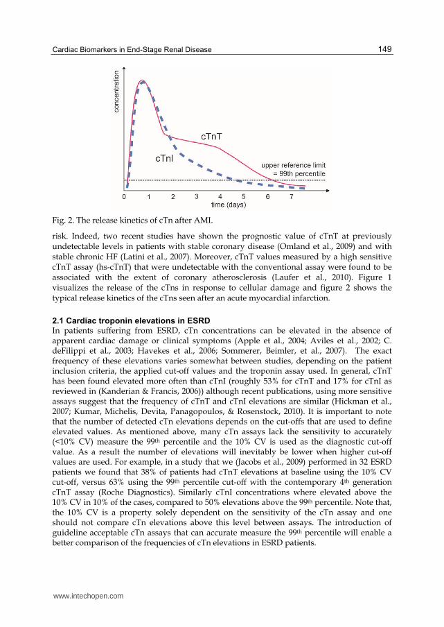
Fig. 3. (A) Biomarkers within the blood circulation follow a bell-shaped Gaussian
distribution within a group of subjects. The diagnostic cut-off concentration for cTn is
defined at the 99th percentile as measured in a healthy reference control group, so 1% of
these subjects with the highest cTn concentrations are defined unhealthy (Apple et al., 2007;
Morrow et al., 2007; Thygesen, Alpert, White, et al., 2007). At the time of definition, cTn
concentrations were undetectable in all healthy individuals, as illustrated by decision limit
‘b’. Improvements in the lower measuring range have lately resulted in cTn reference
concentrations and thus also a true 99th percentile concentration. The lower decision limit is
indicated by ‘a’. However, this improvement in sensitivity = TP/(TP+FN) goes along with a
worsening of the specificity = TN/(TN+FP). TP, number of subjects that were truly assigned
positive; FN, number of subjects that were falsely assigned negative; TN, number of subjects
that were truly assigned negative, FP, number of subjects that were falsely assigned positive;
(B) A typical precision profile of an immunoassay. The diagnostic cutoff concentration
should be measured with sufficient precision (coefficient of variation, CV = SD/mean
<10%)(Panteghini et al., 2004).
In general, the use of more sensitive cTn assays will likely show that the presence of cTn
elevations in ESRD patients might be even more frequent than previously thought. We
found, that by using a more sensitive cTnT assay 94% of our patients had cTnT elevations
above the 99th percentile with the hs-cTnT assay (Jacobs et al., 2009). Others have similarly
found a larger amount of cTnI elevations in ESRD patients by using more sensitive cTnI
assays (Hickman et al., 2007; Kumar et al., 2010).
With respect to the occurrence of cTn elevations in ESRD patients one should also take into
account that the cTn concentrations in ESRD patients can vary over time, even in otherwise
clinically stable patients. For example, by measuring cTn concentrations every two months,
for a period of 6 months an additional number of patients with elevated cTn concentrations
(at least once during the follow-up) could be identified (Jacobs et al., 2009). Similarly Roberts
et al. have also shown longitudinal changes in the presence or absence of cTnT elevations in
ESRD patients during a 1 year follow-up. In their study, cTnT values were measured 5 times
A
cTn (concentration)
CV
(%
)
10
acceptable precision
healthy
population
diseased
patients
decision limita b
TN TP
cTn (concentration)
fre
qu
en
cy
true
99th
perc
entile
FPFN
B
www.intechopen.com
Cardiac Biomarkers in End-Stage Renal Disease
151
and interestingly the patient survival decreased with increasing occurrence of cTnT
elevations, i.e. the 1.7 year patients survival was 100%, 90% and 78% for patients with zero,
one to four, or five out of five concentrations (Roberts et al., 2009). These findings are in line
with above mentioned prognostic value of cTn elevations in ESRD patients. As such,
assessing cTn concentrations at regular points in time would therefore appear as a
sensible tool to increase clinical vigilance for the presence of myocardial damage and as a
means for possible intervention. This is in agreement with previous studies which
provided evidence for the increased ability of serial versus single cTn measurements to
identify patients at risk for an event (Han, Lindsell, Ryan, & Gibler, 2005; Miller et al.,
2007; Ooi, Zimmerman, Graham, & Wells, 2001; Roberts et al., 2004; Wayand, Baum,
Schatzle, Scharf, & Neumeier, 2000).
2.1.1 Mechanisms underlying the cTn elevations
The underlying mechanisms behind these elevations have not been fully elucidated. The high incidence of coronary artery disease in ESRD patients (National Institutes of Health; National Institute of Diabetes and Digestive and Kidney Diseases; Bethesda, 2007) and the close relationship between cTn levels and the severity of coronary artery disease (C. deFilippi et al., 2003; Ooi, Isotalo, & Veinot, 2000) make the presence of subclinical ischemic cardiac damage a possible cause of cTn elevations. In this respect, it is also interesting to mention a study by DeFillipi et al. (C. R. deFilippi, Thorn, et al., 2007) who compared elevated cTnT values in 23 ESRD patients, with evidence of myocardial ischemia gathered by means of cardiovascular magnetic resonance (CMR) with late gadolinium enhancement (C. R. deFilippi, Thorn, et al., 2007). This study found that only a very small number of patients with elevated cTnT had CMR evidence of myocardial damage (0% of patients with cTnT <0.03 µg/L and 23% of patients with cTnT > 0.07 µg/L) (C. R. deFilippi, Thorn, et al., 2007). So, these patients, without known coronary artery disease and virtually no evidence of myocardial ischemia still had elevated cTnT values. These findings suggest there may be other than ischemia related reasons for the elevated levels of cTn. For example, the dialysis process itself can have a direct effect on cTn concentrations. There is some debate as to whether the dialysis process causes a decrease in cTn values (Montagnana et al., 2008) or an increase (Sommerer, Heckele, et al., 2007). In any case, different dialysis modalities like the use of high- or low- flux membranes and the method of vascular access can influence cTn concentrations (Lippi et al., 2008; Sommerer, Heckele, et al., 2007). For this reason, blood sampling times should be taken into account when measuring cTn concentrations and measurements are probably best performed pre-dialysis. Another possible reason for the elevated cTn values in ESRD pertains to a decreased renal clearance of cTn. For example, the cTn half-life was shown to increase with the degree of renal impairment (Wiessner et al., 2007). Diris et al. (Diris et al., 2004) have shown the presence of immunoreactive cTnT fragments, which are small enough to be cleared by the kidneys and which might accumulate in ESRD patients. Others, however, have found only intact cTnT in patients with kidney failure, (Fahie-Wilson et al., 2006) and to date there is still a great deal of debate on the mechanisms underlying the cTn elevation in ESRD patients. Whatever the exact mechanism, the elevations should not be taken lightly as they are highly predictive for adverse cardiovascular events (Apple, Murakami, Pearce, & Herzog, 2002; Khan, Hemmelgarn, Tonelli, Thompson, & Levin, 2005; Sommerer, Beimler, et al., 2007).
www.intechopen.com
Chronic Kidney Disease and Renal Transplantation
152
2.2 Diagnosing AMI in ESRD
The presence of continuously elevated cTn concentrations can frustrate the diagnosis of AMI (eg. when ESRD patients are presenting with clinical symptoms). The National Academy of Clinical Biochemistry (NACB) has recognized this issue and has published guidelines that address this issue (Wu et al., 2007). These guidelines suggest that for patients with chronically elevated concentrations of cTn, changes in cTn (>20%) 6-9 hours after the onset of clinical symptoms are indicative of an AMI. To date, however, little is known about the analytical and biological variations of cTn in ESRD patients and >20% changes might also occur in the absence of clinical symptoms (Miller et al., 2007; Roberts et al., 2004). The lack of detailed knowledge of the biological variation in ESRD patients, in combination with the likely increase in the frequency of chronically elevated cTnT as a result of more sensitive measurements call for further refinement of the current guidelines. As the highly sensitive cTn assays will enable a more accurate assessment of the biological variation, the use of serial measurement in order to detect abnormal changes in cTn values will likely be incorporated into these refinements. A potential approach to incorporate the biological variation into the diagnosis of AMI could come from the use of reference change values (RCV) (Aakre & Sandberg, 2010). The RCV describes the change in a concentration between two time points, than can be perceived as significant, taking into account both the analytical and the individual (biological) variations and is calculated as follows:
2 22 A IRCV z CV CV= × × +
wherein z is the z score, which can be set at the desired level of statistical significance. The analytical variation is described by the CVA and the individual (biological) variation by the CVI (Omar, van der Watt, & Pillay, 2008). To date, there are only a few studies that investigated the RCV values for cTnT (Vasile, Saenger, Kroning, & Jaffe, 2010) and cTnI (Wu, Lu, Todd, Moecks, & Wians, 2009) in healthy subjects and more studies are needed to examine the strengths and weaknesses of using RCV values for the diagnosis of AMI. The CVI for cTn can vary from population to population and can depend on the sampling time-intervals. For example, it has to be investigated if the variations in the cTn measurements seen in dialysis patients (i.e. (Hill, Cleve, Carlisle, Young, & McQueen, 2009; Jacobs et al., 2009)) are comparable to those in “healthy subjects” and if they are diagnostically relevant. So, in order to define the biological variations, clear rules need to be established with respect to the inclusion of subjects, sampling times, storage etc. Moreover, the variations in otherwise healthy subjects may vary from those in diseased populations.
3. The natriuretic peptides
The damage to the heart that is sustained during an AMI, but also other disorders that can impair left ventricular myocardial function, can lead to heart failure (HF). In HF there is a structural or functional cardiac disorder that impairs the ability of the ventricle to fill with or eject blood(Hunt, 2005). In effect, the pump-function of the heart is impaired, which may lead to symptoms of dyspnea, fatigue and fluid retention. In the population over the age of 65, the incidence of HF is about 1 per 100 and within this age group it is the leading cause of hospitalization (in the United States) (Lloyd-Jones et al., 2002). Considering the wide variety of causes underlying HF, the diagnosis and risk stratification in these patients is difficult. Over the years several advances have been made and the use of cardiac biomarkers, notably
www.intechopen.com
Cardiac Biomarkers in End-Stage Renal Disease
153
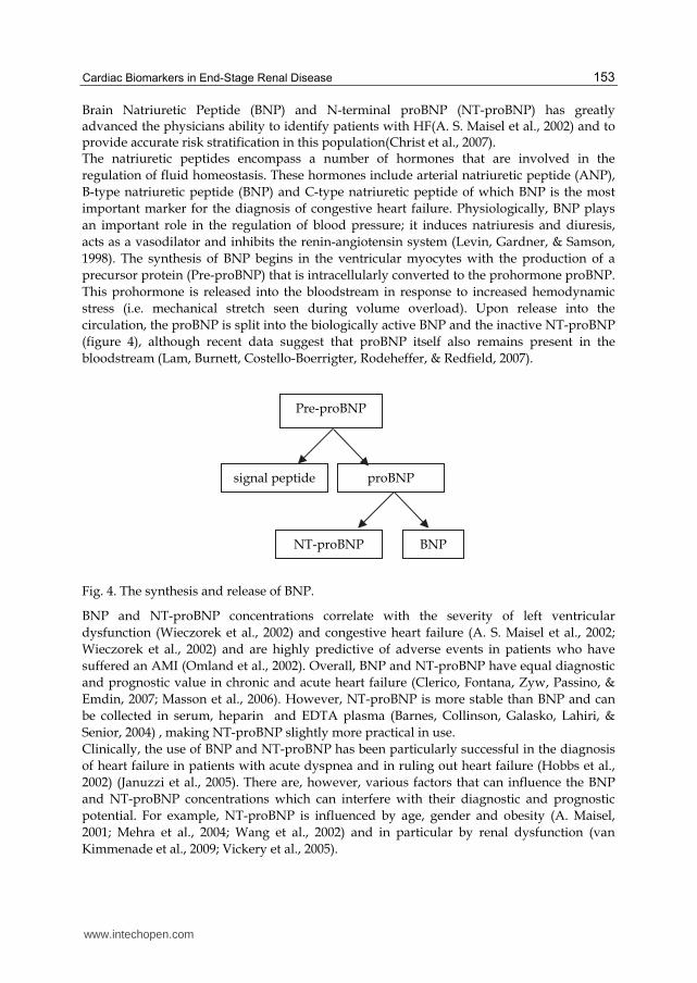
Brain Natriuretic Peptide (BNP) and N-terminal proBNP (NT-proBNP) has greatly advanced the physicians ability to identify patients with HF(A. S. Maisel et al., 2002) and to provide accurate risk stratification in this population(Christ et al., 2007). The natriuretic peptides encompass a number of hormones that are involved in the
regulation of fluid homeostasis. These hormones include arterial natriuretic peptide (ANP),
B-type natriuretic peptide (BNP) and C-type natriuretic peptide of which BNP is the most
important marker for the diagnosis of congestive heart failure. Physiologically, BNP plays
an important role in the regulation of blood pressure; it induces natriuresis and diuresis,
acts as a vasodilator and inhibits the renin-angiotensin system (Levin, Gardner, & Samson,
1998). The synthesis of BNP begins in the ventricular myocytes with the production of a
precursor protein (Pre-proBNP) that is intracellularly converted to the prohormone proBNP.
This prohormone is released into the bloodstream in response to increased hemodynamic
stress (i.e. mechanical stretch seen during volume overload). Upon release into the
circulation, the proBNP is split into the biologically active BNP and the inactive NT-proBNP
(figure 4), although recent data suggest that proBNP itself also remains present in the
bloodstream (Lam, Burnett, Costello-Boerrigter, Rodeheffer, & Redfield, 2007).
Fig. 4. The synthesis and release of BNP.
BNP and NT-proBNP concentrations correlate with the severity of left ventricular
dysfunction (Wieczorek et al., 2002) and congestive heart failure (A. S. Maisel et al., 2002;
Wieczorek et al., 2002) and are highly predictive of adverse events in patients who have
suffered an AMI (Omland et al., 2002). Overall, BNP and NT-proBNP have equal diagnostic
and prognostic value in chronic and acute heart failure (Clerico, Fontana, Zyw, Passino, &
Emdin, 2007; Masson et al., 2006). However, NT-proBNP is more stable than BNP and can
be collected in serum, heparin and EDTA plasma (Barnes, Collinson, Galasko, Lahiri, &
Senior, 2004) , making NT-proBNP slightly more practical in use.
Clinically, the use of BNP and NT-proBNP has been particularly successful in the diagnosis
of heart failure in patients with acute dyspnea and in ruling out heart failure (Hobbs et al.,
2002) (Januzzi et al., 2005). There are, however, various factors that can influence the BNP
and NT-proBNP concentrations which can interfere with their diagnostic and prognostic
potential. For example, NT-proBNP is influenced by age, gender and obesity (A. Maisel,
2001; Mehra et al., 2004; Wang et al., 2002) and in particular by renal dysfunction (van
Kimmenade et al., 2009; Vickery et al., 2005).
Pre-proBNP
signal peptide proBNP
NT-proBNP BNP
www.intechopen.com
Chronic Kidney Disease and Renal Transplantation
154
3.1 BNP and NT-proBNP elevations in ESRD
In patients with ESRD, the BNP and NT-proBNP concentrations are virtually always
elevated above normal cut-off values (Apple et al., 2004; Madsen et al., 2007). There are
several mechanisms that could explain the elevated BNP and NT-proBNP concentrations. In
the first place there might be a lack of renal clearance as BNP and NT-proBNP have been
shown to be inversely correlated to the glomerular filtration rate (reviewed in (C. DeFilippi,
van Kimmenade, & Pinto, 2008). Interestingly, it is not clear if BNP and NT-proBNP are
affected similarly by the reduction in renal clearance. Some reports mention that NT-
proBNP is more strongly influenced by a decrease in renal function than BNP, (Vickery et
al., 2005) whereas others state that they are equally dependent on the renal function for their
clearance (van Kimmenade et al., 2009). It should be mentioned that the discrepancies
between NT-proBNP and BNP increase with a decreasing glomerular filtration rate, as
illustrated by the significantly higher NT-proBNP/BNP ratios in patients with a decreased
renal function (Kemperman, van den Berg, Kirkels, & de Jonge, 2004; Srisawasdi,
Vanavanan, Charoenpanichkit, & Kroll, 2010; van Kimmenade et al., 2009; Vickery et al.,
2005). In particular in patients with a severely reduced renal function (eg. eGFR < 20) the
NT-proBNP/BNP seems to increase exponentially (Srisawasdi et al., 2010; Vickery et al.,
2005). Recent reports in ESRD patients suggest that the NT-proBNP/BNP ratio increases
even further in patients receiving hemodialysis (Jacobs, Mingels, et al., 2010). This increase
in the NT-proBNP ratio might not be the sole result of renal clearance and other mechanism
such as the extra-renal clearance by circulating type-C natriuretic peptide receptor and by
neural endopeptidases (Martinez-Rumayor, Richards, Burnett, & Januzzi, 2008) may play a
role (van Kimmenade et al., 2009). Specifically in ESRD patients, there are several other
factors that can influence BNP or NT-proBNP concentrations. For example, the dialysis
process itself may influence BNP and NT-proBNP concentrations, like the type of dialysis
membrane Interestingly, some find elevated levels of NT-proBNP after dialysis (Sommerer,
Heckele, et al., 2007) whereas others find lower values (Madsen et al., 2007), and for BNP
this might be different than for NT-proBNP (Wahl, Graf, Renz, & Fassbinder, 2004). Other
parameters, related to the dialysis treatment, such as a patient’s volume status could also
affect NT-proBNP and BNP concentrations (Booth, Pinney, & Davenport, 2010; Jacobs, van
de Kerkhof, et al., 2010) whereby, an increase in extracellular volume might induce left
ventricular dilatation and subsequent increases in (NT-pro)BNP concentrations. More
research is needed to understand the renal and extra-renal clearance of BNP and NT-
proBNP and to identify ESRD related differences in the clearance or production of these
peptides.
3.2 The clinical relevance of (NT-pro)BNP in ESRD
Regardless of the fact that virtually all ESRD patients have elevated (NT-pro)BNP values and that there is much uncertainty about the underlying reasons one should keep in mind that the (NT-pro)BNP concentrations still have a strong prognostic value in ESRD patients. BNP and NT-proBNP are related to cardiovascular disease and all-cause mortality and thus their measurement remains of importance for diagnosis and risk stratification in ESRD patients (Apple et al., 2004; Madsen et al., 2007). Importantly the diagnostic and prognostic cut-off values for NT-proBNP in ESRD are significantly elevated compared to the cut-off values in patient with non or mildly impaired renal function. For example, in hemodialysis patients a NT-proBNP cut-off value ≥7200 ng/L could discriminate patients with LVD from
www.intechopen.com
Cardiac Biomarkers in End-Stage Renal Disease
155
those without (David et al., 2008). More in general, DeFilippi et al found an upward shift in the optimal cut-off value for patients with a diminished renal function based on the estimated glomerular filtration rate (eGFR) (C. R. Defilippi, Seliger, Maynard, & Christenson, 2007). The optimal NT-proBNP cut-off for diagnosis of decompensated HF for
patients (n=831) with eGFR <60 and ≥60 mL/min/1.73 m2 was achieved at concentrations as
high as 1200 ng/L and 900/450 ng/L (age ≥50/<50 years), respectively. For BNP, optimal cut-offs for patients with eGFR <30, 30-59, 60-90, >90 mL/min/1.73 m2 were 225, 201, 104, and 71 ng/L, respectively. For optimal diagnostic performance of (NT-pro)BNP in ESRD patients, it is thus of great importance that appropriate cut-off concentrations will be developed.
4. Conclusion
While a reduced renal function and the dialysis treatment itself can have a significant influence on cardiac troponin and (NT-pro)BNP values they are strongly associated with adverse outcomes. Similarly, the cardiac troponins, which are often elevated above the diagnostic cut-off value for AMI provide valuable diagnostic and prognostic information in ESRD patients. Considering the ESRD related processes that can influence the cardiac troponin and natriuretic peptide concentrations, more research is needed to define appropriate cut-off values for the diagnosis of AMI and CHF. As both cTn and (NT-pro)BNP concentrations are independently associated with cardiovascular mortality, their measurement is important for risk stratification and as a tool to increase clinical vigilance.
5. References
Aakre, K. M., & Sandberg, S. (2010). Can changes in troponin results be useful in diagnosing myocardial infarction? Clin Chem, 56(7), 1047-1049.
Apple, F. S., Jesse, R. L., Newby, L. K., Wu, A. H., Christenson, R. H., Cannon, C. P., et al. (2007). National Academy of Clinical Biochemistry and IFCC Committee for Standardization of Markers of Cardiac Damage Laboratory Medicine Practice Guidelines: analytical issues for biochemical markers of acute coronary syndromes. Clin Chem, 53(4), 547-551.
Apple, F. S., Murakami, M. M., Pearce, L. A., & Herzog, C. A. (2002). Predictive value of cardiac troponin I and T for subsequent death in end-stage renal disease. Circulation, 106(23), 2941-2945.
Apple, F. S., Murakami, M. M., Pearce, L. A., & Herzog, C. A. (2004). Multi-Biomarker Risk Stratification of N-Terminal Pro-B-Type Natriuretic Peptide, High-Sensitivity C-Reactive Protein, and Cardiac Troponin T and I in End-Stage Renal Disease for All-Cause Death. Clin Chem, 50, 2233-2235.
Aviles, R. J., Askari, A. T., Lindahl, B., Wallentin, L., Jia, G., Ohman, E. M., et al. (2002). Troponin T levels in patients with acute coronary syndromes, with or without renal dysfunction. N Engl J Med, 346(26), 2047-2052.
Barnes, S. C., Collinson, P. O., Galasko, G., Lahiri, A., & Senior, R. (2004). Evaluation of N-terminal pro-B type natriuretic peptide analysis on the Elecsys 1010 and 2010 analysers. Ann Clin Biochem, 41(Pt 6), 459-463.
www.intechopen.com
Chronic Kidney Disease and Renal Transplantation
156
Booth, J., Pinney, J., & Davenport, A. (2010). N-terminal proBNP--marker of cardiac dysfunction, fluid overload, or malnutrition in hemodialysis patients? Clin J Am Soc Nephrol, 5(6), 1036-1040.
Christ, M., Thuerlimann, A., Laule, K., Klima, T., Hochholzer, W., Perruchoud, A. P., et al. (2007). Long-term prognostic value of B-type natriuretic peptide in cardiac and non-cardiac causes of acute dyspnoea. Eur J Clin Invest, 37(11), 834-841.
Clerico, A., Fontana, M., Zyw, L., Passino, C., & Emdin, M. (2007). Comparison of the diagnostic accuracy of brain natriuretic peptide (BNP) and the N-terminal part of the propeptide of BNP immunoassays in chronic and acute heart failure: a systematic review. Clinical chemistry, 53(5), 813-822.
Cummins, B., Auckland, M. L., & Cummins, P. (1987). Cardiac-specific troponin-I radioimmunoassay in the diagnosis of acute myocardial infarction. Am Heart J, 113(6), 1333-1344.
David, S., Kumpers, P., Seidler, V., Biertz, F., Haller, H., & Fliser, D. (2008). Diagnostic value of N-terminal pro-B-type natriuretic peptide (NT-proBNP) for left ventricular dysfunction in patients with chronic kidney disease stage 5 on haemodialysis. Nephrol Dial Transplant, 23(4), 1370-1377.
DeFilippi, C., van Kimmenade, R. R., & Pinto, Y. M. (2008). Amino-terminal pro-B-type natriuretic peptide testing in renal disease. Am J Cardiol, 101(3A), 82-88.
deFilippi, C., Wasserman, S., Rosanio, S., Tiblier, E., Sperger, H., Tocchi, M., et al. (2003). Cardiac troponin T and C-reactive protein for predicting prognosis, coronary atherosclerosis, and cardiomyopathy in patients undergoing long-term hemodialysis. Jama, 290(3), 353-359.
Defilippi, C. R., Seliger, S. L., Maynard, S., & Christenson, R. H. (2007). Impact of renal disease on natriuretic Peptide testing for diagnosing decompensated heart failure and predicting mortality. Clin Chem, 53(8), 1511-1519.
deFilippi, C. R., Thorn, E. M., Aggarwal, M., Joy, A., Christenson, R. H., Duh, S. H., et al. (2007). Frequency and cause of cardiac troponin T elevation in chronic hemodialysis patients from study of cardiovascular magnetic resonance. Am J Cardiol, 100(5), 885-889.
Diris, J. H., Hackeng, C. M., Kooman, J. P., Pinto, Y. M., Hermens, W. T., & Van Dieijen-Visser, M. P. (2004). Impaired Renal Clearance Explains Elevated Troponin T Fragments in Hemodialysis Patients. Circulation, 109(1), 23-25.
F.S. Apple, M. M. M., D.P.Farris, S.A. Karimi, P.A. Simpson, L.T.Le. (2009). Serum 99th percentile reference value for the high sensitive Singulex cardiac troponin I assay. Clinical Chemistry, 55, A63.
Fahie-Wilson, M. N., Carmichael, D. J., Delaney, M. P., Stevens, P. E., Hall, E. M., & Lamb, E. J. (2006). Cardiac Troponin T Circulates in the Free, Intact Form in Patients with Kidney Failure. Clin Chem, 52(3), 414-420.
Giannitsis, E., & Katus, H. A. (2004). Comparison of cardiac troponin T and troponin I assays--implications of analytical and biochemical differences on clinical performance. Clin Lab, 50(9-10), 521-528.
Giannitsis, E., Kurz, K., Hallermayer, K., Jarausch, J., Jaffe, A. S., & Katus, H. A. (2010). Analytical validation of a high-sensitivity cardiac troponin T assay. Clin Chem, 56(2), 254-261.
www.intechopen.com
Cardiac Biomarkers in End-Stage Renal Disease
157
Han, J. H., Lindsell, C. J., Ryan, R. J., & Gibler, W. B. (2005). Changes in cardiac troponin T measurements are associated with adverse cardiac events in patients with chronic kidney disease. Am J Emerg Med, 23(4), 468-473.
Havekes, B., van Manen, J. G., Krediet, R. T., Boeschoten, E. W., Vandenbroucke, J. P., & Dekker, F. W. (2006). Serum troponin T concentration as a predictor of mortality in hemodialysis and peritoneal dialysis patients. Am J Kidney Dis, 47(5), 823-829.
Herzog, C. A., Ma, J. Z., & Collins, A. J. (1998). Poor long-term survival after acute myocardial infarction among patients on long-term dialysis. N Engl J Med, 339(12), 799-805.
Hickman, P. E., Koerbin, G., Southcott, E., Tate, J., Dimeski, G., Carter, A., et al. (2007). Newer cardiac troponin I assays have similar performance to troponin T in patients with end-stage renal disease. Ann Clin Biochem, 44(Pt 3), 285-289.
Hill, S. A., Cleve, R., Carlisle, E., Young, E., & McQueen, M. J. (2009). Intra-individual variability in troponin T concentration in dialysis patients. Clinical Biochemistry, 42(10-11), 991-995.
Hobbs, F. D., Davis, R. C., Roalfe, A. K., Hare, R., Davies, M. K., & Kenkre, J. E. (2002). Reliability of N-terminal pro-brain natriuretic peptide assay in diagnosis of heart failure: cohort study in representative and high risk community populations. Bmj, 324(7352), 1498.
Hunt, S. A. (2005). ACC/AHA 2005 guideline update for the diagnosis and management of chronic heart failure in the adult: a report of the American College of Cardiology/American Heart Association Task Force on Practice Guidelines (Writing Committee to Update the 2001 Guidelines for the Evaluation and Management of Heart Failure). J Am Coll Cardiol, 46(6), e1-82.
Jacobs, L. H., Mingels, A. M., Wodzig, W. K., van Dieijen-Visser, M. P., Kooman, J. P., Srisawasdi, P., et al. (2010). Renal Dysfunction, Hemodialysis, and the NT-proBNP/BNP Ratio. Am J Clin Pathol, 134(3), 516-517.
Jacobs, L. H., van de Kerkhof, J., Mingels, A. M., Kleijnen, V. W., van der Sande, F. M., Wodzig, W. K., et al. (2009). Haemodialysis patients longitudinally assessed by highly sensitive cardiac troponin T and commercial cardiac troponin T and cardiac troponin I assays. Ann Clin Biochem, 46(Pt 4), 283-290.
Jacobs, L. H., van de Kerkhof, J. J., Mingels, A. M., Passos, V. L., Kleijnen, V. W., Mazairac, A. H., et al. (2010). Inflammation, overhydration and cardiac biomarkers in haemodialysis patients: a longitudinal study. Nephrol Dial Transplant, 25(1), 243-248.
Januzzi, J., James L., Camargo, C. A., Anwaruddin, S., Baggish, A. L., Chen, A. A., Krauser, D. G., et al. (2005). The N-terminal Pro-BNP Investigation of Dyspnea in the Emergency department (PRIDE) study. The American Journal of Cardiology, 95(8), 948-954.
Kanderian, A. S., & Francis, G. S. (2006). Cardiac troponins and chronic kidney disease. Kidney Int, 69(7), 1112-1114.
Katus, H. A., Remppis, A., Looser, S., Hallermeier, K., Scheffold, T., & Kubler, W. (1989). Enzyme linked immuno assay of cardiac troponin T for the detection of acute myocardial infarction in patients. J Mol Cell Cardiol, 21, 1349-1353.
Kemperman, H., van den Berg, M., Kirkels, H., & de Jonge, N. (2004). B-Type Natriuretic Peptide (BNP) and N-Terminal proBNP in Patients with End-Stage Heart Failure Supported by a Left Ventricular Assist Device. Clin Chem, 50(9), 1670-1672.
www.intechopen.com
Chronic Kidney Disease and Renal Transplantation
158
Khan, N. A., Hemmelgarn, B. R., Tonelli, M., Thompson, C. R., & Levin, A. (2005). Prognostic value of troponin T and I among asymptomatic patients with end-stage renal disease: a meta-analysis. Circulation, 112(20), 3088-3096.
Kobayashi, T., Jin, L., & de Tombe, P. P. (2008). Cardiac thin filament regulation. Pflugers Arch, 457(1), 37-46.
Kumar, N., Michelis, M. F., Devita, M. V., Panagopoulos, G., & Rosenstock, J. L. (2010). Troponin I levels in asymptomatic patients on haemodialysis using a high-sensitivity assay. Nephrol Dial Transplant.
Lam, C. S., Burnett, J. C., Jr., Costello-Boerrigter, L., Rodeheffer, R. J., & Redfield, M. M. (2007). Alternate circulating pro-B-type natriuretic peptide and B-type natriuretic peptide forms in the general population. J Am Coll Cardiol, 49(11), 1193-1202.
Latini, R., Masson, S., Anand, I. S., Missov, E., Carlson, M., Vago, T., et al. (2007). Prognostic value of very low plasma concentrations of troponin T in patients with stable chronic heart failure. Circulation, 116(11), 1242-1249.
Laufer, E. M., Mingels, A. M., Winkens, M. H., Joosen, I. A., Schellings, M. W., Leiner, T., et al. (2010). The extent of coronary atherosclerosis is associated with increasing circulating levels of high sensitive cardiac troponin T. Arterioscler Thromb Vasc Biol, 30(6), 1269-1275.
Levin, E. R., Gardner, D. G., & Samson, W. K. (1998). Natriuretic peptides. N Engl J Med, 339(5), 321-328.
Lippi, G., Tessitore, N., Montagnana, M., Salvagno, G. L., Lupo, A., & Guidi, G. C. (2008). Influence of sampling time and ultrafiltration coefficient of the dialysis membrane on cardiac troponin I and T. Arch Pathol Lab Med, 132(1), 72-76.
Lloyd-Jones, D. M., Larson, M. G., Leip, E. P., Beiser, A., D'Agostino, R. B., Kannel, W. B., et al. (2002). Lifetime risk for developing congestive heart failure: the Framingham Heart Study. Circulation, 106(24), 3068-3072.
Madsen, L. H., Ladefoged, S., Corell, P., Schou, M., Hildebrandt, P. R., & Atar, D. (2007). N-terminal pro brain natriuretic peptide predicts mortality in patients with end-stage renal disease in hemodialysis. Kidney Int, 71(6), 548-554.
Maisel, A. (2001). B-type natriuretic peptide levels: a potential novel "white count" for congestive heart failure. J Card Fail, 7(2), 183-193.
Maisel, A. S., Krishnaswamy, P., Nowak, R. M., McCord, J., Hollander, J. E., Duc, P., et al. (2002). Rapid measurement of B-type natriuretic peptide in the emergency diagnosis of heart failure. N Engl J Med, 347(3), 161-167.
Martinez-Rumayor, A., Richards, A. M., Burnett, J. C., & Januzzi, J. L., Jr. (2008). Biology of the natriuretic peptides. Am J Cardiol, 101(3A), 3-8.
Masson, S., Latini, R., Anand, I. S., Vago, T., Angelici, L., Barlera, S., et al. (2006). Direct Comparison of B-Type Natriuretic Peptide (BNP) and Amino-Terminal proBNP in a Large Population of Patients with Chronic and Symptomatic Heart Failure: The Valsartan Heart Failure (Val-HeFT) Data. Clin Chem, 52(8), 1528-1538.
Mehra, M. R., Uber, P. A., Park, M. H., Scott, R. L., Ventura, H. O., Harris, B. C., et al. (2004). Obesity and suppressed B-type natriuretic peptide levels in heart failure. Journal of the American College of Cardiology, 43(9), 1590-1595.
Miller, W. L., Hartman, K. A., Burritt, M. F., Grill, D. E., Rodeheffer, R. J., Burnett, J. C., Jr., et al. (2007). Serial biomarker measurements in ambulatory patients with chronic heart failure: the importance of change over time. Circulation, 116(3), 249-257.
www.intechopen.com
Cardiac Biomarkers in End-Stage Renal Disease
159
Montagnana, M., Lippi, G., Tessitore, N., Salvagno, G. L., Targher, G., Gelati, M., et al. (2008). Effect of hemodialysis on traditional and innovative cardiac markers. J Clin Lab Anal, 22(1), 59-65.
Morrow, D. A., Cannon, C. P., Jesse, R. L., Newby, L. K., Ravkilde, J., Storrow, A. B., et al. (2007). National Academy of Clinical Biochemistry Laboratory Medicine Practice Guidelines: Clinical characteristics and utilization of biochemical markers in acute coronary syndromes. Circulation, 115(13), e356-375.
National Institutes of Health; National Institute of Diabetes and Digestive and Kidney Diseases; Bethesda, M. (2007). U.S. Renal Data System, USRDS 2007 Annual Data Report: Atlas of Chronic Kidney Disease and End-Stage Renal Disease in the United States., 138-154.
Omar, F., van der Watt, G. F., & Pillay, T. S. (2008). Reference change values: how useful are they? J Clin Pathol, 61(4), 426-427.
Omland, T., de Lemos, J. A., Sabatine, M. S., Christophi, C. A., Rice, M. M., Jablonski, K. A., et al. (2009). A sensitive cardiac troponin T assay in stable coronary artery disease. N Engl J Med, 361(26), 2538-2547.
Omland, T., Persson, A., Ng, L., O'Brien, R., Karlsson, T., Herlitz, J., et al. (2002). N-terminal pro-B-type natriuretic peptide and long-term mortality in acute coronary syndromes. Circulation, 106(23), 2913-2918.
Ooi, D. S., Isotalo, P. A., & Veinot, J. P. (2000). Correlation of antemortem serum creatine kinase, creatine kinase-MB, troponin I, and troponin T with cardiac pathology. Clin Chem, 46(3), 338-344.
Ooi, D. S., Zimmerman, D., Graham, J., & Wells, G. A. (2001). Cardiac troponin T predicts long-term outcomes in hemodialysis patients. Clin Chem, 47(3), 412-417.
Panteghini, M. (2006). The new definition of myocardial infarction and the impact of troponin determination on clinical practice. Int J Cardiol, 106(3), 298-306.
Panteghini, M., Pagani, F., Yeo, K. T., Apple, F. S., Christenson, R. H., Dati, F., et al. (2004). Evaluation of imprecision for cardiac troponin assays at low-range concentrations. Clin Chem, 50(2), 327-332.
Pimenta, J., Sampaio, F., Martins, P., Carvalho, B., Rocha-Goncalves, F., Ferreira, A., et al. (2009). Aminoterminal B-type natriuretic peptide (NT-proBNP) in end-stage renal failure patients on regular hemodialysis: does it have diagnostic and prognostic implications? Nephron Clin Pract, 111(3), c182-188.
Roberts, M. A., Fernando, D., Macmillan, N., Proimos, G., Bach, L. A., Power, D. A., et al. (2004). Single and serial measurements of cardiac troponin I in asymptomatic patients on chronic hemodialysis. Clin Nephrol, 61(1), 40-46.
Roberts, M. A., Hare, D. L., Macmillan, N., Ratnaike, S., Sikaris, K., & Ierino, F. L. (2009). Serial increased cardiac troponin T predicts mortality in asymptomatic patients treated with chronic haemodialysis. Ann Clin Biochem, 46(Pt 4), 291-295.
Sommerer, C., Beimler, J., Schwenger, V., Heckele, N., Katus, H. A., Giannitsis, E., et al. (2007). Cardiac biomarkers and survival in haemodialysis patients. Eur J Clin Invest, 37(5), 350-356.
Sommerer, C., Heckele, S., Schwenger, V., Katus, H. A., Giannitsis, E., & Zeier, M. (2007). Cardiac biomarkers are influenced by dialysis characteristics. Clin Nephrol, 68, 392-400.
www.intechopen.com
Chronic Kidney Disease and Renal Transplantation
160
Srisawasdi, P., Vanavanan, S., Charoenpanichkit, C., & Kroll, M. H. (2010). The effect of renal dysfunction on BNP, NT-proBNP, and their ratio. Am J Clin Pathol, 133(1), 14-23.
Thygesen, K., Alpert, J. S., & White, H. D. (2007). Universal definition of myocardial infarction. J Am Coll Cardiol, 50(22), 2173-2195.
Thygesen, K., Alpert, J. S., White, H. D., Jaffe, A. S., Apple, F. S., Galvani, M., et al. (2007). Universal definition of myocardial infarction: Kristian Thygesen, Joseph S. Alpert and Harvey D. White on behalf of the Joint ESC/ACCF/AHA/WHF Task Force for the Redefinition of Myocardial Infarction. Eur Heart J, 28(20), 2525-2538.
Todd, J., Freese, B., Lu, A., Held, D., Morey, J., Livingston, R., et al. (2007). Ultrasensitive Flow-based Immunoassays using Single-Molecule Counting. Clin Chem, 53(11).
van Kimmenade, R. R., Januzzi, J. L., Jr., Bakker, J. A., Houben, A. J., Rennenberg, R., Kroon, A. A., et al. (2009). Renal clearance of B-type natriuretic peptide and amino terminal pro-B-type natriuretic peptide a mechanistic study in hypertensive subjects. J Am Coll Cardiol, 53(10), 884-890.
Vasile, V. C., Saenger, A. K., Kroning, J. M., & Jaffe, A. S. (2010). Biological and analytical variability of a novel high-sensitivity cardiac troponin T assay. Clin Chem, 56(7), 1086-1090.
Vickery, S., Price, C. P., John, R. I., Abbas, N. A., Webb, M. C., Kempson, M. E., et al. (2005). B-type natriuretic peptide (BNP) and amino-terminal proBNP in patients with CKD: relationship to renal function and left ventricular hypertrophy. Am J Kidney Dis, 46(4), 610-620.
Wahl, H. G., Graf, S., Renz, H., & Fassbinder, W. (2004). Elimination of the Cardiac Natriuretic Peptides B-Type Natriuretic Peptide (BNP) and N-Terminal proBNP by Hemodialysis. Clin Chem, 50(6), 1071-1074.
Wang, T. J., Larson, M. G., Levy, D., Leip, E. P., Benjamin, E. J., Wilson, P. W., et al. (2002). Impact of age and sex on plasma natriuretic peptide levels in healthy adults. Am J Cardiol, 90(3), 254-258.
Wayand, D., Baum, H., Schatzle, G., Scharf, J., & Neumeier, D. (2000). Cardiac troponin T and I in end-stage renal failure. Clin Chem, 46(9), 1345-1350.
Wieczorek, S. J., Wu, A. H., Christenson, R., Krishnaswamy, P., Gottlieb, S., Rosano, T., et al. (2002). A rapid B-type natriuretic peptide assay accurately diagnoses left ventricular dysfunction and heart failure: a multicenter evaluation. Am Heart J, 144(5), 834-839.
Wiessner, R., Hannemann-Pohl, K., Ziebig, R., Grubitzsch, H., Hocher, B., Vargas-Hein, O., et al. (2007). Impact of kidney function on plasma troponin concentrations after coronary artery bypass grafting. Nephrol Dial Transplant.
Wu, A. H., Jaffe, A. S., Apple, F. S., Jesse, R. L., Francis, G. L., Morrow, D. A., et al. (2007). National Academy of Clinical Biochemistry Laboratory Medicine Practice Guidelines: Use of Cardiac Troponin and B-Type Natriuretic Peptide or N-Terminal proB-Type Natriuretic Peptide for Etiologies Other than Acute Coronary Syndromes and Heart Failure. Clin Chem, 53(12), 2086-2096.
Wu, A. H., Lu, Q. A., Todd, J., Moecks, J., & Wians, F. (2009). Short- and long-term biological variation in cardiac troponin I measured with a high-sensitivity assay: implications for clinical practice. Clin Chem, 55(1), 52-58.
www.intechopen.com
Chronic Kidney Disease and Renal TransplantationEdited by Prof. Manisha Sahay
ISBN 978-953-51-0003-4Hard cover, 226 pagesPublisher InTechPublished online 10, February, 2012Published in print edition February, 2012
InTech EuropeUniversity Campus STeP Ri Slavka Krautzeka 83/A 51000 Rijeka, Croatia Phone: +385 (51) 770 447 Fax: +385 (51) 686 166www.intechopen.com
InTech ChinaUnit 405, Office Block, Hotel Equatorial Shanghai No.65, Yan An Road (West), Shanghai, 200040, China
Phone: +86-21-62489820 Fax: +86-21-62489821
This valuable resource covers inpatient and outpatient approaches to chronic renal disease and renaltransplant with clinical practicality. This first section of the book discusses chronic disease under distinct topics,each providing the readers with state-of-the-art information about the disease and its management. Itdiscusses the fresh perspectives on the current state of chronic kidney disease. The text highlights not just themedical aspects but also the psychosocial issues associated with chronic kidney disease. The latestapproaches are reviewed through line diagrams that clearly depict recent advances. The second section of thebook deals with issues related to transplant. It provides effective and up-to-date insight into caring for yourtransplant patients.
How to referenceIn order to correctly reference this scholarly work, feel free to copy and paste the following:
Leo Jacobs, Alma Mingels and Marja van Dieijen-Visser (2012). Cardiac Biomarkers in End-Stage RenalDisease, Chronic Kidney Disease and Renal Transplantation, Prof. Manisha Sahay (Ed.), ISBN: 978-953-51-0003-4, InTech, Available from: http://www.intechopen.com/books/chronic-kidney-disease-and-renal-transplantation/cardiac-biomarkers-in-end-stage-renal-disease