Embed Size (px)
Citation preview
SUBJECT TITLE
A Relational Fairness Perspective on Individuality and Individualization in Contemporary Approaches to Compensation in OrganizationsRevised Version
by Martin Lund Petersen, MSc (Econ.)
PhD thesis defended at SDUDepartment of Leadership and Corporate Strategy
Medical Informatics Group
Cardiac Action Potential Prolongation Induced by
Isolated Thioridazine Enantiomers
PhD Thesis byAsk Schou Jensen
River Publishers
Cardiac ActionPotential Prolongation
Induced by IsolatedThioridazine Enantiomers

Cardiac ActionPotential Prolongation
Induced by IsolatedThioridazine Enantiomers
PhD Thesis by
Ask Schou Jensen
Medical Informatics Group,Department of Health Science and Technology,
Aalborg University, Denmark
ISBN: 978-87-93237-42-1 (Ebook)
Published, sold and distributed by:River PublishersNiels Jernes Vej 109220 Aalborg ØDenmark
Tel.: +45369953197www.riverpublishers.com
Copyright for this work belongs to the author, River Publishers have the soleright to distribute this work commercially.
All rights reserved c© 2014 Ask Schou Jensen.
No part of this work may be reproduced, stored in a retrieval system, or trans-mitted in any form or by any means, electronic, mechanical, photocopying,microfilming, recording or otherwise, without prior written permission fromthe Publisher.
1
Contents A. Preface and Acknowledgements ..................................................................................................................3
B. English Summary ..........................................................................................................................................5
C. Danish Summary ...........................................................................................................................................6
D. List of Papers ................................................................................................................................................7
E. Abbreviations ................................................................................................................................................9
Introduction ........................................................................................................................................................... 11
Background .................................................................................................................................................... 11
The Clinical Problem ...................................................................................................................................... 11
Thioridazine Reverses Antimicrobial Resistance ........................................................................................... 11
The Cardiotoxicity of Thioridazine ................................................................................................................. 12
The Cellular Basis of Cardiotoxicity ............................................................................................................... 13
Stereochemistry – is there a Way Forward? ................................................................................................. 15
Investigating Thioridazine Enantiomer Effects .............................................................................................. 15
Objectives of the Thesis ......................................................................................................................................... 17
The Isolated Rabbit Papillary Muscle .................................................................................................................... 19
The Experimental Preparation ....................................................................................................................... 19
The Experimental Setup ................................................................................................................................ 20
Investigation of Drug Effects by Computational Modeling ................................................................................... 23
Modeling of the Action Potential .................................................................................................................. 23
The Hodgkin-Huxley Membrane Current Formulation ................................................................................. 23
Alternative Membrane Channel Formulations .............................................................................................. 25
From Hodgkin and Huxley to the rabbit ventricular cell model .................................................................... 26
The Shannon Model of the Rabbit Ventricular Cell ....................................................................................... 27
Modifications ................................................................................................................................................. 31
References ............................................................................................................................................................. 33
Contributions ......................................................................................................................................................... 39
I: Differential Effects of Thioridazine Enantiomers on Action Potential duration in Rabbit Papillary Muscle ...... 41
II: Model Based Analysis of the Effects of Thioridazine Enantiomers on the Rabbit Papillary Action Potential:
Part 1 ..................................................................................................................................................................... 43
2
III: Model Based Analysis of the Effects of Thioridazine Enantiomers on the Rabbit Papillary Action Potential:
Part 2 ..................................................................................................................................................................... 45
Conclusion ............................................................................................................................................................. 47
3
A. Preface and Acknowledgements
This thesis was submitted in partial fulfillment of the PhD degree at the Department of Health Science and
Technology, Aalborg University. The thesis is based on three papers written during my enrollment as a PhD
student. The work presented would not have been possible without the guidance, support, and opportunities
provided by a number of people which must be acknowledged. Several people from outside Aalborg University
were instrumental in initiating this project. First of all, I would like to thank Jette Elizabeth Kristiansen
associated with the South Danish University for introducing us to thioridazine and creating the opportunity for
carrying out this project. I also must thank her for her many kind words along the way. I would also like to
thank Jørn Bolstad Christensen at Copenhagen University for his technical expertise which produced the
compounds that made the project possible. At Aalborg University, very substantial contributions were made by
Cristian Sevcencu and Cristian Pablo Pennisi. I would like to thank both Cristi and Pablo for the great theoretical
knowledge, technical expertise, practical experience, and problem solving skills with which they have
contributed. Both have showed enormous patience with my incessant questions, and I have greatly enjoyed
their support and the sense of understated humor which they share. I would like to thank the entire
Cardiotechnology group, but especially I want to thank Samuel Emil Schmidt for the opportunities he made
possible and for his infectious optimism and extremely productive intellect. In addition, I have to thank my
current and former office mates Kirstine Rosenbæk Gøeg, Zeinab Mahmoudhi, and Anne Sofie Korsager for
their company and support. More than anyone, however, I would like to thank my supervisor Johannes Jan
Struijk. The contributions which Hans have made to this project are too numerous to be listed here. Instead,
here I will only thank Hans for his exceptionally thoughtful and inquisitive ways, which without fail challenges
anyone to rethink things more deeply and thoroughly than they had. Most importantly, I would like to thank
Hans for his unrelenting and positive focus on future opportunity, which has been instrumental every step of
the way.
Ask Schou Jensen
Aalborg University, May 28, 2014

5
B. English Summary
There is an urgent need for development of new treatments for infectious diseases caused by drug-resistant
strains of bacteria such as mycobacterium tuberculosis (TB). Increasingly resistant microbial strains are causing
disease treatment to become far more difficult, slow, and costly. The discovery of new classes of antimicrobial
drugs has halted, and there are few truly novel compounds in the pipeline.
An alternative approach to the development of conventional antibiotics is the development of compounds that
reverse antimicrobial resistance. Recent discoveries have demonstrated that the antipsychotic drug
thioridazine has both direct antimicrobial activity and the ability to restore sensitivity to conventional
antibiotics. This essentially renders them vulnerable to previously ineffective treatments. Thioridazine was
used successfully in combination with conventional antibiotics to treat extensively drug-resistant tuberculosis
in patients who did not respond to treatment, indicating that thioridazine holds great potential.
There is a problem however. Many compounds within multiple classes of drugs including antipsychotics and
antimicrobials have been shown to cause risk of the potentially fatal cardiac arrhythmia Torsades de Pointes.
The primary indicator of this risk is QT interval prolongation in the ECG, and thioridazine is indeed QT
prolonging. Due to concern over cardiotoxic side effects, branded versions for antipsychotic treatment were
withdrawn from the market, and QT prolongation now presents a major obstacle to the introduction of
thioridazine for antimicrobial treatment.
The purpose of the work presented in thesis was to investigate a possible solution to this problem. Thioridazine
is a chiral compound consisting of a racemic mixture of two similar but chemically distinct molecules:
(-)-thioridazine and (+)-thioridazine. Recent findings show that (-)-thioridazine has a substantially reduced
antipsychotic effect, whereas their antimicrobial effects are similar. In many drugs an antipsychotic effect is
associated with blockade of the cardiac IKr current, which is the primary cause of Torsades de Pointes. This
raises the question of whether (-)-thioridazine also has a reduced cardiotoxic effect.
The primary determinant of QT duration is the ventricular action potential duration (APD). In order to
investigate differences in cardiotoxic effects, an experimental setup was established to test the effects of
(-)-thioridazine, (+)-thioridazine, and racemate on the APD of the isolated rabbit papillary muscle. The results of
this study indicate that both (+)-thioridazine and racemate cause significantly greater prolongation of the APD
than does (-)-thioridazine.
Further analysis of experimentally measured drug effects were carried out using computational modeling of
the rabbit ventricular action potential. The results of this investigation indicate that the differential effect on
the IKr current can explain the difference in APD prolonging effect.
These results provide the first evidence that isolated (-)-thioridazine may have reduced cardiotoxic side effects.
Consequently, (-)-thioridazine may be a safer effective treatment for multidrug-resistant microbial diseases
including tuberculosis.
6
C. Danish Summary
Der er et overhængende behov for udvikling af nye antimikrobielle behandlinger af infektionssygdomme
forårsaget af resistente bakteriestammer, inklusive mycobacterium tuberculosis (TB). Fordi der er en tiltagende
udvikling af resistens mod tilgængelige antibiotika, bliver behandlingen af bakterielle infektionssygdomme
stadigt mere udfordrende, tidskrævende og omkostningsrig. Trods dette er opdagelsen af nye klasser af
antimikrobielle stoffer reelt standset, og kun meget få reelt nye klasser af stoffer er på vej.
Et alternativ til udviklingen af konventionelle antimikrobielle stoffer er udvikling af stoffer der modvirker
antimikrobiel resistens. Nye opdagelser har vist, at det antipsykotiske stof thioridazin har både direkte
antimikrobiel effekt og evnen til at omvende bakteriel resistens og derved gøre allerede resistente bakterier
sårbare mod tidligere ineffektive antibiotika. Thioridazin er nu også blevet brugt i kombination med
konventionelle antibiotika til med succes at behandle patienter med ekstensivt resistent tuberkulose, som ikke
responderede på normal behandling. Disse resultater indikerer at Thioridazine har et stort potentiale som et
antimikrobielt stof.
Der er dog et væsenligt problem. Mange stoffer indenfor flere klasser af medikamenter inklusive antipsykotika
og antimikrobielle stoffer har vist sig at forårsage risiko for tilfælde af den potentielt dødbringende
hjertearrytmi Torsades de Pointes. Den primære kliniske indikator for denne risiko er forlængelse af QT
intervallet i EKGet, og det er påvist at thioridazin er et QT forlængende stof. På grund af bekymring over disse
kardiotoksiske bivirkninger blev brandede versioner af thioridazin trukket tilbage fra markedet. Derfor udgør
kardiotoksiske bivirkninger en forhindring for introduktionen af thioridazin i antimikrobiel behandling.
Formålet med arbejdet præsenteret I denne afhandling var at undersøge en mulig løsning på dette problem.
Thioridazin består af en racemisk 50-50 blanding af to lignende, men stadigt kemisk forskellige molekyler:
(-)-thioridazine og (+)-thioridazine. Nye resultater viser, at (-)-thioridazine har væsentligt mindre antipsykotisk
effekt, en effekt som ofte er relateret til blokade af den kardielle IKr ionstrøm, hvilket er den primære årsag til
risiko for Torsades de Pointes. Til gengæld ser (-)-thioridazine ud til at have mindst lige så effektive
antimikrobielle virkninger som (+)-thioridazine. Disse observationer gør det naturligt at fremsætte hypotesen,
at (-)-thioridazine også har reducerede kardiotoksiske bivirkninger.
Det der primært afgør varigheden af QT intervallet er varigheden af det ventrikulære aktionspotentiale (APD).
For at undersøge forskelle i kardiotoksisk virkning blev en forsøgsopstilling etableret for at teste virkningen af
(-)-thioridazine, (+)-thioridazine og racematen på APD i isolerede papillær muskler fra kaniner. Resultaterne af
dette studie indikerer, at både (+)-thioridazine og racematen forårsager større APD forlængelse end
(-)-thioridazine. Yderligere analyse af målte aktionspotentialer blev udført ved matematisk modellering af
kaninens ventrikulære aktionspotentiale. Resultaterne af dette arbejde indikerer, at blokade af IKr strømmen
kan forklare forskellen i APD forlængende effekt. Disse resultater udgør de første beviser for, at isoleret
(-)-thioridazine kan have reducerede kardiotoksiske bivirkninger. Dermed er (-)-thioridazine potentielt en mere
sikker effektiv behandling mod multiresistente mikrobielle sygdomme inklusive tuberkulose.
7
D. List of Papers
I. Ask Schou Jensen, Cristian Pablo Pennisi, Cristian Sevcencu, Jørn Bolstad Christensen, Jette Elisabeth Kristiansen, Johannes Jan Struijk. Differential Effects of Thioridazine Enantiomers on Action Potential Duration in Rabbit Papillary Muscle
II. Ask Schou Jensen, Cristian Pablo Pennisi, Cristian Sevcencu, Jørn Bolstad Christensen, Jette Elisabeth Kristiansen, Johannes Jan Struijk. Model Based Analysis of the Effects of Thioridazine Enantiomers on the Rabbit Papillary Action Potential: Part 1
III. Ask Schou Jensen, Cristian Pablo Pennisi, Cristian Sevcencu, Jørn Bolstad Christensen, Jette Elisabeth Kristiansen, Johannes Jan Struijk. Model Based Analysis of the Effects of Thioridazine Enantiomers on the Rabbit Papillary Action Potential: Part 2
Paper I investigates differential effects of the thioridazine enantiomers on the APD of the isolated rabbit papillary muscle.
Paper II describes the adaptation of an existing computational model of the rabbit ventricular action potential to baseline recordings with the aim of creating a tool for analysis of drug effects.
Paper III analyzes the physiological mechanisms underlying the effects of the thioridazine enantiomers using the adapted model of the rabbit ventricular action potential.

9
E. Abbreviations
TB: mycobacterium tuberculosis
MDR-TB: multidrug-resistant TB
XDR-TB: extensively drug-resistant TB
TdP: Torsades de Pointes
ECG: electrocardiogram
QT interval: time between Q wave onset and T wave end in the ECG
APD: action potential duration
SR: sarcoplasmic reticulum
sub-SL: nonjunctional subsarcolemmal compartment
CICR: calcium induced calcium release
IKr: rapid component of the delayed rectifier potassium current
IKs: slow component of the delayed rectifier potassium current
HH: Hodgkin-Huxley
INa: fast sodium current
IK1: inward rectifier potassium current
ICaL: L type calcium current
Ito: transient outward potassium current
Itof: fast component of the transient outward potassium current
Itos: slow component of the transient outward potassium current
INaCa: sodium-calcium exchange current
INaK: sodium-potassium pump current
JSRrel: SR calcium release flux
JSRleak: SR calcium leak flux
JSRpump: SR calcium pump flux
INaBk: background sodium leak current
ICl(Ca): calcium dependent chloride current
IClBk: background chloride current
IKp: plateau potassium current
ICaP: sarcolemmal calcium pump current
ICaBk: background calcium current

11
Introduction
Background
This thesis addresses the thioridazine-induced prolongation of the cardiac action potential. Thioridazine, a
member of the phenothiazine family of compounds, was previously used extensively for its antipsychotic
effects. Due to concern over cardiotoxic side effects, thioridazine was eventually withdrawn, as evidence
mounted that it causes substantial QT prolongation in the human ECG related to risk of potentially fatal cardiac
arrhythmia. However, extensive evidence now also shows that thioridazine has potent antimicrobial activity
which makes it effective against multidrug-resistant microbes. This may be very important, as the problems
caused by resistance towards available antimicrobial drugs are becoming increasingly severe. Calls have been
made for global trials of thioridazine in antimicrobial treatment, but the known cardiac side effects present a
substantial barrier towards acceptance of thioridazine for new applications.
The Clinical Problem
There is an urgent need for development of new treatments for infectious diseases caused by drug-resistant
strains of bacteria such as mycobacterium tuberculosis (TB). Over the past two decades, increasingly resistant
strains of TB have appeared - first multidrug-resistant tuberculosis (MDR-TB), then extensively drug-resistant
TB (XDR-TB), and now even TB resistant to all antibiotic drugs (1). The WHO estimates that in 2011 there were
310.000 new cases of pulmonary MDR-TB and that 9 % of these cases were XDR-TB (2). This is a serious
problem, as treatment in cases of drug-resistant TB is far more difficult and likely to fail due to a lack of
effective drugs (3). In addition, development of new antibiotics has dramatically slowed, and most drug
development focuses on improvement of already existing classes of antibiotics. There have been no (as yet)
successful discoveries of new classes of antibiotics since 1987, and few truly novel agents are in the pipeline (4).
Thioridazine Reverses Antimicrobial Resistance
An alternative approach to the development of conventional antibiotics is the development of compounds that
reverse antimicrobial resistance (5). A group of compounds of great interest in this regard is the phenothiazine
neuroleptics, which have been widely used for their antipsychotic effect. It was first discovered in the late 19th
century that methylene blue, the compound from which the phenothiazines were derived, had antimicrobial
activity. Soon after the introduction of the antipsychotic phenothiazine chlorpromazine in the 1950s, anecdotal
reports began to occur that patients suffering from TB improved in condition or were cured when treated with
this drug. However, due to the successful application of antibiotics at the time, focus remained on the
antipsychotic properties of these drugs. In addition, the concentrations required for in vitro antimicrobial effect
were clinically irrelevant and the side effects of chlorpromazine were known to be very severe. (6) However, a
number of developments have renewed interest in antimicrobial application of phenothiazines. First, the
appearance of increasingly drug-resistant microbial strains has caused the need for development of new
treatments. Second, it was shown that far lower concentrations of chlorpromazine were required to inhibit the
12
growth of phagocytized TB (7). During infection, the microbe invades and replicates within human
macrophages, and many phenothiazines are accumulated greatly within these cells (6). Third, chlorpromazine
was eventually replaced with the related antipsychotic compound thioridazine due to the far milder nature of
its side effects, and it was shown that thioridazine has antimicrobial activity similar to that of chlorpromazine (8,
9). This led to a significant amount of research focusing on thioridazine.
It was shown that thioridazine has an in vitro killing effect against phagocytized MDR-TB at a concentration of
0.1 mg L-1, which is well within plasma concentrations reached in chronic antipsychotic treatment (up to 0.5 mg
L-1) (10). Following this, in vivo studies in mice showed that thioridazine alone is highly effective against
infection with drug-susceptible TB (11) as well as against MDR-TB infection when thioridazine is used in
combination with the first-line antibiotic isoniazid (12). In effect, thioridazine is capable of reversing antibiotic
resistance and restoring the effectiveness of a previously ineffective antibiotic.
Based on these and similar findings, thioridazine was used to treat a number of XDR-TB patients on a
compassionate basis and was shown to improve symptoms and clinical outcomes (6). Most notably, 12 XDR-TB
patients not responding to antibiotic treatment were treated with thioridazine in addition to the antibiotics,
and this resulted in 11 of the 12 patients being cured (13, 14). These findings indicate that synergistic use of
thioridazine with conventional antibiotics is indeed an effective treatment for infection of MDR-TB, XDR-TB,
and possibly even totally resistant TB in humans.
It has been argued that the ability of thioridazine to cure XDR-TB is due to three different mechanisms (6): first,
thioridazine causes non-killing macrophages to kill phagocytized TB regardless of resistance by inhibiting
potassium and calcium transport. Second, thioridazine inhibits the expression of genes that code for efflux
pumps that protect TB from noxious compounds. Third, thioridazine inhibits the activity of already existing
efflux pumps. Due to this multiplicity of mechanisms, it has been postulated that resistance to thioridazine may
not develop due to the high fitness cost of the required adaptations (6).
The Cardiotoxicity of Thioridazine
However, while the side effects of thioridazine are in general less serious than those of chlorpromazine, the
cardiotoxicity of thioridazine must be considered a major concern. Initial reports of thioridazine-related ECG
changes and ventricular arrhythmia date back as far as 1963 (15), and in particular, the drug has been
associated with occurrences of the potentially fatal ventricular arrhythmia Torsades de Pointes (TdP). TdP is a
polymorphic ventricular tachycardia which was first described by Dessertenne in 1966 (16). This arrhythmia
presents in the ECG as an irregular and continuously changing pattern of QRS complex morphology, which has
the appearance of the R waves seemingly twisting around an axis (hence the name, which translates as
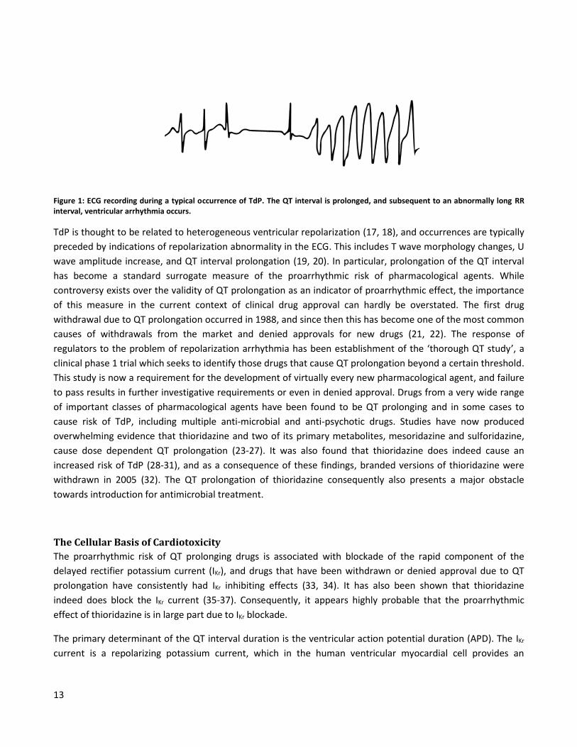
‘twisting of the spikes’ ). An example ECG is shown in Figure 1. While most episodes of TdP spontaneously
revert to sinus rhythm, sudden cardiac death may also occur.
13
Figure 1: ECG recording during a typical occurrence of TdP. The QT interval is prolonged, and subsequent to an abnormally long RR interval, ventricular arrhythmia occurs.
TdP is thought to be related to heterogeneous ventricular repolarization (17, 18), and occurrences are typically
preceded by indications of repolarization abnormality in the ECG. This includes T wave morphology changes, U
wave amplitude increase, and QT interval prolongation (19, 20). In particular, prolongation of the QT interval
has become a standard surrogate measure of the proarrhythmic risk of pharmacological agents. While
controversy exists over the validity of QT prolongation as an indicator of proarrhythmic effect, the importance
of this measure in the current context of clinical drug approval can hardly be overstated. The first drug
withdrawal due to QT prolongation occurred in 1988, and since then this has become one of the most common
causes of withdrawals from the market and denied approvals for new drugs (21, 22). The response of
regulators to the problem of repolarization arrhythmia has been establishment of the ‘thorough QT study’, a
clinical phase 1 trial which seeks to identify those drugs that cause QT prolongation beyond a certain threshold.
This study is now a requirement for the development of virtually every new pharmacological agent, and failure
to pass results in further investigative requirements or even in denied approval. Drugs from a very wide range
of important classes of pharmacological agents have been found to be QT prolonging and in some cases to
cause risk of TdP, including multiple anti-microbial and anti-psychotic drugs. Studies have now produced
overwhelming evidence that thioridazine and two of its primary metabolites, mesoridazine and sulforidazine,
cause dose dependent QT prolongation (23-27). It was also found that thioridazine does indeed cause an
increased risk of TdP (28-31), and as a consequence of these findings, branded versions of thioridazine were
withdrawn in 2005 (32). The QT prolongation of thioridazine consequently also presents a major obstacle
towards introduction for antimicrobial treatment.
The Cellular Basis of Cardiotoxicity
The proarrhythmic risk of QT prolonging drugs is associated with blockade of the rapid component of the
delayed rectifier potassium current (IKr), and drugs that have been withdrawn or denied approval due to QT
prolongation have consistently had IKr inhibiting effects (33, 34). It has also been shown that thioridazine
indeed does block the IKr current (35-37). Consequently, it appears highly probable that the proarrhythmic
effect of thioridazine is in large part due to IKr blockade.
The primary determinant of the QT interval duration is the ventricular action potential duration (APD). The IKr
current is a repolarizing potassium current, which in the human ventricular myocardial cell provides an
14
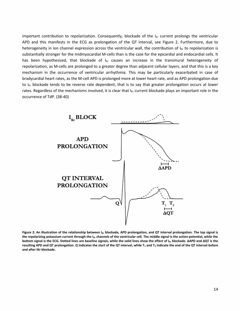
important contribution to repolarization. Consequently, blockade of the IKr current prolongs the ventricular
APD and this manifests in the ECG as prolongation of the QT interval, see Figure 2. Furthermore, due to
heterogeneity in ion channel expression across the ventricular wall, the contribution of IKr to repolarization is
substantially stronger for the midmyocardial M-cells than is the case for the epicardial and endocardial cells. It
has been hypothesized, that blockade of IKr causes an increase in the transmural heterogeneity of
repolarization, as M-cells are prolonged to a greater degree than adjacent cellular layers, and that this is a key
mechanism in the occurrence of ventricular arrhythmia. This may be particularly exacerbated in case of
bradycardial heart rates, as the M-cell APD is prolonged more at lower heart rate, and as APD prolongation due
to IKr blockade tends to be reverse rate dependent, that is to say that greater prolongation occurs at lower
rates. Regardless of the mechanisms involved, it is clear that IKr current blockade plays an important role in the
occurrence of TdP. (38-40)
Figure 2: An illustration of the relationship between IKr blockade, APD prolongation, and QT interval prolongation. The top signal is the repolarizing potassium current through the IKr channels of the ventricular cell. The middle signal is the action potential, while the bottom signal is the ECG. Dotted lines are baseline signals, while the solid lines show the effect of IKr blockade. ΔAPD and ΔQT is the resulting APD and QT prolongation. Q indicates the start of the QT interval, while T1 and T2 indicate the end of the QT interval before and after IKr blockade.
15
Stereochemistry – is there a Way Forward?
In addition to the neurological side effects of thioridazine, proarrhythmic IKr blockade presents a severe
problem for the safety profile of thioridazine in antimicrobial applications. However, newer findings regarding
the stereochemical properties of the compound suggest that unexplored opportunities may exist.
Stereochemistry has now become an important part of most areas of pharmacotherapy, as many chiral
compounds have enantiomers with distinct pharmacological characteristics (41). Thioridazine is such a chiral
compound, although it is normally administered as a 50-50 racemic mixture of two enantiomers. These are
referred to here as (-)-thioridazine and (+)-thioridazine. The antipsychotic effects of thioridazine are due to
blockade of the dopamine D2 receptor, and it has been shown that (+)-thioridazine has 2.7 times the affinity
for this receptor in isolated rat brain preparations (42). Greater affinity for the D2-receptor strongly indicates
that (+)-thioridazine is the more potent antipsychotic (32, 43). However, the antimicrobial properties of the
enantiomers are similar and in some cases stronger for (-)-thioridazine (44-46). In addition, (-)-thioridazine
accumulates to a greater extent in human tissue (47).
The apparent difference between thioridazine enantiomers suggests an interesting hypothesis – that the
enantiomers may also have different cardiotoxicity. This hypothesis is supported by the fact that many D2
dopamine receptor blocking antipsychotics also prolong the QT interval and in some cases cause a risk of TdP
(48, 49). If the lower affinity of (-)-thioridazine for the D2 receptor is associated with a lower affinity for
blockade of the IKr current, then it is quite possible that (-)-thioridazine has both less severe neuroleptic and
cardiotoxic side effects than (+)-thioridazine while having similar antimicrobial activity. In this case, the isolated
(-)-enantiomer may be a safer treatment than the racemate, and an important obstacle to introducing
thioridazine for antimicrobial treatment will have been overcome.
Investigating Thioridazine Enantiomer Effects
It is clear that there is a need for investigation of the effects of the isolated thioridazine enantiomers on
ventricular repolarization. Ultimately, an absolute requirement will be a determination of the effect of the
isolated enantiomers on the QT interval in humans. With the aim of advancing towards this goal, studies first
need to be carried out in an experimental animal model. Such studies may be carried out on several levels,
including that of the ECG arising from the electric activity of the in situ heart, the level of the action potential of
the single cell in isolated heart preparations, and on the level of the individual ion channel current. While full
determination of differences in effect should ideally involve investigation on all these levels, an initial
investigation should aim to verify whether there indeed is a difference in the effect of the enantiomers on the
duration of repolarization. Investigating this through measurement of the individual action potential is a useful
initial approach, as this provides direct indication of the activity of the drug at a relatively detailed level and
under highly controlled conditions. There are a range of animal experimental isolated heart preparations
available in which the effects of compounds may be investigated. The choice of preparation is important and
must be made with the goals of the study in mind.
16
A central aspect that must be considered is the choice of an animal model. The physiology of ventricular
repolarization in the selected animal must be relevant to the information desired about potential effects on
human repolarization. Due to the central role of IKr current blockade in the occurrence of TdP, a central aspect
must necessarily be to investigate differences in the IKr inhibiting effects of the enantiomers. The IKr current
does not play a substantial role in ventricular repolarization in the adult rat and mouse which makes these
species unsuitable. Larger rodents commonly used in experiments and which may be housed at the available
facilities include the guinea pig and rabbit. The slow component of the delayed rectifier potassium current (IKs)
contributes strongly to repolarization in the guinea pig, while the impact of IKs blockade on the QT segment
duration and APD in multicellular ventricular preparations is negligible in the rabbit (50, 51). However, IKr
current blockade does cause measureable APD prolongation in the rabbit (51). This makes the rabbit well
suited for investigating the effects of IKr inhibition. A standard experimental preparation for measuring the
rabbit ventricular action potential is the isolated rabbit papillary muscle. This is the experimental preparation
which was used for the work presented in this thesis and it is described in detail in the section starting on page
19.
While the difference in APD prolonging effect between the thioridazine enantiomers is of central importance,
this measure alone does not directly demonstrate the nature of the exact mechanisms responsible. Over the
past few decades, computational modeling of the cardiac electrophysiology has made substantial leaps forward,
as increasingly detailed and descriptive models of the ventricular cell have been developed using extensive
data from a number of species, including the rabbit (52). A mathematical model may be used to indicate where
existing knowledge is incomplete and to make predictions on which to base further empirical investigation. A
sufficiently descriptive model of the investigated system may aid in integrating empirical data with existing
knowledge in order to further the analysis and understanding of these results. In order to investigate the
physiological basis of the effects of the thioridazine enantiomers, an investigation of the drug effects was
carried out based on the acquired empirical data and a modified version of an existing computational model of
the rabbit ventricular action potential. This model is described in the section starting on page 23.
17
Objectives of the Thesis
The purpose of the work presented in this thesis was to confirm or reject the hypothesis that the enantiomers
of thioridazine have dissimilar effects on the duration of repolarization in the ventricular cell of a
representative animal model and to characterize these differences, if present. This was done using
transmembrane action potential recordings in a standard preparation - the isolated papillary muscle.
The secondary objective was to explore the physiological basis of the differences using computational modeling
of the ventricular papillary muscle cell. In particular, the goal was to test the hypothesis that a difference in
prolonging effect between the enantiomers can be explained by a difference in affinity for blockade of the IKr
current.

19
The Isolated Rabbit Papillary Muscle Prior to the work presented in this thesis, there was no experimental setup available at our department for
investigation of drug effects on the ventricular action potential. For this reason, such a setup was established
for use with the isolated papillary muscle preparation. This chapter provides an overview of the properties of
this preparation and a description of the experimental setup and primary methods. The focus here is on those
aspects which are not covered in detail in the included publications.
The Experimental Preparation
The isolated papillary muscle is a widely used experimental preparation in cardiovascular research, as it is well
suited for investigating the effects of pharmaceutical compounds, ionic imbalances, and hypoxia on contraction
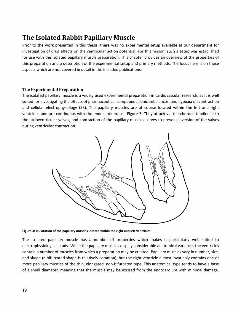
and cellular electrophysiology (53). The papillary muscles are of course located within the left and right
ventricles and are continuous with the endocardium, see Figure 3. They attach via the chordae tendineae to
the atrioventricular valves, and contraction of the papillary muscles serves to prevent inversion of the valves
during ventricular contraction.
Figure 3: Illustration of the papillary muscles located within the right and left ventricles.
The isolated papillary muscle has a number of properties which makes it particularly well suited to
electrophysiological study. While the papillary muscles display considerable anatomical variance, the ventricles
contain a number of muscles from which a preparation may be created. Papillary muscles vary in number, size,
and shape (a bifurcated shape is relatively common), but the right ventricle almost invariably contains one or
more papillary muscles of the thin, elongated, non-bifurcated type. This anatomical type tends to have a base
of a small diameter, meaning that the muscle may be excised from the endocardium with minimal damage.
20
Conversely, the papillary muscles of the left ventricle tend to be relatively short and attach to the endocardium
over a much greater area. An elongated shape allows measurements to be made relatively far from the cutting
surface and the point of electrical stimulation, minimizing undesired interference with the measurement site.
Importantly, the thin diameter of the muscle allows it to be sustained by simple superfusion, as saline solution
is able to diffuse through the muscle (53). In a thicker muscle, the core should be expected to become ischemic
under superfusion due to restriction of diffusion. This also sets the isolated papillary muscle apart from
preparations involving the whole heart or larger sections of the myocardium, which typically require coronary
perfusion. The relatively small size of the papillary muscle also means that muscular contraction results in
relatively limited displacement at any point on the muscle surface. As a consequence, stable impalement of a
single myocardial cell may be maintained in order to directly record the single cell transmembrane potential,
whereas the displacement involved in a whole heart preparation necessitates the recording of monophasic
action potentials instead. For these reasons, the isolated papillary muscle was used in the studies presented in
this thesis.
The Experimental Setup
All procedures involving living animals were carried out by qualified technicians. The animal was sedated and
anesthetized, and once the animal was confirmed to be fully sedated, it was killed by a strong blow to the neck.
After the animal was confirmed to be dead, the heart was rapidly excised and washed in a cold saline solution
in order to remove blood prior to the onset of coagulation. While submerged in cold saline, the right
ventricular wall was opened, and papillary muscles of the elongated type were carefully excised and
transferred to an organ bath for use in the experiment, see Figure 4. For electrophysiological measurements,
the muscle was mounted with fine pins through the base and chordae tendineae in a horizontal position. This
position allowed a microelectrode to be maneuvered to a suitable location on the muscle surface using a
microscope located above the preparation. At all stages, the saline solution was maintained at appropriate
temperature and continuously saturated with a gas appropriate to the buffering agent in order to ensure
oxygenation and proper pH value. It was not possible to gas directly in the bath, as bubbles cause ripples in the
solution. This displaces the microelectrode and may cause instability in the impalement. (53)
The bath was secured on a vibration resistant table, and a peristaltic pump was used to recirculate the solution,
achieving a continuous and stable inflow and outflow of saline solution. A chamber used to gas the solution
and to introduce new solution was located prior to the bath. Between this chamber and the bath was a spiral
contained in a cylinder of heated water. The heating occurring in this spiral maintained the temperature of the
solution in the bath, which was also continuously monitored. A diagram of the experimental setup is shown in
Figure 5.
21
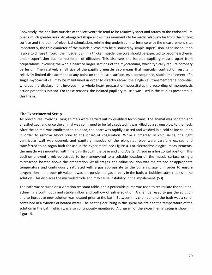
Figure 4: Papillary muscle mounted in organ bath. A: saline inflow. B: temperature probe. C: stimulation electrode. D: action potential recorded near the stimulation site. E: action potential recorded distant from the stimulation site. F: reference electrode. G: saline outflow. H: basal mounting pin. I: chordae tendinae mounting pin. As can be seen, depolarization propagates through the muscle, and greater distance between stimulation site and measurement site results in a longer delay between stimulation artifact and action potential upstroke.
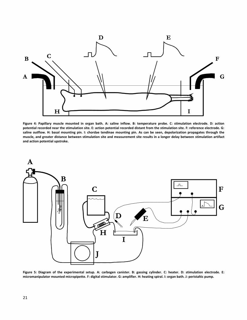
Figure 5: Diagram of the experimental setup. A: carbogen canister. B: gassing cylinder. C: heater. D: stimulation electrode. E: micromanipulator mounted micropipette. F: digital stimulator. G: amplifier. H: heating spiral. I: organ bath. J: peristaltic pump.
22
The papillary muscle was stimulated at the base with a stimulation electrode, causing a wavefront of
depolarization to propagate through the muscle and triggering an action potential at the measurement site.
The stimulation threshold was found manually by varying the pulse amplitude and visually confirming muscle
contraction. The muscle was continuously stimulated with a square pulse with double the amplitude of the
stimulation threshold. Triggered by the stimulator, measurements were carried out using a saline filled, sharp,
high-impedance, glass micropipette created by pulling apart a glass filament with a micropipette puller. The
microelectrode was filled with a 3M KCL solution and mounted on a hydraulic micromanipulator. The electrical
potential was measured with respect to a reference electrode placed in the bath. Using the micromanipulator
and a mounted microscope, the tip of the micropipette was carefully maneuvered to the edge of the papillary
muscle. The muscle was impaled, and by means of very fine and careful movements, a stable impalement of a
single cell was achieved by observation of the resting membrane potential and triggered action potentials. (53)
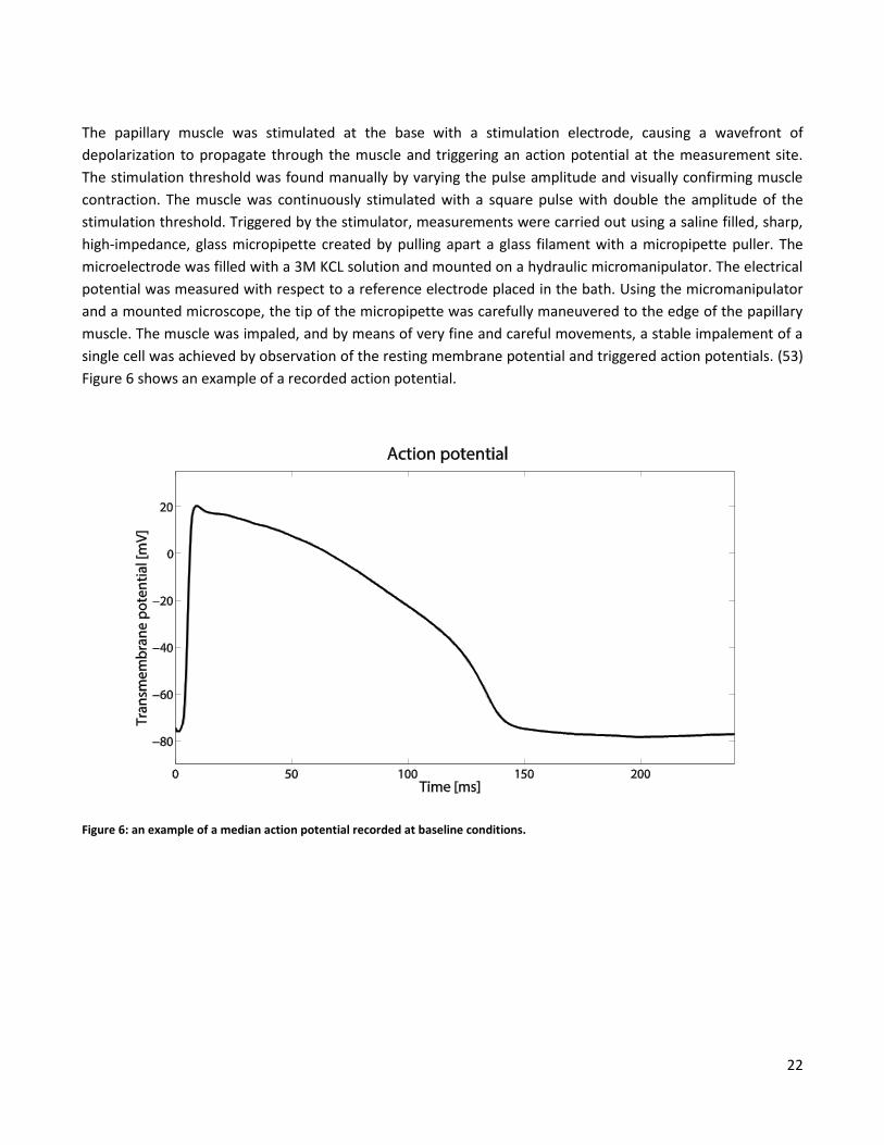
Figure 6 shows an example of a recorded action potential.
Figure 6: an example of a median action potential recorded at baseline conditions.
23
Investigation of Drug Effects by Computational Modeling
Modeling of the Action Potential
The action potential and excitation-contraction coupling of the ventricular cell arises due to a complex system
consisting of many subsystems, each of which has direct or indirect effects on all other subsystems. Each
subsystem may be studied empirically in isolation, but due to interaction between systems, the difficulty of
studying the simultaneous functioning of multiple systems becomes great. At a certain level of complexity, a
potentially more fruitful approach is to use mathematical modeling to characterize each subsystem with
equations that describe laboratory experiments, and then to combine these parts into a more comprehensive
model in which interactions may be studied more readily. While a mathematical model of such complex
interactions can never be perfect, and while a model is not as useful as direct empirical data, nevertheless, an
integrated model may be a powerful tool to improve understanding and guide thought, investigation, and
analysis. (52)
A great number of cardiac action potential models are now available, focusing on different species, cell types,
cell components, behaviors, and levels of complexity. In addition there has been a long history of development
during which models have gradually grown in both complexity and descriptive power as greater understanding
of the modeled mechanics were understood and as more data of higher quality became available. Models now
take into account many aspects of the cardiac cell including membrane voltage, cellular geometry, membrane
channels, pumps, and transporters, ion concentrations in multiple compartments, buffering systems and
binding molecules in both cytoplasm and the sarcoplasmic reticulum (SR), and calcium induced calcium release
(CICR). (54)
The Hodgkin-Huxley Membrane Current Formulation
There are many similarities between the membrane currents of the myocardial cell and the nerve cell. Despite
the ever increasing complexity of new models, modern modeling of the myocardial membrane currents owes
much to Hodgkin and Huxley, whose work resulted in the 1952 publication of the now classic Hodgkin-Huxley
(HH) model, which describes the action potential of the squid giant axon (55). The formulation of the
membrane ion channels in the HH model still represents the basic formulation used in most modern models.
The action potential of course arises due to the net electrical current resulting from ion flux across the cellular
membrane. Individual ions move through specialized pore-forming proteins creating channels through the
membrane, which may be very specific to an ionic species. Such channels may be gated, responding
stochastically to external stimuli by opening for a short time and then rapidly returning to an inactivated state.
Due to the large number of channels across the membrane, this stochastic process gives rise to an observable
and predictable net ion current. (54)
The driving force for movement of charged particles across a passive channel is the result of the
electrochemical gradient across the membrane. Passive net movement of charged particles against the
24
concentration gradient occurs only if the driving voltage difference is sufficiently great. Thus, the most basic
formulation of an ion current across a membrane with conductance selective for that ion is given by Eq. 1. (54)
𝐼 = �̅�(𝑉𝑀 − 𝐸) Eq. 1
Here, �̅� is the maximal current conductance, 𝑉𝑀, is the membrane voltage, and 𝐸 is the (Nernst) reversal
potential for the specific ion.
The maximal conductance of an ion current may be described by Eq. 2.
�̅� = 𝑁�̅�1 Eq. 2
Here, 𝑁 is the number of channels and �̅�1is the unitary conductance - the conductance of a single channel.
As seen in Eq. 1, the driving force for an ionic species is the difference between the membrane voltage and the
reversal potential for that ion. The reversal potential is the potential difference across the membrane at which
no net flux of the ion occurs as the chemical and electrical gradients exactly balance each other out. From the
Nernst equation, the reversal potential is given by Eq. 3. (54)
𝐸 =
𝑅𝑇
𝑧𝐹𝑙𝑛
𝑌𝑜
𝑌𝑖
Eq. 3
Here, 𝑅 is the universal gas constant, 𝑇 is the absolute temperature, 𝐹 is the Faraday constant, 𝑧 is the valence
of the ion in question, and 𝑙𝑛𝑌𝑜
𝑌𝑖 is the natural logarithm of the ratio between extracellular and intracellular ion
concentrations. The greater this ratio, the greater is the membrane voltage required to overcome the
concentration gradient.
Voltage sensitive gating changes the state of the channel to allow or disallow movement of ions. Each gate is
represented by a normalized variable with value between 0 and 1, representing the fraction of channels that
may be found in the open state. For example, in the HH model the Na+ channel current is modeled by three
voltage dependent activation gates and one voltage dependent inactivation gate, see Eq. 4. (54)
𝐼𝑁𝑎 = �̅�𝑁𝑎𝑚3𝑛(𝑉𝑀 − 𝐸𝑁𝑎) Eq. 4
25
Here, 𝑚 is an activation gate and 𝑛 is an inactivation gate. The activation gate opens in response to
depolarization, allowing rapid depolarizing influx of Na+, which triggers the action potential. The inactivation
gate closes in response to depolarization, halting Na+ influx.
Gating variables are described by their own differential equations. For example the change in state of the 𝑚
activation gate is defined by Eq. 5. (54)
𝑑𝑚
𝑑𝑡= 𝛼𝑚(1 − 𝑚) − 𝛽𝑚𝑚 =
𝑚∞(𝑉) − 𝑚
𝜏𝑚(𝑉)
Eq. 5
Here 𝛼𝑚 is the rate of opening, 𝛽𝑚is the rate of closing, 𝑚∞ is the steady state fraction of gates in the open
state given a voltage level, and 𝜏𝑚 is the time constant of state transition. Thus, the change in fraction of gates
in the open state depends on the rate of opening and closing and the fraction of gates already in the open state.
Ultimately, the behavior of the voltage sensing gate is determined by these transition rates, which are voltage
dependent variables. Increasing voltage causes the rate of transition to both the open and inactivated state to
increase, leading to a rapid net opening of channels and subsequent inactivation. The drop in membrane
potential following from repolarization causes a transition away from inactive state to a resting nonconducting
state, from which reopening can occur. The voltage dependence of transition rates typically follows sigmoidal
curves. Due to the voltage regulation of transition rates, the voltage determines both the steady state fraction
of 𝑚 gates in the open state 𝑚∞ and the time constant 𝜏𝑚 with which 𝑚 tracks 𝑚∞.
Alternative Membrane Channel Formulations
The HH channel formulation describes the case of a membrane channel which is selective for a single ionic
species. However, such selectivity may not be ideal, and some currents may carry multiple ionic species with
different conductivities. The Goldman-Hodgkin-Katz equation (or Goldman equation) is a generalization of the
Nernst equation which is used to derive the potential across a membrane with conductivity to multiple ionic
species with different chemical gradients. This equation is often used to model currents that are not assumed
to be ideally selective.
An important alternative to the HH formulation of channel gating is the Markov chain model. In this
formulation, channels may inhabit one of a number of states, with stochastic transitions happening between
states of the channel. For example, in a simple model the channel may inhabit an open, closed, or inactivated
state, and the behavior of the current would be determined by six rates of transition among these three states.
Markov chain models may model certain aspects of channel behavior such as dependence between channel
activation and inactivation more accurately than the HH formulation. However, the complexity and
computational demands are also greater. (54)
26
From Hodgkin and Huxley to the rabbit ventricular cell model
While the HH model provides the basic formulation of ionic currents used in most models, there have been
many advances involved in the development of cardiac cell modeling. These form the basis of the of the rabbit
ventricular cell model used in this study. A brief overview of several important advances leading to the
development of this model will be presented here.
Following the discoveries of Hodgkin and Huxley, extension of this work to describe cardiac cell types
happened very slowly due to experimental challenges and the greater complexity of the cardiac action
potential (56). The first model of a cardiac cell, the 1962 Purkinje cell model published by Noble (57), predates
the discovery of Ca2+ current in cardiac cells. Only by the mid-1970s was data sufficient for the development of
more descriptive cardiac models available, and in 1975 McAllister et al. published a model of the Purkinje fiber
including both inward Ca2+ current and multiple potassium currents (56), which had now been discovered. In
1977, the first model of the ventricular myocardial cell was published by Beeler and Reuter. This was a generic
mammalian model incorporating four ion currents including fast inward sodium (INa), a time-dependent
outward potassium current (which would later become IKs and IKr), a time-independent outward potassium
current (IK1), and a slow inward Ca2+ current (which would later become the L type Ca2+ current, ICaL) (58).
These models were still limited by inadequate experimental techniques and a lack of data on ion
concentrations in the extracellular cleft. With the development of patch clamp techniques in the late 1970s and
early 1980s, single channel recordings became possible, overcoming previous experimental barriers by allowing
direct quantitative description of channel kinetics. In addition, it became possible to control intra- and
extracellular ion concentrations. (59) These important advances led to the development of the 1985 model by
DiFrancesco and Noble of the cardiac Purkinje cell. This model was the first to track K+ and Na+ concentrations
and to include advanced Ca2+ dynamics including Ca2+ sequestration in the SR and also CICR. In addition, the
model was the first to include the Na+-K+ pump, an ATP driven mechanism maintaining ionic gradients by
extruding Na+ in return for K+, and also the first to include the Na+-Ca2+ exchanger, a concentration gradient
driven mechanism which extrudes Ca2+ in return for Na+. (60)
In 1991, Luo and Rudy published the first version of what would in practice become the standard ventricular
cell model (59). This model of the guinea pig ventricular myocyte was the first ventricular model to describe a
specific species rather than a generic mammalian cell. It was based on the model of Beeler and Reuter and
updated the currents of this model with recently acquired data. It also added several currents including plateau
and background K+ currents. The second version was published in 1994 (61). This version included many of the
elements also introduced by DiFranceso and Noble but used new data primarily from the guinea pig ventricular
cell. The ICaL current was reformulated from the slow inward current of the Beeler and Reuter model to include
faster activation and also voltage- and calcium-dependent inactivation. The Na+-K+ pump and Na+-Ca2+
exchanger were included as was a nonspecific Ca2+-activated current and a Ca2+ pump. In addition, the SR was
introduced which was divided into two functional and anatomical compartments, the junctional and the
network SR, comprising 8% and 92% of the total SR volume respectively. An uptake current moved Ca2+ ions
from the cytoplasm to the network SR, from where they would translocate to the junctional SR. CICR occurred
from the junctional SR to the cytoplasm, and a leakage current was present from the network SR to cytoplasm
27
also. The model tracked intracellular K+, Na+, and Ca2+ concentrations and the Ca2+ concentration in the SR as
well. Buffering of Ca2+ was accounted for in both the cytoplasm and SR. Further updates were introduced
through the 1990s and 2000s. In 1995, Zeng et al. divided the time-dependent outward potassium current into
the IKr and IKs currents and updated the Ca2+ buffering (62). In 1999, Viswanathan et al. reformulated IKs and
CICR and introduced heterogeneous IKr and IKs expression to differentiate endocardial-, epicardial-, and
midmyocardial cells (M-cells) (63). In 1999, Clancy and Rudy included a Markov chain model of INa to simulate
the effect of genetic mutations (such as in long QT-3 syndrome and Brugada) (64), and in 2000, Faber and Rudy
introduced Na+ activated K+ current and modified the sarcoplasmic Ca2+ release and Na+-Ca2+ exchange (65).
In the late 1990s, the Winslow group developed a model of the canine ventricular cell based on the Luo-Rudy
model modified using canine data (66, 67). This model represented a major step forward in the modeling of
ventricular Ca2+ homeostasis, as several new discoveries were incorporated (52). This included new
formulations of the Ca2+ inactivation of the ICaL current and Ca2+ regulation of the ryanodine receptor – the
sarcoplasmic channel responsible for CICR. Importantly, this model also included a junctional subspace, an
anatomical volume of the intracellular space between the junctional SR and the T tubule membrane. This
subspace was where the CICR occurred, as ICaL channels and the ryanodine receptors were localized here.
Consequently, the Ca2+ concentration could increase locally to much greater levels than in the bulk cytoplasm.
The model also included troponin binding sites for Ca2+. (66, 67)
The first model of the rabbit ventricular cell was published by Puglisi and Bers in 2001. This model was based
on the Luo-Rudy model but with many currents rescaled to match data from the rabbit ventricle. The model
included the transient inward potassium (Ito) current, which is present in the rabbit but not in the guinea pig. It
also included a Ca2+ activated Cl- current and made modifications to the kinetics of IKr and T type Ca2+ current.
(68)
In 2004, Shannon et al. of the same group published a new Rabbit ventricular cell model. This was based on the
model of Puglisi and Bers and included the advances in Ca2+ modeling introduced by the Winslow group.
Consequently, the Shannon model was of course directly based on the Luo-Rudy model, as this formed the
basis of both previous models. The Shannon model was based on new data acquired from the rabbit ventricle,
and it also included several novel features. A subsarcolemmal compartment was introduced which allowed
membrane channels to sense ion concentrations different from those in the bulk cytoplasm. This model also
introduced a reversible Ca2+ pump in the SR and new formulations of the Na+-Ca2+ exchanger and the ryanodine
receptor. (52)
The Shannon Model of the Rabbit Ventricular Cell
Paper 2 and 3 presented in this thesis were based on the 2004 Shannon model of the ventricular rabbit cell.
This section will present an overview of the structure and components of this model.
28
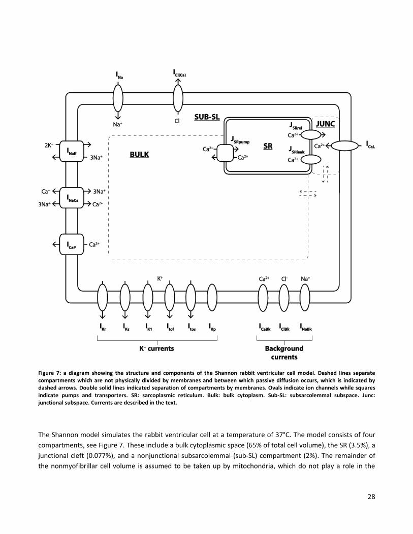
Figure 7: a diagram showing the structure and components of the Shannon rabbit ventricular cell model. Dashed lines separate compartments which are not physically divided by membranes and between which passive diffusion occurs, which is indicated by dashed arrows. Double solid lines indicated separation of compartments by membranes. Ovals indicate ion channels while squares indicate pumps and transporters. SR: sarcoplasmic reticulum. Bulk: bulk cytoplasm. Sub-SL: subsarcolemmal subspace. Junc: junctional subspace. Currents are described in the text.
The Shannon model simulates the rabbit ventricular cell at a temperature of 37°C. The model consists of four
compartments, see Figure 7. These include a bulk cytoplasmic space (65% of total cell volume), the SR (3.5%), a
junctional cleft (0.077%), and a nonjunctional subsarcolemmal (sub-SL) compartment (2%). The remainder of
the nonmyofibrillar cell volume is assumed to be taken up by mitochondria, which do not play a role in the
29
model. The sub-SL compartment is a narrow space just under the remainder of the sarcolemma where
diffusion is restricted due to the presence of mitochondria and myofilaments. The sub-SL is not an anatomical
compartment, but a functional one, where Ca2+ concentrations can exceed those of bulk cytoplasm due to
restricted diffusion. The presence of such a gradient in Ca2+ concentration is indicated by experiments, and this
explains why certain membrane channels are regulated by Ca2+ concentrations higher than those found in the
bulk. Unlike previous models, the SR consists of only a single compartment. The junctional cleft is similar to the
junctional space introduced by the Wilson group. Diffusion between the junction and sub-SL is restricted due to
the narrow boundaries of the junction and the presence of large proteins. (52)
Ions diffuse between the junctional cleft and the sub-SL space and between the sub-SL space and bulk
cytoplasm. The model tracks intracellular Na+ and Ca2+ concentrations in all cytoplasmic compartments and
Ca2+ in the SR. Extracellular ion concentrations and the intracellular K+ concentration is assumed to be constant.
Buffering of Na+ is assumed to be significant only near the sarcolemma and Na+ buffers are thus only present in
the junctional cleft and sub-SL compartments. The model contains a number of Ca2+ buffers and binding
molecules distributed over all compartments. (52)
The SR contains ryanodine receptors giving rise to Ca2+ release flux (JSRrel), a passive Ca2+ leak flux (JSRleak), and
the ATP driven SR Ca2+ pump (JSRpump). Ryanodine receptors are entirely located in the junctional cleft, and all
Ca2+ release occurs into this compartment. The receptor is modeled with a Markov chain of four possible
states: closed, open, inactivated, and resting inactivated. Ca2+influx in the junctional cleft causes Ca2+ to bind to
both fast activation sites and slow inactivation sites, causing channels to transition to the open and then
inactivated states. Ca2+ concentration decline causes a transition to the resting inactivated and finally closed
states. The ryanodine receptor is affected by Ca2+ binding sites inside the SR. Higher Ca2+ load augments
transition to the open state, resulting in greater Ca2+ release. Like the ryanodine receptor, passive leak flux also
occurs between the SR and junctional cleft. ATP driven Ca2+ uptake on the other hand, occurs entirely from the
bulk cytoplasm. The pump uses a novel formulation that includes reversibility as seen in experiments, although
the net flux is always in the same direction. (52)
Membrane channels are evenly distributed between the sub-SL and junctional areas of the sarcolemma. Due to
the difference in membrane surface, these channels are distributed with 89% in sub-SL compartment and 11%
in the junctional cleft. For ICaL however, 90% of channels are concentrated in the junctional cleft, where CICR
occurs. The major membrane currents at 2 Hz steady state pacing are shown in Figure 8 and Figure 9. (52)
Na+ currents include the fast Na+ current (INa) and a background Na+ leak (INaBk). Both used the same equations
as in the Luo Rudy model. The model also includes two Cl- currents, a Ca2+ dependent Cl- current (ICl(Ca)), and a
passive background Cl- current (IClBk). (52)
K+ currents include the IKr, IKs, Ito, and IK1 currents and also a passive plateau K+ current (IKp). All K+ currents
except IKs were modified versions of the formulations in the Luo Rudy model. IKr uses a Hodgkin-Huxley
formulation with a single activation and inactivation gate, and a maximal conductance dependent on
extracellular K+. IKs has two voltage dependent activation gates, the maximal conductance is regulated by
intracellular Ca2+, and the equilibrium potential for IKs also depends on the Na+ gradient across the membrane.
30
The Ito current is divided into two separate components: fast and slow Ito (Itof and Itos), both including voltage
regulated activation and inactivation gates. IK1 is a voltage gated inwardly rectifying current, and similarly to IKr,
the maximal conductance is dependent on extracellular K+ concentration. (52)
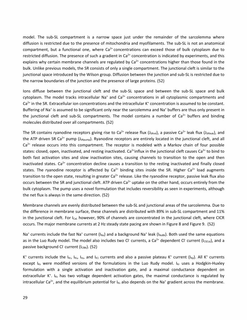
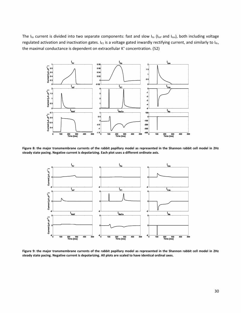
Figure 8: the major transmembrane currents of the rabbit papillary model as represented in the Shannon rabbit cell model in 2Hz steady state pacing. Negative current is depolarizing. Each plot uses a different ordinate axis.
Figure 9: the major transmembrane currents of the rabbit papillary model as represented in the Shannon rabbit cell model in 2Hz steady state pacing. Negative current is depolarizing. All plots are scaled to have identical ordinal axes.
31
Ca2+ currents include the ICaL current, an ATP driven sarcolemmal Ca2+ pump regulated by the Ca2+
concentration near the membrane (ICaP), and a passive background Ca2+ leak current (ICaBk). The model does not
include a T type Ca2+ current, as this is not detectable in normal rabbit myocytes. The ICaL current is a
modification of the formulation in the Luo-Rudy model based on the Goldman-Hodgkin-Katz equation.
However, the permeabilities to Na+ and K+ were decreased to match experimental data when all three ions are
present. The channel is voltage activated and includes both fast and slow voltage regulated inactivation and
Ca2+ inactivation. The Ca2+ inactivation was modified to depend on the Ca2+ binding messenger protein
Calmodulin. (52)
The model also includes the Na+- K+ pump (INaK) and the Na+-Ca2+ exchanger (INaCa). INaK uses the formulation of
the Luo Rudy model and extrudes Na+ in return for K+ in a 3:2 ratio. INaCa is a reversible transporter which uses
the concentration gradient to exchange Na+ for Ca2+ in a 3:1 ratio. In the forward mode it removes Ca2+ from
the cell, which is the primary mode of action, but following Na+ influx it may temporarily operate in the reverse
mode. Despite the forward mode functioning to extrude Ca2+, the current is primarily depolarizing due to the
reverse Na+ influx. The INaCa current in the Shannon model uses a novel formulation. It is activated by binding of
Ca2+ to a site on the inside of the membrane and is deactivated as the Ca2+ transient falls after release and Ca2+
disassociates from the binding site. This causes the exchanger to turn off near the resting level of the
intracellular Ca2+ concentration. Unlike the formulation in the Luo Rudy model, it also accounts for regulation
due to intracellular Na+. (52)
Modifications
The Shannon model was developed using data from a wide variety of sources under different conditions. In
order to more realistically characterize the effects of drugs for the specific preparation used under the specific
circumstances of this experiment, there was a need to adapt the model to better describe action potentials
under baseline conditions. Thus, in the work presented in this thesis, the ion channel currents in the Shannon
model were adapted to model both baseline action potentials and drug effects. Adaptations were carried out
by change in maximal current conductances, which is the product of the number of channels and the unitary
conductance, see Eq. 2. Thus, these adaptations had no direct impact on channel kinetics. Instead, in the case
of adaptation to baseline recordings, this modeled the density of channels present in ventricular cell. In the
case of drug effects, these were consequently modeled as effects on the density of channels able to pass
current and/or effects on the unitary conductance of those channels.
The model action potential depends strongly on multiple factors including pacing rate and inhibition of
membrane currents. A change in these factors induces a change in the steady state, which the modeled action
potential model may require several minutes of simulated time to reach. This greatly increases the
computational cost of investigating the effect of varying parameter values. In order to overcome this barrier,
an additional adaptation was carried out. This did not alter the functioning of the model but only caused the
initial state to be dependent on parameter values, and it enabled an identical steady state to be reached much
faster. It was identified that changes in APD due to changes in pacing rate were almost entirely subsequent to
32
relatively fast changes in sarcoplasmic Ca2+ load (changes over seconds) and very slow changes in bulk cytosolic
Na+ concentration (changes over minutes). In paper III, the ICaL, IKr, and IKs currents were inhibited over multiple
pacing rates. It was identified that intracellular Na+ and Ca2+ concentrations did not depend strongly on IKr or IKs
inhibition, but that they did depend strongly on both pacing rate and ICaL with and interaction between these
dependencies. Consequently, the steady state values of sarcoplasmic Ca2+ load and bulk cytosolic Na+ were
calculated over a grid of combinations of pacing rates and ICaL current inhibition levels, and a polynomial
surface depending on these variables were fitted to the concentrations. This was used to estimate the steady
state level of ion concentrations before starting new simulations, which reduced the time to reach steady state
from minutes to only one or two seconds of simulated time. This greatly increased the efficiency of many
practical tasks but did not alter the internal functioning of the model.
33
References
1. Gandhi NR, Nunn P, Dheda K, Schaaf HS, Zignol M, Van Soolingen D, et al. Tuberculosis 2 multidrug-resistant and extensively drug-resistant tuberculosis: A threat to global control of tuberculosis. Lancet. 2010;375:1830-43.
2. World Health Organization. Global tuberculosis report 2012. 2012.
3. H Gillespie S, Singh K. XDR-TB, what is it; how is it treated; and why is therapeutic failure so high? Recent Patents on Anti-Infective Drug Discovery. 2011;6(2):77-83.
4. Silver LL. Challenges of antibacterial discovery. Clin Microbiol Rev. 2011;24(1):71-109.
5. Kristiansen J, Thomsen V, MARTINS A, Viveiros M, Amaral L. Non-antibiotics reverse resistance of bacteria to antibiotics. In Vivo. 2010;24(5):751-4.
6. Amaral L, Viveiros M. Why thioridazine in combination with antibiotics cures extensively drug-resistant< i> mycobacterium tuberculosis infections. Int J Antimicrob Agents. 2012.
7. Crowle A, Douvas G, May M. Chlorpromazine: A drug potentially useful for treating mycobacterial infections. Chemotherapy. 1992;38(6):410-9.
8. Amaral L, Kristiansen J, Abebe L, Millett W. Inhibition of the respiration of multi-drug resistant clinical isolates of mycobacterium tuberculosis by thioridazine: Potential use for initial therapy of freshly diagnosed tuberculosis. J Antimicrob Chemother. 1996;38(6):1049-53.
9. Bettencourt MV, Bosne-David S, Amaral L. Comparative in vitro activity of phenothiazines against multidrug-resistant mycobacterium tuberculosis. Int J Antimicrob Agents. 2000 Sep;16(1):69-71.
10. Ordway D, Viveiros M, Leandro C, Bettencourt R, Almeida J, Martins M, et al. Clinical concentrations of thioridazine kill intracellular multidrug-resistant mycobacterium tuberculosis. Antimicrob Agents Chemother. 2003;47(3):917-22.
11. Martins M, Viveiros M, Kristiansen JE, Molnar J, Amaral L. The curative activity of thioridazine on mice infected with mycobacterium tuberculosis. In Vivo. 2007;21(5):771-5.
12. van Soolingen D, Hernandez-Pando R, Orozco H, Aguilar D, Magis-Escurra C, Amaral L, et al. The antipsychotic thioridazine shows promising therapeutic activity in a mouse model of multidrug-resistant tuberculosis. PLoS One. 2010;5(9):e12640.
13. Abbate E, Vescovo M, Natiello M, Cufré M, García A, Ambroggi M, et al. Tuberculosis extensamente resistente (XDR-TB) en argentina: Aspectos destacables, epidemiologicos, bacteriologicos, terapeuticos y evolutivos. Revista Argentina de Medicina Respiratoria. 2007;1:19-25.
34
14. Abbate E, Vescovo M, Natiello M, Cufré M, García A, Montaner PG, et al. Successful alternative treatment of extensively drug-resistant tuberculosis in argentina with a combination of linezolid, moxifloxacin and thioridazine. J Antimicrob Chemother. 2012;67(2):473-7.
15. Kelly HG, Fay J, Laverty S. Thioridazine hydrochloride (mellaril): Its effect on the electrocardiogram and a report of two fatalities with electrocardiographic abnormalities. Can Med Assoc J. 1963;89(11):546.
16. Dessertenne F. La tachycardie ventriculaire a deux foyers opposes variables. Arch Mal Coeur. 1966;59(2):263-72.
17. Fenichel RR, Malik M, Antzelevitch C, Sanguinetti M, Roden DM, Priori SG, et al. Drug‐Induced torsades de pointes and implications for drug development. J Cardiovasc Electrophysiol. 2004;15(4):475-95.
18. Belardinelli L, Antzelevitch C, Vos MA. Assessing predictors of drug-induced torsade de pointes. Trends Pharmacol Sci. 2003;24(12):619-25.
19. Yan G, Lankipalli RS, Burke JF, Musco S, Kowey PR. Ventricular repolarization components on the electrocardiogramcellular basis and clinical significance. J Am Coll Cardiol. 2003;42(3):401-9.
20. Kirchhof P, Franz MR, Bardai A, Wilde AM. Giant T–U waves precede torsades de pointes in long QT SyndromeA systematic electrocardiographic analysis in patients with acquired and congenital QT prolongation. J Am Coll Cardiol. 2009;54(2):143-9.
21. Lasser KE, Allen PD, Woolhandler SJ, Himmelstein DU, Wolfe SM, Bor DH. Timing of new black box warnings and withdrawals for prescription medications. JAMA. 2002;287(17):2215-20.
22. Wood AJ, Roden DM. Drug-induced prolongation of the QT interval. N Engl J Med. 2004;350(10):1013-22.
23. Axelsson R, Aspenstrom G. Electrocardiographic changes and serum concentrations in thioridazine-treated patients. J Clin Psychiatry. 1982 Aug;43(8):332-5.
24. Hartigan-Go K, Bateman N, Nyberg G, Mårtensson E, Thomas SHL. Concentration-related pharmacodynamic effects of thioridazine and its metabolites in humans. Clinical Pharmacology & Therapeutics. 1996;60(5):543-53.
25. Reilly J, Ayis S, Ferrier I, Jones S, Thomas S. QTc-interval abnormalities and psychotropic drug therapy in psychiatric patients. The Lancet. 2000;355(9209):1048-52.
26. Harrigan EP, Miceli JJ, Anziano R, Watsky E, Reeves KR, Cutler NR, et al. A randomized evaluation of the effects of six antipsychotic agents on QTc, in the absence and presence of metabolic inhibition. J Clin Psychopharmacol. 2004;24(1):62-9.
27. Salih I, Thanacoody R, McKay G, Thomas S. Comparison of the effects of thioridazine and mesoridazine on the QT interval in healthy adults after single oral doses. Clinical Pharmacology & Therapeutics. 2007;82(5):548-54.
35
28. Mehtonen OP, Aranko K, Mälkonen L, Vapaatalo H. A survey of sudden death associated with the use of antipsychotic or antidepressant drugs: 49 cases in finland. Acta Psychiatr Scand. 1991;84(1):58-64.
29. Ray WA, Meredith S, Thapa PB, Meador KG, Hall K, Murray KT. Antipsychotics and the risk of sudden cardiac death. Arch Gen Psychiatry. 2001;58(12):1161.
30. Reilly J, Ayis S, Ferrier I, Jones S, Thomas S. Thioridazine and sudden unexplained death in psychiatric in-patients. The British Journal of Psychiatry. 2002;180(6):515-22.
31. Hennessy S, Bilker WB, Knauss JS, Margolis DJ, Kimmel SE, Reynolds RF, et al. Cardiac arrest and ventricular arrhythmia in patients taking antipsychotic drugs: Cohort study using administrative data. BMJ: British Medical Journal. 2002;325(7372):1070.
32. HK Thanacoody R. Thioridazine: The good and the bad. Recent Patents on Anti-Infective Drug Discovery. 2011;6(2):92-8.
33. Redfern W, Carlsson L, Davis A, Lynch W, MacKenzie I, Palethorpe S, et al. Relationships between preclinical cardiac electrophysiology, clinical QT interval prolongation and torsade de pointes for a broad range of drugs: Evidence for a provisional safety margin in drug development. Cardiovasc Res. 2003;58(1):32-45.
34. Haverkamp W, Breithardt G, Camm AJ, Janse MJ, Rosen MR, Antzelevitch C, et al. The potential for QT prolongation and pro-arrhythmia by non-anti-arrhythmic drugs: Clinical and regulatory implications. report on a policy conference of the european society of cardiology. Cardiovasc Res. 2000 Aug;47(2):219-33.
35. Drolet B, Vincent F, Rail J, Chahine M, Deschênes D, Nadeau S, et al. Thioridazine lengthens repolarization of cardiac ventricular myocytes by blocking the delayed rectifier potassium current. J Pharmacol Exp Ther. 1999;288(3):1261-8.
36. Tie H, Walker B, Valenzuela S, Breit S, Campbell T. The heart of psychotropic drug therapy. The Lancet. 2000;355(9217):1825.
37. Milnes JT, Witchel HJ, Leaney JL, Leishman DJ, Hancox JC. hERG K< sup> </sup> channel blockade by the antipsychotic drug thioridazine: An obligatory role for the S6 helix residue F656. Biochem Biophys Res Commun. 2006;351(1):273-80.
38. Antzelevitch C. Heterogeneity of cellular repolarization in LQTS: The role of M cells. European Heart Journal Supplements. 2001;3(suppl K):K2-K16.
39. Yan GX, Antzelevitch C. Cellular basis for the normal T wave and the electrocardiographic manifestations of the long-QT syndrome. Circulation. 1998 Nov 3;98(18):1928-36.
40. Gima K, Rudy Y. Ionic current basis of electrocardiographic waveforms: A model study. Circ Res. 2002 May 3;90(8):889-96.
36
41. Baumann P, Zullino DF, Eap CB. Enantiomers’ potential in psychopharmacology—a critical analysis with special emphasis on the antidepressant escitalopram. European neuropsychopharmacology. 2002;12(5):433-44.
42. Svendsen C, Froimowitz M, Hrbek C, Campbell A, Kula N, Baldessarini R, et al. Receptor affinity, neurochemistry and behavioral characteristics of the enantiomers of thioridazine: Evidence for different stereoselectivities at D1 and D2 receptors in rat brain. Neuropharmacology. 1988;27(11):1117-24.
43. Jortani SA, Poklis A. Determination of thioridazine enantiomers in human serum by sequential achiral and chiral high-performance liquid chromatography. J Anal Toxicol. 1993;17(6):374-7.
44. Kristiansen JE, Hendricks O, Delvin T, Butterworth TS, Aagaard L, Christensen JB, et al. Reversal of resistance in microorganisms by help of non-antibiotics. J Antimicrob Chemother. 2007;59(6):1271-9.
45. Hendricks O, Christensen JB, Kristiansen JE. Antibakterielle eigenschaften der phenothiazine. eine behandlungsoption für die zukunft? Chemotherapie Journal. 2004;13(5):203-5.
46. Hendricks O. Antimicrobial effects of selected non-antibiotics on sensitivity and invasion of gram-positive bacteria. Ugeskr Laeger. 2007;169(6):524.
47. Jortani SA, Valentour JC, Poklis A. Thioridazine enantiomers in human tissues. Forensic Sci Int. 1994;64(2):165-70.
48. Glassman AH, Bigger JT. Antipsychotic drugs: Prolonged QTc interval, torsade de pointes, and sudden death. Am J Psychiatry. 2001;158(11):1774-82.
49. Ray WA, Chung CP, Murray KT, Hall K, Stein CM. Atypical antipsychotic drugs and the risk of sudden cardiac death. N Engl J Med. 2009;360(3):225-35.
50. Lu Z, Kamiya K, Opthof T, Yasui K, Kodama I. Density and kinetics of IKr and IKs in guinea pig and rabbit ventricular myocytes explain different efficacy of IKs blockade at high heart rate in guinea pig and rabbit. Circulation. 2001;104(8):951-6.
51. Lengyel C, Iost N, Virág L, Varró A, Lathrop DA, Papp JG. Pharmacological block of the slow component of the outward delayed rectifier current (IKs) fails to lengthen rabbit ventricular muscle QTc and action potential duration. Br J Pharmacol. 2001;132(1):101-10.
52. Shannon TR, Wang F, Puglisi J, Weber C, Bers DM. A mathematical treatment of integrated ca dynamics within the ventricular myocyte. Biophys J. 2004;87(5):3351-71.
53. Dhein S. Isolated papillary muscles. In: Dhein S, Mohr FW, Delmar M, editors. Practical Methods in Cardiovascular Research. 1st ed. Springer Berlin Heidelberg; 2005. p. 190-7.
54. Fenton FH, Cherry EM. Models of cardiac cell. Scholarpedia. 2008;3(8):1868.
37
55. Hodgkin AL, Huxley AF. A quantitative description of membrane current and its application to conduction and excitation in nerve. J Physiol. 1952 Aug;117(4):500-44.
56. McAllister RE, Noble D, Tsien RW. Reconstruction of the electrical activity of cardiac purkinje fibres. J Physiol. 1975 Sep;251(1):1-59.
57. Noble D. A modification of the hodgkin--huxley equations applicable to purkinje fibre action and pace-maker potentials. J Physiol. 1962 Feb;160:317-52.
58. Beeler GW, Reuter H. Reconstruction of the action potential of ventricular myocardial fibres. J Physiol. 1977 Jun;268(1):177-210.
59. Luo CH, Rudy Y. A model of the ventricular cardiac action potential. depolarization, repolarization, and their interaction. Circ Res. 1991 Jun;68(6):1501-26.
60. DiFrancesco D, Noble D. A model of cardiac electrical activity incorporating ionic pumps and concentration changes. Philos Trans R Soc Lond B Biol Sci. 1985 Jan 10;307(1133):353-98.
61. Luo CH, Rudy Y. A dynamic model of the cardiac ventricular action potential. I. simulations of ionic currents and concentration changes. Circ Res. 1994 Jun;74(6):1071-96.
62. Zeng J, Laurita KR, Rosenbaum DS, Rudy Y. Two components of the delayed rectifier K+ current in ventricular myocytes of the guinea pig type. theoretical formulation and their role in repolarization. Circ Res. 1995 Jul;77(1):140-52.
63. Viswanathan PC, Shaw RM, Rudy Y. Effects of IKr and IKs heterogeneity on action potential duration and its rate dependence: A simulation study. Circulation. 1999 May 11;99(18):2466-74.
64. Clancy CE, Rudy Y. Linking a genetic defect to its cellular phenotype in a cardiac arrhythmia. Nature. 1999;400(6744):566-9.
65. Faber GM, Rudy Y. Action potential and contractility changes in [na+]I overloaded cardiac myocytes: A simulation study. Biophys J. 2000;78(5):2392-404.
66. Jafri MS, Rice JJ, Winslow RL. Cardiac ca2 dynamics: The roles of ryanodine receptor adaptation and sarcoplasmic reticulum load. Biophys J. 1998;74(3):1149-68.
67. Winslow RL, Rice J, Jafri S, Marban E, O'Rourke B. Mechanisms of altered excitation-contraction coupling in canine tachycardia-induced heart failure, II: Model studies. Circ Res. 1999 Mar 19;84(5):571-86.
68. Puglisi JL, Bers DM. LabHEART: An interactive computer model of rabbit ventricular myocyte ion channels and ca transport. Am J Physiol Cell Physiol. 2001 Dec;281(6):C2049-60.

39
Contributions
Paper I: Differential Effects of Thioridazine Enantiomers on Action Potential duration in Rabbit Papillary
Muscle Page 41
Paper II: Model Based Analysis of the Effects of Thioridazine Enantiomers on the Rabbit Papillary Action
Potential: Part 1 Page 43
Paper III: Model Based Analysis of the Effects of Thioridazine Enantiomers on the Rabbit Papillary Action
Potential: Part 1 Page 45

41
I: Differential Effects of Thioridazine Enantiomers on Action
Potential duration in Rabbit Papillary Muscle
Ask S. Jensen1, Cristian P. Pennisi1, Cristian Sevcencu1, Jørn B. Christensen2, Jette E. Kristiansen3, Johannes J. Struijk1
1Aalborg University, Denmark. 2University of Copenhagen, Denmark. 3University of Southern Denmark, Denmark
Published by the European Journal of Pharmacology

43
II: Model Based Analysis of the Effects of Thioridazine
Enantiomers on the Rabbit Papillary Action Potential: Part 1
Ask S. Jensen1, Cristian P. Pennisi1, Cristian Sevcencu1, Jørn B. Christensen2, Jette E. Kristiansen3, Johannes J. Struijk1
1Aalborg University, Denmark. 2University of Copenhagen, Denmark. 3University of Southern Denmark, Denmark
Submitted for publication


45
III: Model Based Analysis of the Effects of Thioridazine
Enantiomers on the Rabbit Papillary Action Potential: Part 2
Ask S. Jensen1, Cristian P. Pennisi1, Cristian Sevcencu1, Jørn B. Christensen2, Jette E. Kristiansen3, Johannes J. Struijk1
1Aalborg University, Denmark. 2University of Copenhagen, Denmark. 3University of Southern Denmark, Denmark
Submitted for publication

47
Conclusion The studies presented in this thesis focus on potential differential effects of the thioridazine enantiomers on
cardiac repolarization in the isolated rabbit papillary muscle using transmembrane action potential
measurements. It was found that (-)-thioridazine causes significantly less APD prolongation than either
(+)-thioridazine or the racemate. This in itself is an indication that (-)-thioridazine may also cause less QT
interval prolongation and may have reduced cardiotoxic side effects. However, thioridazine has been reported
to have a multitude of effects on ion channels, and indeed the effects observed in the papillary muscles were
more complex than a dose-dependent APD prolongation alone. Depending on drug and stimulus frequency, a
shortening of the APD also occurred. The specific mechanisms responsible for the observed differences are of
great importance to the potential cardiotoxicity of the compounds, and the occurrence of APD shortening also
requires further investigation. The mechanisms underlying the observed effects were investigated using
computational modeling of the rabbit ventricular papillary electrophysiology.
The action potential is the product of a balance between a number of depolarizing and repolarizing ionic
transmembrane currents, which may include voltage gated channels, energy-dependent pumps, and ionic
exchangers operating in complex, mutually interacting systems. Consequently, a direct investigation of effects
on these mechanisms requires channel-level observations. However, the membrane currents have
characteristic effects on the action potential, and therefore, drug exposure may also yield such characteristic
effects. By incorporating action potential level observations with a sufficiently descriptive mathematical model
of the cellular physiology, it is possible to demonstrate whether a hypothetical mechanism is in agreement with
the data, and to make predictions about which mechanisms best match the observations. Due to the multitude
of potential effects due to thioridazine, a model-based study based exclusively on action potential recordings
cannot provide a comprehensive and accurate description of the effects occurring at the membrane channel
level. Instead, the model-based studies presented in this thesis used a simplified model of drug effects to
demonstrate that the difference in APD prolongation can plausibly be described by a differential blockade of
the IKr current. This indicates that there is good reason to believe that the differential effect on APD is also
associated with a difference in cardiotoxicity.
The results do not provide conclusive evidence that (-)-thioridazine is less likely to cause ventricular arrhythmia
than (+)-thioridazine or the racemate. They do, however, provide the first indication that this may indeed be
the case and that further investigation is an urgent and worthwhile endeavor. A number of questions are left
unanswered which require further study. Firstly, there is a need to directly investigate the effects of the
isolated enantiomers on the level of the individual membrane current through patch clamp studies. This may
provide an accurate assessment of differences in affinity for inhibition of both IKr and other currents. Secondly,
it is necessary to investigate the effects of the isolated enantiomers on the corrected QT interval in humans, as
this is by far the best established and most widely used surrogate measure of proarrhythmic risk, and because
it is a requirement in the regulatory process. However, the corrected QT interval is indeed a surrogate measure
which has been the subject of great deal of controversy, as some drugs cause substantial QT prolongation but
are not proarrhythmic, or are less proarrhythmic than drugs which cause less QT prolongation. IKr inhibition and
risk of Torsades de Pointes is associated with several types of morphological T wave changes, and recent
48
studies have demonstrated that scoring of T wave morphology changes provides a more reliable indicator of
arrhythmic risk. Consequently, a better indication of potential differences in cardiotoxicity may be obtained by
investigating not only the effects of the isolated thioridazine enantiomers on corrected QT interval
prolongation, but also on changes of T wave morphology scores.
There is an urgent need for progress in the fight against the global problem of increasing antimicrobial drug-
resistance, as previously curable diseases are becoming increasingly difficult to treat in many parts of the world.
The primary reason for the unnecessarily rapid rate development of antibiotic resistance is incorrect use of
antibiotics. Exposure to an antibiotic agent produces a selection pressure which preferentially allows certain
microbial strains to further proliferate. Excessive, preemptive, and unnecessary usage of antibiotics serves to
drastically increase this selection pressure, and incomplete treatment and addition of new drugs to failing
regimens allow resistant strains to survive and develop resistance to an increasing number of compounds. It is
clear that the current rate of development of new antibiotics is too slow to effectively reverse this tide of
increasing drug-resistance. The first line of defense must be guidelines which promote prudent, careful, and
thorough application of antimicrobial treatment. However, counteracting the proliferation of drug-resistance is
a slow and difficult process which requires control of antimicrobial treatment on a global scale. For these
reasons there is a need for focus on new approaches to combat currently existing and future multi-drug
resistant strains. The development of helper compounds that reverse antimicrobial resistance shows great
promise and may become an important new tool in such efforts. Thioridazine is particularly interesting in this
regard. However, it is also a well-known compound which has been extensively used in the past, and which has
cardiotoxic side effects that have already caused the drug to be withdrawn. Consequently, there is much
skepticism, both towards the use of an antipsychotic drug in any other role than that of antipsychotic
treatment, but also towards the reintroduction of a drug which has already been found to not live up to the
current standards of cardiac safety.
The outcomes of this thesis pose new questions, but they have substantial potential implications for addressing
safety concerns and for the prospects of introduction of isolated (-)-thioridazine in antimicrobial treatment.
This represents the first step towards full determination of the cardiotoxicity of the isolated thioridazine
enantiomers. If further studies confirm that (-)-thioridazine does indeed have reduced affinity for blockade of
IKr and that it causes reduced QT interval prolongation and T wave morphology alterations, this will
demonstrate that (-)-thioridazine is very likely to be safer treatment than the racemate. In such case, a
substantial obstacle on the path to approval of the isolated (-)-thioridazine enantiomer for antimicrobial
treatments will be removed. The result may be the introduction of a new effective tool in the treatment of
drug-resistant microbial disease.