Embed Size (px)
Citation preview
CARBON PARTITIONING IN NITROGEN-FIXING
ROOT NODULES
Dissertation
zur Erlangung des Doktorgrades
der Mathematisch-Naturwissenschaftlichen Fakultäten
der Georg-August-Universität zu Göttingen
vorgelegt von
Maria Schubert
aus St. Petersburg, Russland
Göttingen 2002
D 7
Referent: Prof. Dr. Hans-Walter Heldt
Korreferentin: Prof. Dr. Christiane Gatz
Tag der mündlichen Prüfung: 30.10.2002
Content
1. Introduction.............................................................................................................1
1.1. Degradation of sucrose in sink organs................................................................ 1 1.1.1. The role of sucrose in the plant cell................................................................. 1
1.1.1.1. Sucrose synthase .................................................................................... 2 1.1.1.2. Invertases ................................................................................................ 2
1.2. Transport of sugars ............................................................................................... 4 1.2.1. Phloem loading................................................................................................ 4 1.2.2. Phloem unloading and post-phloem transport................................................. 5
1.2.2.1. Symplastic sugar transport ...................................................................... 5 1.2.2.2. Apoplastic phloem unloading and post-phloem transport........................ 6 1.2.2.3. Sugar transporters in apoplastic sugar transport..................................... 7
1.3. The regulation of phloem unloading and sink strength in different systems .. 9 1.3.1. Developmental sinks ....................................................................................... 9 1.3.2. Optional sinks................................................................................................ 10
1.4. Nitrogen-fixing root nodule symbioses ............................................................. 10 1.4.1. Nitrogen fixation in nodules ........................................................................... 11 1.4.2. Infection of plants and nodule formation........................................................ 12 1.4.3. Nodule structure ............................................................................................ 13 1.4.4. Phylogenetic relationship of root nodule symbioses...................................... 15 1.4.5. Carbon sources supplied by the host plant to nitrogen-fixing microsymbionts.. ...................................................................................................................... 15 1.4.6. Carbon transport and metabolism in nitrogen-fixing root nodules ................. 17
1.5. Aim of this thesis ................................................................................................ 18
2. Materials and methods………………………………………………………………..20
2.1. Materials................................................................................................................ 20 2.1.1. Plant material ................................................................................................ 20 2.1.2. Bacterial and yeast strains ............................................................................ 20 2.1.3. Oligonucleotides (Primers) ............................................................................ 21 2.1.4. Plasmides...................................................................................................... 21 2.1.5. Enzymes........................................................................................................ 22
2.1.5.1. Restriction enzymes................................................................................ 22 2.1.5.2. Other enzymes and kits .......................................................................... 22
2.1.6. Chemicals...................................................................................................... 23
Content ii
2.1.7. Other materials and devices.......................................................................... 24 2.1.8. Culture media ................................................................................................ 24
2.1.8.1. Plant media ............................................................................................. 24 2.1.8.2. Bacterial media ....................................................................................... 25 2.1.8.3. Media for Saccharomyces cerevisiae ..................................................... 27 2.1.8.4. Medium additives .................................................................................... 28
2.2. Plant culture methods and growth conditions .................................................. 28
2.3. RNA isolation from plant tissue.......................................................................... 29 2.3.1. RNA isolation from Medicago truncatula and Datisca glomerata (modified after Burgos et al., 1995)............................................................................... 29 2.3.2. RNA isolation from Casuarina glauca............................................................ 30
2.4. Isolation of plasmid DNA from bacteria and yeast ........................................... 31 2.4.1. Plasmid mini-preparation protocol „ Triton Boiling“........................................ 31 2.4.2. Mini preparation of bacterial plasmid DNA for the sequence analysis .......... 31 2.4.3. Maxi preparation of bacterial plasmid DNA ................................................... 32 2.4.4. Preparation of plasmid DNA from yeast ........................................................ 32
2.5. Concentration and purification of DNA or RNA solutions ............................... 32 2.5.1. Precipitation of nucleic acids ......................................................................... 32 2.5.2. Phenol-chloroform extraction and precipitation of DNA................................. 33
2.6. Electrophoretic separation of DNA and RNA .................................................... 33 2.6.1. TEA-Agarose gel electrophoresis.................................................................. 33 2.6.2. Separation of RNA on agarose gel for Northern blots ................................... 34
2.7. Northern blot hybridization ................................................................................. 34 2.7.1. RNA transfer to nylon membranes (Northern blotting) .................................. 34 2.7.2. DNA probe labelling with α-[32P]-dATP.......................................................... 35 2.7.3. Hybridization.................................................................................................. 35
2.8. First strand cDNA synthesis (Reverse transcription)....................................... 36
2.9. Amplification of DNA fragments ......................................................................... 36 2.9.1. Polymerase chain reaction (PCR) ................................................................. 37 2.9.2. Design of synthetic oligonucleotide primers .................................................. 37
2.10. Rapid amplification of cDNA ends (RACE): 5´-RACE ....................................... 38
2.11. DNA sequencing................................................................................................... 39
2.12. Cloning methods .................................................................................................. 40 2.12.1. Digestion with restriction enzymes ................................................................ 40 2.12.2. Phosphatase treatment ................................................................................. 40 2.12.3. Filling-in of 5´ overhanging ends with Klenow fragment ................................ 41
Content iii
2.12.4. Isolation of DNA fragments from agarose gels.............................................. 41 2.12.5. Ligation.......................................................................................................... 42
2.13. Transformation of Escherichia coli .................................................................... 42 2.13.1. Preparation of competent E.coli cells ............................................................ 42 2.13.2. Transformation of competent E.coli cells....................................................... 43 2.13.3. Characterisation of transformants ................................................................. 44
2.14. Yeast transformation ........................................................................................... 45
2.15. Bacterial and yeast glycerol cultures................................................................. 46
2.16. Extraction of sugars............................................................................................. 46 2.16.1. Chloroform-methanol extraction .................................................................... 46 2.16.2. Preparative isolation of unknown sugars / sugar derivates from Datisca ...... 47 2.16.3. Ethanol extraction.......................................................................................... 48 2.16.4. Perchlorate extraction.................................................................................... 49 2.16.5. Acetone extraction......................................................................................... 49
2.17. Sugar analysis by high-performance liquid chromatography (HPLC) ............ 49
2.18. Sugar analysis ...................................................................................................... 50 2.18.1. Acid hydrolysis .............................................................................................. 50 2.18.2. Enzymatic hydrolysis ..................................................................................... 51
2.19. Sucrose synthase extraction and assay ............................................................ 52 2.19.1. Extraction of sucrose synthase (SuSy).......................................................... 52 2.19.2. Sucrose synthase activity assay.................................................................... 52
2.20. Invertase extraction and assay ........................................................................... 53 2.20.1. Extraction of invertase................................................................................... 53 2.20.2. Invertase activity assay ................................................................................. 54
2.21. In situ glucose and acid (apoplastic) invertase activity staining .................... 55
2.22. Total protein isolation from plant material ........................................................ 55
2.23. Bradford protein concentration determination ................................................. 56
2.24. Sodiumdodecyl sulfate polyacrylamide gel electrophoresis (SDS-PAGE)..... 57 2.24.1. Buffers and solutions for analytic SDS-PAGE............................................... 58 2.24.2. Pouring the SDS-Polyacrylamide gel ............................................................ 58 2.24.3. Probe preparation and SDS polyacrylamide gel electrophoresis (SDS-PAGE) ...................................................................................................................... 60 2.24.4. Coomassie staining of SDS polyacrylamide gels .......................................... 60
2.25. Western blot.......................................................................................................... 61
Content iv
2.25.1. Protein transfer to a nitrocellulose membrane............................................... 61 2.25.2. Staining of the blot......................................................................................... 61 2.25.3. Immunological detection of transferred proteins............................................ 62
2.26. Measurement of sugar uptake in yeast .............................................................. 63
2.27. Statistic evaluation of experimental data…………………………………………..64
3. Results…………………………………………………………………………………...65
3.1. Sugar contents in roots and nodules................................................................. 65 3.1.1. Sugar contents in roots and nodules of Medicago truncatula........................ 65 3.1.2. Sugar contents in Casuarina glauca.............................................................. 67 3.1.3. Sugar contents in Datisca glomerata............................................................. 67
3.1.3.1. Isolation and characterization of two unknown carbohydrates from Datisca glomerata.................................................................................. 67 3.1.3.2. Sugar contents in roots, nodules and leaves of Datisca after determination of the molecular mass of the novel sugars ..................... 74
3.2. Activities of sucrose cleaving enzymes in roots and root nodules ................ 75 3.2.1. Invertase activities......................................................................................... 75 3.2.2. Sucrose synthase activities ........................................................................... 77 3.2.3. In situ localization of acid invertase activity ................................................... 78
3.2.3.1. In situ localization of glucose in roots and nodules ............................... 79 3.2.3.2. In situ localization of acid invertase in roots and nodules...................... 79
3.3. Expression levels of sucrose synthase and sugar transporters in roots and nodules.................................................................................................................. 81
3.3.1. Expression levels of sucrose synthase (RNA gel blot hybridization analysis)82 3.3.2. Levels of sucrose synthase protein (Protein blot analysis)............................ 83 3.3.3. Expression levels of sugar transporters in roots, nodules and leaves........... 84
3.4. Isolation and characterization of hexose transporter cDNAs.......................... 85 3.4.1. Full size cDNA of the Medicago truncatula hexose transporter (MtHT) ........ 85 3.4.2. Full size cDNA of the Datisca glomerata hexose transporter (DgHT) ........... 88 3.4.3. Protein sequence analysis of DgHT and MtHT ............................................. 91
3.5. Functional characterization of the hexose transporters by expression in yeast 94
3.5.1. Construction of shuttle vectors for the heterologous expression of MtHT and DgHT in yeast................................................................................................ 94
3.5.1.1. Cloning of MtHT in pNEV-X................................................................... 95 3.5.1.2. Cloning of DgHT in pNEV-E .................................................................. 95
3.5.2. Sugar uptake studies with yeast strains expressing MtHT and DgHT........... 95 3.5.2.1. Functional characterization of MtHT...................................................... 97 3.5.2.2. Functional characterization of DgHT ..................................................... 97
Content v
4. Discussion……………………………………………………………………………..101
4.1. Cytological information on putative mechanism of carbon transport in root nodules................................................................................................................ 101
4.1.1. Medicago ..................................................................................................... 102 4.1.2. Cytology of roots and nodules of Datisca glomerata ................................... 102 4.1.3. Cytology of roots and nodules of Casuarina glauca .................................... 103
4.2. Expression levels and patterns of hexose transporter genes in roots and nodules................................................................................................................ 104
4.2.1. Medicago ..................................................................................................... 104 4.2.2. Datisca ........................................................................................................ 105 4.2.3. Casuarina .................................................................................................... 105
4.3. Activities and localization of sucrose degrading enzymes in roots and nodules................................................................................................................ 106
4.3.1. Invertases.................................................................................................... 106 4.3.2. Sucrose synthase (SuSy) ............................................................................ 108
4.4. The Datisca nodule paradox: hexose transporter expression in the absence of apoplastic invertase activity. Could hexoses be the carbon sources for symbiotic Frankia in Datisca nodules?............................................................ 111
4.5. Hexose transporters from nodules................................................................... 112
4.6. Nodule and root sugar contents ....................................................................... 114 4.6.1. Sucrose, glucose and fructose in nodules and roots................................... 114 4.6.2. Datisca contains novel non-structural rhamnosyl saccharides.................... 116
4.6.2.1. Free rutinose and methylrutinose are found in Datisca sugar extracts 116 4.6.2.2. Possible ways of rutinose and methylrutinose synthesis..................... 117 4.6.2.3. Are rutinose and methylrutinose carbon storage or transport forms?.. 118
5. Summary……………………………………………………………………………….122
6. Abbreviations………………………………………………………………………….124
7. References……………………………………………………………………………..127
1. Introduction 1
1. Introduction One of the key features of plants is their ability to reduce carbon dioxide to sugars in the
presence of sunlight and water as well as the subsequent transport of assimilated carbon
to non-photosynthetic tissues. Partitioning of photosynthetic assimilates among organs is
a critical process in plant development. Based on their ability/need to either produce or
import assimilates, plant organs are classified as sources (photosynthetically active leaves
and, to a less extent, shoots) or sinks. There are two types of sink organs/tissues –
developmental, such as roots, tubers, flowers, seeds and fruits, and optional, like
pathogen infection sites, e.g. tumours induced by Agrobacterium tumefaciens, or
symbiotic structures, e.g. nitrogen-fixing root nodules induced by symbiotic nitrogen-fixing
bacteria. The subject of this thesis is the study of sugar partitioning mechanisms in one
type of optional sinks, namely nitrogen-fixing root nodules. Phloem unloading, post-
phloem transport and the utilisation of sucrose, the main transport sugar of plants, are
compared in nodules (optional sinks) and roots (developmental sinks).
1.1. Degradation of sucrose in sink organs
1.1.1. The role of sucrose in the plant cell
The major part of the organic carbon produced during photosynthesis is channelled into
the synthesis of sucrose. Sucrose is a highly soluble sugar which can attain considerable
concentrations without an apparent inhibitory effect on most biochemical reactions in the
cell. This capacity makes sucrose a useful component contributing to the regulation of
osmotic pressure and flow of water between cellular compartments. Sucrose can be
transported across biological membranes, such as the plasmalemma and the tonoplast
(reviewed by Sauer and Tanner, 1993). Being neutral, it does not interact electrostatically
with other charged molecules in the cell. As a nonreducing molecule, it is relatively inert
from interactions with other functional groups, particularly primary amines and oxidizing
agents encountered in the biological milieu. The free energy of hydrolysis of sucrose is
close to that of the γ-phosphoryl group of ATP. Therefore, the metabolic utilisation of the
α-glucosyl moiety in sucrose is much more efficient in energy conservation than most
other glycosides. As a major transportable metabolite in the plant, sucrose is delivered
from the photosynthetic tissues to all plant organs, there to serve as source of organic
carbons for building permanent structural elements and as metabolic fuel for the energy
1. Introduction 2
production. A large portion of the sucrose itself may accumulate and can be stored at high
concentrations in sink organs, e.g. sugar beet (Saftner et al., 1983).
Utilization of sucrose as a source of carbon and energy depends on its cleavage into
hexoses, and in plants this reaction is catalysed either by sucrose synthase (EC 2.4.1.13)
or by invertase (EC 3.2.1.26).
1.1.1.1. Sucrose synthase
Sucrose synthase (SuSy) is recognized as an important enzyme of sucrose utilization in
plant sink tissues (Ho, 1988). In spite of its name, SuSy is not active in sucrose
biosynthesis in vivo, but in degradation. SuSy is a homotetrameric enzyme consisting of
92 to 93 kDa subunits and catalyses the reversible, UDP-dependent cleavage of sucrose
into UDP-glucose and fructose in a variety of plant sink tissues such as tubers, developing
leaves and seeds, and root nodules. The resulting UDP-glucose (or glucose-1-phosphate)
and fructose are used in various important metabolic pathways, including glycolysis,
starch biosynthesis, and the synthesis of cellulose and callose (Delmer and Amor, 1995;
Verma and Hong, 2001).
The highest activity of SuSy often occurs during rapid growth (e.g. in elongating
maize leaves; Nguyen-Quoc et al., 1990) or during storage product deposition (e.g. in
developing seeds; King et al., 1997). SuSy is a globular protein and generally considered
to be a soluble cytosolic enzyme. However, a certain fraction of the enzyme was found to
be associated with the plasma membrane (Amor et al., 1995; Carlson and Chourey,
1996), perhaps in a specific complex with the integral membrane protein glucane
synthase (Verma and Hong, 2001). There is evidence suggesting that reversible
phosphorylation of SuSy at Ser-15 (Huber et al., 1996) may control the binding to the
plasma membrane, with the dephosphorylated enzyme being primarily associated with the
membrane (Winter et al., 1997).
1.1.1.2. Invertases
Invertases (EC 3.2.1.26; β-fructosidases) catalyse the irreversible hydrolysis of sucrose to
glucose and fructose. Three different types of invertase have been identified that are
characterized by their intracellular location, pH optima and isoelectric points (Avigad,
1982), namely vacuolar, apoplastic and cytosolic.
Most plant species contain at least two isoforms of vacuolar invertase, which
represent soluble proteins (soluble acid invertases) in the lumen of this acidic
1. Introduction 3
compartment (Sturm, 1999). Also, several isoforms of extracellular invertase (cell wall
invertases, apoplastic invertases) have been detected that are ionically bound to the cell
wall (Sturm, 1996; Tymowska-Lalanne and Kreis, 1998). Vacuolar and cell wall invertases
share some biochemical properties, e.g. they cleave sucrose most efficiently between pH
4.5 and 5.0 and attack the disaccharide from the fructose residue. Thus, these so-called
acid invertases are β-fructofuranosidases and also hydrolyze other β-fructose-containing
oligosaccharides such as raffinose and stachyose. Additionally, plants have at least two
isoforms of cytoplasmic invertase with pH optima for sucrose cleavage in the neutral or
slightly alkaline range. Neutral and alkaline invertases are less well characterized but, in
contrast to the acid invertases, these enzymes appear to be specific for sucrose (Sturm,
1999).
The following functions have been proposed for invertases:
- provision of the cells with substrates for respiration and with carbon skeletons for
biosynthetic metabolism
- regulation of long-distance transport of sucrose by generating a sucrose gradient
between source and sink tissues
- regulation of cell elongation, because the invertase reaction products, glucose and
fructose, increase the osmotic pressure of cells
Different functions have been assigned to the different isoenzymes. High activity of an
extracellular invertase is usually associated with rapidly growing tissues, and it has been
suggested that this invertase is important for phloem unloading (Eschrich, 1980) and for
determining sink strength (Ho, 1988). The role of extracellular invertase for sink/source
regulation and sink metabolism is supported by a study on cultured cells of Chenopodium
rubrum: the enzyme activity and mRNA levels of apoplastic invertase increased when
heterotrophic metabolism was induced in photoautotrophic suspension culture cells
(Roitsch et al., 1995).
The developmental functions of different invertases are reflected by their
expression patterns. The analysis of isoform-specific steady-state transcript levels of five
genes for acid invertases in carrot showed markedly different expression patterns specific
for organ and stage of development (Sturm et al., 1995). Similar results were obtained for
tomato (Godt and Roitsch, 1997). In addition to the organ- and development-specific
regulation of gene expression, alterations in sugar composition and concentration
markedly affect the expression of some of the acid invertases. In maize, Xu and co-
workers (1996) identified two gene classes for soluble acid invertase with contrasting
sugar responses. One class is up-regulated by increasing carbohydrate supply, whereas
the second class in the same gene family is repressed by sugars and up-regulated by
depletion of this resource. In contrast, in suspension cells of Chenopodium rubrum, the
1. Introduction 4
expression of soluble acid invertase was not affected by sugars, whereas the expression
of cell wall invertase was enhanced (Roitsch et al., 1995). Acid invertases are also
regulated by plant hormones, wounding and in response to pathogen infection (Ehneß
and Roitsch, 1997; Rozenkranz et al., 2001; Sturm, 1999).
Another factor that has to be taken into account regarding invertase activities is the
existence of specific inhibitors. A proteinaceous inhibitor of 17 kDa, inhibiting cell wall
invertase in vitro, was isolated from a suspension culture of tobacco (Weil et al., 1994),
and related amino acid sequences were also found in other plant genomes. A purified
invertase inhibitor could also inhibit vacuolar acid invertase activity in vitro, but since the
inhibitor is located in the apoplast in vivo, it should only be able to interact with the cell
wall enzyme (Sander et al., 1996). Binding of the inhibitor to cell wall invertase is
necessary but not sufficient for inhibition of catalytic activity. Thus, even though the two
proteins are associated, for example, during a 40-d culture period of tobacco cells, the
inhibition of invertase activity is only apparent during the later stages of the culture
(Krausgrill et al., 1998). The inhibitor protein binds to the invertase and inhibits it at very
low concentrations of sucrose. Thus, it is possible that this system protects the plant cell
from sucrose exhaustion during sugar starvation. A vacuolar invertase inhibitor has been
identified as well (Greiner et al., 1999).
1.2. Transport of sugars
1.2.1. Phloem loading
Long-distance transport of carbohydrates occurs in the phloem and is driven by
differences in solute concentrations and osmotic potentials (Ho, 1988). In most plants, the
most abundant carbon compound in the phloem is the disaccharide sucrose
(Zimmermann and Ziegler, 1975). However, for a minority of plant species, the main
translocated sugars fall into two groups, the sugar alcohols (mannitol and sorbitol) and
oligosaccharides of the raffinose family (raffinose, stachyose and verbascose; Zamski and
Schaffer, 1996). When such unusual carbohydrates predominate, sucrose is present as
well. In dicotyledonous plants, a correlation between sugar types transported in the
phloem and the anatomy and type of companion cells in minor veins, where phloem
loading takes place, was demonstrated (Gamalei, 1984). Plants transporting mainly
members of the raffinose family of oligosaccharides or sugar alcohols contain abundant
symplastic connections between companion cells and bundle sheath cells, while plants
transporting mainly sucrose show scarce or no symplastic connections. It is concluded
that sucrose is transported into the companion cell-sieve element complex apoplastically
1. Introduction 5
via membrane transporters, while unusual carbohydrates are transported symplastically
via plasmodesmata. Thus, based on their companion cell anatomy and their transport
sugars, plants can be classified as apoplastic or symplastic phloem loaders, respectively.
As a rule, phloem loading mechanisms are specific to plant families.
Interestingly, most symplastic phloem loaders represent woody plants, while amongst
apoplastic phloem loaders herbaceous species dominate (Gamalei, 1984).
1.2.2. Phloem unloading and post-phloem transport
At the sink end of the phloem path, assimilates move from the sieve element-companion
cell complexes to the sites of utilisation/storage of carbohydrates (phloem unloading). In
most sinks, phloem unloading occurs symplastically, but in some cases this is interrupted
by an apoplastic step (Patrick, 1997). The same mechanisms are applied for the transport
of nutrients from cell to cell within tissues.
In constrast to phloem loading mechanisms, phloem unloading mechanisms are not
specific for plant families. Unloading mechanisms can depend on the type of plant sink
organ and on the developmental stage of the plant/organ. For example, during the
elongation phase of potato stolon growth apoplastic sucrose unloading predominates,
while during the first observable phases of tuberization a transition from apoplastic to
symplastic phloem unloading occurs (Viola et al., 2001). Also during tomato fruit
development, the mechanism of sucrose unloading is not constant. During the first stage
of development, 0 to 35 days after anthesis, sucrose is unloaded symplastically through
numerous plasmodesmata (N’tchobo et al., 1999). Later the symplastic connections are
lost, and, sucrose is unloaded apoplastically (Ruan and Patrick, 1995).
1.2.2.1. Symplastic sugar transport
Sucrose can be transported symplastically via plasmodesmata (Figure 1.1; for review see
Patrick, 1997). A diffusive efflux of assimilates from sieve elements to sink tissues, or from
one sink tissue cell to another is driven by concentration differences of assimilates
between both tissues/cells. Metabolism and intracellular compartmentation determine the
cytoplasmic concentrations of sucrose in the sink cells. In sucrose-storing sinks, active
vacuolar accumulation by a tonoplast sucrose/H+ antiporter (Greutert and Keller, 1993)
could function to sustain low cytoplasmic sucrose concentrations that favour phloem
unloading. Such a mechanism would depend on osmotic adjustment between the
cytoplasm and vacuole to prevent vacuolar swelling. In contrast, for hexose utilizing or
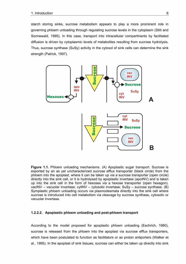
1. Introduction 6
starch storing sinks, sucrose metabolism appears to play a more prominent role in
governing phloem unloading through regulating sucrose levels in the cytoplasm (Stitt and
Sonnewald, 1995). In this case, transport into intracellular compartments by facilitated
diffusion is driven by cytoplasmic levels of metabolites resulting from sucrose hydrolysis.
Thus, sucrose synthase (SuSy) activity in the cytosol of sink cells can determine the sink
strength (Patrick, 1997).
Sucrose
SuSyapo INV
cyt INV
Sucrose
A
B
vac INV
SuSycyt INV
vac INV
Figure 1.1. Phloem unloading mechanisms. (A) Apoplastic sugar transport. Sucrose is exported by an as yet uncharacterized sucrose efflux transporter (black circle) from the phloem into the apoplast, where it can be taken up via a sucrose transporter (open circle) directly into the sink cell, or it is hydrolysed by apoplastic invertase (apoINV) and is taken up into the sink cell in the form of hexoses via a hexose transporter (open hexagon). vacINV – vacuolar invertase; cytINV – cytosolic invertase; SuSy – sucrose synthase. (B) Symplastic phloem unloading occurs via plasmodesmata directly into the sink cell where sucrose is introduced into cell metabolism via cleavage by sucrose synthase, cytosolic or vacuolar invertase.
1.2.2.2. Apoplastic phloem unloading and post-phloem transport
According to the model proposed for apoplastic phloem unloading (Eschrich, 1980),
sucrose is released from the phloem into the apoplast via sucrose efflux transporters,
which have been postulated to function as facilitators or as proton antiporters (Walker et
al., 1995). In the apoplast of sink tissues, sucrose can either be taken up directly into sink
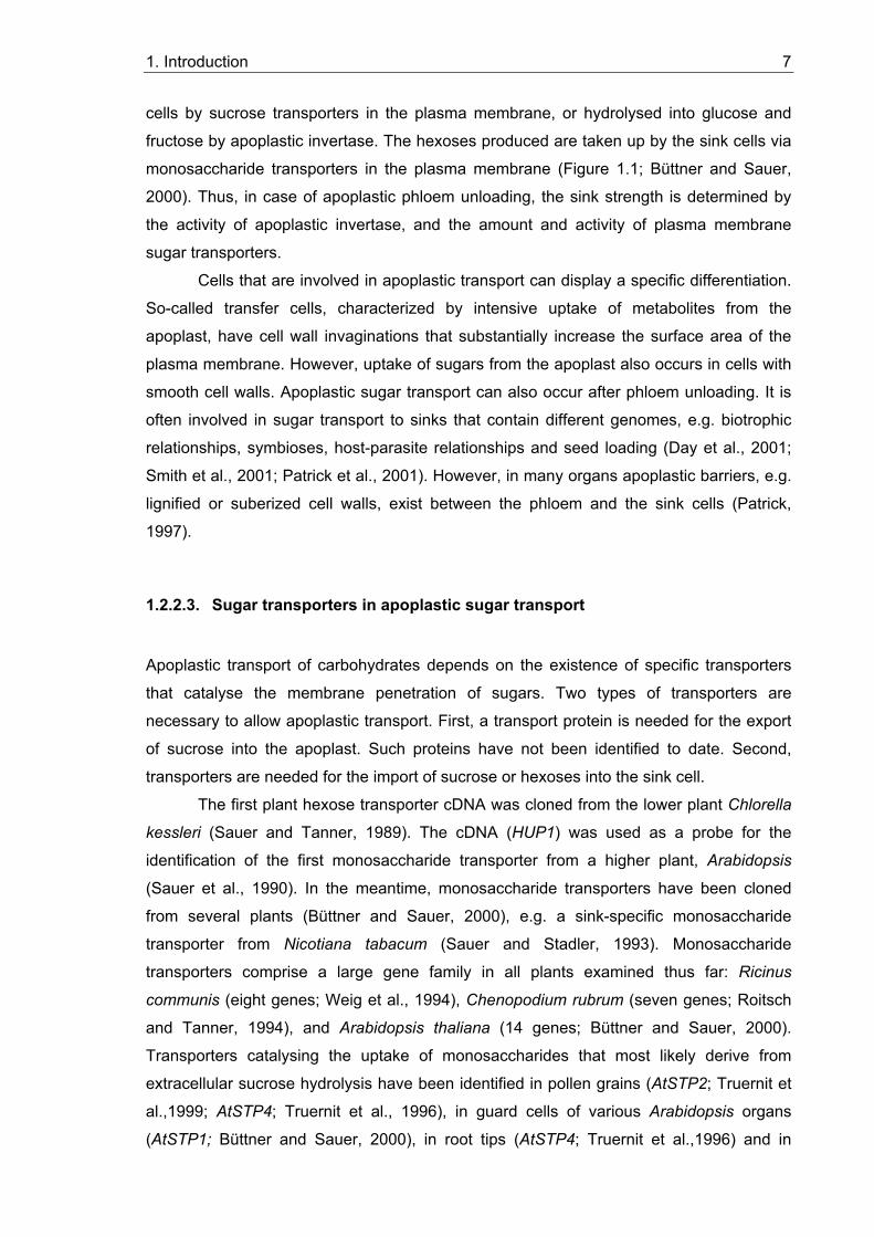
1. Introduction 7
cells by sucrose transporters in the plasma membrane, or hydrolysed into glucose and
fructose by apoplastic invertase. The hexoses produced are taken up by the sink cells via
monosaccharide transporters in the plasma membrane (Figure 1.1; Büttner and Sauer,
2000). Thus, in case of apoplastic phloem unloading, the sink strength is determined by
the activity of apoplastic invertase, and the amount and activity of plasma membrane
sugar transporters.
Cells that are involved in apoplastic transport can display a specific differentiation.
So-called transfer cells, characterized by intensive uptake of metabolites from the
apoplast, have cell wall invaginations that substantially increase the surface area of the
plasma membrane. However, uptake of sugars from the apoplast also occurs in cells with
smooth cell walls. Apoplastic sugar transport can also occur after phloem unloading. It is
often involved in sugar transport to sinks that contain different genomes, e.g. biotrophic
relationships, symbioses, host-parasite relationships and seed loading (Day et al., 2001;
Smith et al., 2001; Patrick et al., 2001). However, in many organs apoplastic barriers, e.g.
lignified or suberized cell walls, exist between the phloem and the sink cells (Patrick,
1997).
1.2.2.3. Sugar transporters in apoplastic sugar transport
Apoplastic transport of carbohydrates depends on the existence of specific transporters
that catalyse the membrane penetration of sugars. Two types of transporters are
necessary to allow apoplastic transport. First, a transport protein is needed for the export
of sucrose into the apoplast. Such proteins have not been identified to date. Second,
transporters are needed for the import of sucrose or hexoses into the sink cell.
The first plant hexose transporter cDNA was cloned from the lower plant Chlorella
kessleri (Sauer and Tanner, 1989). The cDNA (HUP1) was used as a probe for the
identification of the first monosaccharide transporter from a higher plant, Arabidopsis
(Sauer et al., 1990). In the meantime, monosaccharide transporters have been cloned
from several plants (Büttner and Sauer, 2000), e.g. a sink-specific monosaccharide
transporter from Nicotiana tabacum (Sauer and Stadler, 1993). Monosaccharide
transporters comprise a large gene family in all plants examined thus far: Ricinus
communis (eight genes; Weig et al., 1994), Chenopodium rubrum (seven genes; Roitsch
and Tanner, 1994), and Arabidopsis thaliana (14 genes; Büttner and Sauer, 2000).
Transporters catalysing the uptake of monosaccharides that most likely derive from
extracellular sucrose hydrolysis have been identified in pollen grains (AtSTP2; Truernit et
al.,1999; AtSTP4; Truernit et al., 1996), in guard cells of various Arabidopsis organs
(AtSTP1; Büttner and Sauer, 2000), in root tips (AtSTP4; Truernit et al.,1996) and in
1. Introduction 8
leaves (AtSTP3; Büttner et al., 2000). The expression of plant monosaccharide
transporter genes is not only developmentally regulated, but also controlled by
environmental stimuli, such as pathogen infection (AtSTP4) or wounding (AtSTP3 and
AtSTP4; Truernit et al., 1996; Büttner et al., 2000).
The hydropathy profiles for all plant monosaccharide transporters are very similar
and suggest 12 membrane-spanning domains, a typical feature of all members of the
major facilitator superfamily (MFS; Marger and Saier, 1993). Sequence analysis indicates
that they may have evolved by gene duplication from an ancestral gene coding for a six-
transmembrane helix transporter (Büttner and Sauer, 2000).
The kinetic properties of monosaccharide transporters have been studied by their
heterologous expression in yeast or in Xenopus oocytes (Bush, 1993; Büttner and Sauer,
2000). All eleven of the investigated monosaccharide transporters were shown to be
sensitive to uncouplers of the transmembrane proton gradient and to accumulate their
substrates inside the cells to concentrations exceeding the extracellular concentrations,
indicating that they catalyse active transport. They all represent H+-symporters and accept
pyranose monosaccharides (Büttner and Sauer, 2000).
The first sucrose transporter cDNA was isolated from spinach (SoSUT; Riesmeier
et al., 1992). Meanwhile, sucrose transporters have been described in a wide variety of
systems and termed SUT (from sucrose transporter) or SUC (from sucrose carrier). Eight
sucrose transporter genes have been found in Arabidopsis (Williams et al., 2000;
www.tigr.org). The SUT genes encode highly hydrophobic proteins. Like monosaccharide
transporters, sucrose transporters contain twelve membrane-spanning domains. In fact,
they are distantly related to the hexose transporter family (Sauer et al., 1994). In
apoplastic phloem loading, sucrose transporters accumulate sucrose in the sieve element-
companion cell (SE/CC) complex to drive long-distance transport. In some plants these
sucrose transporters have been detected in sieve elements (Kühn et al., 1997) or in
companion cells (Stadler et al., 1995). Sucrose transporters have also been found in
particular root cells: DcSUT2 is expressed in storage parenchyma cells of carrot tap-roots
where it seems to import sucrose for storage (Shakya and Sturm, 1997). In Arabidopsis
roots, strong expression was found for the sucrose transporter SUC1, and weak
expression levels were shown for SUC2 (Sauer and Stolz, 1994). The latter has been
detected in phloem cells all over the plant, including root and stem phloem, where it may
retrieve sucrose that has leaked into the apoplast or possibly catalyses the efflux of
sucrose from the phloem (Truernit and Sauer, 1995) and participates in phloem unloading
(Sauer et al., 1994).
1. Introduction 9
1.3. The regulation of phloem unloading and sink strength in different systems
Mechanisms of phloem unloading and sink strength regulation have been examined in
several systems, including developmental sinks like seeds, storage organs and fruits, and
optional sinks, like pathogen infection sites.
1.3.1. Developmental sinks
The systems best examined for phloem unloading mechanisms so far are seeds of
different plants, including legumes (Weber et al., 1997). For instance, the transport
mechanisms and the cellular pathways of sucrose were analyzed in detail in fava bean
seeds at the filling stage (Patrick, 1997). The sieve elements of the phloem end in the
seed coat and are symplastically connected with the coat cells. Phloem unloading and
post-phloem transport through the seed coat occur via the symplast. From the coat cells,
sugars are unloaded into the seed apoplast and must be taken up by the apoplastically
isolated embryo. Active transport systems are present in different parts of the seed
(Gahrtz et al., 1996). Transfer cell formation (see 1.2.2.2) is correlated with strong
induction of the expression of the Vicia faba sucrose transporter gene VfSUT1 (Weber et
al., 1997). The sucrose uptake system changes from the invertase-mediated unloading
process, which provides the sink strength during the prestorage phase, to the storage
phase, which probably allows the development of the cotyledons (Weber et al., 1996).
The fava bean seed system is a good example for the different roles in sink
development that were proposed for apoplastic invertase and sucrose synthase.
Expression of apoplastic invertase gene expression, which is usually associated with
tissues characterized by high mitotic activity, was found here during embryo development,
whereas expression of sucrose synthase was found during storage product deposition in
the developing cotyledons (Heim et al., 1993; King et al., 1997). Possibly, the epidermal
contact of the cotyledons with the seed coat provides a signal for transfer cell
differentiation. An interesting analogy can be drawn from studies of pathogen-plant
interactions. The parasites Orobanche and Cuscuta retrieve sugars from their hosts,
probably by establishing a sucrose uptake system involving transfer cells, which only form
after contact with the host vascular tissue (Ayres et al., 1996).
1. Introduction 10
1.3.2. Optional sinks
The formation of optional sinks represents a facultative event for the plant. Examples for
optional sinks are sites of pathogen infection, for instance tumors induced by
Agrobacterium tumefaciens, sites of wounding, or symbiotic structures like nitrogen-fixing
root nodules. In order to understand the regulation of phloem unloading and post-phloem
transport, it is important to know which sugar transport mechanisms take place in optional
sinks.
The large number of additional metabolic tasks in wounded or infected tissues cannot be
sufficiently energized by the endogenous resources of the respective cells. Carbohydrates
have to be imported into the affected tissues, which can be achieved by the rapid and
simultaneous induction of genes for extracellular invertases (Sturm and Chrispeels, 1990)
and plasma membrane monosaccharide transporters (Truernit et al., 1996; Büttner et al.,
2000). In case of symplastic phloem unloading and post-phloem transport to the
facultative sink, the activities of cytoplasmic or vacuolar sucrose-degrading enzymes have
to be increased.
An example for optional sinks displaying symplastic phloem unloading and post-phloem
transport are plant tumours induced by Agrobacterium tumefaciens that contain a well
developed vascular system (Pradel et al., 1999). The distribution of the fluorescent dye
carboxyfluorescein (CF) in tumours induced on three host plants, Nicotiana benthamiana,
Cucurbita maxima and Ricinus communis, demonstrated a clear symplastic pathway
between the phloem of the host stem and the tumour parenchyma, and also a
considerable capacity for subsequent cell-to-cell transport between tumor parenchyma
cells (Pradel et al., 1999).
Symbiotic nitrogen-fixing root nodules represent another example of optional sinks with
high metabolic activity that requires large amounts of carbohydrates. The analysis of the
sink function of these nodules is the topic of this thesis.
1.4. Nitrogen-fixing root nodule symbioses
Nitrogen is the element that most commonly restricts plant growth. The biosphere nitrogen
is continuously depleted by denitrification. Only some prokaryotes can form the enzyme
complex nitrogenase that reduces dinitrogen to ammonia and thereby introduces it into
the biosphere. Since nitrogenase is highly oxygen-sensitive, its formation has to be tightly
controlled, or oxygen protection systems have to be provided. Some prokaryotes fix
nitrogen in symbiosis with higher plants. In root nodule symbioses, nitrogen-fixing soil
1. Introduction 11
bacteria induce the formation of special organs, the root nodules, on their plant hosts.
Inside nodule cells, the bacteria fix nitrogen and deliver the products of nitrogen fixation to
the plant, while being supplied with carbon sources by the plant. Two groups of soil
bacteria are able to enter root nodule symbiosis with higher plants: rhizobia - unicellular,
Gram-negative bacteria, and actinomycetes of the genus Frankia – Gram-positive bacteria
that grow in hyphal form. Rhizobia enter symbioses with legumes and one non-legume,
Parasponia sp., a member of the Ulmaceae family (Pueppke and Broughton, 1999),
whereas Frankia strains can interact with a diverse group of mostly woody dicotyledonous
plants that are members of eight different families, collectively called actinorhizal plants
(Benson and Silvester, 1993).
1.4.1. Nitrogen fixation in nodules
The biological reduction of dinitrogen to ammonia is carried out by two enzymes,
dinitrogenase and dinitrogenase reductase. Together, these enzymes are often referred to
as nitrogenase. Dinitrogenase is encoded by genes nifD and nifK, whereas dinitrogenase
reductase is encoded by nifH. The name nif of these genes is derived from “nitrogen
fixation”. Nitrogenase is very sensitive to oxygen (Shaw and Brill, 1977), while the process
of nitrogen fixation requires large amounts of energy provided by respiratory processes,
leading to the so-called "oxygen dilemma" of nitrogen fixation. Therefore, the expression
of nif-genes is strongly regulated and takes place only under nitrogen-limiting conditions
and at very low oxygen concentrations, which may be achieved by the formation of
oxygen diffusion barriers.
In legume nodules, an oxygen-diffusion barrier in combination with the high metabolic
activity of the infected cells results in a low concentration of free oxygen in the area of
nitrogen-fixing infected cells (Denison and Layzell, 1991). Within infected cells high levels
of oxygen-binding proteins, leghemoglobins, provide an efficient transport of oxygen to the
sites of respiration (Appleby, 1984). The oxygen barrier is present in the nodule
parenchyma, which surrounds the inner tissue and encloses the peripheral nodule
vascular system (Figure 1.2). The nodule parenchyma consists of layers of cells more or
less devoid of intercellular spaces (Minchin, 1997).
Frankia can grow on dinitrogen as sole nitrogen source in the soil (Benson and Silvester,
1993). In order to provide oxygen protection for nitrogenase, Frankia forms vesicles at the
end of hyphae or at short side branches of hyphae. These vesicles are surrounded by a
multilayered envelope containing hopanoids, bacterial steroid lipids (Berry et al., 1993),
and in them nitrogenase is formed in a low-oxygen environment (Meesters et al., 1985;
Parsons et al., 1987). Several diverse mechanisms have evolved for oxygen control in
1. Introduction 12
actinorhizal nodules formed by different plant families. In actinorhizal nodules of
Casuarina, Frankia does not form vesicles but nitrogen fixation takes place in
differentiated hyphae (Berg and McDowell, 1987a), and an oxygen diffusion barrier is
achieved by lignification of the walls of infected and adjacent uninfected cells (Berg and
McDowell, 1987b). Casuarina nodules also contain high amounts of nodule-specific
hemoglobin, thus facilitating oxygen transport to the sites of respiration (Fleming et al.,
1987). In other actinorhizal plants, e.g. Datisca sp., no hemoglobin is present in nodules,
and Frankia forms vesicles for nitrogen fixation (Benson and Silvester, 1993).
1.4.2. Infection of plants and nodule formation
Bacteria can enter plant roots either intracellularly or intercellularly (Pawlowski and
Bisseling, 1996). During intracellular infections, the bacteria are always surrounded by a
membrane of plant origin, allowing the plant to control the microsymbiont's access to
nutrients.
Most herbaceous legumes, including Medicago truncatula, are infected intracellularly via
root hairs. Bacterial signal factors, the Nod factors, induce the deformation of root hairs
and the induction of cortical cell divisions that ultimately lead to the formation of a nodule
meristem (reviewed by Mylona et al., 1995). When a Rhizobium is trapped in a curled root
hair, a localized hydrolysis of the root hair cell wall is induced, the plasma membrane
invaginates, and new cell wall material is deposited (Kijne, 1992). This leads to the
formation of a tubular structure, an infection thread, which grows to a nodule primordium,
afterwards the bacteria are released into the cells by a process resembling endocytosis
(Basset et al., 1977). During this process, rhizobia become enclosed by a plant-derived
membrane, the peribacteroid membrane (PBM), that results in the formation of the
symbiosome (Roth and Stacey, 1989). In the symbiosomes, rhizobia differentiate into their
endosymbiotic form, the bacteroids, and start to fix nitrogen (Newcomb, 1981). In several
other rhizobial symbioses (Parasponia and some primitive legumes) the bacteria are not
released from the infection threads and, the infection cells are filled with the branching
infection threads (reviewed by Pawlowski and Bisseling, 1996).
In intracellularly infected actinorhizal plants like Casuarina glauca, bacterial hyphae also
enter the plant root via infection-thread like structures (Torrey, 1976), which grow toward
the dividing cells in cortex and infect them by intense branching within the cells, forming
the so-called prenodule. Nodule primordium formation in actinorhizal plants is induced in
the root pericycle and not in the root cortex as in legumes, therefore infection thread-like
structures grow further from the prenodule to the pericycle and infect primordium cells,
leading to nodule formation.
1. Introduction 13
During intercellular infection, rhizobia can penetrate through the gaps in the epidermis that
can form where lateral roots or adventitious roots emerge from the main root or stem,
respectively (Chandler et al., 1982; James et al., 1992), while Frankia hyphae can
penetrate the roots between root epidermal cells. In actinorhizal plants Frankia hyphae
colonize the apoplastic space of the root cortex growing through the middle lamellae while
cortical cells secrete electron-dense material rich in pectin and proteins into the
intercellular spaces (Miller and Baker, 1985; Racette and Torrey, 1989; Liu and Berry,
1991). Hyphal tips enter primordium cells as the host cell plasma membrane invaginates.
Surrounded by the invaginated host plasma membrane and embedded in fibrillar host-
derived material, the hyphae proliferate inside the infected primordium cells (Miller and
Baker, 1985).
Both infection pathways occur in legumes as well as in actinorhizal symbioses. Similar to
the nodule structure, the type of infection is determined by the host plant, but not by the
microsymbiont. For example, Medicago and Casuarina are infected intracellularly, while
Datisca is assumed to be infected intercellularly (Pawlowski, 2002).
1.4.3. Nodule structure
Mature legume nodules have a stem-like organization in that they have a peripheral
vascular system (Figure 1.2), with infected cells in the inner tissue. Actinorhizal nodules,
as well as nodules induced by rhizobia on Parasponia, are composed of modified lateral
roots without root caps, a central vascular system (Figure 1.2) and infected cells in the
expanded cortex. It has been suggested that the structural differences between legume
and other root nodules may be due to the availability of a unique developmental program
in legumes (Joshi et al., 1993; Hirsch and LaRue, 1997). Some legume cultivars can
spontaneously form bacteria-free nodules (Truchet et al., 1989; Blauenfeldt et al., 1994)
which are rich in amyloplasts, indicating that nodules may originally have developed as
carbon storage organs (Joshi et al., 1993).
There are two types of legume nodules: determinate and indeterminate. Which type of
nodule is formed depends on the host plant (Mylona et al., 1995; Doyle et al., 1997).
Indeterminate nodules contain an apical meristem. As a consequence, a developmental
gradient of infected cells is established along the nodule axis (Figure 1.2; Vasse et al.,
1990) that can be divided into five zones. Directly below the meristem (I), cells become
infected in the prefixation zone (II). At the distal end of the infection zone, rhizobia are
released from infection threads, enclosed by plasma membrane-derived
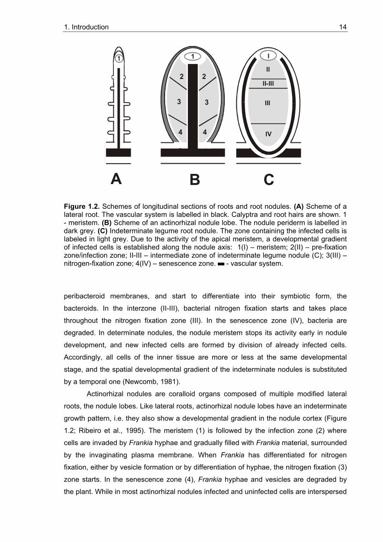
1. Introduction 14
I
II
II-III
III
IV
1
2
4 4
3 3
2
B CA
Figure 1.2. Schemes of longitudinal sections of roots and root nodules. (A) Scheme of a lateral root. The vascular system is labelled in black. Calyptra and root hairs are shown. 1 - meristem. (B) Scheme of an actinorhizal nodule lobe. The nodule periderm is labelled in dark grey. (C) Indeterminate legume root nodule. The zone containing the infected cells is labeled in light grey. Due to the activity of the apical meristem, a developmental gradient of infected cells is established along the nodule axis: 1(I) – meristem; 2(II) – pre-fixation zone/infection zone; II-III – intermediate zone of indeterminate legume nodule (C); 3(III) – nitrogen-fixation zone; 4(IV) – senescence zone. ▀▀ - vascular system.
peribacteroid membranes, and start to differentiate into their symbiotic form, the
bacteroids. In the interzone (II-III), bacterial nitrogen fixation starts and takes place
throughout the nitrogen fixation zone (III). In the senescence zone (IV), bacteria are
degraded. In determinate nodules, the nodule meristem stops its activity early in nodule
development, and new infected cells are formed by division of already infected cells.
Accordingly, all cells of the inner tissue are more or less at the same developmental
stage, and the spatial developmental gradient of the indeterminate nodules is substituted
by a temporal one (Newcomb, 1981).
Actinorhizal nodules are coralloid organs composed of multiple modified lateral
roots, the nodule lobes. Like lateral roots, actinorhizal nodule lobes have an indeterminate
growth pattern, i.e. they also show a developmental gradient in the nodule cortex (Figure
1.2; Ribeiro et al., 1995). The meristem (1) is followed by the infection zone (2) where
cells are invaded by Frankia hyphae and gradually filled with Frankia material, surrounded
by the invaginating plasma membrane. When Frankia has differentiated for nitrogen
fixation, either by vesicle formation or by differentiation of hyphae, the nitrogen fixation (3)
zone starts. In the senescence zone (4), Frankia hyphae and vesicles are degraded by
the plant. While in most actinorhizal nodules infected and uninfected cells are interspersed
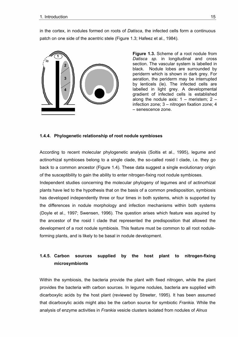
1. Introduction 15
in the cortex, in nodules formed on roots of Datisca, the infected cells form a continuous
patch on one side of the acentric stele (Figure 1.3; Hafeez et al., 1984).
4
3
2
1le
4
3
2
1le
1.4.4. Phylogenetic relationship of root nodule symbioses
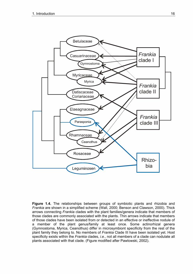
According to recent molecular phylogenetic analysis (Soltis et al., 1995), legume and
actinorhizal symbioses belong to a single clade, the so-called rosid I clade, i.e. they go
back to a common ancestor (Figure 1.4). These data suggest a single evolutionary origin
of the susceptibility to gain the ability to enter nitrogen-fixing root nodule symbioses.
Independent studies concerning the molecular phylogeny of legumes and of actinorhizal
plants have led to the hypothesis that on the basis of a common predisposition, symbiosis
has developed independently three or four times in both systems, which is supported by
the differences in nodule morphology and infection mechanisms within both systems
(Doyle et al., 1997; Swensen, 1996). The question arises which feature was aquired by
the ancestor of the rosid I clade that represented the predisposition that allowed the
development of a root nodule symbiosis. This feature must be common to all root nodule-
forming plants, and is likely to be basal in nodule development.
1.4.5. Carbon sources supplied by the host plant to nitrogen-fixing microsymbionts
Within the symbiosis, the bacteria provide the plant with fixed nitrogen, while the plant
provides the bacteria with carbon sources. In legume nodules, bacteria are supplied with
dicarboxylic acids by the host plant (reviewed by Streeter, 1995). It has been assumed
that dicarboxylic acids might also be the carbon source for symbiotic Frankia. While the
analysis of enzyme activities in Frankia vesicle clusters isolated from nodules of Alnus
Figure 1.3. Scheme of a root nodule fromDatisca sp. in longitudinal and cross section. The vascular system is labelled in black. Nodule lobes are surrounded by periderm which is shown in dark grey. For aeration, the periderm may be interrupted by lenticels (le). The infected cells are labelled in light grey. A developmental gradient of infected cells is established along the nodule axis: 1 – meristem; 2 –infection zone; 3 – nitrogen fixation zone; 4 – senescence zone.
1. Introduction 16
Myrica
DatiscaceaeCoriariaceae
Gymnostoma
Frankiaclade I
Frankiaclade II
Frankiaclade III
Rhizo-bia
Ceanothus
Parasponia
Figure 1.4. The relationships between groups of symbiotic plants and rhizobia and Frankia are shown in a simplified scheme (Wall, 2000; Benson and Clawson, 2000). Thick arrows connecting Frankia clades with the plant families/genera indicate that members of those clades are commonly associated with the plants. Thin arrows indicate that members of those clades have been isolated from or detected in an effective or ineffective nodule of a member of the plant genus/family at least once. Some actinorhizal genera (Gymnostoma, Myrica, Ceanothus) differ in microsymbiont specificity from the rest of the plant family they belong to. No members of Frankia Clade III have been isolated yet. Host specificity exists within the Frankia clades, i.e., not all members of a clade can nodulate all plants associated with that clade. (Figure modified after Pawlowski, 2002).
1. Introduction 17
and Hippophae yielded results consistent with this hypothesis (Akkermans et al., 1983),
experiments on 14CO2 uptake of detached Alnus nodules were inconclusive (Huss-
Danell, 1990; see review by Huss-Danell, 1997). The carbon preferences of Frankia in the
free-living state might provide a clue. Frankia strains have been divided into two genomic
groups A and B based on DNA homology (Lechevallier and Lechevallier, 1990), and into
four different clades based on 16S rRNA sequences (Normand et al., 1996). Clade IV
comprises Frankia-like strains that cannot fix nitrogen; hence, there are three different
clades of symbiotic Frankia strains. This grouping does not map with the differentiation
into genomic groups. Furthermore, the 16S rRNA taxonomy includes non-culturable
Frankia strains that could not be assigned to a genomic group. In the free-living state,
Frankia strains of genomic group A tend to grow fairly on both sugars and organic acids,
while strains of genomic group B grow variably on organic acids and poorly, if at all, on
sugars. The carbon source mostly used in culturing Frankia strains is propionic acid.
However, the endosymbionts of several actinorhizal plants, including Datisca, have not
been isolated yet. Based on their 16S rRNA and nifH gene sequences, it has been
concluded that many non-isolated Frankia strains including the endosymbiont of Datisca
do not belong to any group of Frankia strains isolated so far (Mirza et al., 1994; Normand
et al., 1996; Figure 1.4). Hence it is unclear whether this group of strains in particular, or
Frankia strains in general, are fed other carbon sources than dicarboxylates during
symbiosis.
1.4.6. Carbon transport and metabolism in nitrogen-fixing root nodules
Root nodules represent strong carbon sinks. They need assimilates as energy source for
the microsymbionts, to provide carbon skeletons for the assimilation of the ammonium
produced by the microsymbionts, for growth and, with the exception of Casuarina (E.
Duhoux, personal communication) for starch biosynthesis.
In legume root nodules, several lines of evidence indicate that SuSy is essential for the
cleavage of sucrose translocated from the shoots to the roots in support of nodule
metabolism. First, the levels of SuSy mRNA and protein in soybean nodules are 10-20
times higher than those in root tissues (Thummler and Verma, 1987). Second, the SuSy
gene is known to be predominantly expressed in nodules from Vicia faba and Medicago
truncatula compared to uninfected roots, stems, leaves and other organs (Küster et al.,
1993; Hohnjec et al., 1999). Third, several authors have demonstrated that various abiotic
stress conditions (e.g. drought, salt, nitrate treatments) that decrease symbiotic nitrogen
fixation in soybean nodules also dramatically reduce the mRNA, protein and activity levels
1. Introduction 18
of SuSy (Gordon et al., 1997; González et al., 1995). Finally, a pea mutant with severely
reduced activity of nodule-enhanced SuSy was incapable of an effective root nodule
symbiosis (Gordon et al., 1999). These results, however, do not preclude that invertases
present in nodules (Gordon, 1991) play an important role in nodule carbon metabolism in
specific stages of development.
Few studies have been performed on the possibilities of symplastic and apoplastic carbon
transport in nodules. Ultrastructural investigation of nodules of 27 genera of legumes
demonstrated plasmodesmal connections between sieve element and pericycle cells and
also showed that pericyle cells, like infected cells and endodermal cells, were
symplastically connected (Pate et al., 1969). For seven genera of legumes, transfer cells
were described in the pericycle, but precise investigation showed that they are localized
next to xylem elements and cell wall protuberances are especially developed on the walls
contiguous with the xylem (Pate et al., 1969). Light and electron microscopy of
indeterminate Vicia faba nodules also revealed a block between vascular tissue and inner
cortex, represented by Casparian bands on the radial cell walls of the vascular
endodermis and the so-called nodule endodermis, a cell layer in the nodule cortex
surrounding the inner tissue and the nodule vascular system, and by the suberin coat of
endodermis cells (Abd-Alla et al., 2000).
But symplastic metabolite passage is possible due to high plasmodesmal frequencies
found between the vascular endodermis and the inner cortex (Abd-Alla et al., 2000). It
was also demonstrated that there are very few plasmodesmata between infected cells of
the central tissue, whereas uninfected cells can represent a symplastic continuity (Abd-
Alla et al., 2000).
1.5. Aim of this thesis
One very basal feature in root nodule development is the establishment and maintenance
of the carbon sink function of the nodule. The aim of this thesis was to characterize sugar
partitioning mechanisms in nitrogen-fixing root nodules. Three diverse model systems
were chosen (Table 1.1): one legume symbiosis (Medicago truncatula – Sinorhizobium
meliloti) and two actinorhizal symbioses from different Frankia subclades (Figure 1.4;
Datisca glomerata, Casuarina glauca). The possilibities were (a) that carbon partitioning
mechanisms in nodules would resemble those established in roots. If all three model
plants would be similar here, root carbon partitioning mechanisms might represent the
common basic feature that was required for the establishment of nitrogen-fixing
symbiosis. If all three would be different, the symbiosis would put no specific requirements
1. Introduction 19
on carbon partitioning mechanisms. On the other hand, if (b) carbon partitioning
mechanisms in roots of the three model systems differed, while those in nodules were
similar, the symbiosis would put specific requirements on carbon partition. Either way, the
results of the comparison would deepen our understanding of the interaction between
plants and nitrogen-fixing bacteria.
Table 1.1. Characteristics of the studied model systems for root nodule symbioses.
Characteristics Medicago truncatula Datisca glomerata Casuarina glauca
Microsymbiont Infection mechanism Oxygen-carrying protein Oxygen barrier Microsymbiont in free culture Phloem loading mechanism
Sinorhizobium meliloti
Intracellular
Leghemoglobin
Nodule parenchyma
+
Apoplastic
Frankia subclade III
Intercellular
No hemoglobin found
Mitochondria at vesicle base
Not isolated
Not examined
Frankia subclade I
Intracellular
Symbiotic hemoglobin
Strong lignification of infected cells
+
Symplastic
To characterize carbon partitioning mechanisms in these three systems, the following
aspects were examined:
• Contents and identity of dominating sugars in nodules compared to roots
• Activities of SuSy and of the different invertases
• Localization of sucrose-cleaving enzymes
• Expression levels of SuSy and sugar transporters
• Isolation of the cDNAs of hexose transporters expressed in nodules, and
biochemical characterization of their protein products
The last of these aims was proposed since in nodules of Datisca, the expression of a
monosaccharide transporter had been localized by in situ hybridization specifically to the
infected cells (Wabnitz, 1998).
2. Materials and methods 20
2. Materials and methods
For all methods, except for biochemical methods, only sterile pipette tips, glass and plastic
ware were used. All solutions for molecular biological methods were prepared using water
of MilliQ grade (dd H2O), autoclaved or filter sterilised, if not otherwise described.
2.1. Materials
2.1.1. Plant material
Medicago truncatula: cv. Jemalong, genotype A-17 (Barker et al., 1990) Datisca glomerata: seeds were kindly provided by Alison M. Berry, University
of California (Davis, CA, USA)
Casuarina glauca: seeds were supplied by the Desert Development Centre
(Cairo, Egypt)
2.1.2. Bacterial and yeast strains
Plant infection:
Host plant_____________ Symbiont__________________Reference___________
Medicago truncatula Sinorhizobium meliloti 1021 Meade et al., 1982 Datisca glomerata actinomycetes from crushed
nodules
Casuarina glauca Frankia strain Thr Girgis et al., 1990
Cloning: Escherichia coli DH5α (Woodcock et al., 1989).
Genotype: F- (φ80dlacZ∆M15) recA1 endA1 gyrA96 thi-1 hsdR17(rk-mk
+) supE44 relA1
deoR ∆(lacZYA-argF) U169
Analysis of transport activity for plant hexose transporters: Saccharomyces cerevisiae strain EBY.VW4000 (Wieczorke et al., 1999)
2. Materials and methods 21
Genotype: hxt13∆::loxP hxt15∆::loxP htx16∆::loxP htx14∆::loxP htx12∆::loxP htx9∆::loxP
htx11∆::loxP htx10∆::loxP htx8∆::loxP htx514∆::loxP htx2∆::loxP htx367∆::loxP gal2∆
stl1∆::loxP agt1∆::loxP ydl247w∆::loxP yjir 160c∆::loxP
2.1.3. Oligonucleotides (Primers)
Name Sequence
DgHT fs5 5´-CCC TTC TCT CTT TAT CTC C-3´
DgHT fs3 5´-CAG ATA CAT CAA ATT TGA CCC-3´
DgHT 5´race1 5´-GTC GGG AAG GAG TAG CGA-3´
DgHT 5´race2 5´-CTC CTC TGT ACT TGT AGG GA-3´
for 5´-GTA AAA CGA CGG CCA GT-3´
MtHT fs 5´-CCC AAC CAG CAT AGA ATT AAA CTA ACA GTG-3´
MtHT 5´race1 5´-GAA GAA CAC GAC CCA AAA TGA GC-3´
MtHT 5´race2 5´-ATC CTT AGA CAA AAC AGC GTT CGA-3´
MM1 5´- GGC CAC GCG TCG ACT AGT ACG GGI IGG GII GGG IIG - 3´
MM2 5´- GGC CAC GCG TCG ACT AGT AC - 3´
MM3 5´- CTC GAG GAT CCG CGG CCG CT18 - 3´
MM4 5´- CTC GAG GAT CCG CGG CCG C - 3´
PmDgHT-for 5´-CCG GAA TTC AAG CTT GTA AAA GAA ATG CCG GCC GTC
GGA GG-3´
PmDgHT-rev 5´-CTA AAA ATA ACC CTC CCC CCA AAC-3´
PmMtHT-for 5´-CCG CTC GAG AAG CTT GTA AAA GAA ATG GCT GGT GGG
GTT TTA CCA GTG-3´
PmMtHT-rev 5´-TTC ACT AGT GAT TCT CGA GGA TCC GC-3´
Rev 5´-AGC GGA TAA CAA TTT CAC ACA GGA-3´
T3 5´- TAA CCC TCA CTA AAG GGA - 3´
T7 5´- TAA TAC GAC TCA CTA TAG GG - 3´
dT20 5´-TTTTTTTTTTTTTTTTTTTT-3´
2.1.4. Plasmids
p-Bluescript® II KS (+) AmpR Stratagene, La Jolla, CA, USA
p-GEM ®-T Easy AmpR Promega, Madison,WI, USA
pNEV-E AmpR; URA3 Sauer and Stolz (1994)
2. Materials and methods 22
pNEV-X AmpR; URA3 Sauer and Stolz (1994)
pP001-VS AmpR B.Reiß, personal
communication
pRT 105 AmpR Topfer et al. (1993)
pSPORT AmpR Gibco BRL, Eggenstein,
Germany
2.1.5. Enzymes
2.1.5.1. Restriction enzymes
Restriction enzymes XbaI, EcoR321 (EcoRV), ACC65I (KpnI), SmaI, PstI, SdaI
(Sse8387I), Eco105I (SnaBI) were obtained from MBI Fermentas (Vilnius, Lithuania) and
BamHI, HindIII, EcoRI, SstI, SstII, SalI, PstI from Gibco BRL (Eggenstein, Germany).
2.1.5.2. Other enzymes and kits
Taq-Polymerase Biometra, Göttingen, Germany
DNA-Polymerase I Large Fragment MBI Fermentas, Vilnius, Lithuania
(Klenow Fragment)
MLV Reverse Transcriptase MBI Fermentas, Vilnius, Lithuania
ThermoscriptTM -RT-PCR System Gibco BRL, Eggenstein, Germany
RNase A Sigma, Deisenhofen, Germany
T4 DNA-Ligase Gibco BRL, Eggenstein, Germany
Lysozyme Sigma, Deisenhofen, Germany
Mung bean nuclease Epicentre Technologies, Madison,
WI, USA
ABI PRISM dRhodamine Terminator PE Applied Biosystems, Weiterstadt,
Cycle Sequencing Ready Reaction Kit Germany
Qiagen Gel Extraction Kit Qiagen, Hilden, Germany
Qiaprep Spin Miniprep Kit Qiagen, Hilden, Germany
Qiaprep Spin Maxiprep Kit Qiagen, Hilden, Germany
Peq Lab E.Z.N.A. Plasmid Peq Lab Biotechnologie GmbH,
Miniprep II Kit Erlangen, Germany
Invisorb Spin-Plant-RNA Mini Kit Invitek, Berlin, Germany
2. Materials and methods 23
2.1.6. Chemicals
Agar Gibco BRL, Eggenstein, Germany
Agarose Gibco BRL, Eggenstein, Germany
Ampicillin Sigma, Deisenhofen, Germany
Arbutin Sigma, Deisenhofen, Germany
Bovine Extract Difco, Becton Dickinson GmbH,
Heidelberg, Germany
Casamino Acids (CAA) Difco, Becton Dickinson GmbH,
Heidelberg, Germany
Coomassie Brilliant Blue G 250 Serva, Heidelberg, Germany
5-Bromo-4-chloro-3-indolyl β-D- Roche Molecular Biochemicals,
galactopyranoside (X-Gal) Mannheim, Germany
Dimethylformamide (DMF) Sigma, Deisenhofen, Germany
Dimethyl sulfoxide (DMSO) Sigma, Deisenhofen, Germany
Deoxynucleotide triphosphates (dNTPs) Roche Molecular Biochemicals,
Mannheim, Germany
Dithiothreitol (DTT) Gibco BRL, Eggenstein, Germany
Ethidium bromide Sigma, Deisenhofen, Germany
Ethylendiamine tetraacetic acid (EDTA) Merck, Darmstadt, Germany
D(+)-Glucose Merck, Darmstadt, Germany
Yeast extract Gibco BRL, Eggenstein, Germany
Yeast nitrogen base w/o amino acids Difco, Becton Dickinson GmbH,
Heidelberg, Germany
Isopropyl β-D-thiogalactopyranoside AppliChem, Darmstadt, Germany
(IPTG)
Kanamycin Sigma, Deisenhofen, Germany
β-Mercaptoethanol Merck, Darmstadt, Germany
Orange G Sigma, Deisenhofen, Germany
Polyclar AT Serva, Heidelberg, Germany
D(+)-Sucrose Carl Roth GmbH & Co, Karlsruhe, Germany
Select Pepton 140 Gibco BRL, Eggenstein, Germany
Trishydroxymethylaminomethane (Tris) Sigma, Deisenhofen, Germany
Other chemicals Merck, Darmstadt, Germany
Sigma, Deisenhofen, Germany
2. Materials and methods 24
2.1.7. Other materials and devices
Filters and membranes: Hybond N Amersham Pharmacia Biotech, Freiburg,
Germany
3MM-filter paper Whatman, Madistone, Kent, UK
Miracloth Schütt Labortechnik GmbH, Göttingen,
Germany
Devices: Transilluminator FLX-20M Vilber Lourmat, Marne La-Vallet, France
ABI Prism 310 Genetic Analyser PE Applied Biosystems, Foster City, CA, USA
Phospho-Imager Fuji BAS-1000 Raytest, Sprockhövel, Germany
Thermocycler PTC-100 Biozym, Hessisch Oldendorf, Germany
Personal Cycler Biometra, Göttingen, Germany
Liquid scintillation analyzer 1900TR Canberra Packard GmbH, Dreieich, Germany
Centrifuges:
Megafuge 1.0 Heraeus Sepatech, Osterode, Germany
Biofuge pico Heraeus Sepatech, Osterode, Germany
Eppendorf centrifuge 5415 R Eppendorf AG, Hamburg, Germany
SORVALL®-RC-3B Refrigerated Sorvall GmbH, Bad Homburg, Germany
Superspeed Centrifuge
Ultracentrifuge L-80 Beckman Instruments, München, Germany
2.1.8. Culture media
2.1.8.1. Plant media
Hydroculture medium (Hoagland und Arnon, 1938)
Strength ½ ¼ Macro element stock solutions: per 1l: 2 ml 1 ml 1 M KH2SO4 4 ml 2 ml 1 M MgSO4 ·7H2O 10 ml 5 ml 1 M KNO3 10 ml 5 ml 1 M Ca (NO3)2·4H2O
2. Materials and methods 25
2 ml 1 ml Micro elements stock solution 2.86 g/l H3BO3
1.81 g/l MnCl2·4H2O 0.22 g/l ZnSO4·7H2O 0.08 g/l CuSO4·5H2O 0.025 g/l Na2MoO4·2H2O 0.025 g/l CoCl2·6H2O 10 ml 5 ml Fe-EDTA-stock solution: 5.56 g/l FeSO4·7H2O 7.45 g/l Na2-EDTA adjust pH to 5.5
Aeroponic culture medium (Lullien et al., 1987) 5,5 mM K-PO4-buffer pH 7.0 1 mM CaCl2 0.52 mM K2SO4 0.25 mM MgSO4 0.04 µM CoCl2 0.2 µM CuSO4 0.7 µM ZnSO4 10 µM MnSO4 30 µM H3BO3 50 µM Na2-EDTA 50 µM FeSO4 1 µM Na2Mo4
K-PO4-buffer was prepared as sterile 100× stock solution, pH 7.0, containing 58.92 g/l
K2HPO4 and 41.08 g/l KH2PO4. For the other components 1000× stocks were prepared
separately and autoclaved. FeSO4 solution should be made fresh.
Fåhraeus Medium (Fåhraeus, 1957) For 1 l of medium (nodulation nitrogen-free medium):
0.5 ml 1,0 M MgSO4 1.0 ml 0,7 M KH2PO4 2.0 ml 0,4 M NaH2PO4 2.5 ml 20 mM Fe EDTA 100 µl 1 mg/l MnSO4 100 µl 1 mg/l CuSO4 100 µl 1 mg/l ZnSO4 100 µl 1 mg/l H3BO4 100 µl 1 mg/l Na2MoO4
The pH was adjusted to 6.5 with H2SO4 and then 1.25 ml of 1 M CaCl2 was added.
2.1.8.2. Bacterial media
• Media for Escherichia coli:
2. Materials and methods 26
LB Medium (Luria-Bertani Medium) (Sambrook et al., 1989) Per liter: 5 g Yeast Extract
10 g Select Pepton 140 10 g NaCl
The pH was adjusted to 7.5 with 5 N NaOH. For solid LB medium, 1.5 % (w/v) agar per
liter were added.
SOC-Medium (Sambrook et al., 1989) All glassware used for the preparation of SOC medium was autoclaved filled with dd H2O
to remove remains of detergents.
Per liter: 5 g Yeast Extract
20 g Select Pepton 140
10 ml 1 M NaCl
2.5 ml 1 M KCl
The volume was adjusted to 1 liter with dd H2O. After autoclaving for 10 minutes filter
sterilized solutions were added:
10 ml 1 M MgCl2 10 ml 1 M MgSO4 10 ml 1 M Glucose TB Medium [Terrific Broth] (Tartof and Hobbs, 1987) Per liter:
To 900 ml of dd H2O were added: 12 g Select Pepton 140 24 g Yeast Extract 4 ml Glycerol After autoclaving for 20 minutes, the solution was cooled to 60 ºC or less, and 100 ml of a
sterile solution of K-PO4-buffer (0.17 M KH2PO4, 0.72 M K2HPO4) were added. For K-PO4-
buffer 2.31 g KH2PO4 and 12,54 g K2HPO4·3H2O were dissolved in 90 ml of dd H2O, the
volume was adjusted to 100 ml with dd H2O.
• Media for Sinorhizobium meliloti: TY-medium Per liter: 5 g Select Pepton 140 3 g Yeast Extract 0.9 g CaCl2·2H2O
2. Materials and methods 27
YEB-medium (van Larebeke et al., 1977) Per liter: 5 g Bovine Extract 1 g Yeast Extract 5 g Select Pepton 140 5 g Sucrose
The pH was adjusted to 7.2-7.3 with NaOH
• Medium for Frankia:
BAP Medium (Fontaine et al., 1986)
Stock solutions:
Per liter: 10 ml 13.5 mM CaCl2⋅2H2O 5 ml 40.5 mM MgSO4⋅7H2O 1 ml 195 mM FeNa2EDTA 5 ml 1 M NH4Cl 5 ml 1 M Sodium propionate 1 ml Oligoelements 1 ml Vitamins 10 ml K-PO4-buffer
Oligoelements: 2.86 g/l H3BO3 1.81 g/l MnCl2 0.22 g/l ZnSO4⋅7H2O 0.08 g/l CuSO4⋅5H2O
Vitamin stocks: 11.21 mg/10 ml Thiamin HCl 50 mg/10 ml Pyridoxin HCl 50 mg/10 ml Nicotinic acid 10 mg/10 ml Calcium pantothenate 10 mg/10 ml Folic acid 10 mg/10 ml Riboflavin 9 mg/0.5 ml Biotin, dissolved in 0.5 ml KOH, then diluted to 4 ml with dd H2O Vitamin stocks were stored at –20 ºC. To prepare 100 ml vitamin mix 1 ml of each stock was used. K-PO4- buffer: 560 ml 1 M KH2PO4 320 ml 1 M K2HPO4⋅3H2O pH was adjusted to 6.7 with 10 N KOH 2.1.8.3. Media for Saccharomyces cerevisiae
YPM – medium 1 % (w/v) Yeast extract
2 % (w/v) Peptone
2 % (w/v) Maltose
2. Materials and methods 28
CAA – medium 1 % (w/v) Casamino Acids
2 % (w/v) Maltose
0.67 % (w/v) YNB w/o amino acids
Supplements: 1:50 (v/v) 1 mg/ml Tryptophan stock solution
For agar plates 2 % agar (w/v) were added before autoclaving. L-Tryptophan was
dissolved at 60-70 °C. The solution was filter-sterilized and stored at 4 °C. Tryptophan
was added to CAA-medium at 50 °C before making plates.
2.1.8.4. Medium additives
Additives Solvent Stock solution
concentration
Final
concentration
Ampicillin Kanamycin IPTG X-Gal
H2O H2O H2O DMF
100 mg/ml 50 mg/ml
0,1 M 2 % (w/v)
100 µg/ml 50 µg/ml 0,2 mM 0,004 %
Stock solutions dissolved in H2O were filter sterilized. All medium additives were kept at
–20 ºC.
2.2. Plant culture methods and growth conditions
Datisca glomerata and Casuarina glauca seeds were germinated and grown for three and
five to six months, respectively, in soil (T 25 Frühstorfer Erde®) mixed with one third
volume of sand. Then they were transferred into aerated hydroculture in ¼ strength
Hoagland‘s medium (Hoagland and Arnon, 1938; see 2.1.8.1.).
Noninfected plants of Medicago truncatula genotype A-17 (cv. Jemalong) were
grown in a soil-sand mixture (see above) for three weeks and then transferred either to
hydroculture in Fåhraeus Medium (Fåhraeus, 1957; see 2.1.8.1.) for root material or in
aeroponic culture with Lullien medium for root and nodule material (Lullien et al., 1987;
see 2.1.8.1.). After one week in aeroponic culture they could be infected with
Sinorhizobium meliloti 1021 cells (Meade et al., 1982) grown in TY medium and washed
with dd H2O. Nodules were harvested after 10-14 days (aeroponic culture).
2. Materials and methods 29
D. glomerata was infected with actinomycetes from crushed nodules of D.
glomerata plants grown in soil and C. glauca was infected with Frankia strain Thr (Girgis
et al., 1990) grown in BAP Medium (Fontaine et al., 1986, see 2.1.8.2.) and washed with
dd H2O 3-4 times. Nodules of D. glomerata and C. glauca were harvested 4-8 weeks after
infection.
The greenhouse conditions were: temperature 22 °C/18 °C (day/night), light
intensity 200 µmol photons×m-2×s-1 in winter and 400 µmol photons m-2 s-1 in summer, 60-
65 % humidity.
2.3. RNA isolation from plant tissue
2.3.1. RNA isolation from Medicago truncatula and Datisca glomerata (modified after Burgos et al., 1995)
Homogenization Buffer (HB): 100 mM Tris/HCl , pH 9.0
100 mM NaCl
15 mM EDTA
0.5 % N-Lauroylsarcosin (Sigma)
Store at room temperature in 50 ml aliquots
Plant tissue was ground in liquid nitrogen into a fine powder and transferred into 50
ml Falcon tubes, containing 5 ml homogenization buffer, 5 ml phenol/chloroform mixture
(1:1) and 35 µl ß-mercaptoethanol per 1 g of plant material. After homogenizing for 2
minutes using a IKA Ultra-Turrax T-25 Basic Homogenizer (Ika-Werke GmbH & Co.
KG, Staufen, Germany), 350 µl 3 M sodium acetate (pH 5.4) were added per g tissue, and
the mixture was homogenized again for 30 sec. Then the homogenate was poured into
phenol/chloroform resistant 13 ml tubes and centrifuged in a SORVALL®-RC-3B
Refrigerated Superspeed Centrifuge (SORVALL GmbH, Bad Homburg, Germany) in a
SS 34 rotor at 10 krpm for 10 min at 4 °C.
After centrifugation, the proteins of the cell homogenate formed a solid interphase
between the water phase and the organic phase. The water phase was transferred into a
new 13 ml tube, an equal volume of phenol/chloroform was added, and the tube was
vortexed and centrifuged again at 10 krpm for 10 min at 4 °C. Then the supernatant was
transferred again into a new 13 ml tube and an equal volume of isopropanol was added.
After vortexing, the mixture was incubated at – 20 °C for 20-60 minutes. At this step
2. Materials and methods 30
nucleic acids precipitated and could be pelleted by a 10 min centrifugation step in the SS
34 rotor at 10 krpm and 4 °C.
The pellet was washed with 2 ml 70 % (v/v) ethanol to remove salts and
centrifuged for 5 min in a SS 34 rotor at 10 krpm and 4 °C. The pellet was resuspended in
900 µl dd H2O and transferred into an 1.5 ml Eppendorf tube for extraction with 500 µl
phenol/chloroform mixture (1:1) followed by an extraction of supernatant with 500 µl
chloroform. The supernatant was then transferred to a new Eppendorf tube and the
volume was adjusted to 900 µl. 300 µl (1/3 Vol.) 8 M LiCl were added and RNA was
precipitated on ice for 3 hours, followed by 10 min of centrifugation at 4 °C and 13 krpm.
The pellet was washed with 70 % (v/v) ethanol, resuspended in 50–100 µl H2O at 65 °C
for 5 min and then cooled on ice. The insoluble material was removed by a 5 min
centrifugation step. The supernatant was transferred to a new Eppendorf tube, and the
intactness and concentration of the isolated RNA was tested on a 1 % (w/v) agarose gel
(see 2.6.1.) in a detergent-treated gel chamber.
2.3.2. RNA isolation from Casuarina glauca
The RNA isolation from Casuarina is difficult because of the high polyphenol content of
the plant that interferes with nucleic acid isolation procedures. Therefore different methods
and kits were applied for Casuarina RNA isolation. The best results were obtained with
the „Invisorb Spin Plant RNA Mini Kit“(Invitek, Berlin, Germany). The purification
procedure was conducted according to recommendations of the manufacturer with a few
adaptations.
About 100 mg plant material were ground in liquid nitrogen with ca. 50 mg of
Polyclar AT (Serva, Heidelberg, Germany) to a fine powder and transferred into a 2 ml
Eppendorf tube. After the liquid nitrogen had evaporated, 500-800 µl of Lysis Solution RP
were added to the plant powder followed by vortexing for 1 min and 2 min of incubation at
room temperature. To remove cell debris, the sample was centrifuged for 5 min at 13
krpm. The supernatant was pipetted onto a DNA-Binding Spin Filter which was placed in a
1.5 ml Eppendorf tube and incubated for 1 min at room temperature. After a centrifugation
for 1 min at 10 krpm at room temperature, ½ vol. of absolute ethanol was added to the
RNA-containing flow-through and the mix was transferred onto an RNA-Binding Spin Filter
in a 1.5 ml Eppendorf tube. After 1 min incubation at room temperature, the sample was
centrifuged for 30 sec at 10 krpm in order to bind the RNA to the filter. The flow-through
was discarded, and the filter was washed with 600 µl wash buffer R1 by a centrifugation
for 30 sec at 10 krpm. This step was repeated twice with 500 µl of wash buffer R2. Then
the filter was centrifuged again for the removal of remains of wash buffer at 12 krpm for 2
2. Materials and methods 31
min and placed in a new Eppendorf tube. The RNA was eluted by direct pipetting of 40-60
µl elution buffer onto the membrane, followed by 2 min of incubation at room temperature
and a centrifugation for 1 min at 10 krpm. After this step, the RNA was kept on ice before
testing the RNA concentration on an 1 % agarose gel (see 2.6.1.), or stored at –80 °C.
2.4. Isolation of plasmid DNA from bacteria and yeast
2.4.1. Plasmid mini-preparation protocol „ Triton Boiling“
This method comprised an effective and fast method for the isolation of plasmid DNA from
bacteria for a quick characterization with restriction enzymes.
4 ml of LB medium (see 2.1.8.2.), containing an appropriate antibiotic, were
inoculated with a single bacterial colony and incubated overnight at 37 °C with vigorous
shaking. 1.5 ml of overnight culture were poured into an Eppendorf tube and spun down at
13 krpm for 1 min. The remainder of the overnight culture could be stored at 4 °C for 1-2
days. The supernatant was removed to leave the bacterial pellet as dry as possible.
Bacterial pellets were resuspended by vortexing in 150 µl cold STEL buffer (8 % sucrose;
5 % Triton X 100; 50 mM Tris-HCl, pH 8.0; 50 mM EDTA; 0.5 mg/ml lysozyme) and boiled
for 30 sec at 100 °C. Cell debris with genomic DNA was sedimented by 20 min of
centrifugation at 13 krpm at room temperature. The pellet was removed with a sterile
toothpick, and the supernatant was mixed with 180 µl isopropanol and centrifuged again
at 13 krpm for 5 min to pellet the plasmid DNA. The pellet was then washed with 500 µl 70
% ethanol, spun down for 5 min at 13 krpm, dried at 37 °C and finally resuspended in 50
µl RNase-H2O (0.2 mg/ml RNase A) for 10 min at 37 °C. DNA was stored at –20 °C.
2.4.2. Mini preparation of bacterial plasmid DNA for the sequence analysis
For sequence analysis, the plasmid DNA was isolated using a QIAprep® Spin Miniprep Kit
(Qiagen, Hilden, Germany), which allows a fast preparation of pure DNA. The QIAprep
miniprep procedure is based on the alkaline lysis of bacterial cells followed by the
adsorption of DNA on a silica gel membrane in the presence of a high salt concentration.
The DNA was eluted from the column with 50 µl dd H2O.
2. Materials and methods 32
2.4.3. Maxi preparation of bacterial plasmid DNA
This method was used for the preparation of large amounts of plasmid DNA from bacteria.
For this procedure, 50 ml of TB medium (see 2.1.8.2.), containing an appropriate
antibiotic, were inoculated with a single bacterial colony and incubated overnight at 37 °C
with vigorous shaking. The overnight culture was distributed over four 13 ml centrifugation
tubes and centrifuged for 10 min at 4.3 krpm in a Megafuge 1.0 (Heraeus Sepatech,
Osterode, Germany). The supernatant was discarded, and the pellet could be stored at
-20 °C.
For DNA preparation the Peq Lab E.Z.N.A. Plasmid Miniprep Kit II (Peq Lab
Biotechnologie GmbH, Erlangen, Germany) was used, which allows to isolate large
amounts of very pure plasmid DNA. This procedure, conducted as described in the
standard Peq Lab protocol, is based also on the alkaline lysis of bacterial cells followed by
the adsorption of DNA to a silicagel membrane. 13 ml of overnight culture was used per
preparation. The plasmid DNA was eluted from the column with 120 µl dd H2O with a 5
min incubation step at 37 °C before the last centrifugation.
2.4.4. Preparation of plasmid DNA from yeast
For the quick preparation of plasmid DNA from yeast, a medium size yeast colony (Ø ca.
3 mm) was picked and transferred to 200 µl of lysis buffer (100 mM NaCl, 10 mM Tris (pH
8.0), 1 mM EDTA, 0.1% (w/v) SDS). Then an equal volume of glass beads (Ø 0.45 mm)
was added, and the sample was vortexed at top speed for 1 min. After an extraction with
200 µl of a phenol/chloroform mixture (2:1), the plasmid DNA was precipitated with
ethanol (see 2.5.1.) and finally resuspended in 50 µl of dd H2O. To obtain enough DNA for
the characterization of the plasmid by digestion with restriction enzymes, E.coli was
transformed with 5-10 µl of plasmid DNA prepared from yeast (2.13.2.) and a plasmid
DNA minipreparation (2.4.1.) was performed.
2.5. Concentration and purification of DNA or RNA solutions
2.5.1. Precipitation of nucleic acids
DNA or RNA precipitation was used to concentrate very dilute samples of nucleic acids or
to change salt/buffer composition. For this purpose, the DNA (or RNA) sample was mixed
2. Materials and methods 33
with 0.1 volume of 3 M sodium acetate (pH 5.2) and 2.5 volumes of absolute ethanol, and
precipitated at –20 °C for at least one hour. After centrifugation for 15 min at 13 krpm, the
supernatant was taken off and the pellet was washed with 70 % ethanol to remove excess
salt. After centrifugation for 10 min at 13 krpm, the pellet was dried at 37 °C to remove the
remains of ethanol. Finally, the pellet was resuspended in an appropriate volume of dd
H2O or buffer.
2.5.2. Phenol-chloroform extraction and precipitation of DNA
To remove proteins from DNA solution the same volume (at least 200 µl) of a
phenol/chloroform mixture (1:1, v/v) was added. The mixture was vortexed and
centrifuged for 5 min at 13 krpm. Then the supernatant was extracted the same way with
one volume of chloroform. After this step, 0.1 volume of 3 M sodium acetate (pH 5.4) and
2.5 volume absolute ethanol were added to the supernatant to precipitate DNA as
described above (2.5.1.).
This method was used to purify for instance the linearised form of a cloning vector
from restriction enzymes and buffer salts before ligation.
2.6. Electrophoretic separation of DNA and RNA
2.6.1. TEA-Agarose gel electrophoresis
DNA and RNA concentrations or fragment size were tested on 1 % (w/v) agarose gels in
1× TEA buffer (40 mM Tris-acetate pH 8.3; 2 mM EDTA) as gel- and running buffer. The
gel was supplied with 0.25 µg/ml ethidium bromide (Sigma, Deisenhofen, Germany) to
make nucleic acids visible under UV-light (366 nm wave length; Transilluminator FLX-
20M, Vilber Lourmat, Marne La-Vallet, France).
1 µl of DNA or RNA solution was mixed with 2 µl of orange loading dye (50 % glycerol; 10
mM NaH2PO4, pH 7.0; 0.4 % Orange G (Sigma, Deisenhofen, Germany)). The
electrophoresis was conducted at 70 V. 1 µg of λ-Phage DNA, restricted with PstI was
used as length and quantity standard. Gel documentation was performed using the
program PhotoFinish 3.0 (WordStar Atlante Technology Center Inc., Marietta, GA,
USA).
2. Materials and methods 34
For quantification and quality estimation of RNA, gel box, combs and gel carrier
were pre-treated with 1 % SDS solution (w/v) for 1 h to denature RNases. Afterwards they
were thoroughly rinsed with tap water to remove remains of SDS, which bind ethidium
bromide.
2.6.2. Separation of RNA on agarose gel for Northern blots
For Northern blots, RNA samples were separated on denaturing formaldehyde agarose
gels (1.2 % (w/v) agarose). For a gel of 13.5×16 cm, 3 g agarose were dissolved in 150 ml
dd H2O by heating in a microwave oven. After cooling to 70 °C, 20 ml 10× MEN buffer
(200 mM MOPS, 10 mM EDTA, 80 mM sodium acetate, pH adjusted to 7.0 with NaOH)
and 33 ml 37 % formaldehyde solution were added and the gel was poured.
Per slot, ca. 10 µg of RNA in 9 µl H2O were denatured at 65 °C for 10 min together
with 21 µl of Northern mix (3 µl 10× MEN; 12 µl deionised formamide; 3 µl 37 %
formaldehyde solution; 3 µl blue marker-mix, prepared from 15 µl 10 mg/ml ethidium
bromide stock and 85 µl blue loading dye (0,1 % (w/v) xylene cyanol; 0,2 g/ml sucrose;
250 mM EDTA; 0,5 mg/ml bromphenol blue)). After loading of the RNA samples, all slots
were filled with 1× MEN running buffer. Electrophoresis was performed for ca. 2.5 hours at
100-130 V. Ca.1 h after start of the electrophoresis, the gel was photographed to
document the quantities of RNA loaded.
2.7. Northern blot hybridization
2.7.1. RNA transfer to nylon membranes (Northern blotting)
The gel with electrophoretically separated RNA samples (see 2.6.2.) was put on blotting
paper (3 MM, Watman, Maidstone, UK) that had been soaked in 20× SSC (3M NaCl, 300
mM sodium citrate, pH 7.0) and the ends of which were placed in 20× SSC solution. Then
a nylon membrane (HybondTM-N, Amersham Pharmacia Biotech, Buckinghamshire, UK),
wetted in dd H2O was incubated for 15-20 min in 20× SSC, placed on the gel and covered
with 3 layers of whatman 3 MM paper soaked in 20× SSC and finally with a dry, about 5
cm thick stack of paper towels of about 5 cm height. A weight of about 1 kg was placed on
the paper towel stack and the capillary transfer of RNA onto the membrane proceeded
2. Materials and methods 35
overnight. The Hybond-N membrane was washed in 2× SSC, followed by baking for 2-3
hours at 80 °C. The membrane was stored in a plastic bag at 4 °C before use.
2.7.2. DNA probe labelling with α-[32P]-dATP
About 25 ng of DNA fragment isolated from a gel were denatured for 5 min at 100 °C in a
total volume of 27 µl and then put on ice. 6.5 µl OLB-buffer, 1 µl 20 µg/µl of BSA, 1 µl
(2U/µl) Klenow Fragment (DNA-Polymerase I Large Fragment; MBI Fermentas, Vilnius,
Lithuania) and 2 µl α- [32P]-dATP (ca. 20 µCi, specific activity 111 Tbq/mmol; Hartmann
Analytic, Braunschweig, Germany) were added. After incubation for 45-60 min at 37 °C,
nonincorporated nucleotides were removed using the ConcertTM Rapid PCR Purification
System (Gibco BRL, Eggenstein, Germany). The labelled probe was boiled for 5 min at
95 °C for denaturation and then shortly cooled down on ice before being added to the
hybridization buffer (see 2.7.3.).
OLB-buffer: mix solutions A : B : C as 10 : 25 : 30
Solution O: 1.25 M Tris/HCl, pH 8.0 0.125 M MgCl2 Solution A: 1 ml solution O 18 µl β-Mercaptoethanol 5 µl 100 mM dCTP
5 µl 100 mM dGTP 5 µl 100 mM dTTP
Solution B: 2 M HEPES, pH 6.6 adjusted with NaOH
Solution C: 21.6 OD260 units/ml Hexadeoxynucleotides (Amersham
Pharmacia Biotech, Freiburg, Germany), dissolved in
TE (10 mM Tris/HCl, pH 7.5; 1 mM EDTA)
2.7.3. Hybridization
A Northern blot of ca. 4×7 cm2 was sealed into a plastic bag containing 13 ml of
hybridization buffer and incubated in a shaking water bath at 65 °C for at least 45 min
before the denatured radio-labelled probe (see 2.7.2.) was added. Hybridization was
performed for at least 16 hours at 65 °C in a shaking water bath. After hybridization, the
membrane was washed twice at 65 °C for 20 min in 2× SSC, 0.1 % SDS (low stringency
wash) or once for 20 min in 2× SSC, 0.1 % SDS and once for 20 min in 0.5× SSC, 0.1 %
2. Materials and methods 36
SDS (high strigency wash) at 65 °C to remove non-specifically bound DNA. Then the
membrane was dried on 3MM paper, wrapped up in plastic foil and exposed in a
phosphoimager cassette (Fuji BAS-1000; Raytest, Sprockhoevel, Germany) for a
maximum of 72 hours. The image was analyzed with the computer program Tina 2.0
(Raytest, Sprockhoevel, Germany).
Hybridization buffer: 5× SSC, 5× Denhardt’s, 0.5 % SDS
Stock solutions: 20× SSC: 3 M NaCl, 0.3 M sodium citrate, pH 7.0 50× Denhardt’s: 1% (w/v) Ficoll 400 1% (w/v) PVP 40 1% (w/v) BSA
The hybridization buffer and stock solutions were stored at –20 °C
2.8. First strand cDNA synthesis (Reverse transcription)
For cDNA synthesis ca. 2-3 µg RNA and 500 pmol of 5´-specific primer or oligo(dT20)
primer (see 2.1.3.) were incubated for 5 min at 65 °C in a volume of 11 µl to denature the
secondary structure of RNAs and to allow the primer to bind to the mRNA, and then
placed on ice. For 5´-RACE, a gene specific primer was used on 20 µg total RNA.
Afterwards, the cDNA synthesis mix from ThermoScriptTMRT-PCR System (Gibco
BRL, Karlsruhe, Germany) was added:
4 µl 5× cDNA synthesis buffer 1 µl 100 mM DTT 2 µl dNTP-mix (10 mM each) 1 µl RNase OUTTM (RNase inhibitor) 1 µl ThermoScript RT
The reaction mixture was incubated for 1 hour at 55 °C and then the reaction was
stopped by 5 min heating at 85 °C. cDNA was kept at -20 °C until use.
2.9. Amplification of DNA fragments
DNA sequences localized between two known sequences can be amplified using the
polymerase chain reaction (PCR; Mullis and Faloona, 1987)
2. Materials and methods 37
2.9.1. Polymerase chain reaction (PCR)
A typical program for PCR includes denaturing, annealing and elongation steps.
Denaturation occurs at 94 °C, which separates both strands of the DNA. The following
cooling to the annealing temperature, for example 55 °C, allows the hybridization of
primers to single stranded DNA molecules. Then at 72 °C, the optimal temperature for
Taq polymerase (from the thermophilic bacterium Thermus aquaticus), the primer is
elongated, forming a double-stranded DNA molecule, identical to the original DNA. The
multiple repetition of this cycle leads to the exponential amplification of the sequence
between the primer binding sites.
For a standard PCR the following components were pipetted together:
5 µl 10× Buffer (500 mM KCl,100 mM Tris-HCl pH 9.0) 1.5 µl 25 mM MgCl2 1 µl dNTP-Mix (10 mM each) 1 µl forward primer (100 pmol/µl) 1 µl reverse primer (100 pmol/µl) 2-5 µl DNA template 1 µl Taq-polymerase (1 U) (Promega, Mannheim, Germany) dd H2O was adjusted to 50 µl
The programmed DNA thermal cyclers MJ Research PTC 100 (Biozym,
Hessisch Oldendorf, Germany) or TGradient (Biometra, Göttingen, Germany) were used
with the standard program:
1. 94 °C 5 min 2. 94 °C 1 min 3. TAnn 1 min 25-35 cycles 4. 72 °C n min (ca.1 min per 1 kb product) 5. 72 °C 10 min 6. 4 °C ∞
TAnn is the annealing temperature of the primer pair used and it should be 2-3 °C
lower than the melting temperature (Tm) of this primer pair (see 2.9.2.). After the reaction,
5 µl of the reaction mixture were analyzed on a TEA-agarose gel (see 2.6.1.).
2.9.2. Design of synthetic oligonucleotide primers
PCR primers should be composed of 18-30 nucleotides, with a GC content of 40-60 %
and end on a G or C nucleotide. The primers used together in one PCR should have
approximately the same melting temperature. This temperature can be calculated as
follows:
Tm[°C] = 69.3 + 0.41 * [% G/C] – 650/n,
2. Materials and methods 38
where n is the number of nucleotides in the primer. The primers should not be able to form
any strong secondary structures in order to allow successful hybridisation with the
complementary DNA sequence.
2.10. Rapid amplification of cDNA ends (RACE): 5´-RACE
The RACE (Frohman et al., 1988) is used to obtain full size cDNA sequences, when only
a partial cDNA is available. To amplify the sequence between the known fragment and the
5´-end, a 5´-RACE is performed.
At first, a reverse transcription reaction is performed with a gene-specific primer
derived from the known part of the sequence. This is followed by cDNA tailing with dATP
or dCTP using terminal deoxynucleotidyl transferase. The homopolymeric tails of the
cDNAs are used to bind an anchor primer (5´– CTC GAG GAT CCG CGG CCG CT18 – 3´
or 5´-GGC CAC GCG TCG ACT AGT ACG GGI IGG GII GGG IIG-3´, respectively) in the
following PCR, together with a second gene-specific primer that binds within the known
sequence upstream of the primer used for reverse transcription. Here, ca. 20 µg total RNA
were used for cDNA synthesis according to 2.8.
To remove unincorporated nucleotides after the RT reaction, the ConcertTM Rapid
PCR Purification System (Gibco BRL, Eggenstein, Germany) was used. The cDNA was
eluted from the column with 50 µl of 1× TdT buffer. The purified cDNA was used in the
terminal deoxynucleotidyl transferase (TdT) reaction (Roychoudhury et al., 1976). The
reaction mixture contained:
50 µl cDNA in 1× TdT buffer (Promega, Madison, WI, USA) 1 µl 5× TdT buffer
1,5 µl 1 mM dCTP 1 µl TdT (15 U/µl; Promega, Madison, WI, USA)
1,5 µl H2O The reaction was carried out at 37 °C for 30 min and then stopped by 5 min
incubation at 70 °C.
The PCR reaction for amplification of the 5´-part of the cDNA could be conducted
in two different ways. For one step PCR, the anchor primer (see above) was used
together with the second gene specific primer. For two-step PCR, first the template was
amplified in a reaction involving the anchor primer and the first gene-specific primer, and
then a second reaction was performed with a primer hybridizing to the non-homopolymeric
part of the anchor primer (MM4 or MM4 new, see 2.1.3.) and the second gene specific
primer.
2. Materials and methods 39
In each case, 5 µl of the TdT-reaction was used for the first PCR. The second PCR
was based on 2 µl of the first PCR-mixture product.
2.11. DNA sequencing
DNA sequencing was based on the principle of Sanger et al. (1977), using a kit containing
dideoxynucleotides labelled with dRhodamin („ABI PRISM dRhodamine Terminator Cycle
Sequencing Ready Reaction Kit“; Perkin Elmer Applied Biosystems, Weiterstadt,
Germany). The differently labelled ddNTPs have different absorption and emission
spectra from 450 to 650 nm, so that each of the four nucleotide types could be detected
individually on a sequencer. The following reaction mixture for the polymerase reaction
was used:
2 µl Terminator Ready Reaction Mix
200-300 ng Plasmid DNA
5 pmol Sequencing primer
ad 10 µl dd H2O
Mostly, vector specific primers were used as sequencing primers (T3, T7, for (M13
forward primer), rev (M13 reverse primer); see 2.1.3.). The polymerase reaction was
conducted in the programmed Thermocycler PTC (Biozym, Hessisch Oldendorf,
Germany) with the following program:
96°C 10 sec
50°C 5 sec 25 cycles
60°C 4 min
After the reaction, un-incorporated nucleotides were removed via an ethanol
precipitation. 1 µl Na-acetate/EDTA buffer (1.5 M Na acetate; 250 mM EDTA, pH ≥ 8.0)
and 36 µl of 99.6 % ethanol were added to the reaction mixture, mixed with a pipette and
incubated for at least 45 min on ice. The precipitate was collected by centrifugation for 15
min at 13 000 rpm, washed in 100 µl of 70 % ethanol and centrifuged again for 10 min at
13 000 rpm. Afterwards the supernatant was discarded. The pellet was dried at 90 °C for
1 min, resuspended in 20 µl of Template Suppression Reagent (Perkin Elmer Applied
Biosystems, Weiterstadt, Germany) and finally denatured at 95 °C for 2 min. The sample
was sequenced automatically in an ABI Prism 310 Genetic Analyser (Perkin Elmer Life
Sciences Ltd, UK).
2. Materials and methods 40
Amino acid and nucleotide sequences of known cDNAs, proteins and vectors were
obtained from GenBank at the National Center for Biotechnology Information (Altschul et
al., 1990; http://www.ncbi.nlm.nih.gov). For the alignment of sequences, the Wisconsin
Package programm from Genetics Computer Group (GCG; Madison, WI, USA) was
applied which was available through the Gesellschaft für wissenschaftliche
Datenverarbeitung Göttingen (GWDG). The programs seqed, map, bestfit, lineup,
assemble, peptidesort and translate were used.
The full length sequences of the M. truncatula and D. glomerata hexose
transporter cDNAs were obtained from SEQLAB Sequence Laboratories Göttingen GmbH
(Göttingen, Germany).
2.12. Cloning methods
2.12.1. Digestion with restriction enzymes
Restriction enzymes recognize sequences in double-stranded DNA and cut them
corresponding to enzyme specificity, producing „blunt ends“, or cohesive („sticky“) ends of
DNA with overhanging 5´- or 3´-ends. Reaction conditions were as recommended by the
manufacturers. For characterization of plasmid DNA mini-preparation (see 2.4.1.) 5 U
restriction enzyme were used per 1 µg DNA. For fragment isolation from clone vector or
vector linearization 20 U enzyme per 10 µg DNA were used. Digestion was carried out for
at least 1 hour at 37 °C.
2.12.2. Phosphatase treatment
To increase the probability of the insertion of a fragment into a plasmid during ligation, the
digested plasmids were in some cases treated with alkaline phosphatase. This enzyme
removes the phosphate residue on the 5´-end of a DNA double strand, and thus prevents
vector religation. Afterwards only ligation with an insert carrying an intact 5´-phosphate
group is possible. Phosphatase treatment can be very helpful when ligating (see 2.12.5.) vector and insert with “blunt” ends, which otherwise results in very low insertion efficiency.
After digestion (2.12.1) with a restriction enzyme, the reaction mixture was
supplied with 0.1 volume of 10× dephosphorylation buffer and 1 µl of alkaline phosphatase
(20 U/µl, stored at 4 °C; Roche Molecular Biochemicals, Mannheim, Germany). Then
incubation followed at 37 °C for 30 min. Before ligation, the linearized plasmid was purified
2. Materials and methods 41
from restriction enzyme and phosphatase by phenol-chloroform extraction (2.5.2.) and
ethanol precipitation (2.5.1.).
10× dephosphorylation buffer: Tris/HCl pH 8.5 0.5 M
EDTA 1 mM
2.12.3. Filling-in of 5´ overhanging ends with Klenow fragment
When a fragment had to be cloned, but no compatible restriction site was available in the
vector, blunt-end cloning was performed. In this case, restriction enzymes producing
either blunt or 5’ overhanging ends were used, and for the latter, single-stranded DNA
ends were filled-in using Klenow fragment (MBI Fermentas, Vilnius, Lithuania) to obtain
blunt ends. A fill-in reaction contained:
10× Klenow buffer 4 µl (MBI Fermentas)
Klenow fragment 2 U/µl 1 µl (ca. 1 U per µg DNA)
10 mM dNTPs 4 µl
DNA 2 µg
dd H2O ad 40 µl
The reaction took place at 37 °C for no longer than 20 min to prevent that Klenow
polymerase, after using up the free dNTPs, acted as 3’→5’ exonuclease. The DNA
fragment was purified from enzyme and nucleotides via an agarose gel using the
QIAquick Gel Extraction Kit (see 2.12.4.).
2.12.4. Isolation of DNA fragments from agarose gels
After the digestion of DNA with restriction enzymes or after PCR, the fragments produced
could be separated on an agarose gel (see 2.6.1.) with large slots holding up to 50 µl of
reaction mixture. The DNA band of interest was excised with a scalpel under UV-light and
then extracted from the gel using the QIAquick Gel Extraction Kit (Qiagen, Hilden,
Germany). This kit is based on the selective binding of nucleic acids to kiesel gel particles
at high salt concentrations. DNA was eluted with 25 µl dd H2O, and the DNA
concentration in the eluate was determined by running 1/10 volume on an agarose gel.
2. Materials and methods 42
2.12.5. Ligation
Ligation is the formation of a phosphodiester bond between the 5´-end and 3´-end of one
or two DNA molecules, catalyzed by DNA ligase. DNA ligase requires a free -OH group at
the 3´-end of one DNA chain and a phosphate group at the 5´-end of the other. If either
both fragments or open plasmid have “blunt” ends or if they have compatible cohesive
ends, ligation is possible.
Ligations were conducted at 15 °C overnight in a volume of 10 µl, containing about
10 ng vector, 100 ng insert, 2 µl 5× ligation buffer (660 mM Tris/HCl, pH 7.6; 50 mM
MgCl2; 10 mM DTT; 10 mM ATP) and 1-5 units of T4-Ligase (Gibco BRL, Eggenstein,
Germany).
PCR products were ligated into the cloning vector pGEM-T Easy (Promega,
Madison, WI, USA). This vector has 3´ overhanging T-residues on both sides of the
insertion site which makes it suitable for the cloning of PCR products because Taq and
Tth polymerases leave overhanging A residues on the 3´-ends of amplified DNA fragments. In this case a ligation reaction contained:
0.5 µl pGEM-T Easy Vector (50 ng/µl) 5.0 µl 2× Ligase buffer (Promega) 1.0 µl T4 DNA ligase (Promega) 3.5 µl PCR product
2× T4 DNA ligase buffer: 60 mM Tris/HCl pH 7.8 20 mM MgCl2 20 mM DTT 2 mM ATP
Five µl of a ligation reaction were used for transformation of E.coli DH5α (see
2.13.2.), the rest of the reaction was kept for a few days at 4 °C in case that the
transformation had to be repeated.
2.13. Transformation of Escherichia coli
2.13.1. Preparation of competent E.coli cells
To obtain optimal competent cells, the glassware used in the protocol had to be free of
any detergents. For this purpose, glassware was first autoclaved filled with dd H2O to
remove traces of detergents, and then autoclaved again empty.
The preparation of competent E.coli cells (strain DH5α, see 2.1.2. for genotype) was
conducted according to the method of Inoue et al. (1990) with some modifications. This
2. Materials and methods 43
method produces competent cells with high transformation efficiency, which could be
stored for a long time at –80 °C.
Five ml SOC medium in a 50 ml Falcon tube were inoculated with DH5α and
incubated (shaking) overnight at 37 °C. Then the overnight culture was transferred into a 2
l Erlenmeyer flask containing 200 ml SOC medium and incubated further at 37 °C up to an
OD600 of 0.2. Then the incubation proceeded at 18 °C until an OD600 of 0.3-0.4 was
reached and the cells could be harvested. For this purpose the culture was divided into
four Falcon tubes and centrifuged at 4 000 rpm in a Megafuge 1.0 (Heraeus Sepatech,
Osterode, Germany) at 4 °C for 10 min. Each of the bacterial pellets was resuspended on
ice in 15 ml of cold transformation buffer, followed by incubation on ice for 15 min.
Afterwards, the cell suspensions were collected into two Falcon tubes and centrifuged for
10 min at 4 000 rpm and 4 °C. Each pellet was resuspended in 10 ml of cold
transformation buffer and supplied with 350 µl of DMSO under light shaking. After 5 min
incubation on ice, 350 µl of DMSO were added again to the cell suspension under light
shaking, followed again by 5 min incubation on ice. Then the competent cells were
aliquoted into sterile Eppendorf tubes (200 µl per tube) and shock-frozen in liquid nitrogen.
Competent cells were stored at –80 °C.
Transformation buffer: 10 mM PIPES 15 mM CaCl2
250 mM KCl pH adjusted to 6.7 with KOH ad 55 mM MnCl2 sterile filtration
2.13.2. Transformation of competent E.coli cells
To transform E.coli DH5α, a 200 µl aliquot of competent cells was thawed on ice. Then 5
µl of ligation mixture (see 2.12.5.) or 0.5 - 1 µl of plasmid DNA was added to the
competent cells, mixed by pipetting and incubated on ice for 20 min. After a 30 sec heat
shock at 42 °C, the sample was transferred back on ice and 1 ml SOC medium (see
2.1.8.2.) was added. In SOC medium, the bacteria were incubated for 1 hour at 37 °C.
Then, they were plated on two plates (200 and 1000 µl of culture, respectively) for
selection on LB-agar with the appropriate antibiotics and incubated overnight at 37 °C.
2. Materials and methods 44
2.13.3. Characterization of transformants
The appearance of antibiotic resistant colonies shows the success of the transformation.
The presence of the insert in the vector has to be shown by plasmid isolation and
restriction.
Some vectors (for example, pGEM-T Easy (Promega, Madison, WI, USA) or
pBluescript II KS (+/-) (Stratagene, La Jolla, CA, USA)) allow a blue-white colony
selection on LB-Amp-IPTG-X-Gal indicator plates (see 2.1.8.2. and 2.1.8.4.). These
vectors contain a poly-linker in the coding region of E.coli gene lacZ’, encoding the α
peptide of β-galactosidase, and part of the lac operon promoter. Bacteria containing a
vector without insert are characterized by an intact lacZ gene. IPTG induces the
expression of the lac promoter, i.e. of β-galactosidase, which metabolises the colourless
X-Gal into a blue product, so colonies containing the empty vector can be recognised by
their blue colour. Successful cloning of an insert in pGEM-T Easy or pBluescript II KS
(+/-) interrupts the coding sequence of the β-galactosidase, and therefore, recombinant
clones can usually be identified by their white colour on IPTG/X-Gal containing plates. In
some cases, however, blue colonies can result from inserts that are cloned in-frame with
the lacZ gene.
For plasmid characterization, plasmid DNA has to be isolated (see 2.4.1.) from
selected clones followed by digestion with (an) appropriate restriction enzyme(s) (see
2.12.1.). This reveals the presence and the size of insert after electrophoretic separation
of the digested DNA on an agarose gel.
If white-blue selection of clones is not possible, a PCR (see 2.9.1.) can be applied
for the identification of positive (insert-containing) colonies: the so-called “colony PCR”. At
the same time, colony PCR can be used to determine the orientation of the insert in the
vector (sense or antisense), using an insert-specific and vector-specific primer. For this
purpose, single growing bacterial colonies were picked with a toothpick and each was first
used to inoculate 10 µl dd H2O in 0.5 ml Eppendorf tubes, and then 4 ml LB medium with
the appropriate antibiotic. For a screening 20-40 colonies were picked. LB cultures were
incubated overnight at 37 °C with shaking. The Eppendorf tubes containing dd H2O with
bacteria were used for colony PCR. Insert- or vector-specific primers were used.
After the PCR program was finished the products could be electrophoretically
separated on an agarose gel (see 2.6.1.) to choose a clone with an insert of the expected
size/orientation for mini-preparation of plasmid DNA (see 2.4.1.).
2. Materials and methods 45
Colony PCR reaction mixture: PCR program:
2.5 µl 10× Taq-buffer 94 °C 5 min (lysis of bacteria) 1.5 µl 25 mM MgCl2 0.5 µl 10 mM dNTP 94 °C 30 sec 0.5 µl 3´-primer TAnn 30 sec 30 cycles 0.5 µl 5´-primer 72 °C 1 min/1kb product 0.5 µl Taq polymerase
10 µl template (dd H2O 72 °C 10 min inoculated with bacteria) ad 25 µl H2O
2.14. Yeast transformation
The yeast strain Saccharomyces cerevisiae EBY.VW.4000 (Wieczorke et al., 1999; see
2.1.2. for genotype) was used to investigate in vivo transport activity of plant hexose
transporters. Yeast transformation was carried out corresponding to the protocol of Sauer
and Stolz (2000).
Cells were grown overnight in 25 ml of complete medium YPM (see 2.1.8.3.) on a
rotary shaker at 29 °C. Then the OD600 of the cells was determined and they were diluted
into 50 ml of pre-warmed (29 °C) YPM to a final OD600 of 0.5. Afterwards, cells were
grown to an OD600 of 2.0 and harvested by centrifugation at 3 000 × g for 3 min. The
medium was discarded, and cells were resuspended in 25 ml ddH2O and centrifuged
again. After the supernatant had been discarded, cells were resuspended in 1 ml of 100
mM lithium acetate and transferred to a 1.5 ml microcentrifuge tube. The cells were
pelleted at top speed for 15 sec. The supernatant was removed with a pipette. Finally,
cells were resuspended in 0.5 ml of 100 mM lithium acetate and divided into 10 portions of
50 µl each, yielding 10 samples for 10 transformations. The cells were pelleted for 15 sec
and, after the supernatant had been removed with a pipette, to each tube was added in
the given order:
240 µl 50 % (w/v) PEG
36 µl 1 M lithium acetate
25 µl carrier DNA (see below)
50 µl dd H2O with 1-5 µg of plasmid DNA
Each tube was vortexed vigorously to resuspend the cells and then incubated first
at 30 °C for 30 min, and then at 42 °C for 20 min. After centrifugation for 60 sec at 5 000 ×
g, the supernatant was removed with a pipette and the pellet was resuspended in 1 ml of
dd H2O by pipetting up and down. Aliquots of 2-200 µl were plated on selection medium
(CAA for NEV-constructs; see 2.1.8.3.), and plates were incubated at 29 °C for 2-4 days.
2. Materials and methods 46
Carrier DNA: High molecular weight DNA from salmon testes was added to TE
buffer (10 mM Tris/HCl pH 8.0, 1 mM EDTA) to a final concentration of 2 mg/ml and
dispersed by repeated pipetting and vigorous stirring on a magnetic stirrer. Stirring was
stopped when the viscosity of the solution had decreased. Aliquots were stored at – 20 °C
and heat-denatured by boiling at 100 °C for 5 min prior to use, followed by quick chilling
on ice.
2.15. Bacterial and yeast glycerol cultures
Original strains of E. coli, S. meliloti or S. cerevisiae, or strains containing characterized
plasmids were grown overnight in 5–20 ml of full medium supplemented with the
appropriate antibiotic(s) (for bacterial strains) at the appropriate temperature (37°C for E.
coli, 28-30 °C for yeast). 200 µl of 87% glycerol were pipetted into plastic tubes with
screw tops and autoclaved. 800 µl of bacterial or yeast culture were added, vortexed,
shock-frozen in liquid nitrogen and stored at –80 °C.
2.16. Extraction of sugars
2.16.1. Chloroform-methanol extraction
To isolate water-soluble sugars from roots and nodules, chloroform-methanol extraction
was used. Nodules or roots were frozen in liquid nitrogen, stored at –80 °C and ground to
a fine powder in liquid nitrogen using mortar and pestle. About 300 mg of powder were put
in a pre-cooled 10 ml plastic tube on ice containing 5 ml of a chloroform/methanol mixture
(1.5 : 3.5 v/v), mixed shortly and incubated on ice for 30 min. Then 3 ml of dd H2O were
added to the homogenate, and the sample was mixed vigorously for 2 min. The
separation of two phases (the upper water/methanol phase and the lower chloroform
phase) was achieved by centrifugation of the samples at 5000 rpm for 5 min in a Hettich
EBA 3 S centrifuge (Hettich, Tuttlingen, Germany). The upper (aqueous) phase was
saved in a round-bottom evaporation glass and the rest was extracted once again with 3
ml of water. The aqueous phases were combined and dried completely in a rotary
evaporator at 37 °C. The dried residues were dissolved in 1.5 ml ultra pure H2O
(Millipore), filtered through a syringe with a cellulose-nitrate membrane filter (0.45 µm;
Schleicher and Schuell, Dassel,Germany) and stored at –80 °C.
2. Materials and methods 47
2.16.2. Preparative isolation of unknown sugars / sugar derivates from Datisca
Analysis of the sugars from Datisca glomerata roots and nodules showed these organs
contained high concentrations of an unknown sugar derivate (up to 6 and 13.5 µmol/g FW
in roots and nodules, respectively, when calculated on the basis of molecular weight of
ribitol, a sugar with the nearest retention time to the unknown HPLC peak). This unknown
metabolite (“D”) was isolated for the analysis of its chemical structure by NMR, conducted
at the Institute for Organic Chemistry, University of Göttingen.
The method of Pharr et al. (1987) was used with some modifications. 150 g of D.
glomerata were ground to a powder in liquid nitrogen and extracted in 1.5 l of a
chloroform/methanol mixture on ice for 30 min (see 2.16.1.). Then, 900 ml dd H2O were
added and phases were separated by centrifugation for at least 20 min at 11 600 × g
(Sorvall DuPont, Dreieich, Germany; rotor GS3). The upper (aqueous) phase containing
the water-soluble metabolites was saved. The rest was extracted once again with 900 ml
dd H2O. The upper phases from both centrifugation steps were combined and the volume
was reduced to 100 ml in a rotary evaporator at 37 °C. Then, cations were removed from
the extract by adding 20 g of a cation exchanger Dowex 50W×8 (200-400 mesh, in H+-
form) (Serva Electrophoresis, Heidelberg, Germany) and stirring for 30 min. The extract
was filtered through a paper to remove Dowex particles, and the procedure was repeated
with 1/10 volume of Polyclar AT (Serva) to remove polyphenols and polysaccharides.
Polyclar AT was pelleted by 10 min centrifugation in a Megafuge 1.0 (Heraeus
Sepatech, Osterode, Germany). The volume of the supernatant was reduced to 10 ml in a
rotary evaporator at 37 °C and the concentrate was applied to an anion exchange column
of 50 cm length and 2.8 cm diameter filled with Dowex 1×8 (100-200 mesh, OH--form). Passing the extract through the column allowed to remove anions and at the same time
provided a separation of the extract into fractions with different sugar composition. The
elution was done with 0.2 M NaOH at a rate of 2.5 ml/min. 200 fractions of 5 ml per
fraction were collected. Sugar composition of the fractions was determined by HPLC (see
2.17.). “D” eluted after about 2 hours and was found in the fractions No. 59 to 70. No
other sugars were detectable in these fractions. The “D”-containing fractions were
combined and neutralized using 5 M HCl. This step was necessary because the presence
of 0.2 M NaOH in the eluate might lead to the condensation (caramelisation) of sugars
under alkaline conditions during the following concentration of the combined fractions.
Then, the volume of the combined fractions was reduced to 10 ml in the rotary evaporator
at 37oC. In this step a very high concentration of NaCl (up to 2M) was achieved which
could disturb the following sugar analysis. Therefore, NaCl and “D” were separated by
descendent paper chromatography. Two pieces of 18 cm x 45 cm chromatography paper
2. Materials and methods 48
Whatman Chr 17 were used. The running solution was n-propanol : ethyl acetate : H2O =
7 : 1 : 2 (v/v). For the detection of the isolated sugars, thin strips were cut off and
developed in detection solution (2.5 ml of a saturated AgNO3 solution in 500 ml acetone)
for 2.5 min. The strips were then transferred to the stop solution (10 g NaOH in 15 ml H2O
added to 500 ml of 96% ethanol). NaCl moved along the chromatogram faster than “D”.
The part of the chromatogram containing “D” was cut into pieces and “D” was eluted with
200 ml dd H2O in an ultrasonication bath for 20 min (Branson Sonifier B 15, Branson
Ultrasonics Corp., Geneva, Switzerland). The elution was repeated three times. The
eluates (600 ml in total) were combined and the volume was reduced in a rotary
evaporator at 37 oC to concentrate “D”. Then, the end concentration of “D” was
determined by HPLC (see 2.17.). A 1 ml aliquot of this solution was dried in a Speed Vac
concentrator and used for analysis of the chemical structure by NMR at the Institute for
Organic Chemistry at the University of Göttingen.
Analysis of the sugar contents of Datisca glomerata roots and nodules revealed
also a high concentration of another sugar related compound with a HPLC retention time
close to that of myo-inositol, called “M”. Its preparative isolation was carried out
simultaneously with the isolation of “D” (described above) from the same extract. “M” was
detected in fractions No. 30 to 50. No other sugars were detectable in these fractions.
Isolation proceeded as described above. Aliquots of M were also dried in a Speed Vac
Concentrator (Bachofer, Reutlingen, Germany) and used for the analysis of its chemical
structure by nuclear magnetic resonance spectroscopy (NMR) and mass spectroscopy,
and the NMR correlation spectroscopy methods H,H-COSY and C,H-COSY by Dr. R.
Fortte at the Institute for Organic Chemistry at the University of Göttingen (in laboratory of
Prof. Tietze).
2.16.3. Ethanol extraction
About 300 mg root material was frozen in liquid nitrogen and then transferred into 8 ml
scintillation tubes with 2 ml 10 mM HEPES buffer in 80 % ethanol (pH 7.0) prewarmed to
80 °C, and incubated in a shaking water bath at 80 °C for 30 min. Then the supernatant
was transferred to a round-bottom evaporation glass and the rest was extracted again
with 1 ml 10 mM HEPES buffer in 70 % ethanol (pH 7.0). The supernatant was combined
with the extract in a round-bottom evaporation glass, and the pellet was extracted again
with 1 ml 10 mM HEPES buffer in 50 % ethanol (pH 7.0) for 30 min in a shaking water
bath at 80 °C. All supernatants were combined in the evaporation glass and dried
completely in a rotary evaporator at 37 °C. The dried residues were dissolved in 2 ml ultra
2. Materials and methods 49
pure H2O (Millipore), filtered through a syringe with a cellulose-nitrate membrane filter
(0.45 µm; Schleicher and Schuell) and stored at –80 °C.
2.16.4. Perchlorate extraction
About 300 mg root material was frozen in liquid nitrogen, stored at –80 °C and ground to a
fine powder in liquid nitrogen using mortar and pestle. After the liquid nitrogen was
evaporated, 1 ml perchloric acid solution (10 % HClO4, 5 mM EGTA) was added to the
powder and ground further without cooling. After the sample had melted, it was
transferred into an Eppendorf tube using a pipette with a cut-off tip and centrifuged for 3
min at 13 krpm. The supernatant was transferred into new 2 ml Eppendorf tubes,
neutralized to pH 7.0-7.5 using potassium hydroxide-triethanolamine solution (5M KOH,
1M TEA) and incubated for 15 min on ice. Then precipitated KClO4 was spun down by 10
min centrifugation at 13 krpm, and the supernatant was saved in a new Eppendorf tube
and stored at -80 °C.
2.16.5. Acetone extraction
Roots were frozen in liquid nitrogen, stored at –80 °C and ground to a fine powder in liquid
nitrogen using mortar and pestle. About 650 mg of powder were put in a pre-cooled 10 ml
plastic tube on ice containing 5 ml of 90 % acetone. Sugars were extracted overnight by
shaking at 4 °C. Afterwards, the sample was centrifuged at 4300 rpm in a Megafuge 1.0
(Heraeus Sepatech, Osterode, Germany) at 4 °C for 10 min. The supernatant was
transferred to a round-bottom evaporation glass and dried completely in a rotary
evaporator at 37 °C. The dried residues were dissolved in 1.5 ml ultra pure H2O
(Millipore), filtered through a syringe with a cellulose-nitrate membrane filter (0.45 µm;
Schleicher and Schuell) and stored at –80 °C.
2.17. Sugar analysis by high-performance liquid chromatography (HPLC)
Sugars in root and nodule extracts were assayed by HPLC using pulse amperometrical
detection. The sugars were separated over an anion exchange column MA1
(CarboPAC10; Dionex Corp, Sunnyvale, CA, USA) with a pre-column (CarboPAC10
Guard; Dionex Corp), which allowed a better resolution of polyols and cyclitols, and eluted
with 600 mM NaOH using a LC-9A pump from Shimatzu (Kyoto, Japan) with a flow rate of
2. Materials and methods 50
0.4 ml min-1. Sodium hydroxide solution was prepared using Millipore water purified with
helium for 15 min to prevent the formation of NaHCO3. NaOH (50 %) of the purest
commercially available quality was used (Malinckrodt Baker B.V., Deventer, The
Netherlands) in order to obtain a stable baseline.
The autosampler (# 2157, LKB Pharmacia, Gräfelfing, Germany) was
thermostated at 12 °C. Sugars were detected by a thin layer amperometric cell (ESA,
Model 5200, Chelmsford, MA, USA) with a gold electrode. A Pulse Amperometric Detector
(Coulochem II, ESA, Chelmsford, MA, USA) was set according to the following scheme:
pulse mode voltage duration Measure
Measurement 50 mV 500 ms 400 ms
Clearance 700 mV 540 ms
Regeneration -800 mV 540 ms
The calibration was done with sugar concentrations of 50 µM, 100 µM, 250 µM and
500 µM. Myo-inositol, sorbitol, ribitol, mannitol, glucose, fructose, ribose, sucrose,
raffinose, verbascose and stachyose were used as standards and showed a linear range
of the detector response between 50 and 500 µM (r2 = 0.99). Sugar hydrolysis products
were compared with another calibrated standard, containing myo-inositol, arabitol, xylitol,
fucose, ribose, galactose, glucose, fructose and rhamnose. Plant samples were diluted
before measurements to provide concentrations within the linear range. The evaluation of
chromatograms was performed with the integrated program Peaknet 5.1 (Dionex, Idstein,
Germany).
2.18. Sugar analysis
To identify the unknown saccharides isolated from Datisca glomerata and termed "M" and
"D" (see 2.16.2), the identity of the sugar residues and the conformation of the bonds had
to be determined. For this purpose, M and D were hydrolyzed using both hydrochloric acid
and with different glycosidases.
2.18.1. Acid hydrolysis
For hydrolysis of D and M by hydrochloric acid, each saccharide was boiled for 1 hour in 1
M HCl in the total volume of 50 µl with a substrate concentration of about 0.5-1 mM. In
preparation for HPLC analysis, the sample was twice vacuum evaporated and then
dissolved in 50 µl dd H2O. For HPLC analysis the sample was diluted 1:2.
2. Materials and methods 51
2.18.2. Enzymatic hydrolysis
To determine whether the isolated saccharides represented α-galactosylated compounds,
an α-galactosidase reaction was performed in 30 mM sodium citrate buffer (pH 4.6) with a
substrate concentration in a range of 0.5-1 mM. The reaction volume was 50 µl, and the
reaction was started by adding 1 µl α-galactosidase from green coffee bean (0.05 U/µl;
Sigma). The reaction was stopped after 5, 10 and 20 min, respectively, by boiling for 3
min. The samples were centrifuged for 5 min at 13 000 rpm, and the supernatants were
diluted 1:2 for HPLC analysis (see 2.17.).
To determine whether the isolated saccharides contained glucose at the non-
reducing end, they were treated with α- and β-glucosidases, which hydrolyze terminal
non-reducing α- and β-D-glucose residues, respectively. The reactions were performed in
acetate buffer (71 mmol/l; EDTA 0.96 mmol/l) with pH 6.0 for the α-glucosidase reaction
and 5.0 for the β-glucosidase reaction. The acetate buffer stock solution (0.1 M; EDTA
1.35 mM) was prepared the following way: 0.57 ml acetic acid (96 %) were added to 80 ml
of dd H2O, then 50 mg EDTA disodium salt monohydrate was dissolved in this solution,
the pH was adjusted to 6.0 with 1 N sodium hydroxide and the final volume was adjusted
to 100 ml with dd H2O. The reaction was performed in a volume of 50 µl with a substrate
concentration in the range of 0.5-1 mM and was started by adding either 1 µl α-
glucosidase from Saccharomyces sp. (0.05 U/µl; ICN, Irvine, CA, USA) or β-glucosidase
from almonds (0.05 U/µl; Sigma). In each case, the reaction was stopped after 5, 10 and
20 min, respectively, by boiling for 3 min. The samples were centrifuged for 5 min at 13
krpm, and the supernatants were diluted 1:2 for HPLC analysis (see 2.17.).
To prove the bond configuration between rhamnose and glucose in the isolated
sugars, as well as the position of rhamnose at the non-reducing end, they were
hydrolyzed with naringinase (α-rhamnosidase) from Penicillum decumbens (0.05 U/µl;
Sigma). The reaction was conducted in 50 µl of 0.1 M sodium acetate buffer (pH 4) with a
substrate concentration in the range of 0.5-1 mM at 40 °C, and started by adding 1 µl
enzyme. The reaction was stopped after 5, 10 and 20 min, respectively, by boiling for 3
min. The samples were centrifuged for 5 min at 13 000 rpm, and the supernatants were
diluted 1:2 for HPLC analysis (see 2.17.).
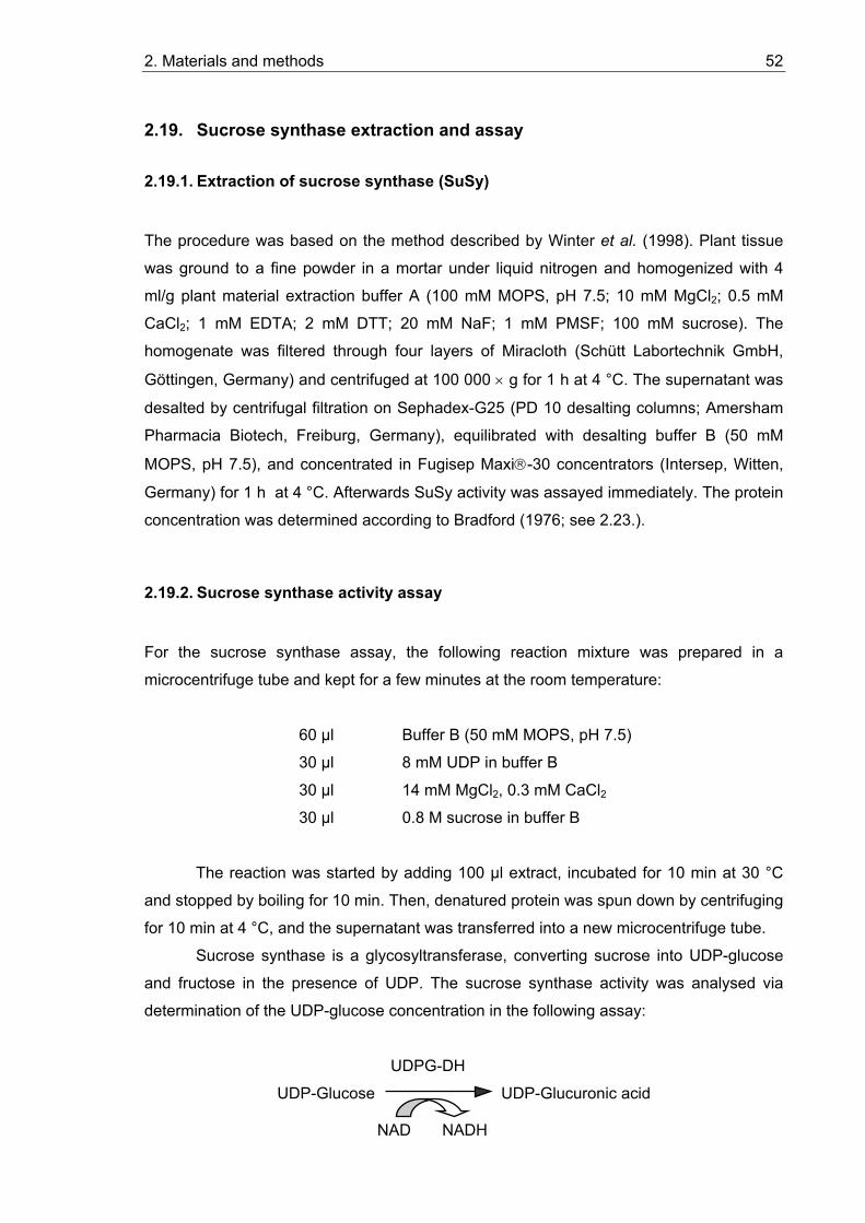
2. Materials and methods 52
2.19. Sucrose synthase extraction and assay
2.19.1. Extraction of sucrose synthase (SuSy)
The procedure was based on the method described by Winter et al. (1998). Plant tissue
was ground to a fine powder in a mortar under liquid nitrogen and homogenized with 4
ml/g plant material extraction buffer A (100 mM MOPS, pH 7.5; 10 mM MgCl2; 0.5 mM
CaCl2; 1 mM EDTA; 2 mM DTT; 20 mM NaF; 1 mM PMSF; 100 mM sucrose). The
homogenate was filtered through four layers of Miracloth (Schütt Labortechnik GmbH,
Göttingen, Germany) and centrifuged at 100 000 × g for 1 h at 4 °C. The supernatant was
desalted by centrifugal filtration on Sephadex-G25 (PD 10 desalting columns; Amersham
Pharmacia Biotech, Freiburg, Germany), equilibrated with desalting buffer B (50 mM
MOPS, pH 7.5), and concentrated in Fugisep Maxi-30 concentrators (Intersep, Witten,
Germany) for 1 h at 4 °C. Afterwards SuSy activity was assayed immediately. The protein
concentration was determined according to Bradford (1976; see 2.23.).
2.19.2. Sucrose synthase activity assay
For the sucrose synthase assay, the following reaction mixture was prepared in a
microcentrifuge tube and kept for a few minutes at the room temperature:
60 µl Buffer B (50 mM MOPS, pH 7.5)
30 µl 8 mM UDP in buffer B
30 µl 14 mM MgCl2, 0.3 mM CaCl2
30 µl 0.8 M sucrose in buffer B
The reaction was started by adding 100 µl extract, incubated for 10 min at 30 °C
and stopped by boiling for 10 min. Then, denatured protein was spun down by centrifuging
for 10 min at 4 °C, and the supernatant was transferred into a new microcentrifuge tube.
Sucrose synthase is a glycosyltransferase, converting sucrose into UDP-glucose
and fructose in the presence of UDP. The sucrose synthase activity was analysed via
determination of the UDP-glucose concentration in the following assay:
UDPG-DH
UDP-Glucose UDP-Glucuronic acid
NAD NADH
2. Materials and methods 53
For the spectrophotometric assay of UDP-glucose, 900 µl of assay buffer (200 mM
glycine, pH 8.9; 5 mM MgCl2), 20 µl 0.1 M NAD+ and 100 µl of sample were mixed in a
cuvette. The reaction was started by adding 10 mU UDP-Glucose-Dehydrogenase
(Sigma). The measurements were conducted on a two wavelength spectrophotometer
(Sigma ZFP 22; Sigma instrumente GmbH, Berlin, Germany). The activity was calculated
as the increase in absorbance due to the formation of NADPH using a molar extinction
coefficient for NADPH of 6.18 mmol-1cm-1 at 334 nm.
2.20. Invertase extraction and assay
2.20.1. Extraction of invertase
Plant tissue was ground to a fine powder in liquid nitrogen and homogenized with
homogenization buffer (2 ml/g fresh weight (FW) tissue). PVPP (200 mg/g FW) was added
to D. glomerata and C. glauca powder to bind polyphenols and polysaccharides. The
homogenate was centrifuged at 6000 × g for 10 min at 4 °C. The supernatant (cytosolic
and vacuolar fraction) represented the soluble enzyme preparation. The pellet (cell-wall
fraction) was washed three times with dd H2O at 4 °C and finally resuspended again in dd
H2O. Both enzyme preparations were dialyzed against 12.5 mM K-Phosphate buffer (pH
7.4 adjusted with KH2PO4) for 45 min twice and then overnight at 4 °C. Afterwards the
probes were collected into Eppendorf tubes and assayed for invertase activity (see 2.20.2.) or stored at – 80 °C before analysis.
Homogenisation buffer: for 40 ml:
200 mM HEPES/NaOH, pH 7.5 16 ml 0.5 M stock 3 mM DTT 240 µl 0.5 M stock 3 mM MgCl2 120 µl 1 M stock 1 mM EDTA 80 µl 0.5 M stock 2 % glycerol 920 µl 87 % stock
10 ml aliquots of the homogenization buffer were kept at –20 °C. PMSF (Merck,
Darmstadt, Germany), dissolved in ethanol, and benzamidine (Sigma-Aldrich,
Taufkirchen, Germany), dissolved in dd H2O, were added to the homogenisation buffer to
the final concentrations of 0.1 mM and 1 mM, respectively, directly before use.
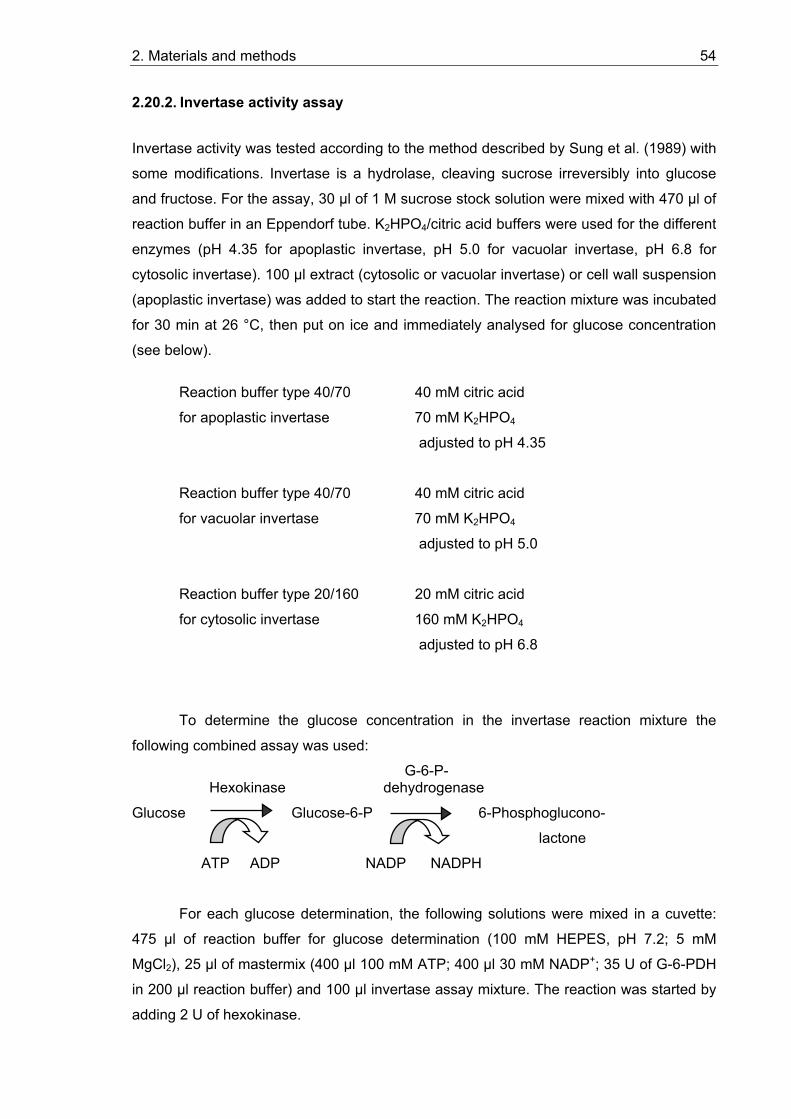
2. Materials and methods 54
2.20.2. Invertase activity assay
Invertase activity was tested according to the method described by Sung et al. (1989) with
some modifications. Invertase is a hydrolase, cleaving sucrose irreversibly into glucose
and fructose. For the assay, 30 µl of 1 M sucrose stock solution were mixed with 470 µl of
reaction buffer in an Eppendorf tube. K2HPO4/citric acid buffers were used for the different
enzymes (pH 4.35 for apoplastic invertase, pH 5.0 for vacuolar invertase, pH 6.8 for
cytosolic invertase). 100 µl extract (cytosolic or vacuolar invertase) or cell wall suspension
(apoplastic invertase) was added to start the reaction. The reaction mixture was incubated
for 30 min at 26 °C, then put on ice and immediately analysed for glucose concentration
(see below).
Reaction buffer type 40/70 40 mM citric acid
for apoplastic invertase 70 mM K2HPO4
adjusted to pH 4.35
Reaction buffer type 40/70 40 mM citric acid
for vacuolar invertase 70 mM K2HPO4
adjusted to pH 5.0
Reaction buffer type 20/160 20 mM citric acid
for cytosolic invertase 160 mM K2HPO4
adjusted to pH 6.8
To determine the glucose concentration in the invertase reaction mixture the
following combined assay was used: G-6-P- Hexokinase dehydrogenase
Glucose Glucose-6-P 6-Phosphoglucono-
lactone
ATP ADP NADP NADPH
For each glucose determination, the following solutions were mixed in a cuvette:
475 µl of reaction buffer for glucose determination (100 mM HEPES, pH 7.2; 5 mM
MgCl2), 25 µl of mastermix (400 µl 100 mM ATP; 400 µl 30 mM NADP+; 35 U of G-6-PDH
in 200 µl reaction buffer) and 100 µl invertase assay mixture. The reaction was started by
adding 2 U of hexokinase.
2. Materials and methods 55
The concentration of NADPH formed was detected photometrically on a double-
beam photometer Uvikon 932 (Kontron, Milan, Italy). The glucose concentration was
calculated as the increase in absorbance due to the equivalent formation of NADPH using
a molar extinction coefficient for NADPH of 6.22 mmol-1cm-1 at 340 nm. The reaction was
considered complete when there was no more increase of the OD340.
The amount of glucose formed in the invertase reaction is equivalent to the amount
of sucrose cleaved in this reaction. Therefore, results of invertase activity assay are
presented as nmol sucrose cleaved per milligram protein per minute. Protein content in
extracts was determined according to the method of Bradford (1976, see 2.23.). BSA was
used for the calibration curve.
2.21. In situ glucose and acid (apoplastic) invertase activity staining
Glucose can be detected via the formation of a coloured reaction product by glucose
oxidase and peroxidase in the presence of diaminobenzidine (DAB). About 200 µm thick
tissue slices were hand cut. For glucose staining the slices were denatured at 65 °C in dd
H2O for 3 -10 min and transferred to reaction medium. To stain the sites of invertase
activity the slices were washed extensively in dd H2O to remove endogenous glucose and
then transferred into reaction medium, supplied with 100 mM sucrose. The reaction was
carried out for 3 - 4 hours at 37 °C (or overnight at room temperature).
After the incubation, the slices were washed with dd H2O and then photographed
under a microscope. Brown precipitate indicates the presence of glucose or acid
(apoplastic) invertase activity, respectively.
Reaction medium: 200 mM Tris/citrate pH adjusted to 6.0 with a citric acid 20 µg/ml glucose oxidase (Sigma Chemical, St. Louis, MO,
USA) 250 µg/ml horseradish peroxidase (Sigma) 300 µg/ml diaminobenzidine (DAB) (Sigma)
2.22. Total protein isolation from plant material
Plant tissue (50 -100 mg) was ground to a fine powder in a mortar under liquid nitrogen,
transferred into a 1.5 ml Eppendorf tube and immediately supplied with 200 µl extraction
buffer (see below). An identical volume of TE-saturated phenol was added to the sample,
vortexed and incubated on ice for at least 5 minutes. For phase separation, the
2. Materials and methods 56
homogenate was centrifuged for 10 min at 10 000 × g at 4 °C. The phenol phase (upper
phase) was transferred into a new Eppendorf tube. Protein precipitation was achieved by
adding five volumes of 100 mM ammonium acetate (w/v) in methanol and incubating for
16 h at – 20 °C. After a centrifugation step at 10 000 × g for 10 min at 4 °C the
supernatant was discarded and the pellet was washed with 80 % acetone (v/v) and dried
under vacuum in a Speed Vac Concentrator (Bachofer, Reutlingen, Germany). The pellet
was dissolved in a small volume (20-50 µl, depending on protein quantity) of denaturing
buffer and incubated for 1 min at 100 °C. Then protein concentration of the sample was
determined according to Bradford (see 2.23.). Protein samples were stored at – 20 °C.
Extraction buffer 700 mM sucrose
500 mM Tris-HCl
100 mM KCl
50 mM EDTA
pH 9.5
2 % (v/v) β-mercaptoethanol, freshly added
Denaturing buffer 10 mM Tris-HCl
2 % (w/v) SDS
pH 9.5
2 % (v/v) β-mercaptoethanol, freshly added
2.23. Bradford protein concentration determination
The Bradford test (Bradford, 1976) is a colorimetric method to quantify the protein content
of solutions. The amount of Coomassie Brilliant Blue G 250 (Serva, Heidelberg, Germany) binding to protein is determined. The staining develops within 5 -10 min. Then the protein
complex is stable for about 30 min. By comparison with a calibration curve obtained with a
standard protein (fatless BSA), the protein concentration in a sample can be estimated.
Every experiment requires a new calibration curve.
5 µl of the sample was filled up with dd H2O to a total volume of 500 µl. Then 500
µl staining solution was added to the sample, mixed and incubated for 10 min at room
temperature. The OD595 was measured in a spectrophotometer (Uvikon 932; Kontron,
Milan, Italy). As a reference, 5 µl of sample buffer was taken and prepared as described
for the protein sample.
2. Materials and methods 57
Staining solution: 70 mg/50 ml abs. EtOH Coomassie Brilliant Blue G-250
600 ml H2O
100 ml 85% H3PO4
volume was adjusted to 1000 ml with dd H2O
and the solution was filtered through 3MM paper
2.24. Sodiumdodecyl sulfate polyacrylamide gel electrophoresis (SDS-PAGE)
The SDS-PAGE method is based on the method of Laemmli (1970). SDS is an anionic
detergent that has two effects on proteins: first, it leads to disbanding of oligomeric
structures, resulting in a relaxed conformation and, second, it binds in excess in a
constant relation (1.4 g SDS/1 g protein) to the protein, which leads to an equal negative
charge per mass unit. Therefore, in the presence of SDS, proteins can be separated
based on their molecular weight.
The negatively charged SDS-protein complexes run towards the anode in an
electrical field. To separate these complexes, the molecular-sieve properties of
polyacrylamide gel (PAA-Gel) are used, where the mobility of most proteins is reversely
proportional to the decimal logarithm of their molecular weight. The discontinuous buffer
system of Laemmli (1970) leads to the concentration of probes on the transition state
between a stacking gel and a separation gel, resulting in a higher resolution of the
following separation of proteins. To determine the molecular weight of unknown proteins,
a marker containing some proteins with molecular weights of 14-97 kDa („Low Molecular
Weight Calibration Kit“; Amersham Pharmacia, Freiburg, Germany) or 9 to 185 kDa
(“Bench Mark Prestained Protein Ladder”, Invitrogen, Groningen, Netherlands) was
loaded on the same gel. The pore size of a PAA-gel depends on relation between the
concentrations of acrylamide to cross-linking N-N´-methylenbisacrylamide. The
acrylamide quota and the cross-linking degree are the values that characterize a
polyacrylamide gel. To separate total protein extracts and detect sucrose synthase
(SuSy), consisting of 92- to 93-kDa subunits (Hohnjec et al., 1999), a PAA-gel containing
6 or 7.5 % polyacrylamide in the separating gel and 4 % acrylamide in the stacking gel
were prepared. The separating range was 60 to 200 kDa in 6 % and 40 to 200 kDa in 7.5
% PAA-gel. Gel plates, spacer and combs as well as gel camera were ordered from
Biometra (Göttingen, Germany).

2. Materials and methods 58
2.24.1. Buffers and solutions for analytic SDS-PAGE
Acrylamide / Bisacrylamide 29.2 % (w/v) acrylamide
(30 % T/2.7 % C) stock solution (30%) 0.8 % (w/v) N,N´-methylene bisacrylamide
Dissolve in 100 ml dd H2O and filter through
3 MM paper
Store at 4-10 oC
4 × Stacking gel buffer 500 mM Tris-HCl pH 6.8
0.4 % (w/v) SDS
4 × Separating gel buffer 1.5 M Tris-HCl pH 8.8
0.4 % (w/v) SDS
10 % Ammonium persulfate 10 % (w/v) Ammonium persulfate (in dd
H2O) (APS) solution stored at 10 oC
10 × SDS Electrophoresis buffer 250 mM Tris
1.92 M Glycine
1 % (w/v) SDS
pH 8.2-8.4 (not adjusted)
5 × Sample buffer 60 mM Tris-HCl (pH 6.8)
2 % (v/v) β-Mercaptoethanol
25 % (v/v) Glycerol
2 % (w/v) SDS
0.1 % (v/v) Bromphenol blue
2.24.2. Pouring the SDS-Polyacrylamide gel
Glass gel plates were cleaned with acetone or ethanol, silicon spacer tubes and combs
were cleaned with water. The glass plates were joined together to form the cassette and
clamped in vertical position. Solutions for stacking and separating gel were mixed as
described in the table below. To start the polymerisation, TEMED (N,N,N‘,N‘-
Tetramethylene diamine) solution was added to the separating gel solution. The gel
solution was mixed and poured into the gel chamber up to 1 cm under the ends of the
2. Materials and methods 59
comb slots. To ensure that the gel polymerized with a smooth surface, a 2 mm layer of dd
H2O was carefully poured on the gel using a Pasteur pipette. Because of the great
difference in density between dd H2O and the gel solution, the water is spread across the
surface of the gel without much mixing. When the gel was set, a clear refractive index
change could be seen between the polymerised gel and the overlaying water. Then the
overlaying water was poured off and TEMED was added to the stacking gel solution to
start polymerisation. The stacking gel was poured into the gel cassette until the solution
reached the upper edge and the slot-forming comb was put into the solution before
leaving the gel to set. 30 min to 1 hour later, the comb was removed from the gel, and the
gel slots were rinsed with electrophoresis buffer to remove any rests of non-polymerised
acrylamide solution.
The data of the following table are based on the stock solutions and buffers given
in 2.24.1. The volume is sufficient for two mini gels (8.5 cm × 7.5 cm).
Separating gel, 7.5 % (15 ml): Acrylamide stock solution (30 %) 3.750 ml
4 × Separating gel buffer 3.750 ml
dd H2O 7.410 ml
10 % APS solution 75 µl
TEMED (add before pouring the gel) 15 µl
Stacking gel, 4 % (5 ml): Acrylamide stock solution (30 %) 0.670 ml
4 × Stacking gel buffer 1.250 ml
dd H2O 3.050 ml
10 % APS solution 30 µl
TEMED (add before pouring the gel) 5 µl
The resulting end concentrations were:
Stacking gel Separating gel
Tris/HCl pH 6.8 0.13 M -
Tris/HCl pH 8.8 - 0.38 M
SDS 0.1 % (w/v) 0.1 % (w/v) Acrylamide 4.6 % (w/v) 7.5 % (w/v) N,N-Methylene bisacrylamide 0.12 % (w/v) 0.33 % (w/v)
APS 0.06 % (w/v) 0.05 % (w/v)
2. Materials and methods 60
2.24.3. Probe preparation and SDS polyacrylamide gel electrophoresis (SDS-PAGE)
Ca. 15 µg protein per slot were used for immunoblotting and 10-30 µg protein for
Coomassie stained gels.
The samples were supplemented with sample buffer to a final concentration of ≥
1× sample buffer to ensure proper solubilisation of the proteins and solubilised for 15 min
at RT by sucking the solution up and down with a pipette. Then the samples were boiled
at 95 °C for 3 min and centrifuged afterwards at top speed (13 000 rpm) for 5 min in a
table top microcentrifuge to sediment insoluble material. After removing the spacers, the
gel cassette was assembled with the electrophoresis unit, and first the top and then the
bottom reservoir were filled with 1× SDS electrophoresis buffer (see 2.24.1.). Then the
appropriate volume of sample was filled up to 10 µl with sample buffer (see 2.24.1.) and
loaded into the SDS gel slot using a microsyringe. Every empty slot on the gel was filled in
with 10 µl sample buffer. The electrophoresis was run under a constant current of 25 mA
(500 V, 100 W) and was continued until the Bromphenol blue reached the bottom of the
gel (about 1.5 hours). Then the gel cassette was removed from the electrophoresis unit
and opened, and the gel was taken out to stain in Coomassie or to mount a Western blot.
2.24.4. Coomassie staining of SDS polyacrylamide gels
Coomassie staining serves to detect proteins in an SDS polyacrylamide gel. For this
purpose, the gel was fixed after electrophoresis and stained in staining solution (see
below) for at least 30 min and then destained in wash solution for about 1-2 hours until the
gel background was colourless. After washing, the gel could be stored for a long time in
dd H2O. Alternatively, it could first be soaked in 10 % glycerol for 30 min and then dried
under vacuum.
Coomassie staining solution 0.25 % (w/v) Coomassie Brilliant Blue R 250
40 % (v/v) Methanol
10 % (v/v) Acetic acid
Washing solution 50 % (v/v) Methanol
10 % (v/v) Acetic acid
2. Materials and methods 61
2.25. Western blot
With Western blotting it is possible to identify and quantify proteins in a total protein
extract by use of specific antibodies.
2.25.1. Protein transfer to a nitrocellulose membrane
After the separation of protein probes by SDS-PAGE (see 2.24.3.) the gel was fixed in a
Western blot camera. The mounting order of the blot was the following (from anode to
kathode):
- three layers of blotting paper (3MM), wetted in blotting buffer
- SDS gel, equilibrated in blotting buffer for 15 min
- nitrocellulose membrane (Hybond ECL Nitrocellulose Membrane; Amersham
Pharmacia Biotech, Freiburg, Germany), wetted in blotting buffer, cut
corresponding to gel size.
- three layers of wet 3MM blotting paper
Protein transfer was performed overnight at 4 oC in the Western blot chamber filled
with blotting buffer at a constant current of 90 mA.
Blotting buffer: Tris 25 mM
Glycine 192 mM
Methanol 20 % (v/v)
2.25.2. Staining of the blot
The nitrocellulose membrane was taken out of the blotting system and stained for two
minutes in Ponceau red solution to check the protein transfer. The staining is reversible,
so after washing with dd H2O the bands of marker proteins and the sample order were
marked with a pencil on the protein side of the membrane. Then, the blot was cut off to
the required size.
Ponceau red solution: Ponceau S 2 % (w/v)
Trichloroacetic acid 3 % (w/v)
2. Materials and methods 62
2.25.3. Immunological detection of transferred proteins
Specific proteins were identified on the nitrocellulose membrane by the indirect
immunological method. First a non-labelled primary antibody binds to the antigen. In the
second step, this antigen-antibody complex is bound by the secondary antibody which
represents the bacterial protein A that binds immunoglobulins of many species. The
secondary antibody is conjugated with an enzyme, horseradish peroxidase. This allows
visualising the labelled proteins after incubation with a horseradish peroxidase substrate,
which is converted into a coloured or luminescent product.
Reaction with antibodies
To reduce the unspecific binding capacity of nitrocellulose, the membrane was
incubated with the primary antibodies in an appropriate dilution (1:1000 to 1:5000) in the
presence of Block buffer for 2 hours at room temperature on a swinging shaker. Then the
blot was washed once shortly and three times for 15 min with TBS-T to remove
unspecifically bound IgG molecules. Then followed an incubation with secondary
antibodies conjugated with peroxidase (1:3000 to 1:10000 in TBS-T) “Anti Rabbit-Protein
A Horseradish Peroxidase Conjugate”; Bio-Rad, München, Germany) for 1 h at room
temperature. Afterwards the wash steps with TBS-T were repeated: shortly once and
three times for 15 min and finally one 15 min wash step with TBS.
TBS: Tris/HCl pH 7.5 50 mM
NaCl 150 mM
TBS-T: Triton-X-100 0.1 % (v/v)
in TBS
Block buffer: Dry milk powder 1 % (w/v)
in TBS-T
Peroxidase detection by “Enhanced Chemiluminiscence”
The method of “Enhanced Chemiluminiscence” (ECL) serves as a sensitive
detection of a protein A-coupled peroxidase. Under alkaline conditions, peroxidase
catalyses the oxidation of luminol by H2O2, which results in the emission of light. This
reaction is enhanced by phenol. The half time of this reaction is about 1 h.
The nitrocellulose membrane was put flat, without air bubbles, on a thin plastic foil
with the protein side up. 2 ml ECL-Solution 1 and 2 ml ECL-solution 2 (Amersham
2. Materials and methods 63
Pharmacia Biotech, Freiburg, Germany) were mixed, poured on the membrane and
incubated for 1 min under swinging. After the reaction, the liquid was removed and the
membrane was covered by plastic foil without air bubbles and immediately exposed to
Hyperfilm ECL (Amersham Pharmacia Biotech) for 1 min in the dark. Another exposition
for 10 min was performed and based on the results a third exposition was performed for
the optimal time period (10 sec, 30 sec or 1 hour). After taking off, the film was developed
in Kodak Develop and Fix Solutions (Kodak, Rochester, NY, USA).
2.26. Measurement of sugar uptake in yeast
Yeast cells transformed with a pNEV-based expression construct with the putative hexose
transporter (2.14.) were grown overnight in the appropriate selection medium (2.1.8.3.) up
to an OD600 of 1. Then the cells were harvested by centrifugation for 5 min at room
temperature in a Megafuge 1.0 at 4500 rpm, and the pellet was washed twice with 50 mM
Na-Pi-buffer pH 5.5 (for determination of the pH optimum, buffers with pH in the range of
3.0 – 7.3 had been tested) with centrifugation steps as described above. Finally, the cells
were resuspended in the same buffer to an OD600 of 10. Before sugar uptake
measurements, the cell suspension was stored on ice for a maximum of 10 hours.
For a series of measurements, 1 ml of cell suspension was pre-incubated in a 25
ml Erlenmeyer flask for 1-2 min at 30 °C under vigorous shaking. Then 11 µl of 10 mM
[14C]-labelled substrate was added to a final substrate concentration of 100 µmol in the
mixture to start the sugar uptake. The labelled substrate solution was prepared by mixing
10 µl 10 mM of non-radioactive (“cold”) substrate and 1 µl of [14C]-labelled substrate (for
example, ca. 0.2 µCi [14C]-glucose from Amersham Pharmacia Biotech, Little Chalfont,
UK).
Samples of 100 µl were taken at given intervals after adding the substrate (10 sec,
30 sec, 1, 2, 3, 5, 10 min), filtered on nitrocellulose filters (0.8 µm pore size; Hölzer,
Dorfen, Germany) and washed with an excess of dd H2O. Incorporation of radioactivity
was determined by scintillation counting. For every measurement series, 100 µl of the
total sample was counted to obtain the value for overall radioactivity. To calculate the
concentration of substrate accumulated in yeast cells, the cell volume (in µl of packed
cells, p.c.) was determined using Hematocrite tubes for the centrifugation of 1 ml cell
sample for 5 min at 3000 × g. 30 % of the total volume was taken up by the intracellular
volume.
2. Materials and methods 64
For analyzing energization, 10 µl of absolute ethanol were added after 1 min of
pre-incubation, and incubated for one min before the uptake test was started. To analyze
the influence of inhibitors on the uptake of glucose, the plasma membrane uncouplers CCCP (carbonyl cyanide m-chlorophenylhydrazone) and DNP (2,4-Dinitrophenol) were
added to the sample to a final concentration of 50 µM before the uptake reaction was
started.
2.27. Statistic evaluation of experimental data
The results shown represent either the typical results of the all experimental series
conducted or the summary results. In the last case the arithmetic average value ± the
standard deviation (SD) are performed. The number of the conducted experiments was at
least 3, when it is not otherwise noted in the text.
Σ Arguments of single measurements Average value = Number of measurements (n)
SDx x
n
ii
n
=−
−=∑ ( )²
1
1
3. Results 65
3. Results
3.1. Sugar contents in roots and nodules
In most plants, sucrose is the main form of carbon transported in the phloem from source
to sink organs. After phloem unloading, it can reach the sink cells in the form of sucrose or
as its component hexoses, glucose and fructose (Sonnewald and Willmitzer, 1992). In
addition to supporting sink cell metabolism in roots, carbon allocated to the roots also
supports nodule formation and the growth of symbiotic microorganisms in root nodules. To
get an overview of the sugar metabolism in roots and root nodules, it was important first to
analyse the soluble sugars in roots and nodules of the three model systems: one legume
symbiosis (Medicago truncatula – Sinorhizobium meliloti) and two actinorhizal symbioses
from different Frankia subclades (see 1.5). Extracts of soluble sugars from roots and
nodules were obtained using the chloroform-methanol extraction (2.16.1). Sugar contents
were determined via HPLC (2.17).
3.1.1. Sugar contents in roots and nodules of Medicago truncatula
The main sugars detected in Medicago extracts were glucose and sucrose. The relative
amounts of these two sugars were similar in roots and nodules, with the sucrose
concentrations being about 2 times higher than the glucose concentrations (Figure 3.1).
The contents of fructose were below the detection limit. The only compound with
increased amounts in nodules compared to roots was a substance that had similar
retention time to sugar-alcohols in analysis by HPLC, but which could not be identified
with available standards (“unknown M1” in Figure 3.1). This compound might represent
pinitol (1-D-3-O-methyl-chiro-inositol), the major cyclitol of legumes, which is present in all
organs of the plant (Smith and Phillips, 1980) or D-ononitol (4-O-methyl-myo-inositol),
which has been described as a nodule-specific sugar alcohol for Pisum sativum (Skøt and
Egsgaard, 1984; Romanov et al., 1995). Another unknown sugar (“unknown M2” in Figure
3.1) was detected only in Medicago root extracts, not in nodule extracts.
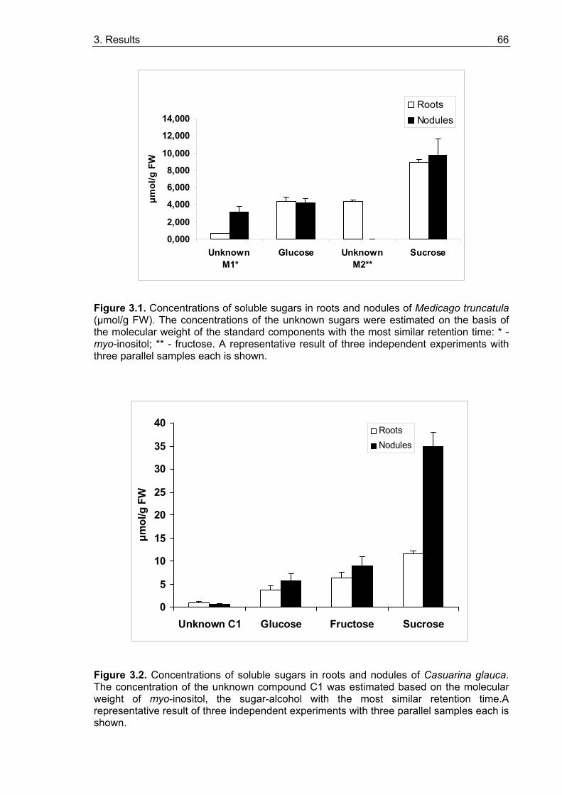
3. Results 66
0,000
2,000
4,000
6,000
8,000
10,000
12,000
14,000
Unknown M1*
Glucose UnknownM2**
Sucrose
µmol
/g F
W
RootsNodules
Figure 3.1. Concentrations of soluble sugars in roots and nodules of Medicago truncatula (µmol/g FW). The concentrations of the unknown sugars were estimated on the basis of the molecular weight of the standard components with the most similar retention time: * - myo-inositol; ** - fructose. A representative result of three independent experiments with three parallel samples each is shown.
0
5
10
15
20
25
30
35
40
Unknown C1 Glucose Fructose Sucrose
µmol
/g F
W
RootsNodules
Figure 3.2. Concentrations of soluble sugars in roots and nodules of Casuarina glauca. The concentration of the unknown compound C1 was estimated based on the molecular weight of myo-inositol, the sugar-alcohol with the most similar retention time.A representative result of three independent experiments with three parallel samples each is shown.
3. Results 67
3.1.2. Sugar contents in Casuarina glauca
In extracts of roots and nodules of Casuarina glauca, glucose, fructose, sucrose and small
amounts of sugar-alcohols were detected (Figure 3.2).
Concentrations of glucose and fructose were not significantly different in roots and
nodules. The main sugar compound in roots as well as in nodules of Casuarina was
sucrose. Moreover, sucrose contents were strongly increased in nodules compared to
roots, with a nodule sucrose concentration of about 35 µmol/g FW, compared to about 12
µmol/g FW in roots.
3.1.3. Sugar contents in Datisca glomerata
The sugar profile of Datisca glomerata (Figure 3.3) was very different from those of
Medicago and Casuarina. Altogether, the total sugar content was higher in Datisca
nodules compared to roots. In nodules, glucose and sucrose concentrations were similar
(about 4.5-4.8 µmol/g FW), whereas fructose concentrations were as high as 8 µmol/g
FW. As with Medicago nodules, two unknown compounds were found, the concentrations
of which were strongly increased in Datisca nodules compared to roots. One of these
substances was termed “M”, because its retention time in HPLC was most similar to that
of myo-inositol (Figure 3.3; Figure 3.4).
The other major unidentifiable component of Datisca soluble sugar extracts had a
retention time close to that of ribitol (Figure 3.3; Figure 3.4). Based on the molecular
weight of ribitol, the concentrations of the unknown sugar in roots and in nodules were
about 5 and 13.5 µmol/g FW, respectively. This compound was termed “D”, from Datisca.
3.1.3.1. Isolation and characterization of two unknown carbohydrates from Datisca glomerata
For identification of the two unknown compounds in soluble sugar extracts from Datisca,
large-scale sugar isolation was performed for D and M as described in 2.16.2, using
chloroform-methanol extraction, separation of sugar extract into fractions using anion
exchange column and sodium hydroxide as eluate and paper chromatography purification
from sodium chloride formed after neutralisation.
The detailed analysis of the chemical structure of D and M was conducted by
nuclear magnetic resonance spectroscopy (NMR) and mass spectroscopy, and the NMR
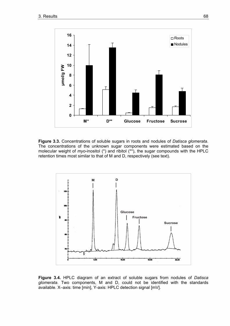
3. Results 68
0
2
4
6
8
10
12
14
16
M* D** Glucose Fructose Sucrose
µmol
/g F
W
RootsNodules
Figure 3.3. Concentrations of soluble sugars in roots and nodules of Datisca glomerata. The concentrations of the unknown sugar components were estimated based on the molecular weight of myo-inositol (*) and ribitol (**), the sugar compounds with the HPLC retention times most similar to that of M and D, respectively (see text).
M
GlucoseFructose
Sucrose
Figure 3.4. HPLC diagram of an extract of soluble sugars from nodules of Datisca glomerata. Two components, M and D, could not be identified with the standards available. X–axis: time [min], Y-axis: HPLC detection signal [mV].
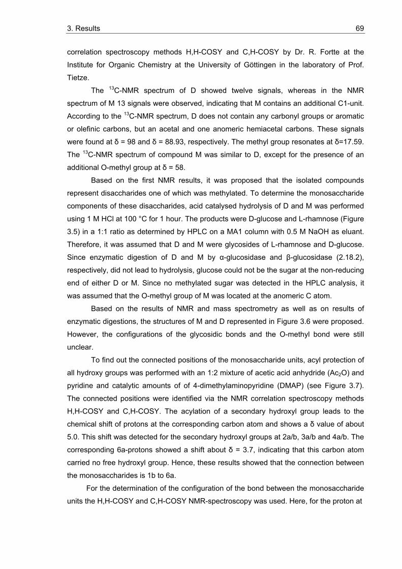
3. Results 69
correlation spectroscopy methods H,H-COSY and C,H-COSY by Dr. R. Fortte at the
Institute for Organic Chemistry at the University of Göttingen in the laboratory of Prof.
Tietze.
The 13C-NMR spectrum of D showed twelve signals, whereas in the NMR
spectrum of M 13 signals were observed, indicating that M contains an additional C1-unit.
According to the 13C-NMR spectrum, D does not contain any carbonyl groups or aromatic
or olefinic carbons, but an acetal and one anomeric hemiacetal carbons. These signals
were found at δ = 98 and δ = 88.93, respectively. The methyl group resonates at δ=17.59.
The 13C-NMR spectrum of compound M was similar to D, except for the presence of an
additional O-methyl group at δ = 58.
Based on the first NMR results, it was proposed that the isolated compounds
represent disaccharides one of which was methylated. To determine the monosaccharide
components of these disaccharides, acid catalysed hydrolysis of D and M was performed
using 1 M HCl at 100 °C for 1 hour. The products were D-glucose and L-rhamnose (Figure
3.5) in a 1:1 ratio as determined by HPLC on a MA1 column with 0.5 M NaOH as eluant.
Therefore, it was assumed that D and M were glycosides of L-rhamnose and D-glucose.
Since enzymatic digestion of D and M by α-glucosidase and β-glucosidase (2.18.2),
respectively, did not lead to hydrolysis, glucose could not be the sugar at the non-reducing
end of either D or M. Since no methylated sugar was detected in the HPLC analysis, it
was assumed that the O-methyl group of M was located at the anomeric C atom.
Based on the results of NMR and mass spectrometry as well as on results of
enzymatic digestions, the structures of M and D represented in Figure 3.6 were proposed.
However, the configurations of the glycosidic bonds and the O-methyl bond were still
unclear.
To find out the connected positions of the monosaccharide units, acyl protection of
all hydroxy groups was performed with an 1:2 mixture of acetic acid anhydride (Ac2O) and
pyridine and catalytic amounts of of 4-dimethylaminopyridine (DMAP) (see Figure 3.7).
The connected positions were identified via the NMR correlation spectroscopy methods
H,H-COSY and C,H-COSY. The acylation of a secondary hydroxyl group leads to the
chemical shift of protons at the corresponding carbon atom and shows a δ value of about
5.0. This shift was detected for the secondary hydroxyl groups at 2a/b, 3a/b and 4a/b. The
corresponding 6a-protons showed a shift about δ = 3.7, indicating that this carbon atom
carried no free hydroxyl group. Hence, these results showed that the connection between
the monosaccharides is 1b to 6a.
For the determination of the configuration of the bond between the monosaccharide
units the H,H-COSY and C,H-COSY NMR-spectroscopy was used. Here, for the proton at
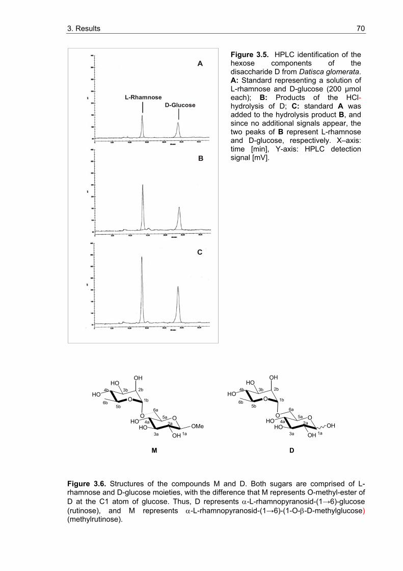
3. Results 70
D-GlucoseL-Rhamnose
A
B
C
O OHOHO
OHOMe
M D
O
HO
HO
OH
1a
2a
3a
4a5a
6a
1b
2b3b4b
5b6b
O OHOHO
OHOH
O
HO
HO
OH
1a
2a
3a
4a5a
6a
1b
2b3b4b
5b6b
Figure 3.6. Structures of the compounds M and D. Both sugars are comprised of L-rhamnose and D-glucose moieties, with the difference that M represents O-methyl-ester of D at the C1 atom of glucose. Thus, D represents α-L-rhamnopyranosid-(1→6)-glucose (rutinose), and M represents α-L-rhamnopyranosid-(1→6)-(1-O-β-D-methylglucose) (methylrutinose).
Figure 3.5. HPLC identification of the hexose components of the disaccharide D from Datisca glomerata. A: Standard representing a solution of L-rhamnose and D-glucose (200 µmol each); B: Products of the HCl-hydrolysis of D; C: standard A was added to the hydrolysis product B, and since no additional signals appear, the two peaks of B represent L-rhamnose and D-glucose, respectively. X–axis: time [min], Y-axis: HPLC detection signal [mV].
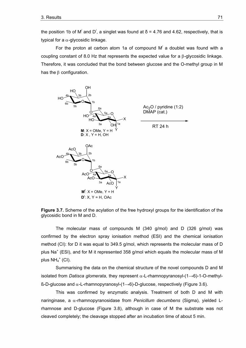
3. Results 71
the position 1b of M/ and D/, a singlet was found at δ = 4.76 and 4.62, respectively, that is
typical for a α-glycosidic linkage.
For the proton at carbon atom 1a of compound M/ a doublet was found with a
coupling constant of 8.0 Hz that represents the expected value for a β-glycosidic linkage.
Therefore, it was concluded that the bond between glucose and the O-methyl group in M
has the β configuration.
M: X = OMe, Y = HD: X , Y = H, OH
Ac2O / pyridine (1:2)DMAP (cat.)rt 24 h
M/: X = OMe, Y = HD/: X , Y = H, OH
O OHOHO
OHX
O
HO
HO
OH
1a
2a
3a
4a5a
6a
1b
2b3b4b
5b6b
O OAcOAcO
AcOX
O
AcO
AcO
OAc
1a
2a
3a
4a5a
6a
1b
2b3b4b
5b6b
Y
Y
Figure 3.7. Scheme of the acylation of the free hydroxyl groups for the identification of the glycosidic bond in M and D.
The molecular mass of compounds M (340 g/mol) and D (326 g/mol) was
confirmed by the electron spray ionisation method (ESI) and the chemical ionisation
method (CI): for D it was equal to 349.5 g/mol, which represents the molecular mass of D
plus Na+ (ESI), and for M it represented 358 g/mol which equals the molecular mass of M
plus NH4+ (CI).
Summarising the data on the chemical structure of the novel compounds D and M
isolated from Datisca glomerata, they represent α-L-rhamnopyranosyl-(1→6)-1-O-methyl-
ß-D-glucose and α-L-rhamnopyranosyl-(1→6)-D-glucose, respectively (Figure 3.6).
This was confirmed by enzymatic analysis. Treatment of both D and M with
naringinase, a α-rhamnopyranosidase from Penicillum decumbens (Sigma), yielded L-
rhamnose and D-glucose (Figure 3.8), although in case of M the substrate was not
cleaved completely; the cleavage stopped after an incubation time of about 5 min.
RT 24 h
D/: X, Y = H, OAc
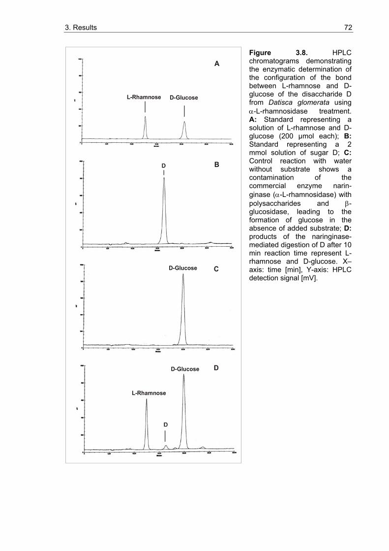
3. Results 72
L-Rhamnose D-Glucose
A
B
CD-Glucose
D-Glucose
L-Rhamnose
Figure 3.8. HPLC chromatograms demonstrating the enzymatic determination of the configuration of the bond between L-rhamnose and D-glucose of the disaccharide D from Datisca glomerata using α-L-rhamnosidase treatment. A: Standard representing a solution of L-rhamnose and D-glucose (200 µmol each); B:Standard representing a 2 mmol solution of sugar D; C:Control reaction with water without substrate shows a contamination of the commercial enzyme narin-ginase (α-L-rhamnosidase) with polysaccharides and β-glucosidase, leading to the formation of glucose in the absence of added substrate; D:products of the naringinase-mediated digestion of D after 10 min reaction time represent L-rhamnose and D-glucose. X–axis: time [min], Y-axis: HPLC detection signal [mV].
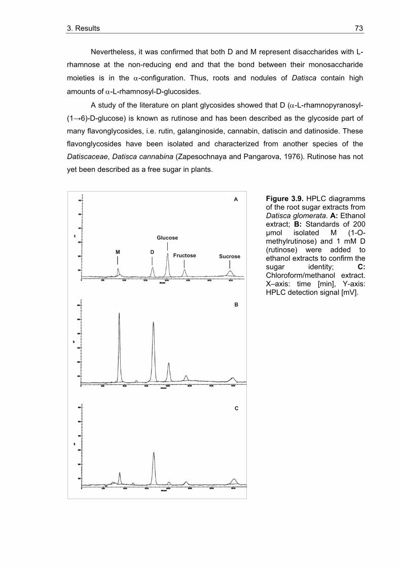
3. Results 73
Nevertheless, it was confirmed that both D and M represent disaccharides with L-
rhamnose at the non-reducing end and that the bond between their monosaccharide
moieties is in the α-configuration. Thus, roots and nodules of Datisca contain high
amounts of α-L-rhamnosyl-D-glucosides.
A study of the literature on plant glycosides showed that D (α-L-rhamnopyranosyl-
(1→6)-D-glucose) is known as rutinose and has been described as the glycoside part of
many flavonglycosides, i.e. rutin, galanginoside, cannabin, datiscin and datinoside. These
flavonglycosides have been isolated and characterized from another species of the
Datiscaceae, Datisca cannabina (Zapesochnaya and Pangarova, 1976). Rutinose has not
yet been described as a free sugar in plants.
A
M
Glucose
Fructose Sucrose
B
C
Figure 3.9. HPLC diagramms of the root sugar extracts from Datisca glomerata. A: Ethanol extract; B: Standards of 200 µmol isolated M (1-O-methylrutinose) and 1 mM D (rutinose) were added to ethanol extracts to confirm the sugar identity; C:Chloroform/methanol extract. X–axis: time [min], Y-axis: HPLC detection signal [mV].
3. Results 74
Accordingly, M (α-L-rhamnopyranosyl-(1→6)-1-O-methyl-ß-D-glucose) could be
named 1-O-methylrutinose. Like rutinose, this sugar is not known among free plant
metabolites. To ensure that the methylation of the anomeric C-atom was no artefact of the
isolation method, namely the chloroform/methanol extraction (2.16.2), alternative methods
for total sugar extractions were applied to confirm the natural occurence of 1-O-
methylrutinose. HPLC analysis of Datisca root extracts obtained with ethanol, perchlorate
or acetone (2.16.3-2.16.5) confirmed the occurrence of 1-O-methylrutinose in total sugar
extracts independently of the presence of a potential methyl donor. The HPLC diagram of
the ethanol extract is shown in Figure 3.9, whereas the results for perchlorate and
acetone extractions are not shown.
Moreover, methylrutinose and ethylrutinose were chemically synthesised from
rutinose by Dr. R. Fortte at the Institute for Organic Chemistry at the University of
Göttingen in laboratory of Prof. Tietze. The comparison of the synthesised alkylated
products with rutinose and 1-O-methylrutinose isolated from Datisca via NMR and thin
layer chromatography (TCL) on silicagel plates showed that during chemical synthesis,
only α-methyl- and α-ethylglycosides of rutinose were formed, whereas the natural
component represents a β-methylglycoside (see above). Hence, it was confirmed that O-
methylrutinose does not represent a chemical artefact in Datisca sugar extracts.
3.1.3.2. Sugar contents in roots, nodules and leaves of Datisca after determination of the molecular mass of the novel sugars
Sugars were extracted with chloroform/methanol for HPLC analysis. The concentrations of
the two unknown components termed M and D in the total sugar extracts had previously
been estimated based on the molecular weight of myo-inositol and ribitol, respectively, as
components with the closest retention time (see 3.1.3). After the isolation and
determination of chemical structure and molecular mass of the unknown sugar
components M (1-O-methylrutinose) and D (rutinose) (see 3.1.3.1), new concentration
standards were prepared to estimate the real concentrations of M and D and to compare
them with concentrations of glucose, fructose and sucrose in the same extracts.
Additionally, leaf extracts were analysed for the presence of methylrutinose and rutinose
to find out whether these two sugars are only present in sink- or also in source organs.
The results are presented in Figure 3.10.
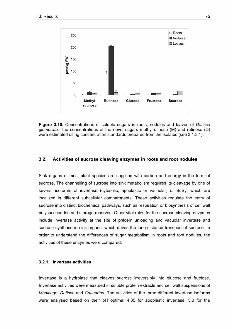
3. Results 75
0
50
100
150
200
250
Methyl-rutinose
Rutinose Glucose Fructose Sucrose
µmol
/g F
W
RootsNodulesLeaves
Figure 3.10. Concentrations of soluble sugars in roots, nodules and leaves of Datisca glomerata. The concentrations of the novel sugars methylrutinose (M) and rutinose (D) were estimated using concentration standards prepared from the isolates (see 3.1.3.1).
3.2. Activities of sucrose cleaving enzymes in roots and root nodules
Sink organs of most plant species are supplied with carbon and energy in the form of
sucrose. The channelling of sucrose into sink metabolism requires its cleavage by one of
several isoforms of invertase (cytosolic, apoplastic or vacuolar) or SuSy, which are
localized in different subcellular compartments. These activities regulate the entry of
sucrose into distinct biochemical pathways, such as respiration or biosynthesis of cell wall
polysaccharides and storage reserves. Other vital roles for the sucrose-cleaving enzymes
include invertase activity at the site of phloem unloading and vacuolar invertase and
sucrose synthase in sink organs, which drives the long-distance transport of sucrose. In
order to understand the differences of sugar metabolism in roots and root nodules, the
activities of these enzymes were compared.
3.2.1. Invertase activities
Invertase is a hydrolase that cleaves sucrose irreversibly into glucose and fructose.
Invertase activities were measured in soluble protein extracts and cell wall suspensions of
Medicago, Datisca and Casuarina. The activities of the three different invertase isoforms
were analysed based on their pH optima: 4.35 for apoplastic invertase; 5.0 for the
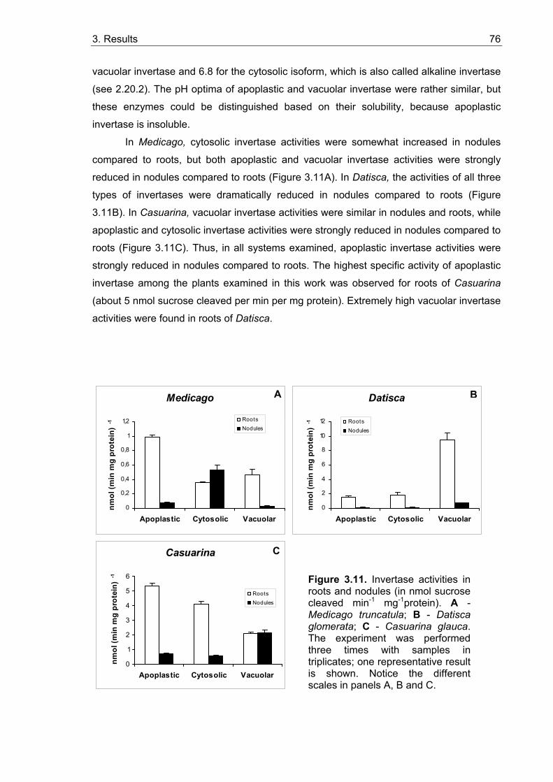
3. Results 76
vacuolar invertase and 6.8 for the cytosolic isoform, which is also called alkaline invertase
(see 2.20.2). The pH optima of apoplastic and vacuolar invertase were rather similar, but
these enzymes could be distinguished based on their solubility, because apoplastic
invertase is insoluble.
In Medicago, cytosolic invertase activities were somewhat increased in nodules
compared to roots, but both apoplastic and vacuolar invertase activities were strongly
reduced in nodules compared to roots (Figure 3.11A). In Datisca, the activities of all three
types of invertases were dramatically reduced in nodules compared to roots (Figure
3.11B). In Casuarina, vacuolar invertase activities were similar in nodules and roots, while
apoplastic and cytosolic invertase activities were strongly reduced in nodules compared to
roots (Figure 3.11C). Thus, in all systems examined, apoplastic invertase activities were
strongly reduced in nodules compared to roots. The highest specific activity of apoplastic
invertase among the plants examined in this work was observed for roots of Casuarina
(about 5 nmol sucrose cleaved per min per mg protein). Extremely high vacuolar invertase
activities were found in roots of Datisca.
Medicago
0
0,2
0,4
0,6
0,8
1
1,2
Apoplastic Cytosolic Vacuolar
nmol
(min
mg
prot
ein)
-1 Roots
Nodules
Datisca
0
2
4
6
8
10
12
Apoplastic Cytosolic Vacuolar
nmol
(min
mg
prot
ein)
-1 Roots
Nodules
Casuarina
0
1
2
3
4
5
6
Apoplastic Cytosolic Vacuolar
nmol
(min
mg
prot
ein)
-1
RootsNodules
A B
C
Figure 3.11. Invertase activities in roots and nodules (in nmol sucrose cleaved min-1 mg-1protein). A -Medicago truncatula; B - Datisca glomerata; C - Casuarina glauca. The experiment was performed three times with samples in triplicates; one representative result is shown. Notice the different scales in panels A, B and C.
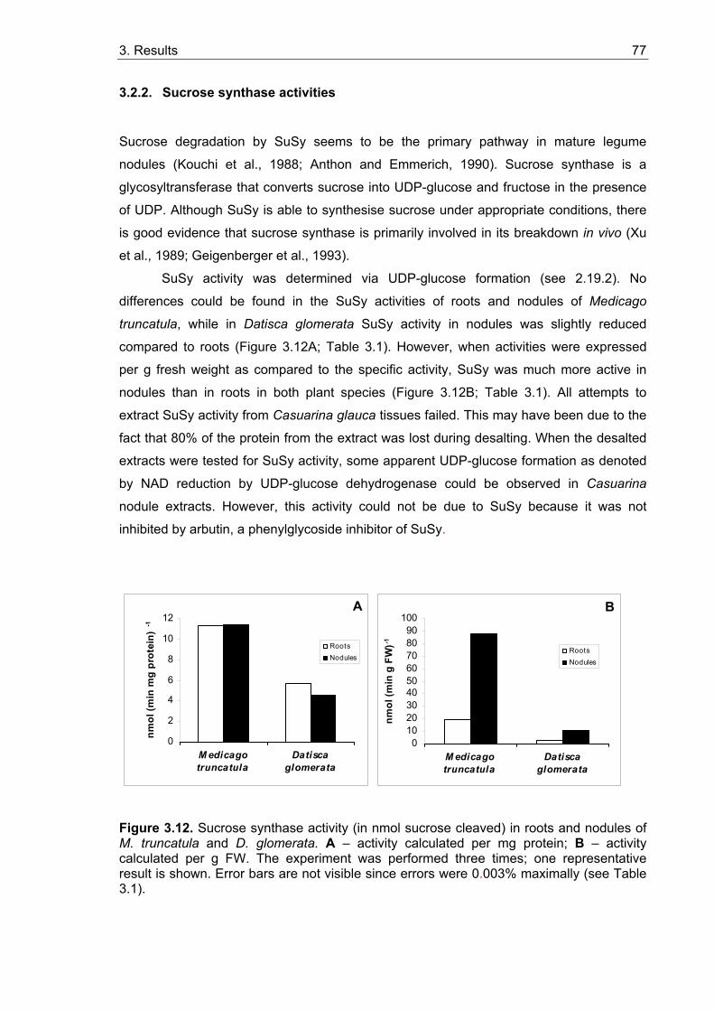
3. Results 77
3.2.2. Sucrose synthase activities
Sucrose degradation by SuSy seems to be the primary pathway in mature legume
nodules (Kouchi et al., 1988; Anthon and Emmerich, 1990). Sucrose synthase is a
glycosyltransferase that converts sucrose into UDP-glucose and fructose in the presence
of UDP. Although SuSy is able to synthesise sucrose under appropriate conditions, there
is good evidence that sucrose synthase is primarily involved in its breakdown in vivo (Xu
et al., 1989; Geigenberger et al., 1993).
SuSy activity was determined via UDP-glucose formation (see 2.19.2). No
differences could be found in the SuSy activities of roots and nodules of Medicago
truncatula, while in Datisca glomerata SuSy activity in nodules was slightly reduced
compared to roots (Figure 3.12A; Table 3.1). However, when activities were expressed
per g fresh weight as compared to the specific activity, SuSy was much more active in
nodules than in roots in both plant species (Figure 3.12B; Table 3.1). All attempts to
extract SuSy activity from Casuarina glauca tissues failed. This may have been due to the
fact that 80% of the protein from the extract was lost during desalting. When the desalted
extracts were tested for SuSy activity, some apparent UDP-glucose formation as denoted
by NAD reduction by UDP-glucose dehydrogenase could be observed in Casuarina
nodule extracts. However, this activity could not be due to SuSy because it was not
inhibited by arbutin, a phenylglycoside inhibitor of SuSy.
0
2
4
6
8
10
12
M edicagotruncatula
Datiscaglomerata
nmol
(min
mg
prot
ein)
-1
RootsNodules
0102030405060708090
100
M edicagotruncatula
Datiscaglomerata
nmol
(min
g F
W)-1
RootsNodules
Figure 3.12. Sucrose synthase activity (in nmol sucrose cleaved) in roots and nodules of M. truncatula and D. glomerata. A – activity calculated per mg protein; B – activity calculated per g FW. The experiment was performed three times; one representative result is shown. Error bars are not visible since errors were 0.003% maximally (see Table 3.1).
A B
3. Results 78
Table 3.1. Sucrose synthase activities in roots and nodules of M. truncatula and D. glomerata.
Roots Nodules
Host plant nmol sucrose cleaved/min/mg protein
nmol sucrose cleaved/min/g FW
nmol sucrose cleaved/min/mg protein
nmol sucrose cleaved/min/g FW
Medicago truncatula 11.3 ± 0.0004 19.4 ± 0.0002 11.4 ± 0.0014 88.1 ± 0.0027
Datisca glomerata 5.7 ± 0.0006 2.6 ± 0.0001 4.5 ± 0.0002 10.6 ± 0.0002
In root extracts, NADH formation could already be observed before the UDP-
glucose determination was started by the addition of UDP-glucose dehydrogenase. This
artefactual activity might be the result of UDP-glucose formation by UDP-glucose
pyrophosphorylase in the crude extract.
The data show that generally, in both roots and nodules of Medicago, SuSy
activities are more than 20-fold higher than cytosolic invertase activities (Figure 3.11A).
SuSy activities were also significantly higher than cytosolic invertase activities in nodules
and roots of Datisca (3 times higher in roots, 34 times higher in nodules; Figure 3.12B).
3.2.3. In situ localization of acid invertase activity
A glucose oxidase reaction in the presence of horseradish peroxidase and
diaminobenzidine (DAB) causes the production of H2O2, which is then used by peroxidase
to oxidize diaminobenzidine (DAB), leading to the formation of a brown precipitate. On
hand sections of plant material, after removal of apoplastic glucose by washing and the
addition of sucrose, this staining method can be used to visualize the places of acid, i.e.
apoplastic or vacuolar invertase activity (Kim et al., 2000). Alternatively, when invertases
are inactivated by heat treatment before the reaction medium is added, this method allows
to visualize sites of glucose accumulation.
The method did not work for both Casuarina roots and nodules, because it was
impossible to identify brown precipitates in the strongly lignified and polyphenol-containing
tissues of this plant. Therefore, only results obtained with Medicago and Datisca are
presented below.
3. Results 79
3.2.3.1. In situ localization of glucose in roots and nodules
There was no significant staining for glucose in roots of Medicago and Datisca (Figures
3.13A and 3.13B). It was very surprising to find strong glucose staining in the nitrogen-
fixing infected cells of Datisca nodules (Figure 3.14A; for Datisca nodule morphology, see
Figure 1.3). However, this staining turned out to be an artefact, because a similar reaction
was also observed when nodules were incubated in reaction mixture without glucose
oxidase (Figure 3.14B). It is likely that in Datisca nodule sections stained for glucose, the
brown precipitate was due to the presence of H2O2 (see above) produced by the symbiotic
bacteria.
As in roots, no significant staining for glucose was found for Medicago nodules,
where only infected cells showed weak glucose staining (Figure 3.15A).
3.2.3.2. In situ localization of acid invertase in roots and nodules
A different pattern of acid invertase activity was observed in roots of Medicago and
Datisca. Medicago roots showed invertase activity in the cortex, whereas Datisca roots
only showed activity in the stele (Figure 3.13). Thus, in Medicago apoplastic hydrolysis of
sucrose in the course of apoplastic sugar transport takes place in the entire root cortex,
while in Datisca, it only takes place in the vascular system, indicating that sugar transport
in the root cortex occurs symplastically.
Invertase activity staining could not be performed with Medicago nodules, since
DAB was reduced in the infected cells independently of the presence of glucose oxidase
(Figure 3.15). Like the glucose staining in Datisca nodules, this could be explained by the
production of H2O2 by the bacteria in infected cells. In Datisca nodules, no acid invertase
activity could be detected (Figure 3.14). In nitrogen-fixing infected cells, a light brown
staining was observed sometimes, but it was much weaker than the colouring found in the
same cells during glucose staining (Figure 3.14). This is in accordance with the lack of
invertase activity in Datisca nodules (see 3.2.1.). Thus, either incomplete removal of
glucose in the washing steps, and/or also the production H2O2 by the bacteria could be an
explanation for this weak staining.
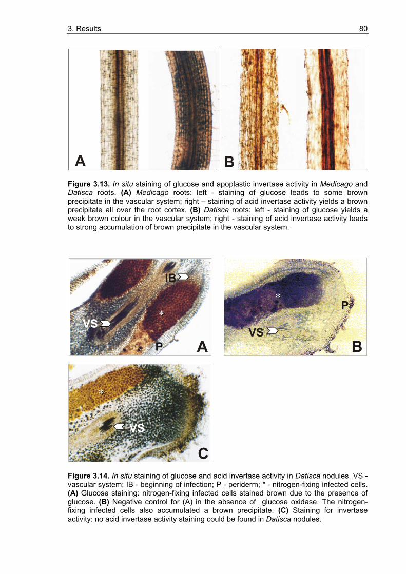
3. Results 80
A B
Figure 3.13. In situ staining of glucose and apoplastic invertase activity in Medicago and Datisca roots. (A) Medicago roots: left - staining of glucose leads to some brown precipitate in the vascular system; right – staining of acid invertase activity yields a brown precipitate all over the root cortex. (B) Datisca roots: left - staining of glucose yields a weak brown colour in the vascular system; right - staining of acid invertase activity leads to strong accumulation of brown precipitate in the vascular system.
P
**
A B
C
**
** P
VSVS
VS
IB
Figure 3.14. In situ staining of glucose and acid invertase activity in Datisca nodules. VS - vascular system; IB - beginning of infection; P - periderm; * - nitrogen-fixing infected cells. (A) Glucose staining: nitrogen-fixing infected cells stained brown due to the presence of glucose. (B) Negative control for (A) in the absence of glucose oxidase. The nitrogen-fixing infected cells also accumulated a brown precipitate. (C) Staining for invertase activity: no acid invertase activity staining could be found in Datisca nodules.
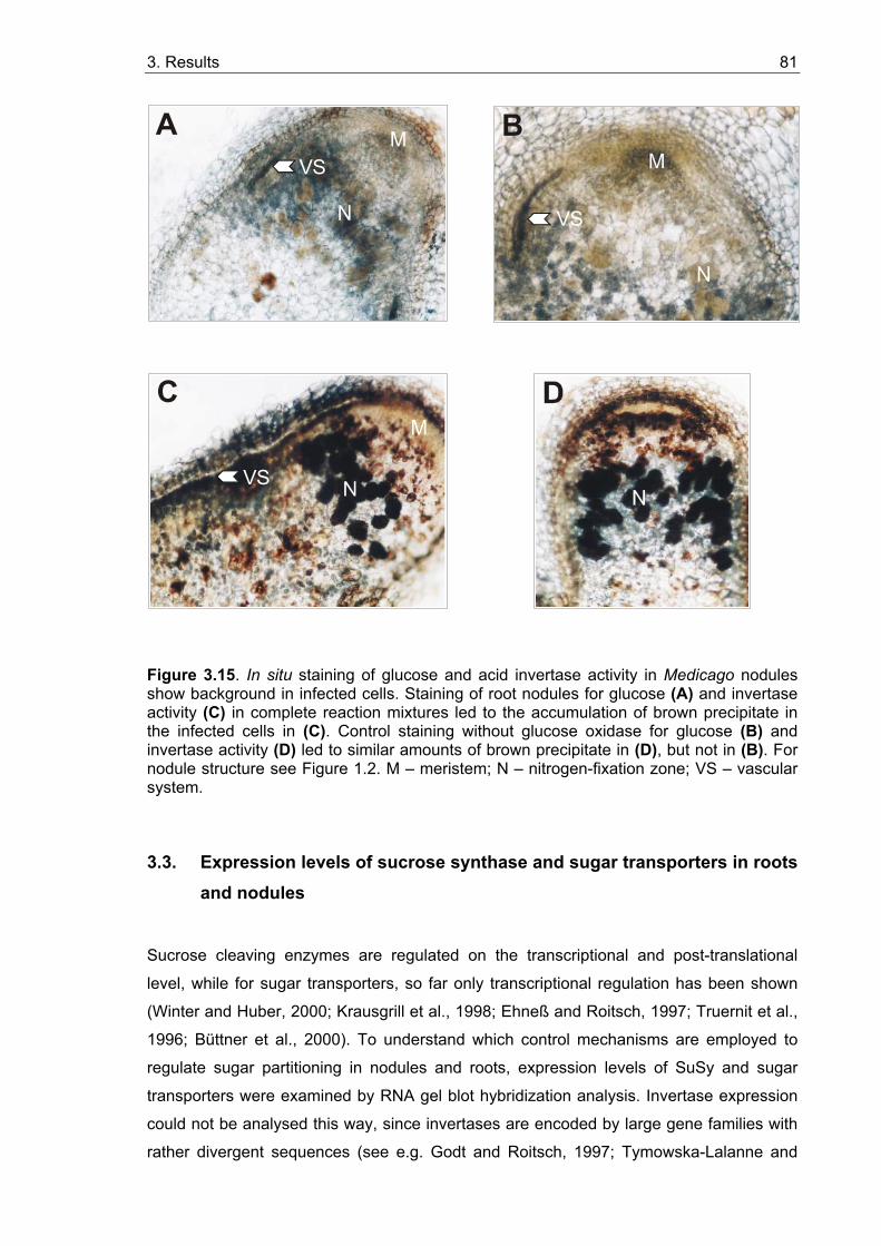
3. Results 81
C
VS
B
VS
AVS
Figure 3.15. In situ staining of glucose and acid invertase activity in Medicago nodules show background in infected cells. Staining of root nodules for glucose (A) and invertase activity (C) in complete reaction mixtures led to the accumulation of brown precipitate in the infected cells in (C). Control staining without glucose oxidase for glucose (B) and invertase activity (D) led to similar amounts of brown precipitate in (D), but not in (B). For nodule structure see Figure 1.2. M – meristem; N – nitrogen-fixation zone; VS – vascular system.
3.3. Expression levels of sucrose synthase and sugar transporters in roots and nodules
Sucrose cleaving enzymes are regulated on the transcriptional and post-translational
level, while for sugar transporters, so far only transcriptional regulation has been shown
(Winter and Huber, 2000; Krausgrill et al., 1998; Ehneß and Roitsch, 1997; Truernit et al.,
1996; Büttner et al., 2000). To understand which control mechanisms are employed to
regulate sugar partitioning in nodules and roots, expression levels of SuSy and sugar
transporters were examined by RNA gel blot hybridization analysis. Invertase expression
could not be analysed this way, since invertases are encoded by large gene families with
rather divergent sequences (see e.g. Godt and Roitsch, 1997; Tymowska-Lalanne and
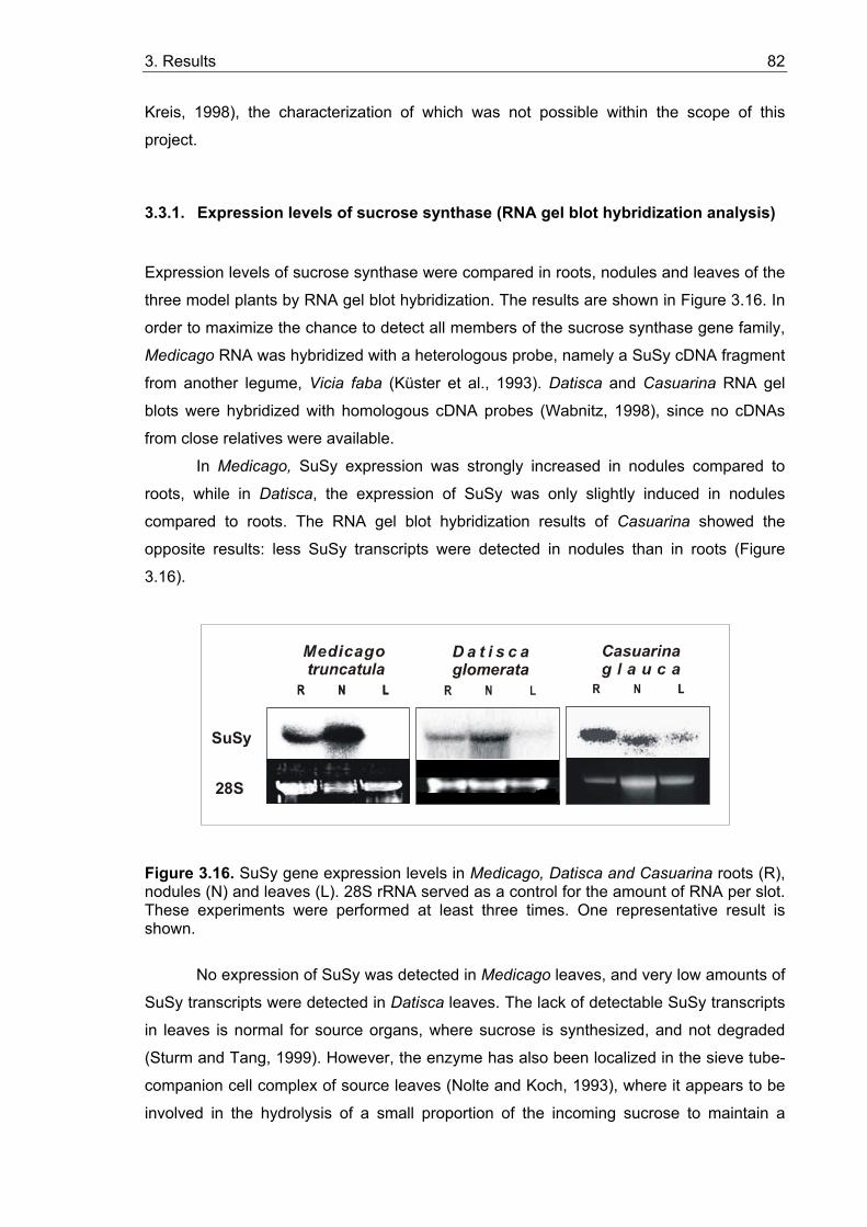
3. Results 82
Kreis, 1998), the characterization of which was not possible within the scope of this
project.
3.3.1. Expression levels of sucrose synthase (RNA gel blot hybridization analysis)
Expression levels of sucrose synthase were compared in roots, nodules and leaves of the
three model plants by RNA gel blot hybridization. The results are shown in Figure 3.16. In
order to maximize the chance to detect all members of the sucrose synthase gene family,
Medicago RNA was hybridized with a heterologous probe, namely a SuSy cDNA fragment
from another legume, Vicia faba (Küster et al., 1993). Datisca and Casuarina RNA gel
blots were hybridized with homologous cDNA probes (Wabnitz, 1998), since no cDNAs
from close relatives were available.
In Medicago, SuSy expression was strongly increased in nodules compared to
roots, while in Datisca, the expression of SuSy was only slightly induced in nodules
compared to roots. The RNA gel blot hybridization results of Casuarina showed the
opposite results: less SuSy transcripts were detected in nodules than in roots (Figure
3.16).
D a t i s c a glomerata
SuSy
28S
Figure 3.16. SuSy gene expression levels in Medicago, Datisca and Casuarina roots (R), nodules (N) and leaves (L). 28S rRNA served as a control for the amount of RNA per slot. These experiments were performed at least three times. One representative result is shown.
No expression of SuSy was detected in Medicago leaves, and very low amounts of
SuSy transcripts were detected in Datisca leaves. The lack of detectable SuSy transcripts
in leaves is normal for source organs, where sucrose is synthesized, and not degraded
(Sturm and Tang, 1999). However, the enzyme has also been localized in the sieve tube-
companion cell complex of source leaves (Nolte and Koch, 1993), where it appears to be
involved in the hydrolysis of a small proportion of the incoming sucrose to maintain a
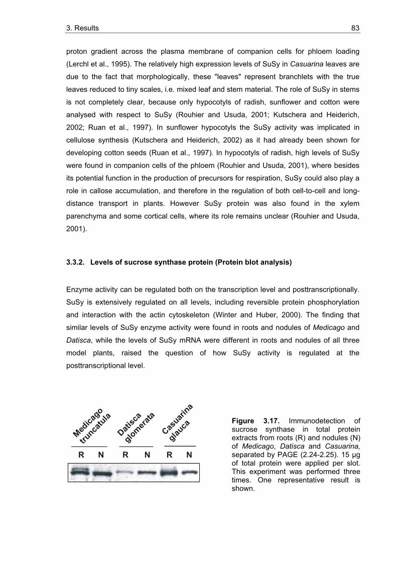
3. Results 83
proton gradient across the plasma membrane of companion cells for phloem loading
(Lerchl et al., 1995). The relatively high expression levels of SuSy in Casuarina leaves are
due to the fact that morphologically, these "leaves" represent branchlets with the true
leaves reduced to tiny scales, i.e. mixed leaf and stem material. The role of SuSy in stems
is not completely clear, because only hypocotyls of radish, sunflower and cotton were
analysed with respect to SuSy (Rouhier and Usuda, 2001; Kutschera and Heiderich,
2002; Ruan et al., 1997). In sunflower hypocotyls the SuSy activity was implicated in
cellulose synthesis (Kutschera and Heiderich, 2002) as it had already been shown for
developing cotton seeds (Ruan et al., 1997). In hypocotyls of radish, high levels of SuSy
were found in companion cells of the phloem (Rouhier and Usuda, 2001), where besides
its potential function in the production of precursors for respiration, SuSy could also play a
role in callose accumulation, and therefore in the regulation of both cell-to-cell and long-
distance transport in plants. However SuSy protein was also found in the xylem
parenchyma and some cortical cells, where its role remains unclear (Rouhier and Usuda,
2001).
3.3.2. Levels of sucrose synthase protein (Protein blot analysis)
Enzyme activity can be regulated both on the transcription level and posttranscriptionally.
SuSy is extensively regulated on all levels, including reversible protein phosphorylation
and interaction with the actin cytoskeleton (Winter and Huber, 2000). The finding that
similar levels of SuSy enzyme activity were found in roots and nodules of Medicago and
Datisca, while the levels of SuSy mRNA were different in roots and nodules of all three
model plants, raised the question of how SuSy activity is regulated at the
posttranscriptional level.
R N R N R N
Medica
go
trunca
tula
Datisc
a
glomerata
Casuari
na
glauca
Figure 3.17. Immunodetection of sucrose synthase in total protein extracts from roots (R) and nodules (N) of Medicago, Datisca and Casuarina, separated by PAGE (2.24-2.25). 15 µg of total protein were applied per slot. This experiment was performed three times. One representative result is shown.
3. Results 84
The amounts of SuSy were analysed by the protein blot analysis of total protein
extracts from roots and nodules (2.25) from the three plant systems with an antibody
raised against SuSy from cotyledons of Vicia faba (Ross and Davies, 1992; kindly
provided by N. Hohnjec, University of Bielefeld). The results are shown in Figure 3.17.
Increase of SuSy protein levels in nodules compared to roots could be found only in
nodules of Datisca, which contain a single immunoreactive protein of approximately 92-
kDa. For Casuarina glauca, also a single immunoreactive protein of the same mass was
detected, but the amounts of SuSy were reduced in nodules compared to roots. So, for
Datisca and Casuarina the amounts of protein are in accordance with mRNA expression
levels. However, in Medicago nodules, only a slight increase in the amount of the
immunoreactive 92 kDa protein was detected compared to roots. This does not reflect the
mRNA levels, but is in accordance with SuSy enzyme activities in both organs (Figure
3.12A). A second immunoreactive protein of slightly higher molecular weight was detected
in extracts of uninfected roots of Medicago, which is consistent with the results obtained
by Hohnjec et al. (1999).
3.3.3. Expression levels of sugar transporters in roots, nodules and leaves
The organ-specific expression of sucrose and hexose transporters was analysed by RNA
gel blot hybridisation in roots, nodules and leaves of the three model plants. For the
sucrose transporter of Medicago, a heterologous probe from Vicia faba was used (Weber
et al., 1997), while a homologous probe was used for the hexose transporter of Medicago
(Wabnitz, 1998). The expression levels of the sucrose transporter (MtST) were strongly
reduced in nodules compared to roots, and the expression levels of the hexose
transporter (MtHT) in nodules were below the detection limit (Figure 3.18). For Datisca
only homologous probes were used (Wabnitz, 1998). Here, the expression of the hexose
transporter was strongly induced in nodules compared to roots, while sucrose transporter
expression was below the detection limit in both nodules and roots as well as in leaves
(Figure 3.18; data not shown for the sucrose transporter). For Casuarina, only hexose
transporter expression was examined since no sucrose transporter cDNA fragment could
be amplified by PCR with the degenerate primers that had been used successfully for
Datisca (Wabnitz, 1998). Casuarina hexose transporter expression was strongly induced
in nodules compared to roots, as in Datisca (Figure 3.18). Interestingly, in both Casuarina
and Datisca, hexose transporter expression levels were very low in roots, while in
Medicago roots high expression levels of both hexose and sucrose transporters were
found, indicating that root sugar partitioning mechanisms differ significantly between the
legume Medicago and the two actinorhizal plants.
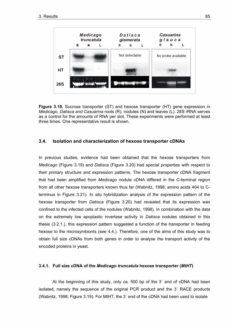
3. Results 85
D a t i s c a glomerata
28S
ST
HT
Figure 3.18. Sucrose transporter (ST) and hexose transporter (HT) gene expression in Medicago, Datisca and Casuarina roots (R), nodules (N) and leaves (L). 28S rRNA serves as a control for the amounts of RNA per slot. These experiments were performed at least three times. One representative result is shown.
3.4. Isolation and characterization of hexose transporter cDNAs
In previous studies, evidence had been obtained that the hexose transporters from
Medicago (Figure 3.19) and Datisca (Figure 3.20) had special properties with respect to
their primary structure and expression patterns. The hexose transporter cDNA fragment
that had been amplified from Medicago nodule cDNA differed in the C-terminal region
from all other hexose transporters known thus far (Wabnitz, 1998; amino acids 404 to C-
terminus in Figure 3.21). In situ hybridization analysis of the expression pattern of the
hexose transporter from Datisca (Figure 3.20) had revealed that its expression was
confined to the infected cells of the nodules (Wabnitz, 1998). In combination with the data
on the extremely low apoplastic invertase activity in Datisca nodules obtained in this
thesis (3.2.1.), this expression pattern suggested a function of the transporter in feeding
hexose to the microsymbionts (see 4.4.). Therefore, one of the aims of this study was to
obtain full size cDNAs from both genes in order to analyse the transport activity of the
encoded proteins in yeast.
3.4.1. Full size cDNA of the Medicago truncatula hexose transporter (MtHT)
At the beginning of this study, only ca. 500 bp of the 3´ end of cDNA had been
isolated, namely the sequence of the original PCR product and the 3´ RACE products
(Wabnitz, 1998; Figure 3.19). For MtHT, the 3´ end of the cDNA had been used to isolate
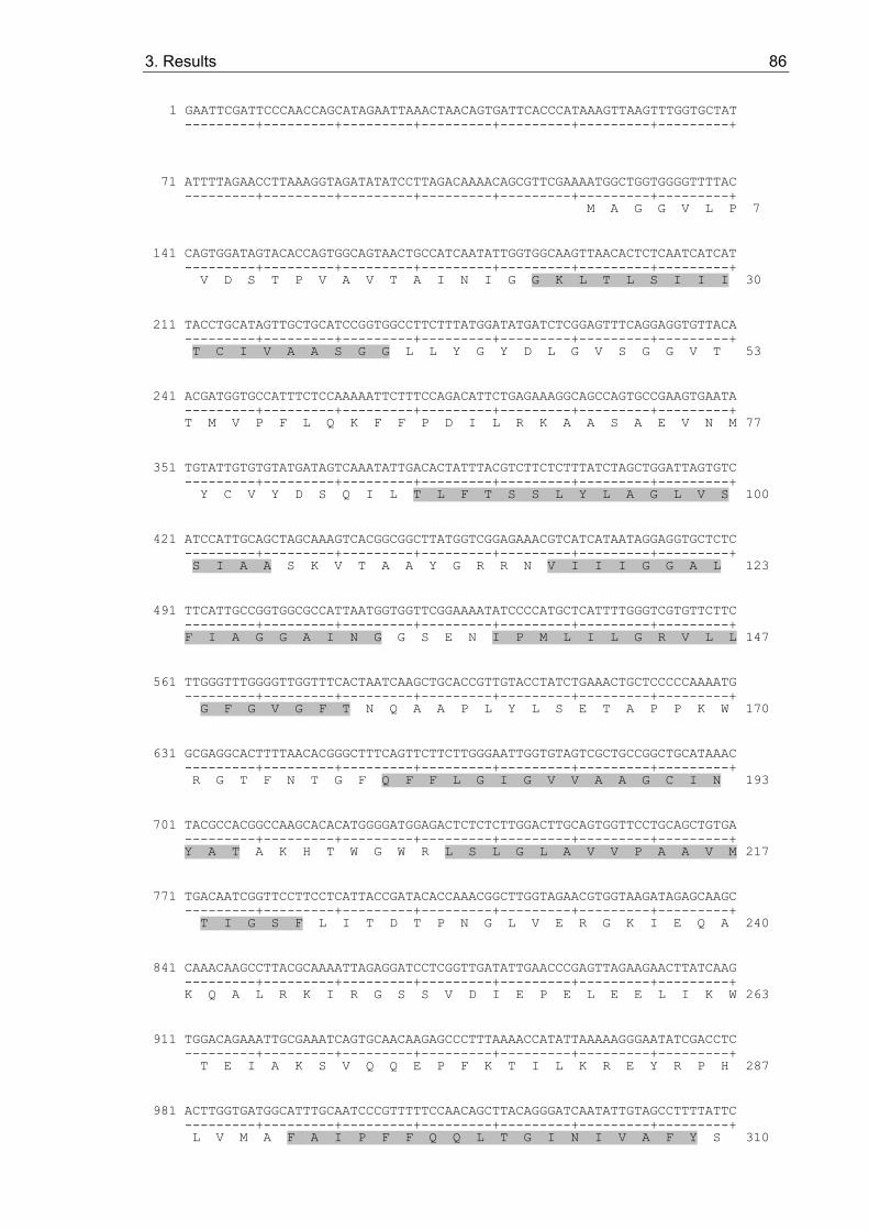
3. Results 86
1 GAATTCGATTCCCAACCAGCATAGAATTAAACTAACAGTGATTCACCCATAAAGTTAAGTTTGGTGCTAT ---------+---------+---------+---------+---------+---------+---------+
71 ATTTTAGAACCTTAAAGGTAGATATATCCTTAGACAAAACAGCGTTCGAAAATGGCTGGTGGGGTTTTAC ---------+---------+---------+---------+---------+---------+---------+ M A G G V L P 7
141 CAGTGGATAGTACACCAGTGGCAGTAACTGCCATCAATATTGGTGGCAAGTTAACACTCTCAATCATCAT ---------+---------+---------+---------+---------+---------+---------+ V D S T P V A V T A I N I G G K L T L S I I I 30
211 TACCTGCATAGTTGCTGCATCCGGTGGCCTTCTTTATGGATATGATCTCGGAGTTTCAGGAGGTGTTACA ---------+---------+---------+---------+---------+---------+---------+ T C I V A A S G G L L Y G Y D L G V S G G V T 53
241 ACGATGGTGCCATTTCTCCAAAAATTCTTTCCAGACATTCTGAGAAAGGCAGCCAGTGCCGAAGTGAATA ---------+---------+---------+---------+---------+---------+---------+ T M V P F L Q K F F P D I L R K A A S A E V N M 77
351 TGTATTGTGTGTATGATAGTCAAATATTGACACTATTTACGTCTTCTCTTTATCTAGCTGGATTAGTGTC ---------+---------+---------+---------+---------+---------+---------+ Y C V Y D S Q I L T L F T S S L Y L A G L V S 100
421 ATCCATTGCAGCTAGCAAAGTCACGGCGGCTTATGGTCGGAGAAACGTCATCATAATAGGAGGTGCTCTC ---------+---------+---------+---------+---------+---------+---------+ S I A A S K V T A A Y G R R N V I I I G G A L 123
491 TTCATTGCCGGTGGCGCCATTAATGGTGGTTCGGAAAATATCCCCATGCTCATTTTGGGTCGTGTTCTTC ---------+---------+---------+---------+---------+---------+---------+ F I A G G A I N G G S E N I P M L I L G R V L L 147
561 TTGGGTTTGGGGTTGGTTTCACTAATCAAGCTGCACCGTTGTACCTATCTGAAACTGCTCCCCCAAAATG ---------+---------+---------+---------+---------+---------+---------+ G F G V G F T N Q A A P L Y L S E T A P P K W 170
631 GCGAGGCACTTTTAACACGGGCTTTCAGTTCTTCTTGGGAATTGGTGTAGTCGCTGCCGGCTGCATAAAC ---------+---------+---------+---------+---------+---------+---------+ R G T F N T G F Q F F L G I G V V A A G C I N 193
701 TACGCCACGGCCAAGCACACATGGGGATGGAGACTCTCTCTTGGACTTGCAGTGGTTCCTGCAGCTGTGA ---------+---------+---------+---------+---------+---------+---------+ Y A T A K H T W G W R L S L G L A V V P A A V M 217
771 TGACAATCGGTTCCTTCCTCATTACCGATACACCAAACGGCTTGGTAGAACGTGGTAAGATAGAGCAAGC ---------+---------+---------+---------+---------+---------+---------+ T I G S F L I T D T P N G L V E R G K I E Q A 240
841 CAAACAAGCCTTACGCAAAATTAGAGGATCCTCGGTTGATATTGAACCCGAGTTAGAAGAACTTATCAAG ---------+---------+---------+---------+---------+---------+---------+ K Q A L R K I R G S S V D I E P E L E E L I K W 263
911 TGGACAGAAATTGCGAAATCAGTGCAACAAGAGCCCTTTAAAACCATATTAAAAAGGGAATATCGACCTC ---------+---------+---------+---------+---------+---------+---------+ T E I A K S V Q Q E P F K T I L K R E Y R P H 287
981 ACTTGGTGATGGCATTTGCAATCCCGTTTTTCCAACAGCTTACAGGGATCAATATTGTAGCCTTTTATTC ---------+---------+---------+---------+---------+---------+---------+ L V M A F A I P F F Q Q L T G I N I V A F Y S 310

3. Results 87
1051 ACCCAACCTCTTTCACTCTGTGGGTTTTGGACACGACGGAGCTTTACTTTCCGCCATTATACTTGGATCT ---------+---------+---------+---------+---------+---------+---------+ P N L F H S V G F G H D G A L L S A I I L G S V 333
1121 GTTAGCCTTTTGTCTAACCTTATCTCTGCTGGTATTGTTGATCGAATTGGTCGAAGATTCTTGTTCATAT ---------+---------+---------+---------+---------+---------+---------+ S L L S N L I S A G I V D R I G R R F L F I S 357
1191 CTGGGGGAATTATGATGCTTGTCTGCTTGATTGCTGTGTCCATTGTCCTGGCAGTGGTGACTGGTGTTGA ---------+---------+---------+---------+---------+---------+---------+ G G I M M L V C L I A V S I V L A V V T G V D 380
1261 TGGTACAAAGGACATATCCAAGGGCAATGCAATAGTGGTATTGGTGCTATTGTGTTTCTACTCTGCAGGT ---------+---------+---------+---------+---------+---------+---------+ G T K D I S K G N A I V V L V L L C F Y S A G F 403
1331 TTTGGTTGGTCATGGGGTCCATTAACATGGCTTATTCCAAGTGAGATTTTCCCAGTAAAAATAAGAACCA ---------+---------+---------+---------+---------+---------+---------+ G W S W G P L T W L I P S E I F P V K I R T T 427
1401 CAGGACAAAGCATAGCTGTTGCTGTGCAATTCATAATAATCTTTGTGTTATCGCAAACATTCTTGACAAT ---------+---------+---------+---------+---------+---------+---------+ G Q S I A V A V Q F I I I F V L S Q T F L T M 450
1471 GTTATGCCACATGAAGTTTGGGGCCTTCGTCTTCTATGCTTTTTGGGTTATTGTGATGACTCTGTTTGTT ---------+---------+---------+---------+---------+---------+---------+ L C H M K F G A F V F Y A F W V I V M T L F V I 473
1541 ATCTTCTTCTTGCCTGAGACCAAAGGAATTCCTTTGGAGTCAATGTACACTATATGGGGCAGACACTGGT ---------+---------+---------+---------+---------+---------+---------+ F F L P E T K G I P L E S M Y T I W G R H W F 497
1611 TTTGGTCTCGGTATGTTAAAGGACAAGAGGTCCTAGAAAATCTCCCATGAAGTTTTTAACACTTTTTATG ---------+---------+---------+---------+---------+---------+---------+ W S R Y V K G Q E V L E N L P * 512
1681 ATTGATTTATTATTTTTAAAGAAGTCAATGCACATGCATTATATCTTGACAAAATTGTGCAGCATTGGCT ---------+---------+---------+---------+---------+---------+---------+
1751 TCATAAAAATAAATCAATAATAAAAGAAATGTAATCAATAATAACGTCTCAAAAATAATGTATTGATTGT ---------+---------+---------+---------+---------+---------+---------+
1821 TAATGTTAAAAAAAAAAAAAAAAAAAAAGCGGCCGCGGATCCTCGAGAATCACTAGTGAATTC ---------+---------+---------+---------+---------+---------+---
Figure 3.19. DNA sequence and deduced amino acid sequence of the M. truncatula hexose transporter (MtHT). Shadowed amino acid sequences represent the membrane-spanning domains predicted by the method of Rost et al. (1995; 1996). Numbers to the right refer to the predicted amino acid sequence of MtHT. Underlining denotes potential phosphorylation consensus sequences. The amino acid sequences that had been used to design the original degenerate oligonucleotide primers are underlined and labelled by bold print.
3. Results 88
a cDNA clone from a cDNA library made from RNA of young Medicago nodules
(MtHTN15;Wabnitz, 1998; Gamas et al., 1996). However, this clone was not full size but
started at nucleotide 442 of the full size sequence that was finally obtained in this thesis.
Thus, 5´ RACEs had to be performed for cDNA. The cDNA sequences available were
used for generating primers for 5´-RACEs. The sequences of all primers used are
presented in 2.1.3.
For MtHT 5´-RACE, reverse transcription was performed with the first gene-
specific primer MtHT 5´race1. Based on the C-tailed first-strand cDNA (2.10.), two PCRs
were performed using for the first PCR the anchor primer MM1 and primer MtHT 5´race1
(PCR program was 94 °C 30 s, 35 cycles of 94 °C 30 s, Tann= 61 °C 30 s, 72 °C 90 s,
followed by 72 °C for 10 min in the presence of 1.5 mM MgCl2). For the second PCR the
anchor-specific primer MM2 and second gene-specific primer MtHT 5´race2 were used
(PCR program as described above, except that the Tann was 58 °C). A PCR product of
about 500 bp was cloned in pGEM-T Easy and sequenced. The 5´-end sequence was
used to design another primer, MtHT fs, which in combination with the primer MM4 was
used to amplify the full size cDNA. First strand cDNA was transcribed with primer MM3
from nodule mRNA, and the full size cDNA was amplified with the following PCR program:
94 °C 5 min, 35 cycles of 94 °C 1 min, 60 °C 1 min, 72 °C 2 min, followed by 72 °C for 10
min. The full size cDNA was cloned in pGEM-T Easy and both strands were sequenced.
The DNA sequence and the deduced amino acid sequence of M. truncatula
hexose transporter (MtHT) are presented in Figure 3.19. The full size clone comprises
1883 nucleotides, 1536 of which represent an ORF that is predicted to encode a protein of
512 amino acids with an estimated molecular mass of 55.41 kDa. The 5´-untranslated
region includes 121 nucleotides, and 3´-untranslated region is composed of 226
nucleotides.
The protein encoded by MtHT is predicted to be an extremely hydrophobic integral
membrane protein with 12 transmembrane domains according to a hydropathy plot
(Figure 3.19; Rost et al., 1995; 1996).
3.4.2. Full size cDNA of the Datisca glomerata hexose transporter (DgHT)
At the beginning of this study, only ca. 500 bp of the 3´ end of cDNA had been isolated
(Wabnitz, 1998; Figure 3.20). The cDNA sequences available were used for generating
primers for 5´-RACEs. The sequences of all primers used are presented in 2.1.3.
For DgHT 5´-RACE, reverse transcription was performed with a first gene-specific
primer DgHT 5´race1. Based on the C-tailed first-strand cDNA (2.10.), the first PCR was
performed using an anchor primer MM1 and above-mentioned primer DgHT 5´race1 (PCR
3. Results 89
1 GAATTCGATTCCCTTCTCTCTTTATCTCCTTTTCATCACTCCCCTCTTTCTCTCCTCGCAAAAGAAGTCA ---------+---------+---------+---------+---------+---------+---------+
71 GAGATAAAGGACGAAAACATGCCGGCCGTCGGAGGAATCGTTGTCGGTGGCAGTAAAAAGGAGTATCCCG ---------+---------+---------+---------+---------+---------+---------+ M P A V G G I V V G G S K K E Y P G 18
141 GCAACCTTACTCCTTATGTCACCATAACATGCATTGTTGCCGCTATGGGTGGTCTGATTTTCGGTTACGA ---------+---------+---------+---------+---------+---------+---------+ N L T P Y V T I T C I V A A M G G L I F G Y D 41
211 TATTGGAATTTCAGGTGGGGTGACGTCAATGGATTCATTCTTGAAGAAATTCTTCCCGGCGGTTTACCGG ---------+---------+---------+---------+---------+---------+---------+ I G I S G G V T S M D S F L K K F F P A V Y R 64
241 AAAAAAGAGTTGGATTCGACGACGAACCAGTACTGTCAGTACGACAGTCAGACTCTGACGATGTTCACGT ---------+---------+---------+---------+---------+---------+---------+ K K E L D S T T N Q Y C Q Y D S Q T L T M F T S 88
351 CTTCTCTTTATTTGGCTGCTCTGCTCGCTTCGATAGTGGCTTCCACCATCACCCGTAAATTCGGCAGGAG ---------+---------+---------+---------+---------+---------+---------+ S L Y L A A L L A S I V A S T I T R K F G R R 111
421 ATTATCCATGCTCTTCGGCGGCATTCTGTTCTGTGCCGGTGCTATCATCAATGGCTTCGCCCAGGCTGTT ---------+---------+---------+---------+---------+---------+---------+ L S M L F G G I L F C A G A I I N G F A Q A V 134
491 TGGATGCTCATTCTCGGTCGTATGTTTCTCGGTTTTGGTATTGGGTTTTCCAATCAGTCTGTGCCACTCT ---------+---------+---------+---------+---------+---------+---------+ W M L I L G R M F L G F G I G F S N Q S V P L Y 158
561 ACCTCTCAGAGATGGCTCCCTACAAGTACAGAGGAGCATTAAACATAGGCTTCCAATTATCAATCACAAT ---------+---------+---------+---------+---------+---------+---------+ L S E M A P Y K Y R G A L N I G F Q L S I T I 181
631 TGGAATATTGGTAGCAAATGTGTTAAATTACTTCTTTGCCAAGATCAGGGGCGGCTGGGGATGGCGGTTG ---------+---------+---------+---------+---------+---------+---------+ G I L V A N V L N Y F F A K I R G G W G W R L 204
701 AGCTTAGGTGGCGCAATGGTCCCAGCCCTTATCATCACGGTCGGATCGCTACTCCTTCCCGACACACCCA ---------+---------+---------+---------+---------+---------+---------+ S L G G A M V P A L I I T V G S L L L P D T P N 228
771 ACTCATTAATCGAGCGTGGCAACCGAGACGAAGCCCGCAGCAAACTCCAAAGGGTTCGAGGCGTTGATGA ---------+---------+---------+---------+---------+---------+---------+ S L I E R G N R D E A R S K L Q R V R G V D D 251
841 CGTGGACGAGGAGTTTAACGATCTGGTGGCGGCAAGTGAAGAGTCAAAGCAAGTGGAACATCCTTGGACT ---------+---------+---------+---------+---------+---------+---------+ V D E E F N D L V A A S E E S K Q V E H P W T 274
911 AACCTGTTGAGGAGGAAGTACAGACCTCATCTTGCAATGGCTATACTAATTCCTTTCTTCCAGCAACTTA ---------+---------+---------+---------+---------+---------+---------+ N L L R R K Y R P H L A M A I L I P F F Q Q L T 298
981 CCGGCATCAATGTGATTATGTTTTACGCGCCTGTTTTGTTTAACACCATTGGGTTTGGAAGTGATGCTTC ---------+---------+---------+---------+---------+---------+---------+ G I N V I M F Y A P V L F N T I G F G S D A S 321

3. Results 90
1051 GCTCATGTCGGCTGTGATTACTGGCTGTGTTAATGTCGCTGGGACTTTGGTTTCTATTTATGGGGTTGAT ---------+---------+---------+---------+---------+---------+---------+ L M S A V I T G C V N V A G T L V S I Y G V D 344
1121 AAGTGGGGAAGGAGGTTCCTTTTCCTTGAGGGTGGATTTCAAATGTTGATTTGCCAGGCGGTTGTAGCAG ---------+---------+---------+---------+---------+---------+---------+ K W G R R F L F L E G G F Q M L I C Q A V V A A 368
1191 CTGCAATTGGTGCTAAATTTGGAGTAAATGGAAATCCAGGAGAACTGCCAAAATGGTATGCGATAGTGGT ---------+---------+---------+---------+---------+---------+---------+ A I G A K F G V N G N P G E L P K W Y A I V V 391
1261 GGTGCTGTTCATATGCATATATGTAGCAGGGTTCTCTTGGTCATGGGGTCCTCTAGGTTGGCTAGTGCCA ---------+---------+---------+---------+---------+---------+---------+ V L F I C I Y V A G F S W S W G P L G W L V P 414
1331 AGTGAAAGTTTCCCCTTAGAAATACGGTCAGCTGCTCAAAGTATCAATGTCTCTGTCAACATGATCTTCA ---------+---------+---------+---------+---------+---------+---------+ S E S F P L E I R S A A Q S I N V S V N M I F T 438
1401 CATTTGCTATAGCTCAAATCTTCTTGACAATGCTTTGCCACTTGAAATTCGGTTTATTCATTTTCTTCGC ---------+---------+---------+---------+---------+---------+---------+ F A I A Q I F L T M L C H L K F G L F I F F A 461
1471 CTTCTTCGTGGTTGTCATGTCTATCTTCGTCTACTTCTTCTTGCCCGAGACTAAGGGAATCCCGATCGAA ---------+---------+---------+---------+---------+---------+---------+ F F V V V M S I F V Y F F L P E T K G I P I E 484
1541 GAGATGGGCAGAGTATGGAAGTCACATTGGTACTGGTCAAGATTTGTTACTGATGCTGATTACACTATTG ---------+---------+---------+---------+---------+---------+---------+ E M G R V W K S H W Y W S R F V T D A D Y T I G 508
1611 GATCAGGAGTTGAGATGGGAAAGGGAGCGCAGGAGCTTAAGAATATTTAAGTTTAATGTTTGGGGGGAGG ---------+---------+---------+---------+---------+---------+---------+ S G V E M G K G A Q E L K N I * 523
1681 GTTATTTTTAGATTATTTTTTTCTATCTAGTAAATAGAAGTTTGGGTCAATATTAAATTAAGAACATATG ---------+---------+---------+---------+---------+---------+---------+
1751 AATGGGTCAAATTTGATGTATCTGAATCACTAGTGAATTC 1790 ---------+---------+---------+---------+
Fig. 3.20. DNA sequence and deduced amino acid sequence of the D. glomerata hexose transporter (DgHT). Shadowed amino acid sequences represent the membrane-spanning domains predicted by the method of Rost et al. (1995; 1996; see 3.4.3.). Numbers to the right refer to the predicted amino acid sequence of DgHT. Underlining denotes potential phosphorylation consensus sequences. The amino acid sequences that had been used to design the original degenerate oligonucleotide primers are underlined and labeled by bold print.
3. Results 91
program was 94 °C 30 s, 35 cycles of 94 °C 30 s, 58 °C 30 s, 72 °C 30 s, followed by 72
°C for 10 min in the presence of 1.5 mM MgCl2). For the second PCR another anchor-
specific primer MM2 and second gene-specific primer DgHT 5´race2 were used (PCR
program as described above). A 480 bp PCR product was cloned into pGEM-T Easy and
sequenced. The 5´-end sequence was used as a template to design primer DgHT fs5,
which together with the primer from the 3´-end sequence DgHT fs3 was used to amplify
the full size cDNA with the following PCR program: 94 °C 5 min, 35 cycles of 94 °C 1 min,
52 °C 1 min, 72 °C 2 min, followed by 72 °C for 10 min. For this purpose, first strand
cDNA was transcribed with primer dT20 from nodule mRNA. The full size cDNA was
cloned in pGEM-T Easy and both strands were sequenced.
The cDNA sequence and the deduced amino acid sequences of the hexose
transporter (DgHT) from Datisca nodules are shown in Figure 3.20. The full size cDNA
clone comprises 1790 nucleotides, 1569 of which represent an ORF encoding a protein of
523 amino acids with an estimated molecular mass of 57.64 kDa. The 5´-untranslated
region consists of 88 and the 3´-untranslated region of 133 nucleotides. Like the other
members of the hexose transporter protein family, the protein encoded by DgHT is
predicted to be an extremely hydrophobic integral membrane protein with 12
transmembrane domains (Rost et al., 1995; 1996).
3.4.3. Protein sequence analysis of DgHT and MtHT
The deduced amino sequences of DgHT and MtHT exhibit similarity to known plant
hexose transporters, with the highest levels of identity for DgHT to the hexose
transporters AtSTP1 from Arabidopsis and MtSTP1 from Medicago (83.3 and 83.2%,
respectively), and for MtHT to AtSTP1 from Arabidopsis (55.1%; Table 3.2, Figure 3.21;
references see figure legend).
Hydropathy analysis reveals that like the other plant hexose transporters, DgHT and MtHT
are extremely hydrophobic and predicted to represent integral membrane proteins with 12
membrane spanning domains (Figures 3.19 and 3.20), a typical feature of transporters
belonging to the major facilitator superfamily described by Marger and Saier (1993). The
MtHT and DgHT sequences also contain conserved sequence motifs previously identified
among sugar transporters, including PETKG at the end of membrane-spanning domain 12
and two potential phosphorylation consensus sequences (Figures 3.19 and 3.20).
3. Results 92
* * * * * * DgHT : MPAVGGIVVGGSKKEYPGNLTPYVTITCIVAAMGGLIFGYDIGISGGVTSMDSFLK : 56 MtSTP1 : MAGGGIPIGGGNKEYPGNLTPFVTITCIVAAMGGLIFGYDIGISGGVTSMDPFLK : 55 AtSTP1 : MPAGGFVVGDGQKAYPGKLTPFVLFTCVVAAMGGLIFGYDIGISGGVTSMPSFLK : 55 AtSTP4 : MAGGFVSQTPGVRNYNYKLTPKVFVTCFIGAFGGLIFGYDLGISGGVTSMEPFLE : 55 MtHT : MAGGVLPVDSTPVAVTAINIGGKLTLSIIITCIVAASGGLLYGYDLGVSGGVTTMVPFLQ : 60 AtSTP3 : MVAEEARKEAMAKSVSGGKITYFVVASCVMAAMGGVIFGYDIGVSGGVMSMGPFLK : 56 AtSTP2 : MAVGSMNVEEGTKAFPAKLTGQVFLCCVIAAVGGLMFGYDIGISGGVTSMDTFLL : 55 * * * * * * DgHT : KFFPAVYRKKELD-----STTNQYCQYDSQTLTMFTSSLYLAALLASIVASTITRKFGRR : 111 MtSTP1 : KFFPAVYRKKNKD-----KSTNQYCQYDSQTLTMFTSSLYLAALLSSLVASTITRRFGRK : 110 AtSTP1 : RFFPSVYRKQQED-----ASTNQYCQYDSPTLTMFTSSLYLAALISSLVASTVTRKFGRR : 110 AtSTP4 : EFFPYVYKKMKS------AHENEYCRFDSQLLTLFTSSLYVAALVSSLFASTITRVFGRK : 109 MtHT : KFFPDILRKAAS------AEVNMYCVYDSQILTLFTSSLYLAGLVSSIAASKVTAAYGRR : 114 AtSTP3 : RFFPKVYKLQEEDRRRRGNSNNHYCLFNSQLLTSFTSSLYVSGLIATLLASSVTRSWGRK : 116 AtSTP2 : DFFPHVYEKKHR------VHENNYCKFDDQLLQLFTSSLYLAGIFASFISSYVSRAFGRK : 109 * * * * * * DgHT : LSMLFGGILFCAGAIINGFAQAVWMLILGRMFLGFGIGFSNQS----------------- : 154 MtSTP1 : LSMLFGGLLFLVGALINGFANHVWMLIVGRILLGFGIGFANQP----------------- : 153 AtSTP1 : LSMLFGGILFCAGALINGFAKHVWMLIVGRILLGFGIGFANQA----------------- : 153 AtSTP4 : WSMFLGGFTFFIGSAFNGFAQNIAMLLIGRILLGFGVGFANQS----------------- : 152 MtHT : NVIIIGGALFIAGGAINGGSENIPMLILGRVLLGFGVGFTNQA----------------- : 157 AtSTP3 : PSIFLGGVSFLAGAALGGSAQNVAMLIIARLLLGVGVGFANQS----------------- : 159 AtSTP2 : PTIMLASIFFLVGAILNLSAQELGMLIGGRILLGFGIGFGNQVSCQTLKTFFYLSGFLCF : 169 * * * * * * DgHT : -------------------------------------VPLYLSEMAPYKYRGALNIGFQL : 177 MtSTP1 : -------------------------------------VPLYLSEMAPYKYRGALNIGFQL : 176 AtSTP1 : -------------------------------------VPLYLSEMAPYKYRGALNIGFQL : 176 AtSTP4 : -------------------------------------VPVYLSEMAPPNLRGAFNNGFQV : 175 MtHT : -------------------------------------APLYLSETAPPKWRGTFNTGFQF : 180 AtSTP3 : -------------------------------------VPLYLSEMAPAKYRGAISNGFQL : 182 AtSTP2 : HLGFLCFHLGFPLFLCLNSSCFVLFCLLTLKAILLQTVPLFISEIAPARYRGGLNVMFQF : 229 * * * * * * DgHT : SITIGILVANVLNYFFAKIRGGWGWRLSLGGAMVPALIITVGSLLLPDTPNSLIER-GNR : 236 MtSTP1 : SITIGILVANVLNYFFAKIKGGWGWRLSLGGAMVPALIITIGSLVLPDTPNSMIER-GDR : 235 AtSTP1 : SITIGILVAEVLNYFFAKIKGGWGWRLSLGGAVVPALIITIGSLVLPDTPNSMIER-GQH : 235 AtSTP4 : AIIFGIVVATIINYFTAQMKGNIGWRISLGLACVPAVMIMIGALILPDTPNSLIER-GYT : 234 MtHT : FLGIGVVAAGCINYATAKHT--WGWRLSLGLAVVPAAVMTIGSFLITDTPNGLVER-GKI : 237 AtSTP3 : CIGIGFLSANVINYETQNIK--HGWRISLATAAIPASILTLGSLFLPETPNSIIQTTGDV : 240 AtSTP2 : LITIGILAASYVNYLTSTLKN--GWRYSLGGAAVPALILLIGSFFIHETPASLIER-GKD : 286 * * * * * * DgHT : DEARSKLQRVRG-VDDVDEEFNDLVAASEESKQVEH-PWTNLLRRKYRPHLAMAILIPFF : 294 MtSTP1 : DGAKAQLKRIRG-IEDVDEEFNDLVAASEASMQVEN-PWRNLLQRKYRPQLTMAVLIPFF : 293 AtSTP1 : EEAKTKLRRIRG-VDDVSQEFDDLVAASKESQSIEH-PWRNLLRRKYRPHLTMAVMIPFF : 293 AtSTP4 : EEAKEMLQSIRG-TNEVDEEFQDLIDASEESKQVKH-PWKNIMLPRYRPQLIMTCFIPFF : 292 MtHT : EQAKQALRKIRGSSVDIEPELEELIKWTEIAKSVQQEPFKTILKREYRPHLVMAFAIPFF : 297 AtSTP3 : HKTELMLRRVRG-TNDVQDELTDLVEASSGSDTDSN-AFLKLLQRKYRPELVMALVIPFF : 298 AtSTP2 : EKGKQVLRKIRG-IEDIELEFNEIKYATEVATKVKSPFKELFTKSENRPPLVCGTLLQFF : 345 * * * * * * DgHT : QQLTGINVIMFYAPVLFNTIGFGSDASLMSAVITGCVNVAGTLVSIYGVDKWGRRFLFLE : 354 MtSTP1 : QQFTGINVIMFYAPVLFNSIGFKDDASLMSAVITGVVNVVATCVSIYGVDKWGRRALFLE : 353 AtSTP1 : QQLTGINVIMFYAPVLFNTIGFTTDASLMSAVVTGSVNVGATLVSIYGVDRWGRRFLFLE : 353 AtSTP4 : QQLTGINVITFYAPVLFQTLGFGSKASLLSAMVTGIIELLCTFVSVFTVDRFGRRILFLQ : 352 MtHT : QQLTGINIVAFYSPNLFHSVGFGHDGALLSAIILGSVSLLSNLISAGIVDRIGRRFLFIS : 357 AtSTP3 : QQVTGINVVAFYAPVLYRTVGFGESGSLMSTLVTGIVGTSSTLLSMLVVDRIGRKTLFLI : 358 AtSTP2 : QQFTGINVVMFYAPVLFQTMGSGDNASLISTVVTNGVNAIATVISLLVVDFAGRRCLLME : 405
3. Results 93
* * * * * * DgHT : GGFQMLICQAVVAAAIGAKFGVNGN----------------------PGELPKWYAIVVV : 392 MtSTP1 : GGAQMLICQVAVAAAIGAKFGTSGN----------------------PGNLPEWYAIVVV : 391 AtSTP1 : GGTQMLICQAVVAACIGAKFGVDGT----------------------PGELPKWYAIVVV : 391 AtSTP4 : GGIQMLVSQIAIGAMIGVKFGVAG-----------------------TGNIGKSDANLIV : 389 MtHT : GGIMMLVCLIAVSIVLAVVTGVDG-----------------------TKDISKGNAIVVL : 394 AtSTP3 : GGLQMLVSQVTIGVIVMVADVHDG-------------------------VIKEGYGYAVV : 393 AtSTP2 : GALQMTATQVSFFFFFFACVTWYTYSYLDNKLTYDNWRHSLSSLEASWSYYWPCRALIVL : 465 * * * * * * DgHT : LFICIYVAGFSWSWGPLGWLVPSESFPLEIRSAAQSINVSVNMIFTFAIAQIFLTMLCHL : 452 MtSTP1 : LFICIYVAGFAWSWGPLGWLVPSEIFPLEIRSAAQSVNVSVNMLFTFLVAQVFLIMLCHM : 451 AtSTP1 : TFICIYVAGFAWSWGPLGWLVPSEIFPLEIRSAAQSITVSVNMIFTFIIAQIFLTMLCHL : 451 AtSTP4 : ALICIYVAGFAWSWGPLGWLVPSEISPLEIRSAAQAINVSVNMFFTFLVAQLFLTMLCHM : 449 MtHT : VLLCFYSAGFGWSWGPLTWLIPSEIFPVKIRTTGQSIAVAVQFIIIFVLSQTFLTMLCHM : 454 AtSTP3 : VLVCVYVAGFGWSWGPLGWLVPSEIFPLEIRSVAQSVTVAVSFVFTFAVAQSAPPMLCKF : 453 AtSTP2 : ILICVYVSGFAWSWGPLGWLVPSEIYPLEVRNAGYFCAVAMNMVCTFIIGQFFLSALCRF : 525 * * * * * * DgHT : KFGLFIFFAFFVVVMSIFVYFFLPETKGIPIEEMGR-VWKSHWYWSRFVTDADYTIGSGV : 511 MtSTP1 : KFGLFLFFAFFVLVMSIYVFFLLPETKGIPIEEMDR-VWKSHPFWSRFVEHGDHGN--GV : 508 AtSTP1 : KFGLFLVFAFFVVVMSIFVYIFLPETKGIPIEEMGQ-VWRSHWYWSRFVEDGEYGN--AL : 508 AtSTP4 : KFGLFFFFAFFVVIMTIFIYLMLPETKNVPIEEMNR-VWKAHWFWGKFIPDEAVNMG-AA : 507 MtHT : KFGAFVFYAFWVIVMTLFVIFFLPETKGIPLESMYT-IWGRHWFWSRYVKGQEVLENLP : 512 AtSTP3 : RAGIFFFYGGWLVVMTVAVQLFLPETKNVPIEKVVG-LWEKHWFWRRMTSKRDIQETTIL : 512 AtSTP2 : RSLLFFFFGIMNIIMGLFVVFFLPETKGVPIEEMAEKRWKTHPRWKKYFKD : 576 * DgHT : EMGKGAQELKNI : 523 MtSTP1 : EMGKGAPKNV : 518 AtSTP1 : EMGKNSNQAGTKHV : 522 AtSTP4 : EMQQKSV : 514 AtSTP3 : SH : 514 Figure 3.21. Alignment of the deduced amino acid sequences of DgHT and MtHT with amino acid sequences of known monosaccharide transporters from Arabidopsis and Medicago: AtSTP1 (Sauer et al., 1990), AtSTP2 (GenBank Accession No. AAF79565.1), AtSTP3 (Büttner et al., 2000) and AtSTP4 (Truernit et al., 1996) from Arabidopsis and MtSTP1 from Medicago roots (Harrison, 1996). – indicates gaps introduced to maximize the homology, inverse print indicates identical amino acids at conserved positions in all proteins. Amino acids conserved in six of the proteins are labelled in dark grey with inverse print, those conserved in four or five proteins are shaded in light grey. Sequence alignment was performed using the program ClustalW from EMBL, Heidelberg (http://dot.imgen.bcm.tmc.edu:9331/multi-align/Options/clustalw.html). The alignment was presented using the editor GeneDoc (Nicholas et al., 1997).
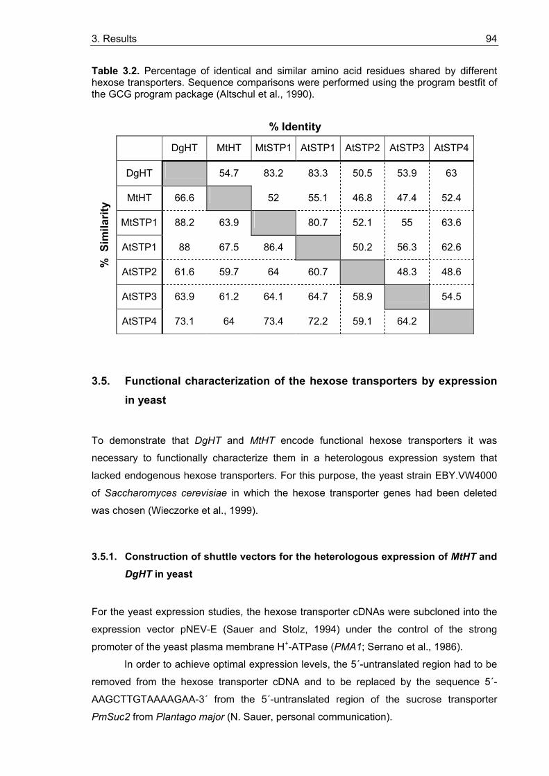
3. Results 94
Table 3.2. Percentage of identical and similar amino acid residues shared by different hexose transporters. Sequence comparisons were performed using the program bestfit of the GCG program package (Altschul et al., 1990).
% Identity
DgHT MtHT MtSTP1 AtSTP1 AtSTP2 AtSTP3 AtSTP4
DgHT 54.7 83.2 83.3 50.5 53.9 63
MtHT 66.6 52 55.1 46.8 47.4 52.4
MtSTP1 88.2 63.9 80.7 52.1 55 63.6
AtSTP1 88 67.5 86.4 50.2 56.3 62.6
AtSTP2 61.6 59.7 64 60.7 48.3 48.6
AtSTP3 63.9 61.2 64.1 64.7 58.9 54.5
% S
imila
rity
AtSTP4 73.1 64 73.4 72.2 59.1 64.2
3.5. Functional characterization of the hexose transporters by expression in yeast
To demonstrate that DgHT and MtHT encode functional hexose transporters it was
necessary to functionally characterize them in a heterologous expression system that
lacked endogenous hexose transporters. For this purpose, the yeast strain EBY.VW4000
of Saccharomyces cerevisiae in which the hexose transporter genes had been deleted
was chosen (Wieczorke et al., 1999).
3.5.1. Construction of shuttle vectors for the heterologous expression of MtHT and DgHT in yeast
For the yeast expression studies, the hexose transporter cDNAs were subcloned into the
expression vector pNEV-E (Sauer and Stolz, 1994) under the control of the strong
promoter of the yeast plasma membrane H+-ATPase (PMA1; Serrano et al., 1986).
In order to achieve optimal expression levels, the 5´-untranslated region had to be
removed from the hexose transporter cDNA and to be replaced by the sequence 5´-
AAGCTTGTAAAAGAA-3´ from the 5´-untranslated region of the sucrose transporter
PmSuc2 from Plantago major (N. Sauer, personal communication).
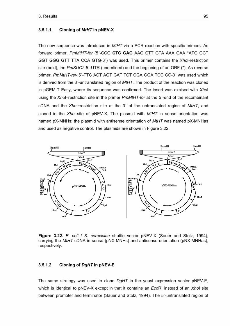
3. Results 95
3.5.1.1. Cloning of MtHT in pNEV-X
The new sequence was introduced in MtHT via a PCR reaction with specific primers. As
forward primer, PmMtHT-for (5´-CCG CTC GAG AAG CTT GTA AAA GAA *ATG GCT
GGT GGG GTT TTA CCA GTG-3´) was used. This primer contains the XhoI-restriction
site (bold), the PmSUC2-5´-UTR (underlined) and the beginning of an ORF (*). As reverse
primer, PmMtHT-rev 5´-TTC ACT AGT GAT TCT CGA GGA TCC GC-3´ was used which
is derived from the 3´-untranslated region of MtHT. The product of the reaction was cloned
in pGEM-T Easy, where its sequence was confirmed. The insert was excised with XhoI
using the XhoI-restriction site in the primer PmMtHT-for at the 5´-end of the recombinant
cDNA and the XhoI-restriction site at the 3´ of the untranslated region of MtHT, and
cloned in the XhoI-site of pNEV-X. The plasmid with MtHT in sense orientation was
named pX-MNHs; the plasmid with antisense orientation of MtHT was named pX-MNHas
and used as negative control. The plasmids are shown in Figure 3.22.
XhoI MtHT
pNX-MNHs
BamHI BamHI BamHI
XhoI MtHT
pNX-MNHas
BamHI
Figure 3.22. E. coli / S. cerevisiae shuttle vector pNEV-X (Sauer and Stolz, 1994), carrying the MtHT cDNA in sense (pNX-MNHs) and antisense orientation (pNX-MNHas), respectively.
3.5.1.2. Cloning of DgHT in pNEV-E
The same strategy was used to clone DgHT in the yeast expression vector pNEV-E,
which is identical to pNEV-X except in that it contains an EcoRI instead of an XhoI site
between promoter and terminator (Sauer and Stolz, 1994). The 5´-untranslated region of
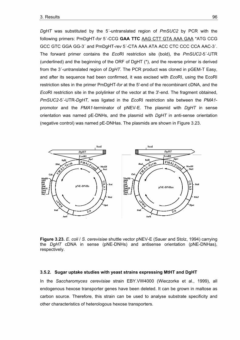
3. Results 96
DgHT was substituted by the 5´-untranslated region of PmSUC2 by PCR with the
following primers: PmDgHT-for 5´-CCG GAA TTC AAG CTT GTA AAA GAA *ATG CCG
GCC GTC GGA GG-3´ and PmDgHT-rev 5´-CTA AAA ATA ACC CTC CCC CCA AAC-3´.
The forward primer contains the EcoRI restriction site (bold), the PmSUC2-5´-UTR
(underlined) and the beginning of the ORF of DgHT (*), and the reverse primer is derived
from the 3´-untranslated region of DgHT. The PCR product was cloned in pGEM-T Easy,
and after its sequence had been confirmed, it was excised with EcoRI, using the EcoRI
restriction sites in the primer PmDgHT-for at the 5'-end of the recombinant cDNA, and the
EcoRI restriction site in the polylinker of the vector at the 3'-end. The fragment obtained,
PmSUC2-5´-UTR-DgHT, was ligated in the EcoRI restriction site between the PMA1-
promotor and the PMA1-terminator of pNEV-E. The plasmid with DgHT in sense
orientation was named pE-DNHs, and the plasmid with DgHT in anti-sense orientation
(negative control) was named pE-DNHas. The plasmids are shown in Figure 3.23.
ScaI
EcoRI DgHT
pNE-DNHs
ScaI
EcoRI DgHT
pNE-DNHas
Figure 3.23. E. coli / S. cerevisiae shuttle vector pNEV-E (Sauer and Stolz, 1994) carrying the DgHT cDNA in sense (pNE-DNHs) and antisense orientation (pNE-DNHas), respectively.
3.5.2. Sugar uptake studies with yeast strains expressing MtHT and DgHT
In the Saccharomyces cerevisiae strain EBY.VW4000 (Wieczorke et al., 1999), all
endogenous hexose transporter genes have been deleted. It can be grown in maltose as
carbon source. Therefore, this strain can be used to analyse substrate specificity and
other characteristics of heterologous hexose transporters.
3. Results 97
3.5.2.1. Functional characterization of MtHT
Hexose uptake studies with EBY.VW4000(pNX-MNHs) and EBY.VW4000(pNX-MNHas)
did not reveal any uptake activity of MtHT. Of several monosaccharides tested as
potential substrates (D-glucose, 3-O-methylglucose, D-galactose, D-fructose, D-mannose,
D-xylose and D-ribose), none were taken up by either EBY.VW4000(pE-MNHs) or
EBY.VW4000(pE-MNHas). This may be due to a PCR error in the sequence of MtHT
since the original cDNA sequence was derived from a PCR product (3.4.1), the error
might have accured already during the original amplification. It is also possible that MtHT
does not in fact represent a hexose transporter. It has been shown that a member of the
PTR family of nitrate and oligopeptide transporters transports malate (Jeong et al.,
submitted). However, a PCR error is the most likely explanation.
3.5.2.2. Functional characterization of DgHT
Hexose uptake studies (see 2.26.) with EBY.VW4000(pNE-DNHs) and
EBY.VW4000(pNE-DNHas) showed that DgHT accepts several monosaccharides as
substrates (D-glucose >> D-galactose ≥ D-xylose > D-mannose; see Figure 3.24). Under
the same conditions no uptake of D-ribose, D-fructose and of the non-metabolizable
glucose analogue 3-O-methylglucose (3-OMG) could be detected.
The Km value of DgHT for glucose was 43 µM (Figure 3.25), which is in the range
of Km values of other plant sugar transporters, such as AtSTP1 in Arabidopsis (20 µM;
Stolz et al., 1994), VfSTP1 in Vicia faba (30 µM; Weber et al., 1997), RcHEX3 in Ricinus
communis (80 µM; Weig et al., 1994). The Vmax of DgHT amounts to 2 nmol µl-1p.c. h-1
(p.c., packed cells; see 2.26).
The energy dependence of monosaccharide transport by DgHT was analysed
using D-glucose. No glucose transport was demonstrated in yeast cells expressing DgHT
in antisense orientation (Figure 3.26). The results presented in Figure 3.26 show that
DgHT catalyzes the uptake of glucose in an energy-dependent manner: (i) uptake rates
for glucose increased in the presence of added energy souce, namely the 1% ethanol,
while (ii) uncouplers of transmembrane proton gradients, namely 50 µM 2,4-dinitro phenol
(DNP) or 50 µM carbonyl cyanide-m-chlorophenylhydrazone (CCCP), completely
abolished glucose uptake by DgHT.
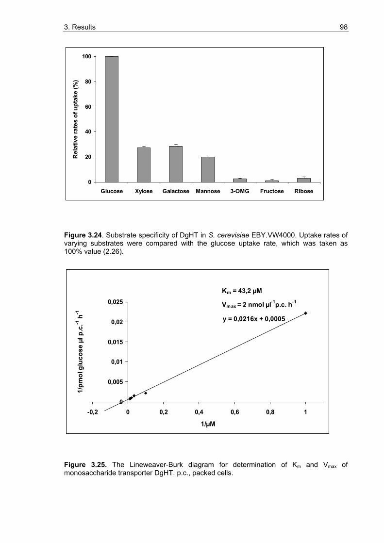
3. Results 98
0
20
40
60
80
100
Glucose Xylose Galactose Mannose 3-OMG Fructose Ribose
Rel
ativ
e ra
tes
of u
ptak
e (%
)
Figure 3.24. Substrate specificity of DgHT in S. cerevisiae EBY.VW4000. Uptake rates of varying substrates were compared with the glucose uptake rate, which was taken as 100% value (2.26).
y = 0,0216x + 0,0005
0
0,005
0,01
0,015
0,02
0,025
-0,2 0 0,2 0,4 0,6 0,8 1
1/µM
1/pm
ol g
luco
se µ
l p.c
.-1 h
-1
Km = 43,2 µM
Vmax = 2 nmol µl-1p.c. h-1
Figure 3.25. The Lineweaver-Burk diagram for determination of Km and Vmax of monosaccharide transporter DgHT. p.c., packed cells.
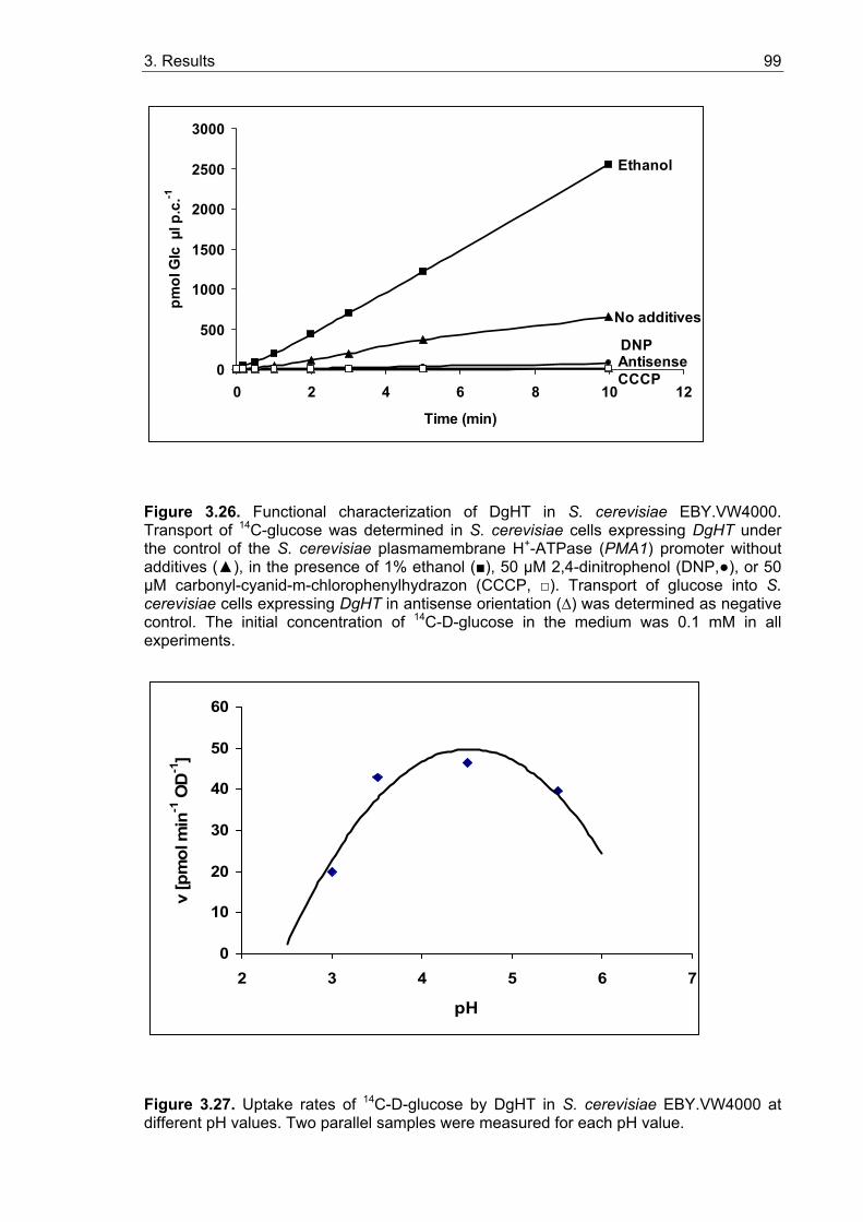
3. Results 99
No additives
Antisense
Ethanol
DNP
CCCP0
500
1000
1500
2000
2500
3000
0 2 4 6 8 10 12
Time (min)
pmol
Glc
µl p
.c.-1
Figure 3.26. Functional characterization of DgHT in S. cerevisiae EBY.VW4000. Transport of 14C-glucose was determined in S. cerevisiae cells expressing DgHT under the control of the S. cerevisiae plasmamembrane H+-ATPase (PMA1) promoter without additives (▲), in the presence of 1% ethanol (■), 50 µM 2,4-dinitrophenol (DNP,●), or 50 µM carbonyl-cyanid-m-chlorophenylhydrazon (CCCP, □). Transport of glucose into S. cerevisiae cells expressing DgHT in antisense orientation (∆) was determined as negative control. The initial concentration of 14C-D-glucose in the medium was 0.1 mM in all experiments.
0
10
20
30
40
50
60
2 3 4 5 6 7
pH
v [p
mol
min
-1 O
D-1
]
Figure 3.27. Uptake rates of 14C-D-glucose by DgHT in S. cerevisiae EBY.VW4000 at different pH values. Two parallel samples were measured for each pH value.
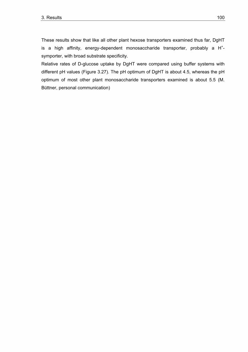
3. Results 100
These results show that like all other plant hexose transporters examined thus far, DgHT
is a high affinity, energy-dependent monosaccharide transporter, probably a H+-
symporter, with broad substrate specificity.
Relative rates of D-glucose uptake by DgHT were compared using buffer systems with
different pH values (Figure 3.27). The pH optimum of DgHT is about 4.5, whereas the pH
optimum of most other plant monosaccharide transporters examined is about 5.5 (M.
Büttner, personal communication)
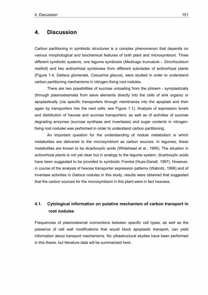
4. Discussion 101
4. Discussion Carbon partitioning in symbiotic structures is a complex phenomenon that depends on
various morphological and biochemical features of both plant and microsymbiont. Three
different symbiotic systems, one legume symbiosis (Medicago truncatula – Sinorhizobium
meliloti) and two actinorhizal symbioses from different subclades of actinorhizal plants
(Figure 1.4; Datisca glomerata, Casuarina glauca), were studied in order to understand
carbon partitioning mechanisms in nitrogen-fixing root nodules.
There are two possibilities of sucrose unloading from the phloem - symplastically
(through plasmodesmata from sieve elements directly into the cells of sink organs) or
apoplastically (via specific transporters through membranes into the apoplast and then
again by transporters into the next cells; see Figure 1.1). Analysis of expression levels
and distribution of hexose and sucrose transporters, as well as of activities of sucrose
degrading enzymes (sucrose synthase and invertases) and sugar contents in nitrogen-
fixing root nodules was performed in order to understand carbon partitioning.
An important question for the understanding of nodule metabolism is which
metabolites are delivered to the microsymbiont as carbon sources. In legumes, these
metabolites are known to be dicarboxylic acids (Whitehead et al., 1995). The situation in
actinorhizal plants is not yet clear but in analogy to the legume system, dicarboxylic acids
have been suggested to be provided to symbiotic Frankia (Huss-Danell, 1997). However,
in course of the analysis of hexose transporter expression patterns (Wabnitz, 1998) and of
invertase activities in Datisca nodules in this study, results were obtained that suggested
that the carbon sources for the microsymbiont in this plant were in fact hexoses.
4.1. Cytological information on putative mechanism of carbon transport in root nodules
Frequencies of plasmodesmal connections between specific cell types, as well as the
presence of cell wall modifications that would block apoplastic transport, can yield
information about transport mechanisms. No ultrastructural studies have been performed
in this thesis, but literature data will be summarized here.
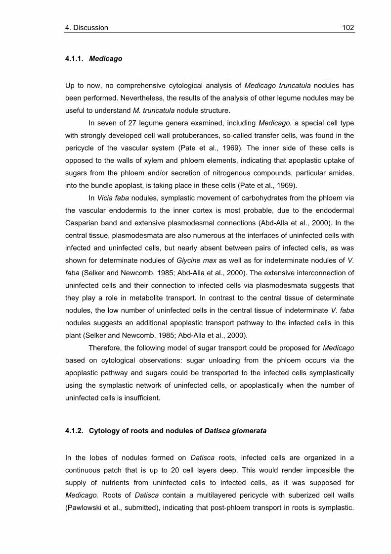
4. Discussion 102
4.1.1. Medicago
Up to now, no comprehensive cytological analysis of Medicago truncatula nodules has
been performed. Nevertheless, the results of the analysis of other legume nodules may be
useful to understand M. truncatula nodule structure.
In seven of 27 legume genera examined, including Medicago, a special cell type
with strongly developed cell wall protuberances, so-called transfer cells, was found in the
pericycle of the vascular system (Pate et al., 1969). The inner side of these cells is
opposed to the walls of xylem and phloem elements, indicating that apoplastic uptake of
sugars from the phloem and/or secretion of nitrogenous compounds, particular amides,
into the bundle apoplast, is taking place in these cells (Pate et al., 1969).
In Vicia faba nodules, symplastic movement of carbohydrates from the phloem via
the vascular endodermis to the inner cortex is most probable, due to the endodermal
Casparian band and extensive plasmodesmal connections (Abd-Alla et al., 2000). In the
central tissue, plasmodesmata are also numerous at the interfaces of uninfected cells with
infected and uninfected cells, but nearly absent between pairs of infected cells, as was
shown for determinate nodules of Glycine max as well as for indeterminate nodules of V.
faba (Selker and Newcomb, 1985; Abd-Alla et al., 2000). The extensive interconnection of
uninfected cells and their connection to infected cells via plasmodesmata suggests that
they play a role in metabolite transport. In contrast to the central tissue of determinate
nodules, the low number of uninfected cells in the central tissue of indeterminate V. faba
nodules suggests an additional apoplastic transport pathway to the infected cells in this
plant (Selker and Newcomb, 1985; Abd-Alla et al., 2000). Therefore, the following model of sugar transport could be proposed for Medicago
based on cytological observations: sugar unloading from the phloem occurs via the
apoplastic pathway and sugars could be transported to the infected cells symplastically
using the symplastic network of uninfected cells, or apoplastically when the number of
uninfected cells is insufficient.
4.1.2. Cytology of roots and nodules of Datisca glomerata
In the lobes of nodules formed on Datisca roots, infected cells are organized in a
continuous patch that is up to 20 cell layers deep. This would render impossible the
supply of nutrients from uninfected cells to infected cells, as it was supposed for
Medicago. Roots of Datisca contain a multilayered pericycle with suberized cell walls
(Pawlowski et al., submitted), indicating that post-phloem transport in roots is symplastic.
4. Discussion 103
However, since the vascular system of nodules does not contain cells with suberized
walls, both apoplastic and symplastic phloem unloading and post-phloem transport are
possible here. However, the presence of fluorescent material, i.e. polyphenolics, in the
walls of the endodermis of the nodule vascular system (Figure 4.1) indicates that here,
apoplastic transport is blocked, so symplastic transport is needed to cross this cell layer.
4.1.3. Cytology of roots and nodules of Casuarina glauca
In the cortex of nodules of Casuarina, infected and uninfected cells are interspersed. Cells
infected by Frankia are characterized by strong lignification of their cell walls (Berg and
McDowell, 1987b) that together with the low volume of intercellular space in the infection
zone (Zeng et al., 1989) restricts oxygen diffusion and therefore protects the oxygen-
sensitive bacterial nitrogenase. These morphological features disable apoplastic transport
into infected cells. The cell walls of uninfected cells of the nodule cortex and cells of the
multilayered pericycle of the nodule vascular system are not lignified (Pawlowski, 2002).
No analysis of the distribution of plasmodesmata has been performed for Casuarina
nodules to date, but the density of infected cells in the cortex is too high to postulate
apoplastic sugar transport to the uninfected cells and symplastic transport from there to
the infected cells. Altogether, based on current cytological information, symplastic
transport of nutrients is expected to take place in the nodule cortex, while both symplastic
Figure 4.1. Dark field micrograph of a longitudinal section of a Datisca nodule lobe photographed under fluorescent light. No fluorescent material can be detected in the walls of the phloem (P) of the nodule vascular bundle, but the cell walls of the endodermis (E) show bright autofluorescence. The photo-graphed region is located next to the fixation zone of the infected cells (∗). X – xylem (Courtesy from K. Pawlowski, unpublished).
E
P
X ∗
4. Discussion 104
and apoplastic pathways are possible for phloem unloading and post-phloem transport to
the nodule cortex.
4.2. Expression levels and patterns of hexose transporter genes in roots and nodules
According to the apoplastic model of phloem unloading, this pathway depends on the
presence of several types of specific transporters that catalyse the membrane penetration
of sugars (Eschrich, 1980; Sauer et al., 1994; Figure 1.1A). Therefore, apoplastic
transport activities can be implied from the expression levels and patterns of sugar
transporter genes.
4.2.1. Medicago
It has been shown in this study that in Medicago, apoplastic sugar transport as signified
by the expression levels of hexose and sucrose uptake transporter genes (MtHT and
MtST; Figure 3.18) plays a major role in roots. Similarly, M. Harrison (1996) has shown
previously that the expression levels of the hexose transporter gene MtSTP1 which is
homologous but not identical with MtHT, were higher in roots of Medicago than in stems
or in young or old leaves. The situation in nodules was different. The sucrose transporter
MtST was expressed at extremely low levels in nodules, and expression of the hexose
transporter MtHT in nodules could not be detected by RNA gel blot hybridization (Figure
3.18). In previous experiments, Medicago nodules had been grown in hydroponic culture
in Fåhraeus medium (Fåhraeus, 1957) where nodules needed two times longer to develop
than in aeroponic culture in Lullien's medium (Lullien et al., 1987). These plants had
displayed the same high expression levels for MtHT and MtST in nodules as in roots
(Wabnitz, 1998; Schubert et al., submitted). Therefore, while in roots the expression levels
of sugar transporter genes seem to be independent of the growth conditions, in nodules
this process is regulated, probably by oxygen supply. The data obtained indicate that
sugar partitioning mechanisms in Medicago change from apoplastic in roots to symplastic
in nodules in aeroponic culture. There are only few studies in wheat on the modulation of
plasmodesmal-mediated cell-to-cell transport roots under anaerobic stress, that do not
give a final answer if the plasmodesmata and assimilate transport become occluded under
anaerobic stress conditions. On the one hand, it was shown that the electrical coupling
ratio, which is a measure of plasmodesmatal resistance, between cortical cells of weat
4. Discussion 105
roots was not affected by the low oxygen treatment, suggesting that solute transport
through the cytoplasmic annulus of plasmodesmata was not affected (Zhang and
Tyerman, 1997). Moreover, the size exclusion limit (SEL) of the plasmodesmata rises
under anaerobiosis (Cleland et al., 1994). On the other hand, there is a hypothesis that
the transport through the plasmodesmata does not only involve the cytoplasmic
compartment, but also the desmotubule which is part of the endoplasmic reticulum (ER)
(Gamalei, 1994). In this context, Davies et al. (1987) have observed that the ER
condenses into whorls when the energy charge is reduced by hypoxia. This may well
account for the observation that there is a significant increase in molecular SEL, described
by Cleland et al. (1994).
4.2.2. Datisca
Analysis of the expression levels of sugar transport genes in Datisca showed a very
strong induction of hexose transporter gene expression in nodules compared to roots
(Figure 3.18). In situ localization of sugar transporter gene expression had been
performed previously by Wabnitz (1998). Expression of the hexose transporter gene
DgHT was found only in infected cells, while sucrose transporter (DgST) gene expression
was detected at very low levels in the pericycle of the nodule vascular system adjacent to
the senescence zone of infected cells. Expression levels of DgST in roots, nodules and
leaves were below the detection limit for RNA gel blot hybridization. Hence, the sucrose
transporter does not seem to play a major role in the Datisca – Frankia symbiosis.
However, the hexose transporter gene expression pattern seems to indicate that infected
cells take up large amount of hexoses from the apoplast.
In roots of Datisca low expression levels of sugar transporter genes were found,
that indicating that here, sugar transport mostly occurs symplastically, which is consistent
with the cytological data (4.1.2.).
4.2.3. Casuarina
RNA gel blot hybridization analysis of hexose transporter gene (CgHT) expression in
Casuarina showed that it was somewhat induced in nodules compared to roots,
resembling the situation in Datisca. However, all attempts to determine the expression
pattern of CgHT in situ failed. Since the walls of these cells are lignified (Berg and
McDowell, 1987b), which would block apoplastic transport, it is extremely unlikely that
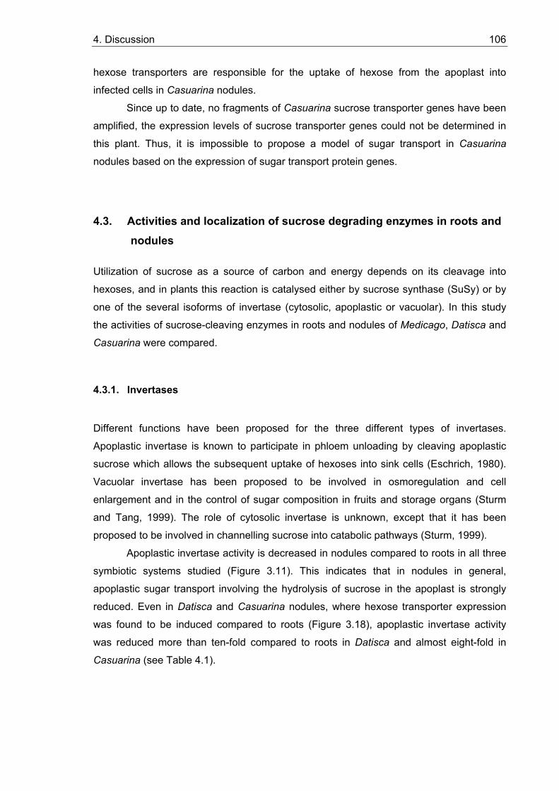
4. Discussion 106
hexose transporters are responsible for the uptake of hexose from the apoplast into
infected cells in Casuarina nodules.
Since up to date, no fragments of Casuarina sucrose transporter genes have been
amplified, the expression levels of sucrose transporter genes could not be determined in
this plant. Thus, it is impossible to propose a model of sugar transport in Casuarina
nodules based on the expression of sugar transport protein genes.
4.3. Activities and localization of sucrose degrading enzymes in roots and nodules
Utilization of sucrose as a source of carbon and energy depends on its cleavage into
hexoses, and in plants this reaction is catalysed either by sucrose synthase (SuSy) or by
one of the several isoforms of invertase (cytosolic, apoplastic or vacuolar). In this study
the activities of sucrose-cleaving enzymes in roots and nodules of Medicago, Datisca and
Casuarina were compared.
4.3.1. Invertases
Different functions have been proposed for the three different types of invertases.
Apoplastic invertase is known to participate in phloem unloading by cleaving apoplastic
sucrose which allows the subsequent uptake of hexoses into sink cells (Eschrich, 1980).
Vacuolar invertase has been proposed to be involved in osmoregulation and cell
enlargement and in the control of sugar composition in fruits and storage organs (Sturm
and Tang, 1999). The role of cytosolic invertase is unknown, except that it has been
proposed to be involved in channelling sucrose into catabolic pathways (Sturm, 1999).
Apoplastic invertase activity is decreased in nodules compared to roots in all three
symbiotic systems studied (Figure 3.11). This indicates that in nodules in general,
apoplastic sugar transport involving the hydrolysis of sucrose in the apoplast is strongly
reduced. Even in Datisca and Casuarina nodules, where hexose transporter expression
was found to be induced compared to roots (Figure 3.18), apoplastic invertase activity
was reduced more than ten-fold compared to roots in Datisca and almost eight-fold in
Casuarina (see Table 4.1).
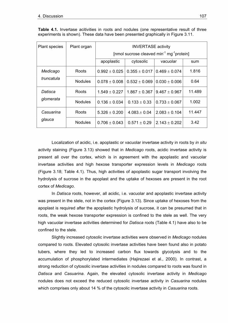
4. Discussion 107
Table 4.1. Invertase acitivities in roots and nodules (one representative result of three experiments is shown). These data have been presented graphically in Figure 3.11.
INVERTASE activity
[nmol sucrose cleaved min-1 mg-1protein]
Plant species Plant organ
apoplastic cytosolic vacuolar sum
Roots 0.992 ± 0.025 0.355 ± 0.017 0.469 ± 0.074 1.816 Medicago
truncatula Nodules 0.078 ± 0.008 0.532 ± 0.069 0.030 ± 0.006 0.64
Roots 1.549 ± 0.227 1.867 ± 0.367 9.467 ± 0.967 11.489 Datisca
glomerata Nodules 0.136 ± 0.034 0.133 ± 0.33 0.733 ± 0.067 1.002
Roots 5.326 ± 0.200 4.083.± 0.04 2.083 ± 0.104 11.447 Casuarina
glauca Nodules 0.706 ± 0.043 0.571 ± 0.29 2.143 ± 0.202 3.42
Localization of acidic, i.e. apoplastic or vacuolar invertase activity in roots by in situ
activity staining (Figure 3.13) showed that in Medicago roots, acidic invertase activity is
present all over the cortex, which is in agreement with the apoplastic and vacuolar
invertase activities and high hexose transporter expression levels in Medicago roots
(Figure 3.18; Table 4.1). Thus, high activities of apoplastic sugar transport involving the
hydrolysis of sucrose in the apoplast and the uptake of hexoses are present in the root
cortex of Medicago.
In Datisca roots, however, all acidic, i.e. vacuolar and apoplastic invertase activity
was present in the stele, not in the cortex (Figure 3.13). Since uptake of hexoses from the
apoplast is required after the apoplastic hydrolysis of sucrose, it can be presumed that in
roots, the weak hexose transporter expression is confined to the stele as well. The very
high vacuolar invertase activities determined for Datisca roots (Table 4.1) have also to be
confined to the stele.
Slightly increased cytosolic invertase activities were observed in Medicago nodules
compared to roots. Elevated cytosolic invertase activities have been found also in potato
tubers, where they led to increased carbon flux towards glycolysis and to the
accumulation of phosphorylated intermediates (Hajirezaei et al., 2000). In contrast, a
strong reduction of cytosolic invertase activities in nodules compared to roots was found in
Datisca and Casuarina. Again, the elevated cytosolic invertase activity in Medicago
nodules does not exceed the reduced cytosolic invertase activity in Casuarina nodules
which comprises only about 14 % of the cytosolic invertase activity in Casuarina roots.
4. Discussion 108
Vacuolar invertase activity is reduced in Medicago and Datisca nodules compared
to roots, but is similar in roots and nodules of Casuarina. It should be pointed out that
although vacuolar invertase activity is dramatically reduced in Datisca nodules compared
to roots, the remaining activity in Datisca nodules is still about 20 times higher than in
Medicago roots (Table 4.1). The high vacuolar invertase activities in Casuarina and
Datisca nodules, compared to nodules of Medicago (see Table 4.1) may be related to
different requirements for turgor control in actinorhizal versus legume nodules. The
mechanism of cell infection is different in actinorhizal nodules than in Medicago. In
actinorhizal nodules, no complete endocytotic process takes place but the perisymbiont
space remains continuous with the apoplast (Mylona et al., 1995). This aspect might
necessitate specific mechanisms for turgor control. However, more symbiotic systems
have to be examined before such a conclusion can be drawn.
To summarize the data on invertase activities, Medicago is characterized by
comparatively low activities of all three invertase types – apoplastic, cytosolic and
vacuolar – compared to Datisca and Casuarina. The reduction of total invertase activities
in nodules compared to roots, however, is about three times stronger in Datisca than in
the two other systems. The in situ staining results for acidic invertase activity in roots
indicate that phloem unloading involves apoplastic transport in Datisca (Figure 3.13B). However, post-phloem transport mechanisms in Medicago and Datisca roots obviously
differ, with apoplastic mechanisms dominating in Medicago. This is consistent with the
results about sugar transporter gene expression levels.
The roots of the two actinorhizal plants differ in respect to their invertase activity
distribution among isoforms. In Datisca roots, vacuolar invertase plays a major role,
whereas in Casuarina roots activities of both apoplastic and cytosolic invertases are high,
and vacuolar invertase activity is lower (2.1 ± 0.1 nmol sucrose cleaved/min/mg protein
(vacuolar) in comparison to 4.1 ± 0.04 (cytosolic) and 5.3 ± 0.2 (apoplastic) activity).
As was mentioned above, the common feature of all systems studied here is a
clear reduction of apoplastic invertase activity in nodules compared to roots. This means
that the apoplastic transport of sucrose followed by hydrolysis in the apoplast is strongly
reduced in nodules compared to roots.
4.3.2. Sucrose synthase (SuSy)
SuSy is recognized as an important enzyme of sucrose utilization in plant sink tissues (Ho
et al., 1988). SuSy is a globular protein and thus is generally considered to be soluble in
the cytosol. However, some percentage of the enzyme is known to be associated with the
plasma membrane, perhaps in a specific complex with glucan synthase, producing
4. Discussion 109
cellulose and/or callose (Amor et al., 1995; Carlson and Chourey, 1996). Part of the
soluble fraction of sucrose synthase seems to be associated with the actin cytoskeleton
(Winter et al., 1998).
Since in all three types of nodules, invertase activities were reduced in nodules compared
to roots, it can be concluded that SuSy has a dominant function in nodule sugar
metabolism. This is in agreement with results of Gordon et al. (1999) on a pea mutant with
a defect in the SuSy gene expressed in nodules, which caused a dramatic reduction of
nitrogenase activity, and thus led to an inefficient symbiosis. SuSy activities are expected
to be increased in nodules compared to roots, since a strong increase of sucrose
synthase transcription levels in nodules has been reported for the SuSy genes of soybean
(Thummler and Verma, 1987), Medicago (Hohnjec et al., 1999) and broad bean (Küster et
al., 1993). However, in this study, more or less equal SuSy activities were found in
nodules and roots of Medicago and Datisca (see 3.2.2). This is particularly suprising when
considering that there is clear transcriptional induction of SuSy in both systems (Figure
3.16) and that in Datisca, a corresponding increase of SuSy protein amounts was found in
nodules compared to roots (Figure 3.17). However, there was no increase of SuSy protein
in Medicago nodules compared to roots in spite of the transcriptional induction of the
SuSy gene (Figures 3.16 and 3.17). A similar situation has already been described for the
SuSy gene sh1 in maize roots, where the increase of sh1-mRNA levels under anaerobic
conditions was not reflected to the same degree in SS1 protein amounts which showed no
detectable increase (McElfresh and Chourey, 1988; Taliercio and Chourey, 1989). In
contrast, the transcription levels of the other maize gene coding for sucrose synthase
SS2, sus1, decreased during anaerobic stress as indicated by the reduced amounts of
sus1 mRNA in root tissue, while SS2 protein levels actually increased in the root tip
(McElfresh and Chourey, 1988). In summary, for maize roots it has been shown that the
expression of both SuSy genes is regulated at both the transcriptional and the
translational level, and that both levels of regulation may act against each other
(McElfresh et al., 1988; Taliercio and Chourey, 1989). Thus, translational regulation of
SuSy mRNA could also occur in root nodules, particularly in the infected zone where
oxygen-limited conditions are established for nitrogenase function (Vance and Heichel,
1991).
Moreover, recently it was shown that pea contains three different isoforms of
sucrose synthase (Sus1, Sus2 and Sus3), and that the genes encoding these different
isoforms have distinct patterns of expression in different organs of the plant as well as
during organ development (Barratt et al., 2001). The presence of at least two SuSy
isoforms was detected immunologically in Medicago roots using antibodies raised against
SuSy from Vicia faba (Ross and Davies, 1992), while only one immunologically active
4. Discussion 110
SuSy isoform was found in nodules (see 3.3.2; Figure 3.17). One of the isoforms detected
was somewhat larger than the major isoform. This confirms the results of Hohnjec et al.
(1999), who found only one SuSy isoform in nodules of eight different legumes, but two
isoforms of SuSy in root and stem extracts of Medicago. Studying of the partial cDNA
sequences from Medicago truncatula at www.tigr.org shows that Medicago contains at
least five different SuSy genes, with DNA homologies to the Vicia faba probe used in this
study ranging from 67% to 91% (data not shown). Taking into account the existence of
multiple SuSy isoforms, one could also propose that discrepancy between the SuSy
expression pattern on the mRNA level and on the level of protein amount and enzyme
activity is the consequence of incomplete detection of isoformes in Medicago using a
cDNA probe from Vicia faba (Küster et al., 1993). To date, only a single isoform of SuSy
has been characterized from Vicia faba (Ross and Davies, 1992), whereas for Pisum
sativum it has been shown that one of its three SuSy isoforms, Sus2, has less sequence
similarity with the two other isoforms than they share among each other (only 70-71 %
identity; Barratt et al., 2001). However, Sus3 was found to be only expressed at very low
levels. On RNA gel blots containing total RNA, only expression levels in flowers and
young testas were high enough for detection, while analysis of Sus3 expression using
RNA gel blots with poly(A+) RNA revealed that the transcript was also present in nodules,
but not in roots (Barratt et al., 2001). Hence, if Medicago nodules would contain two larger
very homologous SuSy isoforms, while roots contained only one of them, an induction of
SuSy expression in nodules compared to roots would be apparent in RNA gel blot
hybridizations, but not on the protein level. Furthermore, considering the translational
regulation of SuSy (Winter and Huber, 2000), it is possible that only one of the mRNAs for
the major isoforms is translated, leading to similar protein levels and enzyme activities of
SuSy in Medicago roots and nodules in spite of an apparent transcriptional induction.
A study of literature shows that for SuSy a broad analysis of gene expression,
protein quantities and presence of isoforms has been performed in different legumes (see
e.g. Thummler and Verma, 1987; Buchner et al., 1998; Hohnjec et al., 1999; Küster et al.,
1993; Ross and Davies, 1992; Barratt et al., 2001). However, comparisons of SuSy
enzyme activities were performed only on nodules grown under different nutrient
conditions and stresses, which led to the conclusion that SuSy activity is sufficient for
normal nodule function (e.g. González et al., 1995; Gordon et al., 1997). Except for this
thesis, SuSy activities in roots and nodules have never been compared so far.
One of the known mechanisms of posttranslational regulation of SuSy activity is
the reversible protein phosphorylation (Winter and Huber, 2000). Based on the results for
phosphorylation of recombinant SuSy, it has been suggested that phosphorylation
specifically activates sucrose cleavage (Nakai et al., 1998). Apart from its effects on SuSy
4. Discussion 111
activity, phosphorylation/dephosphorylation of SuSy seems to play a role in the
distribution of the enzyme between cytosol, plasma membrane, and actin cytoskeleton
(Winter et al., 1997; Winter et al., 1998). Taking into account that the Km of non-
phosphorylated recombinant SuSy protein for sucrose is several folds higher than that of
phosphorylated enzyme, it is possible to explain the lack of increase in SuSy activity in
Datisca nodules compared to roots by dephosphorylation of part of the total SuSy protein,
causing plasma membrane association and a decrease in enzyme activity. It is known that
SuSy protein associated with the plasma membrane is involved in cell wall biosynthesis
(Amor et al., 1995). The uptake of the microsymbiont into infected cells and its embedding
in a polysaccharide-rich cell wall-like matrix necessitates higher activities of synthesis of
cell wall-like material in nodules than in roots. Therefore, a change in the distribution of
SuSy protein in nodules relative to roots, with an increased fraction of the total SuSy
protein being associated with the plasma membrane may reflect the demands of the root
nodule symbiosis.
4.4. The Datisca nodule paradox: hexose transporter expression in the absence of apoplastic invertase activity. Could hexoses be the carbon sources for symbiotic Frankia in Datisca nodules?
As mentioned above, in nodules of Datisca apoplastic invertase activity was practically
absent (Figure 3.11B), indicating that no apoplastic sugar transport occurs that relies on
the hydrolysis of sucrose in the apoplast and the uptake of hexoses. However, in spite of
the absence of apoplastic invertase activities, a hexose transporter was found to be
expressed in nodules at high levels (Figure 3.18), and in situ hybridization showed that it
was expressed specifically in the infected cortical cells (Wabnitz, 1998). These data could
be interpreted to mean that the hexose transporter is working as an exporter to supply
endosymbiotic Frankia with carbon sources.
In analogy to legume nodules, where the bacteria are supposed to be supplied with
dicarboxylic acids by the host plant (see e.g. Streeter, 1995), it has been assumed that
dicarboxylic acids might also be the carbon source for symbiotic Frankia. While the
analysis of enzyme activities in Frankia vesicle clusters isolated from nodules of Alnus
and Hippophae yielded results consistent with this hypothesis (Akkermans et al., 1983),
experiments on 14CO2 uptake of detached Alnus nodules were inconclusive (Huss-Danell,
1990; summarized by Huss-Danell, 1997). Isolated Frankia strains have been divided into
two genomic groups based on DNA reassociation kinetics (Lechevalier and Lechevalier,
1990). In the free-living state, Frankia strains of genomic group A tend to grow fairly on
4. Discussion 112
both sugars and organic acids, while strains of genomic group B grow variably on organic
acids and poorly, if at all, on sugars (Lechevalier and Lechevalier, 1990; Benson and
Schultz, 1990). The endosymbiont of Datisca, however, has not been isolated yet and
based on its 16S rRNA and nifH gene sequences, does not belong to any group of
Frankia strains isolated so far (Mirza et al., 1994; Normand et al., 1996; Figure 1.4). Thus,
based on the data available, the carbon preferences of the endosymbiont of Datisca
cannot be determined, and the strains might be able to grow on hexoses as carbon
sources.
In summing up, there is no physiological evidence to contradict the hypothesis that
this group of strains in particular, or Frankia strains in general, are fed hexoses during
symbiosis. Based on the results presented in this study and in situ hybridisation data of
Wabnitz (1998), it is conceivable that in analogy to symbiotic nitrogen-fixing cyanobacteria
in Gunnera (R. Parsons, personal communication), endosymbiotic Frankia is supplied with
hexoses in nodules of Datisca.
4.5. Hexose transporters from nodules
As described above, an unusual function has been suggested for the hexose transporter
from Datisca nodules. Preliminary sequence information had also indicated that the
hexose transporter from Medicago nodules belongs to a different subfamily than all other hexose transporters published thus far (see sequence alignment in Figure 3.21; Table
3.2). Therefore, the full length cDNA sequences of the hexose transporters MtHT and
DgHT were amplified from Medicago and Datisca nodules and sequenced for further
analysis.
The amino acid sequences and the predicted secondary structure of DgHT were
highly similar to those of other monosaccharide transporters from plants, with the highest
homology to AtSTP1 from Arabidopsis (Table 3.2; Sauer et al., 1990). The analysis of the
full sequence of MtHT confirmed that this hexose transporter belongs indeed to a different
subfamily of transporters than AtSTP1, -2, -3 or -4, but its sequence is less dissimilar to
those of other hexose transporters than that of Arabidopsis AtSTP2. AtSTP2 contains two
insertions compared to other known hexose transporters from Arabidopsis as well as to
MtSTP1, MtHT and DgHT (see sequence alignment in Figure 3.21; Table 3.2).
To analyse the functional characteristics of DgHT , the cDNA was cloned in yeast
expression vectors and fused to the strong promoter of the yeast plasmamembrane H+-
ATPase (PMA1; Serrano et al., 1986) and the 5'-UTR of the sucrose transporter PmSuc2
from Plantago major which has been shown to result in high translation activities in yeast
4. Discussion 113
(N. Sauer, personal communication). DgHT was shown to represent a hexose transporter
with a broad substrate specificity, with the highest affinity to glucose, but also transporting
to a less extent D-galactose, D-xylose and D-mannose. DgHT did not transport fructose or
L-rhamnose. Regarding substrate specificity, DgHT has more in common with the hexose
transporter AtSTP3 from Arabidopsis than with AtSTP1 (Büttner and Sauer, 2000).
However, AtSTP3 has a Km of 2 mM for glucose, while other hexose transporters have Km
values for glucose in the micromolar range, like DgHT with 43 µM (Figure 3.25). Hence,
regarding substrate affinity DgHT does not resemble AtSTP3 at all. Furthermore, AtSTP3
was the first monosaccharide transporter not to be expressed in sink tissues (Büttner et
al., 2000), while DgHT is expressed in at least two types of sink organs, namely at high
levels in Datisca nodules and and at low levels in roots, while no DgHT transcripts were
detected in leaves by RNA gel blot hybridization (Figure 3.18). At any rate, these
comparisons show that hexose transporters of the same sequence group can have
different substrate specificities.
The most unusual feature of DgHT is its pH optimum which is more acidic (about
4.5; Figure 3.27) than it is known for other monosaccharide transporters with the pH
optima lying in the range of 5.5 (M. Büttner, personal communication). It is known that the
peribacteroid space in infected cells of legume nodules is more acidic than the apoplast
(Blumwald et al., 1985), which is also the case for the periarbuscular space in infected
cells of arbuscular mycorrhizal roots (Guttenberger, 2000). In analogy, it can be assumed
that the perisymbiont space in infected cells of actinorhizal nodules is also more acidic
than the apoplast. A more acidic pH optimum of a transporter specific to the perisymbiont
membrane (as implied by its supposed function in feeding the bacteria) would correspond
with a location in the perisymbiont membrane. This increased pH gradient between
cytosol and perisymbiont space would make the export of sugars in a proton-symport
mechanism more difficult, but facilitate the uptake of sugars by the symbiotic bacteria.
Up to now, plant sugar transporters could only be shown to be active in sugar
uptake (reviewed by Caspari et al., 1994), although activity in sugar export has been
discussed for a sucrose transporter (Truernit and Sauer, 1995) based on its expression
pattern and on comparisons of metabolite concentrations in apoplast and cytosol (Lohaus
et al., 1995). Interestingly, the only hexose transporter tested in vivo and in vitro (HUP1
from Chlorella kessleri), was found to catalyse strict H+-monosaccharide symport (uptake)
when expressed in yeast but was facilitating glucose diffusion in both directions when
used in vitro in artificial vesicles (Caspari et al., 1996). Another example that raises
questions about the reliability of transport directions determined in yeast is a recently
identified zinc transporter from the peribacteroid membrane of soybean nodules, GmZIP1
(Moreau et al., 2002). When expressed in yeast, it only catalyzes zinc uptake, but this
4. Discussion 114
direction of transport is extremely unlikely in the homologous system where the plant
supplies the microsymbiont with micronutrients and not vice versa. Thus, the membrane
environment seems to be able to influence a transporter's activity, which might allow the
hexose transporter of Datisca to perform glucose export when present in the perisymbiont
membrane of infected nodule cortical cells.
4.6. Nodule and root sugar contents
The steady state levels of soluble sugars in a plant organ can yield information about
metabolism. To complete the analysis of sugar partitioning in nodules, soluble sugars
were extracted from nodules and roots of the three model plants and analysed by HPLC
(Figures 3.1 - 3.3).
4.6.1. Sucrose, glucose and fructose in nodules and roots
A comparison of the amounts of sucrose, glucose and fructose in roots and nodules of the
three model plants shows several interesting aspects.
Unusually high sucrose concentrations were found in Casuarina nodules (Figure
3.2; Table 4.2). This phenomenon is probably due to the fact that in contrast with
Medicago and Datisca nodules, no starch biosynthesis takes place in Casuarina nodules
which means that sucrose may serve as carbon storage form. In other systems, sucrose
has been shown to be able to serve as long term carbon storage form, e.g. in sugar beet
(Reinefeld et al., 1986) or as transient carbon storage form, e.g. in barley leaves (Riens et
al., 1994).
The absence of fructose in extracts of Medicago nodules and roots (Figure 3.1;
Table 4.2) is probably caused by high fructokinase (FK) activities, which are often linked
with the operation of sucrose syntase (SuSy). A similar situation has been found in potato
tubers, where fructokinase has been suggested to reduce cytosolic fructose contents in
order to maintain a high SuSy-catalysed net flux of sucrose to phosphorylated hexoses
(Renz and Stitt, 1993; Appeldoorn et al., 1997). Both FK and SuSy genes are expressed
in similar fashion during the development of sink tissues and both enzymes are inhibited
by elevated fructose concentrations (Schaffer and Petreikov, 1997). Therefore, it is likely
to be important to effectively remove fructose to maintain high SuSy activities, which are
essential for nitrogen fixation in nodules (Gordon et al., 1999).
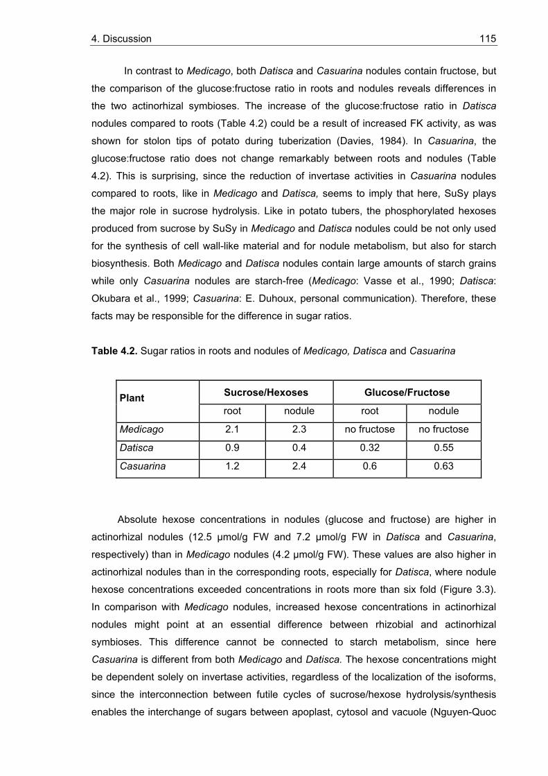
4. Discussion 115
In contrast to Medicago, both Datisca and Casuarina nodules contain fructose, but
the comparison of the glucose:fructose ratio in roots and nodules reveals differences in
the two actinorhizal symbioses. The increase of the glucose:fructose ratio in Datisca
nodules compared to roots (Table 4.2) could be a result of increased FK activity, as was
shown for stolon tips of potato during tuberization (Davies, 1984). In Casuarina, the
glucose:fructose ratio does not change remarkably between roots and nodules (Table
4.2). This is surprising, since the reduction of invertase activities in Casuarina nodules
compared to roots, like in Medicago and Datisca, seems to imply that here, SuSy plays
the major role in sucrose hydrolysis. Like in potato tubers, the phosphorylated hexoses
produced from sucrose by SuSy in Medicago and Datisca nodules could be not only used
for the synthesis of cell wall-like material and for nodule metabolism, but also for starch
biosynthesis. Both Medicago and Datisca nodules contain large amounts of starch grains
while only Casuarina nodules are starch-free (Medicago: Vasse et al., 1990; Datisca:
Okubara et al., 1999; Casuarina: E. Duhoux, personal communication). Therefore, these
facts may be responsible for the difference in sugar ratios.
Table 4.2. Sugar ratios in roots and nodules of Medicago, Datisca and Casuarina
Sucrose/Hexoses Glucose/Fructose Plant root nodule root nodule
Medicago 2.1 2.3 no fructose no fructose
Datisca 0.9 0.4 0.32 0.55
Casuarina 1.2 2.4 0.6 0.63
Absolute hexose concentrations in nodules (glucose and fructose) are higher in
actinorhizal nodules (12.5 µmol/g FW and 7.2 µmol/g FW in Datisca and Casuarina,
respectively) than in Medicago nodules (4.2 µmol/g FW). These values are also higher in
actinorhizal nodules than in the corresponding roots, especially for Datisca, where nodule
hexose concentrations exceeded concentrations in roots more than six fold (Figure 3.3).
In comparison with Medicago nodules, increased hexose concentrations in actinorhizal
nodules might point at an essential difference between rhizobial and actinorhizal
symbioses. This difference cannot be connected to starch metabolism, since here
Casuarina is different from both Medicago and Datisca. The hexose concentrations might
be dependent solely on invertase activities, regardless of the localization of the isoforms,
since the interconnection between futile cycles of sucrose/hexose hydrolysis/synthesis
enables the interchange of sugars between apoplast, cytosol and vacuole (Nguyen-Quoc
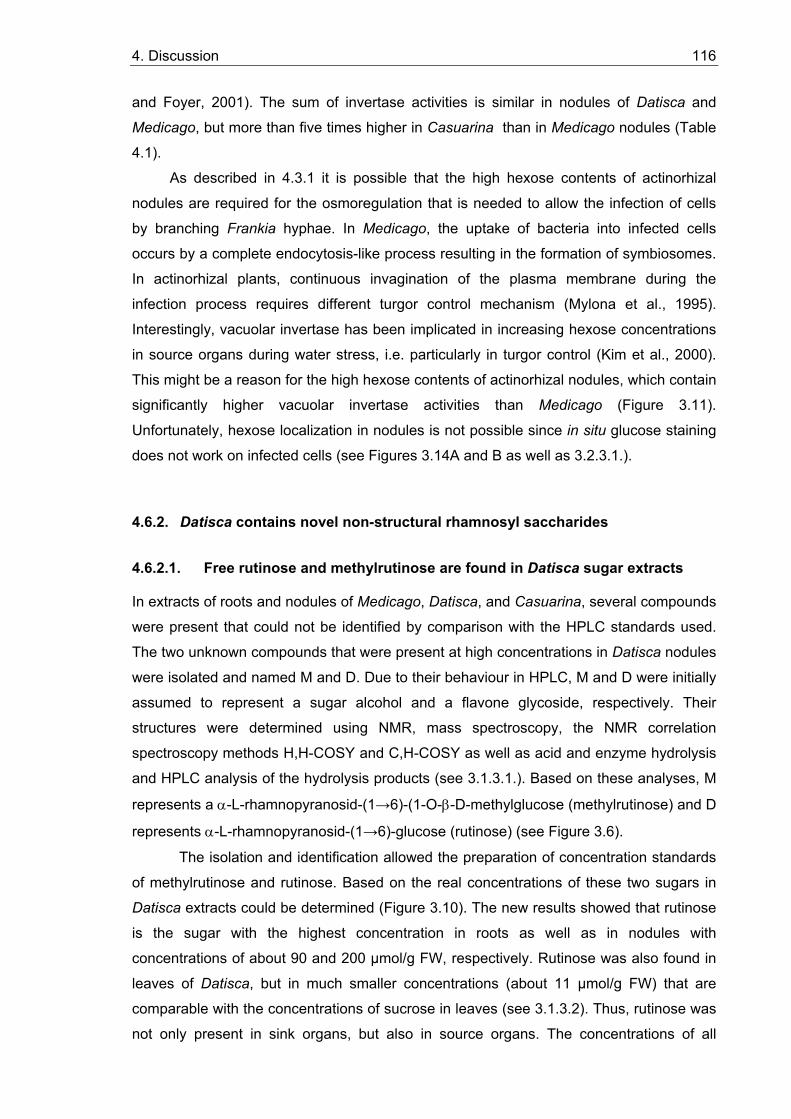
4. Discussion 116
and Foyer, 2001). The sum of invertase activities is similar in nodules of Datisca and
Medicago, but more than five times higher in Casuarina than in Medicago nodules (Table
4.1).
As described in 4.3.1 it is possible that the high hexose contents of actinorhizal
nodules are required for the osmoregulation that is needed to allow the infection of cells
by branching Frankia hyphae. In Medicago, the uptake of bacteria into infected cells
occurs by a complete endocytosis-like process resulting in the formation of symbiosomes.
In actinorhizal plants, continuous invagination of the plasma membrane during the
infection process requires different turgor control mechanism (Mylona et al., 1995).
Interestingly, vacuolar invertase has been implicated in increasing hexose concentrations
in source organs during water stress, i.e. particularly in turgor control (Kim et al., 2000).
This might be a reason for the high hexose contents of actinorhizal nodules, which contain
significantly higher vacuolar invertase activities than Medicago (Figure 3.11).
Unfortunately, hexose localization in nodules is not possible since in situ glucose staining
does not work on infected cells (see Figures 3.14A and B as well as 3.2.3.1.).
4.6.2. Datisca contains novel non-structural rhamnosyl saccharides
4.6.2.1. Free rutinose and methylrutinose are found in Datisca sugar extracts
In extracts of roots and nodules of Medicago, Datisca, and Casuarina, several compounds
were present that could not be identified by comparison with the HPLC standards used.
The two unknown compounds that were present at high concentrations in Datisca nodules
were isolated and named M and D. Due to their behaviour in HPLC, M and D were initially
assumed to represent a sugar alcohol and a flavone glycoside, respectively. Their
structures were determined using NMR, mass spectroscopy, the NMR correlation
spectroscopy methods H,H-COSY and C,H-COSY as well as acid and enzyme hydrolysis
and HPLC analysis of the hydrolysis products (see 3.1.3.1.). Based on these analyses, M
represents a α-L-rhamnopyranosid-(1→6)-(1-O-β-D-methylglucose (methylrutinose) and D
represents α-L-rhamnopyranosid-(1→6)-glucose (rutinose) (see Figure 3.6).
The isolation and identification allowed the preparation of concentration standards
of methylrutinose and rutinose. Based on the real concentrations of these two sugars in
Datisca extracts could be determined (Figure 3.10). The new results showed that rutinose
is the sugar with the highest concentration in roots as well as in nodules with
concentrations of about 90 and 200 µmol/g FW, respectively. Rutinose was also found in
leaves of Datisca, but in much smaller concentrations (about 11 µmol/g FW) that are
comparable with the concentrations of sucrose in leaves (see 3.1.3.2). Thus, rutinose was
not only present in sink organs, but also in source organs. The concentrations of all
4. Discussion 117
sugars in Datisca extracts except for rutinose are presented in Figure 4.2 to allow a more
precise comparison using a smaller scale than necessitated by the high concentrations of
rutinose in Figure 3.10. It can be seen that the concentrations of methylrutinose do not
differ dramatically from those of the other sugars in the corresponding plant organs,
except that leaves contain more sucrose than methylrutinose and nodules vice versa
(Figure 3.10).
0,0
5,0
10,0
15,0
20,0
25,0
Methyl-rutinose
Glucose Fructose Sucrose
µmol
/g F
W
RootsNodulesLeaves
Figure 4.2. Concentrations of methylrutinose, glucose, fructose and sucrose in roots, nodules and leaves of Datisca. Data for rutinose are omitted (see Figure 3.10). Average values of three parallel samples are shown.
4.6.2.2. Possible ways of rutinose and methylrutinose synthesis
A study of the literature on plant glycosides showed that rutinose has not been described
as free plant sugar, but only as glycoside part of many flavone glycosides, e.g. rutin,
galanginoside, cannabin, datiscin and datinoside, which have been isolated and
characterised from the only other Datisca species besides Datisca glomerata, Datisca
cannabina (Zapesochnaya and Pangarova, 1976). Datiscin represents 5,7,2'-
trihydroxyflavone-3-O-rutinose (Zapesochnaya et al., 1982). Methylrutinose is also not
known among free plant metabolites.
Many plant glycosides such as flavonoides, cardenolides, phenols, sterols,
coumarins and others, contain a disaccharidyl unit and sometimes even a larger
oligosaccharide. The synthesis of these glycosides involves stepwise, sequential
transglycosylations from the proper sugar nucleotide donors. The degradation of the
glycosides is the sequential removal of the monosaccharides by exoglycosidases.
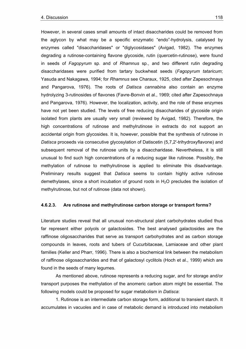
4. Discussion 118
However, in several cases small amounts of intact disaccharides could be removed from
the aglycon by what may be a specific enzymatic “endo”-hydrolysis, catalysed by
enzymes called "disaccharidases" or "diglycosidases" (Avigad, 1982). The enzymes
degrading a rutinose-containing flavone glycoside, rutin (quercetin-rutinose), were found
in seeds of Fagopyrum sp. and of Rhamnus sp., and two different rutin degrading
disaccharidases were purified from tartary buckwheat seeds (Fagopyrum tataricum;
Yasuda and Nakagawa, 1994; for Rhamnus see Charaux, 1925, cited after Zapesochnaya
and Pangarova, 1976). The roots of Datisca cannabina also contain an enzyme
hydrolyzing 3-rutinosides of flavones (Favre-Bonvin et al., 1969; cited after Zapesochnaya
and Pangarova, 1976). However, the localization, activity, and the role of these enzymes
have not yet been studied. The levels of free reducing disaccharides of glycoside origin
isolated from plants are usually very small (reviewed by Avigad, 1982). Therefore, the
high concentrations of rutinose and methylrutinose in extracts do not support an accidental origin from glycosides. It is, however, possible that the synthesis of rutinose in
Datisca proceeds via consecutive glycosylation of Datiscetin (5,7,2'-trihydroxyflavone) and
subsequent removal of the rutinose units by a disaccharidase. Nevertheless, it is still
unusual to find such high concentrations of a reducing sugar like rutinose. Possibly, the
methylation of rutinose to methylrutinose is applied to eliminate this disadvantage.
Preliminary results suggest that Datisca seems to contain highly active rutinose
demethylases, since a short incubation of ground roots in H2O precludes the isolation of
methylrutinose, but not of rutinose (data not shown).
4.6.2.3. Are rutinose and methylrutinose carbon storage or transport forms?
Literature studies reveal that all unusual non-structural plant carbohydrates studied thus
far represent either polyols or galactosides. The best analysed galactosides are the
raffinose oligosaccharides that serve as transport carbohydrates and as carbon storage
compounds in leaves, roots and tubers of Cucurbitaceae, Lamiaceae and other plant
families (Keller and Pharr, 1996). There is also a biochemical link between the metabolism
of raffinose oligosaccharides and that of galactosyl cyclitols (Hoch et al., 1999) which are
found in the seeds of many legumes.
As mentioned above, rutinose represents a reducing sugar, and for storage and/or
transport purposes the methylation of the anomeric carbon atom might be essential. The
following models could be proposed for sugar metabolism in Datisca:
1. Rutinose is an intermediate carbon storage form, additional to transient starch. It
accumulates in vacuoles and in case of metabolic demand is introduced into metabolism
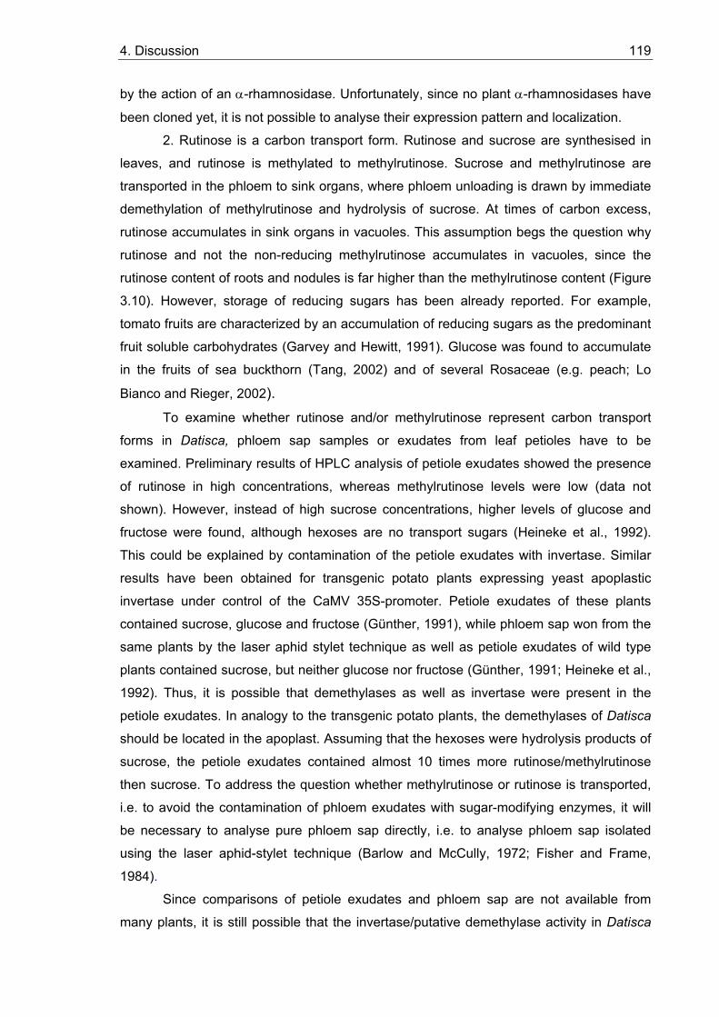
4. Discussion 119
by the action of an α-rhamnosidase. Unfortunately, since no plant α-rhamnosidases have
been cloned yet, it is not possible to analyse their expression pattern and localization.
2. Rutinose is a carbon transport form. Rutinose and sucrose are synthesised in
leaves, and rutinose is methylated to methylrutinose. Sucrose and methylrutinose are
transported in the phloem to sink organs, where phloem unloading is drawn by immediate
demethylation of methylrutinose and hydrolysis of sucrose. At times of carbon excess,
rutinose accumulates in sink organs in vacuoles. This assumption begs the question why
rutinose and not the non-reducing methylrutinose accumulates in vacuoles, since the
rutinose content of roots and nodules is far higher than the methylrutinose content (Figure
3.10). However, storage of reducing sugars has been already reported. For example,
tomato fruits are characterized by an accumulation of reducing sugars as the predominant
fruit soluble carbohydrates (Garvey and Hewitt, 1991). Glucose was found to accumulate
in the fruits of sea buckthorn (Tang, 2002) and of several Rosaceae (e.g. peach; Lo
Bianco and Rieger, 2002). To examine whether rutinose and/or methylrutinose represent carbon transport
forms in Datisca, phloem sap samples or exudates from leaf petioles have to be
examined. Preliminary results of HPLC analysis of petiole exudates showed the presence
of rutinose in high concentrations, whereas methylrutinose levels were low (data not
shown). However, instead of high sucrose concentrations, higher levels of glucose and
fructose were found, although hexoses are no transport sugars (Heineke et al., 1992).
This could be explained by contamination of the petiole exudates with invertase. Similar
results have been obtained for transgenic potato plants expressing yeast apoplastic
invertase under control of the CaMV 35S-promoter. Petiole exudates of these plants
contained sucrose, glucose and fructose (Günther, 1991), while phloem sap won from the
same plants by the laser aphid stylet technique as well as petiole exudates of wild type
plants contained sucrose, but neither glucose nor fructose (Günther, 1991; Heineke et al.,
1992). Thus, it is possible that demethylases as well as invertase were present in the
petiole exudates. In analogy to the transgenic potato plants, the demethylases of Datisca
should be located in the apoplast. Assuming that the hexoses were hydrolysis products of
sucrose, the petiole exudates contained almost 10 times more rutinose/methylrutinose
then sucrose. To address the question whether methylrutinose or rutinose is transported,
i.e. to avoid the contamination of phloem exudates with sugar-modifying enzymes, it will
be necessary to analyse pure phloem sap directly, i.e. to analyse phloem sap isolated
using the laser aphid-stylet technique (Barlow and McCully, 1972; Fisher and Frame,
1984).
Since comparisons of petiole exudates and phloem sap are not available from
many plants, it is still possible that the invertase/putative demethylase activity in Datisca
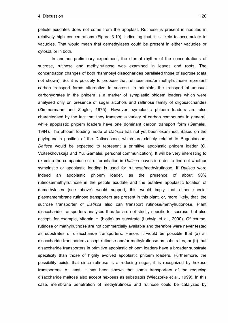
4. Discussion 120
petiole exudates does not come from the apoplast. Rutinose is present in nodules in
relatively high concentrations (Figure 3.10), indicating that it is likely to accumulate in
vacuoles. That would mean that demethylases could be present in either vacuoles or
cytosol, or in both.
In another preliminary experiment, the diurnal rhythm of the concentrations of
sucrose, rutinose and methylrutinose was examined in leaves and roots. The
concentration changes of both rhamnosyl disaccharides paralleled those of sucrose (data
not shown). So, it is possibly to propose that rutinose and/or methylrutinose represent
carbon transport forms alternative to sucrose. In principle, the transport of unusual
carbohydrates in the phloem is a marker of symplastic phloem loaders which were
analysed only on presence of sugar alcohols and raffinose family of oligosaccharides
(Zimmermann and Ziegler, 1975). However, symplastic phloem loaders are also
characterised by the fact that they transport a variety of carbon compounds in general,
while apoplastic phloem loaders have one dominant carbon transport form (Gamalei,
1984). The phloem loading mode of Datisca has not yet been examined. Based on the
phylogenetic position of the Datiscaceae, which are closely related to Begoniaceae,
Datisca would be expected to represent a primitive apoplastic phloem loader (O.
Voitsekhovskaja and Yu. Gamalei, personal communication). It will be very interesting to
examine the companion cell differentiation in Datisca leaves in order to find out whether
symplastic or apoplastic loading is used for rutinose/methylrutinose. If Datisca were
indeed an apoplastic phloem loader, as the presence of about 90%
rutinose/methylrutinose in the petiole exudate and the putative apoplastic location of
demethylases (see above) would support, this would imply that either special
plasmamembrane rutinose transporters are present in this plant, or, more likely, that the
sucrose transporter of Datisca also can transport rutinose/methylrutionose. Plant
disaccharide transporters analysed thus far are not strictly specific for sucrose, but also
accept, for example, vitamin H (biotin) as substrate (Ludwig et al., 2000). Of course,
rutinose or methylrutinose are not commercially available and therefore were never tested
as substrates of disaccharide transporters. Hence, it would be possible that (a) all
disaccharide transporters accept rutinose and/or methylrutinose as substrates, or (b) that
disaccharide transporters in primitive apoplastic phloem loaders have a broader substrate
specificity than those of highly evolved apoplastic phloem loaders. Furthermore, the
possibility exists that since rutinose is a reducing sugar, it is recognized by hexose
transporters. At least, it has been shown that some transporters of the reducing
disaccharide maltose also accept hexoses as substrates (Wieczorke et al., 1999). In this
case, membrane penetration of methylrutinose and rutinose could be catalyzed by
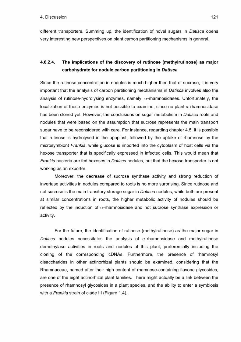
4. Discussion 121
different transporters. Summing up, the identification of novel sugars in Datisca opens
very interesting new perspectives on plant carbon partitioning mechanisms in general.
4.6.2.4. The implications of the discovery of rutinose (methylrutinose) as major carbohydrate for nodule carbon partitioning in Datisca
Since the rutinose concentration in nodules is much higher then that of sucrose, it is very
important that the analysis of carbon partitioning mechanisms in Datisca involves also the
analysis of rutinose-hydrolysing enzymes, namely, α-rhamnosidases. Unfortunately, the
localization of these enzymes is not possible to examine, since no plant α-rhamnosidase
has been cloned yet. However, the conclusions on sugar metabolism in Datisca roots and
nodules that were based on the assumption that sucrose represents the main transport
sugar have to be reconsidered with care. For instance, regarding chapter 4.5. it is possible
that rutinose is hydrolysed in the apoplast, followed by the uptake of rhamnose by the
microsymbiont Frankia, while glucose is imported into the cytoplasm of host cells via the
hexose transporter that is specifically expressed in infected cells. This would mean that
Frankia bacteria are fed hexoses in Datisca nodules, but that the hexose transporter is not
working as an exporter.
Moreover, the decrease of sucrose synthase activity and strong reduction of
invertase activities in nodules compared to roots is no more surprising. Since rutinose and
not sucrose is the main transitory storage sugar in Datisca nodules, while both are present
at similar concentrations in roots, the higher metabolic activity of nodules should be
reflected by the induction of α-rhamnosidase and not sucrose synthase expression or
activity.
For the future, the identification of rutinose (methylrutinose) as the major sugar in
Datisca nodules necessitates the analysis of α-rhamnosidase and methylrutinose
demethylase activities in roots and nodules of this plant, preferentially including the
cloning of the corresponding cDNAs. Furthermore, the presence of rhamnosyl
disaccharides in other actinorhizal plants should be examined, considering that the
Rhamnaceae, named after their high content of rhamnose-containing flavone glycosides,
are one of the eight actinorhizal plant families. There might actually be a link between the
presence of rhamnosyl glycosides in a plant species, and the ability to enter a symbiosis
with a Frankia strain of clade III (Figure 1.4).
5. Summary
122
5. Summary
In this thesis sucrose-cleaving enzymes and sugar transport proteins were studied to
determine the way of sugar transport and their role in the function of nitrogen-fixing root
nodules. Soluble sugar metabolite profile was analysed to reveal dominating sugars,
which could be important for the symbiotic function of root nodules. To find out a possible
common feature of root nodule symbioses, different model systems, one legume
symbiosis (Medicago truncatula – Sinorhizobium meliloti) and two actinorhizal symbioses
from different Frankia subclades, Datisca glomerata and Casuarina glauca were chosen.
In order to reveal whether sugar partitioning mechanisms represent system-specific
adaptations, i.e. resemble sugar partitioning mechanisms established in roots or are
common to all nitrogen-fixing root nodules, all experiments included comparisons between
roots and nodules.
The main results obtained can be summarized as follows:
• Legume and actinorhizal symbioses differ with respect to sugar transporter (HT)
expression: induction of HT expression was found in nodules of actinorhizal plants
Datisca and Casuarina, but expression levels of HT in Medicago nodules were
strongly reduced.
• Full-size cDNA sequences for hexose transporters were isolated from Medicago
(MtHT) and Datisca (DgHT) nodules. Based on their amino acid sequence, the
encoded proteins belonged to different classes of sugar transporters. The
functional characterization of DgHT in a heterologous system (yeast)
demonstrated that DgHT is a high affinity, energy-dependent monosaccharide
transporter, probably a H+-symporter, with broad substrate specificity.
• Expression levels of sucrose transporter (ST) in Medicago nodules were strongly
reduced compared to roots.
• The total (sum of apoplastic, cytosolic and vacuolar) invertase activity is strongly
reduced in nodules compared to roots of all three symbiotic systems studied.
• Different localizations of acid invertase activity suggest that sugar transport in
Medicago roots occurs apoplastically, whereas in Datisca roots it takes place
symplastically. There may be apoplastic transport in vascular parenchyma of
Datisca roots.
• The major sucrose-cleaving activity in Medicago and Datisca roots and nodules is
represented by sucrose synthase (SuSy). Datisca and Casuarina possess a
5. Summary
123
single immunoreactive protein for SuSy of about 92 kDa, Medicago contains an
additional, somewhat larger SuSy protein in roots, but not in nodules. SuSy
activities were similar in the soluble protein fractions in both roots and nodules of
Medicago. In Datisca nodules they were lower compared to roots.
• Two unknown sugars were isolated from Datisca and identified using biochemical
methods, NMR and mass spectroscopy. They were shown to represent rutinose
(α-L-rhamnopyranosyl-(1→6)-D-glucose) and methylrutinose (α-L-
rhamnopyranosyl-(1→6)-1-O-methyl-ß-D-glucose). Both of them were present in
large amounts in roots, nodules and leaves of Datisca.
• Sucrose is the principal sugar of Medicago and Casuarina nodules. The major
sugar of Datisca root and nodule extract is rutinose.
The data presented in this thesis show significant differences between carbon
partitioning mechanisms in the three symbiotic systems examined. However, some
common features were also noted.
In all systems studied there was a decrease of total invertase activity,
determined mostly by apoplastic invertase, in nodules compared to roots that could be
an evidence for symplastic transport of sugars in nodules. In Medicago, this was
underlined by the strong reduction of sugar transporter expression. This implies a shift
toward symplastic transport mechanisms in nodules, which is likely to represent a
symbiosis-specific adaptation in that symplastic transport allows the plant to control
the microsymbiont's access to carbon sources.
Both actinorhizal symbioses showed strong induction of hexose transporter
expression in nodules combined with a considerable reduction of apoplastic invertase
activity which would be needed to provide these transporters with substrates. The last
circumstance allows to propose a special role of the hexose transporter in supplying
the microsymbiont with carbon sources in nodules.
6. Abbreviations
124
6. Abbreviations
Amp ampicillin
AmpR ampicillin resistance
ATP adenosine 5´ triphosphate
bp base pairs
BSA bovine serum albumin
CAA casamino acids
CC companion cell
CCCP carbonyl cyanide-m-chlorophenylhydrazone
cDNA complementary DNA
CgHT Casuarina glauca hexose transporter
CW cell wall
DAB diaminobenzidine
dCTP deoxycytosine 5´ triphosphate
dd H2O double distilled water
DgHT Datisca glomerata hexose transporter
DMF dimethylformamide
DMSO dimethylsulfoxide
DNA deoxyribonucleic acid
DNase deoxyribonuclease
DNP 2,4-dinitrophenol
dNTP deoxynucleotide triphosphate
dNTPs deoxyribonucleotides
DTT dithiothreitol
ECL enhanced chemiluminescence
EDTA Ethylenediamine tetraacetic acid
EGTA Ethyleneglycole-bis-(2’-aminoester)-N,N,N’,N’-tetraacetate
EtOH ethanol
FW fresh weight
G-6-PDH glucose-6-phosphate dehydrogenase
GWDG Gesselschaft für wissenschaftliche Datenverarbeitung Göttingen
HEPES hydroxyethyl-piperazinethane sulfonic acid
HPLC high performance liquid chromatography
IPTG isopropyl-β-D-thiogalactopyranoside
kb kilobase pairs
6. Abbreviations
125
kDa kilodalton
Km kanamycin
Km Michaelis-Menten constant
KmR kanamycin resistance
MEN MOPS-sodium acetate-EDTA
MOPS morpholinopropane sulfonic acid
MtHT Medicago truncatula hexose transporter
MtST Medicago truncatula sucrose transporter
NAD nicotine adenine dinucleotide (oxidized)
NADH nicotine adenine dinucleotide (reduced)
NADP nicotine adenine dinucleotide phosphate (oxydized)
NADPH nicotine adenine dinucleotide phosphate (reduced)
NMR nuclear magnetic resonance
OD optical density
ORF open reading frame
p.c. packed cells
PAGE polyacrylamide-gel electrophoresis
PCR polymerase chain reaction
PEG polyethylenglycol
PMSF phenylmethylsulfonylfluoride
PVP polyvinylpyrrolidone
PVPP polyvinylpolypyrrolidone
RACE rapid amplification of cDNA ends
RNA ribonucleic acid
RNase ribonuclease
rpm rounds per minute
RT reverse transcriptase
SDS sodium dodecylsulfate
SE sieve element
SE-CC complex sieve element-companion cell complex
ST sucrose transporter
STEL sucrose-Triton-X-100-Tris-EDTA-lysozyme buffer
SuSy sucrose synthase
Tann annealing temperature
Taq Thermus aquqticus
TdT terminal deoxynucleotidyl transferase
TEA triethanolamine
6. Abbreviations
126
TEMED N,N,N’,N’-tetramethylene diamine
Tm melting temperature
Tris tris-(hydroxymethyl)-aminomethane
U enzyme activity unit (1 unit corresponds to the conversion of 1
µmol substrate min-1)
UDP uridine 5´ diphosphate
UDPG- DH uridine diphosphate glucose dehydrogenase
UDP-glucose uridine diphosphate glucose
UTP uridine 5´ triphosphate
UV ultraviolet light
v/v volume/volume
w/v weight/volume
X-Gal 5-bromo-4-chloro-3-indolyl-β-D-galactopyranoside
YNB yeast nitrogen base
7. References 127
7. References
Abd-Alla MH, Koyro H-W, Schubert S, Peiter E (2000) Functional structure of the indeterminate Vicia faba L. root nodules: implications for metabolite transport. J Plant Physiol 157: 335-343.
Akkermans ADL, Roelofsen W, Blom J, Huss-Danell K, Harkink R (1983) Utilization of carbon and nitrogen compounds by Frankia in synthetic media and in root nodules of Alnus glutinosa, Hippophae rhamnoides, and Datisca cannabina. Can J Bot 61: 2793-2800.
Altschul SF, Gish W, Miller W, Myers EW, Lipman DJ (1990) Basic local alignment search tool. J Mol Biol 215: 403-410.
Amor Y, Haigler CH, Jonson S, Wainscott M, Delmer DP (1995) A membrane-associated form of sucrose synthase and its potential role in synthesis of cellulose and callose in plants. Proc Natl Acad Sci USA 92: 9353-9357.
Angelini R, Manes F, Federico R (1990) Spatial and functional correlation between diamine-oxydase and peroxidase activities and their dependence upon deetiolation and wounding in chickpea stems. Planta 182: 89-96.
Anthon GE, Emmerich DW (1990) Developmental regulation of enzymes of sucrose and hexose metabolism in effective and ineffective soybean nodules. Plant Physiol 92: 346-351.
Appeldoorn NJG, de Bruijn SM, Koot-Gronsveld EAM, Visser RGF, Vreugdenhil D, van der Plas LHW (1997) Developmental changes of enzymes involved in conversion of sucrose to hexose-phosphate during early tuberisation of potato. Planta 202: 220-226.
Appleby CA (1984) Leghemoglobin and rhizobial respiration. Annu Rev Plant Physiol 35: 443-478.
Arai M, Mori H, Imaseki H (1992) Expression of the gene for sucrose synthase during growth of mung bean seedlings. Plant Cell Physiol 33: 503-506.
Avigad G (1982) Sucrose and disaccharides. In: Encyclopedia of Plant Physiology, Lowus FA and Tanner W, eds. (Berlin: Springer Verlag), pp 217-347.
Ayres PG, Press MC, Spencer-Phillips PTN (1996) Effects of pathogens and parasitic plants on source-sink relationships. In: Photoassimilate Distribution in Plants and Crops, E Zamski and AA Schaffner, eds. (New-York: Marcel Dekker), pp 479-499.
Barker DG, Bianchi S, Blondon F, Datté Y, Duc G, Flament P, Gallusci P, Génier P, Guy P, Muel X, Tourneur J, Dénarié J, Huguet T (1990) Medicago truncatula, a model plant for studying the molecular genetics of the Rhizobium-legume symbiosis. Plant Mol Biol Rep 8:40-49.
Barlow CA, Mc Cully ME (1972) The ruby laser as an instrument for cutting the stylets of feeding aphids. Can J Zool 50: 1497-1498.
Barratt DHP, Barber L, Kruger NJ, Smith AM, Wang TL, Martin C (2001) Multiple, distinct isoforms of sucrose synthase in pea. Plant Physiol 127: 655-664.
Basset B, Goodman RN, Novacky A (1977) Ultrastructure of soybean nodules. I. Release of rhizobia from the infection thread. Can J Microbiol 23: 573-582.
Batut J, Boistard P (1994) Oxygen control in Rhizobium. Anthonie Van Leeuwenhoek 66: 129-150.

7. References 128
Benson DR, Clawson ML (2000) Evolution of the actinorhizal plant symbiosis. In: Prokaryotic Nitrogen Fixation: A Model System for Analysis of a Biological Process (Symondham: Horizon Scientific Press), pp 207-224.
Benson DR, Schultz NA (1990) Physiology and biology of Frankia in culture. In: The Biology of Frankia and Actinorhizal Plants, Schwintzer CR and Tjepkema JD, eds. (San Diego: Academic Press), pp 107-127.
Benson DR, Silvester WB (1993) Biology of Frankia strains, actinomycete symbionts of actinorhizal plants. Microbiol Rev 57: 293-319.
Berg RH, McDowell L (1987a) Endophyte differentiation in Casuarina actinorhizae. Protoplasma 136: 104-117.
Berg RH, McDowell L (1987b) Cytochemistry of the wall of infected cells in Casuarina actinorhizae. Can J Bot 66: 2038-2047.
Berry AM, Harriot OT, Moreau RA, Osman SF, Benson DR, Jones AD (1993) Hopanoid lipids compose the Frankia vesicle envelope, presumptive barrier of oxygen diffusion to nitrogenase. Proc Natl Acad Sci USA 90: 6091-6094.
Blauenfeldt J, Joshi PA, Gresshoff PM, Caetano-Anollés G (1994) Nodulation of white clover (Trifolium repens) in the absence of Rhizobium. Protoplasma 179: 106-110.
Blumwald E, Fortin MG, Rea PA, Verma DPS, Poole R (1985) Presence of host-plasma membrane type H+-ATPase in the membrane envelope enclosing the bacteroids in soybean root nodules. Plant Physiol 78: 665-672.
Bradford MM (1976) A rapid and sensitive method for the determination of microgramm quantities of protein utilizing the principle of protein-dye binding. Anal Biochem 72:248-254.
Buchner P, Poret M, Rochat C (1998) Cloning and characterization of a cDNA (Accession No. AJ001071) encoding a second sucrose synthase gene in pea (Pisum sativum L.). Plant Physiol 117: 719.
Burgos RC, Chiang VL, Zhang X-H, Campbell ER, Podila GK, Champbell WH (1995) RNA isolation from plant tissues resistant to extraction in guanidine. Bio Techniques 19: 734-737.
Bush DR (1993) Proton-coupled sugar and amino acid transporters in plants. Annu Rev Physiol Plant Mol Biol 44: 513-542.
Büttner M, Sauer N (2000) Monosaccharide transporters in plants: structure, function and physiology. Biochim Biophys Acta 1465: 263-274.
Büttner M, Truernit E, Baier K, Scholz-Starke J, Sontheim M, Lauterbach C, Huss VAR, Sauer N (2000) AtSTP3, a green leaf-specific, low affinity monosaccharide-H+ symporter of Arabidopsis thaliana. Plant Cell 23: 175-184.
Carlson SJ, Chourey PS (1996) Evidence for plasma membrane-associated forms of sucrose synthase in maize. Mol Gen Genet 252:303-310.
Caspari T, Robl I, Stolz J, Tanner W (1996) Purification of the Chlorella HUP1 hexose-proton symporter to homogeneity and its reconstitution in vitro. Plant J 10: 1045-1053.
Caspari T, Will A, Opekarova M, Sauer N, Tanner W (1994) Hexose/H+ symporters in lower and higher plants. J Exp Biol 196: 483-491.
Chandler MR, Date RA, Roughley RJ (1982) Infection and root nodule development in Stilosanthes species by Rhizobium. J Exp Bot 33: 47-57.
Cleland RE, Fujiwara T, Lucas WJ (1994) Plasmodesmal-mediated cell-to-cell transport in weat roots is modulated by anaerobic stress. Protoplasma 178: 81-85.
7. References 129
Davies DD, Kenworthy P, Mocquot D, Roberts K (1987) The effects of anoxia on the ultrastructure of pea roots. In: Plant Life in Aquatic and Amphibious Habitats, Crawford RMM, ed (Oxford: Blackwell Scientific Publication), pp 265-277.
Davies HV (1984) Sugar metabolism in stolon tips during early tuberization. Z Pflanzenphysiol 113: 377-381.
Day DA, Kaiser BN, Thomson R, Udvardi MK, Moreau S, Puppo A (2001) Nutrient transport across symbiotic membranes from legume nodules. Aust J Plant Physiol 28: 667-674.
Delmer DP, Amor Y (1995) Cellulose biosynthesis. Plant Cell 7: 987-1000. Denison RF, Layzell DB (1991) Measurement of legume nodule respiration and O2
permeability by nonivasive spectrophotometry of leghemoglobin. Plant Physiol 96: 137-143.
Doyle JJ (1994) Phylogeny of the legume family: an approach to understanding the origins of nodulation. Annu Rev Ecol Syst 25: 325-349.
Doyle JJ, Doyle JL, Ballenger JA, Dickson EE, Kajita T, Ohashi H (1997) A phylogeny of the chloroplast gene rbcL in the Leguminosae – taxonomic correlations and insights into the evolution of nodulation. Amer J Bot 84: 541-554, 1997.
Ehneß R, Roitsch T (1997) Co-ordinated induction of mRNAs for extracellular invertase and glucose transporter in Chenopodium rubrum by cytokinins. Plant J 11: 539-548.
Eschrich W (1980) Free space invertase, its possible role in phloem unloading. Ber Dtsch Bot Ges 93: 363-378.
Fåhraeus G (1957) The infection of clover root hairs by nodule bacteria studied by simple glass technique. J Gen Microbiol 16: 374-381.
Fisher DB, Frame JM (1984) A guide to the use of the exuding-stylet technique in phloem physiology. Planta 161: 385-393.
Fleming AI, Wittenberg JB, Wittenberg BA, Dudman WF, Appleby CA (1987) The purification, characterization and ligand-binding kinetics of hemoglobins from root nodules of the non-leguminous Casuarina glauca – Frankia symbiosis. Biochim Biophys Acta 911: 209-220.
Fontaine MS, Young PH, Torrey JG (1986) Effect of long-term preservation of Frankia strains on infectivity, effectivity and in vitro nitrogenase activity. Appl Environ Microbiol 51: 694-698.
Frohman MA, Dush MK, Martin GR (1988) Rapid production of full-length cDNAs from rare transcripts: Amplification using a single gene-specific oligonucleotide primer. Proc Natl Acad Sci USA 85: 8998-9002.
Gahrtz M, Schmelzer E, Stolz J, Sauer N (1996) Expression of the PmSUC1 sucrose carrier gene from Plantago major L. is induced during seed development. Plant J 9: 93-100.
Gamalei YV (1984) The structure of leaf minor veins and the types of translocated carbohydrates. Dokl Akad Nauk (in Russ) 277: 1513-1516.
Gamalei YV (1994) The endoplasmic reticulum of plants: its origin, structure and functions. BIN RAN, St-Petersburg.
Gamas P, de Carvalho Niebel F, Lescure N, Cullimore JV (1996) Use of a subtractive hybridization approach to identify new Medicago truncatula genes induced during root nodule development. Mol Plant-Microbe Interact 9: 233-242.
7. References 130
Garvey TC, Hewitt JD (1991) Starch and sugar accumulation in two accessions of Lycopersicon cheesmanii. J Am Soc Hortic Sci 116:77-79.
Geigenberger P, Langerberger S, Wilke I, Heineke D, Heldt H and Stitt M (1993) Sucrose is metabolised by sucrose synthase and glycolysis within phloem complex of Ricinus communis L. seedlings. Planta 190:446-453.
Geigenberger P, Lerchl J, Stitt M, Sonnewald U (1996) Phloem-specific expression of pyrophosphatase inhibits long distance transport of carbohydrates and amino acids in tobacco plants. Plant Cell Environ 19: 43-45.
Girgis ZMG, Ishac ZI, El-Haddad M, Saleh AE, Diem HG, Dommergues RY (1990) First report on isolation and culture of effective Casuarina- compatible strain of Frankia from Egypt. In: Advance in Casuarina Research and Utilisation, El-Lakany HM, Turnbull WJ and Brewbaker JL, eds. (Kairo: Desert Development Center, A.U.C.), pp156-164.
Godt DE, Roitsch T (1997) Regulation and tissue-specific distribution of mRNAs for three extracellular invertase isoenzymes of tomato suggest an important function in establishing and maintaining sink metabolism. Plant Physiol 115: 273-282.
González EM, Gordon AJ, James CL, Arrese-Igor C (1995) The role of sucrose synthase in the response of soybean nodules to drought. J Exp Bot, 46: 1515-1523.
Gordon AJ (1991) Enzyme distribution between the cortex and the infected region of soybean nodules. J Exp Bot 42: 961-967.
Gordon AJ, Minchin FR, James CL, Komina O (1999) Sucrose synthase is essential for nitrogen fixation. Plant Physiol 120: 867-877.
Gordon AJ, Minchin FR, Skøt L, James CL (1997) Stress-induced declines in soybean N2 fixation are related to nodule sucrose synthase activity. Plant Physiol 114: 937-946.
Greiner S, Rausch T, Sonnewald U, Herbers K (1999) Ectopic expression of a tobacco invertase inhibitor homolog prevents cold-induced sweetening of potato tubers. Nat Biotechnol 17: 708-711.
Greutert H, Keller F (1993) Further evidence for stachyose and sucrose/H+ antiporters on the tonoplast of Japanese artichoke (Stachys sieboldii) tubers. Plant Physiol 101: 1317-1322.
Günther G (1991) Vergleichende Untersuchungen von Stoffwechselintermediaten in Blättern von transgenen Kartoffelpflanzen. Diplomarbeit, Universität Göttingen.
Guttenberger M (2000) Arbuscules of vesicular-arbuscula mycorrhizal fungi inhabit an acidic compartement within plant roots. Planta 211: 299-304.
Hafeez F, Akkermans ADL, Chaudhary AH (1984) Observations on the ultrastructure of Frankia sp. in root nodules of Datisca cannabina L. Plant Soil 79: 383-402.
Hajirezaei M-R, Takahata Y, Trethewey RN, Willmitzer L, Sonnewald U (2000) Impact of elevated cytosolic and apoplastic invertase activity on carbon metabolism during potato tuber development. J Exp Bot 51: 439-445.
Harrison MJ (1996) A sugar transporter from Medicago truncatula: altered expression pattern in roots during vesicular-arbuscular (VA) mycorrhizal associations. Plant J 9: 491-503.
Heim U, Weber H, Bäumlein H, Wobus U (1993) A sucrose-synthase gene of Vicia faba L.: Expression pattern in developing seeds in relation to starch synthesis and metabolic regulation. Planta 191: 394-401.
7. References 131
Heineke D, Sonnewald U, Büssis D, Günther G, Leidreiter K, Wilke I, Raschke K, Willmitzer L, Heldt HW (1992) Apoplastic expression of yeast-derived invertase in potato: effects on photosynthesis, leaf solute composition, water relations, and tuber composition. Plant Physiol 100: 301-308.
Hirsch AM, LaRue TA (1997) Is the legume nodule a modified root or stem or an organ sui generis? CRC Crit Rev Plant Sci 16: 361-392.
Ho LC (1988) Metabolism and compartmentation of imported sugars in sink organs in relation to sink strength. Annu Rev Plant Physiol Plant Mol Biol 39: 355-378.
Hoagland DR, Arnon DT (1938) The water-culture method for growing plants without soil. California Agriculture Experiment Station Circular 347.
Hoch G, Peterbauer T, Richter A (1999) Purification and characterization of stachiose synthase from lentil (Lens cilunaris) seeds. Galactopinitol and stachyose synthesis. Arch Biochem Biophys 366:75-81.
Hohnjec N, Becker JD, Pühler A, Perlick AM, Küster H (1999) Genomic organization and expression properties of the MtSucS1 gene, which encodes a nodule-enhanced sucrose synthase in the model legume Medicago truncatula. Mol Gen Genet 261: 514-522.
Huber SC, Huber JL, Liao P-C, Gage DA, McMichael RW, Chourey PS, Hannah LD, Koch K (1996) Phosphorylation of serine-15 of maize leaf sucrose synthase. Occurrence in vivo and possible regulatory significance. Plant Physiol 112: 793-802.
Huss-Danell K (1990) The physiology of atinorhizal nodules. In: The biology of Frankia and actinorhizal plants, Schwintzer CR, Tjepkema JD, eds. (San Diego: Acad Press), pp 129-156.
Huss-Danell K (1997) Tansley Review No. 93. Actinorhizal symbioses and their N2 fixation. New Phytol 136, 375-405.
Inoue H, Nojima H, Okayama H (1990) High efficiency transformation of Escherichia coli with plasmids. Gene 96: 23-28.
James EK, Sprent JI, Sutherland JM, McInroy SG, Minchin FR (1992) The structure of nitrogen fixing root nodules on the aquatic mimosoid legume Neptunia plena. Ann Bot (Lond) 69: 173-180.
Jeong J, Suh SJ, Guan C, Tsay Y-F, Moran N, Pawlowski K, Lee Y (2002) A nodule-specific dicarboxylate transporter from Alnus glutinosa. Proc Natl Acad Sci USA, submitted.
Joshi PA, Caetano-Anollés G, Graham ET, Gresshoff PM (1993) Ultrastructure of transfer cells in spontaneous nodules of alfalfa (Medicago sativa). Protoplasma 172: 64-76.
Keller F, Pharr DM (1996) Metabolism of carbohydrates in sinks and sources: galactosyl-sucrose oligosaccharides. In: Photoassimilate distribution in plants and crops, Zamski E and Schaffer AA, eds. (New York: Marcel Dekker), pp 115-184.
Kijne JW (1992) The Rhizobium infection process. In: Biological Nitrogen Fixation, G Stacey, RH Burris, and HJ Evans, eds. (New York: Chapman and Hall), pp 349-398.
Kim J-Y, Mahe A, Brangeon J, Prioul J-L (2000) A maize vacuolar invertase, IVR2, is induced by water stress. Organ/tissue specificity and diurnal modulation of expression. Plant Physiol 124: 71-84.
King SP, Lunn JE, Furbank RT (1997) Carbohydrate content and enzyme metabolism in developing canola siliques. Plant Physiol 114: 153-160.
7. References 132
Kouchi H, Katsuhiko F, Katagiri H, Minamisawa K, Tajima S (1988) Isolation and enzymological characterization of infected and uninfected cell protoplasts from root nodules of Glycine max. Physiol Plant 73: 327-334.
Krausgrill S, Greiner S, Kröger U, Vogel R, Rausch T (1998) In transformed tobacco cells the apoplasmic invertase inhibitor operates as a regulatory switch of cell wall invertase. Plant J 13: 275-280.
Kühn C, Franceschi VR, Schulz A, Lemoine R, Frommer WB (1997) Macromolecular trafficking indicated by localization and turnover of sucrose transporters in enucleate sieve elements. Science 275: 1298-1300.
Küster H, Frühling M, Perlick AM, Pühler A (1993) The sucrose synthase gene is predominantly expressed in the root nodule tissue of Vicia faba. Mol Plant-Microbe Interact 6: 507-514.
Kutschera U, Heiderich A (2002) Sucrose metabolism and cellulose biosynthesis in sunflower hypocotyls. Physiol Plant 114: 372-379.
Laemmli UK (1970) Cleavage of structural proteins during assembly of the head of bacteriophage T4. Nature 227: 680-685.
Laplaze L, Duhoux E, Franche C, Frutz T, Svistoonoff S, Bisseling T, Bogusz D, Pawlowski K (2000) Casuarina glauca prenodule cells display the same differentiation as the corresponding nodule cells. Mol Plant Microbe Interact 13:107-112.
Lechevalier MP, Lechevalier HA (1990) Systematics, isolation, culture of Frankia. In: The Biology of Frankia and Actinorhizal Plants, Schwintzer CR and Tjepkema JD, eds. (San Diego: Academic Press), pp 35-60.
Lerchl J, Geigenberger P, Stitt M, Sonnewald U (1995) Impaired photoassimilate partitioning caused by phloem-specific removal of pyrophosphate can be complemented by a phloem-specific cytosolic yeast-derived invertase in transgenic plants. Plant Cell 7: 259-270.
Liu Q, and Berry AM (1991) Localization and characterization of pectic polysaccharides in roots and root nodules of Ceanothus spp. during intercellular infection by Frankia. Protoplasma 164: 93-101.
Lo Bianco R, Rieger M (2002) Partitioning of sorbitol and sucrose catabolism within peach fruit. J Amer Soc Hortic Sci127:115-121.
Ludwig A, Stolz J, Sauer N (2000) Plant sucrose-H+ symporters mediate the transport of vitamin H. Plant J 24: 503-509.
Lullien V, Barker DG, Lajudie P, Huguet T (1987) Plant gene expression in effective and ineffective root nodules of alfalfa (Medicago sativa). Plant Mol Biol 9: 469-478.
Marger MD, Saier MH Jr (1993) A major superfamily of transmembrane facilitators that catalyze uniport, symport and antiport. Trends Biochem Sci 18: 13-20.
McElfresh KC, Chourey PS (1988) Anaerobiosis induces transcription but not translation of sucrose synthase in maize. Plant Physiol 87: 542-546.
Meade HM, Long SR, Ruvkun GB, Brown SE, Ansubel FM (1982) Physical and genetic characterization of symbiotic and auxotrophic mutants of Rhizobium meliloti induced by transposon Tn5 mutagenesis. J Bacteriol 149:114-122.
Meesters TM, Genesen ST, Akkermans ADL (1985) Growth, acetylene reduction activity and localization of nitrogenase in relation to vesicle formation in Frankia strains Cc1.17 and Cp1.2. Arch Microbiol 143: 137-142.
Miller IM, Baker DD (1985) The initiation, development and structure of root nodules in Elaeagnus angustifolia L. (Elaeagnaceae). Protoplasma 128: 107-119.
7. References 133
Minchin FR (1997) Regulation of oxygen diffusion in legume nodules. Soil Biol Biochem 29: 881-888.
Mirza MS, Hahn D, Dobritsa SV, Akkermans ADL (1994) Phylogenetic studies on uncultured Frankia populations in nodules of Datisca cannabina. Can J Microbiol 40: 315-318.
Mullis KB, Faloona FA (1987) Specific synthesis of DNA in vitro via a polymerase-catalysed chain reaction. Methods Enzymol 155: 335-350.
Murry MA, Zhongze Z, Torrey JG (1985) Effect of O2 on vesicle formation, acetylene reduction, and O2-uptake kinetics in Frankia sp. HFPCcI3 isolated from Casuarina cunninghamiana. Can J Microbiol 31: 804-809.
Mylona P, Pawlowski K, Bisseling T (1995) Symbiotic nitrogen fixation. Plant Cell 7: 869-885.
Nakai T, Konishi T, Zhang X-Q, Chollet R, Tonouchi N, Tsuchida T, Yoshinaga F, Mori H, Sakai F, Hayashi T (1998) An increase in apparent affinity for sucrose of mung bean sucrose synthase is caused by in vitro phosphorylation or directed mutagenesis of Ser11. Plant Cell Physiol 39: 1337-1341.
Newcomb W (1981) Nodule morphogenesis and differentiation. Int Rev Cytol Suppl 13: S247-S297.
Newcomb W, Wood SM (1987) Morphogenesis and fine structure of Frankia (Actinomycetales): The microsymbiont of nitrogen-fixing actinorhizal root nodules. Int Rev Cytol 109: 1-88.
Nguyen-Quoc B, Foyer CH (2001) A role of “futile cycles” involving invertase and sucrose synthase in sucrose metabolism of tomato fruit. J Exp Bot 52: 881-889.
Nguyen-Quoc B, Krivitzky M, Huber SC, Lecharny A (1990) Sucrose synthase in developing maize leaves. Regulation of activity by protein level during the import to export transition. Plant Physiol 94: 516-523.
Nicholas KB, Nicholas HB Jr, Deerfield DW (1997) GeneDoc: Analysis and Visualization of Genetic Variation. EMBnet.NEWS 4: 14.
Nolte KD, Koch KE (1993) Companion-cell specific localization of sucrose synthase in zones of phloem loading and unloading. Plant Physiol 101: 899-905.
Normand P, Orso S, Cournoyer B, Jeannin P, Chapelon J, Dawson J, Evtushenko L, Misra AK (1996) Molecular phylogeny of the genus Frankia and related genera and emendation of the family Frankiaceae. Int J Syst Bacteriol 46: 1-9.
N’tchobo H, Dali N, Nguyen-Quoc B, Foyer CH, Yelle S (1999) Starch synthesis in tomato remains constant throughout fruit development and is dependent on sucrose supply and sucrose activity. J Exp Bot 50: 1457-1463.
Okubara PA, Pawlowski K, Murphy TM, Berry AM (1999) Symbiotic root nodules of the actinorhizal plant Datisca glomerata express Rubisco activase mRNA. Plant Physiol 120: 411-420.
Parsons R, Silvester WB, Harris S, Gruijters WTM, Bullivant S (1987) Frankia vesicles provide inducible and absolute protection for nitrogenase. Plant Physiol 83: 728-731.
Pate JS, Gunning BES, Briarty LG (1969) Ultrastructure and functioning of the transport system of the leguminous root nodule. Planta 85: 11-34.
Patrick JW (1997) Phloem unloading: sieve element unloading and post-sieve element transport. Annu Rev Plant Physiol Plant Mol Biol 48: 191-222.
7. References 134
Patrick JW, Zhang W, Tyerman SD, Offler CE, Walker NA (2001) Role of membrane transport in phloem translocation of assimilates and water. Aust J Plant Physiol 28: 695-707.
Pawlowski K (2002) Actinorhizal symbioses. In: Nitrogen Fixation at the Millenium; Leigh GJ (ed), Elsevier Science, Pergamon Press, Amsterdam, in press.
Pawlowski K, Bisseling T (1996) Rhizobial and actinorhizal symbioses: What are the shared features? Plant Cell 8: 1899-1913.
Pawlowski K, Swensen SM, Guan C, Hadri A-E, Berry AM, Bisseling T (2002) Distinct patterns of symbiosis-related gene expression and metabolic specialization in actinorhizal nodules from different plant families. Mol Plant-Microbe Interact, submitted.
Pharr DM, Hendrix DL, Robbins NS, Gross KC and Sox HN (1987) Isolation of galactinol from leaves of Cucumis sativus. Plant Sci 50:21-26.
Pradel KS, Ullrich CI, Santa Cruz S, Oparka KJ (1999) Symplastic continuity in Agrobacterium tumefaciens-induced tumors. J Exp Bot 50: 183-192.
Pueppke SG, Broughton WJ (1999) Rhizobium sp. strain NGR234 and R. fredii USDA257 share exceptionally broad, nested host ranges. Mol Plant Microbe Interact 12: 293-318.
Racette S, Torrey JG (1989) Root nodule initiation in Gymnostoma (Casuarinaceae) and Shepherdia (Elaeagnaceae) induced by Frankia strain HFPGpI1 Can J Bot 67: 2873-2879.
Reinefeld E, Emmerich A, Burba M, Possiel M (1986) Evaluation of sugarbeet quality in terms of corrected sugar content, particularly for evaluation of the importance of classes of involved substances. Zuckerindustrie, 111, 730-738.
Renz A, Stitt M (1993) Substrate specificity and product inhibition of different forms of fructokinases and hexokinases in developing potato tubers. Planta 190: 166-175.
Ribeiro A, Akkermans ADL, van Kammen A, Bisseling T, Pawlowski K (1995) A nodule-specific gene encoding a subtilisin-like protease is expressed in early stages of actinorhizal nodule development. Plant Cell 7: 785-794.
Riens B, Lohaus G, Winter H, Heldt WH (1994) Production and diurnal utilization in leaves of spinach (Spinacia oleracea L.) and barley (Hordeum vulgare L.). Planta 192: 497-501.
Riesmeier JW, Willmitzer L, Frommer WB (1992) Isolation and characterization of a sucrose carrier cDNA from spinach by functional expression in yeast. EMBO J 11: 4705-4713.
Roitsch T, Bittner M, Godt DE (1995) Induction of apoplastic invertase of Chenopodium rubrum by D-glucose and a glucose analog and tissue-specific expression suggest a role in sink-source regulation. J Plant Physiol 108: 285-294.
Roitsch T, Tanner W (1994) Expression of a sugar transporter gene family in a photoautotrophic suspension culture of Chenopodium rubrum L. Planta 193: 365-371.
Romanov VI, Gordon AJ, Minchin FR, Witty JF, Skøt L, James CL, Borisov AY, Tikhonovich IA (1995) Anatomy, physiology and biochemistry of Sprint-2 Fix-, a symbiotically defective mutant of pea (Pisum sativum L.) J Exp Bot 46: 1809-1816.
Ross HA, Davies HV (1992) Purification and characterization of sucrose synthase from the cotyledons of Vicia faba L. Plant Physiol 100: 1008-1013.
Rost B, Casadio R, Fariselli P, Sander C (1995) Transmembrane helices predicted at 95% accuracy. Protein Sci 4: 521-533.
7. References 135
Rost B, Fariselli P, Casaido R (1996) Topology prediction for helical transmembrane proteins at 86% accuracy. Protein Science, 5:1704-1718.
Roth LF, Stacey G (1989) Bacterium release into host cells of nitrogen-fixing soybean nodules: the symbiosome membrane comes from three sources. Eur J Cell Biol 49: 13-23.
Rouhier H, Usuda H (2001) Spatial and temporal distribution of sucrose synthase in the radish hypocotyls in relation to thickening growth. Plant Cell Physiol 42: 583-593.
Roychoudhury R, Jay E, Wu, R (1976) Terminal labelling and addition of homopolymer tracts to duplex DNA fragments by terminal deoxynucleotidyl transferase. Nucleic Acids Res 3: 863-877.
Rozenkranz H, Vogel R, Greiner S, Rausch T (2001) In wounded sugar beet (Beta vulgaris L.) tap-root, hexose accumulation correlates with the induction of a vacuolar invertase isoform. J Exp Bot 52: 2381-2385.
Ruan Y-L, Chourey PS, Delmer DP, Perez-Grau L (1997) The differential expression of sucrose synthase in relation to diverse patterns of carbon partitioning in developing cotton seed. Plant Physiol 115: 375-385.
Ruan Y-L, Patrick JW (1995) The cellular pathway of post-phloem sugar transport in developing tomato fruit. Planta 196: 434-444.
Saftner RA, Daie J, Wyse RE (1983) Sucrose uptake and compartmentation in sugar beet taproot tissue. Plant Physiol 72: 1-6.
Sambrook J, Fritsch EF, Maniatis T (1989) Molecular cloning: A laboratory manual. Second edition (New York: Cold Spring Harbor).
Sander A, Krausgrill S, Greiner S, Weil M, Rausch T (1996) Sucrose protects cell wall invertase but not vacuolar invertase against proteinaceous inhibitors. FEBS Lett 385: 171-175.
Sauer N, Baier K, Gahrtz M, Stadler R, Stolz J, Truernit E (1994) Sugar transport across the plasma membranes of higher plants. Plant Mol Biol 26: 1671-1679.
Sauer N, Friedländer K, Gräml-Wicke U (1990) Primary structure, genomic organization and heterologous expression of a glucose transporter from Arabidopsis thaliana. EMBO J 9: 3045-3050.
Sauer N, Stadler R (1993) A sink-specific H+/monosaccharide co-transporter from Nicotiana tabacum: cloning and heterologous expression in baker’s yeast. Plant J 4: 601-610.
Sauer N, Stolz J (1994) SUC1 and SUC2: two sucrose transporters from Arabidopsis thaliana; expression and characterization in baker’s yeast and identification of the histidine-tagged protein. Plant J 6: 67-77.
Sauer N, Stolz J (2000) Expression of foreign transport protein in yeast. In: Membrane transport. A practical approach (Oxford: Oxford University Press), 79-105.
Sauer N, Tanner W (1989) The hexose carrier from Chlorella. cDNA cloning of a eucariotic H+ cotransporter. FEBS Lett 258: 43-36.
Sauer N, Tanner W (1993) Molecular biology of sugar transporters in plants. Bot Acta 106: 277-286.
Schaffer AA, Petreikov M (1997) Inhibition of fructokinase and sucrose synthase by cytological levels of fructose in young tomato fruit undergoing transient accumulation of starch. Physiol Plant 101: 800-806.
Selker JML, Newcomb EH (1985) Spatial relationships between uninfected and infected cells in root nodules of soybean. Planta 165: 446-454.
7. References 136
Senser M, Kandler O (1967) Vorkommen und verbreitung von galactinol in blättern höherer pflanzen. Phytochemistry 7: 1533-1540.
Serrano R, Kielland-Brandt MC, Fink GR (1986) Yeast plasma membrane ATPase is essential for growth and has homology with (Na+ + K+), K+- and Ca2+-ATPases. Nature 319: 689-693.
Shakya R, Sturm A (1997) Characterization of source and sink specific sucrose/H+ symporters from carrot. Plant Physiol 118: 1473-1480.
Shaw VK, Brill WJ (1977) Isolation of an iron-molybdenum cofactor from nitrogenase. Proc Natl Acad Sci USA 74: 1493-1497.
Silvester WB, Langenstein B, Berg RH (1999) Do mitochondria provide the oxygen diffusion barrier in root nodules of Coriaria and Datisca? Can J Bot 77: 1358-1366.
Skøt L, Egsgaard H (1984) Identification of ononitol and O-methyl-scyllo-inositol in pea root nodules. Planta 161: 32-36.
Smith AE, Phillips DV (1980) Occurrence of pinitol in foliage of several forage legume species. Crop Sci 20: 75-77.
Smith SE, Dickson S, Smith FA (2001) Nutrient transfer in arbuscular mycorrhizas: how are fungal and plant processes integrated? Aust J Plant Physiol 28: 685-696.
Soltis DE, Soltis PS, Morgan DR, Swensen SM, Mullin BC, Dowd JM, Martin PG (1995) Chloroplast gene sequence data suggest a single origin of the predisposition for symbiotic nitrogen fixations in angiosperms. Proc Natl Acad Sci USA 92: 2647-2651.
Sonnewald U, Willmitzer L (1992) Molecular approaches to sink-source interactions. Plant Physiol 99: 1267-1270.
Stadler R, Brandner J, Schulz A, Gahrtz M, Sauer N (1995) Phloem loading by the PmSUC2 sucrose carrier from Plantago major occurs into companion cells. Plant Cell 7: 1545-1554.
Stitt M, Sonnewald U (1995) Regulation of metabolism in transgenic plants. Annu Rev Plant Physiol Plant Mol Biol 46: 341-368.
Stolz J, Stadler R, Opekarová M, Sauer N (1994) Functional reconstitution of the solubilized Arabidopsis thaliana STP1 monosaccharide-H+ symporter in lipid vesicles and purification of the histidine tagged protein from transgenic Saccharomyces cerevisiae. Plant Journal 6: 225-233.
Streeter JG (1995) Recent developments in carbon transport and metabolism in symbiotic systems. Symbiosis 19: 175-196.
Sturm A (1996) Molecular characterization and functional analysis of sucrose cleaving enzymes in carrot (Daucus carota L.). J Exp Bot 47: 1187-1192.
Sturm A (1999) Invertases. Primary structures, functions, and roles in plant development and sucrose partitioning. Plant Physiol 121: 1-7.
Sturm A, Chrispeels MJ (1990) cDNA cloning of carrot extracellular β-fructosidase and its expression in response to wounding and bacterial infection. Plant Cell 2: 1107-1119.
Sturm A, Šebková V, Lorenz K, Hardegger M, Lienhard S, Unger C (1995) Development- and organ-specific expression of the β-fructofuranosidase in carrot. Planta 195: 601-610.
Sturm A, Tang GQ (1999) The sucrose-cleaving enzymes of plants are crucial for development, growth and carbon partitioning. Trends in Plant Science 4: 401-407.
7. References 137
Sung S-JS, Xu D-P, Black CC (1989) Identification of actively filling sucrose sinks. Plant Physiol 89: 1117-1121.
Swensen S (1996) The evolution of actinorhizal symbioses: Evidence for multiple origins of the symbiotic association. Am J Bot 83: 1503-1512.
Taliercio EW, Chourey PS (1989) Posttranscriptional control of sucrose synthase expression in anaerobic seedlings of maize. Plant Physiol 90:1359-1364.
Tang XR (2002) Intrinsic change of physical and chemical properties of sea buckthorn (Hippophae rhamnoides) and implications for berry maturity and quality. J Hortic Sci Biotech 77:177-185.
Tartof KD, Hobbs CA (1987) Improved media for growing plasmid and cosmid clones. Bethesda Res Lab Focus 9: 12.
Thummler F, Verma DPS (1987) Nodulin-100 of soybean ist the subunit of sucrose synthase regulated by the availability of free heme in nodules. J Biol Chem 262: 14730-14736.
Topfer R, Maas C, Horicke-Grandpierre C, Schell J, Steinbiss HH (1993) Expression vectors for high-level gene expression in dicotyledonous and monocotyledonous plants. Methods Enzymol 217:67-78.
Torrey JG (1976) Initiation and development of root nodules of Casuarina (Casuarinaceae). Amer J Bot 63: 335-344.
Truchet G, Barker DG, Calmut S, de Billy F, Vasse J, Huguet T (1989) Alfalfa nodulation in the absence of Rhizobium. Mol Gen Genet 219: 65-68.
Truernit E, Sauer N (1995) The promoter of the Arabidopsis thaliana SUC2 sucrose-H+ symporter gene directs expression of β-glucuronidase to the phloem: Evidence for phloem loading and unloading by SUC2. Planta 196: 564-570.
Truernit E, Schmid J, Epple P, Illig J, Sauer N (1996) The sink-specific and stress-regulated Arabidopsis STP4 gene: enhanced expression of a gene encoding a monosaccharide transporter by wounding, elicitors, and pathogen challenge. Plant Cell 8: 2169-2182.
Truernit E, Stadler R, Baier K, Sauer N (1999) A male gametophyte-specific monosaccharide transporter in Arabidopsis. Plant J 17: 191-201.
Tymowska-Lalanne Z, Kreis M (1998) Expression of the Arabidopsis thaliana invertase gene family. Planta 207: 259-265.
Van Larebeke N, Genetello C, Hernalsteen J, De Picker A, Zaenen I, Messens E, Van Montagu M, Schell J (1977) Transfer of Ti plasmids between Agrobacterium strains by mobilisation with conugative plasmid RP4. Mol Gen Genet 152: 119-124.
Vance CP, Heichel GH (1991) Carbon in N2 fixation: limitation or exquisite adaptation. Annual Review of Plant Physiology & Plant Molecular Biology 42: 329-392.
Vasse J, de Billy F, Camut S, Truchet G (1990) Correlation between ultrastructural differentiation of bacteroids and nitrogen fixation in alfalfa nodules. J Bacteriol 172: 4295-4306.
Verma DPS, Hong ZL (2001) Plant callose synthase complexes. Plant Mol Biol 47: 693-701.
Viola R, Roberts AG, Haupt S, Gazzani S, Hancock RD, Marmiroli N, Machray GC, Oparka KJ (2001) Tuberization in potato involves a switch from apoplastic to symplastic phloem unloading. The Plant Cell 13: 385-398.
7. References 138
Wabnitz P (1998) Untersuchung über die Verteilung von Zuckern in Stickstoff-fixierenden Wurzelknöllchen. Diploma thesis. Georg-August-University, Göttingen, Germany.
Walker NA, Patric JW, Zhang W, Fieuw S (1995) Identification and characterization of a phloem-specific β-amylase. Plant Physiol 109: 743-750.
Wall LG (2000) The actinorhizal symbiosis. J Plant Growth Regul 19: 167-182. Weber H, Borisjuk L, Wobus U (1996) Controlling seed development and seed size in
Vicia faba: A role for seed coat-associate invertases and carbohydrate state. Plant J 10: 101-112.
Weber H, Borisjuk L, Heim U, Sauer N, Wobus U (1997) A role for sugar transporters during seed development: molecular characterization of a hexose transporter and a sucrose carrier in Vicia faba. Plant Cell 9: 895-908.
Weig A, Franz J, Sauer N, Komor E (1994) Isolation of a family of cDNA clones from Ricinus communis L. with close homology to the hexose carriers. J Plant Physiol 143: 178-183.
Weil M, Krausgrill S, Schuster A, Rausch T (1994) A 17-kDa Nicotiana tabacum cell-wall peptide acts as an in-vitro inhibitor of the cell wall isoform of acid invertase. Planta 193: 438-445.
Whitehead LF, Tyermann SD, Salom CL, Day DA (1995) Transport of fixed nitrogen across symbiotic membranes of legume nodules. Symbiosis 19: 141-154.
Wieczorke R, Krampe S, Weierstall T, Freidel K, Hollenberg CP, Boles E (1999) Concurrent knock-out of at least 20 transporter genes is required to block uptake of hexoses in Saccharomyces cerevisiae. FEBS Lett 464:123-128.
Wieczorke R, Krampe S, Weierstall T, Freidel K, Hollenberg CP, Boles E (1999) Concurrent knock-out of at least 20 transporter genes is required to block uptake of hexoses in Saccharomyces cerevisiae. FEBS Lett 464: 123-128.
Williams LE, Lemoine R, Sauer N (2000) Sugar transporters in higher plants – a diversity of roles and complex regulation. Trends Plant Sci 5: 283-290.
Winter H, Huber JL, Huber SC (1997) Membrane association of sucrose synthase: changes during the graviresponse and possible control by protein phosphorylation. FEBS Lett 420:151-155.
Winter H, Huber JL, Huber SC (1998) Identification of sucrose synthase as an actin-binding protein. FEBS Lett 430: 205-208.
Winter H, Huber SC (2000) Regulation of sucrose metabolism in higher plants: Localization and regulation of activity of key enzymes. Crit Rev Plant Sci 19: 31-67.
Wisniewski J-P, Rathbun EA, Knox JP, Brewin NJ (2000) Involvement of diamine oxydase and peroxidase in insolubilization of the extracellular matrix: implications for pea nodule initiation by Rhizobium leguminosarum. Mol Plant-Microbe Interact 13: 413-420.
Woodcock DM, Crowther PJ, Doherty J, Jefferson S, Decruz E, Noyer C, Weidner M, Smith SS, Michael MZ, Graham MW (1989) Quantitative evaluation of Escherichia coli host strains for tolerance to cytosine methylation in plasmid and phage recombinants. Nucleic Acids Res 17: 3469-3478.
Xu D-P, Sung S-J, Loboda T, Komarnik PP, Black CC (1989) Characterization of sucrolysis via the uridine diphosphate and pyrophosphate-dependent sucrose synthase pathway. Plant Physiol 90: 635-642.
7. References 139
Xu J, Avigne WT, McCarty DR, Koch K (1996) A similar dichotomy of sugar modulation and developmental expression affects both path of sucrose metabolism: evidence from a maize invertase gene family. Plant Cell 8: 1209-1220.
Yasuda T, Nakagawa H (1994) Purification and characterization of the rutin-degrading enzymes in tartary buckwheat seeds. Phytochemistry 37: 133-136.
Zamski E, Schaffner AA (1996) Photoassimilates distribution in plants and crops: source-sink relationships (New York: Marcel Decker).
Zapesochnaya GG and Pangarova TT (1976) Structure of flavonoids from Datisca cannabina L. Rastitelnye Resursy (in Russ) 12: 237-241.
Zeng S, Tjepkema JD, Berg RH (1989) Gas diffusion pathway in nodules of Casuarina cunninghamiana. Plant Soil 118: 119-123.
Zhang WH, Tyerman SD (1997) Effect of low oxygen concentration on the electrical properties of cortical cells of weat roots. J Plant Physiol 150: 567-572.
Zimmermann MH, Ziegler H (1975) List of sugars and sugar alcohols in sieve-tube exudates. In: Transport in Plants, Encyclopedia of Plant Physiology, New Series Vol.1 I. Phloem Transport, MH Zimmermann and JA Milburn, eds. (New York: Springer-Verlag), pp 245-271.
Danksagung
Frau Dr. Katharina Pawlowski danke ich, dass ich dieses spannende Projekt übernehmen
konnte und sie mich bei meiner Dissertation stets unterstützte. Dabei halfen mir ihre
umfangreichen praktischen und theoretischen Erfahrungen, meine wissenschaftlichen
Kenntnisse zu erweitern.
Prof. Dr. Hans-Walter Heldt danke ich für die Möglichkeit, die Arbeit in seiner Abteilung
durchführen zu können und für seine kritischen Kommentare.
Bei Frau PD Dr. Gertrud Lohaus bedanke ich mich für die Unterstützung bei allen HPLC-
Analysen.
Frau Prof. Renate Scheibe, Dr. Heike Winter und Daniela Holtgräwe von der Universität
Osnabrück bin ich sehr dankbar für die Einführung in die Saccharosesynthase-
Aktivitätsmessungen.
Prof. Dr. Norbert Sauer und Dr. Michael Büttner danke ich für die Möglichkeit, die Hexosen-
Aufnahmetests an der Universität Erlangen durchführen zu können, und ihre Unterstützung
bei den Experimenten.
Prof. Dr. Lutz Tietze und Dr. Rocco Fortte vom Institut für Organische Chemie in Göttingen
möchte ich danken für die NMR-Analyse und Strukturaufklärung von bis dahin unbekannten
Zuckern aus Datisca.
Frau Prof. Dr. Christiane Gatz bin ich sehr dankbar für die Übernahme des Korreferates
sowie dafür, dass ich jederzeit die Geräte ihrer Abteilung benutzen durfte. Den Mitarbeitern
der Abteilung Gatz danke ich für ihre Hilfsbereitschaft.
Prof. Dr. Dieter Heineke, Frau Dr. Sigrun Reumann, Dr. Christian Knop und Jens Tilsner
danke ich für die guten praktischen Ratschläge bei der täglichen Laborarbeit und die
kritische Durchsicht meiner Dissertation.
Allen Kollegen aus der Abteilung Biochemie der Pflanze, besonders Anne Brandeck, Andrea
Nickel, Monika Raabe, Melanie Hußmann, Dr. Michaela Strauß, Dr. Olga Voitsekhovskaja,
Dr. Kirill Demchenko, Dr. Anke Sirrenberg und Anita Stottmeister möchte ich danken für ihre
große Hilfsbereitschaft bei allen meinen Fragen und für das angenehme Arbeitsklima.
Herrn Uwe Wedemeyer danke ich für die sorgfältige Betreuung meiner Pflanzen und Herrn
Gerd Mader für die Hilfe bei allen technischen Problemen.
Meinen Eltern möchte ich herzlich danken für ihre große Unterstützung meiner Promotion in
Deutschland.
Meinem Mann Marcel danke ich von ganzem Herzen für seine Geduld und für sein
freundliches Warten auf den Abschluss meiner Promotion sowie sein Verständnis und seinen
Trost in schwierigen Situationen.
Lebenslauf
09.01.1975 geboren in Leningrad (Russland),
Staatsangehörigkeit: russisch
1982-1992 Mittelschule Nr. 98 St. Petersburg
Juni 1992 Abschlußzeugnis
1992-1996 Biologiestudium an der Universität St. Petersburg
Juni 1996 Qualifikationsgrad: Bakkalaureus der Biologie mit der
Qualifikationsabschlussarbeit zum Thema:
„Benutzung von Baitan für die Selektion von
Tomatenpflanzen mit verändertem
Isoprenoidmetabolismus“ unter Anleitung von
Dr. L.T. Khodjajova
Juni 1996-November 1998 Biologiestudium (Hauptfach: Genetik, Aufbaustudium für
Magister-Grad) an der Universität St. Petersburg
Magisterarbeit über: „Erzeugung einer Form von Tomaten
(Lycopersicon esculentum L.) mit unspezifischer
Resistenz gegen den Phitophtoroseerreger Phytophthora
infestans“ in der Arbeitsgruppe von Prof. Dr. L.A. Lutova
November 1998 Akademischer Grad: Magister der Biologie
Januar-April 1999 Promotionsstudium an der Universität St. Petersburg
(Fortsetzung des Themas der Magisterarbeit)
Ab Juni 1999 Promotionsstudium an der Universität Göttingen
Abteilung Prof. Dr. H.-W. Heldt, in der Arbeitsgruppe von
Dr. K.Pawlowski, Projekt „Zuckerverteilung in Stickstoff-
fixierenden Wurzelknöllchen“