Embed Size (px)
Citation preview

Capturing directed molecular motion inthe nuclear pore complex of live cellsFrancesco Cardarellia, Luca Lanzanob, and Enrico Grattonb,1
aCenter for Nanotechnology Innovation at National Enterprise for nanoScience and nanoTechnology, Istituto Italiano di Tecnologia, 56127 Pisa, Italy; andbLaboratory for Fluorescence Dynamics, Department of Biomedical Engineering, University of California, Irvine, CA 92697
Edited by* Jennifer Lippincott-Schwartz, National Institutes of Health, Bethesda, MD, and approved May 9, 2012 (received for review January 10, 2012)
Nuclear pore complexes (NPCs) are gateways for nucleocytoplasmicexchange. Intrinsically disordered nucleoporins (Nups) form a selec-tive filter inside the NPC, taking a central role in the vital nucleo-cytoplasmic transport mechanism. How such intricate meshworkrelates to function and gives rise to a transport mechanism is stillunclear. Here we set out to tackle this issue in intact cells by anestablished combination of fluorescence correlation spectroscopyand real-time tracking of the center of mass of single NPCs. We findthe dynamics of nucleoporin Nup153 to be regulated so as toproduce rapid, discrete exchange between two separate positionswithin the NPC. A similar behavior is also observed for both karyo-pherinβ1 transport-receptor and cargoes destined to nuclearimport. Thus, we argue that directed Nup-mediated molecularmotion may represent an intrinsic feature of the overall selectivegating through intact NPCs.
fluctuation spectroscopy ∣ particle tracking
Nuclear pore complexes (NPCs) regulate the exchange ofmacromolecules between the nucleus and the cytoplasm
(1, 2). Each eukaryotic NPC is an approximately 120-MDa supra-molecular complex of about 30 different polypeptides designatednucleoporins (Nups) (3) approximately one-third of which arerich in natively unfolded Phe-Gly (FG) repeat domains (4). Theyform a selective filter inside the NPC that inhibits the efficienttranslocation of large cargo molecules (>40 kD) (5), unless theyare chaperoned by transport receptors known as karyopherins(Kaps; also called importins and exportins) (6). In spite of theprogress made in understanding NPC structure and its implica-tions for nucleocytoplasmic transport, several aspects of how in-dividual NPCs facilitate cargo translocation remain unresolvedand represent a formidable challenge in present research. Suchuncertainties stem from the complex nature of NPCs and fromlimitations associated with the current experimental approachesused to probe the NPC mechanism at the nanomolecular leveland under real-time trafficking conditions. One of the controver-sies surrounding the field involves the origin of the “paradoxical”selective gating mechanism, which remains largely unknown.Different figurative translocation models have been proposed sofar (for a review see ref. 2). In the “virtual gating” model (7), theNPC allocates a large channel decorated at both the cytoplasmicand nucleoplasmic entrances with several FG-Nup fibrils. In thisscheme, flexible and largely unstructured FG-domains limitavailable space in the NPC near field, thus restricting accessof nontransport substrates to the channel. Conversely, FG-Nupsincrease the residence time of transport complexes in the centralaperture of the pore by binding to nuclear transport receptors. Inthis way, FG-Nups facilitate diffusion of transport complexesinto the central channel. In the “affinity gradient” model (8),FG-Nups are arranged along the NPC channel that their affinityfor FG-binding molecules increases along the NPC axis. FG-bind-ing molecules would permeate the NPC by being handed overfrom one nucleoporin with lower affinity to the neighboring onewith higher affinity. In the “reduction of dimensionality” modelthe filaments and the central channel of the NPC are lined by acoherent FG surface, while a selectivity filter restricts the passage
of neutral molecules to a narrow tube in the channel center (9):Here transport complexes first bind to filaments at the channelentrance and then slide on the FG surface by a two-dimensionalsearch for the channel exit. In contrast, the “selective phase”model describes Kap movement across the pore as a “permea-tion/melting through a gel-like selective phase” (5). In fact, theauthors demonstrated that yeast FG-Nups can form a supra-molecular hydrogel in vitro, with properties reminiscent of thephysiological NPC permeability barrier (10–12). This gel-like be-havior of the NPC is directly contrasted by the nanoscale mechan-ochemical properties of selected FG-Nups measured in vitro (13,14). In this case, in fact, the FG repeats of nucleoporin Nup153are proved to form a noncohesive brush in vitro, which is able tobe reversibly collapsed by interactions with the Karyopherinβ1(Kapβ1) receptor. The emerging complex picture of NPC struc-ture and function is somewhat taken into account within the “hy-brid”model recently proposed by Yamada et al. (15). The authorsfound that the yeast FG domains are structurally and chemicallyheterogeneous, adopting either globular, collapsed coil config-urations and more dynamic, extended coil conformations. In ad-dition, several FG-Nups can feature both types of structures in abimodal distribution along the polypeptide chain. Notably, thesefeatures are not randomly distributed, as they are assumed toform a tubular gate structure or transporter at the NPC centerfeaturing two separate zones of traffic with distinct physicochem-ical properties.
In our opinion, an intriguing part of such ambiguities fall be-yond the overall structural aspects of an NPC and instead pertainto how individual components of the pore (e.g., FG-Nups) affect/regulate nucleocytoplasmic transport. In general, within the pro-posed models, diffusion and directed motion are opposed as thetwo candidate mechanisms responsible for propelling the cargo(and receptor) molecules through the pore. Diffusion and direc-ted motion, however, have different spatiotemporal behaviorsand we should be able to separate these two possibilities withproperly designed experiments. To this end, we recently set upa microscope that combines fluorescence correlation spectro-scopy (FCS) with real-time orbital tracking of single pores in livecells (16). From the privileged observation point located in thereference system of the NPC we can study the pore behavior withhigh temporal and spatial resolution. It is clear that the elucida-tion of complex nanoscale effects, such as those described above,necessitate the need to probe and understand the behavior of in-dividual components of the NPC. As a first relevant molecule weanalyze a C-terminally GFP-tagged version of Nup153. Nup153 isa well-studied FG-nucleoporin, which is known to be critical forboth nuclear import and nuclear export (for a review see ref. 17).
Author contributions: F.C. and E.G. designed research; F.C. and L.L. performed research; F.C.and L.L. analyzed data; and F.C. and E.G. wrote the paper.
The authors declare no conflict of interest.
*This Direct Submission article had a prearranged editor.
Freely available online through the PNAS open access option.1To whom correspondence should be addressed. E-mail: [email protected].
This article contains supporting information online at www.pnas.org/lookup/suppl/doi:10.1073/pnas.1200486109/-/DCSupplemental.
www.pnas.org/cgi/doi/10.1073/pnas.1200486109 PNAS ∣ June 19, 2012 ∣ vol. 109 ∣ no. 25 ∣ 9863–9868
BIOPH
YSICSAND
COMPU
TATIONALBIOLO
GY

Nup153 is anchored to the nuclear basket of the NPC through itsN-terminal domain (18). At the same time it is hypothesized thatthe long, mobile FG-rich C-terminal domain of Nup153 is able tobind cargo located anywhere within its reach, even close to thecytoplasmic side of the pore (18). By rapidly orbiting aroundthe center of mass of Nup153-GFP distribution, we detect a pre-viously hidden dynamic behavior regulated so as to produce ra-pid, discrete exchange of the GFP tag between two separatepositions within the NPC. Next, we show that this highly regulatedvectorial exchange is shared by the Kapβ1 receptor during trans-port. Finally, by cross-correlation of the two signals, we show thatNup153 activity is compatible with the functional nuclear importof a classical NLS-bearing cargo. Based on these evidences, wepropose that Nup-mediated directed motion may contribute tothe selective gating of molecules through intact NPCs. We dobelieve our results pave the way for future investigations ofNPC function in intact cells.
Results and DiscussionAnalysis of Single Nup153-GFP Dynamics Within the NPC. To investi-gate the barrier-like behavior of the FG domains duringtransport, we use a C-terminally GFP-labeled adduct of nucleo-porin Nup153 transiently transfected into live CHO-K1 cells(Fig. 1A). It has been demonstrated by others that GFP-variantsof Nup153 are correctly targeted and incorporated into NPCs(19–21). In addition, it was shown that Nup153-GFP dynamicallyinteracts with the pore, with two characteristic residence times inthe range of minutes (19, 20). This overall slow turnover definesthe lifetime of Nup153 association with the NPC and is wellseparated from the characteristic timescale (millisecond range)of nucleocytoplasmic transport we want to address here. Duringtransport, as schematically shown in Fig. 1B, the flexible FG-repeat carboxy-terminal domain (here tagged to GFP) extendstowards the central channel where it is supposed to interact withsoluble transport receptors [i.e., Kapβ1, (14)] and promote theirtranslocation to the nucleus (18). A schematic representation of
Fig. 1. (A) Cell expressing Nup153-GFP (scale bar: 5 μm) imaged at different z positions. Nup153-GFP accumulation on single pores is better shown in theBottom (scale bar: 1 μm). (B) Schematic representation of the accepted model of Nup153 activity: It binds to the Kap-cargo complex on the cytoplasmic face ofthe NPC (I), collapses towards the nucleus (II), and is finally released by RanGTP (III). (C) The PSF is scanned along a 64-points orbit (R ¼ 120 nm) around the pore,as described in the text. (D) The ACF is displayed in a pseudo-color carpet in which the x coordinate corresponds to the points along the orbit and the ycoordinate to the autocorrelation time (log-scale). (E) The average ACF of columns 8–24 quantitatively describes the hump of the cytoplasmic arc. (F) Thepair correlation function at the distance of 32 pixels along the orbit [pCFðþ32Þ] identifies two characteristic times of Nup153 molecular movement at thepore: a fast cytoplasm-to-nucleus collapse (peak position: 3.1 ms; FWHM: 1.8) and a slower nucleus-to-cytoplasm release (region highlighted by the red squarein the pCF carpet and in C; correlation peak position: 5 ms; FWHM: 2.4). By contrast, almost no correlation is detected at the same distance but quite per-pendicular to the channel (regions highlighted by the green and blue squares in the pCF carpet and in C). (G) The collapse/release components are herecompared to the total ACF (dashed line).
9864 ∣ www.pnas.org/cgi/doi/10.1073/pnas.1200486109 Cardarelli et al.
the experiment is shown in Fig. 1C: A circular light envelope isformed around the pore by a scanning laser spot [point spreadfunction (PSF)] around the pore in 0.5–1 ms, while the real-timetracking routine keeps the center of mass of the NPC always atthe center of the orbit* (see Materials and Methods). The scanstart point is known (Fig. S1) and positioned along the nuclearenvelope plane (black dot in Fig. 1C), with the first half of theorbit scanning through the cytoplasmic face of the NPC. The orbitradius is usually set to approximately 120 nm, which we found tobe optimal for the accuracy of tracking (see Materials and Meth-ods). The intensity along the orbit is measured at 64 points in theNPC reference system and fluctuation analysis is performed toextract information on single Nup153-GFP molecules dynamicswithin the pore. The autocorrelation function (ACF) carpet un-equivocally shows a spatiotemporal regulation of Nup153-GFPdynamics, which produces a peculiar “double-arc” shape of thecorrelation profile (Fig. 1D). As shown in the plot of Fig. 1E,the hump of the arc (e.g., columns 8–24) depicts a narrow distri-bution of correlation times in the ACF. Notably, these featuresare reminiscent of what we observed in the ACFanalysis of Kapβ1shuttling (16). Thus we are prompted to interpret the hump as acharacteristic time of Nup153-GFP movement at the pore, withthe same GFP being detected in one position along the orbit attime t and again in the same location with a certain delay τ thatdetermines the time position of the hump. Such a signature be-havior is found in all the measured pores (additional examples inFig. S2) with an average timing of 7.5� 2.2 ms (N ¼ 25 cells,Table 1). This recalls the molecular model of Nup153 activity(2), described as the result of the controlled collapse and releaseof its C-terminal FG-rich domain. However, the ACF cannot pro-vide the directionality of the motion, but only the characteristictime of the overall collapse-release cycle. To separate the putativecollapsing and releasing components of Nup153 movement weuse the pair correlation function (pCF) analysis (22). In this ana-lysis we perform a cross-correlation calculation of the time se-quence at a pair of points along the orbit. At the distance of32 pixels along the orbit, the pCF algorithm detects a sharp dis-tribution of Nup153-GFP transit times in both the nucleus-to-cy-toplasm and cytoplasm-to-nucleus directions (Fig. 1F). As anegative control, almost no correlation is detected at the samedistance but perpendicular to the NPC channel (Fig. 1F andFig. S3). The pCF analysis reveals that the directional movementof Nup153-GFP from cytoplasm to nucleus (3.0� 0.7 ms,N ¼ 25; Table 1) is slightly faster compared to the one from nu-cleus to cytoplasm (5.1� 0.9 ms; Table 1) (see also Fig. S4).These data are compatible with the idea that Nup153-GFP actsas a molecular spring alternating a fast collapse into compact mo-lecular conformations and a slightly slower release into extendedconformations. Remarkably, atomic force microscope measure-ments showed similar properties of Nup153 in an in vitro recon-stituted transport assay (14). In the same report, the authors showby immunogold electron microscopy how the FG-carboxy-term-
inal domain of Nup153 (here tagged to GFP) topologically mapstwo positions separated by approximately 100 nmwithin the NPC,corresponding to its extended and collapsed conformations (14),in good agreement with our findings.
Kapβ1-GFP Directed Motion Through the NPC. If the Nup153-GFPdynamics highlighted above truly reflect functional pore activity,then Nup153 vectorial motion should correlate with the motionof the molecules transported by the same mechanism. We re-cently showed that, similarly to Nup153, the transport of the clas-sical Kapβ1 receptor at the NPC is regulated so as to produce apeculiar hump in the ACF (16) (Table 1). Here we report resultsfrom a two-color data acquisition on cotransfected Kapβ1-GFPand mCherry (Fig. 2 A and B) in which we concomitantly measureactive and passive fluxes at the NPC. The pCF analysis demon-strates that Kapβ1 movement through the NPC must be directed,as it is univocally described by a single time of transport fromcytoplasm to nucleus (Fig. 2 C and D) and from nucleus to cyto-plasm (Fig. 2 E and F). Untagged mCherry yields instead a broaddistribution of transit times in both directions (Fig. 2 C–F) (i.e.,passive diffusion is not able to order molecules in time andspace). As a further control, the pair cross-correlation function(pcCF) was calculated to look for any possible coupling betweentranslocating Kapβ1 and mCherry molecules. As expected no(pair cross) correlation was detected in both the cytoplasm-to-nu-cleus (Fig. 2C, Right carpet ; Fig. 2D, blue curve) and nucleus-to-cytoplasm direction (Fig. 2E, Right carpet; Fig. 2F, blue curve).The individual average transit times for active and passive trans-port (Table 1) are in agreement with reported values (23–25) andwith the pCF-based estimate obtained by sampling over severalmicrons across the nuclear envelope (26). It is worth noting that,contrary to Nup153, Kapβ1 shows a highly symmetric pCF profilein the two directions of transport (Table 1). In light of what wasobserved so far, this result suggests that Kapβ1 may be affected bytwo independent, although similar, mechanisms: one operatingfrom cytoplasm to nucleus [possibly mediated by Nup153, (14)]and the other from nucleus to cytoplasm (yet to be identified).
Nup153-Mediated Cargo Transport Across the NPC. To probe the in-volvement of Nup153 in promoting molecular translocationsfrom cytoplasm to nucleus, we performed an experiment in whichNup153-GFP is coexpressed with a mCherry-tagged nuclear lo-calization sequence (NLS) (Fig. 3A). NLS-mCherry is activelytransported into the nucleus by a Kapβ1-driven process whileit moves back to the cytoplasm by passive diffusion (27) (Fig. 3B).Under these conditions Nup153-GFP behaves as expected interms of ACF profile (Fig. 3C), cytoplasm-to-nucleus “collapse”(Fig. 3D, Left carpet; Fig. 3E, green curve), and nucleus-to-cyto-plasm “release” (Fig. 3F, Left carpet; Fig. 3G, green curve). Con-comitantly, NLS-mCherry yields two characteristic distributionsof transit times (activeþ passive) from cytoplasm to nucleus(Fig. 3D,Middle carpet ; Fig. 3E, red curve), but only one (passive)from nucleus to cytoplasm (Fig. 3F, Middle carpet; Fig. 3G, redcurve). Remarkably, by pCF we show that NLS-mCherry motionis correlated in time and space with that of Nup153-GFP alongthe cytoplasm-to-nucleus direction of the NPC (Fig. 3D, Right
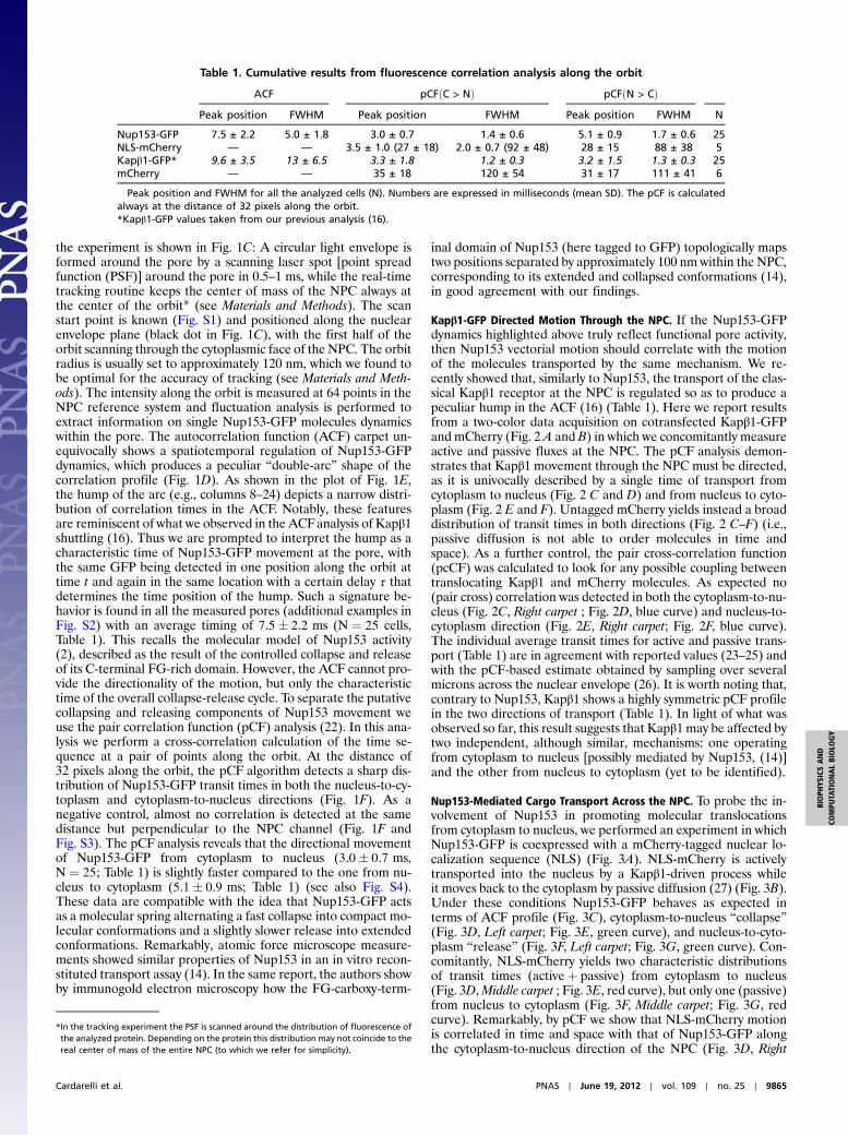
Table 1. Cumulative results from fluorescence correlation analysis along the orbit
ACF pCFðC > NÞ pCFðN > CÞPeak position FWHM Peak position FWHM Peak position FWHM N
Nup153-GFP 7.5 ± 2.2 5.0 ± 1.8 3.0 ± 0.7 1.4 ± 0.6 5.1 ± 0.9 1.7 ± 0.6 25NLS-mCherry — — 3.5 ± 1.0 (27 ± 18) 2.0 ± 0.7 (92 ± 48) 28 ± 15 88 ± 38 5Kapβ1-GFP* 9.6 ± 3.5 13 ± 6.5 3.3 ± 1.8 1.2 ± 0.3 3.2 ± 1.5 1.3 ± 0.3 25mCherry — — 35 ± 18 120 ± 54 31 ± 17 111 ± 41 6
Peak position and FWHM for all the analyzed cells (N). Numbers are expressed in milliseconds (mean SD). The pCF is calculatedalways at the distance of 32 pixels along the orbit.*Kapβ1-GFP values taken from our previous analysis (16).
*In the tracking experiment the PSF is scanned around the distribution of fluorescence ofthe analyzed protein. Depending on the protein this distribution may not coincide to thereal center of mass of the entire NPC (to which we refer for simplicity).
Cardarelli et al. PNAS ∣ June 19, 2012 ∣ vol. 109 ∣ no. 25 ∣ 9865
BIOPH
YSICSAND
COMPU
TATIONALBIOLO
GY

carpet; Fig. 3E, blue curve). The positive pair-cross-correlationsignal suggests that at least a subpopulation of NLS-mCherry mo-lecules may be effectively exploiting Nup153-GFP to move fromcytoplasm to nucleus across the NPC. A relevant control for thisconclusion is that no pcCF signal can be observed in the oppositedirection (Fig. 3F, Right carpet ; Fig. 3G, blue curve), whereNLS-mCherry passive diffusion is not coupled to Nup153-GFPdynamics.
ConclusionsAn accurate picture of how selective gating is achieved by theFG-Nups remains unclear due to a general lack of understandingwith regard to their behavior within the NPC. The source of thisambiguity stems in part from the difficulty in addressing FG-Nupsdynamics in the intact NPC. The thought-provoking idea here isthat we can overcome these limitations by a combination of track-ing and fluctuation analysis to study the behavior of single porecomponents with high spatial and temporal resolution in live,minimally perturbed cells. The crucial point in our results isthe observation of a previously hidden dynamics of nucleoporinNup153, characterized by a rapid, discrete exchange between twoseparate positions within the NPC of intact cells. The nanome-chanical mechanism suggested by our results highlights apparentdifferences with some macroscopic views proposed thus far that
deserve consideration. The bulk-like hydrogel meshwork model(5), for instance, places a greater importance on the hydrophobicinteractions between neighbor FG-Nups, as they are able to forma sieve-like meshwork in vitro (10). Transport could then occurthrough binding of transport receptors to FG-repeats, causinga local gel-to-liquid transition and allowing the receptor to cat-alyze its own diffusion (28). Our data do not exclude that theseinteractions play a role in transport; however, they suggest thatthe most kinetically relevant events in the intact NPC have thecharacteristics of directed transport, not of unbiased diffusion.It must be noted that Nup153 belongs to the FxFG-rich familyof nucleoporins, which generally display noncohesive properties(29). At the same time, however, it was recently reported thatC-terminal fragments of Nup153 can exist in a collapsed statebut are concomitantly able to form a hydrogel in vitro (30). Be-sides highlighting the multifaceted character of Nup153, thesecontrasting results may also reflect a more general complexityof the NPC function, with a not yet completely understood bal-ance/interplay between cohesive (i.e., gel-like) and noncohesive(i.e., “spring-like”) properties, as also emerges from the hybridmodel recently proposed by Yamada and coworkers (15). Basedon our results on intact cells, we propose that molecular transportthrough the NPC can be powered, at least in part, by the directedmotion of specific nucleoporins (here Nup153). Further experi-
Fig. 2. (A) Kapβ1-GFP is cotransfected with untagged mCherry (scale bar: 1 μm). (B) In the schematic diagram, an unlabeled Nup153 (the endogenous one, inthis experiment) mediates Kapβ1-GFP movement towards the nucleus (I and II); a symmetric process must exist to account for Kapβ1 movement towards thecytoplasm (III and IV). The mCherry molecule passively diffuses back and forth with no interaction with Nup153. (C and D) The pCFðþ32Þ from cytoplasm tonucleus shows a sharp distribution of Kapβ1 transolaction times (Left carpet, green curve) opposed to a much broader distribution of mCherry translocationtimes (Middle carpet, red curve). The pcCFðþ32Þ shows that Kapβ1-GFP and mCherry are not translocating together from cytoplasm to nucleus (Right carpet,blue curve). (E and F) The pCF and pcCF at the distance of 32 pixels from nucleus to cytoplasm reveal the symmetry of the observed behaviors: Kapβ1 rapidlytranslocates, independently from mCherry passive diffusion.
9866 ∣ www.pnas.org/cgi/doi/10.1073/pnas.1200486109 Cardarelli et al.

ments on additional constituents of the pore (e.g., more cohesiveNups of the central channel) are needed to achieve more generalconclusions about the intimate nature of selective molecular gat-ing through the NPC. Finally, it is worth noting that the observeddirected-motion mechanism shares many biochemical and physi-cal characteristics with several spring-like molecular systems pre-sent in nature (31). We shall argue that they represent ancientand commonplace eukaryotic molecular engines.
Materials and MethodsCell Culture, Plasmids, and Treatments. CHO-K1 cells were grown in Ham’sF12K medium supplemented with 10% of fetal bovine serum at 37 °C andin 5% CO2. Freshly split cells were plated on imaging dishes and transientlytransfected using Lipofectamine 2000 according to manufacturer’s protocol,24 h before the experiment. The plasmid encoding for Nup153-GFP was akind gift from Nathalie Daigle (Cell Biology and Biophysics Unit, EuropeanMolecular Biology Laboratory, Heidelberg, Germany) (20). The plasmid en-coding for human Kapβ1-GFP was a generous gift from Marilena Ciciarello(Istituto di Biologia e Patologia Molecolari, Consiglio Nazionale delle Ri-cerche) (32). The plasmid encoding for NLS-mCherry has been described ina previous publication (27).
Single NPC Tracking Setup. Tracking of single NPCs was performed using ahome-built microscope capable of single particle tracking, whose details havebeen already described (33). Briefly, the microscope is built around an Olym-pus X71 body. A Chameleon Ultra (tunable) Ti:Sapphire laser (Coherent)tuned at 940 nm was used for two-photon excitation of the GFP (andmCherry) constructs. The scanning of the excitation light was obtained inthe x–y plane and in the z axis through two galvano-motor driven mirrors(Cambridge Technology) and a piezo-objective positioner (Phisik Instrument)respectively, both driven by a computer card (three-axis card, ISS). Fluores-cence emission was collected by a 1.2-NA water objective (Olympus UplanSA-po 60x), split by a dichroic mirror at 570 nm and detected in the 500–550 nm(GFP) and 575–645 nm (mCherry) spectral ranges by two GaAS detectorsH7241P (Hamamatsu). During the tracking procedure, the two scanning mir-rors are moved independently by π∕2-phase shifted sine wave voltages gen-erated in the card so that the laser beam moves in a circular path around theparticle. The position of the scanning center is determined by the offset va-lues of the sine waves. The position of the center is updated at each trackingcycle according to the fast Fourier transform (FFT)-based algorithm previouslydescribed (34). From the FFTof the intensity trace along the orbit, we get theaverage intensity or dc as the zeroth term in a Fourier series and the ac as thecoefficient of the first harmonic term. The angular coordinate of the particleis given directly by the phase of the ac term, and its distance from the center
Fig. 3. (A) Nup153-GFP is cotransfected with NLS-mCherry (scale bar: 1 μm). (B) In the schematic diagram, Nup153-GFP drives NLS-mCherry towards the nucleus(I, II); the intervention of RanGTP detaches NLS-mCherry and releases Nup153-GFP (III). (C) The ACF yields the double-arc, characteristic of Nup153 activity at theNPC. (D and E) The pCFðþ32Þ analysis in the cytoplasm-to-nucleus direction yields the expected distribution of Nup153 collapse times (Left carpet, green curve);NLS-mCherry instead shows a bimodal distribution of transit times, in keeping with the presence of two translocation mechanism (activeþ passive). Only thefaster component (active) pair cross-correlates with Nup153 movement in this direction (Right carpet, blue curve). (F and G) The pCFðþ32Þ in the oppositedirection shows the expected distribution of Nup153 release times (Left carpet, green curve) opposed to a broader distribution of NLS-mCherry passive trans-location times (Middle carpet, red curve). The pcCFðþ32Þ analysis shows that Nup153-GFP and NLS-mCherry are not moving bound together in this direction, asexpected (Right carpet, blue curve).
Cardarelli et al. PNAS ∣ June 19, 2012 ∣ vol. 109 ∣ no. 25 ∣ 9867
BIOPH
YSICSAND
COMPU
TATIONALBIOLO
GY

can be calculated from themodulation of the signal, defined asmod ¼ ac∕dc,so that its position can be recovered. The tracking routine changes the co-ordinates of the center of the scanning orbit in such a way to keep the mod-ulation at a minimum, i.e., to keep the particle always at the center. Thetracking procedure started by acquiring a raster scan image of the sample,focusing on the equatorial section of the nuclear envelope. Then we clickedon a location on the image corresponding to an isolated NPC. The fluores-cence intensity is collected at 64 points along a circular orbit around the pore,with a period of 0.5–1 ms and a calibrated scanning start point. The orbitradius (R) is usually set to 120 nm, which we found to be optimal for the track-ing. The optimal radius for tracking a point-like particle should be on theorder of half the size of the PSF (34), but in our case this value is slightly largerdue to the finite size of the NPC. The position of the center of the scanningorbit is updated typically every 32–64 orbits (that is defined as the cycle),which is fast enough to follow the NPC movement. The acquisition timefor a single NPC tracking measurement typically varied from 15 s up to100 s. For each NPC we acquired the intensity along the orbit for one ortwo channels and the trajectory of the center of mass. From the recordedtrace of the fluorescence intensity along the orbit the value of the modula-tion of the signal can be calculated at each cycle. The analysis of the mod-
ulation allows to check a posteriori if the particle has been tracked correctlyfor the entire acquisition or to exclude the portions of the dataset where theparticle was temporarily or definitely lost. We selected only the part of thedata acquired with the orbit centered and stationary with respect to the cen-ter of mass of the NPC.
Fluctuation Analysis. The fluctuation analysis of the data was performed withthe SimFCS software (www.lfd.uci.edu, University of California at Irvine)using the scanning-FCS analysis tool, as thoroughly described in previous pub-lications (22, 26). Briefly, the ACF, pCF(pixels), and pcCF(pixels) are displayedin pseudo colors in a carpet in which the x coordinate corresponds to thepoint along the orbit and the vertical coordinate corresponds to the correla-tion time in a log-scale.
ACKNOWLEDGMENTS. We thank Milka Stakic for helping in cultivating andtransfecting the CHO-K1 cells. This work was supported by the National Cen-ter for Research Resources (5P41RR003155), the National Institute of GeneralMedical Sciences (8P41GM103540), divisions of the National Institutes ofHealth (NIH), and NIH 5P50 GM076516.
1. Weis K (2003) Regulating access to the genome: Nucleocytoplasmic transport through-out the cell cycle. Cell 112:441–451.
2. Fahrenkrog B, Aebi U (2003) The nuclear pore complex: Nucleocytoplasmic transportand beyond. Nat Rev Mol Cell Biol 4:757–66.
3. Rout M-P, Aitchison J-D (2001) The nuclear pore complex as a transport machine. J BiolChem 276:16593–16596.
4. Denning D-P, Patel S-S, Uversky V, Fink A-L, Rexach M (2003) Disorder in the nuclearpore complex: The FG repeat regions of nucleoporins are natively unfolded. Proc NatlAcad Sci USA 100:2450–2455.
5. Ribbeck K, Gorlich D (2002) The permeability barrier of nuclear pore complexes ap-pears to operate via hydrophobic exclusion. EMBO J 21:2664–2671.
6. Conti E, Muller C-W, Stewart M (2006) Karyopherin flexibility in nucleocytoplasmictransport. Curr Opin Struct Biol 16:237–244.
7. Rout M-P, et al. (2000) The yeast nuclear pore complex: Composition, architecture, andtransport mechanism. J Cell Biol 148:635–651.
8. Ben-Efraim I, Gerace L (2001) Gradient of increasing affinity of importin beta for nu-cleoporins along the pathway of nuclear import. J Cell Biol 152:411–417.
9. Peters R (2005) Translocation through the nuclear pore complex: Selectivity and speedby reduction-of-dimensionality. Traffic 6:421–427.
10. Frey S, Richter R-P, Gorlich D (2006) FG-rich repeats of nuclear pore proteins form athree-dimensional meshwork with hydrogel-like properties. Science 314:815–817.
11. Frey S, Gorlich D (2009) FG/FxFG as well as GLFG repeats form a selective permeabilitybarrier with self-healing properties. EMBO J 28:2554–2567.
12. Frey S, Gorlich D (2007) A saturated FG-repeat hydrogel can reproduce the permeabil-ity properties of nuclear pore complexes. Cell 130:512–523.
13. Lim R-Y, et al. (2006) Flexible phenylalanine-glycine nucleoporins as entropic barriersto nucleocytoplasmic transport. Proc Natl Acad Sci USA 103:9512–9517.
14. Lim R-Y, et al. (2007) Nanomechanical basis of selective gating by the nuclear porecomplex. Science 318:640–643.
15. Yamada J, et al. (2010) A bimodal distribution of two distinct categories of intrinsicallydisordered structures with separate functions in FG nucleoporins.Mol Cell Proteomics9:2205–2224.
16. Cardarelli F, Lanzano L, Gratton E (2011) Nanoscale fluorescence correlation spectro-scopy of intact nuclear pore complexes. Biophys J 101:L27–29.
17. Ball J-R, Ullman K-S (2005) Versatility at the nuclear pore complex: Lessons learnedfrom the nucleoporin Nup153. Chromosoma 114:319–330.
18. Fahrenkrog B, et al. (2002) Domain-specific antibodies reveal multiple-site topology ofNup153 within the nuclear pore complex. J Struct Biol 140:254–267.
19. Rabut G, Doye V, Ellenberg J (2004) Mapping the dynamic organization of the nuclearpore complex inside single living cells. Nat Cell Biol 6:1114–1121.
20. Daigle N, et al. (2001) Nuclear pore complexes form immobile networks and have avery low turnover in live mammalian cells. J Cell Biol 154:71–84.
21. Pante N, Thomas F, Aebi U, Burke B, Bastos R (2000) Recombinant Nup153 incorporatesin vivo into Xenopus oocyte nuclear pore complexes. J Struct Biol 129:306–312.
22. Digman M-A, Gratton E (2009) Imaging barriers to diffusion by pair correlation func-tions. Biophys J 97:665–673.
23. YangW, Gelles J, Musser S-M (2004) Imaging of single-molecule translocation throughnuclear pore complexes. Proc Natl Acad Sci USA 101:12887–12892.
24. Kubitscheck U, et al. (2005) Nuclear transport of single molecules: Dwell times at thenuclear pore complex. J Cell Biol 168:233–243.
25. Dange T, Grunwald D, Grunwald A, Peters R, Kubitscheck U (2008) Autonomy and ro-bustness of translocation through the nuclear pore complex: A single-molecule study. JCell Biol 183:77–86.
26. Cardarelli F, Gratton E (2010) In vivo imaging of single-molecule translocation throughnuclear pore complexes by pair correlation functions. PLoS One 5:e10475.
27. Cardarelli F, Bizzarri R, Serresi M, Albertazzi L, Beltram F (2009) Probing nuclear loca-lization signal-importin alpha binding equilibria in living cells. J Biol Chem284:36638–36646.
28. Eisele N-B, Frey S, Piehler J, Gorlich D, Richter R-P (2010) Ultrathin nucleoporin phe-nylalanine-glycine repeat films and their interaction with nuclear transport receptors.EMBO Rep 11:366–372.
29. Patel S-S, Belmont B-J, Sante J-M, Rexach M-F (2007) Natively unfolded nucleoporinsgate protein diffusion across the nuclear pore complex. Cell 129:83–96.
30. Milles S, Lemke E-A (2011) Single molecule study of the intrinsically disordered FG-re-peat nucleoporin 153. Biophys J 101:1710–1719.
31. Mahadevan L, Matsudaira P (2000) Motility powered by supramolecular springs andratchets. Science 288:95–100.
32. Ciciarello M, et al. (2004) Importin beta is transported to spindle poles during mitosisand regulates Ran-dependent spindle assembly factors in mammalian cells. J Cell Sci117:6511–6522.
33. Levi V, Ruan Q, Gratton E (2005) 3-D particle tracking in a two-photon microscope:Application to the study of molecular dynamics in cells. Biophys J 88:2919–2928.
34. Kis-Petikova K, Gratton E (2004) Distance measurement by circular scanning of theexcitation beam in the two-photon microscope. Microsc Res Tech 63:34–49.
9868 ∣ www.pnas.org/cgi/doi/10.1073/pnas.1200486109 Cardarelli et al.