Embed Size (px)
Citation preview

Governors State UniversityOPUS Open Portal to University Scholarship
All Capstone Projects Student Capstone Projects
Fall 2011
Capture and Analysis of Cells Using MagneticBeadsAlexandro DominguezGovernors State University
Follow this and additional works at: http://opus.govst.edu/capstones
Part of the Analytical Chemistry Commons
For more information about the academic degree, extended learning, and certificate programs of Governors State University, go tohttp://www.govst.edu/Academics/Degree_Programs_and_Certifications/
Visit the Governors State Analytical Chemistry DepartmentThis Project Summary is brought to you for free and open access by the Student Capstone Projects at OPUS Open Portal to University Scholarship. Ithas been accepted for inclusion in All Capstone Projects by an authorized administrator of OPUS Open Portal to University Scholarship. For moreinformation, please contact [email protected].
Recommended CitationDominguez, Alexandro, "Capture and Analysis of Cells Using Magnetic Beads" (2011). All Capstone Projects. 13.http://opus.govst.edu/capstones/13

1
Capture and Analysis of Cells using Magnetic Beads
A project
Submitted
To
Governors State University
By
Alexandro Dominguez
In the partial fulfillment of the
Requirement for the Degree
of
Master of Science
In
Analytical Chemistry
December 2011
Governors State University
University park, IL-60484

2
DEDICATED TO MY PARENTS

3
Acknowledgement
I would like to thank my advisor Dr. Walter Henne for his encouragement and guidance
throughout the project work. His support was critical in achieving a successful project. I
appreciate all his time, impute and assistance with continuous improvement throughout the
development of this project. Dr. Walter Henne reinforced key values such as dedication,
determination, and ambition. I thank Dr. Patty Fu and Professor Kent, for their support and
guidance during the project. In addition, I would like to thank my graduate professors, Dr.
Addison, Dr. Kumar, and Dr. D’Arcy for their support and advice during my academic years at
Governors State University. I am very thankful to the Governors State University for providing
a stronger educational foundation. I would like thank my family for all their support throughout
my academic endeavors and accomplishments. I am most thankful to my wife for her
understanding and patience’s during my higher studies.

4
Table of Content
Abstract ………………………………………………………………………………………...5
Introduction…………………………………………………………………………………..5-6
Background……………………………………………………………………………….......6-7
Example of Experiments………………………………………………………………….........7
Magnetic Capture of Mycobacterium Avium subsp. Paratuberculosis…………………….7-13
Figure 1. Magnetic Separation Method Capture Efficiency………………………….……...10
Figure 2. Improved Capture Efficiency with Combination of Paramagnetic Beads ....…....12
Magnetic Cell Sorting of Parasitized Erythrocyte…………………………............................14
Figure 3. Magnetic Deposition Microscopy…………..….……………………….……….......14
High Gradient Magnetic Separation of Infected Red Blood Cells ……..………..............14-15
Magnetic Filter of Leukocytes from Tumor Cells……………………...……..……..........15-18
Figure 4. Magnetic Separation Method Capture Efficiency………………………….….…..16
Figure 5. Magnetic Filtration of Leukocytes from Tumor Cells…………………………….17
Figure 6. Direct Labeling and Fluorescence Micrographs……….……..................................18
Self-Assembled Magnetic Array ………………………………….......................................18-23
Figure 7. Ephesia …………………………….……………………………………………........19
Figure 8. Histogram of Granulocyte …………………………………………………….........23
Figure 9. Negative Enrichment of Neutrophil Granulocyte …….……………………...……24
Conclusion…………………………………………………………………………………...24-25
References………………………………………………………………………………..…..26-29

5
Abstract
Magnetic separation of cells and bacterium is a fascinating field of study. There are
tremendous capabilities in separation, selectivity, and sensitivity.1 Magnetic separation reduces
complex techniques, time consuming preparation, and complicated multistep methods of
separation.2 The separation of cells, bacterium, and macromolecules can be extremely
challenging. Fortunately, magnetically tagged cells, bacterium and macromolecules can be easily
separated without great expertise. Antibody magnetic micro beads are specific to a cell,
bacterium, or macromolecules. Separating a mixture can lead to a better, more accurate analysis
of samples that can be crucial in the medical field, therapeutic drug delivery systems, and in
some circumstances lifesaving.3 The ability to positively identify a disease in hours instead of
days results in faster recovery times and minimizes the spread of the disease.4 Faster recovery
time can be lifesaving in many countries because then the disease can be treated quickly.
Introduction
Magnetic cell sorting can lead to a better and more comprehensive understanding of the
human body. A better understanding of the human body would lead to better medical treatments,
smarter prevention, and new medical intervention for diseases. A variety of cells can be more
thoroughly studied with fluorochromes incorporated with magnetic separation.5 Magnetic
separation purity can be increased by various methods. One method is uses a double column
where the second column further purifies and separates the macromolecules from the solution.
Having several micro bead antigens can also increase the purity of the sample. Finally, stronger
magnets can polarize the molecules to a better separation due to magnetization.6

6
The ability to be exceptionally selective is a concept that is important to the scientific
community. Cells, bacterium, and macromolecules are composed of very unique molecules.
These molecules polymerize into macromolecules, and macromolecules have unique binding
sites that can be utilized in magnetic separation. Magnetic micro beads attach to the binding sites
and are magnetically separated from the solution. Magnetic micro beads can be purchased from
several pharmaceutical companies. There is a huge demand for synthesizing specific magnetic
beads because of their capabilities in separation. It is important to note that separation techniques
are based on several factors such as affinity, size, binding, stereochemistry, van der waal forces,
etcetera.7 Micro magnetic beads are prepared based on what is being separated, and the best
method of separation. Different samples have different physical characteristics. Though there is a
strong inclination for the magnetic bead to only attach to the target cells, physical forces from
non-target cells can also influence magnetic interaction.8
Background
There are two methods of magnetic labeling; direct and indirect magnetic labeling. Direct
labeling is a one-step process and is usually the simplest to perform. The specific antibody micro
bead is directly labeled to the cell. There are numerous antibody micro beads such as biotinylated
antibody, streptavidin micro bead, and fluorochrome-conjugated antibodies available in the
market. Indirect magnetic labeling is usually completed when no direct micro beads are available
for the cell. A cocktail of antibodies can be used to remove undesired cells from the desired cell.9
A pharmaceutical company can produce an antibody micro bead solution that is specific to a
particular antigen on the cell where several antibody micro beads will attach to the cell.
Magnetically labeled cells can then be separated with magnetization. There are two methods of
separation, one being positive selection where the target cells are magnetically labeled and are

7
magnetically retained in cell fraction. This strategy is the easiest method resulting in outstanding
purity, excellent recovery, and fast results. The second method is the untouched isolation, where
the undesired cells are removed. This method works well if there is no specific antibody
available for target cells, and if binding to the target cell is not desired.10
There are several methods of micro magnetic separation. One piece of ground-breaking
technology that can be seen here are magnetic-activated cell sorting (MACS) separators. The
MACS high gradient magnetic separation columns can separate large cells up to 50 um in
diameter. The capacity max is 2x10⁸ total cells and 1x10
7 magnetically labeled cells.
11 The large
cell separation columns are engineered for positive selection of human and animal cells. There is
a wide range of applications from small proteins to cells. The column itself has a hydrophilic
coating. The buffers suggested for rinsing are PBS, EDTA, or BSA.11
The column is washed to
remove unlabeled cells, and the cell-magnet complex is separated. Then the flow resistor is
removed, and the target cells elute from the column. The MAC column demonstrates how
routine the separation really is. There are several automatic magnetic separator instruments,
manual magnetic separators, magnetic separator kits, and micro beads available in the market.11
Examples of Experiments
Magnetic Capture of Mycobacterium Avium subsp. Paratuberculosis
Magnetic separation allows enormous specificity, sensitivity, and separation of
bacterium. Mycobacterium avium subsp. paratuberculosis is an example of a bacterium that was
captured effectively with magnetic separation. The immunomagnetic (IMS)-phage assay yields
excellent detection of M. avium subsp. paratuberculosis. The recovery was <10 CFU/ml from
both spiked broth and milk.12
Table one lists several different paramagnetic beads and their

8
corresponding vendors (the paramagnetic beads are coated in-house with the appropriate
antigen.)12
TABLE 1. In-house-prepared paramagnetic-bead-coating-antigen combinations evaluated.
Coating antigena
Paramagnetic beads
Polyclonal
antibody S624
aMp3
peptide
aMptD
peptide
Biotinylated
aMp3 peptide
Biotinylated
aMptD peptide
Dynabeads, M280 sheep
anti-rabbit IgGb
+ − − − −
Magnabind carboxyl
derivatized beadsc
− + + − −
Amine-coated magnetic
hollow glass
microspheresd
+ + + − −
Dynabeads, MyOne
Carboxylic acidb
+ + + − −
Dynabeads, MyOne
Tosylactivatedb
+ + + + +
Dynabeads, MyOne
Streptavidin-T1b
− − − + +
Dynabeads, M280 − − − + +

9
Coating antigena
Paramagnetic beads
Polyclonal
antibody S624
aMp3
peptide
aMptD
peptide
Biotinylated
aMp3 peptide
Biotinylated
aMptD peptide
Streptavidinb
a+, tested; −, not tested.
bFrom Invitrogen, Life Technologies Corporation.
cFrom Pierce Protein Research Products, Thermo Scientific.
dFrom Microsphere Technology Limited, Adare, County Limerick, Republic of Ireland.
There are numerous paramagnetic beads and the coating antigens are specifically
synthesized for the target bacterium. Bacterial species have abundant binding sites that can be
selected for optimal binding and separation. Binding sites are based on abundance, accessibility,
and cost effective generation of the respective antibody. There is an undesired effect of having
antibody micro beads attached to several bacteria for a positive selection. A false
positive/negative can lead to massive recalls of milk, food, and the health of the consumer being
compromised. Therefore, it is important to know the medium/interferences, and use the micro
beads that specifically bind to the target bacterium. The immunomagnetic (IMS)-phage assay
plays an important role because there is minimal nonspecific binding from other mycobacteria.
10
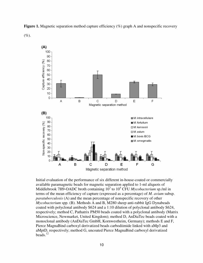
Figure 1. Magnetic separation method capture efficiency (%) graph A and nonspecific recovery
(%).
Initial evaluation of the performance of six different in-house-coated or commercially
available paramagnetic beads for magnetic separation applied to 1-ml aliquots of
Middlebrook 7H9-OADC broth containing 103 to 10
4 CFU Mycobacterium sp./ml in
terms of the mean efficiency of capture (expressed as a percentage) of M. avium subsp.
paratuberculosis (A) and the mean percentage of nonspecific recovery of other
Mycobacterium spp. (B). Methods A and B, M280 sheep anti-rabbit IgG Dynabeads
coated with polyclonal antibody S624 and a 1:10 dilution of polyclonal antibody S624,
respectively; method C, Pathatrix PM50 beads coated with a polyclonal antibody (Matrix
Microscience, Newmarket, United Kingdom); method D, AnDiaTec beads coated with a
monoclonal antibody (AnDiaTec GmbH, Kornwestheim, Germany); methods E and F,
Pierce MagnaBind carboxyl derivatized beads carbodiimide linked with aMp3 and
aMptD, respectively; method G, uncoated Pierce MagnaBind carboxyl derivatized
beads.12

11
(See Figure 1) Graph A displays the commercially available Pathatrix PM50 beads
coated with polyclonal antibody. Method C has the most efficiency capture of M. avium subsp.
paratuberculosis at a mean of 50.3% with a deviation of + 8.4%. Method A has the M289
Dynabeads coated with polyclonal antibody S624 has a mean of 29.2% to 34.2%. The graphs
revealed reasonable capture for method E, A and F. Poor capture was demonstrated in methods B
and D. (See Figure 1) Graph B displays Pathatrix PM50 beads coated with polyclonal antibody
(method C) with the most nonspecific recovery percentage at 22% + 11%. The antibody micro
beads interact with various bacteria. Graph B displayed less than 10% nonspecific recovery for
all groups except group C. Remember, life is composed of the same building blocks. Hence, the
different bacteria can display the same polypeptide or a similar polypeptide with the same
physical forces. There was no M. avium subsp. paratuberculosis capture at 100%. It is of utmost
importance to be as close as possible to 100% capture efficiency.

12
Figure 2. Improved capture efficiency with combination of paramagnetic bead-coating antigen.
(See Figure 2) The addition of extra paramagnetic bead coating antigen significantly
increased capture efficiency. The most efficient capture of M. avium subsp. paratuberculosis is
91.5% + 5.0% by PMS with a 50:50 ratio of MyOne Tosylactivated Dynabeads coated with
biotinylated aMp3 and biotinylated aMptD. The MyOne Tosylactivated Dynabeads, bars 11
through 15, exhibit impressible improved capture efficiency. The amine-coated magnetic hollow
glass beads coated with polyclonal antibody S624, aMp3, and aMptD presented capture
efficiency below five percent. Group nine and ten with M280 streptavidin Dynabeads coated
with biotinylated aMp3 and biotinylated aMptD had less than three percent capture efficiency.
Different characteristics that determine the selectivity of capture are the coating antigen

13
polyconal, monoclonal, antibody, peptide or biotinylated. In addition, the bead characteristics
such as composition, size concentration and surface play a role in capture efficiency.12
In the use of a combination of several paramagnetic bead coating antigens, the automated
and manual PMS did not have significant variance in efficient magnetic capture, however, the
mean recovery of nonspecific recovery by M. bovis BCG by automated IMS (AIMS) was noted
as less than the manual IMS. It is suggested that AIMS moves the beads from tube to tube during
processing. That movement of beads most likely leaves non target mycobacterium behind on
surface of tubes versus the manual process which leaves beads in the same tube while
processing.12
Immunomagnetic separation of pathogenic mycobacterium was accomplished. Magnetic
beads with genus specific polyclonal and mouse monoclonal antibodies complexes were tagged
with anti-mouse biotinylated antibody. The addition of quantum dots resulted in a fluorescent
detection. The limit of detection was 104 bacteria/ml and 10
3 bacteria/ml with the usage of a
spectrofluorometer.13
Immunomagnetic isolation of CD4+CD25
+FoxP3
+ natural T regulatory
lymphocytes is a more complicated and longer process. The T regulatory cells were isolated
from leukapheresis products via double negative selection of anti-CD8 and anti-CD19
monoclonal antibody continued with positive selection of anti-CD25 monoclonal antibody. The
final cell fraction, CD4+/CD25
+,
resulted in a mean purity of 93.6% with a standard deviation of
+ 1.1. The recovery efficiency was 81.52% + 7.4%.14
Immunomagnetic separation can be a series
of steps that finally leads to a purified product.

14
Magnetic Cell Sorting of Parasitized Erythrocytes
Magnetic cell sorting is a fast and accurate method of analyzing diseases. Magnetic
sorting depends on interaction between cell surface antigens, antibodies, and magnetic particles.
Magnetic deposition microscopy (MDM) (See Figure 3) captures parasitized erythrocytes in a
magnetic field and the sample is placed on a slide. The sample on the slide can then be stained
and viewed immediately.15
Figure 3. (A) Components of the malaria MDM device and the sample flow path. The
location of the expected magnetic cell deposit band next to the magnet pole piece tips.
(B) An unaided eye appearance of the magnetic deposition, collected in the interpolar gap
area (Panel A and B), from a P. falciparum parasitized blood sample.15
High Gradient Magnetic Separation of Infected Red Blood Cells
High gradient magnetic separation has been utilized for concentrating or eliminating
malaria from infected red blood cells (IRBCs) via blood magnetic properties (Fe content). The

15
column was loaded with 5x10⁸ RBCs. The average yield for the six experiments was 12.1x10
6 +
2.6x10⁶ IRBC with a purity of 95.74% + 1.38%. Purities ranged from 94.23% to 98.26%.
16 Any
particle with a higher magnetic capacity will replace a particle with lower magnetic capacity.
Optimizing the column load can significantly increase the purity on the eluted sample. In
addition, using a second column can further purifying the product. 16
Magnetic Filter of Leukocytes from Tumor Cells
Another approach to immunomagnetic separation is the use of a magnetic filter. The
magnetic filter attracts magnetically tagged cells from solution with alternating magnetic dipoles.
The magnetic filter was first tested by sorting magnetic beads from nonmagnetic beads. (See
Figure 4) High capture yields were attained with approximately 90% of nonmagnetic beads
eluted from the filtration assembly whereas magnetic beads were preferentially captured. The
magnetic filter enhanced the removal of nonmagnetic beads by a factor greater than 105. A strong
magnetic force can achieve high capture efficiency on a moderate flow rate of 1 ml/hr. The
magnetic field decays rapidly with distance creating large gradients. The length of the magnetic
field can be adjusted with the size of the grains.17
Therefore, much of the separation occurs
rapidly and the separation rate progressively approaches zero with time. The filtration assembly
was tested on the basis of sorting magnetic beads from non-magnetic polystyrene beads. The
self-assembled magnetic filter also demonstrated proficient sorting of cells.

16
Figure 4. The graphs below represent flow cytometry before and after self-assembled magnetic
filter with the enrichment recovery ratio of polystyrene and magnetic beads.
The self-assembled magnetic filter was examined by filtering a population of magnetic
beads from polystyrene beads. Flow cytometry quantified the bead population before and after
the filtration. The enrichment and recovery ratio were measured at several flow rates.
Another example uses negatively enriching tumor cells from leukocytes solution that
were tagged with CD-45 magnetic beads (MACS, Militenyi Biotec).17
(See Figure 5) The self-
assembled filtration enriched the population of tumor cells to leukocytes by a factor greater than
103. The fraction of tumor cells that passed through the system was approximately 90%. The
leukocytes were stained with green dye. Tumor cells were incubated with magnetic beads labeled

17
with anti-CD45 antibodies and fluorescent antibodies. (See Figure 6) The cells were observed
through fluorescence micrographs depicting ratios of 1/10, 1/100 and 1/1000
(tumor/leukocytes).17
The solution filtered through self-assembled magnetic and concentrated on
an integrated micropore filter. Fluorescence micrographs of the solution before the filter and
after the micropore filter display a significant difference. After filtration there is a minimal
amount of green fluorescent that is visible.17
Figure 5. Magnetic filtration of leukocytes from tumor cells.

18
Figure 6. The graphs below illustrate direct labeling and fluorescent micrographs.
Self-Assembled Magnetic Arrays
The concept noted as “Ephesia,” is another method for immunomagnetic separation.
Super paramagnetic beads are assembled in microfluidic channel on an array of magnetic traps.
The magnetic beads are injected into the microfluidic channel and the beads align together when
the magnetic field is switched on. (See Figure 7) The array is divided into a series of rows with
magnetic ink that attracts the beads within a magnetic field.18

19
Figure 7. A) Principle of magnetic self-assembly. A hexagonal array of magnetic ink is
patterned at the bottom of a microfluidic channel. Beads coated with an antibody are
injected in the channel. Beads undergo to Brownian motion. The application of an
external vertical magnetic field induces the formation of a regular array of bead columns
localized on top of the ink dots. (B) Two levels PDMS integrated microchip. Channels
were filled with colored water. Delivery and separation channels for the cells appear in
yellow. Inlets ports appear in orange. The separation channel is the longer vertical
branch. The area bearing magnetic posts is marked by the dotted white box. Channels in
the upper PDMS layer, controlling the opening and closing of the inlet channels, appear

20
in blue. The green wire is a thermocouple for in situ control of the temperature in the
system. (Scale bar: 0.5 cm.) (C) Magnetically assembled array of columns of 4.5 µm
beads coated with anti-CD19 mAb (specifically retaining Raji B-Lymphocytes). Typical
column shapes are shown in the insets. (Scale bar: 80 µm.) (D) Optical micrograph of the
columns after the passage of 1,000 Jurkat cells. No cell can be seen. (E) After the passage
of 400 Raji cells, numerous ones are captured and rosetted on the columns (Scale bar:
80 µm.)18
The cell capture correlates to a function of flow rate. The flow rate can increase or
decrease the amount of beads that are retained. A channel with the width of 500 um, a channel
height of 50 um and a 100 um/s flow correlates to a flow rate of a few ul/min with a throughput
in the vicinity of ten to hundred cells. Positive and negative cell sorting yielded 97+ 2% Raji
cells and over 98% Jurkat cells eluted out. There were 612 cells analyzed, 31% Raji and 69%
Jurkat that yielded a capture of 97% Raji and 0.02% capture of Jurkat. Resulting in 97% in purity
of Raji.18
Magnetic cell sorting of circulating tumor cells (CTCs) was completed by removing
normal blood cells by erythrocyte lysis and pan-leukocyte marker (CD45) antibody tagging.15
Immunomagnetic nano beads were implemented for detection of circulating tumor cells
in several patients. The characterization of CTCs was the underlying task. The purity and
recovery of spiked SW620 was analyzed with three enrichment methods. (See Table 2) The
method was CD45 depletion, positive enrichment and CD45 depletion with positive enrichment.
The results of the performance enrichment after spiking 100 SW620 cells in 5ml of peripheral
blood are displayed in table two. The CD45 depletion displays the highest recovery at 58%.19

21
Table 2. The CD45 depletion, positive enrichment and CD45 depletion with positive
enrichment.
Method Total number of leukocytes Recovery Purity
Before
enrichment
After
enrichment
Average
(%)
Range
(%)
Average
(%)
CD45 depletion 3 × 10⁷ 6.0 × 10
3 58 50-66 0.97%
Positive enrichment 3 × 10⁷ 2.0 × 10
3 25 24-26 1.25%
CD45 depletion + positive
enrichment
3 × 10⁷ 1.5 × 10
3 22.5 20-25 1.50%
Table 3. The detection rate of CTCs in 84 blood samples from 48 epithelial cancer patients and
30 samples from 22 melanoma patients.
Carcinoma
Number of blood
samples
Number of
patients
Positivity of blood
samples
Positivity of
patients
Gastric 5 3 80% (4/5) 67% (2/3)
Colon 25 11 44% (11/25) 64% (7/11)
Ovarian 8 6 50% (4/8) 50% (3/6)
Breast 21 10 52% (11/21) 60% (6/10)

22
Carcinoma
Number of blood
samples
Number of
patients
Positivity of blood
samples
Positivity of
patients
Cervix 11 7 64% (7/11) 86% (6/7)
NSCLC 4 3 75% (3/4) 100% (3/3)
SCCHN 10 8 70% (7/10) 75% (6/8)
Melanoma 32 22 53% (17/32) 64% (14/22)
(See Table 3) The detection rate of CTCs ranged from 44% to 80%. It can be challenging
and difficult to detect rare CTCs. Immunomagnetic separation is providing an alternative to
identification of CTCs and can eventually lead to a more accurate estimation of CTCs.19
Immunomagnetic bead separation of mononuclear cells contaminating granulocytes in
blood samples was also accomplished. The anti-CD15 micro beads were effective due to
increased numbers of CD15 binding sites. (See Figure 8) Histogram A with anti-CD-ECD
antibody displays 58% granulocytes, histogram B with histopaque displays 69% and histogram C
displays 1%.20
Histogram C is after magnetic separation. The histopaque procedure does not
remove granulocytes. A high level of separation was attained with magnetic beads.

23
Figure 8. The Histograms with magnetic separation and without magnetic separation are
displayed.

24
Immunomagnetic negative enrichment of neutrophil granulocyte from bone marrow was
accomplished. Polymorphonuclear neutrophils (PMN) mediate early immunity infection and
bone marrow is a known greater source of PMN. (See Figure 9) A negative cocktail was
implemented to eliminate any direct binding to PMN. Positive selection of PMN was not
completed due to previously recorded data of low detection of PMN complex.21
Figure 9. Represents a negative antibody cocktail versus percent of positive cells.
Conclusion
The underlying task of magnetic separation relies on novel separation techniques,
purification, and analytical analyses, understanding what needs to be isolated is the first step in
choosing a method of magnetic separation. There are a variety of magnetic separation techniques
that can be chosen to optimize separation efficiency. Immunomagnetic sorting has broad and
near limitless application across a spectrum of scientific fields. There are numerous complex
cells, bacterium, and macromolecules that can be separated via magnetic separation. Magnetic
separation can also purify small molecules. Otherwise, a cell separation would be time

25
consuming, complex and a multistep process. Reducing the time of a test and having accurate
results in imperative especially for disease detection. Future progress with fabrication of
complex magnetic structures and magnetic molds will ultimately aid in the development of new
techniques and the improvement of existing methods. The molds can be shaped to a specific
shape and filled with magnetic material. It is only a matter of magnetic interaction interwoven
with selectivity that is the basis for these methods.

26
References
1. N. Jothikumar, Dean O. Cliver, and Tadesse W. Mariam. Immunomagnetic Capture PCR for
Rapid Concentration and Detection of Hepatitis A Virus from Environmental Samples. Applied
and Environmental Microbiology. February 1998 vol. 64 no. 2 504-508.
2. Stacy J. Favrin1, Sabah A. Jassim1, and Mansel W. Griffiths. Development and Optimization
of a Novel Immunomagnetic Separation- Bacteriophage Assay for Detection of Salmonella
enterica Serovar Enteritidis in Broth. Applied and Enviromental Microbiology. 2001 January;
67(1): 217–224.
3. Huang Z, Pei N, Wang Y, Xie X, Sun A, Shen L, Zhang S, Liu X, Zou Y, Qian J, Ge J. Deep
magnetic capture of magnetically loaded cells for spatially targeted therapeutics. Biomaterials.
2010 Mar;31 (8):2130-40. Epub 2009 Dec 7.
4. Vanhee LM, Meersseman W, Lagrou K, Maertens J, Nelis HJ, Coenye T.J. Rapid and direct
quantification of viable Candida species in whole blood by use of immunomagnetic separation
and solid-phase cytometry. Clin Microbiol. 2010 Apr;48(4):1126-31. Epub 2010 Feb 3.
5. C. V. Durgadas, Chandra P. Sharma and K. Sreenivasan Fluorescent and superparamagnetic
hybrid quantum clusters for magnetic separation and imaging of cancer cells from blood.
Nanoscale, 2011, 3, 4780-4787
6. David W. Inglis, Robert Riehn, James C. Sturm, and Robert H. Austin. J. Microfluidic high
gradient magnetic cell separation. Appl. Phys. 99, 08K101 (2006); doi:10.1063/1.2165782

27
7. Magni F, Van Der Burgt YE, Chinello C, Mainini V, Gianazza E, Squeo V, Deelder AM,
Kienle MG. Biomarkers discovery by peptide and protein profiling in biological fluids based on
functionalized magnetic beads purification and mass spectrometry. Blood Transfus. 2010 Jun;8
Suppl 3:s92-7.
8. Kutay Icoz, Cagri Savran.Nanomechanical biosensing with immunomagnetic separation.
Applied Physics letters 97, 123701 (2010).
9. Christine E. Probst, Pavel Zrazhevskiy, and Xiaohu Gao. Rapid Multitarget Immunomagnetic
Separation through Programmable DNA Linker Displacement. J. Am. Chem. Soc., 2011, 133
(43), pp 17126–17129.
10. Chen-Lin Chen, Ken-Chao Chen, Yu-Cheng Pan, Tai-Ping Lee, Lo-Chang Hsiung, Cheng-
Ming Lin, Chang-Yu Chen, Ching-Hung Lin, Bor-Luen Chiang and Andrew M. Wo. Separation
and detection of rare cells in a microfluidic disk via negative selection. Lab Chip, 2011, 11, 474-
483
11. http://www.miltenyibiotec.com/en/default.aspx
12. Foddai A, Elliott CT, Grant IR. Maximizing capture efficiency and specificity of magnetic
separation for Mycobacterium avium subsp. paratuberculosis cells. Appl Environ Microbiol.
2010 Nov;76(22):7550-8. Epub 2010 Sep 17.

28
13. Liandris E, Gazouli M, Andreadou M, Sechi LA, Rosu V, Ikonomopoulos Detection of
pathogenic mycobacteria based on functionalized quantum dots coupled with immunomagnetic
separation. J. PLoS One. 2011;6(5):e20026. Epub 2011 May 27.
14. Di Ianni M, Del Papa B, Cecchini D, Bonifacio E, Moretti L, Zei T, Ostini RI, Falzetti F,
Fontana L, Tagliapietra G, Maldini C, Martelli MF, Tabilio A. Immunomagnetic isolation of
CD4+CD25+FoxP3+ natural T regulatory lymphocytes for clinical applications. Clin Exp
Immunol. 2009 May;156(2):246-53. Epub 2009 Mar 9.
15. Maciej Zborowski and Jeffrey J. Chalmers. Rare Cell Separation and Analysis by Magnetic
Sorting. Anal. Chem., 2011, 83 (21), pp 8050–8056.
16. Bhakdi SC, Ottinger A, Somsri S, Sratongno P, Pannadaporn P, Chimma P, Malasit P,
Pattanapanyasat K, Neumann HP. Optimized high gradient magnetic separation for isolation of
Plasmodium-infected red blood cells. Malar J. 2010 Feb 2;9:38.
17. Issadore D, Shao H, Chung J, Newton A, Pittet M, Weissleder R, Lee H. Self-assembled
magnetic filter for highly efficient immunomagnetic separation. Lab Chip. 2011 Jan 7;11(1):147-
51. Epub 2010 Oct 14.
18. Saliba AE, Saias L, Psychari E, Minc N, Simon D, Bidard FC, Mathiot C, Pierga JY, Fraisier
V, Salamero J, Saada V, Farace F, Vielh P, Malaquin L, Viovy JL. Microfluidic sorting and

29
multimodal typing of cancer cells in self-assembled magnetic arrays. Proc Natl Acad Sci U S A.
2010 Aug 17;107(33):14524-9. Epub 2010 Aug 2.
19. Liu Z, Fusi A, Klopocki E, Schmittel A, Tinhofer I, Nonnenmacher A, Keilholz U.Negative
enrichment by immunomagnetic nanobeads for unbiased characterization of circulating tumor
cells from peripheral blood of cancer patients. J Transl Med. 2011 May 19;9:70.
20. Preobrazhensky SN, Bahler DW. Immunomagnetic bead separation of mononuclear cells
from contaminating granulocytes in cryopreserved blood samples. Cryobiology. 2009
Dec;59(3):366-8. Epub 2009 Sep 18.
21. Hasenberg M, Köhler A, Bonifatius S, Borucki K, Riek-Burchardt M, Achilles J, Männ L,
Baumgart K, Schraven B, Gunzer M. Rapid immunomagnetic negative enrichment of neutrophil
granulocytes from murine bone marrow for functional studies in vitro and in vivo. PLoS One.
2011 Feb 23;6(2):e17314.